
Fatal Case of a Child Harboring Enterobius vermicularis
 ,
,  , , , and
Abstract
:1. Introduction
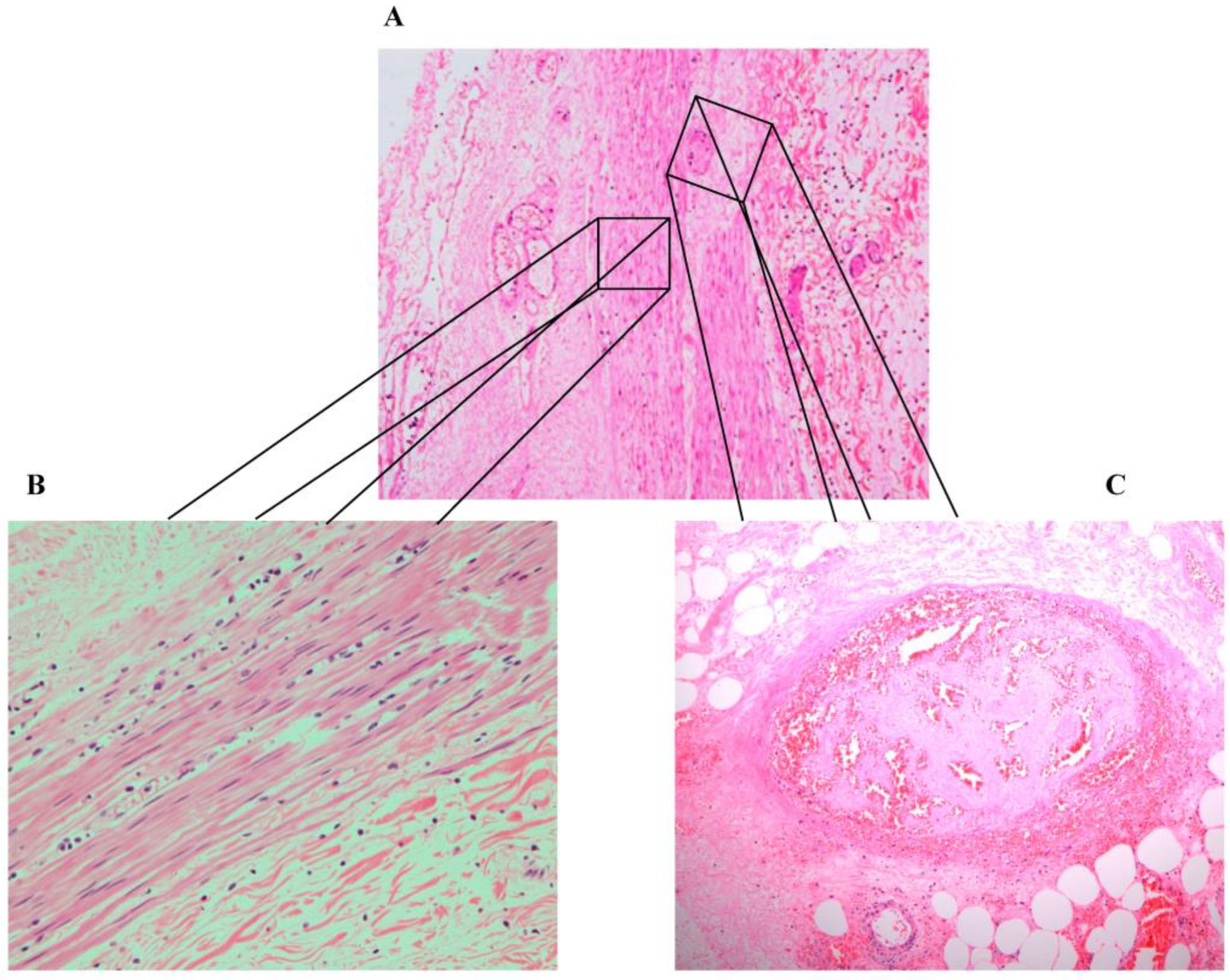
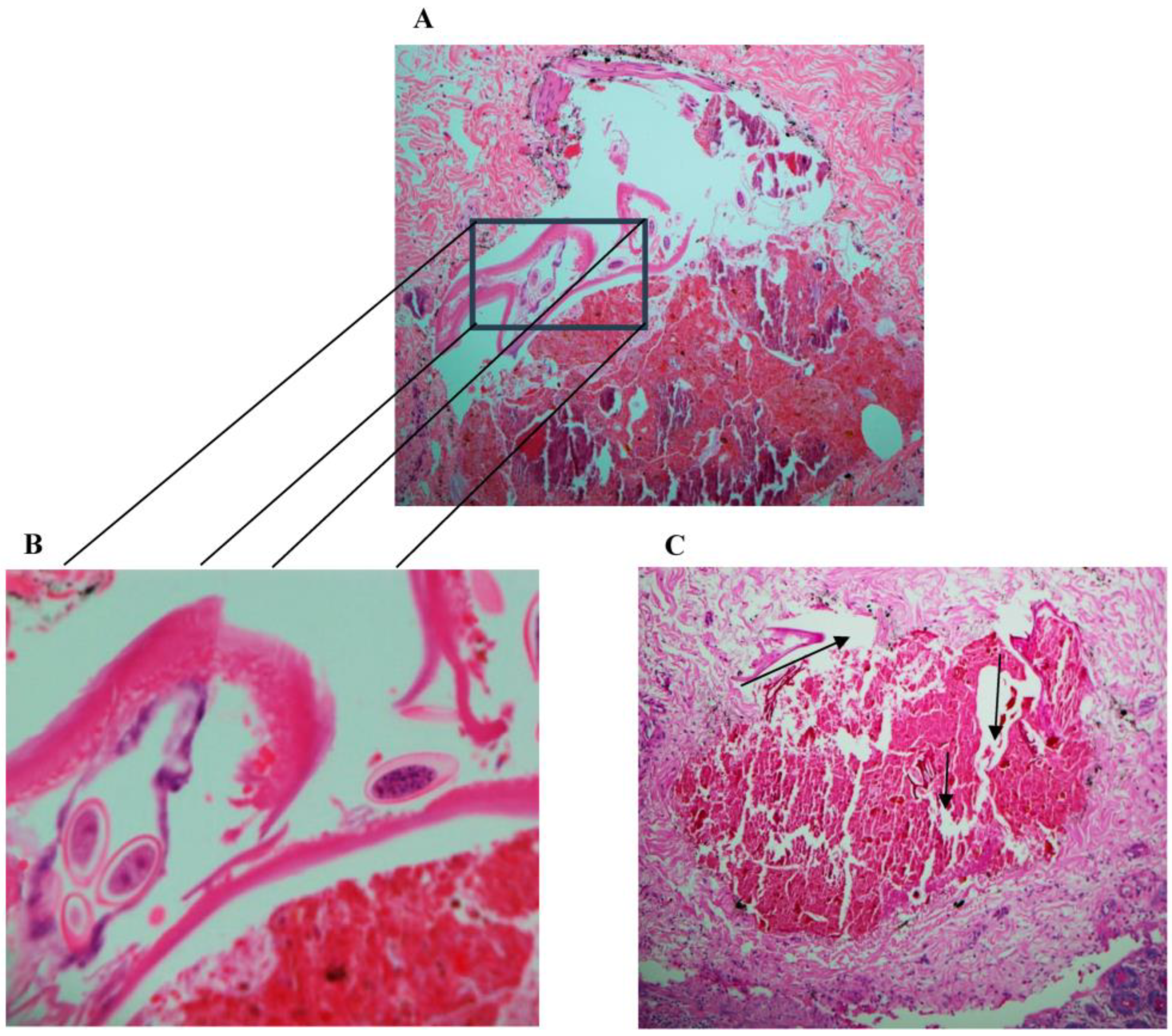
2. Case Description
3. Discussion
4. Conclusions
Author Contributions
Funding
Institutional Review Board Statement
Informed Consent Statement
Data Availability Statement
Acknowledgments
Conflicts of Interest
References
- Neva, F.A.; Brown, H.W. Basic Clinical Parasitology, 6th ed.; McGraw-Hill Education/Medical: New York, NY, USA, 1994. [Google Scholar]
- Lamps, L.W. Surgical Pathology of the Gastrointestinal System: Bacterial, Fungal, Viral, and Parasitic Infections, 1st ed.; Springer: New York, NY, USA, 2010. [Google Scholar]
- Wendt, S.; Trawinski, H.; Schubert, S.; Rodloff, A.C.; Mössner, J.; Lübbert, C. The Diagnosis and Treatment of Pinworm Infection. Dtsch. Arztebl. Int. 2019, 116, 213–219. [Google Scholar] [CrossRef]
- Stĕrba, J.; Vlcek, M.; Noll, P.; Vorel, F. Contribution to the question of relationships between Enterobius vermicularis (L.) and inflammatory processes in the appendix. Folia Parasitol. 1985, 32, 231–235. [Google Scholar]
- Kucik, C.J.; Martin, G.L.; Sortor, B.V. Common intestinal parasites. Am. Fam. Physician 2004, 69, 1161–1168. [Google Scholar]
- Arkoulis, N.; Zerbinis, H.; Simatos, G.; Nisiotis, A. Enterobius vermicularis (pinworm) infection of the liver mimicking malignancy: Presentation of a new case and review of current literature. Int. J. Surg. Case Rep. 2012, 3, 6–9. [Google Scholar] [CrossRef] [Green Version]
- Isik, B.; Yilmaz, M.; Karadag, N.; Kahraman, L.; Sogutlu, G.; Yilmaz, S.; Kirimlioglu, V. Appendiceal Enterobius vermicularis infestation in adults. Int. Surg. 2007, 92, 221–225. [Google Scholar]
- Zahariou, A.; Karamouti, M.; Papaioannou, P. Enterobius vermicularis in the male urinary tract: A case report. J. Med. Case Rep. 2007, 1, 137. [Google Scholar] [CrossRef] [Green Version]
- Sammour, Z.M.; Gomes, C.M.; Tome, A.L.; Bruschini, H.; Srougi, M. Prolonged irritative voiding symptoms due to Enterobius vermicularis bladder infestation in an adult patient. Braz. J. Infect. Dis. Off. Publ. Braz. Soc. Infect. Dis. 2008, 12, 352. [Google Scholar] [CrossRef] [PubMed] [Green Version]
- Cateau, E.; Yacoub, M.; Tavilien, C.; Becq-Giraudon, B.; Rodier, M.H. Enterobius vermicularis in the kidney: An unusual location. J. Med. Microbiol. 2010, 59, 860–861. [Google Scholar] [CrossRef] [PubMed] [Green Version]
- Dick, L.; Hannay, J. Enterobius vermicularis presentation during laparoscopic cholecystectomy. J. Surg. Case Rep. 2017, 2017, rjw239. [Google Scholar] [CrossRef]
- Ngui, R.; Ravindran, S.; Ong, D.B.; Chow, T.K.; Low, K.P.; Nureena, Z.S.; Rajoo, Y.; Chin, Y.T.; Amir, A.; Ahmad, A.F.; et al. Enterobius vermicularis salpingitis seen in the setting of ectopic pregnancy in a Malaysian patient. J. Clin. Microbiol. 2014, 52, 3468–3470. [Google Scholar] [CrossRef] [Green Version]
- Mentessidou, A.; Theocharides, C.; Patoulias, I.; Panteli, C. Enterobius vermicularis-Associated Pelvic Inflammatory Disease in a Child. J. Pediatr. Adolesc. Gynecol. 2016, 29, e25–e27. [Google Scholar] [CrossRef] [PubMed]
- Babady, N.E.; Awender, E.; Geller, R.; Miller, T.; Scheetz, G.; Arguello, H.; Weisenberg, S.A.; Pritt, B. Enterobius vermicularis in a 14-year-old girl’s eye. J. Clin. Microbiol. 2011, 49, 4369–4370. [Google Scholar] [CrossRef] [PubMed] [Green Version]
- Hammood, Z.D.; Salih, A.M.; Mohammed, S.H.; Kakamad, F.H.; Salih, K.M.; Omar, D.A.; Hassan, M.N.; Sidiq, S.H.; Mustafa, M.Q.; Habibullah, I.J.; et al. Enterobius vermicularis causing acute appendicitis, a case report with literature review. Int. J. Surg. Case Rep. 2019, 63, 153–156. [Google Scholar] [CrossRef] [PubMed]
- Taghipour, A.; Olfatifar, M.; Javanmard, E.; Norouzi, M.; Mirjalali, H.; Zali, M.R. The neglected role of Enterobius vermicularis in appendicitis: A systematic review and meta-analysis. PLoS ONE 2020, 15, e0232143. [Google Scholar] [CrossRef]
- Efared, B.; Atsame-Ebang, G.; Soumana, B.M.; Tahiri, L.; Hammas, N.; El Fatemi, H.; Chbani, L. Acute suppurative appendicitis associated with Enterobius vermicularis: An incidental finding or a causative agent? A case report. BMC Res. Notes 2017, 10, 494. [Google Scholar] [CrossRef]
- Rajendran, S.; Carmody, E.; Murphy, M.; Barry, B. Enterobius granulomas as a cause of abdominal pain. BMJ Case Rep. 2015, 2015, bcr2015210464. [Google Scholar] [CrossRef] [Green Version]
- Mansueto, G.; De Simone, M.; Ciamarra, P.; Capasso, E.; Feola, A.; Campobasso, C.P. Infections Are a Very Dangerous Affair: Enterobiasis and Death. Healthcare 2021, 9, 1641. [Google Scholar] [CrossRef]
- Mendos, A.; Mathison, B.A.; Pritt, B.S.; Lamps, L.W.; Pai, S.A. Intramural Ova of Enterobius vermicularis in the Appendix—An Egg-Topic Location! Int. J. Surg. Pathol. 2022, 30, 214–216. [Google Scholar] [CrossRef]
- Harumatsu, T.; Baba, T.; Orokawa, T.; Sunagawa, H.; Ieiri, S. A rare case of acute appendicitis with Enterobius vermicularis. Pediatr. Int. 2022, 64, e15195. [Google Scholar] [CrossRef]
- Liu, L.X.; Chi, J.; Upton, M.P.; Ash, L.R. Eosinophilic colitis associated with larvae of the pinworm Enterobius vermicularis. Lancet 1995, 346, 410–412. [Google Scholar] [CrossRef]
- Rajamanickam, A.; Usmani, A.; Suri, S.; Dimov, V. Chronic diarrhea and abdominal pain: Pin the pinworm. J. Hosp. Med. 2009, 4, 137–139. [Google Scholar] [CrossRef] [PubMed] [Green Version]
- Sousa, J.; Hawkins, R.; Shenoy, A.; Petroze, R.T.; Mustafa, M.M.; Taylor, J.A.; Larson, S.D.; Islam, S. Enterobius vermicularis-associated appendicitis: A 22-year case series and comprehensive review of the literature. J. Pediatr. Surg. 2021, 57, 1494–1498. [Google Scholar] [CrossRef] [PubMed]
- Hamdona, S.M.; Lubbad, A.M.; Al-Hindi, A.I. Histopathological study of Enterobius vermicularis among appendicitis patients in Gaza strip, Palestine. J. Parasit. Dis. Off. Organ Indian Soc. Parasitol. 2016, 40, 176–183. [Google Scholar] [CrossRef] [Green Version]
- Ames, P.R.; Aloj, G.; Gentile, F. Eosinophilia and thrombosis in parasitic diseases: An overview. Clin. Appl. Thromb. Hemost. Off. J. Int. Acad. Clin. Appl. Thromb. Hemost. 2011, 17, 33–38. [Google Scholar] [CrossRef] [Green Version]
- Moxon, C.A.; Alhamdi, Y.; Storm, J.; Toh, J.M.H.; McGuinness, D.; Ko, J.Y.; Murphy, G.; Lane, S.; Taylor, T.E.; Seydel, K.B.; et al. Parasite histones are toxic to brain endothelium and link blood barrier breakdown and thrombosis in cerebral malaria. Blood Adv. 2020, 4, 2851–2864. [Google Scholar] [CrossRef] [PubMed]
- Waters, M.; Krajden, S.; Kim, C.; Elsobky, R.; Lychacz, B.; Cheung, M.; Crowther, M.; Keystone, J. Case Report: Two Cases of Strongyloidiasis Presenting with Thrombotic Events. Am. J. Trop. Med. Hyg. 2019, 101, 418–421. [Google Scholar] [CrossRef]
- Nowak, S.P.; Pielok, Ł.; Stefaniak, J. Thrombosis of inferior vena cava in the course of advanced alveolar echinococcosis. Pol. Arch. Intern. Med. 2019, 129, 204–205. [Google Scholar] [CrossRef]
- Ozsay, O.; Gungor, F.; Karaisli, S.; Kokulu, I.; Dilek, O.N. Hydatid cyst of the pancreas causing both acute pancreatitis and splenic vein thrombosis. Ann. R. Coll. Surg. Engl. 2018, 100, e178–e180. [Google Scholar] [CrossRef]
- Kaur, J.; Gupta, A.; Wadhwa, N. Hepatic Visceral Larva Migrans Causing Hepatic Venous Thrombosis and Prolonged Fever. Indian Pediatr. 2017, 54, 882–884. [Google Scholar] [CrossRef]
- Takeda, A.; Hayashi, S.; Teranishi, Y.; Imoto, S.; Nakamura, H. Portomesenteric Vein Thrombosis After Excision of Parasitic Peritoneal Myomas. J. Minim. Invasive Gynecol. 2017, 24, 501–505. [Google Scholar] [CrossRef]
- Mosli, M.H.; Chan, W.W.; Morava-Protzner, I.; Kuhn, S.M. Schistosomiasis Presenting as a Case of Acute Appendicitis with Chronic Mesenteric Thrombosis. Can. J. Infect. Dis. Med. Microbiol. 2016, 2016, 5863219. [Google Scholar] [CrossRef] [PubMed] [Green Version]
- Leite, L.A.C.; de Cássia dos Santos Ferreira, R.; Hatzlhofer, B.L.D.; Correia, M.C.B.; Bandeira, Â.P.; Owen, J.S.; Lima, V.; Domingues, A.L.C.; Lopes, E. Portal vein thrombosis associated with protein C deficiency and elevated Factor VIII in hepatosplenic schistosomiasis. Blood Coagul. Fibrinolysis Int. J. Haemost. Thromb. 2016, 27, 210–212. [Google Scholar] [CrossRef] [PubMed]
- Abo-Salem, E.S.; Ramadan, M.M. A huge thrombosed pulmonary artery aneurysm without pulmonary hypertension in a patient with hepatosplenic schistosomiasis. Am. J. Case Rep. 2015, 16, 140–145. [Google Scholar] [PubMed] [Green Version]
- Singla, N.; Gupta, M.; Singh, R.; Kumar, A. Atypical neurological manifestations of malaria. BMJ Case Rep. 2014, 2014, bcr2014204807. [Google Scholar] [CrossRef] [PubMed] [Green Version]
- Olveda, D.U.; Olveda, R.M.; Montes, C.J.; Chy, D.; Abellera, J.M.B., III; Cuajunco, D.; Lam, A.K.; McManus, D.P.; Li, Y.; Ross, A.G.P. Clinical management of advanced schistosomiasis: A case of portal vein thrombosis-induced splenomegaly requiring surgery. BMJ Case Rep. 2014, 2014, bcr2014203897. [Google Scholar] [CrossRef]
- Patsantara, G.G.; Piperaki, E.T.; Tzoumaka-Bakoula, C.; Kanariou, M.G. Immune responses in children infected with the pinworm Enterobius vermicularis in central Greece. J. Helminthol. 2016, 90, 337–341. [Google Scholar] [CrossRef]


| Location of Enterobius vermicularis Infection | Age, Gender, Country of Origin | Therapy | Outcome | Ref |
|---|---|---|---|---|
| Urinary tract | 65-year-old male from Greece | Mebendazole 100 mgs for 3 days followed by two more courses with 3 week time interval. | Complete resolution of symptoms. | [8] |
| Bladder | 54-year-old female from Brazil | Mebendazole 200 mgs for 3 days. | Complete resolution of the voiding symptoms and normalization of urinalysis. | [9] |
| Kidney | 51-year-old female from France | Nephrectomy and amoxicillin; gentamicin. | Complete resolution of symptoms. | [10] |
| Biliary tree | 73-year-old female from UK | Laparoscopic cholecystectomy with intra-operative cholangiogram | Recovered after surgery. | [11] |
| Fallopian tube | 23-year-old female from Malaysia | Laparotomy with a right salpingo-oophorectomy. Albendazole for a week upon discharge. | Recovered after surgery. | [12] |
| Pelvis | 11-year-old female from Greece | Laparotomy was performed. Intravenous cefoxitin, amikacin, and metronidazole postoperatively. | Recovered after Surgery. | [13] |
| Nares and ocular orbit | 14-year-old female from the United States | Mebendazole 300 mg twice daily for 3 days. | Complete resolution of symptoms. | [14] |
| Blood Cell Count | Patient’s Results | Reference Range |
|---|---|---|
| Total white blood cell | 1.1 × 10⁹/L | 4.5–11.0 × 10⁹/L |
| Lymphocytes | 1.5–4.0 × 10⁹/L | |
| Neutrophils | 2–7.5 × 10⁹/L | |
| Eosinophils | 0.1 × 10⁹/L | 0.0–0.4 × 10⁹/L |
| Monocytes | 0.2–1.0 × 10⁹/L | |
| Basophils | 0.0–0.5 × 10⁹/L |
Disclaimer/Publisher’s Note: The statements, opinions and data contained in all publications are solely those of the individual author(s) and contributor(s) and not of MDPI and/or the editor(s). MDPI and/or the editor(s) disclaim responsibility for any injury to people or property resulting from any ideas, methods, instructions or products referred to in the content. |
© 2023 by the authors. Licensee MDPI, Basel, Switzerland. This article is an open access article distributed under the terms and conditions of the Creative Commons Attribution (CC BY) license (https://creativecommons.org/licenses/by/4.0/).
Share and Cite
Al-Shouli, S.T.; Barry, M.; Binkhamis, K.; AlHogail, N.; Alafaleq, N.O.; Dufailu, O.A.; Aljerian, K. Fatal Case of a Child Harboring Enterobius vermicularis. Healthcare 2023, 11, 917. https://doi.org/10.3390/healthcare11060917
Al-Shouli ST, Barry M, Binkhamis K, AlHogail N, Alafaleq NO, Dufailu OA, Aljerian K. Fatal Case of a Child Harboring Enterobius vermicularis. Healthcare. 2023; 11(6):917. https://doi.org/10.3390/healthcare11060917
Chicago/Turabian StyleAl-Shouli, Samia T., Mazin Barry, Khalifa Binkhamis, Nourah AlHogail, Nouf Omar Alafaleq, Osman Adamu Dufailu, and Khaldoon Aljerian. 2023. "Fatal Case of a Child Harboring Enterobius vermicularis" Healthcare 11, no. 6: 917. https://doi.org/10.3390/healthcare11060917