

Accidental Injury or “Shaken Elderly Syndrome”? Insights from a Case Report

 , ,
, ,  ,
, 
Abstract
:1. Introduction
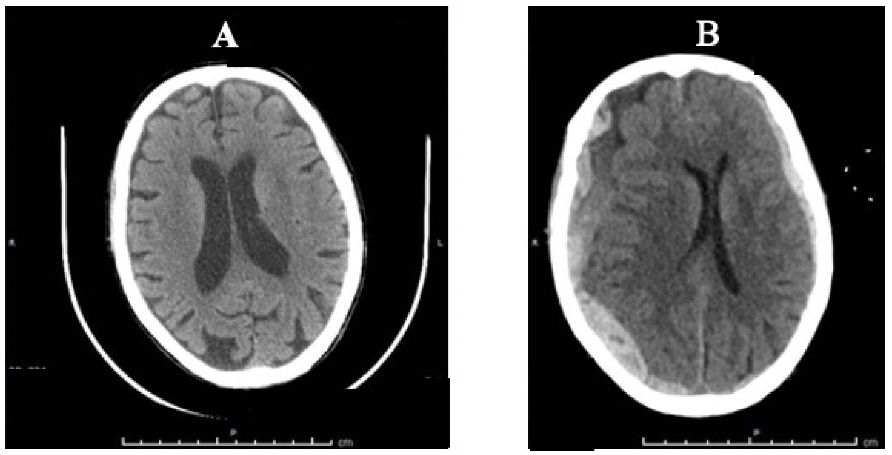
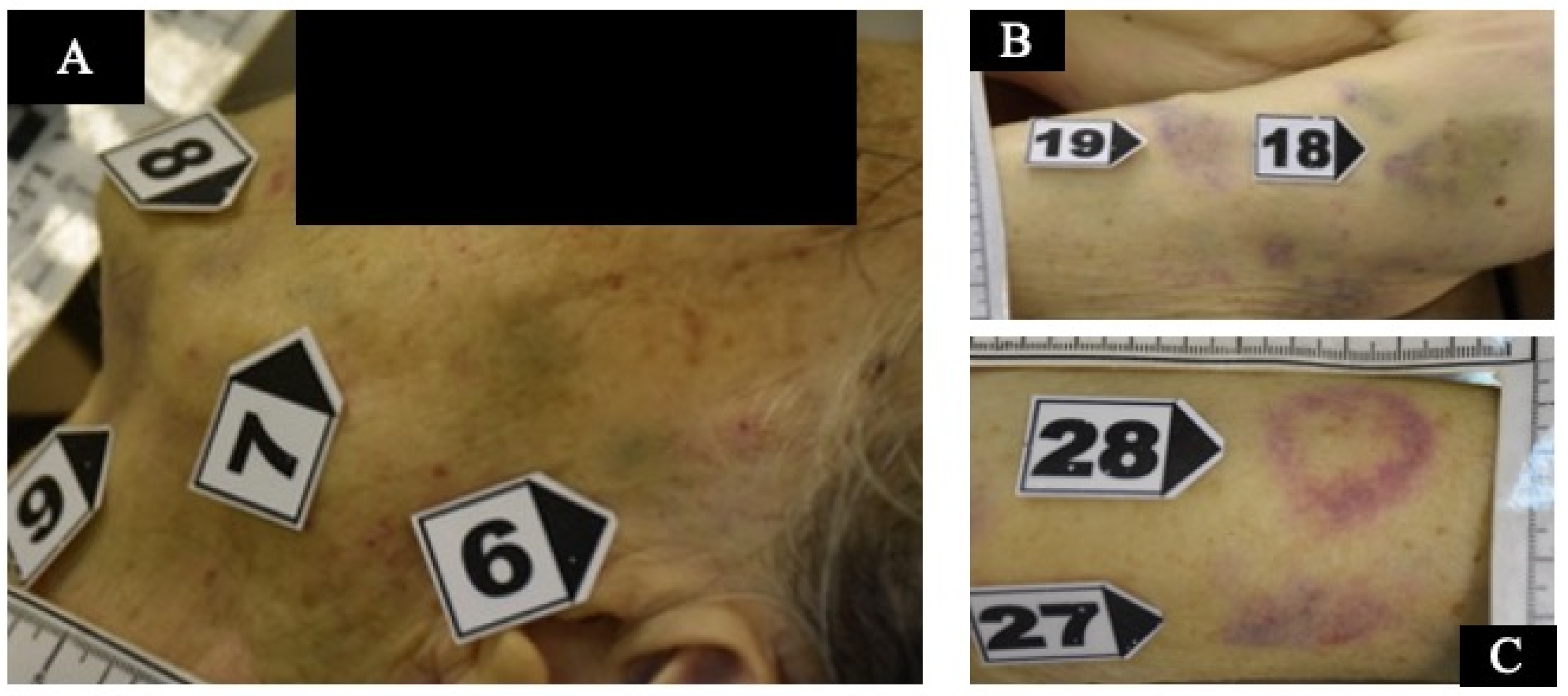
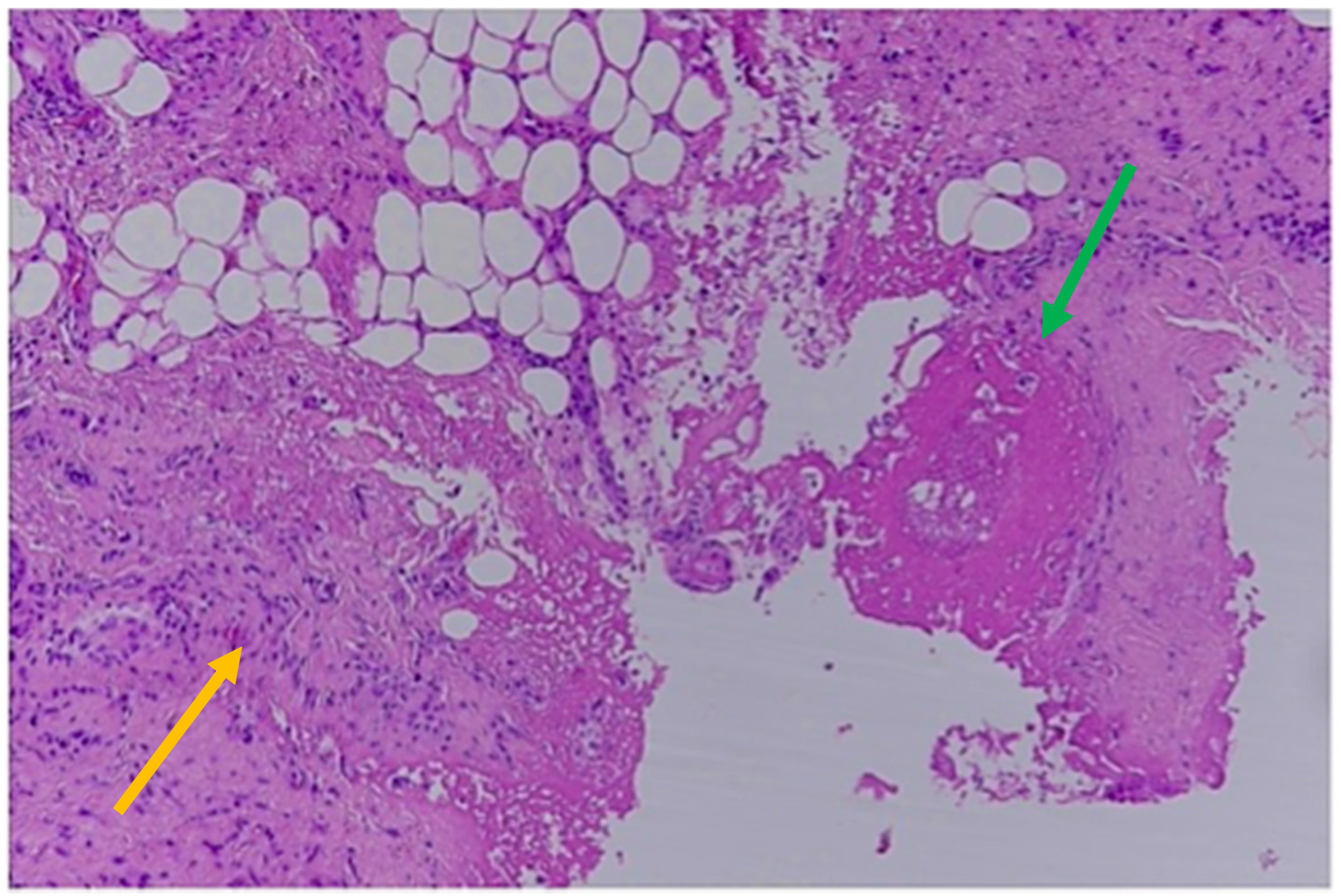
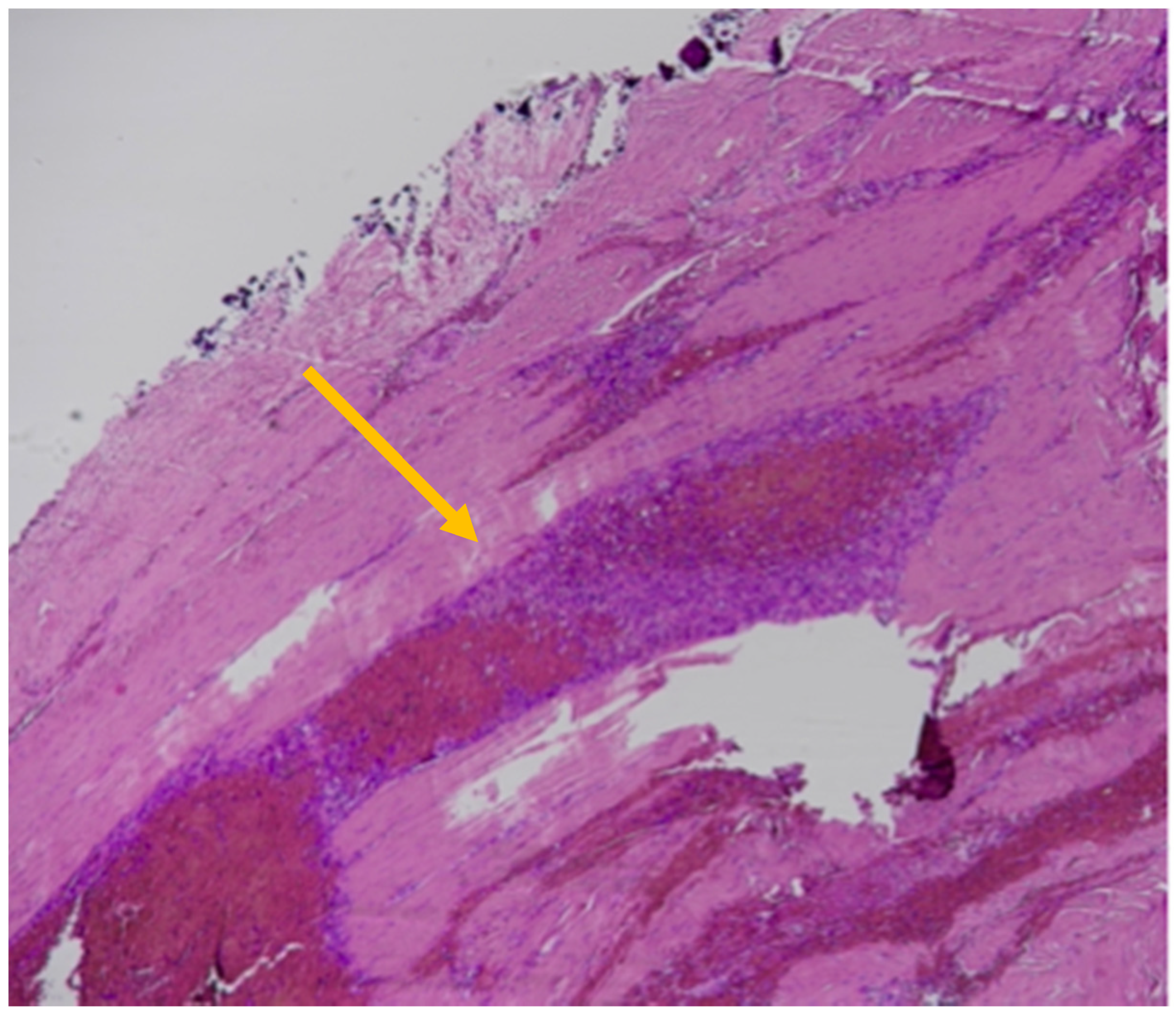
2. Case Report
3. Discussion
4. Conclusions
Author Contributions
Funding
Institutional Review Board Statement
Informed Consent Statement
Data Availability Statement
Conflicts of Interest
References
- Lohr, K.N. (Ed.) Medicare: A Strategy for Quality Assurance; National Academies Press (US): Washington, DC, USA, 1990; Volume 1. [Google Scholar]
- Corbi, G.; Grattagliano, I.; Sabbà, C.; Fiore, G.; Spina, S.; Ferrara, N.; Campobasso, C.P. Elder abuse: Perception and knowledge of the phenomenon by healthcare workers from two Italian hospitals. Intern. Emerg. Med. 2019, 14, 549–555. [Google Scholar] [CrossRef] [PubMed]
- Corbi, G.; Ambrosino, I.; Massari, M.; De Lucia, O.; Simplicio, S.; Dragone, M.; Paolisso, G.; Piccioni, M.; Ferrara, N.; Campobasso, C.P. The potential impact of multidimesional geriatric assessment in the social security system. Aging Clin. Exp. Res. 2018, 30, 1225–1232. [Google Scholar] [CrossRef] [PubMed]
- Available online: https://www.who.int/news-room/fact-sheets/detail/elder-abuse (accessed on 24 December 2022).
- Benbow, S.M.; Bhattacharyya, S.; Kingston, P.; Peisah, C. Invisible and at-risk: Older adults during the COVID-19 pandemic. J. Elder Abus. Negl. 2022, 34, 70–76. [Google Scholar] [CrossRef] [PubMed]
- Du, P.; Chen, Y. Prevalence of elder abuse and victim-related risk factors during the COVID-19 pandemic in China. BMC Public Health 2021, 21, 1096. [Google Scholar] [CrossRef] [PubMed]
- Chang, E.-S.; Levy, B.R. High Prevalence of Elder Abuse during the COVID-19 Pandemic: Risk and Resilience Factors. Am. J. Geriatr. Psychiatry 2021, 29, 1152–1159. [Google Scholar] [CrossRef]
- Makaroun, L.K.; Bachrach, R.L.; Rosland, A.-M. Elder Abuse in the Time of COVID-19—Increased Risks for Older Adults and Their Caregivers. Am. J. Geriatr. Psychiatry 2020, 28, 876–880. [Google Scholar] [CrossRef]
- Hunsaker, D.; Hunsaker, J.C., III. Elder abuse: Challenges for Clinical Forensic Specialists and Forensic Pathologists in the 21st Century. In Forensic Pathology Reviews; Tsokos, M., Ed.; Humana Press Inc.: Totowa, NJ, USA, 2006; Volume 4, pp. 25–62. [Google Scholar]
- Lachs, M.S.; Pillemer, K.A. Elder Abuse. N. Engl. J. Med. 2015, 373, 1947–1956. [Google Scholar] [CrossRef]
- Dong, X.Q. Elder Abuse: Systematic Review and Implications for Practice. J. Am. Geriatr. Soc. 2015, 63, 1214–1238. [Google Scholar] [CrossRef]
- Corbi, G.; Grattagliano, I.; Catanesi, R.; Ferrara, N.; Yorston, G.; Campobasso, C.P. Elderly Residents at Risk for Being Victims or Offenders. J. Am. Med. Dir. Assoc. 2012, 13, 657–659. [Google Scholar] [CrossRef]
- Corbi, G.; Grattagliano, I.; Ivshina, E.; Ferrara, N.; Cipriano, A.S.; Campobasso, C.P. Elderly abuse: Risk factors and nursing role. Intern. Emerg. Med. 2015, 10, 297–303. [Google Scholar] [CrossRef]
- Yon, Y.; Ramiro-Gonzalez, M.; Mikton, C.R.; Huber, M.; Sethi, D. The prevalence of elder abuse in institutional settings: A systematic review and meta-analysis. Eur. J. Public Health 2019, 29, 58–67. [Google Scholar] [CrossRef] [PubMed]
- McCreadie, C.; Bennett, G.; Gilthorpe, M.S.; Houghton, G.; Tinker, A. Elder abuse: Do general practitioners know or care? J. R. Soc. Med. 2000, 93, 67–71. [Google Scholar] [CrossRef] [PubMed] [Green Version]
- Jones, J.S.; Veenstra, T.R.; Seamon, J.P.; Krohmer, J. Elder mistreatment: National survey of emergency physicians. Ann. Emerg. Med. 1997, 30, 473–479. [Google Scholar] [CrossRef]
- Kleinschmidt, K.C. Elder abuse: A review. Ann. Emerg. Med. 1997, 30, 463–472. [Google Scholar] [CrossRef]
- Jones, J.S.; Holstege, C.; Holstege, H. Elder abuse and neglect: Understanding the causes and potential risk factors. Ann. Emerg. Med. 1997, 15, 579–583. [Google Scholar]
- Yon, Y.; Mikton, C.R.; Gassoumis, Z.D.; Wilber, K.H. Elder abuse prevalence in community settings: A systematic review and meta-analysis. Lancet Glob. Health 2017, 5, e147–e156. [Google Scholar] [CrossRef] [Green Version]
- Chandanshive, P.; Subba, S.H.; Parida, S.P.; Mishra, S. Prevalence patterns and associated factors of elder abuse in an urban slum of eastern India. BMC Geriatr 2022, 22, 317. [Google Scholar] [CrossRef]
- Clarke, M.E.; Pierson, W. Management of elder abuse in the emergency department. Emerg. Med. Clin. N. Am. 1999, 17, 631–644. [Google Scholar] [CrossRef] [PubMed]
- Keller, E.; Santos, C.; Cusack, D.; Väli, M.; Ferrara, D.; Ludes, B.; Mangin, P.; Payne-James, J.J.; Vieira, D.N. European council of legal medicine (ECLM) guidelines for the examination of suspected elder abuse. Int. J. Leg. Med. 2019, 133, 317–322. [Google Scholar] [CrossRef]
- Zhu, B.-L.; Oritani, S.; Ishida, K.; Quan, L.; Sakoda, S.; Fujita, M.Q.; Maeda, H. Child and elderly victims in forensic autopsy during a recent 5 year period in the southern half of Osaka city and surrounding areas. Forensic Sci. Int. 2000, 113, 215–218. [Google Scholar] [CrossRef]
- Wiglesworth, A.; Austin, R.; Corona, M.; Schneider, D.; Liao, S.; Gibbs, L.; Mosqueda, L. Bruising as a Marker of Physical Elder Abuse. J. Am. Geriatr. Soc. 2009, 57, 1191–1196. [Google Scholar] [CrossRef] [PubMed]
- Mosqueda, L.; Burnight, K.; Liao, S. The Life Cycle of Bruises in Older Adults. J. Am. Geriatr. Soc. 2005, 53, 1339–1343. [Google Scholar] [CrossRef]
- Collins, K.A. Elder Maltreatment: A Review. Arch. Pathol. Lab. Med. 2006, 130, 1290–1296. [Google Scholar] [CrossRef] [PubMed]
- Akaza, K.; Bunai, Y.; Tsujinaka, M.; Nakamura, I.; Nagai, A.; Tsukata, Y.; Ohya, I. Elder abuse and neglect: Social problems revealed from 15 autopsy cases. Leg. Med. 2003, 5, 7–14. [Google Scholar] [CrossRef] [PubMed]
- Collins, K.A.; Bennett, A.T.; Hanzlick, R.; the Autopsy Committee of the College of American Pathologists. Elder Abuse and Neglect. Arch. Intern. Med. 2000, 160, 1567–1569. [Google Scholar] [CrossRef]
- Paranitharan, P.; Pollanen, M.S. The interaction of injury and disease in the elderly: A case report of fatal elder abuse. J. Forensic Leg. Med. 2009, 16, 346–349. [Google Scholar] [CrossRef]
- Oehmichen, M.; Auer, R.N.; König, H.G. Forensic Neuropathology and Associated Neurology; Springer: Berlin/Heidelberg, Germany, 2006. [Google Scholar]
- Leestma, J.E. Forensic Neuropathology, 3rd ed.; CRC Press: Boca Raton, FL, USA, 2014. [Google Scholar]
- Yee, G.; Jain, A. Geriatric Head Injury. In StatPearls [Internet]; StatPearls Publishing: Treasure Island, FL, USA, 2022. [Google Scholar]
- Saukko, P.; Knight, B. Head and Spinal Injuries. In Knight’s Forensic Pathology, 4th ed.; Saukko, P., Knight, B., Eds.; CRC Press: Broken Sound Parkway, NY, USA, 2016; pp. 185–188. [Google Scholar]
- Dettmeyer, R.B. Forensic Medicine: Fundamentals and Perspectives; Springer: Berlin/Heidelberg, Germany, 2018. [Google Scholar]
- Hung, K.-L. Pediatric abusive head trauma. Biomed. J. 2020, 43, 240–250. [Google Scholar] [CrossRef]
- Payne, F.L.; Fernandez, D.N.; Jenner, L.; Paul, S.P. Recognition and nursing management of abusive head trauma in children. Br. J. Nurs. 2017, 26, 974–981. [Google Scholar] [CrossRef]
- Choudhary, A.K.; Servaes, S.; Slovis, T.L.; Palusci, V.J.; Hedlund, G.L.; Narang, S.K.; Moreno, J.A.; Dias, M.S.; Christian, C.W.; Nelson, M.D.; et al. Consensus statement on abusive head trauma in infants and young children. Pediatr. Radiol. 2018, 48, 1048–1065. [Google Scholar] [CrossRef] [Green Version]
- Pounder, D.J. Shaken Adult Syndrome. Am. J. Forensic Med. Pathol. 1997, 18, 321–324. [Google Scholar] [CrossRef]
- Azari, A.A.; Kanavi, M.R.; Saipe, N.B.; Potter, H.D.; Albert, D.M.; Stier, M.A. Shaken Adult Syndrome. JAMA Ophthalmol 2013, 131, 1468–1470. [Google Scholar] [CrossRef] [PubMed]
- Li, Z.; Wang, J.; Zhang, J.; Jia, M.; Xu, Q.; Chen, M.; Zou, D.; Ma, K.; Chen, Y. Cerebral hemorrhage caused by shaking adult syndrome? Evidence from biomechanical analysis using 3D motion capture and finite element models. Int. J. Leg. Med. 2022, 136, 1621–1636. [Google Scholar] [CrossRef] [PubMed]
- Carrigan, T.D.; Walker, E.; Barnes, S. Domestic violence: The shaken adult syndrome. J. Accid. Emerg. Med. 2000, 17, 138–139. [Google Scholar] [CrossRef] [PubMed]
- Graham, D.I.; Gennarelli, T.A. Trauma. In Greenfield’s Neuropathology, 6th ed.; Graham, D.I., Lantos, P.L., Eds.; Arnold: London, UK, 1997; Volume 1. [Google Scholar]
- Center for Drug Evaluation and Research. Ticlid. Available online: https://www.accessdata.fda.gov/drugsatfda_docs/nda/2001/19-979S018_Ticlid_prntlbl.pdf (accessed on 29 November 2022).
- Center for Drug Evaluation and Research. Ticlopidine. Available online: https://www.accessdata.fda.gov/drugsatfda_docs/anda/99/75161_Ticlopidine%20Hydrochloride_Prntlbl.pdf (accessed on 29 November 2022).
- Martins, W.A.; Teixeira, A.B.; Frigeri, T.M.; Paglioli, E. Spontaneous subdural hematoma associated to Duret hemorrhage. Interdiscip. Neurosurg. 2015, 2, 13–15. [Google Scholar] [CrossRef] [Green Version]
- Maxeiner, H. Detection of ruptured cerebral bridging veins at autopsy. Forensic Sci. Int. 1997, 89, 103–110. [Google Scholar] [CrossRef]
- Sreerama, V.; Ivan, L.P.; Dennery, J.M.; Richard, M.T. Neurosurgical complications of anticoagulant therapy. Can. Med. Assoc. J. 1973, 108, 305–307. [Google Scholar]
- Adhiyaman, V.; Asghar, M.; Ganeshram, K.N.; Bhowmick, B.K. Chronic subdural haematoma in the elderly. Postgrad. Med. J. 2002, 78, 71–75. [Google Scholar] [CrossRef] [Green Version]
- Guyomarc’H, P.; Campagna-Vaillancourt, M.; Kremer, C.; Sauvageau, A. Discrimination of Falls and Blows in Blunt Head Trauma: A Multi-Criteria Approach. J. Forensic Sci. 2010, 55, 423–427. [Google Scholar] [CrossRef]
- Campobasso, C.P.; Laviola, D.; Grattagliano, I.; Strada, L.; Dell’Erba, A.S. Undetected patricide: Inaccuracy of cause of death determination without an autopsy. J. Forensic Leg. Med. 2015, 34, 67–72. [Google Scholar] [CrossRef]
- Feola, A.; De Simone, M.; Ciamarra, P.; Sica, S.; Buonomo, C.; Carfora, A.; Campobasso, C.P. Chronic Encapsulated Intracerebral Hematoma as an Occasional Finding in Sudden Cardiac Death. Healthcare 2022, 10, 2053. [Google Scholar] [CrossRef]


Disclaimer/Publisher’s Note: The statements, opinions and data contained in all publications are solely those of the individual author(s) and contributor(s) and not of MDPI and/or the editor(s). MDPI and/or the editor(s) disclaim responsibility for any injury to people or property resulting from any ideas, methods, instructions or products referred to in the content. |
© 2023 by the authors. Licensee MDPI, Basel, Switzerland. This article is an open access article distributed under the terms and conditions of the Creative Commons Attribution (CC BY) license (https://creativecommons.org/licenses/by/4.0/).
Share and Cite
Bugelli, V.; Campobasso, C.P.; Feola, A.; Tarozzi, I.; Abbruzzese, A.; Di Paolo, M. Accidental Injury or “Shaken Elderly Syndrome”? Insights from a Case Report. Healthcare 2023, 11, 228. https://doi.org/10.3390/healthcare11020228
Bugelli V, Campobasso CP, Feola A, Tarozzi I, Abbruzzese A, Di Paolo M. Accidental Injury or “Shaken Elderly Syndrome”? Insights from a Case Report. Healthcare. 2023; 11(2):228. https://doi.org/10.3390/healthcare11020228
Chicago/Turabian StyleBugelli, Valentina, Carlo Pietro Campobasso, Alessandro Feola, Ilaria Tarozzi, Arturo Abbruzzese, and Marco Di Paolo. 2023. "Accidental Injury or “Shaken Elderly Syndrome”? Insights from a Case Report" Healthcare 11, no. 2: 228. https://doi.org/10.3390/healthcare11020228