
Antigenicity Preservation Is Related to Tissue Characteristics and the Post-Mortem Interval: Immunohistochemical Study and Literature Review

 ,
,
Abstract
:1. Introduction
2. Materials and Methods
2.1. Case Selection
2.2. Histology
2.3. Immunohistochemistry
- (a)
- Vimentin
- (b)
- Ki 67
- (c)
- Cytokeratin
- (d)
- CD20
3. Results
3.1. Morphological Analysis
3.1.1. Skin
3.1.2. Kidney
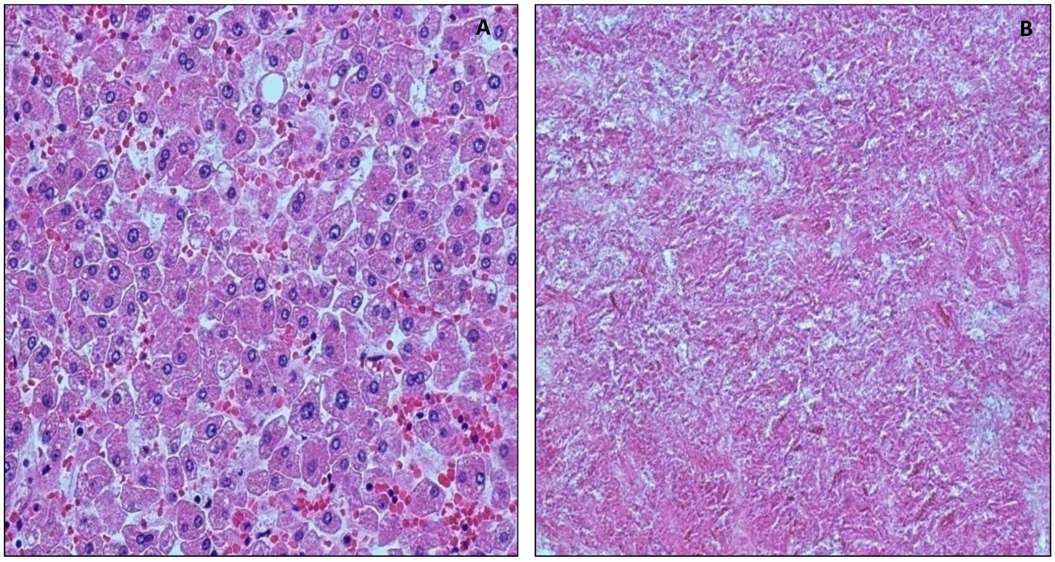
3.1.3. Liver
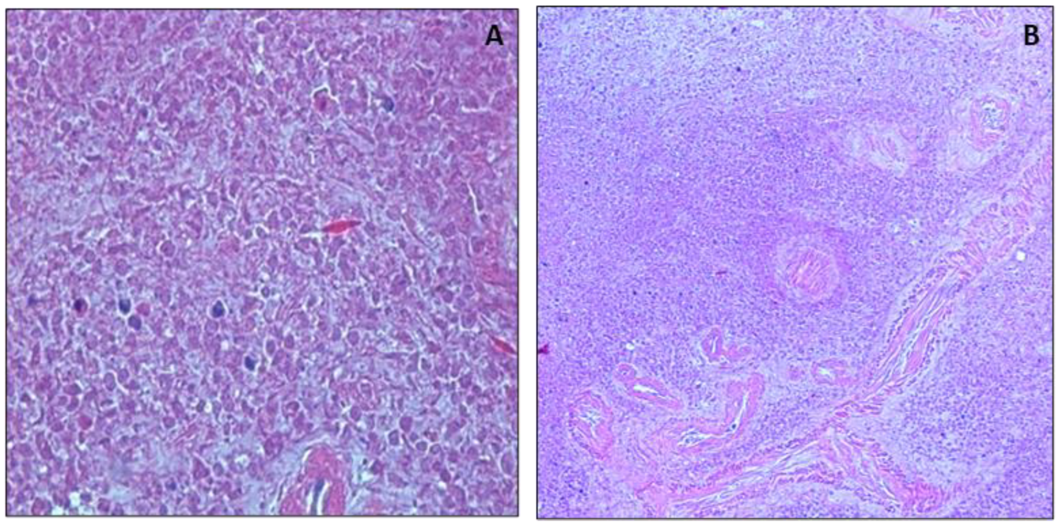
3.1.4. Spleen
3.2. Immunohistochemical Analysis
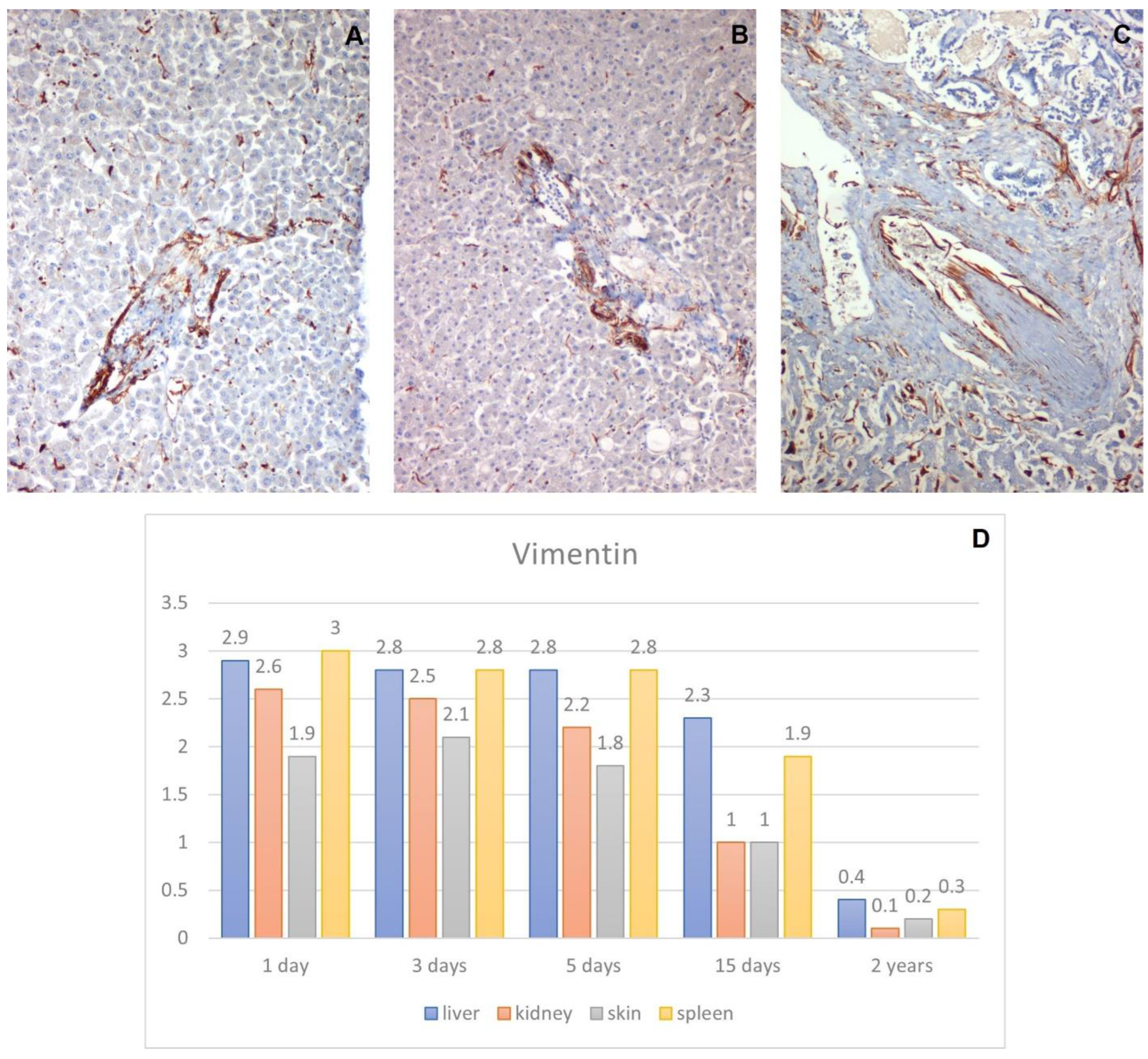
3.2.1. Anti-Vimentin Antibody
3.2.2. Anti-Ki67 Antibody
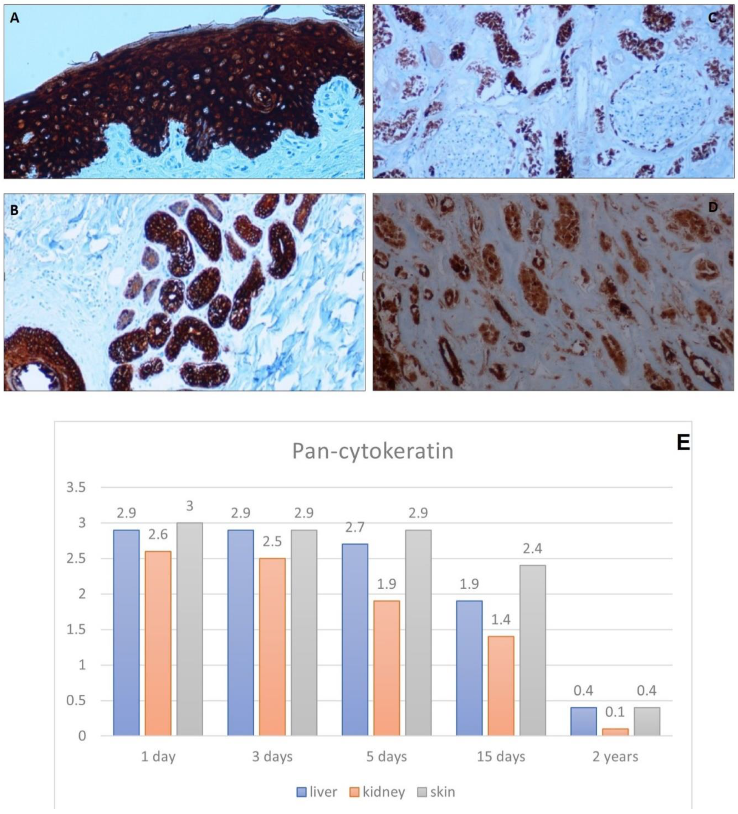
3.2.3. Anti-Pan-Cytokeratin Antibody
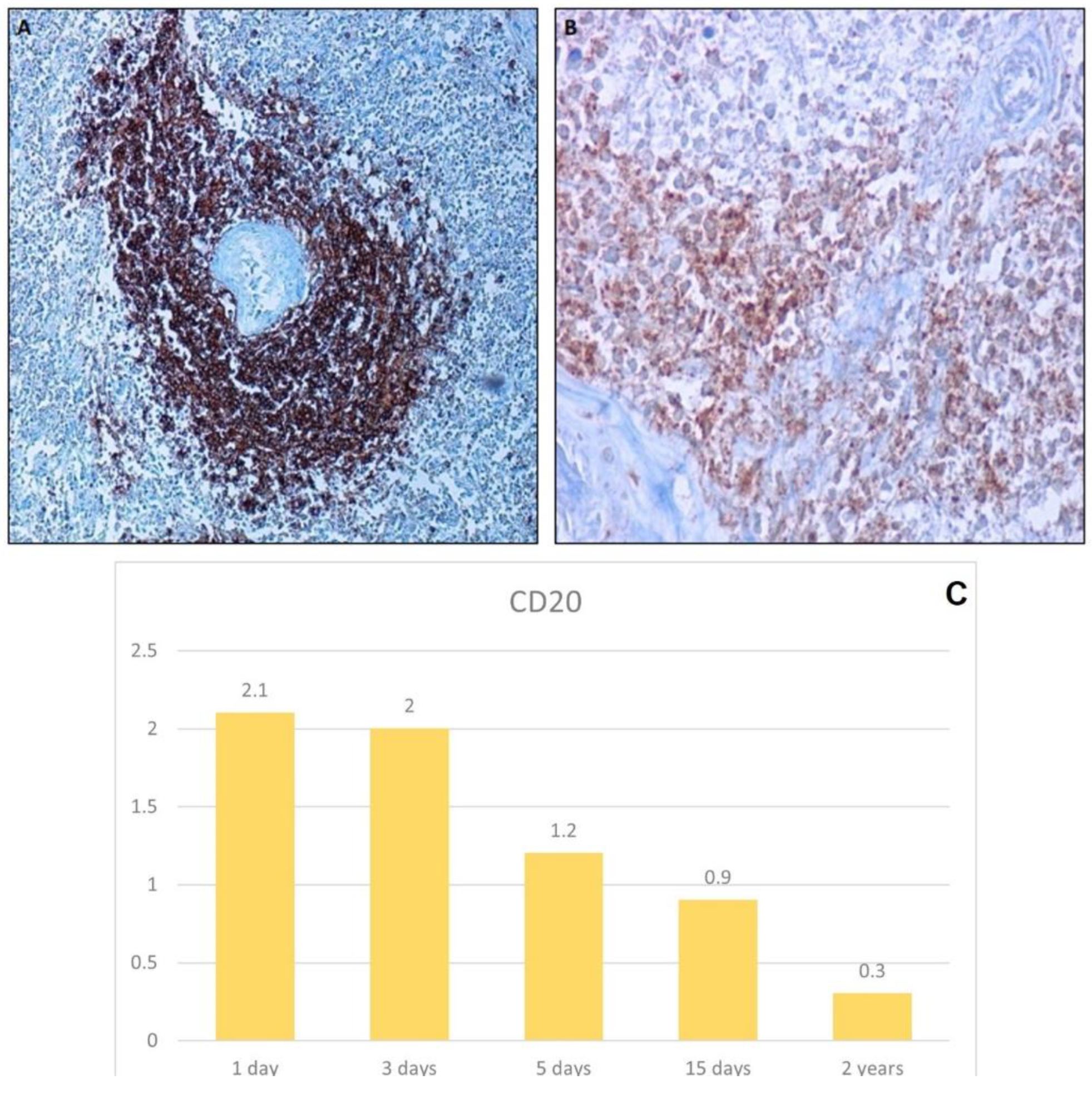
3.2.4. Anti-CD20 Antibody
4. Discussion
5. Conclusions
Author Contributions
Funding
Institutional Review Board Statement
Informed Consent Statement
Data Availability Statement
Conflicts of Interest
References
- Madea, B. Methods for determining time of death. Forensic Sci. Med. Pathol. 2016, 12, 451–485. [Google Scholar] [CrossRef] [PubMed]
- Henssge, C.; Madea, B. Estimation of the time since death. Forensic Sci. Int. 2007, 165, 182–184. [Google Scholar] [CrossRef] [PubMed]
- Wehner, F.; Wehner, H.D.; Subke, J. Delimitation of the time of death by immunohistochemical detection of glucagon in pancreatic alpha-cells. Forensic Sci. Int. 2001, 124, 192–199. [Google Scholar] [CrossRef]
- Pittner, S.; Ehrenfellner, B.; Monticelli, F.C.; Zissler, A.; Sänger, A.M.; Stoiber, W.; Steinbacher, P. Postmortem muscle protein degradation in humans as a tool for PMI delimitation. Int. J. Legal Med. 2016, 130, 1547–1555. [Google Scholar] [CrossRef] [PubMed] [Green Version]
- Zissler, A.; Stoiber, W.; Steinbacher, P.; Geissenberger, J.; Monticelli, F.C.; Pittner, S. Postmortem Protein Degradation as a Tool to Estimate the PMI: A Systematic Review. Diagnostics 2020, 10, 1014. [Google Scholar] [CrossRef] [PubMed]
- Lonergan, E.J.H. The Relationship between the Calpain Enzyme System and the Postmortem Degradation of Selected Myofibrillar Proteins. Retrospective Theses and Dissertations. 1995. Available online: https://lib.dr.iastate.edu/rtd/10928 (accessed on 10 July 2022).
- Sorimachi, Y.; Harada, K.; Yoshida, K. Involvement of calpain in postmortem proteolysis in the rat brain. Forensic Sci. Int. 1996, 81, 165–174. [Google Scholar] [CrossRef]
- Scimeca, M.; Bonfiglio, R.; Varone, F.; Ciuffa, S.; Mauriello, A.; Bonanno, E. Calcifications in prostate cancer: An active phenomenon mediated by epithelial cells with osteoblast-phenotype. Microsc. Res. Tech. 2018, 81, 745–748. [Google Scholar] [CrossRef]
- Scimeca, M.; Giocondo, R.; Montanaro, M.; Granaglia, A.; Bonfiglio, R.; Tancredi, V.; Mauriello, A.; Urbano, N.; Schillaci, O.; Bonanno, E. BMP-2 Variants in Breast Epithelial to Mesenchymal Transition and Microcalcifications Origin. Cells 2020, 9, 1381. [Google Scholar] [CrossRef]
- Urbano, N.; Scimeca, M.; Crocco, A.; Mauriello, A.; Bonanno, E.; Schillaci, O. 18F-Choline PET/CT Identifies High-Grade Prostate Cancer Lesions Expressing Bone Biomarkers. J. Clin. Med. 2019, 8, 1657. [Google Scholar] [CrossRef] [Green Version]
- Chang, L.; Shav-Tal, Y.; Trcek, T.; Singer, R.H.; Goldman, R.D. Assembling an intermediate filament network by dynamic cotranslation. J. Cell Biol. 2006, 172, 747–758. [Google Scholar] [CrossRef] [Green Version]
- Scimeca, M.; Urbano, N.; Bonfiglio, R.; Schillaci, O.; Bonanno, E. Breast osteoblast-like cells: A new biomarker for the management of breast cancer. Br. J. Cancer 2018, 119, 1129–1132. [Google Scholar] [CrossRef] [PubMed] [Green Version]
- Urbano, N.; Scimeca, M.; Di Russo, C.; Mauriello, A.; Bonanno, E.; Schillaci, O. [99mTc]Sestamibi SPECT Can Predict Proliferation Index, Angiogenesis, and Vascular Invasion in Parathyroid Patients: A Retrospective Study. J. Clin. Med. 2020, 9, 2213. [Google Scholar] [CrossRef] [PubMed]
- Brown, D.C.; Gatter, K.C. Monoclonal antibody Ki-67: Its use in histopathology. Histopathology 1990, 17, 489–503. [Google Scholar] [CrossRef]
- Gatti, V.; Fierro, C.; Compagnone, M.; La Banca, V.; Mauriello, A.; Montanaro, M.; Scalera, S.; De Nicola, F.; Candi, E.; Ricci, F.; et al. ΔNp63-Senataxin circuit controls keratinocyte differentiation by promoting the transcriptional termination of epidermal genes. Proc. Natl. Acad. Sci. USA 2022, 119, e2104718119. [Google Scholar] [CrossRef] [PubMed]
- Scimeca, M.; Giannini, E.; Antonacci, C.; Pistolese, C.A.; Spagnoli, L.G.; Bonanno, E. Microcalcifications in breast cancer: An active phenomenon mediated by epithelial cells with mesenchymal characteristics. BMC Cancer. 2014, 14, 286. [Google Scholar] [CrossRef] [Green Version]
- Ordóñez, N.G. Broad-spectrum immunohistochemical epithelial markers: A review. Hum. Pathol. 2013, 44, 1195–1215. [Google Scholar] [CrossRef]
- Scimeca, M.; Bonfiglio, R.; Urbano, N.; Cerroni, C.; Anemona, L.; Montanaro, M.; Fazi, S.; Schillaci, O.; Mauriello, A.; Bonanno, E. Programmed death ligand 1 expression in prostate cancer cells is associated with deep changes of the tumor inflammatory infiltrate composition. Urol. Oncol. 2019, 37, 297.e19–297.e31. [Google Scholar] [CrossRef]
- Middleton, O.; Wheadon, H.; Michie, A. Classical complement pathway. In Encyclopedia of Immunobiology, 1st ed.; Ratcliffe, M.J.H., Ed.; Elsevier: New York, NY, USA, 2006; ISBN 9780123742797. [Google Scholar] [CrossRef]
- Betz, P. Immunohistochemical parameters for the age estimation of human skin wounds. A review. Am. J. Forensic Med. Pathol. 1995, 16, 203–209. [Google Scholar] [CrossRef]
- Gauchotte, G.; Martrille, L.; Plénat, F.; Vignaud, J.M. Les marqueurs de vitalité des blessures en pathologie médicolégale [The markers of wound vitality in forensic pathology]. Ann. Pathol. 2013, 33, 93–101. [Google Scholar] [CrossRef]
- Ortiz-Rey, J.A.; Suárez-Peñaranda, J.M.; San Miguel, P.; Muñoz, J.I.; Rodríguez-Calvo, M.S.; Concheiro, L. Immunohistochemical analysis of P-Selectin as a possible marker of vitality in human cutaneous wounds. J. Forensic Leg Med. 2008, 15, 368–372. [Google Scholar] [CrossRef]
- La Russa, R.; Maiese, A.; Viola, R.V.; De Matteis, A.; Pinchi, E.; Frati, P.; Fineschi, V. Searching for highly sensitive and specific biomarkers for sepsis: State-of-the-art in post-mortem diagnosis of sepsis through immunohistochemical analysis. Int. J. Immunopathol. Pharmacol. 2019, 33, 2058738419855226. [Google Scholar] [CrossRef] [PubMed] [Green Version]
- Ventura Spagnolo, E.; Mondello, C.; Cardia, L.; Minutoli, L.; Puzzolo, D.; Asmundo, A.; Macaione, V.; Alibrandi, A.; Malta, C.; Baldino, G.; et al. Post-Mortem Immunohistochemical Evidence of β2-Adrenergic Receptor Expression in the Adrenal Gland. Int. J. Mol. Sci. 2019, 20, 3065. [Google Scholar] [CrossRef] [Green Version]
- Mansueto, G.; Feola, A.; Zangani, P.; Porzio, A.; Carfora, A.; Campobasso, C.P. A Clue on the Skin: A Systematic Review on Immunohistochemical Analyses of the Ligature Mark. Int. J. Environ. Res. Public Health 2022, 19, 2035. [Google Scholar] [CrossRef] [PubMed]
- Cingolani, M.; Osculati, A.; Tombolini, A.; Tagliabracci, A.; Ghimenton, C.; Ferrara, S.D. Morphology of sweat glands in determining time of death. Int. J. Legal Med. 1994, 107, 132–140. [Google Scholar] [CrossRef]
- Wehner, F.; Wehner, H.D.; Schieffer, M.C.; Subke, J. Delimitation of the time of death by immunohistochemical detection of insulin in pancreatic beta-cells. Forensic Sci. Int. 1999, 105, 161–169. [Google Scholar] [CrossRef]
- Wehner, F.; Wehner, H.D.; Schieffer, M.C.; Subke, J. Delimitation of the time of death by immunohistochemical detection of thyroglobulin. Forensic Sci. Int. 2000, 110, 199–206. [Google Scholar] [CrossRef]
- Wehner, F.; Wehner, H.D.; Subke, J. Delimitation of the time of death by immunohistochemical detection of calcitonin. Forensic Sci. Int. 2001, 122, 89–94. [Google Scholar] [CrossRef]
- Wehner, F.; Steinriede, A.; Martin, D.; Wehner, H.D. Two-tailed delimitation of the time of death by immunohistochemical detection of somatostatin and GFAP. Forensic Sci. Med. Pathol. 2006, 2, 241–247. [Google Scholar] [CrossRef]
- Ortmann, J.; Doberentz, E.; Madea, B. Immunohistochemical methods as an aid in estimating the time since death. Forensic Sci. Int. 2017, 273, 71–79. [Google Scholar] [CrossRef]
- Mazzotti, M.C.; Fais, P.; Palazzo, C.; Fersini, F.; Ruggeri, A.; Falconi, M.; Pelotti, S.; Teti, G. Determining the time of death by morphological and immunohistochemical evaluation of collagen fibers in postmortem gingival tissues. Leg Med. 2019, 39, 1–8. [Google Scholar] [CrossRef]
- Hilbig, H.; Bidmon, H.J.; Oppermann, O.T.; Remmerbach, T. Influence of post-mortem delay and storage temperature on the immunohistochemical detection of antigens in the CNS of mice. Exp. Toxicol. Pathol. 2004, 56, 159–171. [Google Scholar] [CrossRef] [PubMed]
- Libard, S.; Cerjan, D.; Alafuzoff, I. Characteristics of the tissue section that influence the staining outcome in immunohistochemistry. Histochem. Cell Biol. 2019, 151, 91–96. [Google Scholar] [CrossRef] [PubMed] [Green Version]
- Pallocci, M.; Petroni, G.; Treglia, M.; Giammatteo, J.; Marella, G.L.; Arcangeli, M. Law proposal “provisions on the post-mortem body donation and the use of bodies for the purposes of study, scientific research and training”: Comment and analysis of the bill and the historical-juridical-ethical aspects of cadaveric dissection and practice of the donation of a corpse for scientific and medical training purposes. Acta Med. Mediterr. 2020, 36, 999–1005. [Google Scholar] [CrossRef]


| Antibody | Characteristics | Dilution | Retrieval |
|---|---|---|---|
| anti-PanCytokeratin | mouse monoclonal clone [AE1/AE3]; Ventana, Tucson, AZ, USA | Pre-diluted | EDTA citrate pH 7.8 |
| anti-Vimentin | mouse monoclonal clone V9; Ventana, Tucson, AZ, USA | Pre-diluted | EDTA citrate pH 7.8 |
| anti-Ki67 | rabbit monoclonal clone (30-9); Ventana, Tucson, AZ, USA | Pre-diluted | Citrate pH 6.0 |
| anti-CD20 | mouse monoclonal clone L26; Ventana, Tucson, AZ, USA | 1:100 | EDTA citrate pH 7.8 |
| Post Mortem Interval | Hematoxylin—Eosin Staining | Anti-Pan-CK Staining | Anti-Ki 67 Staining | Anti-Vimentin Staining |
|---|---|---|---|---|
| 1 day | Preservation of epidermis and cutaneous annex structure | Positive and specific staining in all samples | Intense staining | Positive and specific staining in all samples |
| 3 days | Preservation of epidermis and cutaneous annex structure | Positive and specific staining in all samples | Intense staining | Positive and specific staining in all samples |
| 5 days | Preservation of epidermis and cutaneous annexes structure | Positive and specific staining in all preparations with reduction in intensity | Quantitative decrease of positive cells | Positive and specific staining in all preparations with reduction in intensity |
| 15 days | Preservation of epidermis and cutaneous annexes structure | Positive and specific staining in all preparations with reduction in intensity | Lack of positive cells. Non-specific background staining for increased cytoplasmic staining of negative cells | Negative staining |
| 2 years | Negative staining | Negative staining | Negative staining | Negative staining |
| Post Mortem Interval | Hematoxylin—Eosin Staining | Anti-Pan-CK Staining | Anti-Vimentin Staining |
|---|---|---|---|
| 1 day | Parenchymal structure clearly detectable with preservation of both glomeruli and tubules | Specific and intense positivity | Specific and intense positivity |
| 3 days | Progressive autolysis of tubular cells with evidence of only isolated tubular structures. Glomeruli still preserved | Specific and intense positivity | Specific and intense positivity |
| 5 days | Parcellular autolysis of glomerular cells | Decreased positivity for autolytic phenomena | Positivity detectable only at the level of some glomerular endothelia while tubules in autolysis are negative |
| 15 days | No longer detectable any cellular structure of the tubules and glomeruli | Non-specific staining | Negative staining |
| 2 years | Negative staining | Negative staining | Negative staining |
| Post Mortem Interval | Hematoxylin—Eosin Staining | Anti Ki-67 Staining | Anti-Vimentin Staining | Anti-CD20 Staining |
|---|---|---|---|---|
| 1 day | Structure of the parenchyma preserved, cellular components are detectable in the red pulp and in the white pulp | Well stained with structure retention | Positive and specific staining in all samples | Positive and specific in all samples |
| 3 days | Lymphatic structures in the white pulp are still well preserved and visible, while a progressive autolysis of red pulp cells is observed | Well stained with structure retention | Negative staining | Positive and specific in all samples |
| 5 days | Progressive autolysis of white pulp lymphocytes appearing as cellular shadows | Rare positive cells | Negative staining | Decreased positivity for autolytic phenomena |
| 15 days | Complete tissue autolysis | Complete tissue autolysis | Negative staining | Non-specific staining |
| 2 years | Negative staining | Negative staining | Negative staining | Negative staining |
| Post Mortem Interval | Hematoxylin—Eosin Staining | Anti-Pan-CK Staining | Anti-Vimentin Staining |
|---|---|---|---|
| 1 day | Well-preserved structure with clear recognition of hepatocytes | Positive and specific staining in all samples | Positive and specific staining in all samples |
| 3 days | Tissue morphology mostly maintained despite foci of hepatocyte autolysis beginning to appear | Positive and specific staining in all samples | Positive and specific staining in all samples |
| 5 days | Enhanced autolysis phenomena | Positive and specific staining in all preparations with reduction in intensity | Positive and specific staining in all preparations with reduction in intensity |
| 15 days | Complete tissue autolysis | Non-specific staining | Negative staining |
| 2 years | Negative staining | Negative staining | Negative staining |
| Author and Year | Tissue Sample | Marker | Sample Size and Study Group |
|---|---|---|---|
| Cingolani M. et al., 1994 [26] | Skin (sweat glands) | S-100 protein, carcinoembryonic antigen (CEA), cytokeratin, actin smooth muscle (ASM) | n = 29 corpses with known time since death; samples were taken at intervals of 3, 6, 9 and 12 h after death |
| Wehner F. et al., 1999 [27] | Pancreas | Insulin | n = 128 corpses with known time since death (between 1 day and 445 days) |
| Wehner F. et al., 2000 [28] | Thyroid | Thyreoglobulin | n = 147 corpses with known time since death (between 1 day and 21 days) |
| Wehner F. et al., 2001 [29] | Thyroid | Calcitonin | n = 136 corpses with known time since death (between 1 day and 21 days) |
| Wehner F. et al., 2001 [3] | Pancreas | Glucagon | n = 214 corpses with known time since death (between 1 day and 21 days) |
| Wehner F. et al., 2006 [30] | Pancreas and brain (frontal cortex) | Glial fibrillary acidic protein (GFAP) and somatostatin | n = 500 corpses with known time since death (between 1 day and 23 days) |
| Ortmann J. et al., 2017 [31] | Pancreas and thyroid | Insulin, glucagon, thyreoglobulin and calcitonin | n = 105 corpses with known time since death (between several hours and 22 days) |
| Mazzotti M.C. et al., 2019 [32] | Gingival tissue, sampled from the superior dental arch | Collagen type I and III | n = 10 corpses with known time since death (between 1 and 9 days) |
Publisher’s Note: MDPI stays neutral with regard to jurisdictional claims in published maps and institutional affiliations. |
© 2022 by the authors. Licensee MDPI, Basel, Switzerland. This article is an open access article distributed under the terms and conditions of the Creative Commons Attribution (CC BY) license (https://creativecommons.org/licenses/by/4.0/).
Share and Cite
Mauriello, S.; Treglia, M.; Pallocci, M.; Bonfiglio, R.; Giacobbi, E.; Passalacqua, P.; Cammarano, A.; D’Ovidio, C.; Marsella, L.T.; Scimeca, M. Antigenicity Preservation Is Related to Tissue Characteristics and the Post-Mortem Interval: Immunohistochemical Study and Literature Review. Healthcare 2022, 10, 1495. https://doi.org/10.3390/healthcare10081495
Mauriello S, Treglia M, Pallocci M, Bonfiglio R, Giacobbi E, Passalacqua P, Cammarano A, D’Ovidio C, Marsella LT, Scimeca M. Antigenicity Preservation Is Related to Tissue Characteristics and the Post-Mortem Interval: Immunohistochemical Study and Literature Review. Healthcare. 2022; 10(8):1495. https://doi.org/10.3390/healthcare10081495
Chicago/Turabian StyleMauriello, Silvestro, Michele Treglia, Margherita Pallocci, Rita Bonfiglio, Erica Giacobbi, Pierluigi Passalacqua, Andrea Cammarano, Cristian D’Ovidio, Luigi Tonino Marsella, and Manuel Scimeca. 2022. "Antigenicity Preservation Is Related to Tissue Characteristics and the Post-Mortem Interval: Immunohistochemical Study and Literature Review" Healthcare 10, no. 8: 1495. https://doi.org/10.3390/healthcare10081495