Article

Biogenic Synthesis, Characterization, and In Vitro Biological Evaluation of Silver Nanoparticles Using Cleome brachycarpa

1
Department of Chemistry, The Women University Multan, Multan 60000, Pakistan
2
Department of Biochemistry and Biotechnology, The Women University Multan, Multan 60000, Pakistan
3
Department of Forestry and Range Management, Bahauddin Zakriya University Multan, Multan 60000, Pakistan
4
Department of Environmental Sciences, The University of Lahore, Lahore 54590, Pakistan
5
Molecular Biology Development and Disease, University College London, London WC1E 6BT, UK
6
Department of Botany and Microbiology, College of Science, King Saud University, Riyadh 11451, Saudi Arabia
*
Author to whom correspondence should be addressed.
Plants 2023, 12(7), 1578; https://doi.org/10.3390/plants12071578
Submission received: 14 January 2023 / Revised: 13 March 2023 / Accepted: 16 March 2023 / Published: 6 April 2023

Abstract

:
The therapeutical attributes of silver nanoparticles (Ag-NPs) in both conditions (in vitro and in vivo) have been investigated using different plants. This study focused on the green chemistry approach that was employed to optimize the synthesis of silver nanoparticles (AgNPs) using Cleome brachycarpa aqueous extract as a reducing and stabilizing agent. The characterization of obtained CB-AgNPs was undertaken using UV-visible spectroscopy, Atomic-force microscopy (AFM), Fourier-Transform Infrared Spectroscopy (FTIR), scanning electron microscopy (SEM), and Energy-Dispersive X-ray (EDX) analysis. Results suggest that CB-AgNPs synthesized via stirring produced small-sized particles with more even distribution. The synthesized silver nanoparticles were spherical with a 20 to 80 nm size range. In vitro studies were used to analyze antioxidant, antidiabetic, and cytotoxic potential under different conditions. The results also indicated that CB-AgNPs may have significant potential as an antidiabetic in low concentrations, but also exhibited potential antioxidant activity at different concentrations. Moreover, the anticancer activity against the breast cell line (MCF-7) with IC50 reached up to 18 μg/mL. These results suggest that green synthesized silver nanoparticles provide a promising phytomedicine for the management of diabetes and cancer therapeutics.

1. Introduction

Nanoparticles are important nanotechnology field products that are used in numerous sizes and shapes as an eco-friendly approach [1,2]. Green nanotechnology is a promising new approach for the efficient synthesis of nanoparticles, including gold, zinc, and silver metals [3]. Most importantly, the conformation and distribution of nanoparticles in solution, along with their enhanced surface areas, help stimulate the physio-chemical properties that are significantly advantageous for anti-microbial growth [4], bio-molecular detection and diagnostics [5], cosmetics [6], biomedical research [7], drug delivery [8], gene delivery [9], and the environment [10]. A diverse range of its applications and advancing capacity to manipulate functions and forms of nanoparticles have increased interest in synthesizing nanoparticles using eco-friendly and effective synthetic techniques.
More recently, the beneficial uses of metal nanoparticles, particularly silver-generated and silver nanoparticles(AgNPs), have proved to carry promising biomedical application potential [11] owing to their antioxidant, antimicrobial, anticancer, and antiplatelet activities and potential angiogenic uses [12,13,14]. However, AgNPs also have numerous biological applications in pharmacology; phytoremediation; photo-electrochemistry; phytomining; single-electron transistors, sensors, and tracers; and wastewater treatment [15,16]. Urban effluent treatment has attracted great attention in developing unique methods including different biological routes to produce nanoparticles of different shapes and sizes. This is important because the physico-chemical methods commonly used to produce nanostructures require a variety of chemicals that are damaging to ecosystem functioning and health [17]. To overcome the challenges of chemical synthesis of AgNPs for biological usage, the present study aimed at testing whether the Cleome species could be used to effectively synthesize and characterize AgNPs.
Previous evidence has shown that plant-based generation of AgNPs (green synthesis nanoparticles) can be accomplished using various plant species and different parts of plants, including leaves, stems, roots, buds, fruit, latex, and bark [18]. By employing spectroscopic analysis, it has been demonstrated that the applications of the most influential phytochemicals involved in reducing silver included flavonoids, phenolic acid, phenols, alkaloids, terpenoids, aldehydes, amides, and ketones [19,20]. Quinone and flavones are among the water-soluble phytochemicals that are considered liable for a speedy reduction. Additionally, reducing the aptitude of secondary metabolites from plant extracts has a higher potential ability to synthesize nanoparticles of small sizes with large surface areas [21]. Using plant extracts seems advantageous in AgNP synthesis due to their function as reducing agents (for instance, Ag+ to Ag0), stabilizing mediators, and avoiding nanoparticle clustering. Therefore, the current study focused on synthesizing AgNPs using Cleome brachycarpa extract, and tested the pharmacological properties of nanoparticles.
Cleome brachycarpa (punwar) is a ethanomedicinal plant of Pakistan [22] found in the semi-arid to subtropical regions of Punjab, Sindh, KPK and adjoining areas [23,24]. It is commonly named wild mustard and tick-weed, and is widely used in conventional medicines, but is also used in cooking (leaves and fresh branches) [25]. Its seed pods are used to make pickles and local delicacies [26]. According to the Ayurvedic medical system, the extracts of Cleome brachycarpa have been used for the treatment of various diseases such as diarrhea, malaria, illness, hypertension, and snakebite. The compounds visconoside, astragalin, kaempferol, quercetin, and kaempferitrin extracted from C. brachycarpa leaves have been shown to display hepatoprotective activity against CCl4-induced hepatotoxicity [27]. Citrus viscosa 3-O-(200-acetyl)-glucoside, also extracted from this plant, is considered to possess antibacterial and anti-inflammatory properties [25].
Previous studies have also used fruit and leaf extract from other plants (Calligonum comosum, Azadirachta indica, and Caesalpinia pulcherrima) to synthesize AgNPs that were tested for antibacterial and cytotoxic effects on human breast cancer cells [28]. However, the investigation of antioxidant properties of plant extracts reported in this study [29] and AgNPs synthesized from C. brachycarpa are unique and have not been reported before. Therefore, in addition to focusing on the synthesis of AgNPs using C. brachycarpa (plant aqueous extract), which functioned as a stabilizer and reducing agent, this study also performed a qualitative pharmacological screening of synthesized AgNPs. Considering the significance of AgNPs and their potential applications in biomedicine, we focused on using the bioreduction method to produce sustainable AgNPs from the aqueous extract of C. brachycarpa leaves. The synthesized AgNPs were characterized using UV–visible spectroscopy for surface plasmon resonance bands, FTIR for functional groups responsible for reduction, and SEM with EDX (Energy-Dispersive X-ray) for a determination of the size of synthesized materials. Furthermore, AgNPs obtained from this process were assessed for antioxidants, antidiabetic action, and in vitro anti-cancer potential. We expected to develop a promising method for the green synthesis of AgNPs using C. brachycarpa extract, and its potential applications were evaluated.

2. Materials and Methods

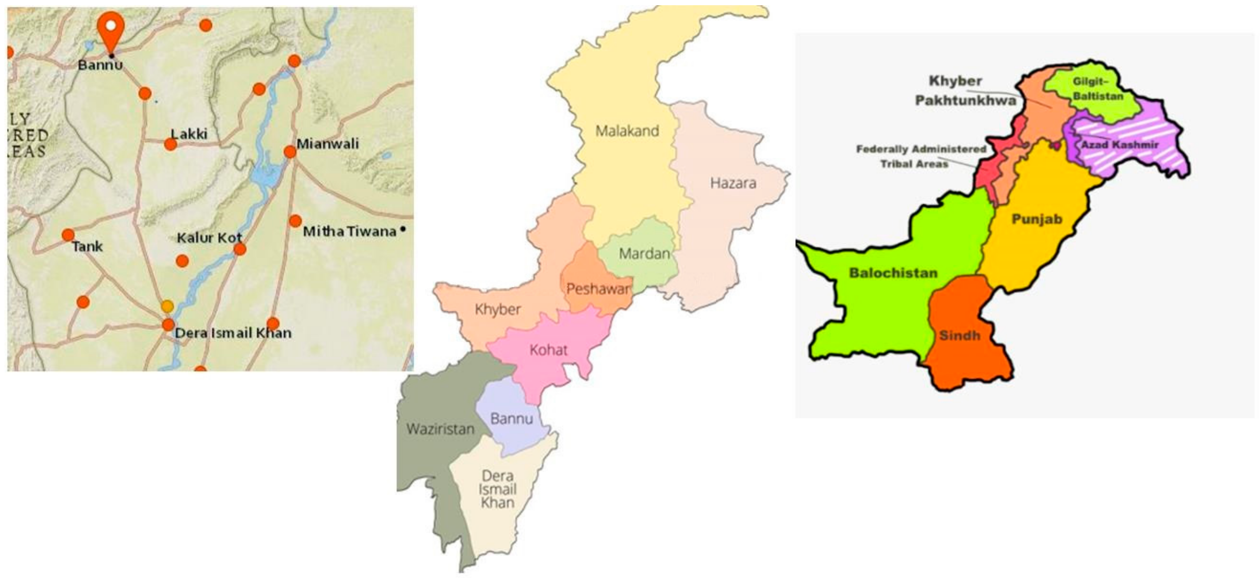
2.1. Collection of Plant Samples

Collection of the C. brachycarpa plant was carried out in March from the KPK province (River Kurram and Hill torrents). The sampled area is located between 32.43 and 33.06 N and between 70.22 and 70.57 E , as shown in the location map (Scheme 1). Plant species were identified and verified based on their vegetative and floral characteristics by Botanist Prof. Dr. Sultan Mehmood from the University of Science and Technology, Bannu (Identification certificate attached in Supplementary Data).

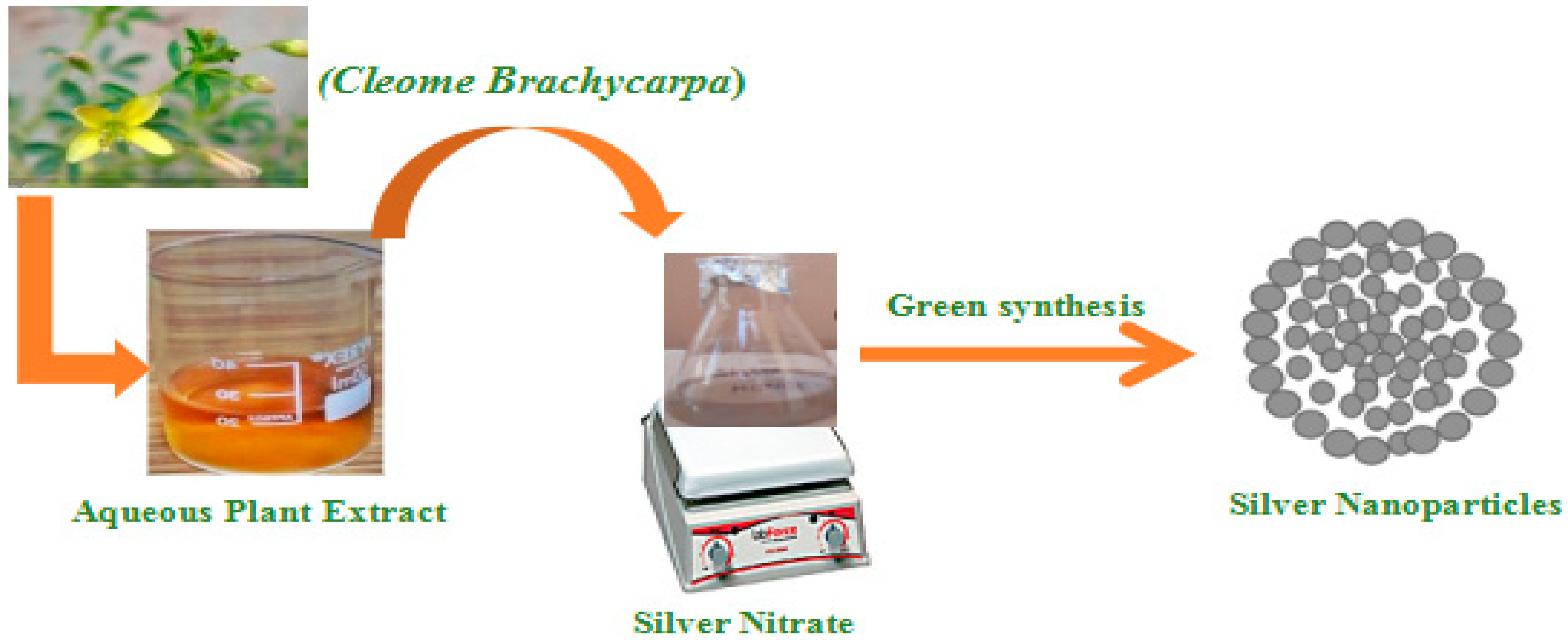
2.2. Aqueous Extraction of C. brachycarpa

The C. brachycarpa extracts were prepared following the procedure reported by Benakashani et al. [30] with some modifications. After washing, plants were dried at room temperature, then ground into powder form and sieved to remove debris and particles. For the extract preparation, about 3 g of powder was macerated at 30 °C, centrifuged at 150 rpm, and then subjected to sonication (model-Elmasonic, E60H) at 40 °C to homogenize the mixture for 2 h in 35 mL distilled water. The brown filtered solution thus obtained was evaporated and lyophilized to acquire pure extract, which was then stored at 4 °C to synthesize nanoparticles.

2.3. Qualitative Phytochemical Study

The extract’s preliminary phytochemical screening was conducted to identify its active constituents, using standard chemical methods reported in the literature [29] and the WHO (World Health Organization) guidelines [31]. A 1% stock solution was prepared for phytochemical screening. The aqueous leaf extract of C. brachycarpa was tested conventionally for its phytoconstituents such as steroids, alkaloids, flavonoids, phenolics, glycosides, tannins, saponins, anthracyanins, terpenoids, etc. Detailed conventional qualitative tests are explained in Table 1.

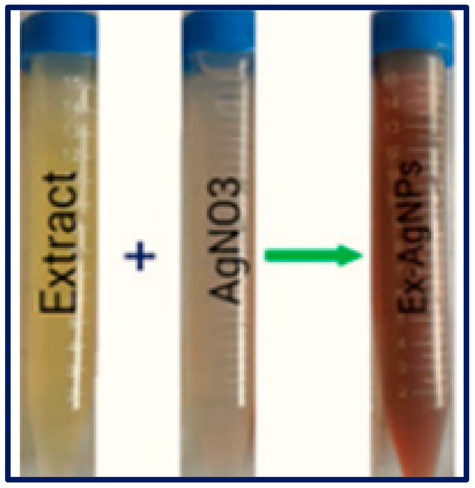
2.4. Silver Nanoparticles SYNTHESIS

The procedures described in the literature on biogenic synthesis were followed in the preparation of silver nanoparticles [32]. Aqueous C. brachycarpa extract (10 mL) was mixed thoroughly with AgNO3 (90 mL, 1 mM) aqueous solution, and the incubation of the mixture was performed at room temperature under static conditions (Scheme 2). The AgNP formation was observed through the color transition from pale yellow to dark brown. The obtained silver nanoparticles were centrifuged at 11,000 rpm for 5 min. The prepared final pellet was dried in an oven at 60 °C as depicted in Scheme 2.

2.5. Characterization Techniques

The biosynthesized AgNPs were morphologically, chemically, and physically characterized by complying with standard procedures provided in the literature using diverse analytic techniques such as UV-visible spectroscopy, Fourier-Transform Infrared Spectroscopy (FTIR), scanning electron microscopy (SEM), Energy-Dispersive X-ray (EDX) analysis, and Atomic Force Spectroscopy (AFM) analysis [33].

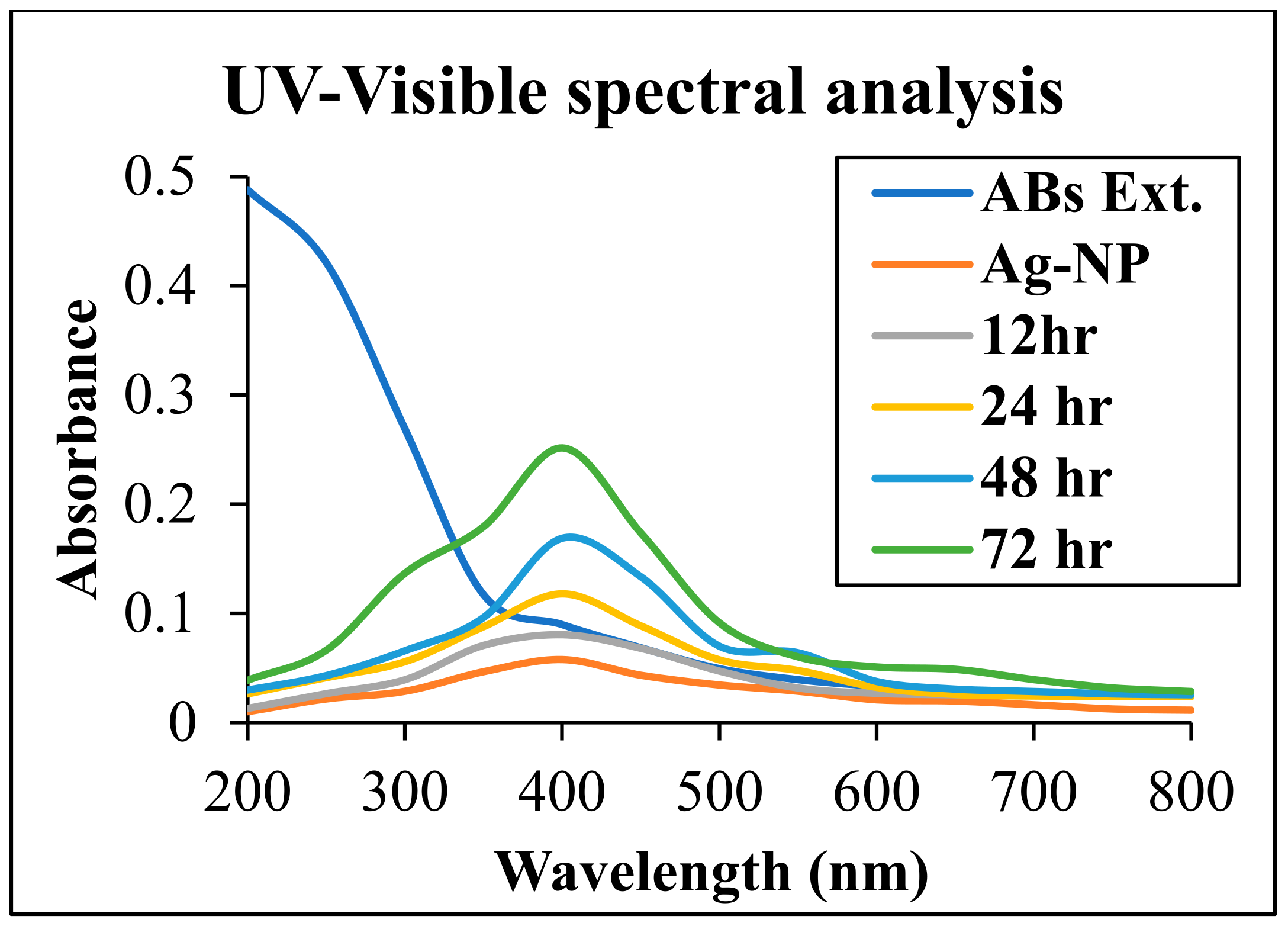
2.5.1. UV–Visible Spectral Analysis

The formation of AgNPs and their stability was examined at different time intervals by measuring absorbance using a double-beam spectrophotometer (HALO, DB-20, COM 1:6,822,0121) for 24 h at a resolution of 2 nm between 200 nm and 700 nm. The solution color was also noted at different intervals.

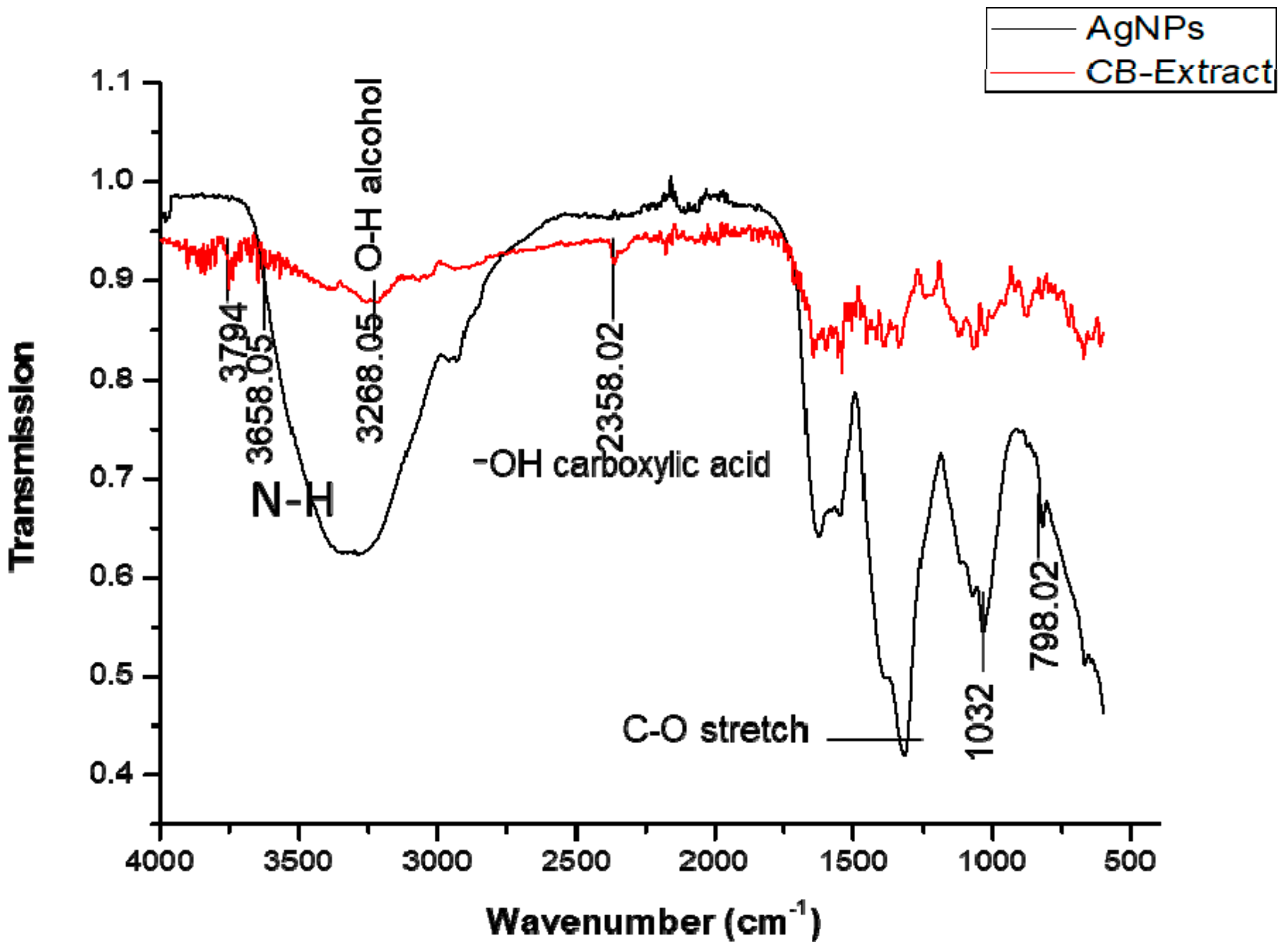
2.5.2. FTIR Analysis

The different spectra were compared with each other in order to analyze the fingerprints of C. brachycarpa represented by the band located at a wavelength range of 400–4000 cm−1 using an FTIR spectrophotometer (Bruker alpha). A total of 2 μg of each mineralized sample was used to form a semitransparent pellet, which allowed light to be transmitted to the detector.

2.5.3. SEM and EDX Analysis

The AgNPs were characterized according to their morphology and size via scanning electron microscopy (SEM) and EDX analysis using a VEGA TESCAN-XMU SEM machine. The AgNPs were evenly spread with carbon tape and sputter-coated with platinum on sample holders. After obtaining SEM images, EDX analysis was performed to confirm the presence of the different elemental compositions of the nanoparticles in the samples, following the method described earlier [34].

2.5.4. Atomic Force Microscope

The AFM was performed using AFM-Agilent 7500 AFM/SPM 3D in the tapping mode. Maximum scan areas were 90 μm × 90 μm. The sample with a cantilever was located by means of a charge-coupled device monitor. Sample imaging related to size was conducted at 10 μm, 5 μm, 2 μm, and 1.2 μm scan widths. The acquired phase and height images thus obtained were analyzed using Asylum Research IGOR PRO-based software.

2.6. Antioxidant Activity

2.6.1. DPPH Scavenging Activity

About 1 mL of different concentrations (25–200 μg/mL) of the compounds were added to methanol to prepare 4 mL of DPPH (0.004%) solution. After incubation for 30 min at room temperature, absorbance at 517 nm against a blank was recorded. The percent inhibition (I (%)) of free radicals by DPPH was calculated as follows [35]:
I (%) = ((Ablank − Asample)/Ablank) × 100.

2.6.2. Reducing Power Assay

The reducing capacities of nanoparticles were assessed with a ferric-reducing antioxidant power assay. About 2.5 mL of phosphate buffer and 2.5 mL potassium ferricyanide (1% w/v) were added to different dilutions of plant extracts. The mixture was incubated at 50 °C for 25 min. The mixture was allowed to cool down, and 2.5 mL of 10% trichloroacetic acid solution was added to it to stop the reaction. The upper layer of the solution (2.5 mL) was equally blended with water and a ferric chloride solution of 0.5 mL. After 30 min, absorbance was measured at 700 nm. The control of BHT of various concentrations (25, 50, 75, 100, and 200 μg/mL) was also prepared without adding the sample.

2.7. In Vitro Antidiabetic Activity

3,5 Dinitrosalicylic Acid Assay (DNSA)
To measure relative enzyme activity, we used a dinitrosalicylic acid assay with some modifications to previous researchers’ methods [36]. The mixture consisted of 500 μL of sodium phosphate buffer (0.02 M with pH 6.9 and 6 mM sodium chloride), salivary amylase (1 mL), and extracts of 400 μL at a concentration between 0.3 and 1.5 mg/mL−1 (w/v) that was incubated for 10 min at 37 °C. After pre-incubation, in the above buffer, 580 μL of 1% (w/v) starch solution was added to each tube. Then, the reaction mixture of 1 mL DNSA reagent was added and placed in a boiling water bath for 5 min, cooled at room temperature, and diluted, and absorbance was measured at 540 nm. A value of 100% enzyme activity was represented by control and plant extract was absent. To remove the absorbance induced by plant extract, the controlled extract was added to the reaction mixture, except for the enzyme. To calculate the percent inhibition of alpha-amylase, the following method was used:
% Relative enzyme activity = (enzyme activity test/enzyme activity control) × 100
% inhibition in the alpha - amylase activity = (100 - % Relative enzyme activity)

2.8. In Vitro Anti-Cancer Potential

2.8.1. Cell Culture

MCF-7 cells were maintained in Dulbecco’s Modified Eagle’s Medium (DMEM) containing 10% fetal bovine serum and 1% penicillin-streptomycin, which was incubated at 37 °C in a humidified incubator. At 80% confluence, cells were reseeded and passaged twice weekly with Trypsin-EDTA [37].

2.8.2. Cell Viability Assay

To assess the cytotoxic impact of AgNPs, an MTT (3-(4, 5-dimethylthiazol-2-yl)-2, 5-diphenyltetrazolium bromide) assay was conducted using 96-well plates [38,39]. Briefly, MCF-7 cells were trypsinized and transferred into 12-well plates at a density of 1 × 10⁵ cells/well in 1 mL of DMEM. After incubation overnight, cells were exposed to different doses (0–100 μg/mL) of the tested AgNPs and incubated again for 24 h. Before undertaking the MTT assay (i.e., addition of 5% MTT (50 μL), samples were added into each well followed by incubation of cells at 37 °C for 4 h. The media were replaced with 100 μL of DMSO for 15 min and, after that, the optical density (OD) at 570 nm was measured using a microplate reader. The percentage of cytotoxicity was measured with the following equation [40]:
% Cell Viability = (OD of test/OD of control) × 100
% Cytotoxicity = 100 - % cell viability
Statistical Analysis:
All the finding results are presented as mean ± SDs. Experiments were conducted in triplicates. One-way ANOVA was used to determine the significances between group subsets, and p-values < 0.05 were considered significant.

3. Results

Various color tests carried out for initial phytochemical analysis of the extract of C. brachycarpa revealed the presence of bioactive contents such as phenolics, flavonoids, alkaloids, Tannins, sapnins, glycosides, terpenoids, and steroids. The concentration of glycosides was not detected in the aqueous extract of C. brachycarpa, as shown in Table 2. In the present study, the biogenic synthesis of silver nanoparticles was performed without adding any external reducing agents where biomolecules act as capping and stabilizing agents. Different techniques characterize these nanoparticles. The basic indication for the formation of AgNPs is a change in color from yellow to dark brown, as shown in Figure 1.

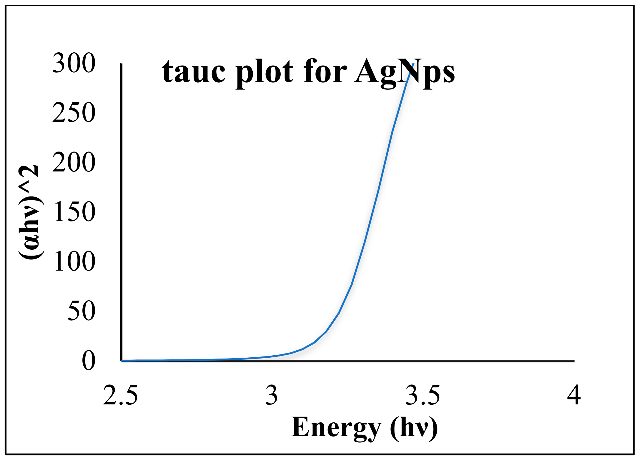
3.1. UV–Visible Spectroscopy

UV–Visible spectroscopy of extracts of the plant was conducted for bio-reduction of Ag+ ions at a temperature of 40 °C. The extract showed maximum absorbance bands at 285 nm and 375 nm. Band I at 375 nm was assigned for the cinnamoyl system of conjugated aromatics, and the band II at 285 nm indicated phenolics (benzoyl system), as shown in Figure 2. A variation in color was also associated with precise peaks characterized by maxima centered at 285 nm and 375 nm. The synthesized silver nanoparticles were interpreted by scanning the UV–visible spectra. The spectrum showed a significant difference in maxima. The highest absorption peak was recorded at 437 nm, while the lowest peak was recorded at 190 nm. The formulated metal nanoparticles were well-established due to the polyphenolic compounds in the extract that re-stained the accumulation. A significant energy band gap of 3.2 eV was calculated by using Tauc plots shown in Figure 3.

3.2. Fourier-Transform Infrared Spectroscopy (FTIR) Analysis

FTIR spectroscopy was used to analyze the surface and functional groups on phytochemicals and their interaction with the AgNPs. The FTIR analysis was carried out within a range of 400–4000 cm−1 (Figure 4). The FTIR spectra of C. brychapara had visible intensities at 3794.05, 3658, 3321, 2952, 1647, 1541, 1389, 1183, 1045, and 711 cm−1. Meanwhile, for Ag nanoparticles, the observable bands were at 3293, 2918, 1610, 1589, 1262, 1032, and 798.02 cm−1. The intense peak between 1800 and 1600 cm−1 indicated the presence of phenolics and the flavonoids group. The peaks shown at 1389 cm−1 correspond to C-N of the amide group, indicating the presence of proteins. The peak at 1189 cm−1 reduced and shifted toward 1032, which represents the phenol cluster. The observed peak at 798.02 described the formation of cation stabilization from oxygen. The strong infrared band (IR) near 3293 cm−1 indicated the hydroxyl group’s O–H vibration of phenols and flavonoids. In conclusion, the FTIR of the extract showed broader peaks that represent various -OH groups in flavonoids and phenolics. On that basis of above mentioned results we can conclude that Quercetin, Gallic acid, and flavone ascorbic acid could be involved in the bioreduction and capping agents for the synthesis of silver nanoparticles.

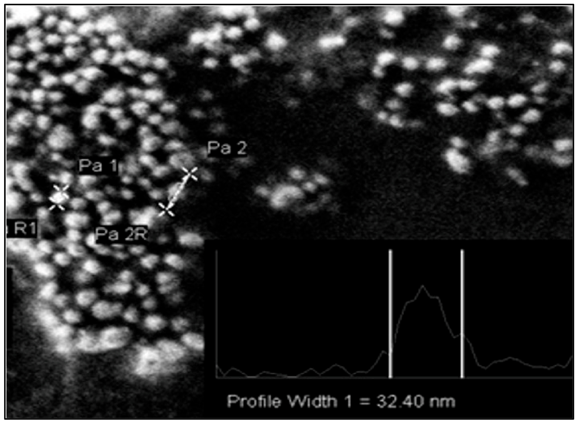
3.3. Scanning Electron Microscope
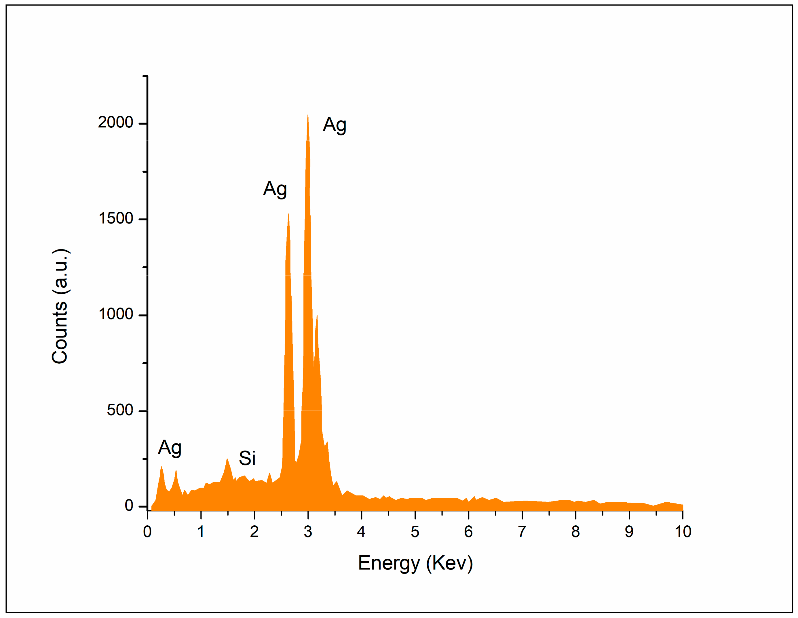
The morphological appearance of biogenically synthesized AgNPs was determined through SEM analysis at 200 nm. It could be perceived that the median particle size of the AgNPs was mainly 20–80 nm, having spherical, uneven shapes due to slight agglomeration (Figure 5).

3.4. EDX Studies

The elemental alignments of biogenically fabricated AgNPs were scanned with EDX, as depicted in Figure 6. The EDX analysis represented the presence of silver along with Si and a few other elements, which might be due to the presence of impurities introduced during the grinding process. The three strong peaks of Ag show the presence of silver at 0.8, 2.7, and 3 keV. These are typical absorption peaks of silver nanomaterials that corroborate with previous studies, as shown in Figure 6.

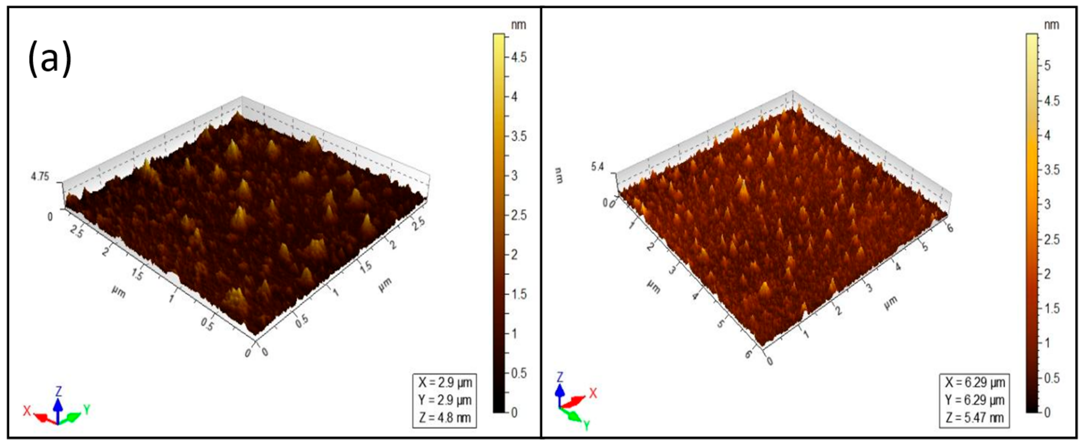
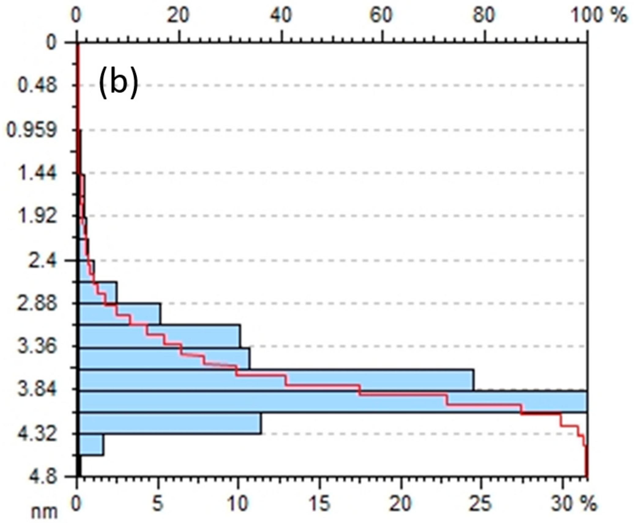
3.5. Atomic Force Microscopy (AFM) Analysis

AFM analysis was employed to characterize the particle size and the morphology of AgNPs. Figure 7a shows the three-dimensional and topographical images of silver nanoparticles at two diverse nanometer ranges. Figure 7b shows the average size range of silver nanoparticles.

3.6. Antioxidant Activity

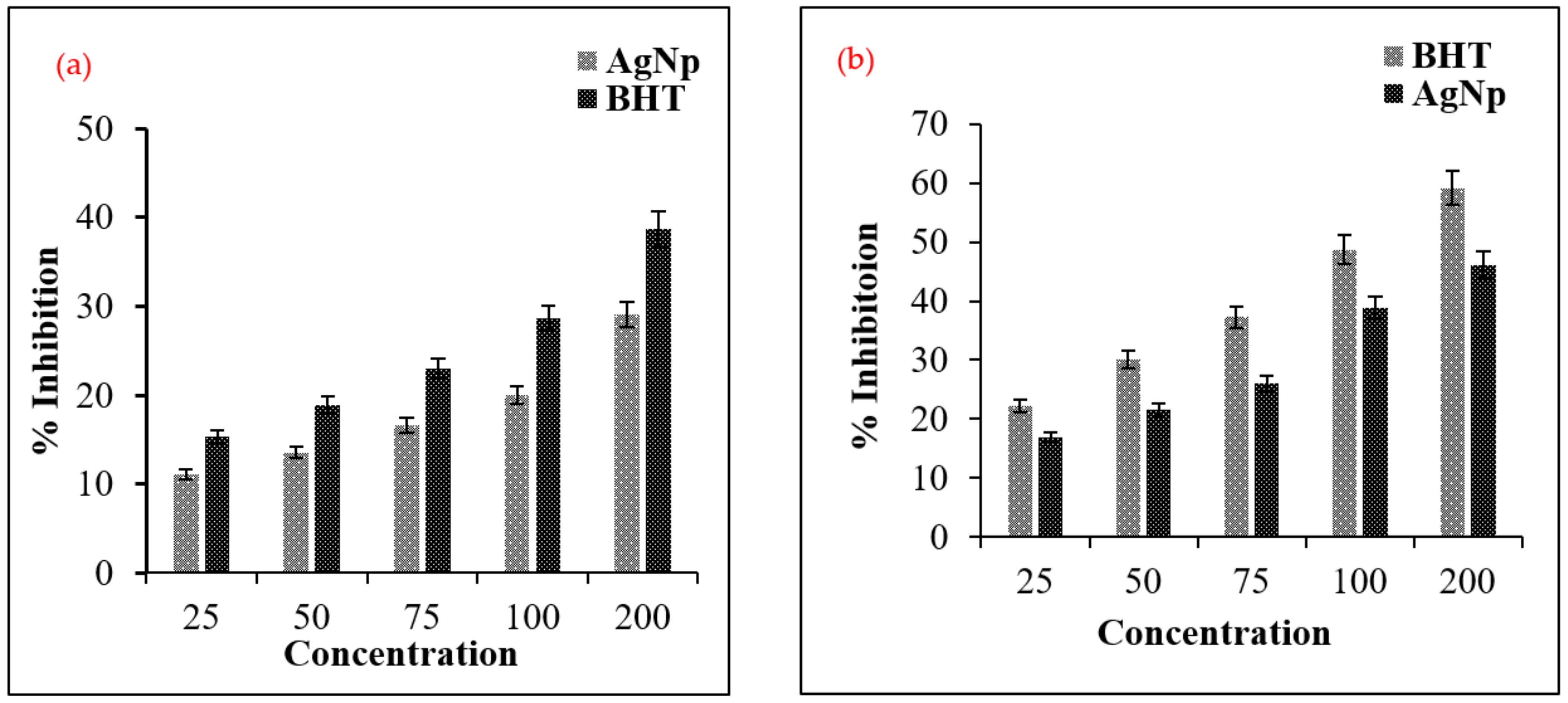
3.6.1. The Antioxidant Activity DPPH and PFRAP

By using DPPH and PFRAP assays, the scavenging potential of eco-friendly synthesized nanoparticles and Cleome brachycarpa extract were assessed, and the BHT was used as a standard. The outcomes from the two assays of antioxidant inhibition are compatible with one another. Our findings indicated that silver nanoparticles and CB extract have dose-dependent activity, as shown in Figure 8. These results show that AgNPs have a potential nearly equal to BHT as a standard at a concentration of 200 μg/mL, as determined through DPPH and PFRAP assays (Figure 9a,b).
For tested samples, the antioxidant potential results indicate the significant value of radical scavenging activity as RSA percent. The purpose of the antioxidant agent is to eliminate free radicals through its reduction by providing hydrogen atoms to free radicals that reduce DPPH radicals (purple color) into diphenylpicrylhydrazine (yellow color), which is the basis for detection using DPPH assay.

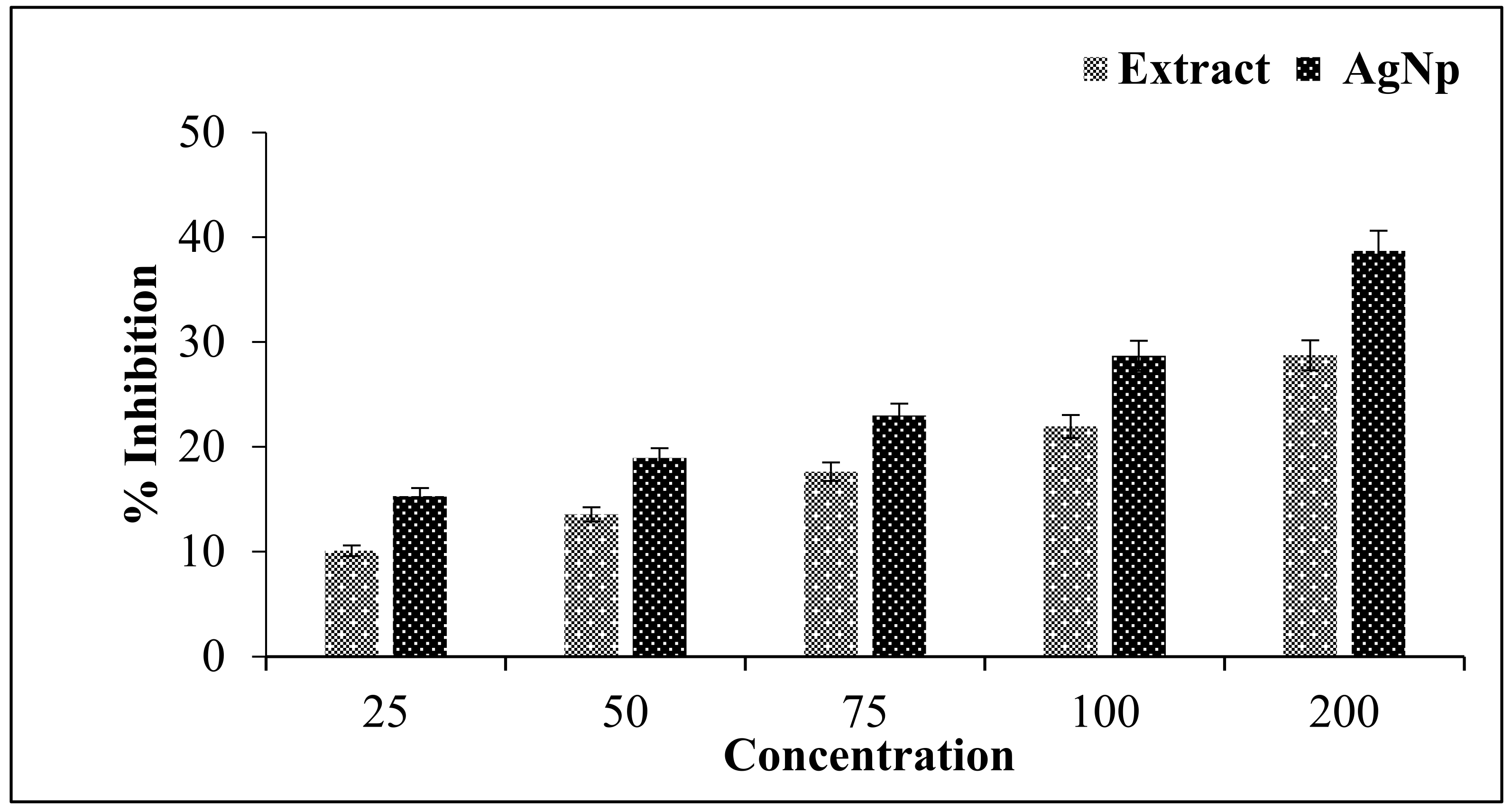
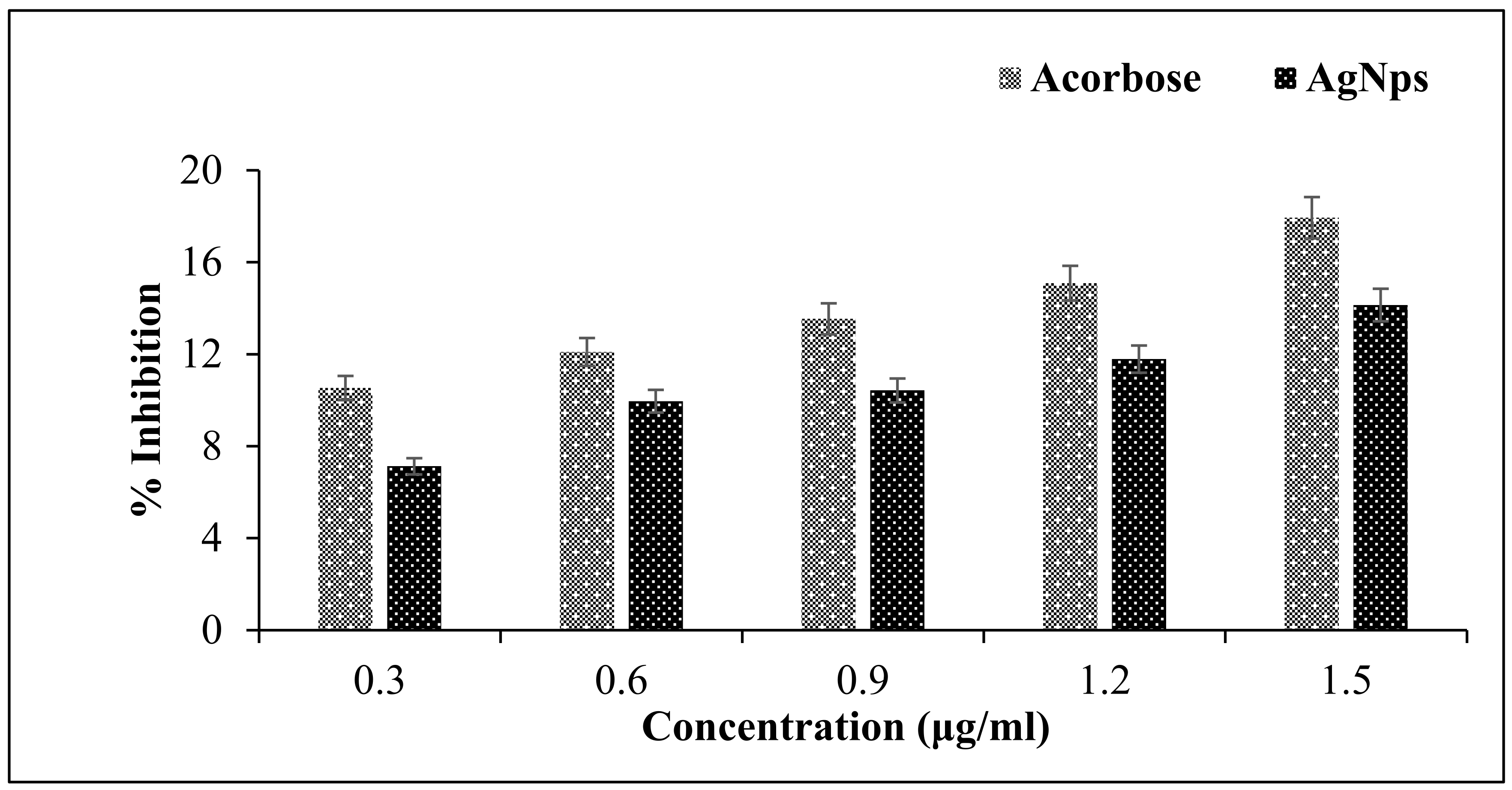
3.6.2. Antidiabetic Activity

AgNps from C. brachycarpa were also assessed for α-amylase activity as a measure of antidiabetic ability. In the stomach, carbohydrate digestive enzymes, i.e., pancreatic α-amylase, are liable for the breakdown of oligosaccharide into monosaccharides suitable for absorption. The outcomes revealed that AgNPs demonstrated the highest percentage value of inhibition on α-amylase activity. The graph in Figure 10 shows the percent inhibition of alpha-amylase according to different concentrations of AgNPs, indicating that α-amylase was significantly inhibited in a concentration-dependent manner.

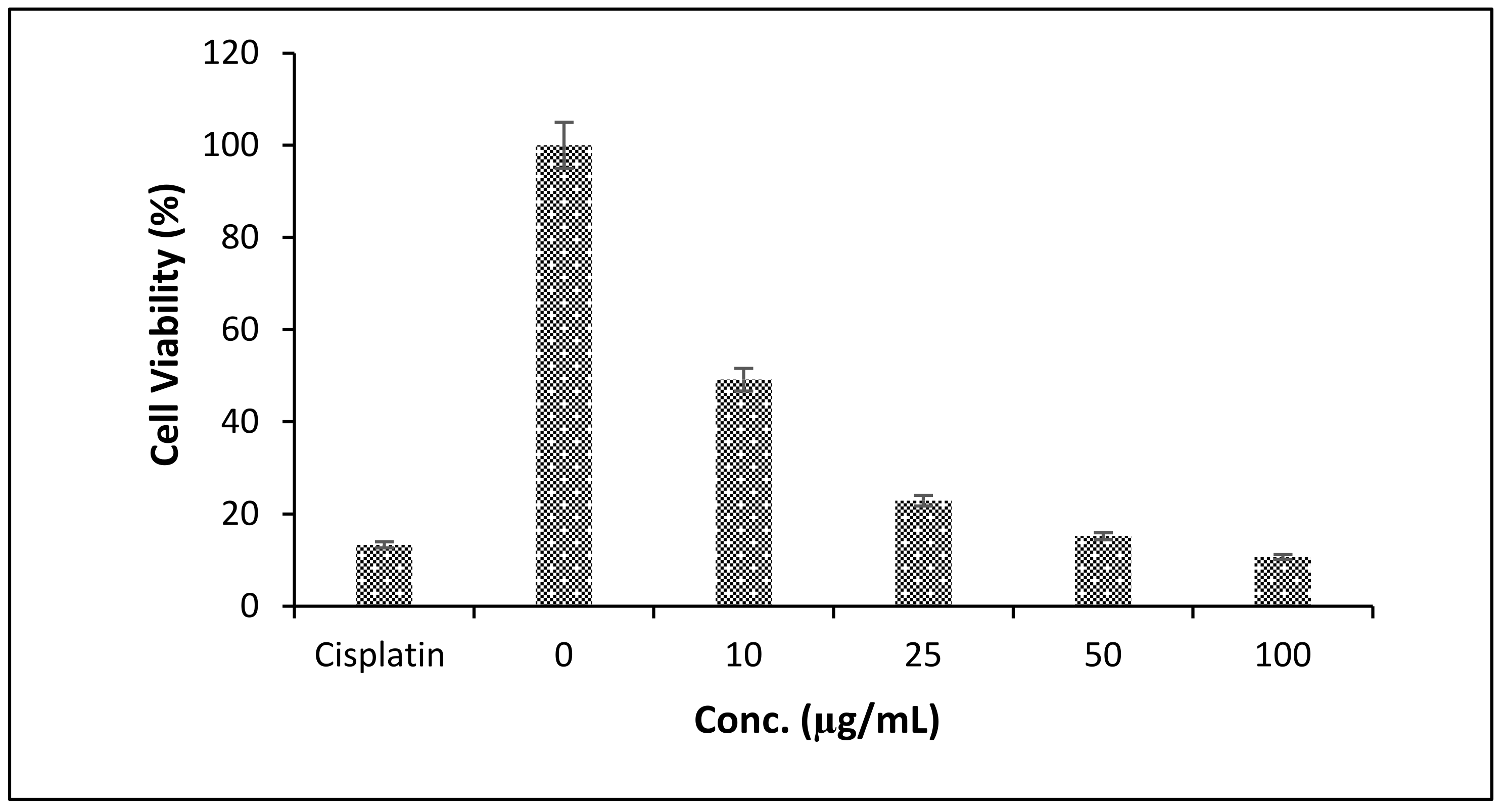
3.6.3. Cytotoxic Potential (MTT Assay)

The AgNPs were evaluated for cytotoxic potential using MTT assay on the basis of the viability of cancer cells (breast cancer cell line (MCF-7)). Figure 11 shows MCF7 cells treated with different concentrations (0–100 μg/mL) of AgNPs and cisplatin for 24 h. Figure 11 shows that results from the MTT assay indicated that a significant reduction in viability was observed in MCF-7 following treatment with different concentrations (0–100 μg/mL) of silver nanoparticles with inhibition concentration (IC50) at 18 μg/mL, while cisplatin, a standard drug, showed a significant reduction of viability at 15 μg/mL.

4. Discussion

Phytochemicals are natural antioxidants derived from plants that can resist and protect themselves from free radicals and their reactive derivatives (ROS). The formation of biogenic AgNPs from plants has been reported by many researchers [6,41,42,43]. In the present work, AgNPs prepared using C. brachycarpa were screened for phytochemicals, including glycosides, alkaloids, flavonoids, proteins, tannins, phenols, terpenoids, steroids, and saponins because the presence of phytochemicals as secondary metabolites (phenolic, flavonoids, and terpenoids) play an important role in the pharmacological activities of plants [44]. Therefore, to produce new drugs to target human diseases, it is important to screen for phytochemical properties. The evidence for diverse phytoconstituents in C. brachycarpa with potential medicinal properties rationalizes our approach to developing novel CB-AgNPs in this study. The primary indication of synthesizing the nanoparticles is the change in color from yellow to dark brown (Figure 1) [33]. The highest absorption peak was observed at 437 nm, while the minimum peak was recorded at 190 nm, as demonstrated in Figure 2. This might be because one of the characteristics of surface plasmon resonance is associated with metal nanostructures. The SPR band caused by the excitation and AgNO3 is responsible for the color transformation of the reaction to reddish-brown. Optical bandgap value for synthesized AgNPs was calculated with a Tauc plot using absorption and wavelength and was converted into energy (Figure 3). The calculated Eg value for silver nanoparticles was 3.2 eV, which demonstrates that visible regions of the spectrum may be able to simulate electron transport in synthesized material [45]. The confirmation of the synthesis of CB-AgNPs from AgNO3 was performed via UV-visible spectral analysis of solution. In this study, to find the functional groups of molecules that are responsible for reducing silver ions and coating, reduced AgNP FTIR analysis was performed. Typically, the FTIR spectrum of synthesized CB-AgNPs shows numerous absorption bands, indicating different stretching and bending modes, including O-H, C-H, C=C, and C-O, as shown in Figure 4. The FTIR analysis shows that the CB-AgNPs are coated with phytochemicals from plant extract that contain a variety of organic molecule functional groups (flavonoids, phenolics, tannins, and terpenoids), as depicted in Figure 4, as reported earlier [46,47,48], because secondary metabolites and phytochemicals in high quantity also contribute to the pharmacological activities of these plants. As previously reported, C-H stretching vibration of the groups (CH2 and CH3) might prompt the absorption peak at 2918 cm−1 [49]. The peaks obtained at wavelengths of 1632 cm−1 and 1500 cm−1 indicate the formation of amide compounds. NH-stretching and peak intensity at a wavelength of 1032 cm−1 is attributed to C-O stretching from plant extracts, as supported by [50]. Scanning electron microscopy revealed the size and morphology of the nanoparticles. The size of CB-AgNPs was 20–80 nm and they were spherical in shape, which might be due to slight agglomeration in the plant extract (Figure 5). Moreover, carbon and oxygen were also detected. EDX analysis showed the maximum percent of Ag (Figure 6), because CB extract (which is specifically rich in flavonoids & phenolic entities) acts as a reducing agent in an artificial process, and this result is in agreement with previous findings [29]. AFM analysis showed the surface area and size distribution of silver nanoparticles, as depicted in Figure 7a,b [51].
The antioxidant properties of purified CB-AgNPs were determined in vitro through analysis reducing power assay, and by measuring DPPH scavenging activity. Figure 8 shows that both CB-AgNPs and extracts were very important in diminishing the scavenging and reduction of free radicals. Similarly, CB-AgNPs with different concentrations compared to a standard showed effective activity of DPPH scavenging. DPPH scavenging assay is an effective method to determine the antioxidant activity (in vitro). DPPH is also used to assess the scavenging capacity following the mechanism that blocks the oxidation of lipids. In addition, it is a stable substance that receives hydrogen from silver nanostructures [52]. Thus, the radical compound is stable and need not be generated. There is a quick response of DPPH with phenols. The conversion of the purple color of the DPPH solution into yellow is due to the allocation of an electron to DPPH radicals from an antioxidant caused by unpaired electrons present in the DPPH solution. This change in coloration was determined via spectrophotometric analysis [53]. CB-AgNPs showed momentous DPPH scavenging activity compared to CB-extract (Figure 8) because of their high secondary metabolites (flavonoids, phenolic, terpenoids, and alkaloids) [29]. It was observed that with the increase in concentration, the antioxidant activity of CB-AgNPs increased (Figure 9a), which was comparable with and in agreement with previous findings [54,55].
A reducing power assay was also used to measure antioxidant potential, in which the reduction of Fe+3 with a yellow color to Fe2+ with a blue color by transfer of an electron instead of a hydrogen atom was observed. The reducing potential also confirmed the antioxidant activity of nanoparticles as compared to extract. The Fe+3 reduced to Fe+2 with an absorption maximum at 593 nm. For determination of the reducing power, CB-AgNPs and BHT were also examined, as shown in Figure 9b. The CB-extract showed a lower reducing ability than CB-AgNPs. Both CB-extract and CB-AgNPs showed a concentration-dependent reducing capacity, as described in previous studies [56]. In the same way, AgNPs were also used to demonstrate in vitro antidiabetic activity, as shown in Figure 10. Diabetes is a category of metabolic disorders characterized by persistently elevated blood glucose levels, which can result in many diseases [57]. Theranostic methods help to decrease hyperglycemia by inhibiting the activity of α-amylase (a carbohydrate-digesting enzyme that increases breakdown into mono-saccharides and contributes to increased blood glucose levels) [57,58]. Acarbose is a glucosidase inhibitor that inhibits human pancreatic amylase. With a delay in carbohydrate digestion, acarbose shows low glucose absorption resulting from the reduction of glucose concentration in blood [59].
Our results show the antidiabetic ability of AgNPs with effective inhibition at low concentrations against carbohydrate digestive enzymes such as α-amylase. This may be a result of the presence of potential α-amylase inhibitors. Previous findings showing the antidiabetic ability of AgNPs with α-glucosidase/amylase inhibition support our results [60,61,62]. In summary, although generally lower compared to corresponding precursors, this study revealed that AgNPs have interesting enzymatic activity, and showed that their significant inhibitory activities are concentration-dependent.
The most significant requirements of cancer therapeutics are selective cytotoxicity and efficacy. A medicine’s ability to cause the apoptosis of cancer cells makes it a successful cancer treatment. Candidate drugs should not negatively influence immune system cells, as these agents attack cancer in conjunction with the immune system. To better understand these effects, silver nanoparticles were tested in MCF-7 cells (human breast cancer cells).The MTT results showed the cytotoxic potential of AgNPs using a colorimetric assay to assess for color changes (purple to yellow) upon reduction of formazan in the presence of mitochondrial succinate dehydrogenase (Figure 11). Similarly, previous findings supported this study regarding the inhibition concentration of AgNPs being 18 μg/mL [40]. In agreement with previous reports, at lower concentrations, toxicity is negligible [47,63,64]. However, the foregoing results suggest that the synthesized AgNPs could be potentially useful in synthesizing diabetes-related drugs, as well as could be a potential substitute for human breast cancer therapy.

5. Conclusions

In short, the biologically active silver nanoparticles were produced via green synthesis in an effective and supportive technique using Cleome brachycarpa extract for the first time. Due to the presence of dynamic phytoconstituents, the nanoparticles provided additional benefits. The synthesized silver nanoparticles were confirmed via different studies. UV-visible spectroscopy showed a maximum absorption peak at 437 nm, which confirms the presence of AgNPs. FTIR showed different functional peaks present in the extract responsible for the reduction and induction of metal–oxygen bonds shown in organic regions. SEM showed the morphology of silver nanoparticles along with elemental composition, as confirmed by EDX. The CB-AgNPs were determined to be valid based on their valuable potential effects on free radicals. Capped phytochemical compounds were responsible for the antioxidant properties of CB-AgNPs, showing the broad potential of silver nanoparticles. Therefore, using CB-AgNPs for a series of applications as a remedial agent to combat free radical-mediated losses shows promise. Furthermore, this study revealed that these results are associated with considerable amounts of phytochemicals such as flavonoids, saponins, tannins, and polyphenolic compounds in CB extract. C. brachycarpa extract-mediated silver nanoparticles may be useful for other biomedical applications, as they showed outstanding efficiency for inhibitory action, scavenging potential, and strong anticancer activity against MCF-7, potentially making them therapeutic alternative agents for human breast cancer treatment. In the future of nanomedicine, AgNPs designed from therapeutic plant extract may be implemented as a medical device.

Supplementary Materials

The following supporting information can be downloaded at: https://www.mdpi.com/article/10.3390/plants12071578/s1, Supplementary Data.

Author Contributions

A.A.S. conceptualized and wrote the main manuscript; M.Z., V.B.-M. and T.S. helped to perform lab analysis; Q.U.Z. analyze the data; G.Y., H.M.A. and M.F.A. reviewed and edited the manuscript in the present form; V.B.-M. supervised and reviewed the manuscript. All authors have read and agreed to the published version of the manuscript.

Funding

This publication was funded by the Researchers Supporting Project number (RSP2023R123), King Saud University, Riyadh, Saudi Arabia.

Institutional Review Board Statement

I, Ghulam Yasin, the corresponding author on behalf of all remaining authors, will conduct myself with integrity, fidelity, and honesty. I will openly take responsibility for my actions and only make agreements that I intend to keep. I will not intentionally engage in or participate in any form of malicious harm to another person or animal.

Data Availability Statement

All data supporting this research article are already included in this manuscript.

Acknowledgments

The authors would like to extend their sincere appreciation to the Researchers Supporting Project number (RSP2023R123), King Saud University, Riyadh, Saudi Arabia. All the contributing authors are grateful to the Department of Chemistry, The Women University Multan, for their financial assistance and for provision of funds. Authors also acknowledge the support of the Molecular Biology Development and Disease laboratory, University College London, United Kingdom.

Conflicts of Interest

The authors report no potential conflict of interest.

References

  1. Ahmad, S.; Munir, S.; Zeb, N.; Ullah, A.; Khan, B.; Ali, J.; Bilal, M.; Omer, M.; Alamzeb, M.; Salman, S.M.; et al. Green nanotechnology: A review on green synthesis of silver nanoparticles—An ecofriendly approach. Int. J. Nanomed. 2019, 14, 5087–5107. [Google Scholar] [CrossRef] [Green Version]
  2. Babayevska, N.; Przysiecka, Ł.; Iatsunskyi, I.; Nowaczyk, G.; Jarek, M.; Janiszewska, E.; Jurga, S. ZnO size and shape effect on antibacterial activity and cytotoxicity profile. Sci. Rep. 2022, 12, 8148. [Google Scholar] [CrossRef]
  3. Lakshmanan, G.; Sathiyaseelan, A.; Kalaichelvan, P.T.; Murugesan, K. Plant-mediated synthesis of silver nanoparticles using fruit extract of Cleome viscosa L.: Assessment of their antibacterial and anticancer activity. Karbala Int. J. Mod. Sci. 2018, 4, 61–68. [Google Scholar]
  4. Nguyen, T.H.A.; Nguyen, V.-C.; Phan, T.N.H.; Le, V.T.; Vasseghian, Y.; Trubitsyn, M.A.; Nguyen, A.-T.; Chau, T.P.; Doan, V.-D. Novel biogenic silver and gold nanoparticles for multifunctional applications: Green synthesis, catalytic and antibacterial activity, and colorimetric detection of Fe(III) ions. Chemosphere 2021, 287, 132271. [Google Scholar] [CrossRef]
  5. Vijayaraghavan, K.; Ashokkumar, T. Plant-mediated biosynthesis of metallic nanoparticles: A review of literature, factors affecting synthesis, characterization techniques and applications. J. Environ. Chem. Eng. 2017, 5, 4866–4883. [Google Scholar] [CrossRef]
  6. Shah, M.; Nawaz, S.; Jan, H.; Uddin, N.; Ali, A.; Anjum, S.; Giglioli-Guivarc’h, N.; Hano, C.; Abbasi, B.H. Synthesis of bio-mediated silver nanoparticles from Silybum marianum and their biological and clinical activities. Mater. Sci. Eng. C 2020, 112, 110889. [Google Scholar] [CrossRef]
  7. Mobaraki, F.; Momeni, M.; Jahromi, M.; Kasmaie, F.M.; Barghbani, M.; Yazdi, M.E.T.; Meshkat, Z.; Shandiz, F.H.; Hosseini, S.M. Apoptotic, antioxidant and cytotoxic properties of synthesized AgNPs using green tea against human testicular embryonic cancer stem cells. Process Biochem. 2022, 119, 106–118. [Google Scholar] [CrossRef]
  8. Busatto, S.; Pham, A.; Suh, A.; Shapiro, S.; Wolfram, J. Organotropic drug delivery: Synthetic nanoparticles and extracellular vesicles. Biomed. Microdevices 2019, 21, 46. [Google Scholar] [CrossRef]
  9. Ansari, M.A.; Yadav, M.K.; Rathore, D.; Svedberg, A.; Karim, Z. Applications of Nanostructured Polymer Composites for Gene Delivery. In Nanostructured Polymer Composites for Biomedical Applications; Elsevier: Amsterdam, The Netherlands, 2019; pp. 211–226. [Google Scholar] [CrossRef]
  10. Dikshit, P.; Kumar, J.; Das, A.; Sadhu, S.; Sharma, S.; Singh, S.; Gupta, P.; Kim, B. Green Synthesis of Metallic Nanoparticles: Applications and Limitations. Catalysts 2021, 11, 902. [Google Scholar] [CrossRef]
  11. Shumail, H.; Khalid, S.; Ahmad, I.; Khan, H.; Amin, S.; Ullah, B. Review on Green Synthesis of Silver Nanoparticles through Plants. Endocr. Metab. Immune Disord.—Drug Targets 2021, 21, 994–1007. [Google Scholar] [CrossRef]
  12. Ali, Z.A.; Yahya, R.; Sekaran, S.D.; Puteh, R. Green Synthesis of Silver Nanoparticles Using Apple Extract and Its Antibacterial Properties. Adv. Mater. Sci. Eng. 2016, 2016. [Google Scholar] [CrossRef] [Green Version]
  13. Baharara, J.; Namvar, F.; Ramezani, T.; Mousavi, M.; Mohamad, R. Silver Nanoparticles Biosynthesized Using Achillea biebersteinii Flower Extract: Apoptosis Induction in MCF-7 Cells via Caspase Activation and Regulation of Bax and Bcl-2 Gene Expression. Molecules 2015, 20, 2693–2706. [Google Scholar] [CrossRef] [Green Version]
  14. Salehi, S.; Shandiz, S.A.; Ghanbar, F.; Darvish, M.R.; Ardestani, M.S.; Mirzaie, A.; Jafari, M. Phytosynthesis of silver nanoparticles using Artemisia marschalliana Sprengel aerial part extract and assessment of their antioxidant, anticancer, and antibacterial properties. Int. J. Nanomed. 2016, 11, 1835–1846. [Google Scholar]
  15. Aktepe, N.; Baran, A. Fast and low-cost biosynthesis of AgNPs with almond leaves: Medical applications with biocompatible structures. Prog. Nutr. 2021, 23, e2021271. [Google Scholar]
  16. Yazdi, M.E.T.; Amiri, M.S.; Akbari, S.; Sharifalhoseini, M.; Nourbakhsh, F.; Mashreghi, M.; Yousefi, E.; Abbasi, M.R.; Modarres, M.; Es-Haghi, A. Green Synthesis of Silver Nanoparticles Using Helichrysum graveolens for Biomedical Applications and Wastewater Treatment. Bionanoscience 2020, 10, 1121–1127. [Google Scholar] [CrossRef]
  17. Anbarasu, A.; Karnan, P.; Deepa, N.; Usha, R. Carica papaya mediated green synthesized silver nanoparticles. Int. J. Curr. Pharm. Res. 2018, 10, 15–20. [Google Scholar] [CrossRef]
  18. Moteriya, P.; Chanda, S. Green Synthesis of Silver Nanoparticles from Caesalpinia pulcherrima Leaf Extract and Evaluation of Their Antimicrobial, Cytotoxic and Genotoxic Potential (3-in-1 System). J. Inorg. Organomet. Polym. Mater. 2020, 30, 3920–3932. [Google Scholar] [CrossRef]
  19. Yarrappagaari, S.; Gutha, R.; Narayanaswamy, L.; Thopireddy, L.; Benne, L.; Mohiyuddin, S.S.; Vijayakumar, V.; Saddala, R.R. Eco-friendly synthesis of silver nanoparticles from the whole plant of Cleome viscosa and evaluation of their characterization, antibacterial, antioxidant and antidiabetic properties. Saudi J. Biol. Sci. 2020, 27, 3601–3614. [Google Scholar] [CrossRef]
  20. Rai, M.; Ingle, A.P.; Gade, A.; Duran, N. Synthesis of silver nanoparticles by Phoma gardeniae and in vitro evaluation of their efficacy against human disease-causing bacteria and fungi. IET Nanobiotechnology 2015, 9, 71–75. [Google Scholar] [CrossRef]
  21. Ahmed, R.H.; Mustafa, D.E. Green synthesis of silver nanoparticles mediated by traditionally used medicinal plants in Sudan. Int. Nano Lett. 2020, 10, 1–14. [Google Scholar] [CrossRef] [Green Version]
  22. Ahmad, V.U.; Qazi, S.; Bin Zia, N.; Xu, C.; Clardy, J. Cleocarpone, a triterpenoid from Cleome brachycarpa. Phytochemistry 1990, 29, 670–672. [Google Scholar] [CrossRef]
  23. Rassouli, E.; Dadras, O.G.; Bina, E.; Asgarpanah, J. The Essential Oil Composition of Cleome brachycarpa Vahl ex DC. J. Essent. Oil Bear. Plants 2014, 17, 158–163. [Google Scholar] [CrossRef]
  24. Hameed, M.; Ashraf, M.; Al-Quriany, F.; Nawaz, T.; Ahmad, M.S.A.; Younis, A.; Naz, N. Medicinal flora of the Cholistan desert: A review. Pak. J. Bot. 2011, 43, 39–50. [Google Scholar]
  25. Chand, J.; Panda, S.R.; Jain, S.; Murty, U.; Das, A.M.; Kumar, G.J.; Naidu, V. Phytochemistry and polypharmacology of cleome species: A comprehensive Ethnopharmacological review of the medicinal plants. J. Ethnopharmacol. 2021, 282, 114600. [Google Scholar] [CrossRef]
  26. Facciola, S. Cornucopia: A Source Book of Edible Plants; Kampong Publications: Vista, CA, USA, 1990. [Google Scholar]
  27. Nguyen, T.P.; Tran, C.L.; Vuong, C.H.; Do, T.H.T.; Le, T.D.; Mai, D.T.; Phan, N.M. Flavonoids with hepatoprotective activity from the leaves of Cleome viscosa L. Nat. Prod. Res. 2017, 31, 2587–2592. [Google Scholar] [CrossRef]
  28. Upadhyay, R. Cleome viscosa Linn: A natural source of pharmaceuticals and pesticides. Int. J. Green Pharm. 2015, 9, 71. [Google Scholar] [CrossRef]
  29. Saleem, T.; Sumra, A.; Khan, S.; Zain, M.; Hassan, W.; Mehdi, S.; Wahid, N.-U.; Kanwal, S.; Gull, T. A Green Nutraceutical Study of Antioxidants Extraction in Cleome brachycarpa—An Ethnomedicinal Plant. Sains Malays. 2020, 49, 1915–1924. [Google Scholar] [CrossRef]
  30. Benakashani, F.; Allafchian, A.; Jalali, S. Biosynthesis of silver nanoparticles using Capparis spinosa L. leaf extract and their antibacterial activity. Karbala Int. J. Mod. Sci. 2016, 2, 251–258. [Google Scholar] [CrossRef] [Green Version]
  31. World Health Organization. Quality Control Methods for Medicinal Plant Materials; World Health Organization: Geneva, Switzerland, 1998. [Google Scholar]
  32. Al-Rajhi, A.M.; Salem, S.S.; Alharbi, A.A.; Abdelghany, T. Ecofriendly synthesis of silver nanoparticles using Kei-apple (Dovyalis caffra) fruit and their efficacy against cancer cells and clinical pathogenic microorganisms. Arab. J. Chem. 2022, 15, 103927. [Google Scholar] [CrossRef]
  33. Das, G.; Patra, J.K.; Debnath, T.; Ansari, A.; Shin, H.-S. Investigation of antioxidant, antibacterial, antidiabetic, and cytotoxicity potential of silver nanoparticles synthesized using the outer peel extract of Ananas comosus (L.). PLoS ONE 2019, 14, e0220950. [Google Scholar] [CrossRef] [Green Version]
  34. Patra, J.K.; Das, G.; Kumar, A.; Ansari, A.; Kim, H.; Shin, H.-S. Photo-mediated Biosynthesis of Silver Nanoparticles Using the Non-edible Accrescent Fruiting Calyx of Physalis peruviana L. Fruits and Investigation of its Radical Scavenging Potential and Cytotoxicity Activities. J. Photochem. Photobiol. B Biol. 2018, 188, 116–125. [Google Scholar] [CrossRef] [PubMed]
  35. Pyrzynska, K.; Pękal, A. Application of free radical diphenylpicrylhydrazyl (DPPH) to estimate the antioxidant capacity of food samples. Anal. Methods 2013, 5, 4288–4295. [Google Scholar] [CrossRef]
  36. Layer, P.; Rizza, R.A.; Zinsmeister, A.R.; Carlson, G.L.; DiMAGNO, E.P. Effect of a Purified Amylase Inhibitor on Carbohydrate Tolerance in Normal Subjects and Patients with Diabetes Mellitus. Mayo Clin. Proc. 1986, 61, 442–447. [Google Scholar] [CrossRef]
  37. Khashan, K.S.; Sulaiman, G.M.; Hussain, S.A.; Marzoog, T.R.; Jabir, M.S. Synthesis, Characterization and Evaluation of Anti-bacterial, Anti-parasitic and Anti-cancer Activities of Aluminum-Doped Zinc Oxide Nanoparticles. J. Inorg. Organomet. Polym. Mater. 2020, 30, 3677–3693. [Google Scholar] [CrossRef]
  38. Al-Salman, H.N.K.; Ali, E.; Jabir, M.; Sulaiman, G.M.; Al-Jadaan, S.A.S. 2-Benzhydrylsulfinyl-N-hydroxyacetamide-Na extracted from fig as a novel cytotoxic and apoptosis inducer in SKOV-3 and AMJ-13 cell lines via P53 and caspase-8 pathway. Eur. Food Res. Technol. 2020, 246, 1591–1608. [Google Scholar] [CrossRef]
  39. Maskell, L.; Mahadeo, A.; Budhram-Mahadeo, V. POU4F2/Brn-3b transcription factor is associated with survival and drug resistance in human ovarian cancer cells. Oncotarget 2018, 9, 36770–36779. [Google Scholar] [CrossRef] [Green Version]
  40. Gomathi, A.; Rajarathinam, S.X.; Sadiq, A.M.; Rajeshkumar, S. Anticancer activity of silver nanoparticles synthesized using aqueous fruit shell extract of Tamarindus indica on MCF-7 human breast cancer cell line. J. Drug Deliv. Sci. Technol. 2019, 55, 101376. [Google Scholar] [CrossRef]
  41. Zhang, Y.-J.; Gan, R.-Y.; Li, S.; Zhou, Y.; Li, A.-N.; Xu, D.-P.; Li, H.-B. Antioxidant Phytochemicals for the Prevention and Treatment of Chronic Diseases. Molecules 2015, 20, 21138–21156. [Google Scholar] [CrossRef] [Green Version]
  42. Rashmi, B.; Harlapur, S.F.; Avinash, B.; Ravikumar, C.; Nagaswarupa, H.; Kumar, M.A.; Gurushantha, K.; Santosh, M. Facile green synthesis of silver oxide nanoparticles and their electrochemical, photocatalytic and biological studies. Inorg. Chem. Commun. 2019, 111, 107580. [Google Scholar] [CrossRef]
  43. Dhar, S.A.; Alam Chowdhury, R.; Das, S.; Nahian, K.; Islam, D.; Gafur, A. Plant-mediated green synthesis and characterization of silver nanoparticles using Phyllanthus emblica fruit extract. Mater. Today Proc. 2021, 42, 1867–1871. [Google Scholar] [CrossRef]
  44. Saratale, R.G.; Saratale, G.D.; Shin, H.S.; Jacob, J.M.; Pugazhendhi, A.; Bhaisare, M.; Kumar, G. New insights on the green synthesis of metallic nanoparticles using plant and waste biomaterials: Current knowledge, their agricultural and environmental applications. Environ. Sci. Pollut. Res. 2017, 25, 10164–10183. [Google Scholar] [CrossRef] [PubMed]
  45. Sumra, A.A.; Aadil, M.; Ejaz, S.R.; Anjum, S.; Saleem, T.; Zain, M.; Alsafari, I.A. Biological synthesis of nanostructured ZnO as a solar-light driven photocatalyst and antimicrobial agent. Ceram. Int. 2022, 48, 14652–14661. [Google Scholar] [CrossRef]
  46. Qais, F.A.; Shafiq, A.; Khan, H.M.; Husain, F.M.; Khan, R.A.; Alenazi, B.; Alsalme, A.; Ahmad, I. Antibacterial Effect of Silver Nanoparticles Synthesized Using Murraya koenigii (L.) against Multidrug-Resistant Pathogens. Bioinorg. Chem. Appl. 2019, 2019, 4649506. [Google Scholar] [CrossRef] [PubMed] [Green Version]
  47. Sabapathi, N.; Ramalingam, S.; Aruljothi, K.N.; Lee, J.; Barathi, S. Characterization and Therapeutic Applications of Biosynthesized Silver Nanoparticles Using Cassia auriculate Flower Extract. Plants 2023, 12, 707. [Google Scholar] [CrossRef]
  48. Ahmad, A.; Javed, M.S.; Khan, S.; Almutairi, T.M.; Mohammed, A.A.; Luque, R. Green synthesized Ag decorated CeO2 nanoparticles: Efficient photocatalysts and potential antibacterial agents. Chemosphere 2022, 310, 136841. [Google Scholar] [CrossRef]
  49. Alsafari, I.A.; Chaudhary, K.; Warsi, M.F.; Warsi, A.Z.; Waqas, M.; Hasan, M.; Jamil, A.; Shahid, M. A facile strategy to fabricate ternary WO3/CuO/rGO nano-composite for the enhanced photocatalytic degradation of multiple organic pollutants and antimicrobial activity. J. Alloys Compd. 2023, 938, 168537. [Google Scholar] [CrossRef]
  50. Anwar, M.; Alghamdi, K.S.; Zulfiqar, S.; Warsi, M.F.; Waqas, M.; Hasan, M. Ag-decorated BiOCl anchored onto the g-C3N4 sheets for boosted photocatalytic and antimicrobial activities. Opt. Mater. 2023, 135, 113336. [Google Scholar] [CrossRef]
  51. Jain, N.; Jain, P.; Rajput, D.; Patil, U.K. Green synthesized plant-based silver nanoparticles: Therapeutic prospective for anticancer and antiviral activity. Micro Nano Syst. Lett. 2021, 9, 5. [Google Scholar] [CrossRef]
  52. Keerthiga, N.; Anitha, R.; Rajeshkumar, S.; Lakshmi, T. Antioxidant Activity of Cumin Oil Mediated Silver Nanoparticles. Pharmacogn. J. 2019, 11, 787–789. [Google Scholar] [CrossRef] [Green Version]
  53. Phaniendra, A.; Jestadi, D.B.; Periyasamy, L. Free Radicals: Properties, Sources, Targets, and Their Implication in Various Diseases. Indian J. Clin. Biochem. 2015, 30, 11–26. [Google Scholar] [CrossRef] [Green Version]
  54. Das, G.; Shin, H.-S.; Patra, J.K. Comparative Bio-Potential Effects of Fresh and Boiled Mountain Vegetable (Fern) Extract Mediated Silver Nanoparticles. Plants 2022, 11, 3575. [Google Scholar] [CrossRef] [PubMed]
  55. Saravanakumar, A.; Ganesh, M.; Jayaprakash, J.; Jang, H.T. Biosynthesis of silver nanoparticles using Cassia tora leaf extract and its antioxidant and antibacterial activities. J. Ind. Eng. Chem. 2015, 28, 277–281. [Google Scholar] [CrossRef]
  56. Reddy, N.J.; Vali, D.N.; Rani, M.; Rani, S.S. Evaluation of antioxidant, antibacterial and cytotoxic effects of green synthesized silver nanoparticles by Piper longum fruit. Mater. Sci. Eng. C 2014, 34, 115–122. [Google Scholar] [CrossRef] [PubMed]
  57. Saratale, R.G.; Shin, H.S.; Kumar, G.; Benelli, G.; Kim, D.-S.; Saratale, G.D. Exploiting antidiabetic activity of silver nanoparticles synthesized using Punica granatum leaves and anticancer potential against human liver cancer cells (HepG2). Artif. Cells Nanomed. Biotechnol. 2017, 46, 211–222. [Google Scholar] [CrossRef] [PubMed] [Green Version]
  58. Etxeberria, U.; de la Garza, A.L.; Campión, J.; Martínez, J.A.; Milagro, F.I. Antidiabetic effects of natural plant extracts via inhibition of carbohydrate hydrolysis enzymes with emphasis on pancreatic alpha amylase. Expert Opin. Ther. Targets 2012, 16, 269–297. [Google Scholar] [CrossRef] [PubMed] [Green Version]
  59. Wettergreen, S.A.; Sheth, S.; Malveaux, J. Effects of the addition of acarbose to insulin and non-insulin regimens in veterans with type 2 diabetes mellitus. Pharm. Pract. 2016, 14, 832. [Google Scholar] [CrossRef] [Green Version]
  60. Jini, D.; Sharmila, S. Green synthesis of silver nanoparticles from Allium cepa and its in vitro antidiabetic activity. Mater. Today Proc. 2020, 22, 432–438. [Google Scholar] [CrossRef]
  61. Patra, J.K.; Das, G.; Shin, H.S. Facile green biosynthesis of silver nanoparticles using Pisum sativum L. outer peel aqueous extract and its antidiabetic, cytotoxicity, antioxidant, and antibacterial activity. Int. J. Nanomed. 2019, 14, 6679–6690. [Google Scholar] [CrossRef] [Green Version]
  62. Rani, R.; Sharma, D.; Chaturvedi, M.; Yadav, J.P. Green Synthesis, Characterization and Antibacterial Activity of Silver Nanoparticles of Endophytic Fungi Aspergillus terreus. J. Nanomed. Nanotechnol. 2017, 8, 457. [Google Scholar] [CrossRef]
  63. Mani, M.; Okla, M.K.; Selvaraj, S.; Kumar, A.R.; Kumaresan, S.; Muthukumaran, A.; Kaviyarasu, K.; El-Tayeb, M.A.; Elbadawi, Y.B.; Almaary, K.S.; et al. A novel biogenic Allium cepa leaf mediated silver nanoparticles for antimicrobial, antioxidant, and anticancer effects on MCF-7 cell line. Environ. Res. 2021, 198, 111199. [Google Scholar] [CrossRef]
  64. Rashidipour, M.; Heydari, R. Biosynthesis of silver nanoparticles using extract of olive leaf: Synthesis and in vitro cytotoxic effect on MCF-7 cells. J. Nanostructure Chem. 2014, 4, 1–6. [Google Scholar] [CrossRef] [Green Version]
Scheme 1. Study area map and geographical distribution of plants in Pakistan.
Plants 12 01578 sch001
Scheme 2. Schematic diagram for biological synthesis of AgNPs.
Plants 12 01578 sch002
Figure 1. Color change indication of formation of AgNPs.
Plants 12 01578 g001
Figure 2. UV–Visible spectrum at different time durations.
Plants 12 01578 g002
Figure 3. Energy Bandgap determined using Tauc plots.
Plants 12 01578 g003
Figure 4. FTIR spectrum of biogenically synthesized silver nanoparticles with extract.
Plants 12 01578 g004
Figure 5. SEM image of CB-AgNPs obtained from Cleome brachycarpa.
Plants 12 01578 g005
Figure 6. EDX spectrum of CB-AgNPs.
Plants 12 01578 g006
Figure 7. (a) 3D Atomic Force microscopy of silver nanoparticles; (b) average size range of AgNPs.
Plants 12 01578 g007aPlants 12 01578 g007b
Figure 8. Antioxidant Potential of Extract with AgNPs.
Plants 12 01578 g008
Figure 9. The activities of antioxidants using (a) DPPH (b) PFRAP assay.
Plants 12 01578 g009
Figure 10. α-amylase activity of AgNPs.
Plants 12 01578 g010
Figure 11. Anticancer potential of untreated control, cisplatin, and silver nanoparticles at different concentrations against MCF-7.
Plants 12 01578 g011
Table 1. Phytochemical screening tests and their indications.
Sr no.PhytochemicalsScreening TestIndication
1Alkaloids0.5 mL extract +2 mL HCl + 1 mL Dragendroff’s reagent Orange or red precipitates
2Saponins0.5 mL extract + 2 mL distilled H2O + shaken vigorouslyAppearance of foam layer
3Phenols0.5 mL extract + 5 mL distilled H2O + 2–3 drops of neutral 5% FeCl3 solutionBluish-black color
4Flavonoids1 mL extract + 1 mL 10% Pb(C2H3O2)2 solutionYellow precipitates
5Tannins0.5 mL extract + 1 mL 10% K2Cr2O7 solutionYellow precipitates
6Proteins1 mL extract + 2 drops Ninhydrin’s reagent Blue color
7Steroids1 mL extract + 10 mL CHCl3 + 10 mL conc. H2SO4 sides of test tube Upper layer red and H2SO4 layer green fluorescence
8Terpenoids2 mL extract + 2 mL CHCl3 + 3 mL conc. H2SO4 + 2 min heatingReddish-brown color
9Glycosides0.5 mL extract + 2 mL glacial CH3COOH + 1–2 drops 2% FeCl3 + 2 mL conc. H2SO4Brown ring at junction
Table 2. Phytochemicals present in Cleome brachycarpa aqueous extract.
Sr.noPhytochemicalsPresence (+)/Absence (-)
01Phenolics+
02Flavonoids+
03Saponins+
04Alkaloids+
05Tannins+
06Steroids+
07Glycosides-
08Terpenoids+
09Proteins+
Disclaimer/Publisher’s Note: The statements, opinions and data contained in all publications are solely those of the individual author(s) and contributor(s) and not of MDPI and/or the editor(s). MDPI and/or the editor(s) disclaim responsibility for any injury to people or property resulting from any ideas, methods, instructions or products referred to in the content.

