Article

A Plant Biostimulant from Ascophyllum nodosum Potentiates Plant Growth Promotion and Stress Protection Activity of Pseudomonas protegens CHA0

Department of Plant Food and Environmental Sciences, Dalhousie University, Halifax, NS B3H 4R2, Canada
*
Author to whom correspondence should be addressed.
Plants 2023, 12(6), 1208; https://doi.org/10.3390/plants12061208
Submission received: 31 December 2022 / Revised: 28 February 2023 / Accepted: 6 March 2023 / Published: 7 March 2023
(This article belongs to the Special Issue Salinity Stress Tolerance in Plants)

Abstract

:
Abiotic stresses, including salinity stress, affect numerous crops, causing yield reduction, and, as a result, important economic losses. Extracts from the brown alga Ascophyllum nodosum (ANE), and compounds secreted by the Pseudomonas protegens strain, CHA0, can mitigate these effects by inducing tolerance against salt stress. However, the influence of ANE on P. protegens CHA0 secretion, and the combined effects of these two biostimulants on plant growth, are not known. Fucoidan, alginate, and mannitol are abundant components of brown algae and of ANE. Reported here are the effects of a commercial formulation of ANE, fucoidan, alginate, and mannitol, on pea (Pisum sativum), and on the plant growth-promoting activity of P. protegens CHA0. In most situations, ANE and fucoidan increased indole-3-acetic acid (IAA) and siderophore production, phosphate solubilization, and hydrogen cyanide (HCN) production by P. protegens CHA0. Colonization of pea roots by P. protegens CHA0 was found to be increased mostly by ANE and fucoidan in normal conditions and under salt stress. Applications of P. protegens CHA0 combined with ANE, or with fucoidan, alginate, and mannitol, generally augmented root and shoot growth in normal and salinity stress conditions. Real-time quantitative PCR analyses of P. protegens revealed that, in many instances, ANE and fucoidan enhanced the expression of several genes involved in chemotaxis (cheW and WspR), pyoverdine production (pvdS), and HCN production (hcnA), but gene expression patterns overlapped only occasionally those of growth-promoting parameters. Overall, the increased colonization and the enhanced activities of P. protegens CHA0 in the presence of ANE and its components mitigated salinity stress in pea. Among treatments, ANE and fucoidan were found responsible for most of the increased activities of P. protegens CHA0 and the improved plant growth.

1. Introduction

Crop plants can be challenged by various abiotic stress factors, such as salinity, drought, waterlogging, and soil pH. Among these factors, salinity represents a major threat, causing a reduction in the productivity of many crops. Salinity affects almost half of the irrigated areas, severely impacting many important crops that are sensitive to low salinity levels, drastically limiting their yield [1,2,3,4]. Under salinity stress, the accumulation of Na+ and Cl creates an imbalance of nutrients, affecting a wide range of essential physiological activities [5].
Pea (Pisum sativum) is one of the essential leguminous crops in developing countries, due to its high content of proteins, starch, and dietary fibers [6]. Most cultivated pea varieties are sensitive or moderately susceptible to salinity stress. Irrigation with water containing high salt concentrations results in reduced plant growth and an increased root-to-shoot ratio [7]. During the growth season, variations in the concentration of salt in the soil can significantly harm the plants. Several other factors, such as waterlogging, can increase the damage created by salinity stress [8]. Typical symptoms of salinity stress include leaf yellowing, necrosis in older leaves, reduction in germination percentage, early death of the young seedling, decrease in nodulation, and ultrastructural modifications of chloroplasts and mitochondria, as well as changes in photosynthesis and antioxidative metabolism [7,8].
Plant biostimulants, including natural compounds, such as protein hydrolysates, humic and fulvic acids, chitosan, and extracts obtained from seaweed, along with rhizospheric microorganisms that comprise bacteria and fungi have been shown to alleviate the detrimental effects of salinity stress [9,10,11]. Extracts obtained from seaweed, especially from brown macroalgae (Phaeophyceae), are increasingly studied and used for their biostimulant activity [10,11,12,13,14,15,16]. Seaweed extracts can stimulate both the primary and secondary metabolic processes of plants, leading to improved nutrient uptake and assimilation under a variety of stresses [9,14,17]. Several studies indicated that brown macroalgae Ascophyllum nodosum extracts (ANE) can promote the uptake of micronutrients and macronutrients and the accumulation of phytochemicals, such as anthocyanins and phenolics, which are likely to be responsible for the enhancement of plant tolerance against different stressors [9,14,17,18,19]. ANE was shown to affect proline metabolism in common bean plants (Phaseolus vulgaris) and, therefore, to increase the tolerance of plants to drought stress [20]. Soybean (Glycine max) treated with ANE demonstrated improved tolerance to drought stress, including higher relative water content, stomatal conductance, and antioxidant activity; ANE also changed the expression of several stress-responsive genes [16]. Seaweed extracts can induce different plant defense pathways, including the activation of the jasmonic acid-dependent signaling pathway [14,15,21,22]. ANE was also found to mitigate salinity stress in the model organism Arabidopsis [13,14,23], as well as in several crop plants, such as tomato [24], passion fruit [25], and avocado [26]. Seaweed extracts were found to improve the survival of Kentucky bluegrass (Poa pratensis cv. Plush) [27] and of chickpea (Cicer arietinum) [28], due to the accumulation of amino acids, including proline, and enhanced activities of enzymes involved in limiting the effects of oxidative damage [28]. Fucoidan and alginate are major complex polysaccharides found in brown seaweed [19,29,30,31,32,33]. Mannitol, a sugar alcohol, is also highly abundant in these macroalgae, acting as an osmolyte and antioxidant [31,32,33,34]. Though complex brown macroalgae extracts, such as ANE, were found to elicit various mechanisms in plants which can alleviate biotic and abiotic stresses, the potential biostimulant effect of brown seaweed components, such as fucoidan, alginate, and mannitol, have not been sufficiently explored [14,19,29].
Production of phytohormones and of other bioactive molecules, as well as enhanced colonization of the rhizosphere by beneficial soil bacteria, and of plants by endophytic bacteria, can increase plant growth under salinity stress [35,36]. For example, Pseudomonas putida strain S1 and Pseudomonas aeruginosa strain Crg have been shown to mitigate salinity stress in chickpea [37], and Pseudomonas PS01 in Arabidopsis [38]. Also, Pseudomonas sp. possessing 1-aminocyclopropane-1-carboxylate [39] deaminase activity improved tolerance and increased yield under salinity stress in tomato plants [40] and enhanced salt tolerance in Camelina sativa [41]. Seed biopriming with Aneurinibacillus aneurinilyticus and Paenibacillus sp. was found to alleviate salinity stress in French bean [42]. Not only rhizospheric, but also endophytic microorganisms, such as Arthrobacter sp., P. putida and Bacillus sp., can increase salt tolerance in plants (reviewed by [35]).
In the future, several alternative stress mitigation technologies are likely to play a significant role in improving crop production [9,14,19,35]. Several studies pointed out that abiotic stresses, such as salinity stress, can be reduced by using seaweed extracts and various beneficial bacteria. Less is known, however, about the effects of seaweed extracts on rhizosphere bacteria and whether these extracts can influence the interaction between plants and beneficial microbes. The aim of this study was to determine the effects of ANE, fucoidan, alginate, and mannitol on Pseudomonas protegens CHA0 secretion and colonization ability and the combined effects of these seaweed compounds and P. protegens CHA0 on the growth of pea (P. sativum). The effects were tested in the absence and presence of salt stress. The inclusion of fucoidan, alginate, and mannitol in the experiments was done to determine whether, individually, these major components of ANE can have effects similar to those of ANE. Biochemical and gene expression analyses of P. protegens CHA0 and plant growth experiments indicated that ANE and fucoidan have the most pronounced beneficial effects.

2. Results

2.1. Screening of Bacterial Strains for Different Plant Growth-Related Biological Activities

Seventeen bacteria were screened for their activities by plate assays. Among them, P. protegens CHA0 and Sinorhizobium meliloti RM11559 were excellent performers in all activities, including phosphate solubilization, siderophore production, hydrogen cyanide (HCN) production, and indole-3-acetic acid (IAA) production. However, overall, P. protegens CHA0 was better in all the assays compared to S. meliloti RM11559 and the other bacterial cultures (Table 1). As a result, P. protegens CHA0 was selected for further studies.

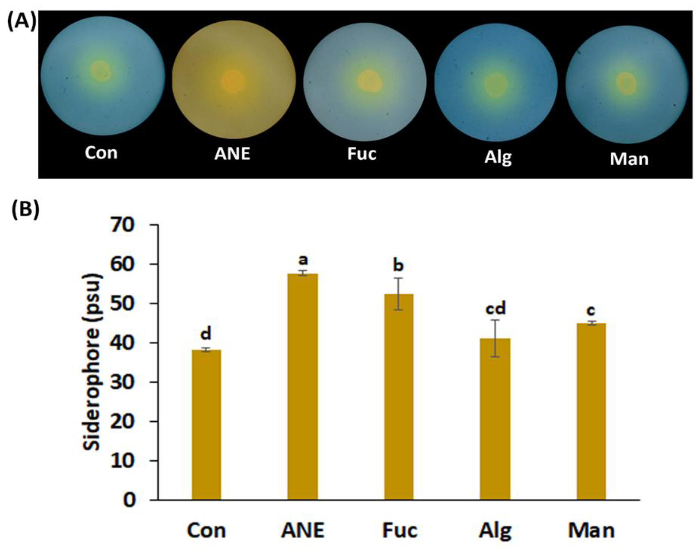
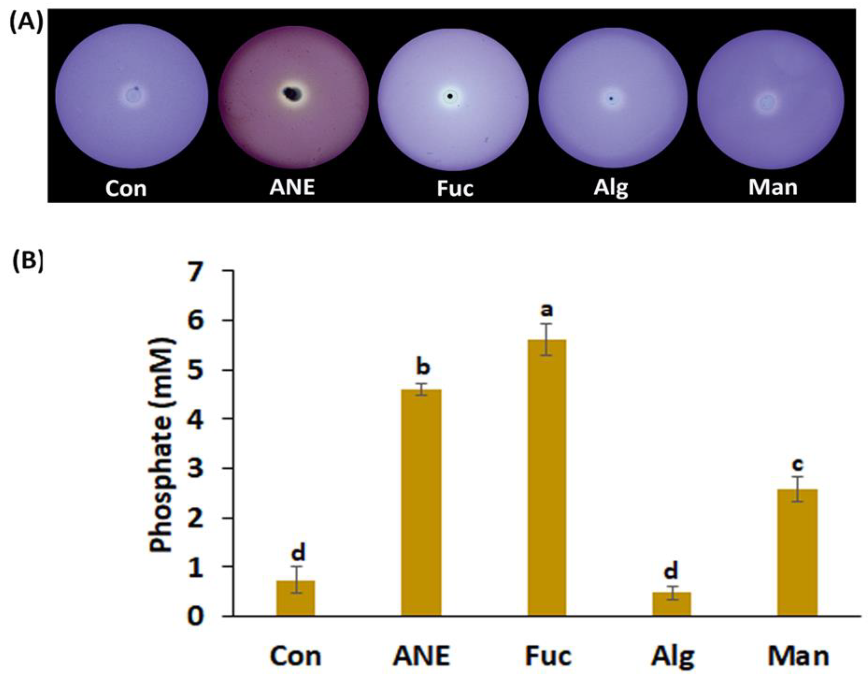
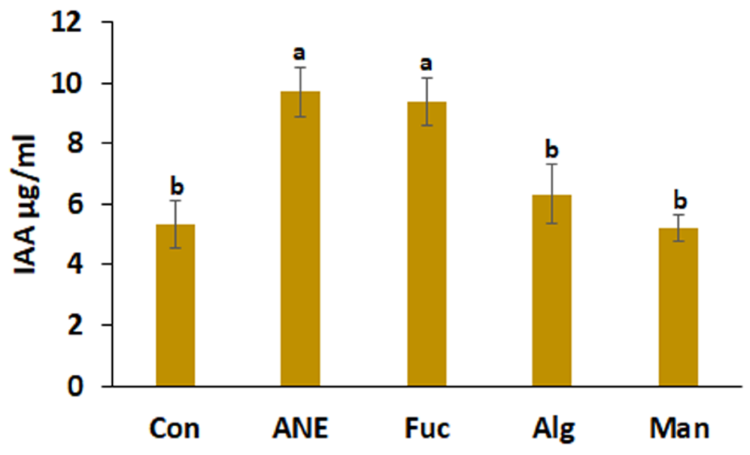
2.2. Effect of ANE, Fucoidan, Alginate, and Mannitol on Siderophore Production, Phosphate Solubilization, HCN Production, and IAA Production by P. protegens CHA0

P. protegens CHA0 was assessed using qualitative and quantitative assays for different activities under salinity stress.
Siderophore activity was determined first by the halo zone formed around the culture inoculums. Concentrations of 0.1% ANE and 0.01% fucoidan, alginate, and mannitol were found to form halo zones of comparable sizes (Figure 1A). However, quantification of siderophore production in the presence of 0.1% ANE and 0.01% fucoidan, alginate, and mannitol showed that the highest amounts were produced in the presence of ANE (57.66 psu), followed by fucoidan, mannitol, and alginate; siderophore amounts were significantly higher in all compounds as compared to control (Figure 1B).
Similar to siderophore production, in the plate assay, the addition of 0.1% ANE and 0.01% fucoidan, alginate, and mannitol was clearly associated with substantial phosphate solubilization (Figure 2A). Quantitatively, fucoidan, ANE, and mannitol were found to be better than the control, while alginate showed no significant change in phosphate solubilization (Figure 2A,B).
HCN production is related to defense mechanisms employed by P. protegens CHA0. HCN production was high in the presence of mannitol, alginate, and fucoidan as compared to ANE and control (Figure 3A,B).
The IAA production assay provides information on the growth promotion activity of P. protegens CHA0. The effect of media amendments was very similar to that observed in phosphate solubilization and siderophore production; that is, ANE (9.6 µg/mL) and fucoidan (9.37 µg/mL) were found to enhance the most IAA production. Alginate was slightly better than the control, while mannitol did not improve IAA production (Figure 4).

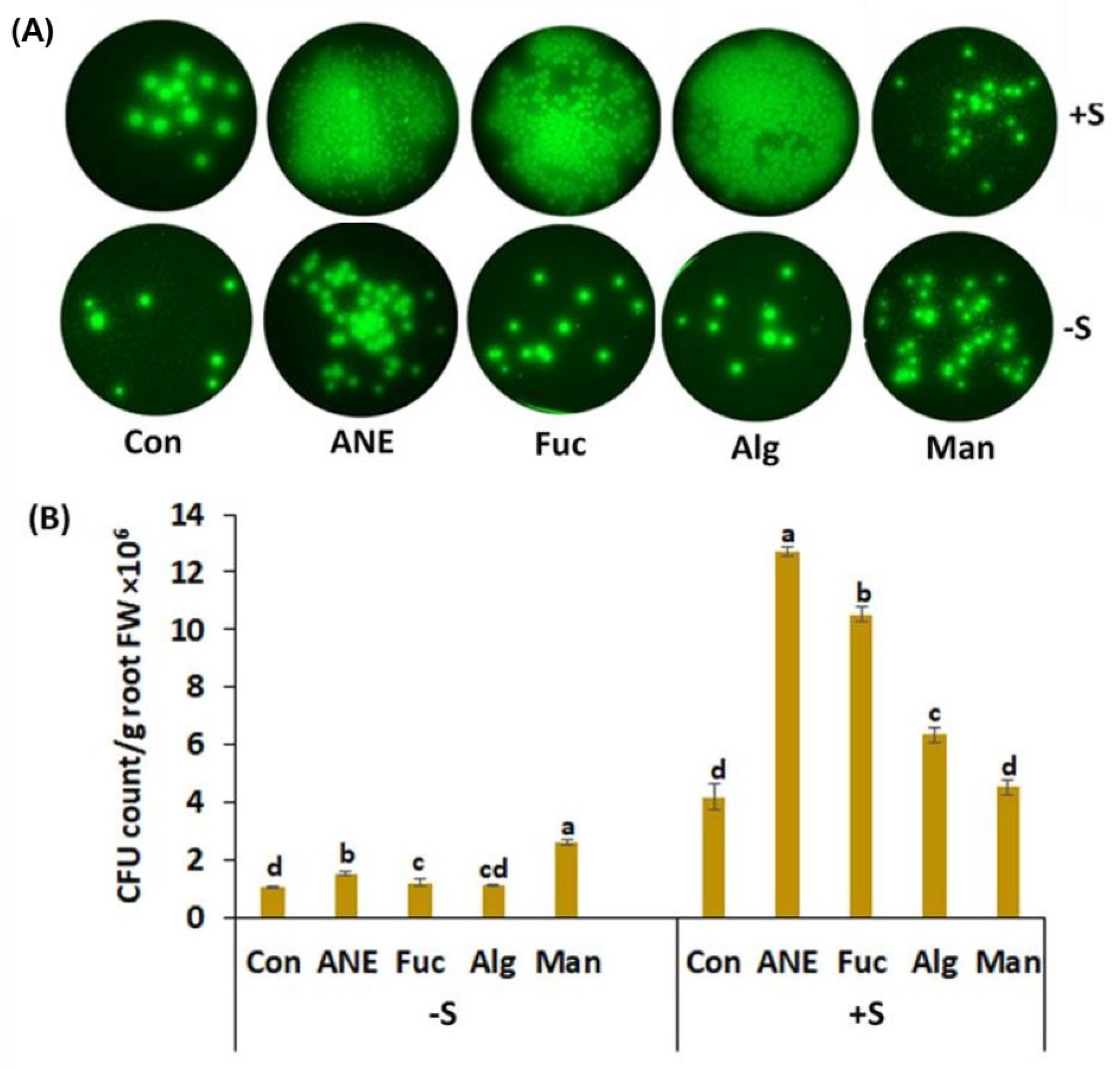
2.3. Effect of ANE, Fucoidan, Alginate, and Mannitol on CFU Count of P. protegens CHA0

The CFU count of P. protegens CHA0 was found to be increased by the addition of A. nodosum components, in the presence and absence of salt stress (Figure 5A,B). The count was found to be increased more than two-fold by 30 mM NaCl. Surprisingly, in salinity conditions, mannitol failed to enhance bacterial growth, though it did so significantly in normal conditions. ANE and fucoidan showed the most consistent effects in both growth conditions (Figure 5B).

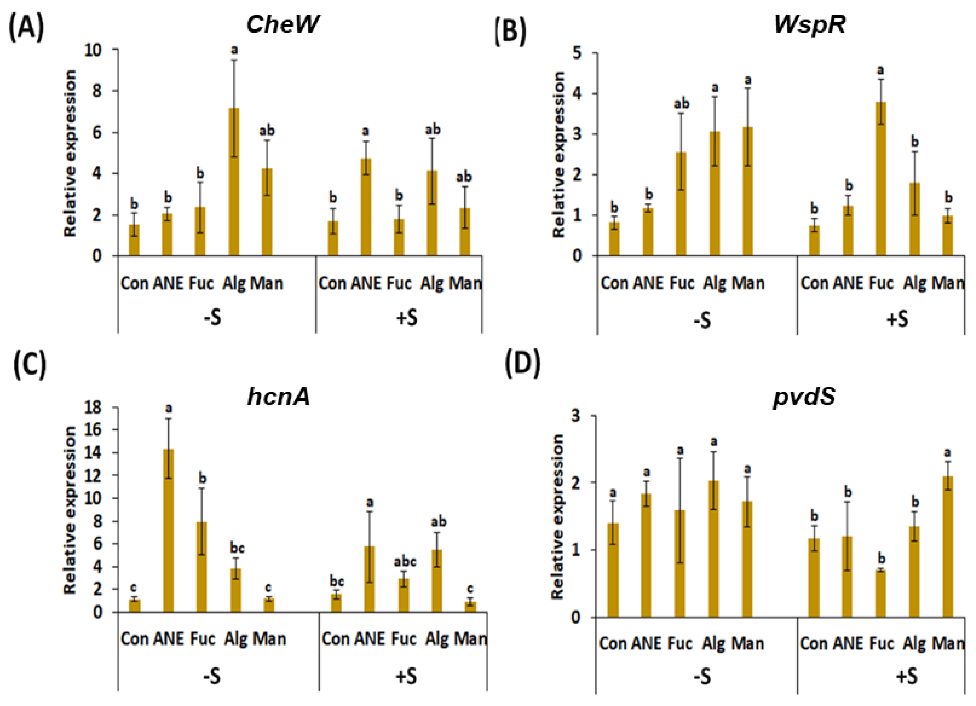
2.4. Effect of ANE, Fucoidan, Alginate, and Mannitol, in the Absence and Presence of Salinity Stress, on Key Genes of P. protegens CHA0 Involved in Plant Growth Promoting Rhizobacteria (PGPR) Activity

The gene cheW codes a protein involved in chemotaxis that is present in a wide range of bacteria [43]. In absence of salt, all treatments induced the gene expression level compared to the control; however, gene up-regulation was significant only in the alginate and mannitol treatments (7.15 fold and 4.27 fold, respectively; Figure 6A). In presence of salt stress, alginate, and ANE treatments showed the highest expression (4.20 fold and 4.74 fold, respectively) compared to the control, while fucoidan and mannitol treatments had almost no effect on gene expression (Figure 6A).
WspR, the product of the WspR gene, is the response regulator of the Wsp chemosensory system in several bacteria [44,45]. The expression of WspR, in absence of salt stress, was found to be significantly increased in mannitol, alginate, and fucoidan treatments (3.17 fold, 3.06 fold, and 2.56 fold, respectively), but much less in ANE treatment. In the presence of salt stress, the expression pattern changed noticeably, with fucoidan treatment having the strongest up-regulatory effect (3.79-fold increase), while ANE, alginate, and mannitol treatments showed only minimal effects. (Figure 6B).
The gene hcnaA codes a hydrogen cyanide synthase that is responsible for the volatile HCN production by various bacteria [46,47,48]. HCN is an antimicrobial compound that is also released by P. protegens CHA0. The highest expression of hcnaA, in absence of salt stress, was observed in the ANE treatment (14.4-fold), followed by the treatments with fucoidan and alginate. Mannitol treatment was not different from the control. In the presence of salt stress, compared to the control, the expression of hcnaA was induced by ANE and alginate treatments (5.72-fold and 5.48-fold, respectively), but much less by fucoidan (2.93-fold). Mannitol treatment was not different from control in this condition as well. Overall, versus control, the expression of hcnaA was less pronounced in all the treatments under salinity conditions, except for the treatment with alginate (Figure 6C).
The gene pvdS is responsible for the production of sigma factor PvdS, which is involved in the positive regulation of secondary metabolite biosynthetic processes, including pyoverdine synthesis [49,50]. In the absence of salt stress, an overall increase in pvdS transcript abundance was observed in treated plants, but these differences were not significant when compared to the control. In the presence of salt stress, the highest expression was observed in mannitol treatment (2.10-fold), but this was the only significant difference compared to the control, among treatments (Figure 6D).

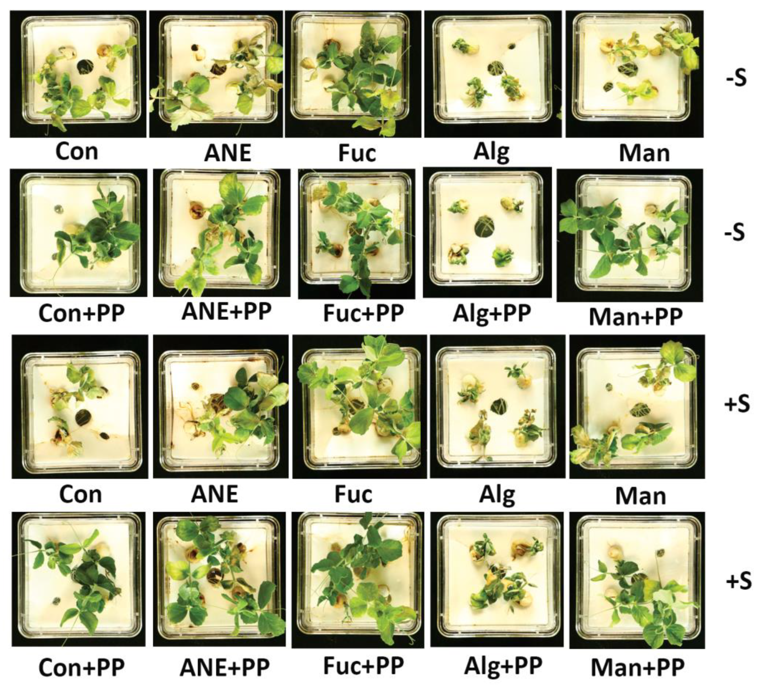
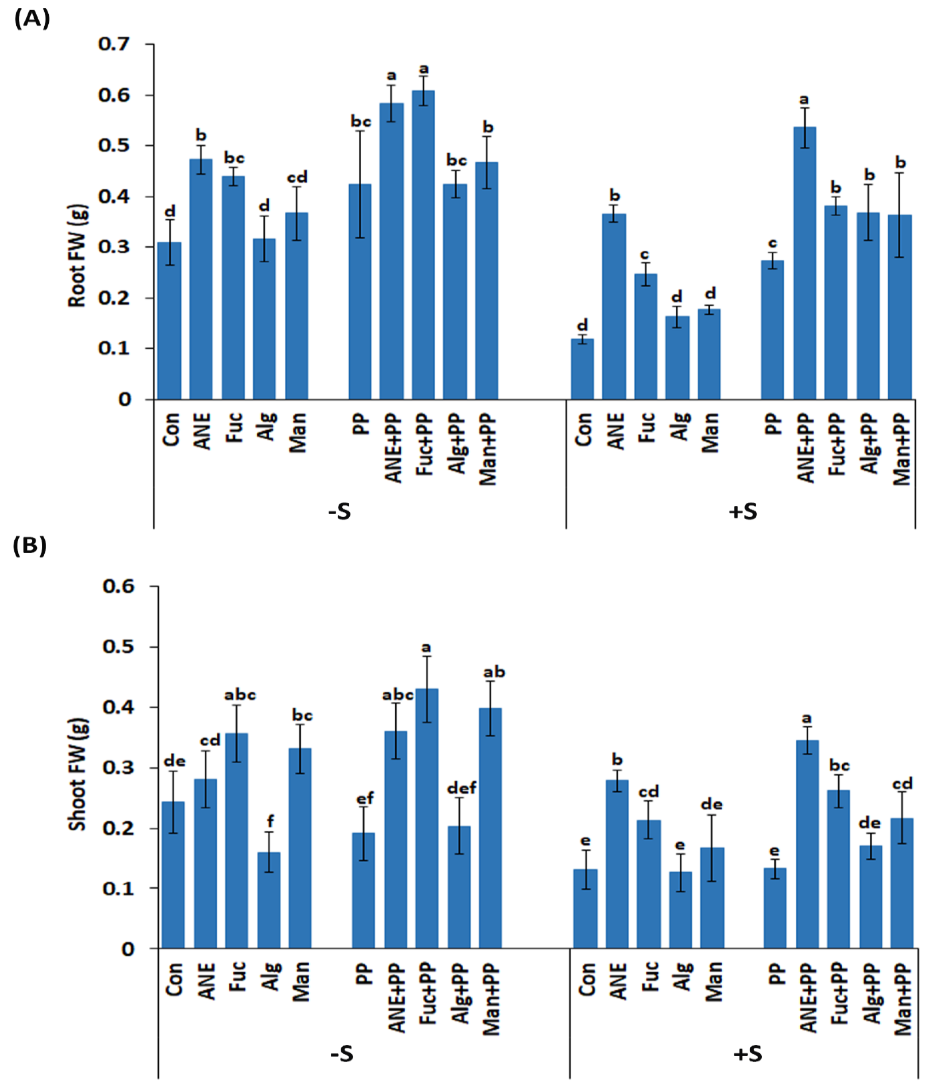
2.5. Effect of P. protegens CHA0 on Different Plant Growth-Related Parameters under Salt Stress

The effects of ANE, fucoidan, alginate, and mannitol on plant growth, in the absence and presence of P. protegens CHA0, and salinity stress, were assessed by growing the pea plants in a hydroponic system (Figure 7). After 7 days in the hydroponic system, the fresh weight of roots and shoots was recorded (Figure 8A,B). All of the treatments and the control showed significantly higher root fresh weight (FW) in the presence of P. protegens CHA0 in the normal and salinity stress conditions (Figure 8A). Among treatments, ANE and fucoidan were better in all conditions (presence or absence of P. protegens CHA0 or/and salinity stress) as compared to alginate and mannitol. In the absence of salt stress and of P. protegens CHA0, only alginate was not different from the control while, in the presence of P. protegens CHA0, alginate and mannitol were not statistically different from the control (Figure 8A).
While the presence of P. protegens CHA0 had a significant effect on root FW, the overall effects on shoot FW were not that obvious (Figure 8A,B). Overall, fucoidan, ANE, and mannitol improved shoot growth, while alginate was found to be better than the control, only in the presence of P. protegens CHA0 and salinity. Also, ANE and fucoidan performed better in the presence of P. protegens CHA0, in both the absence and presence of salt stress (Figure 8B).

3. Discussion

Tritrophic interaction between root-soil and rhizosphere microorganisms is a crucial phenomenon affecting plant growth, health, and defense. Several physiological activities of plants, such as nutrient uptake, can be improved by rhizosphere microbes, which can reduce the use of agrochemicals [51]. Microorganisms are recruited by exudates produced by root cells. Root exudates are a rich source of fixed carbon, such as polysaccharides, and of chemicals related to chemotaxis for the microorganisms found in the rhizosphere. Rhizospheric microorganisms perceive the signals produced by the plant roots and produce a wide range of chemicals in response to these signals, promoting plant growth and enhancing their tolerance against biotic and abiotic stresses [52,53,54].
The results of the current study indicate that ANE, and components present in ANE (fucoidan, alginate, and mannitol), can (i) enhance the production by P. protegens CHA0 of essential products associated with root colonization, (ii) promote the colonization of pea root by P. protegens CHA0, and (iii) mitigate salt stress in pea, a process that is further improved in the presence of P. protegens CHA0. This last effect was found to be more pronounced in root growth promotion than shoot growth. A. nodosum extract (ANE) has been shown to exhibit strong biostimulant activity against several biotic and abiotic stresses [13,14,19,23,55]. However, the effects of fucoidan, alginate, and mannitol, which are main components of A. nodosum, and therefore of ANE, are less characterized, though oligosaccharides and polysaccharides present in ANE have been shown to be elicitors of plant defense responses [56]. The role of ANE in the potentiation of beneficial bacteria growth and root colonization is still unexplored. On the other hand, several species from the Pseudomonas genus are well known to enhance plant defense mechanisms against abiotic and biotic stresses [37,57,58].
The presence of ANE improved the PGPR activities of P. protegens CHA0. Siderophores are well-known high-affinity iron-chelating compounds, which facilitate iron uptake from soil by plants. Plants assimilate iron from the soil through the roots [59,60,61]; siderophore production by P. protegens CHA0 can enhance iron transport across the plasma membrane of root epidermal cells and, therefore, the assimilation process. Results from the current study showed that all treatments improved siderophore production of P. protegens CHA0, though, overall, ANE was found to perform the best. It is likely that the more complex composition of ANE can explain this effect, suggesting potential synergistic effects of the components present in ANE. Pyoverdines are major fluorescent siderophores in Pseudomonas spp. [61,62], and pvdS is a key gene in the regulation of pyoverdine synthesis [49,50]. All treatments, including that with ANE, had a rather limited effect on the expression of pvdS, and the addition of salt in the medium determined changes in its abundance. The lack of a positive correlation between siderophore production by P. protegens CHA0 under different treatments and pvdS expression suggests that several other genes related to pyoverdines biosynthesis, secretion, and iron uptake [50,63] may contribute to the observed phenotype; also, inconsistencies can occur when comparing and integrating transcriptomics, proteomics, and metabolomics data [64,65,66].
Phosphorus (P) is a major limiting factor for plant growth. Insufficient amounts of phosphorus in agricultural soils are often counterbalanced by the excessive use of high-priced P-based chemical fertilizers [67]. Organic acids produced by phosphate-solubilizing bacteria can make the P present in the soil more accessible to plants [68,69]. Among the A. nodosum components analyzed, the phosphate solubilizing activity of P. protegens CHA0 was found to be enhanced in the presence of fucoidan and ANE, suggesting that fucoidan is the main component in ANE responsible for potentiating this activity in P. protegens CHA0.
Beneficial Pseudomonas spp. have been reported to suppress different plant diseases by producing antimicrobial metabolites, such as hydrogen cyanide [48,69]. The production of HCN by P. protegens CHA0 was not influenced by ANE, though mannitol, alginate, and fucoidan were found to increase around 3-fold this activity. It is likely that ANE components, other than polyols and complex polysaccharides, counterbalanced the enhancing effects determined by mannitol, alginate, and fucoidan. Interestingly, in absence of salt stress, the expression of hcnaA was found to be the highest in presence of ANE, a pattern that is opposite to that of HCN production. The hcnaA encodes subunit A of hydrogen cyanide synthase, responsible for the volatile HCN production by bacteria. The functional protein is a heterotrimer, formed by subunits HcnA, HcnB, and HcnC [46,47,48]. It is difficult to rationalize the effects triggered by ANE, which had no effect on HCN production, but enhanced the expression of hcnaA. One possible explanation is that ANE compounds, such as polyphenols, had inhibitory effects on the enzymatic activity of HCN synthase.
Plant growth promotion is one of the important effects of beneficial PGPR. Production of phytohormones, such as IAA, is the key mechanism behind growth promotion by PGPR. Several studies provided support for IAA production by PGPR [70,71,72]. IAA production ability of P. protegens CHA0 was found to be enhanced in the presence of ANE; fucoidan and alginate, but not mannitol, were found to have similar effects. Similarly to the results observed in siderophore production, the better activity of ANE might be explained by the potential synergistic effects of fucoidan and alginate.
Plants are essential sources of nutrition and provide shelter for rhizospheric bacteria. Several factors affect the colonization of microorganisms in the rhizosphere of plants. Rhizobacteria can be attracted and move toward plant roots by several chemoattractants secreted by plants [51,73,74]. Chemotaxis allows bacteria to move along the signal gradient [75], and the final number of cells colonizing the root can be in the range of 1011 cells per gram of root [76]. In absence of salt stress, pea root colonization by P. protegens CHA0 was not strongly enhanced, though mannitol, ANE, and fucoidan had some stimulating effects. In the presence of salt stress, root colonization was promoted by ANE and fucoidan, while the effects of mannitol and alginate were limited. These results indicate that (i) the addition of salt influences the effects of A. nodosum components, and (ii) A. nodosum components influence differently the various P. protegens CHA0 functions related to root colonization. Gene expression analysis of cheW and WspR in P. protegens CHA0, after treatment with ANE and components, provided limited support to pea root colonization; overall, no clear positive correlation between transcript abundance and enhanced physiological responses could be determined. CheW is one of the proteins involved in the transmission of sensory signals from the chemoreceptors to the motor proteins of the flagellar apparatus, which is responsible for the movement of bacteria toward the rhizosphere [43]. The WspR chemosensory system is well characterized in bacteria; WspR is the response regulator of this system. WspR contains the conserved GGDEF domain, which is involved in the formation of the intracellular-signaling molecule cyclic diguanylate (c-diGMP) [44,45]. Increased levels of c-diGMP result in enhanced biofilm formation and, therefore, of root colonization [44]. ANE-enhanced root colonization in the absence and presence of salt stress; however, treatments with ANE determined a large increase of transcript abundance only in the case of cheW, in the presence of salt stress. Fucoidan also had significant effects on root colonization, but determined the up-regulation of WspR. It is likely that, to have a more comprehensive view, at a molecular level, of the effects of ANE and its components on root colonization, more genes have to be analyzed, or different omics approaches should be considered because of the rather complicated relationship between transcriptomics, proteomics, metabolomics, and phenotype data [64,65,66].
The effects of the treatments with ANE, fucoidan, alginate, and mannitol were found to be more pronounced on root growth compared to shoot growth, and these effects on root growth were further enhanced in the presence of salt stress. The addition of P. protegens CHA0 also had a marked effect on root growth, while the overall effects on shoot growth were more limited. ANE is well known for its biostimulant activity [29,77,78,79,80], including improved seed germination, seedling vigor, and plant growth [14,19]. Mannitol is naturally synthesized in numerous bacteria, fungi, algae, and land plants, and functions as an osmolyte, energy storage, and antioxidant [34,81,82]. Mannitol treatment can improve salt tolerance in different plant species [83,84]. However, in the current work, except for shoot-fresh weight under the normal condition, which was marginally enhanced, mannitol treatment did not significantly influence root or shoot growth under both normal and stress conditions. Slightly better results have been observed on shoot growth in the presence of P. protegens CHA0. These results can be ascribed to the low concentration of the mannitol used in this study, that is, 0.01% mannitol; this concentration is equivalent to ~0.5 mM mannitol, which is quite low compared to the concentration of mannitol proved to act as an osmolyte [83,84]. The present study supports these findings, as ANE and fucoidan consistently improved root and shoot growth in the absence and presence of salinity stress, while P. protegens CHA0 further augmented these effects.

4. Materials and Methods

4.1. Plant Material and Growth Conditions

Seeds of pea (Pisum sativum L. cv. Sabre) were used in the experiments. Pea seeds, selected to have a weight of around 0.35 g, were surface-sterilized with 1% of sodium hypochlorite for 3 min, followed by rinsing in a series of distilled water. Seeds were soaked in distilled water overnight. Imbibed seeds were incubated in moist chambers for germination under a 16/8 light and dark period at 22 ± 2 °C. Germinated seeds with a radicle length of 2–4 cm were transferred to the hydroponics system containing ¼ MS media (without sucrose; pH 5.8).

4.2. Source of ANE, Fucoidan, Alginate, and Mannitol

Acadian®, a commercial ANE product, was provided by Acadian Seaplants Limited, Dartmouth, NS, Canada. Fucoidan was obtained from Marinova (Marinova Pty Ltd., Cambridge, TAS, Australia). The sodium salts of alginate and mannitol were from Sigma-Aldrich. (Oakville, ON, Canada).

4.3. Bacterial Cultures Screening

Seventeen beneficial bacterial cultures (Table 1) were characterized for their various biochemical activities, such as phosphate solubilization, siderophore production, IAA production, and HCN production. Bacterial cultures were provided by Dr. Zhenyu Cheng, Dalhousie University, NS, Canada. All the bacterial cultures were grown on their respective media (Table S1) at 28 ± 2 °C.

4.4. Siderophore Production Assay

Siderophore production assay was performed by using the Chrome Azurol S dye (CAS, Sigma-Aldrich) method described by Schwyn and Neilands [85]. This assay estimates siderophore production qualitatively and quantitatively. The qualitative test was done on King’s B agar media containing the CAS solution at a ratio of 1:15. Aqueous suspensions of bacterial cultures with a CFU count of 2.4 × 10⁸ were spotted on the middle of CAS agar plates in triplicate. All Petri dishes were incubated at 28 ± 2 °C temperature for 48 h. The formation of an orange halo zone around the developing bacterial colonies indicated a positive reaction for the assay. For quantitative estimation, 0.5 mL of the culture supernatant was added to 0.5 mL of CAS solution, followed by 10 µL of sulfosalicylic acid (0.2 M) (shuttling reagent). After 30 min of incubation at room temperature, the optical density was recorded at 630 nm. Siderophore production was quantified in terms of percent siderophore units (psu), using the formula: (Ar − As)/Ar × 100; where Ar is the absorbance of the reference solution, and As is the absorbance of samples.

4.5. Phosphate Solubilization Assay

Phosphate solubilization by microbes was assayed using the NBRIP-BPB medium described by Nautiyal [86]. Bacterial strains with CFU count of 2.4 × 10⁸ were inoculated on solid NBRIP-BPB medium in triplicate and incubated for 2–3 days at 28 ± 2 °C. The clear zone around the growing colonies was considered as a positive result for phosphate solubilization.
Quantitative estimation of phosphate solubilization was carried out in broth medium NBRIP, amended with different treatments in triplicates. The amount of released phosphate by the activity of P. protegens CHA0 was estimated using the method described by Fiske and Subbarow [87].

4.6. HCN Production Assay

HCN production assay was performed as described by Lorck [88]. Bacterial strains with a CFU count of 2.4 × 10⁸ were inoculated on the middle of the Petri plate in triplicate. Picrate filter paper (0.5% picric acid in 0.2% w/v Na2CO3) was placed on the lid of the Petri plate. HCN formation determines the change of color from deep yellow to orange, and, finally, to dark brown. Bacterial strains changing the color of the picrate filter paper from deep yellow to brown were considered as positive for HCN production. The plates were sealed by parafilm and incubated for 72 h at 28 °C.
Dissolved, free cyanide ions concentration in the liquid medium was determined using the colorimetric methemoglobin method described by von Rohr et al. [89]. The liquid medium and the methemoglobin reagent were mixed at a 1:1 ratio, and the optical density was recorded at 424 nm.

4.7. Indole-3-Acetic Acid (IAA) Production Assay

The IAA production assay was performed using the modified protocol described by Malik and Sindhu [90]. Bacterial cultures were grown on their respective media for 48 h and 72 h, respectively, at 28 ± 2 °C. Culture suspension (2.4 × 10⁸ CFU) was centrifuged at 3000× g for 30 min, and 2 mL of supernatant was used for the assay. The reaction was performed by the addition of 3 µL of orthophosphoric acid (85%) and 4 mL of Salkowski reagent (50 mL of 35% perchloric acid and 1 mL 0.5 M FeCl3). Optical density was recorded at 530 nm.

4.8. Protease Assay

Protease assay was performed as previously described by Kembhavi et al. [91]. In brief, the bacterial culture was centrifuged at 5000 rpm for 6 min. The supernatant (150 µL) was transferred to a tube and was mixed with 300 µL of 1% (w/v) casein (prepared in 20 mM Tris-HCl buffer, pH 7.4) and incubated at 37 °C for 30 min. Then, 0.45 mL of a 10% (w/v) trichloroacetic acid (TCA) solution was added to stop the proteolysis reaction, followed by incubation of the mixture at room temperature for one hour. Eventually, the reaction mixture was centrifuged at 12,000× g for 5 min, and the absorbance of the supernatant was measured at 280 nm. One unit of protease is defined as the amount of enzyme that hydrolyses casein to produce equivalent absorbance to 1 μmol of tyrosine/min with tyrosine as standard [91].

4.9. Effect of ANE, Fucoidan, Alginate, and Mannitol on Gene Expression Profiles of P. protegens CHA0

P. protegens CHA0 cells were collected after 24 h of growth in the presence and absence of ANE, fucoidan, alginate, or mannitol. RNA isolation was performed using a RNeasy mini kit (Qiagen Inc., Toronto, ON, Canada). Total RNA was quantified with a NanoDrop 2000 spectrophotometer (Thermo Fisher Scientific Inc., Mississauga, ON, Canada). 2 µg of RNA was used to synthesize cDNA using a RevertAid First Strand cDNA Synthesis Kit (Thermo Fisher Scientific Inc). The relative gene expression of pvdS (coding the sigma factor PvdS, which is involved in the regulation of pyoverdine synthesis), cheW (CheW protein is implicated in chemotaxis), wspR (coding the WspR protein with diguanylate cyclase activity, involved in signal transduction, cell adhesion, and biofilm formation) and hcnA (HcnA is an enzyme with glycine dehydrogenase, cyanide-forming activity), was determined by real-time quantitative PCR (RT-qPCR) (QuantStudio 3, Applied Biosystems, Burlington, ON, Canada), using the SYBR® Green Supermix Kit (Bio-Rad Laboratories (Canada) Ltd., Mississauga, ON, Canada). Gene-specific primers were used at a final concentration of 0.1 μM. Primer3 software, version 4.1.0, was used to design the primers used in the experiments (Table S2). RT-qPCR assays were carried out under the following conditions: denaturation at 95 °C for 2 min, 40 repeats at 95 °C for 20 s, 60 °C for 30 s, and 72 °C for 25 s. Transcript abundance was estimated using the 2−ΔΔCT method [92] for relative quantification by normalizing the data against the endogenous gene (rpoC) and the individual with the lowest gene expression in controls. Fold change was calculated with respect to the mean of the CT values of the three biological replicates from treatment C (control). All RT-qPCR experiments were carried out with three biological and three technical replicates.

4.10. Effect of ANE, Fucoidan, Alginate, and Mannitol on the Biochemical Activities and Root Colonization Activity of P. protegens CHA0

The concentration of ANE was adjusted to 0.1% [23], while that of fucoidan, alginate, and mannitol were adjusted to 0.01%, considering that the proportion of these compounds in brown seaweed extracts ranges between 2 and 10% [29,30,31,32]. All biochemical assays were performed as mentioned in the previous sections. P. protegens CHA0 was grown in the King’s B broth, with incubation at 28 ± 2 °C in a shaking incubator.
To study the effect of ANE, fucoidan, alginate, and mannitol on the root colonization activity of P. protegens CHA0, the CFU count was performed using samples taken from the rhizoplane, after 24 h of stress challenge. 0.2 g of the root sample from each treatment was taken for the experiment. Samples were crushed in 1 mL of sterilized, Millipore water and filtered. 100 µL of 10−5 and 10−6 dilution was transferred to Petri dishes containing King’s B medium. The plates were incubated at 28 ± 2 °C, and CFU was counted after 24 h.

4.11. Effect of ANE, Fucoidan, Alginate, and Mannitol on Plant Growth in the Presence of P. protegens CHA0 under Salinity Stress

Treatments, that is, 0.1% ANE, 0.01% fucoidan, mannitol, alginate, and 25 mL of P. protegens CHA0 (2.4 × 10⁷ CFU/mL), were added two days after the germinated seeds were placed in the hydroponic system. Salinity stress was carried out at the same time by the addition of 30 mM NaCl in each treatment. The phenotype of the plants grown in the Magenta jars was observed after eight days of salinity stress challenge. Experiments were repeated 3 times. Each treatment had 5 replicates and there were 4 plants in each replicate.

4.12. Statistical Analysis

Analysis of variance (One-Way ANOVA) was performed by using Minitab Statistical Software Version 21.1 (Minitab Inc., State College, PA, USA). A Tukey post hoc test was used to perform multiple mean comparisons at p ≤ 0.05.

5. Conclusions

The action of ANE and of biological compounds, such as fucoidan, which are found in large amounts in A. nodosum, increased the production of antimicrobial compounds and the synthesis of chemotaxis-related chemicals by P. protegens CHA0. These results suggest that A. nodosum extract, as well as various A. nodosum components, can potentiate the beneficial PGP effects of P. protegens CHA0 in terms of phosphate solubilization, siderophore, HCN, and IAA production. Colonization of pea roots by P. protegens CHA0 was also enhanced, mainly by ANE and fucoidan, under salinity stress. Overall, the combined applications of ANE and P. protegens CHA0 greatly improved the growth and tolerance against salinity of pea plants grown in hydroponic conditions. The current study suggests that ANE and compounds such as fucoidan, derived from A. nodosum, could be used for the improvement of crop growth under salinity stress. New formulations, containing various formulations of ANE supplemented with beneficial microbes, could also be envisioned for improved yield and plant health.

Supplementary Materials

The following supporting information can be downloaded at: https://www.mdpi.com/article/10.3390/plants12061208/s1, Table S1: Media used for growing the different bacterial cultures. Table S2: List of primers used in the qPCR experiment.

Author Contributions

Conceptualization, J.S.P. and B.P.; Methodology, J.S.P., V.S. and T.B.; Validation, J.S.P. and V.S.; Formal Analysis, J.S.P., V.S. and T.B.; Investigation, J.S.P. and V.S.; Resources, B.P.; Data Curation, J.S.P., V.S., P.M., R.B., T.B. and B.P.; Writing–Original Draft Preparation, J.S.P. and T.B.; Writing—Review and Editing, J.S.P., V.S., P.M., R.B., T.B. and B.P.; Visualization, J.S.P. and T.B.; Supervision, B.P.; Project Administration, B.P.; Funding Acquisition, B.P. and T.B. All authors have read and agreed to the published version of the manuscript.

Funding

BP’s research group was funded by an Accelerate Cluster grant (IT08347) from Mitacs, Canada (www.mitacs.ca), awarded to Balakrishnan Prithiviraj, and by Acadian Seaplants Limited (www.acadian.ca). Funding partners did not play any role in the design of the experiments, data collection, analysis, and writing the manuscript.

Data Availability Statement

The data presented in this study are available on request from the corresponding author.

Acknowledgments

Special thanks to Zhenyu Cheng, Dalhousie University, NS, Canada for providing the bacterial strains used in this study.

Conflicts of Interest

The authors declare no conflict of interest. The funders had no role in the design of the study; in the collection, analyses, or interpretation of data; in the writing of the manuscript; or in the decision to publish the results.

References

  1. Boyer, J.S. Plant productivity and environment. Science 1982, 218, 443–448. [Google Scholar] [CrossRef]
  2. Zhu, J.-K. Genetic analysis of plant salt tolerance using Arabidopsis. Plant Physiol. 2000, 124, 941–948. [Google Scholar] [CrossRef] [Green Version]
  3. Munns, R. Comparative physiology of salt and water stress. Plant Cell Environ. 2002, 25, 239–250. [Google Scholar] [CrossRef] [Green Version]
  4. Moghadam, A.A.; Ebrahimie, E.; Taghavi, S.M.; Niazi, A.; Babgohari, M.Z.; Deihimi, T.; Djavaheri, M.; Ramezani, A. How the nucleus and mitochondria communicate in energy production during stress: Nuclear MtATP6, an early-stress responsive gene, regulates the mitochondrial F1F0-ATP synthase complex. Mol. Biotechnol. 2013, 54, 756–769. [Google Scholar] [CrossRef]
  5. Munns, R.; Tester, M. Mechanisms of salinity tolerance. Annu. Rev. Plant Biol. 2008, 59, 651–681. [Google Scholar] [CrossRef] [Green Version]
  6. Dahl, W.J.; Foster, L.M.; Tyler, R.T. Review of the health benefits of peas (Pisum sativum L.). Br. J. Nutr. 2012, 108, S3–S10. [Google Scholar] [CrossRef] [Green Version]
  7. Acosta-Motos, J.R.; Ortuño, M.F.; Bernal-Vicente, A.; Diaz-Vivancos, P.; Sanchez-Blanco, M.J.; Hernandez, J.A. Plant responses to salt stress: Adaptive mechanisms. Agronomy 2017, 7, 18. [Google Scholar] [CrossRef] [Green Version]
  8. Fukao, T.; Barrera-Figueroa, B.E.; Juntawong, P.; Peña-Castro, J.M. Submergence and waterlogging stress in plants: A review highlighting research opportunities and understudied aspects. Front. Plant Sci. 2019, 10, 340. [Google Scholar] [CrossRef]
  9. De Pascale, S.; Rouphael, Y.; Colla, G. Plant biostimulants: Innovative tool for enhancing plant nutrition in organic farming. Eur. J. Hortic. Sci. 2018, 82, 277–285. [Google Scholar] [CrossRef]
  10. Rouphael, Y.; Colla, G. Editorial: Biostimulants in agriculture. Front. Plant Sci. 2020, 11, 40. [Google Scholar] [CrossRef] [Green Version]
  11. Colla, G.; Rouphael, Y. Biostimulants in horticulture. Sci. Hortic. 2015, 196, 1–2. [Google Scholar] [CrossRef]
  12. Goñi, O.; Quille, P.; O’Connell, S. Ascophyllum nodosum extract biostimulants and their role in enhancing tolerance to drought stress in tomato plants. Plant Physiol. Biochem. 2018, 126, 63–73. [Google Scholar] [CrossRef]
  13. Jithesh, M.N.; Shukla, P.S.; Kant, P.; Joshi, J.; Critchley, A.T.; Prithiviraj, B. Physiological and transcriptomics analyses reveal that Ascophyllum nodosum extracts induce salinity tolerance in Arabidopsis by regulating the expression of stress responsive genes. J. Plant Growth Regul. 2019, 38, 463–478. [Google Scholar] [CrossRef]
  14. Shukla, P.S.; Mantin, E.G.; Adil, M.; Bajpai, S.; Critchley, A.T.; Prithiviraj, B. Ascophyllum nodosum-based biostimulants: Sustainable applications in agriculture for the stimulation of plant growth, stress tolerance, and disease management. Front. Plant Sci. 2019, 10, 655. [Google Scholar] [CrossRef] [Green Version]
  15. De Saeger, J.; Van Praet, S.; Vereecke, D.; Park, J.; Jacques, S.; Han, T.; Depuydt, S. Toward the molecular understanding of the action mechanism of Ascophyllum nodosum extracts on plants. J. Appl. Phycol. 2019, 32, 573–597. [Google Scholar] [CrossRef] [Green Version]
  16. Shukla, P.S.; Shotton, K.; Norman, E.; Neily, W.; Critchley, A.T.; Prithiviraj, B. Seaweed extract improve drought tolerance of soybean by regulating stress-response genes. AoBP 2017, 10, plx051. [Google Scholar] [CrossRef]
  17. Xu, C.; Leskovar, D.I. Effects of A. nodosum seaweed extracts on spinach growth, physiology and nutrition value under drought stress. Sci. Hortic. 2015, 183, 39–47. [Google Scholar] [CrossRef]
  18. Fan, D.; Hodges, D.M.; Critchley, A.T.; Prithiviraj, B. A commercial extract of brown macroalga (Ascophyllum nodosum) affects yield and the nutritional quality of spinach in vitro. Commun. Soil Sci. Plant Anal. 2013, 44, 1873–1884. [Google Scholar] [CrossRef]
  19. Battacharyya, D.; Babgohari, M.Z.; Rathor, P.; Prithiviraj, B. Seaweed extracts as biostimulants in horticulture. Sci. Hortic. 2015, 196, 39–48. [Google Scholar] [CrossRef]
  20. Carvalho, M.; Castro, P.; Gaziola, S.; Azevedo, R. Is seaweed extract an elicitor compound? Changing proline content in drought-stressed bean plants. Comun. Sci. 2018, 9, 292–297. [Google Scholar] [CrossRef]
  21. Santaniello, A.; Scartazza, A.; Gresta, F.; Loreti, E.; Biasone, A.; Di Tommaso, D.; Piaggesi, A.; Perata, P. Ascophyllum nodosum seaweed extract alleviates drought stress in Arabidopsis by affecting photosynthetic performance and related gene expression. Front. Plant Sci. 2017, 8, 1362. [Google Scholar] [CrossRef] [Green Version]
  22. Subramanian, S.; Sangha, J.S.; Gray, B.A.; Singh, R.P.; Hiltz, D.; Critchley, A.T.; Prithiviraj, B. Extracts of the marine brown macroalga, Ascophyllum nodosum, induce jasmonic acid dependent systemic resistance in Arabidopsis thaliana against Pseudomonas syringae pv. tomato DC3000 and Sclerotinia sclerotiorum. Eur. J. Plant Pathol. 2011, 131, 237–248. [Google Scholar] [CrossRef]
  23. Shukla, P.S.; Borza, T.; Critchley, A.T.; Hiltz, D.; Norrie, J.; Prithiviraj, B. Ascophyllum nodosum extract mitigates salinity stress in Arabidopsis thaliana by modulating the expression of miRNA involved in stress tolerance and nutrient acquisition. PLoS ONE 2018, 13, e0206221. [Google Scholar] [CrossRef] [Green Version]
  24. Di Stasio, E.; Van Oosten, M.J.; Silletti, S.; Raimondi, G.; dell’Aversana, E.; Carillo, P.; Maggio, A. Ascophyllum nodosum-based algal extracts act as enhancers of growth, fruit quality, and adaptation to stress in salinized tomato plants. J. Appl. Phycol. 2018, 30, 2675–2686. [Google Scholar] [CrossRef]
  25. Sá, J.M.D.; Leitão, E.T.C.; Gomes, C.D.L.; Rodrigues, M.S.; Sousa, V.F.O.; dos Santos, G.L.; de Melo, R.A.P.; de Mendonça Júnior, A.F.; de Lacerda, J.S.P.; Santos, A.d.S. The initial growth of passion fruit plant irrigated with saline water and the application of biostimulants. J. Agric. Sci. 2018, 10, 357–363. [Google Scholar] [CrossRef] [Green Version]
  26. Bonomelli, C.; Celis, V.; Lombardi, G.; Mártiz, J. Salt sress effects on avocado (Persea americana Mill.) plants with and without seaweed extract (Ascophyllum nodosum) application. Agronomy 2018, 8, 64. [Google Scholar] [CrossRef] [Green Version]
  27. Nabati, D.A.; Schmidt, R.E.; Parrish, D.J. Alleviation of salinity stress in Kentucky bluegrass by plant growth regulators and iron. Crop Sci. 1994, 34, 198–202. [Google Scholar] [CrossRef]
  28. Abdel Latef, A.A.H.; Srivastava, A.K.; Saber, H.; Alwaleed, E.A.; Tran, L.-S.P. Sargassum muticum and Jania rubens regulate amino acid metabolism to improve growth and alleviate salinity in chickpea. Sci. Rep. 2017, 7, 10537. [Google Scholar] [CrossRef] [Green Version]
  29. Khan, W.; Menon, U.; Subramanian, S.; Jithesh, M.; Rayorath, P.; Hodges, D.; Critchley, A.; Craigie, J.; Norrie, J.; Prithiviraj, B. Seaweed extracts as biostimulants of plant growth and development. J. Plant Growth Regul. 2009, 28, 386–399. [Google Scholar] [CrossRef]
  30. Black, W.A.P. The seasonal variation in chemical composition of some of the littoral seaweeds common to Scotland. Part I. Ascophyllum nodosum. J. Soc. Chem. Ind. 1948, 67, 355–357. [Google Scholar] [CrossRef]
  31. Black, W.A.P. The seasonal variation in weight and chemical composition of the common British Laminariaceae. J. Mar. Biol. Assoc. UK 1950, 29, 45–72. [Google Scholar] [CrossRef] [Green Version]
  32. Percival, E. The polysaccharides of green, red and brown seaweeds: Their basic structure, biosynthesis and function. Eur. J. Phycol. 1979, 14, 103–117. [Google Scholar] [CrossRef]
  33. Venugopal, V. Sulfated and non-sulfated polysaccharides from seaweeds and their uses: An overview. ECronicon Nutr. 2019, 14, 126–141. [Google Scholar]
  34. Patel, T.K.; Williamson, J.D. Mannitol in Plants, Fungi, and Plant-Fungal Interactions. Trends Plant Sci. 2016, 21, 486–497. [Google Scholar] [CrossRef]
  35. Vaishnav, A.; Shukla, A.K.; Sharma, A.; Kumar, R.; Choudhary, D.K. Endophytic bacteria in plant salt stress tolerance: Current and future prospects. J. Plant Growth Regul. 2019, 38, 650–668. [Google Scholar] [CrossRef]
  36. Samaddar, S.; Chatterjee, P.; Choudhury, A.; Ahmed, S.; Sa, T. Interactions between Pseudomonas spp. and their role in improving the red pepper plant growth under salinity stress. Microbiol. Res. 2019, 219, 66–73. [Google Scholar] [CrossRef]
  37. Sarkar, A.; Patel, J.; Yadav, S.; Sarma, B.; Srivastava, J.; Singh, H. Studies on rhizosphere-bacteria mediated biotic and abiotic stress tolerance in chickpea (Cicer arietinum L.). Vegetos 2014, 27, 158–169. [Google Scholar] [CrossRef]
  38. Chu, T.N.; Tran, B.T.H.; Van Bui, L.; Hoang, M.T.T. Plant growth-promoting rhizobacterium Pseudomonas PS01 induces salt tolerance in Arabidopsis thaliana. BMC Res. Notes 2019, 12, 11. [Google Scholar] [CrossRef] [Green Version]
  39. Chomczynski, P.; Sacchi, N. The single-step method of RNA isolation by acid guanidinium thiocyanate–phenol–chloroform extraction: Twenty-something years on. Nat. Protoc. 2006, 1, 581–585. [Google Scholar] [CrossRef]
  40. Orozco-Mosqueda, M.D.C.; Duan, J.; DiBernardo, M.; Zetter, E.; Campos-García, J.; Glick, B.R.; Santoyo, G. The production of ACC deaminase and trehalose by the plant growth promoting bacterium Pseudomonas sp. UW4 synergistically protect tomato plants against salt stress. Front. Microbiol. 2019, 10, 1392. [Google Scholar] [CrossRef] [Green Version]
  41. Heydarian, Z.; Gruber, M.; Glick, B.R.; Hegedus, D.D. Gene expression patterns in roots of Camelina sativa with enhanced salinity tolerance arising from inoculation of soil with plant growth promoting bacteria producing 1-aminocyclopropane-1-carboxylate deaminase or expression the corresponding acdS gene. Front. Microbiol. 2018, 9, 1297. [Google Scholar] [CrossRef] [PubMed]
  42. Gupta, S.; Pandey, S. ACC deaminase producing bacteria with multifarious plant growth promoting traits alleviates salinity stress in French bean (Phaseolus vulgaris) plants. Front. Microbiol. 2019, 10, 1506. [Google Scholar] [CrossRef] [PubMed]
  43. Sampedro, I.; Parales, R.E.; Krell, T.; Hill, J.E. Pseudomonas chemotaxis. FEMS Microbiol. Rev. 2014, 39, 17–46. [Google Scholar] [CrossRef] [Green Version]
  44. Hickman, J.W.; Tifrea, D.F.; Harwood, C.S. A chemosensory system that regulates biofilm formation through modulation of cyclic diguanylate levels. Proc. Natl. Acad. Sci. USA 2005, 102, 14422–14427. [Google Scholar] [CrossRef] [Green Version]
  45. Martínez-Granero, F.; Navazo, A.; Barahona, E.; Redondo-Nieto, M.; González de Heredia, E.; Baena, I.; Martín-Martín, I.; Rivilla, R.; Martín, M. Identification of flgZ as a flagellar gene encoding a PilZ domain protein that regulates swimming motility and biofilm formation in Pseudomonas. PLoS ONE 2014, 9, e87608. [Google Scholar] [CrossRef] [PubMed] [Green Version]
  46. Pessi, G.; Haas, D. Dual control of hydrogen cyanide biosynthesis by the global activator GacA in Pseudomonas aeruginosa PAO1. FEMS Microbiol. Lett. 2001, 200, 73–78. [Google Scholar] [CrossRef] [PubMed]
  47. Nandi, M.; Selin, C.; Brawerman, G.; Fernando, W.G.D.; de Kievit, T.R. The global regulator ANR is essential for Pseudomonas chlororaphis strain PA23 biocontrol. Microbiology 2016, 162, 2159–2169. [Google Scholar] [CrossRef] [PubMed]
  48. Laville, J.; Blumer, C.; Von Schroetter, C.; Gaia, V.; Défago, G.; Keel, C.; Haas, D. Characterization of the hcnABC gene cluster encoding hydrogen cyanide synthase and anaerobic regulation by ANR in the strictly aerobic biocontrol agent Pseudomonas fluorescens CHA0. J. Bacteriol. 1998, 180, 3187–3196. [Google Scholar] [CrossRef] [Green Version]
  49. Imperi, F.; Tiburzi, F.; Fimia, G.M.; Visca, P. Transcriptional control of the pvdS iron starvation sigma factor gene by the master regulator of sulfur metabolism CysB in Pseudomonas aeruginosa. Environ. Microbiol. 2010, 12, 1630–1642. [Google Scholar] [CrossRef]
  50. Ringel, M.T.; Brüser, T. The biosynthesis of pyoverdines. Microb. Cell 2018, 5, 424–437. [Google Scholar] [CrossRef]
  51. Berendsen, R.L.; Pieterse, C.M.J.; Bakker, P.A.H.M. The rhizosphere microbiome and plant health. Trends Plant Sci. 2012, 17, 478–486. [Google Scholar] [CrossRef] [PubMed]
  52. Jones, D.L.; Hodge, A.; Kuzyakov, Y. Plant and mycorrhizal regulation of rhizodeposition. New Phytol. 2004, 163, 459–480. [Google Scholar] [CrossRef] [PubMed]
  53. Badri, D.; Vivanco, J. Regulation and function of root exudates. Plant Cell Environ. 2009, 32, 666–681. [Google Scholar] [CrossRef] [PubMed]
  54. van Dam, N.M.; Bouwmeester, H.J. Metabolomics in the rhizosphere: Tapping into belowground chemical communication. Trends Plant Sci. 2016, 21, 256–265. [Google Scholar] [CrossRef]
  55. Gunupuru, L.R.; Patel, J.S.; Sumarah, M.W.; Renaud, J.B.; Mantin, E.G.; Prithiviraj, B. A plant biostimulant made from the marine brown algae Ascophyllum nodosum and chitosan reduce Fusarium head blight and mycotoxin contamination in wheat. PLoS ONE 2019, 14, e0220562. [Google Scholar] [CrossRef] [Green Version]
  56. Patier, P.; Yvin, J.-C.; Kloareg, B.; Liénart, Y.; Rochas, C. Seaweed liquid fertilizer from Ascophyllum nodosum contains elicitors of plantd-glycanases. J. Appl. Phycol. 1993, 5, 343–349. [Google Scholar] [CrossRef]
  57. Patel, J.S.; Kharwar, R.N.; Singh, H.B.; Upadhyay, R.S.; Sarma, B.K. Trichoderma asperellum (T42) and Pseudomonas fluorescens (OKC)-enhances resistance of pea against Erysiphe pisi through enhanced ROS generation and lignifications. Front. Microbiol. 2017, 8, 306. [Google Scholar] [CrossRef]
  58. Srivastava, S.; Patel, J.S.; Singh, H.B.; Sinha, A.; Sarma, B.K. Streptomyces rochei SM3 induces stress tolerance in chickpea against Sclerotinia sclerotiorum and NaCl. J. Phytopathol. 2015, 163, 583–592. [Google Scholar] [CrossRef]
  59. Briat, J.F. Iron dynamics in plants. Adv. Bot. Res. 2007, 46, 137–180. [Google Scholar]
  60. Kim, S.A.; Guerinot, M.L. Mining iron: Iron uptake and transport in plants. FEBS Lett. 2007, 581, 2273–2280. [Google Scholar] [CrossRef] [Green Version]
  61. Aznar, A.; Chen, N.W.G.; Rigault, M.; Riache, N.; Joseph, D.; Desmaële, D.; Mouille, G.; Boutet, S.; Soubigou-Taconnat, L.; Renou, J.-P.; et al. Scavenging iron: A novel mechanism of plant immunity activation by microbial siderophores. Plant Physiol. 2014, 164, 2167–2183. [Google Scholar] [CrossRef] [PubMed] [Green Version]
  62. Trapet, P.; Avoscan, L.; Klinguer, A.; Pateyron, S.; Citerne, S.; Chervin, C.; Mazurier, S.; Lemanceau, P.; Wendehenne, D.; Besson-Bard, A. The Pseudomonas fluorescens siderophore pyoverdine weakens Arabidopsis thaliana defense in favor of growth in iron-deficient conditions. Plant Physiol. 2016, 171, 675–693. [Google Scholar] [CrossRef] [PubMed] [Green Version]
  63. Nadal-Jimenez, P.; Koch, G.; Reis, C.R.; Muntendam, R.; Raj, H.; Jeronimus-Stratingh, C.M.; Cool, R.H.; Quax, W.J. PvdP is a tyrosinase that drives maturation of the pyoverdine chromophore in Pseudomonas aeruginosa. J. Bacteriol. 2014, 196, 2681–2690. [Google Scholar] [CrossRef] [PubMed] [Green Version]
  64. Bathke, J.; Konzer, A.; Remes, B.; McIntosh, M.; Klug, G. Comparative analyses of the variation of the transcriptome and proteome of Rhodobacter sphaeroides throughout growth. BMC Genom. 2019, 20, 358. [Google Scholar] [CrossRef] [Green Version]
  65. Liu, Y.; Beyer, A.; Aebersold, R. On the dependency of cellular protein levels on mRNA abundance. Cell 2016, 165, 535–550. [Google Scholar] [CrossRef] [Green Version]
  66. Cavill, R.; Jennen, D.; Kleinjans, J.; Briedé, J.J. Transcriptomic and metabolomic data integration. Brief. Bioinform. 2015, 17, 891–901. [Google Scholar] [CrossRef] [Green Version]
  67. Rasul, M.; Yasmin, S.; Zubair, M.; Mahreen, N.; Yousaf, S.; Arif, M.; Sajid, Z.I.; Mirza, M.S. Phosphate solubilizers as antagonists for bacterial leaf blight with improved rice growth in phosphorus deficit soil. Biol. Control 2019, 136, 103997. [Google Scholar] [CrossRef]
  68. De Werra, P.; Péchy-Tarr, M.; Keel, C.; Maurhofer, M. Role of gluconic acid production in the regulation of biocontrol traits of Pseudomonas fluorescens CHA0. Appl. Environ. Microbiol. 2009, 75, 4162–4174. [Google Scholar] [CrossRef] [Green Version]
  69. Gross, H.; Loper, J.E. Genomics of secondary metabolite production by Pseudomonas spp. Nat. Prod. Rep. 2009, 26, 1408–1446. [Google Scholar] [CrossRef]
  70. Bharti, N.; Pandey, S.S.; Barnawal, D.; Patel, V.K.; Kalra, A. Plant growth promoting rhizobacteria Dietzia natronolimnaea modulates the expression of stress responsive genes providing protection of wheat from salinity stress. Sci. Rep. 2016, 6, 34768. [Google Scholar] [CrossRef] [Green Version]
  71. Khan, N.; Bano, A.; Rahman, M.A.; Guo, J.; Kang, Z.; Babar, M.A. Comparative physiological and metabolic analysis reveals a complex mechanism involved in drought tolerance in chickpea (Cicer arietinum L.) induced by PGPR and PGRs. Sci. Rep. 2019, 9, 2097. [Google Scholar] [CrossRef] [PubMed] [Green Version]
  72. Mantelin, S.; Touraine, B. Plant growth-promoting bacteria and nitrate availability: Impacts on root development and nitrate uptake. J. Exp. Bot. 2004, 55, 27–34. [Google Scholar] [CrossRef] [PubMed] [Green Version]
  73. De Weert, S.; Vermeiren, H.; Mulders, I.H.M.; Kuiper, I.; Hendrickx, N.; Bloemberg, G.V.; Vanderleyden, J.; De Mot, R.; Lugtenberg, B.J.J. Flagella-driven chemotaxis towards exudate components is an important trait for tomato root colonization by Pseudomonas fluorescens. Mol. Plant-Microbe Interact. 2002, 15, 1173–1180. [Google Scholar] [CrossRef] [Green Version]
  74. Feng, H.; Zhang, N.; Fu, R.; Liu, Y.; Krell, T.; Du, W.; Shao, J.; Shen, Q.; Zhang, R. Recognition of dominant attractants by key chemoreceptors mediates recruitment of plant growth-promoting rhizobacteria. Environ. Microbiol. 2019, 21, 402–415. [Google Scholar] [CrossRef] [Green Version]
  75. Bi, S.; Sourjik, V. Stimulus sensing and signal processing in bacterial chemotaxis. Curr. Opin. Microbiol. 2018, 45, 22–29. [Google Scholar] [CrossRef] [PubMed]
  76. Egamberdieva, D.; Kamilova, F.; Validov, S.; Gafurova, L.; Kucharova, Z.; Lugtenberg, B. High incidence of plant growth-stimulating bacteria associated with the rhizosphere of wheat grown on salinated soil in Uzbekistan. Environ. Microbiol. 2008, 10, 1–9. [Google Scholar] [CrossRef] [PubMed]
  77. Bajpai, S.; Shukla, P.S.; Asiedu, S.; Pruski, K.; Prithiviraj, B. A biostimulant preparation of brown seaweed Ascophyllum nodosum suppresses powdery mildew of strawberry. Plant. Pathol. J. 2019, 35, 406–416. [Google Scholar] [CrossRef] [PubMed]
  78. Hong, D.D.; Hien, H.M.; Son, P.N. Seaweeds from Vietnam used for functional food, medicine and biofertilizer. J. Appl. Phycol. 2007, 19, 817–826. [Google Scholar] [CrossRef]
  79. Khan, W.; Hiltz, D.; Critchley, A.; Prithiviraj, B. Bioassay to detect Ascophyllum nodosum extract-induced cytokinin-like activity in Arabidopsis thaliana. J. Appl. Phycol. 2011, 23, 409–414. [Google Scholar] [CrossRef]
  80. Silva, L.D.; Bahcevandziev, K.; Pereira, L. Production of bio-fertilizer from Ascophyllum nodosum and Sargassum muticum (Phaeophyceae). J. Oceanol. Limnol. 2019, 37, 918–927. [Google Scholar] [CrossRef]
  81. Iwamoto, K.; Shiraiwa, Y. Salt-regulated mannitol metabolism in algae. Mar. Biotechnol. 2005, 7, 407–415. [Google Scholar] [CrossRef] [PubMed]
  82. Srivastava, A.K.; Ramaswamy, N.K.; Suprasanna, P.; D’Souza, S.F. Genome-wide analysis of thiourea-modulated salinity stress-responsive transcripts in seeds of Brassica juncea: Identification of signalling and effector components of stress tolerance. Ann. Bot. 2010, 106, 663–674. [Google Scholar] [CrossRef] [PubMed] [Green Version]
  83. Kaya, C.; sonmez, O.; Aydemir, S.; Ashraf, M.; Dikilitas, M. Exogenous application of mannitol and thiourea regulates plant growth and oxidative stress responses in salt-stressed maize (Zea mays L.). J. Plant Interact. 2013, 8, 234–241. [Google Scholar] [CrossRef]
  84. Seckin, B.; Sekmen, A.H.; Türkan, İ. An enhancing effect of exogenous mannitol on the antioxidant enzyme activities in roots of wheat under salt stress. J. Plant Growth Regul. 2009, 28, 12–20. [Google Scholar] [CrossRef]
  85. Schwyn, B.; Neilands, J.B. Universal chemical assay for the detection and determination of siderophores. Anal. Biochem. 1987, 160, 47–56. [Google Scholar] [CrossRef]
  86. Nautiyal, C.S. An efficient microbiological growth medium for screening phosphate solubilizing microorganisms. FEMS Microbiol. Lett. 1999, 170, 265–270. [Google Scholar] [CrossRef]
  87. Fiske, C.H.; Subbarow, Y.J. The colorimetric determination of phosphorus. J. Biol. Chem. 1925, 66, 375–400. [Google Scholar] [CrossRef]
  88. Lorck, H. Production of hydrocyanic acid by bacteria. Physiol. Plant. 1948, 1, 142–146. [Google Scholar] [CrossRef]
  89. Von Rohr, M.R.; Furrer, G.; Brandl, H. Effect of iron and phosphate on bacterial cyanide formation determined by methemoglobin in two-dimensional gradient microcultivations. J. Microbiol. Methods. 2009, 79, 71–75. [Google Scholar] [CrossRef]
  90. Malik, D.K.; Sindhu, S.S. Production of indole acetic acid by Pseudomonas sp.: Effect of coinoculation with Mesorhizobium sp. Cicer on nodulation and plant growth of chickpea (Cicer arietinum). Physiol. Mol. Biol. Plants. 2011, 17, 25–32. [Google Scholar] [CrossRef] [Green Version]
  91. Kembhavi, A.A.; Kulkarni, A.; Pant, A. Salt-tolerant and thermostable alkaline protease from Bacillus subtilis NCIM No. 64. Appl. Biochem. Biotechnol. 1993, 38, 83–92. [Google Scholar] [CrossRef] [PubMed]
  92. Livak, K.J.; Schmittgen, T.D. Analysis of relative gene expression data using real-time quantitative PCR and the 2(-Delta Delta C(T)) Method. Methods 2001, 25, 402–408. [Google Scholar] [CrossRef] [PubMed]
Figure 1. Effect of ANE, fucoidan, alginate, and mannitol on siderophore production by P. protegens CHA0. (A) Images of the plates showing siderophore production under different treatments. (B) Siderophore production quantified using the CAS method. Con, control; ANE, 0.1% Acadian®; Fuc, 0.01% fucoidan; Alg, 0.01% alginate; and Man, 0.01% mannitol. Statistical analysis was done using One-Way ANOVA (p ≤ 0.05), followed by Tukey’s multiple comparison test (p ≤ 0.05). Error bars represent SD. Letter grouping indicates significant differences according to Tukey’s test.
Plants 12 01208 g001
Figure 2. Effect of ANE, fucoidan, alginate, and mannitol on phosphate solubilization by P. protegens CHA0. (A) Images of the plates showing phosphate solubilization under different treatments. (B) Amount of phosphate solubilized by P. protegens CHA0 under different treatments. Con, control; ANE, 0.1% Acadian®; Fuc, 0.01% fucoidan; Alg, 0.01% alginate; and Man, 0.01% mannitol. Statistical analysis was done using One-Way ANOVA (p ≤ 0.05) followed by Tukey’s multiple comparison test (p ≤ 0.05). Error bars represent SD. Letter grouping indicates significant differences according to Tukey’s test.
Plants 12 01208 g002
Figure 3. Effect of ANE, fucoidan, alginate, and mannitol on HCN production by P. protegens CHA0. (A) Images of the plates showing HCN production under different treatments. (B) Relative amounts of HCN produced by P. protegens CHA0 under different treatments. Con, control; ANE, 0.1% Acadian®; Fuc, 0.01% fucoidan; Alg, 0.01% alginate, and Man, 0.01% mannitol. Statistical analysis was done using One-Way ANOVA (p ≤ 0.05), followed by Tukey’s multiple comparison test (p ≤ 0.05). Error bars represent SD. Letter grouping indicates significant differences according to Tukey’s test.
Plants 12 01208 g003
Figure 4. Effect of ANE, fucoidan, alginate, and mannitol on IAA production by P. protegens CHA0. Con, control; ANE, 0.1% Acadian®; Fuc, 0.01% fucoidan; Alg, 0.01% alginate; and Man, 0.01% mannitol. Statistical analysis was done using One-Way ANOVA (p ≤ 0.05), followed by Tukey’s multiple comparison test (p ≤ 0.05). Error bars represent SD. Letter grouping indicates significant differences according to Tukey’s test.
Plants 12 01208 g004
Figure 5. Effect of the ANE, fucoidan, alginate, and mannitol on pea root colonization by P. protegens CHA0. (A) Images of the plates on which P. protegens CHA0 cells were enumerated. (B) Colony-forming units (CFU) counts in the absence and presence of salinity stress. Con, control; ANE, 0.1% Acadian®; Fuc, 0.01% fucoidan; Alg, 0.01% alginate; and Man, 0.01% mannitol; −S, normal conditions (no salt treatment); +S, 30 mM NaCl. Statistical analysis was done using One-Way ANOVA (p ≤ 0.05), followed by Tukey’s multiple comparison test (p ≤ 0.05). Error bars represent SD. Letter grouping indicates significant differences according to Tukey’s test.
Plants 12 01208 g005
Figure 6. Effect of ANE, fucoidan, alginate, and mannitol on gene expression of P. protegens CHA0. (A) CheW, (B) WspR, (C) hcnA, (D) pvdS. Con, control; ANE, 0.1% Acadian®; Fuc, 0.01% fucoidan; Alg, 0.01% alginate; Man, 0.01% mannitol; PP, P. protegens CHA0; −S, normal conditions (no salt treatment); +S, 30 mM NaCl. Statistical analysis was done using One-Way ANOVA (p ≤ 0.05), followed by Tukey’s multiple comparison test (p ≤ 0.05). Error bars represent SD. Letter grouping indicates significant differences according to Tukey’s test.
Plants 12 01208 g006
Figure 7. Effect of ANE, fucoidan, alginate, and mannitol on plant growth in the absence and presence of P. protegens CHA0 and salinity stress. Con, control; ANE, 0.1% Acadian®; Fuc, 0.01% fucoidan; Alg, 0.01% alginate; Man, 0.01% mannitol; PP, P. protegens CHA0; −S, normal conditions (no salt treatment); +S, 30 mM NaCl.
Plants 12 01208 g007
Figure 8. Effect of ANE, fucoidan, alginate, and mannitol on plant growth in the absence and presence of P. protegens CHA0 and/or salinity stress. (A) Root fresh weight. (B) Shoot fresh weight. Con, control; ANE, 0.1% Acadian; Fuc, 0.01% fucoidan; Alg, 0.01% alginate Man, 0.01% mannitol; Pp, P. protegens CHA0; −S, normal conditions (no salt treatment); +S, 30 mM NaCl. Statistical analysis was done using One-Way ANOVA (p ≤ 0.05), followed by Tukey’s multiple comparison test (p ≤ 0.05). Error bars represent SD. Letter grouping indicates significant differences according to Tukey’s test.
Plants 12 01208 g008
Table 1. Screening of the bacteria for the different biochemical activities. (−) no activity; (+) low activity; (++) medium activity; (+++) strongest activity.
Bacterial SpeciesPhosphate
Solubilization
Siderophore
Production
IAA
Production
Protease
Production
HCN
Production
Azospirillum lipoferum 1842+
Azotobacter vinelandii (ATCC12837)+++
Bacillus subtilis+++
Bacillus thuringiensis subsp. oloke+
Bradyrhizobium japonicum 3I1b6+
Enterobacter agglomerans (ATCC23216)++
Enterobacter cloacae CAL2+++
Kluyvera ascorbata SUD165+++
Lactobacillus acidophilus (ATCC 4356)+
Paenibacillus polymyxa K56++
Pseudomonas brassicacearum (ATCC 49054)++
Pseudomonas fluorescens 34-13+++++++++
Pseudomonas protegens CHAO+++
Pseudomonas putida (ATCC 12633)+++
Sinorhizobium fredii (ATCC51808)++++
Sinorhizobium meliloti RM11559++++++
Streptococcus salivarius C699+
Disclaimer/Publisher’s Note: The statements, opinions and data contained in all publications are solely those of the individual author(s) and contributor(s) and not of MDPI and/or the editor(s). MDPI and/or the editor(s) disclaim responsibility for any injury to people or property resulting from any ideas, methods, instructions or products referred to in the content.

