Brief Report

Identification of Lasiodiplodia pseudotheobromae Causing Fruit Rot of Citrus in China

1
Hubei Key Laboratory of Plant Pathology, Huazhong Agricultural University, Wuhan 430070, China
2
National R & D Center for Citrus Postharvest Technology, Huazhong Agricultural University, Wuhan 430070, China
*
Author to whom correspondence should be addressed.
Plants 2021, 10(2), 202; https://doi.org/10.3390/plants10020202
Submission received: 4 December 2020 / Revised: 31 December 2020 / Accepted: 19 January 2021 / Published: 21 January 2021
(This article belongs to the Special Issue Citrus Fungal and Oomycete Diseases)

Abstract

:
Considering the huge economic loss caused by postharvest diseases, the identification and prevention of citrus postharvest diseases is vital to the citrus industry. In 2018, 16 decayed citrus fruit from four citrus varieties—Satsuma mandarin (Citrus unshiu), Ponkan (Citrus reticulata Blanco cv. Ponkan), Nanfeng mandarin (Citrus reticulata cv. nanfengmiju), and Sugar orange (Citrus reticulata Blanco)—showing soft rot and sogginess on their surfaces and covered with white mycelia were collected from storage rooms in seven provinces. The pathogens were isolated and the pathogenicity of the isolates was tested. The fungal strains were identified as Lasiodiplodia pseudotheobromae based on their morphological characteristics and phylogenetic analyses using the internal transcribed spacer regions (ITS), translation elongation factor 1-α gene (TEF), and beta-tubulin (TUB) gene sequences. The strains could infect wounded citrus fruit and cause decay within two days post inoculation, but could not infect unwounded fruit. To our knowledge, this is the first report of citrus fruit decay caused by L. pseudotheobromae in China.

1. Introduction

Citrus is the second-largest fruit crop in China. It is cultivated in over 20 provinces, with an area of approximately 2.5 million ha [1,2]. China has an over 4000-year history of citrus cultivation. There are an abundant and excellent variety of resources in China. Fruit decay is a common disease during postharvest transportation and storage. Generally, the decay rate is 20% to 30%, and sometimes it can reach up to 50%, which causes serious postharvest economic losses [3]. Therefore, disease control is vital for the postharvest period of citrus fruit.
Common postharvest citrus diseases include green mold, blue mold, sour rot, anthracnose, Diplodia stem-end rot, Diaporthe stem-end rot, brown rot, and Alternaria stem-end rot, etc. In 2018, a survey of postharvest citrus diseases in storage rooms found that the average fruit decay rate exceeded 30% in seven provinces. In addition to the common postharvest diseases, 16 decayed fruit showing soft rot and soggy lesions with dense white hyphae were also observed. In this study, the causal agent of the citrus fruit rot disease was determined via morphological observation, molecular identification, and pathogenicity tests.

2. Results

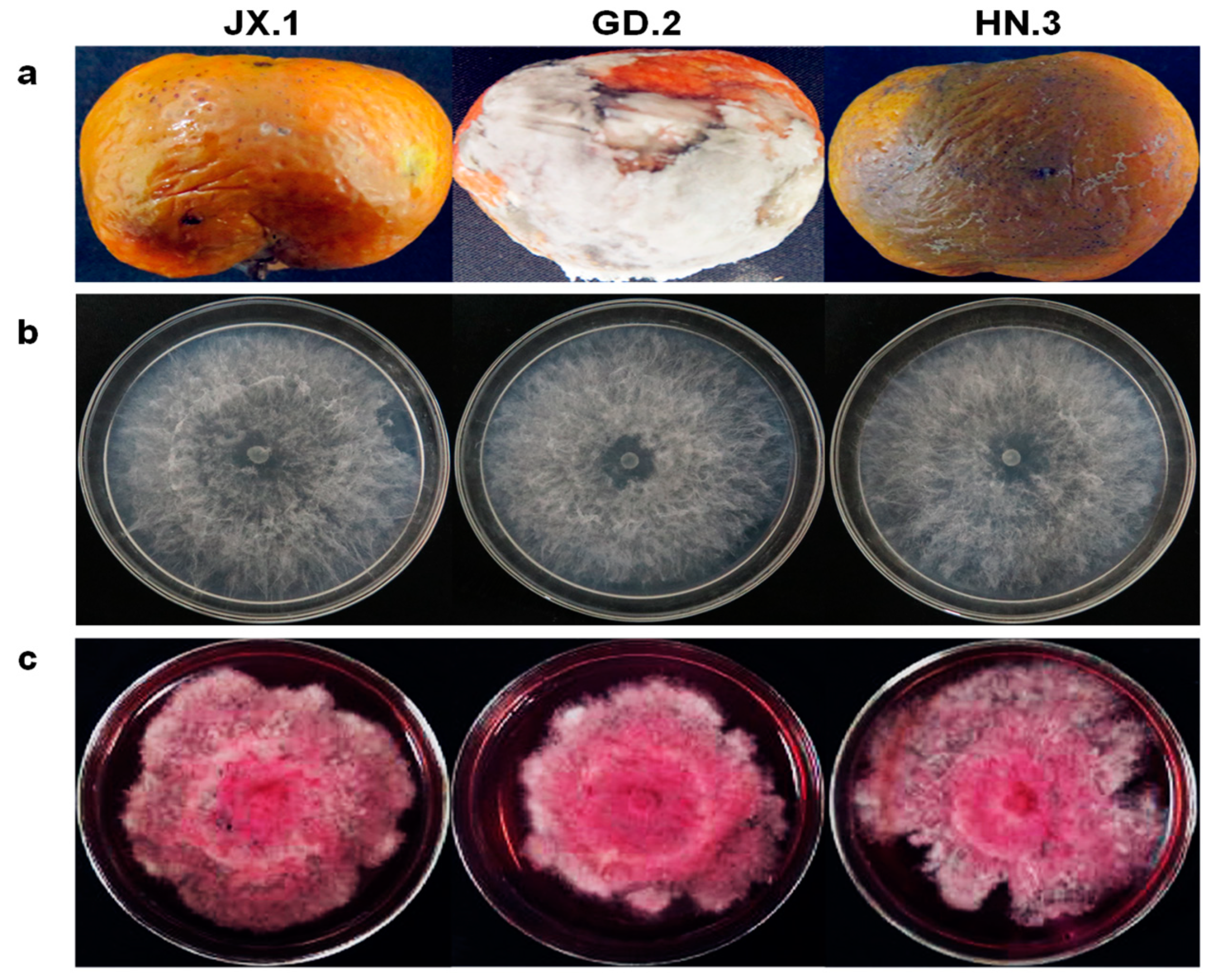
2.1. Fungal Isolates

In 2018, a total of 2402 citrus fruit from four different citrus varieties (Satsuma mandarin (Citrus unshiu), Ponkan (Citrus reticulata Blanco cv. Ponkan), Nanfeng mandarin (Citrus reticulata cv. nanfengmiju), and Sugar orange (Citrus reticulata Blanco)) were taken from storage houses (15 °C) in seven provinces (Guangxi, Hubei, Zhejiang, Hunan, Jiangxi, Fujian, and Guangdong) and investigated. Except for fruit with typical green mold and blue mold symptoms, 402 diseased fruit samples were collected for fungal isolation. From the 402 decayed fruit, 450 fungal strains were isolated. Based on the morphological characteristics and internal transcribed spacer regions (ITS) sequences, most of them were identified as Geotrichum spp., Colletotrichum spp., Diplodia spp., Alternaria spp., etc. In addition, 16 strains belonging to the genus Lasiodiplodia were also isolated from 16 diseased fruits with soft, water-soaked lesions (Figure 1a, Table 1).

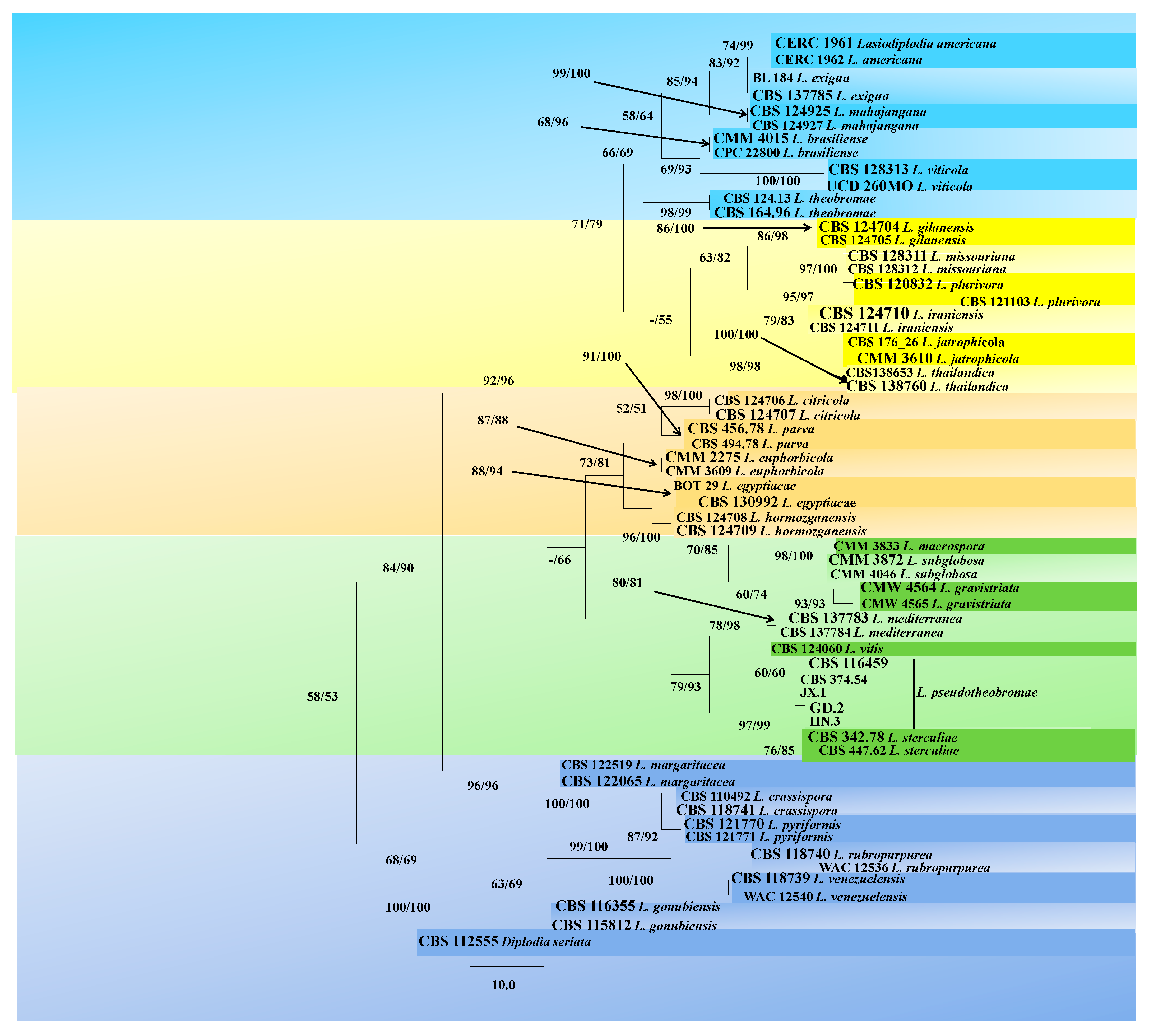
2.2. Molecular Characterization

Based on the multiple gene analyses, 16 strains were identified as Lasiodiplodia pseudotheobromae. A MegaBlast analysis showed that they exhibited 100% identity to the type strain of L. pseudotheobromae (CBS 116459). Since the 16 strains were of the same species, three of them from different citrus varieties and producing areas—JX.1, GD.2, and HN.3—were randomly selected for further study (Table S1). In total, 1727 characters of 62 isolates from 30 Lasiodiplodia species and one outgroup species (Diplodia seriata) were used to construct Maximum likelihood (ML) and Maximum Parsimony (MP) phylogenetic trees. The combined data included 525 characters of ITS (1–525), 550 characters of translation elongation factor 1-α gene (TEF) (530–1079), and 643 characters of beta-tubulin (TUB) (1085–1727). Based on the MP analysis, a single most parsimonious tree was constructed (tree length (TL) = 460, consistency index (CI) = 0.654, retention index (RI) = 0.854, rescaled consistency index (RC) = 0.559, homoplasy index (HI) = 0.346). Of all the characters, 175 characters were parsimony informative, 63 variable characters were parsimony uninformative, and 1,489 characters were consistent. The topology of the ML tree was similar to that of the MP tree. The isolates were grouped with L. pseudotheobromae but were distinguished from L. sterculiae, L. vitis, and L. mediterranea, the species with the closest genetic distance to L. pseudotheobromae (Figure 2).

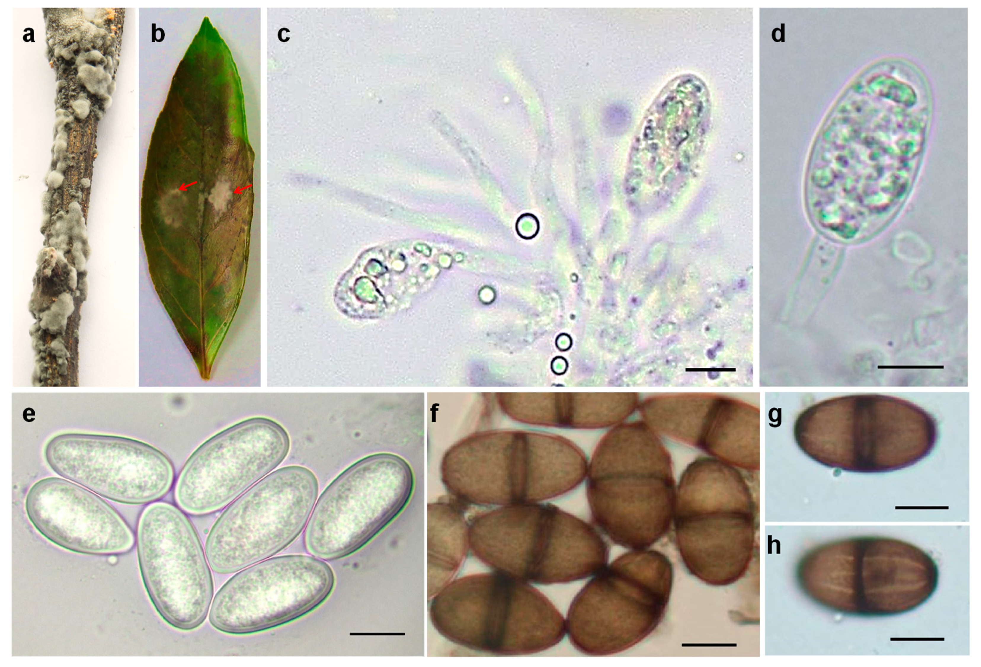
2.3. Morphological Characteristics

At first, the colonies were white with sparse, fluffy aerial mycelia. After one week, they became a pale gray with dark pigment (Figure 1b). Pycnidia were dark brown to black, solitary, globose, and intraepidermal (Figure 3a,b). The pseudparaphyses were hyaline, cylindrical-shaped, aseptate, sometimes branched, end-rounded, and arising among the conidiogenous cells (Figure 3c). Conidiogenous cells were hyaline, smooth, cylindrical, slightly swollen at the base, and holoblastic (Figure 3d). Conidia were ellipsoidal, apex and base-rounded, widest at the middle, and thick-walled. Immature conidia were colorless, hyaline, and aseptate, while mature conidia were dark brown (Figure 3e,f), one-septate with longitudinal striations (Figure 3g,h). The conidial dimensions were (22.5−) 27.8 (−32) µm × (14.5−) 17 (−20) µm ( x ¯ = 27.8 × 17 µm, n = 50).
The strain JX.1 could grow between 10 and 35 °C. The optimal growth temperature was 30 °C (Figure S1). Meanwhile, all three of the strains JX.1, GD.2, and HN.3 produced a reddish pigment at 35 °C (Figure 1c).

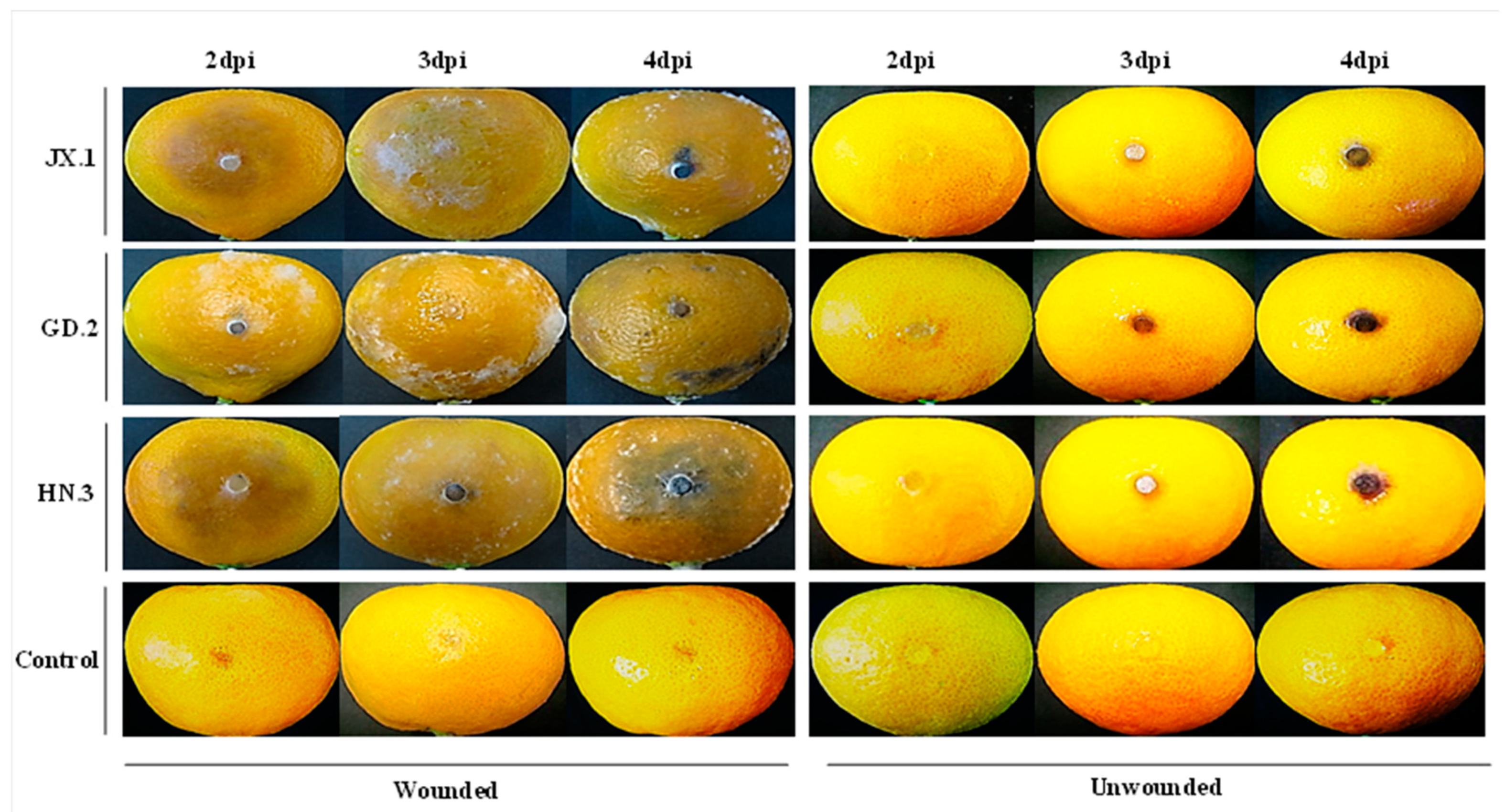
2.4. Pathogenicity Assay

In the pathogenicity assay, mature fruits of the Satsuma mandarin were used to test the virulence of the isolates. Firstly, discoloration and necrosis appeared on the fruit surface. Then, the lesions enlarged rapidly, became soft and rotten, and were covered with sparse white mycelia. The lesions spread to the whole fruit within 3 to 4 days after the occurrence of necrosis. Finally, the fruit became soggy. The onset of lesions emerged on wounded Satsuma mandarin fruit within two days of inoculation (dpi) (Figure 4). No symptoms of decay were observed on the unwounded fruit. The recovered isolates showed identical morphological traits to the original ones.

3. Discussion

In this study, 16 L. pseudotheobromae strains were isolated from rotten postharvest citrus fruit which were collected from storage rooms in seven provinces of China. This is the first report that L. pseudotheobromae causes fruit rot on citrus in China.
The conidiaomata of the isolates formed on citrus twigs, producing slowly maturing conidia with thick walls and longitudinal striations, which are typical morphological features of the genus Lasiodiplodia [4]. Furthermore, the pseudparaphyses were mostly aseptate. The colonies can grow at 10 °C and produce a dark pink pigment at 35 °C on potato dextrose agar (PDA). These morphological characteristics are consistent with those of L. pseudotheobromae [5,6]. Combined with the results of the phylogenetic analysis, these isolates were identified as L. pseudotheobromae.
Based on its morphological and molecular characteristics, L. pseudotheobromae has been distinguished from L. theobromae as a cryptic species [5]. It is a common pathogen of tropical and subtropical plants and has a wide host range, including Acacia confusa, Albizia falcataria, Eucalyptus sp., Mangifera sylvaticaand, Paulownia fortune, and Vitis vinifera, etc. [5,7,8,9,10,11,12,13,14,15,16,17,18]. L. pseudotheobromae was also isolated from the leaf and twig of Magnolia candolii as an endophyte and a saprobe [19]. It has been reported to be associated with citrus stem-end rot disease in Bangladesh and the postharvest fruit rot of lemon in Turkey [20,21].
It is difficult to evaluate the harm of L. pseudotheobromae to citrus fruit during the growth and storage period. Although the fungus can artificially infect citrus twigs and leaves and produce pycnidia, whether it can infect citrus in the field is still unknown. Due to the wide host range of L. pseudotheobromae, the primary inocula of this disease might also come from other plants in the surrounding area. At the same time, since the fungus has also been found to be an endophyte in several tropical and subtropical trees [19,22], whether it could endogenously grow in citrus trees should also be investigated. Follow-up research is needed to clarify the primary inocula and the inoculum source of the disease.

4. Materials and Methods

4.1. Fungal Isolates

In 2018, disregarding fruit with typical green mold and blue mold symptoms, 402 diseased fruit samples of 4 different citrus varieties (Satsuma mandarin (Citrus unshiu), Ponkan (Citrus reticulata Blanco cv. Ponkan), Nanfeng mandarin (Citrus reticulata cv. nanfengmiju), and Sugar orange (Citrus reticulata Blanco) were collected from storage houses (15 °C) in seven provinces (Guangxi, Hubei, Zhejiang, Hunan, Jiangxi, Fujian, and Guangdong). Fungal isolation was performed as previously described [23]. Single-spore isolation was carried out for all the strains after conidiation.

4.2. Molecular Characterization

Genomic DNA was extracted using the CTAB method [24]. Fragments of ITS regions, TEF gene (TEF), and TUB gene were amplified by polymerase chain reaction (PCR) with primer pairs ITS1/ITS4, EF1-688F/EF1-1251R, and Bt2a/Bt2b, respectively [5,25,26]. The annealing temperatures were 54 °C for the partial ITS and 55 °C for the partial TEF and TUB, as mentioned previously. The size of the PCR products was verified by gel electrophoresis in Tris-borate-EDTA (TBE) buffer using 1% agarose gel. The PCR products were purified using E.Z.N.A.® Gel Extraction Kits (Omega Bio-tek, Norcross, GA, USA) and then cloned into the vector pMD19-T (TaKaRa, Dalian, China). Sequencing was carried out at Wuhan Tianyi Huiyuan Biotechnology Co., Ltd. For each amplicon, three clones were selected for sequencing and assembly to avoid point mutations introduced by PCR and sequencing. The forward and reverse sequences were assembled by BioEdit v.7.2.5 [27]; the sequences of the three loci were deposited in GenBank (www.ncbi.nlm.nih.gov).

4.3. Phylogenetic Analysis

Based on a preliminary identification by MegaBlast search for ITS regions, all the available type strains with the closest genetic distance were selected for phylogenetic analysis (Table S1). The sequences of the ITS, TEF, and TUB loci were aligned by the MAFFT online service of the European Bioinformatics Institute [28] with L-INS-I iterative refinement method, and manual adjustment was performed when necessary by BioEdit v.7.2.5. Based on the combined dataset of ITS, TEF, and TUB, the ML tree was constructed with 1000 replicates of bootstrap using RAxML-HPC BlackBox v.8.2.10 [29] on the CIPRES Science Gateway v.3.3 Web Portal (https://www.phylo.org). The MP trees were generated with 1000 replicates using Phylogenetic Analyses Using Parsimony (PAUP*) v.4.0b10 [30]. The TL, cCI, RI, RC, and HI were calculated for parsimony and the bootstrap analyses.

4.4. Morphological Characterization

To observe the colony morphology, all the strains were incubated on PDA at 25 °C in the dark. Meanwhile, to determine their optimal growth temperatures, the strains were incubated at 10, 15, 20, 25, 30, or 35 °C in the dark. After conidiation on autoclaved twig and leaf of a Satsuma mandarin at 25 °C with diffused daylight for 7 days, the morphology of the conidiogenous cells, paraphyses, and conidia were observed. The dimensions of 50 conidia were also measured under a microscope.

4.5. Pathogenicity Assay

To fulfill the Koch’s postulates, the pathogenicity of the strains was tested on the detached fruit of Satsuma mandarin. In brief, healthy, mature citrus fruit were surface-sterilized with 75% ethanol and then rinsed with sterile water. After being air dried, the fruit were wounded by an aseptic puncture. Mycelial plugs (6 mm in diameter) cut off from the margin of the activated colonies were transferred onto the wounded or unwounded citrus fruit. Plugs of PDA media were used as controls. The fruit were then incubated at 25 °C and 95% relative humidity with 12 h of light and 12 h of dark for 4 days. For each strain, 8 wounded and 8 unwounded fruit were inoculated, and the experiments were independently repeated twice. To authenticate the pathogens, the causal agents were reisolated.

Supplementary Materials

The following are available online at https://www.mdpi.com/2223-7747/10/2/202/s1. Figure S1. Effects of ambient temperature on the mycelial growth of strain JX.1. The letters indicate a significant difference (p < 0.05). Table S1. GenBank accession numbers of isolates used in this study.

Author Contributions

Conceptualization, J.C. (Jianghua Chen); validation, J.C. (Jianghua Chen) and Z.Z.; formal analysis, J.C. (Jianghua Chen), Y.F., and Y.L.; funding acquisition, Y.F.; project administration, Y.F.; supervision, Y.F., J.C. (Jiasen Cheng), J.X., and Y.L.; writing—original draft, J.C. (Jianghua Chen), Y.F., and Y.L.; writing—review and editing, J.C. (Jianghua Chen), Y.F., and Y.L. All the authors have read and agreed to the published version of the manuscript.

Funding

This research was funded by the earmarked fund for China Agriculture Research System, grant number CARS-26.

Institutional Review Board Statement

Not applicable.

Informed Consent Statement

Not applicable.

Data Availability Statement

All sequence data are available in NCBI GenBank following the accession numbers in the manuscript.

Conflicts of Interest

The authors declare no conflict of interest.

References

  1. Deng, X.X.; Peng, C.J.; Chen, Z.S.; Deng, Z.N.; Xu, J.G.; Li, J. Citrus Varieties in China; China Agriculture Press: Beijing, China, 2008. [Google Scholar]
  2. FAO. Citrus Fruit—Fresh and Processed Statistical Bulletin 2016; Food and Agriculture Organization of the United Nations: Rome, Italy, 2017. [Google Scholar]
  3. Salunkhe, D.K.; Bolin, H.R.; Reddy, N.R. Storage, Processing, and Nutritional Quality of Fruits and Vegetables, 2nd ed.; CRC Press: Boca Raton, FL, USA, 1991. [Google Scholar]
  4. Punithalingam, E. Plant Diseases Attributed to Botryodiplodia Theobromae Pat; Cramer: Vaduz, Liechtenstein, 1980. [Google Scholar]
  5. Alves, A.; Crous, P.W.; Correia, A.; Phillips, A.J.L. Morphological and molecular data reveal cryptic speciation in Lasiodiplodia theobromae. Fungal Divers. 2008, 28, 1–13. [Google Scholar]
  6. Phillips, A.J.L.; Alves, A.; Abdollahzadeh, J.; Slippers, B.; Wingfield, M.J.; Groenewald, J.Z.; Crous, P.W. The Botryosphaeriaceae: Genera and species known from culture. Stud. Mycol. 2013, 76, 51–167. [Google Scholar] [CrossRef] [PubMed] [Green Version]
  7. Abdollahzadeh, J.; Javadi, A.; Goltapeh, E.M.; Zare, R.; Phillips, A.J.L. Phylogeny and morphology of four new species of Lasiodiplodia from Iran. Persoonia 2010, 25, 1–10. [Google Scholar] [CrossRef] [Green Version]
  8. Begoude, B.A.D.; Slippers, B.; Wingfield, M.J.; Roux, J. Botryosphaeriaceae associated with Terminalia catappa in Cameroon, South Africa and Madagascar. Mycol. Prog. 2010, 9, 101–123. [Google Scholar] [CrossRef] [Green Version]
  9. Correia, K.C.; CÂMara, M.P.S.; Barbosa, M.A.G.; Sales, R.; AgustÍ-Brisach, C.; Gramaje, D.; LeÓN, M.; GarcÍA-JimÉNez, J.; Abad-Campos, P.; Armengol, J.; et al. Fungal trunk pathogens associated with table grape decline in North-eastern Brazil. Phytopathol. Mediterr. 2013, 52, 380–387. [Google Scholar]
  10. Dissanayake, A.J.; Zhang, W.; Mei, L.; Chukeatirote, E.; Yan, J.Y.; Li, X.; Hyde, K.D. Lasiodiplodia pseudotheobromae causes pedicel and peduncle discolouration of grapes in China. Australas. Plant Dis. Notes 2015, 10, 21. [Google Scholar] [CrossRef] [Green Version]
  11. Ismail, A.M.; Cirvilleri, G.; Polizzi, G.; Crous, P.W.; Groenewald, J.Z.; Lombard, L. Lasiodiplodia species associated with dieback disease of mango (Mangifera indica) in Egypt. Australas. Plant Pathol. 2012, 41, 649–660. [Google Scholar] [CrossRef]
  12. Kwon, J.-H.; Choi, O.; Kang, B.; Lee, Y.; Park, J.; Kang, D.-W.; Han, I.; Kim, J. Identification of Lasiodiplodia pseudotheobromae causing mango dieback in Korea. Can. J. Plant Pathol. 2017, 39, 241–245. [Google Scholar] [CrossRef]
  13. Li, G.Q.; Liu, F.F.; Li, J.Q.; Liu, Q.L.; Chen, S.F. Botryosphaeriaceae from Eucalyptus plantations and adjacent plants in China. Persoonia 2018, 40, 63–95. [Google Scholar] [CrossRef] [Green Version]
  14. Netto, M.S.B.; Assunção, I.P.; Lima, G.S.A.; Marques, M.W.; Lima, W.G.; Monteiro, J.H.A.; de Queiroz Balbino, V.; Michereff, S.J.; Phillips, A.J.L.; Câmara, M.P.S. Species of Lasiodiplodia associated with papaya stem-end rot in Brazil. Fungal Divers. 2014, 67, 127–141. [Google Scholar] [CrossRef]
  15. Netto, M.S.B.; Lima, W.G.; Correia, K.C.; da Silva, C.F.B.; Thon, M.; Martins, R.B.; Miller, R.N.G.; Michereff, S.J.; Câmara, M.P.S. Analysis of phylogeny, distribution, and pathogenicity of Botryosphaeriaceae species associated with gummosis of Anacardium in Brazil, with a new species of Lasiodiplodia. Fungal Biol. 2017, 121, 437–451. [Google Scholar] [CrossRef] [PubMed] [Green Version]
  16. Pillay, K.; Slippers, B.; Wingfield, M.J.; Gryzenhout, M. Diversity and distribution of co-infecting Botryosphaeriaceae from Eucalyptus grandis and Syzygium cordatum in South Africa. S. Afr. J. Bot. 2013, 84, 38–43. [Google Scholar] [CrossRef] [Green Version]
  17. Pipattanapuckdee, A.; Boonyakait, D.; Tiyayon, C.; Seehanam, P.; Ruangwong, O.-U. Lasiodiplodia pseudotheobromae causes postharvest fruit rot of longan in Thailand. Australas. Plant Dis. Notes 2019, 14, 21. [Google Scholar] [CrossRef] [Green Version]
  18. Trakunyingcharoen, T.; Lombard, L.; Groenewald, J.Z.; Cheewangkoon, R.; To-anun, C.; Crous, P.W. Caulicolous Botryosphaeriales from Thailand. Persoonia 2015, 34, 87–99. [Google Scholar] [CrossRef] [PubMed] [Green Version]
  19. de Silva, N.I.; Phillips, A.J.L.; Liu, J.-K.; Lumyong, S.; Hyde, K.D. Phylogeny and morphology of Lasiodiplodia species associated with Magnolia forest plants. Sci. Rep. 2019, 9, 14355. [Google Scholar] [CrossRef]
  20. Sultana, R.; Islam, M.S.; Rahman, H.; Alam, M.S.; Islam, M.A.; Sikdar, B. Characterization of Lasiodiplodia pseudotheobromae associated with citrus stem-end rot disease in Bangladesh. Int. J. Biosci. 2018, 13, 252–262. [Google Scholar]
  21. Awan, Q.N.; Akgül, D.S.; Unal, G. First Report of Lasiodiplodia pseudotheobromae Causing Postharvest Fruit Rot of Lemon in Turkey. Plant Dis. 2016, 100, 2327. [Google Scholar] [CrossRef]
  22. Zhao, J.P.; Liang, Q.; Lu, Q.; Zhang, X.Y. Lasiodiplodia pseudotheobromae a new record of pathogenic fungus from some subtropical and tropical trees in southern China. Cryptogam. Mycol. 2010, 31, 431–439. [Google Scholar]
  23. Feng, L.; Qu, Z.; Cheng, J.S.; Xie, J.T.; Fu, Y.P.; Guo, D.Y. First Report of Postharvest Rot Caused by Rhizopus oryzae of Kiwifruit in Shandong Province, China. Plant Dis. 2019, 103, 2951. [Google Scholar] [CrossRef]
  24. Sambrook, J.; Fritsch, E.F.; Maniatis, T. Molecular Cloning: A Laboratory Manual, 2nd ed.; Cold Spring Harbor Laboratory: Cold Spring Harbor, NY, USA, 1989. [Google Scholar]
  25. Glass, N.L.; Donaldson, G.C. Development of primer sets designed for use with the PCR to amplify conserved genes from filamentous ascomycetes. Appl. Environ. Microbiol. 1995, 61, 1323. [Google Scholar] [CrossRef] [Green Version]
  26. White, T.J.; Bruns, T.; Lee, S.; Taylor, J. Amplification and direct sequencing of fungal ribosomal RNA genes for phylogenetics. In PCR Protocols: A Guide to Methods and Applications; Innis, M.A., Gelfand, D.H., Sninsky, J.J., White, T.J., Eds.; Academic Press: San Diego, CA, USA, 1990; pp. 315–322. [Google Scholar]
  27. Hall, A.T. BioEdit: A user-friendly biological sequence alignment editor and analysis program for Windows 95/98/NT. Nucleic Acids Res. 1999, 41, 95–98. [Google Scholar]
  28. Katoh, K.; Rozewicki, J.; Yamada, K.D. MAFFT online service: Multiple sequence alignment, interactive sequence choice and visualization. Brief. Bioinform. 2017, 20, 1160–1166. [Google Scholar] [CrossRef] [PubMed] [Green Version]
  29. Stamatakis, A. RAxML version 8: A tool for phylogenetic analysis and post-analysis of large phylogenies. Bioinformatics 2014, 30, 1312–1313. [Google Scholar] [CrossRef] [PubMed]
  30. Swofford, D.L. PAUP* Phylogenetic Analysis Using Parsimony, (*and other Methods); Version 4.0 b10; Sinauer Associates: Sunderland, MA, USA, 2003. [Google Scholar]
Figure 1. Symptoms of the diseased fruit and colony morphology on potato dextrose agar (PDA). (a) Symptoms of the diseased citrus fruit. (b) Colony morphology of the fungal strains JX.1, GD.2, and HN.3 on PDA incubated at 25 °C in the dark for 2 days; (c) Colony morphology of the fungal strains JX.1, GD.2, and HN.3 on PDA incubated at 35 °C in the dark for 40 days.
Plants 10 00202 g001
Figure 2. The phylogenetic tree generated from the analysis of the combined sequences of three loci, internal transcribed spacer regions (ITS), translation elongation factor 1-α gene (TEF), and beta-tubulin gene (TUB). Bootstrap support values ≥ 50% (Maximum likelihood bootstrap value (MLBS)/ Maximum parsimony bootstrap value (MPBS) are displayed at the nodes. The tree is rooted with Diplodia seriata (CBS 112555). Ex-type, ex-epitype, and ex-isotype cultures are indicated in bold. Isolates obtained in this study are indicated in italics. The codes of the isolates used for the phylogenetic tree are given.
Plants 10 00202 g002
Figure 3. Morphology of pycnidia, conidiogenous cells, and the conidia of the strain JX.1. (a) Pycnidia induced on an autoclaved citrus twig. (b) Pycnidia induced on a citrus leaf. (c) Conidiogenous layer with paraphyses. (d) Concurrently proliferating conidiogenous cells. (e) Hyaline and aseptate conidia. (f) Septate and dark-walled conidia. (g,h) Mature conidium at two focal planes to show the longitudinal striations. Bars = 10 µm.
Plants 10 00202 g003
Figure 4. Pathogenicity of strains JX.1, GD.2, and HN.3 on Satsuma mandarin (Citrus unshiu) fruit. The inoculated fruit were placed in a plastic chamber maintained near 95% relative humidity, incubated at 25 °C with 12 h of light and 12 h of dark for 4 days.
Plants 10 00202 g004
Table 1. Samples used in this study.
Citrus VarietiesLocationLasiodiplodia StrainNo. of Rotten Fruit Except for Those with Green and Blue Mold SymptomsTotal No. of Investigated FruitIncidence (%)
Citrus unshiuGuangxi, Guilin1272250.44
Hunan, Huaihua5763201.56
Hubei, Wuhan1692580.39
Hubei, Yicang1183440.29
Zhejiang, Taizhou2202590.77
Citrus reticulata Blanco cv. PonkanFujiang, Quanzhou17921.09
Citrus reticulata cv. nanfengmijuJiangxi, Nanfeng31115660.53
Citrus reticulata BlancoGuangdong, Quangzhou2743380.59
Publisher’s Note: MDPI stays neutral with regard to jurisdictional claims in published maps and institutional affiliations.

