

Biomolecules of Fermented Tropical Fruits and Fermenting Microbes as Regulators of Human Hair Loss, Hair Quality, and Scalp Microbiota
 ,
,
Abstract
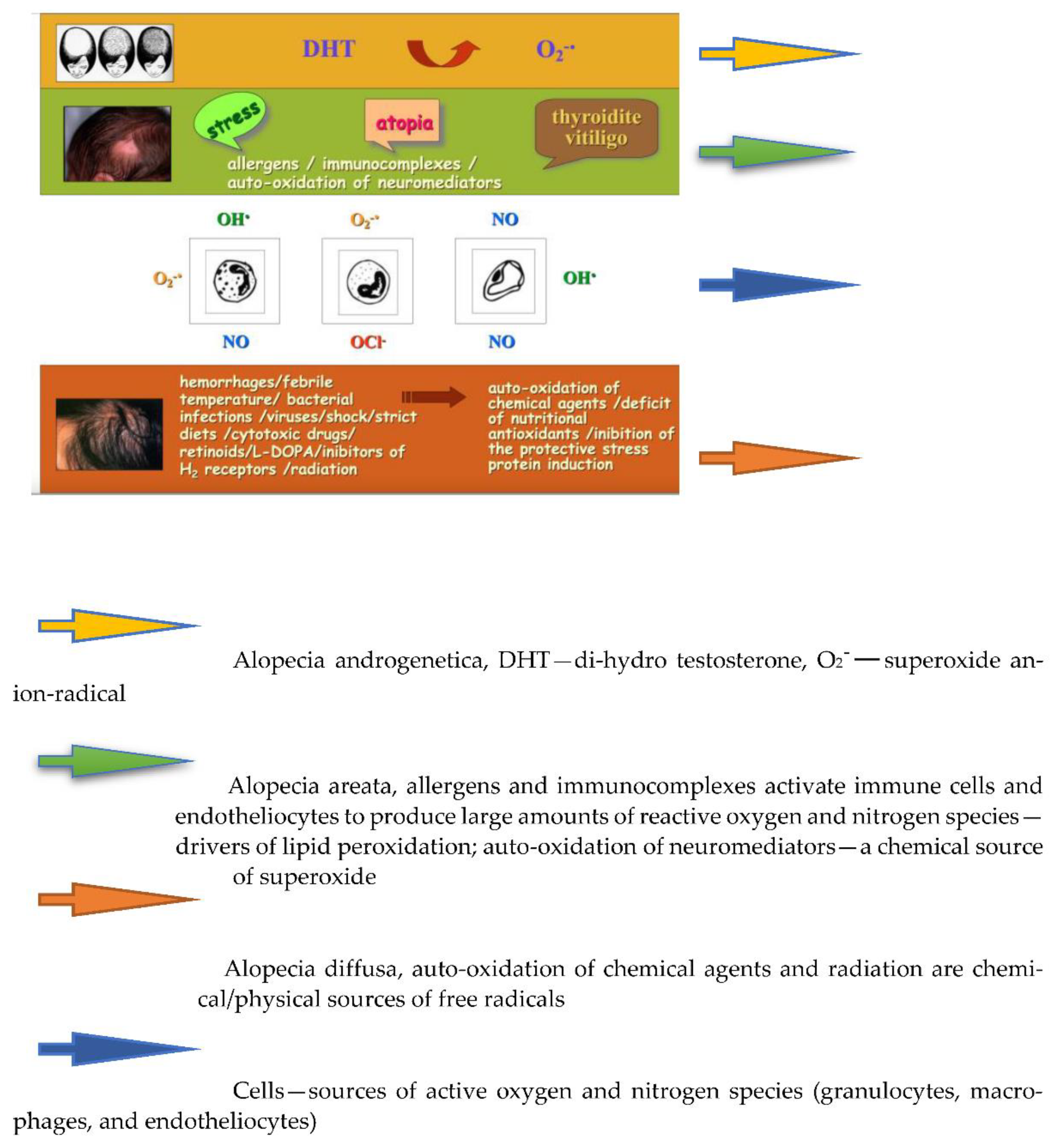
:1. Introduction
2. Materials and Methods
2.1. Patients and Study Design
- ➢
- Regular use of the products for 14 weeks;
- ➢
- Avoid any other product with similar intended use;
- ➢
- In the case of any side effects, they were instructed to immediately discontinue the products and consult a dermatologist.
2.2. Questionnaire for Experts Dermatologists/Trichologists
2.3. Cosmetic Products under Investigation and Protocols of the Application
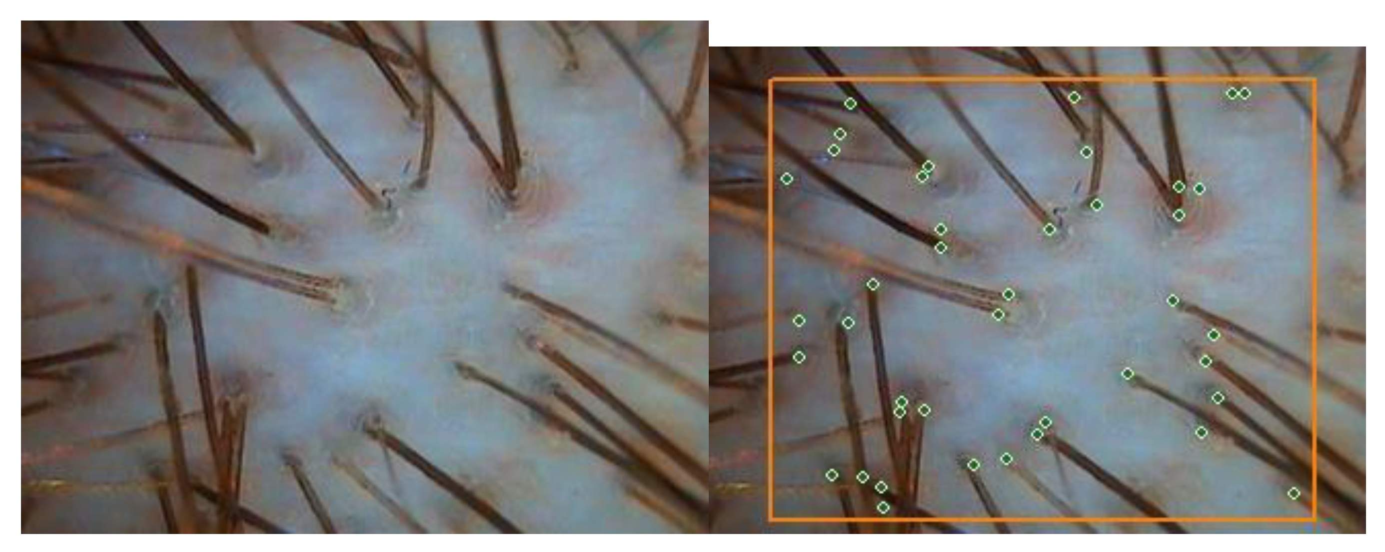
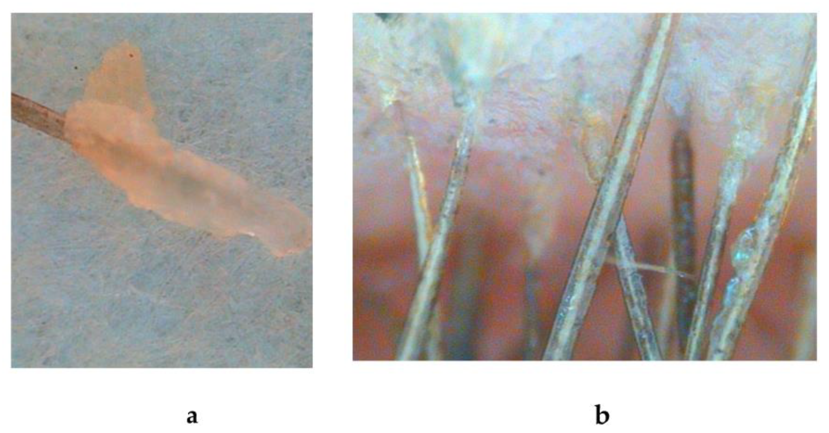
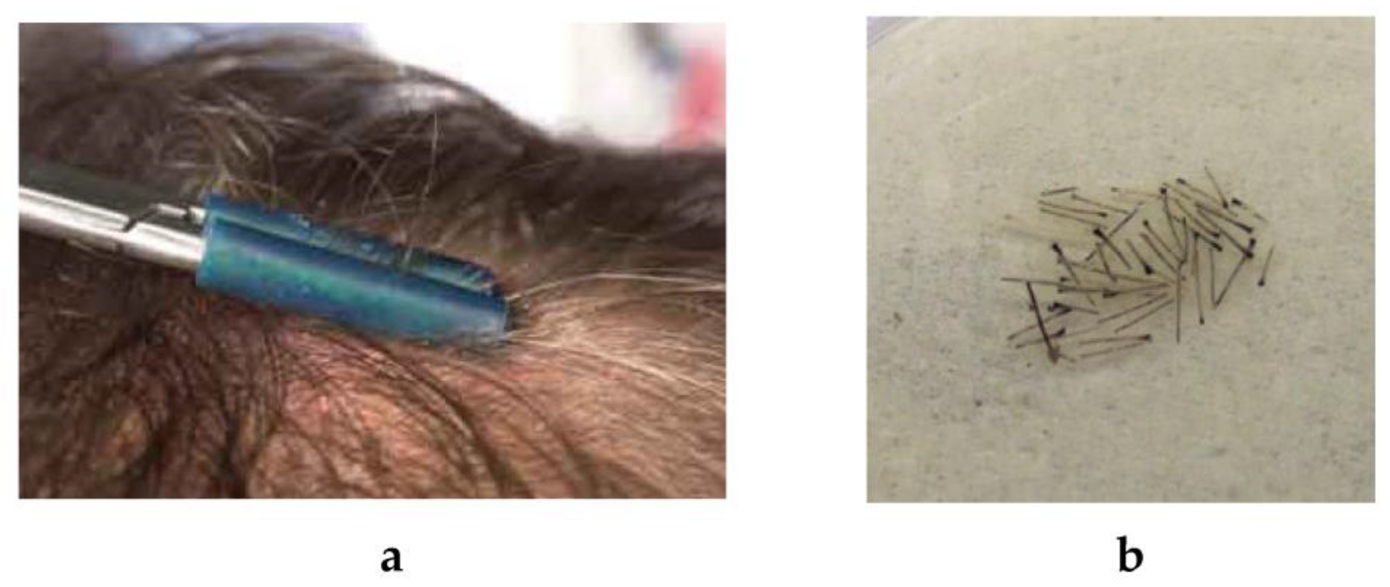
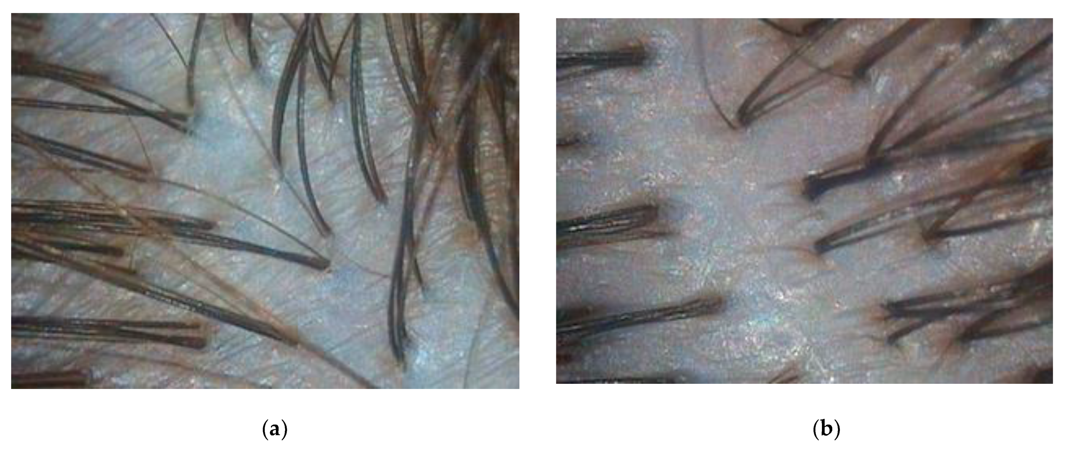
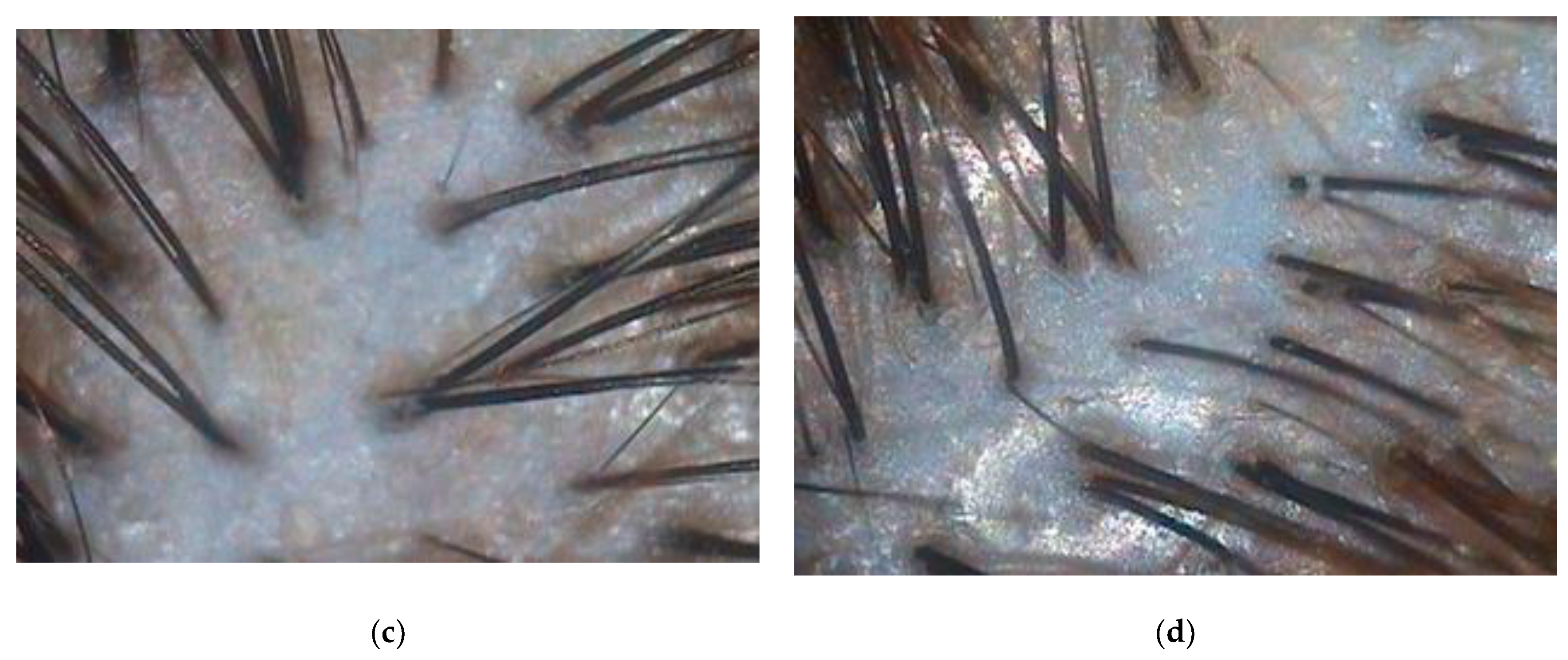
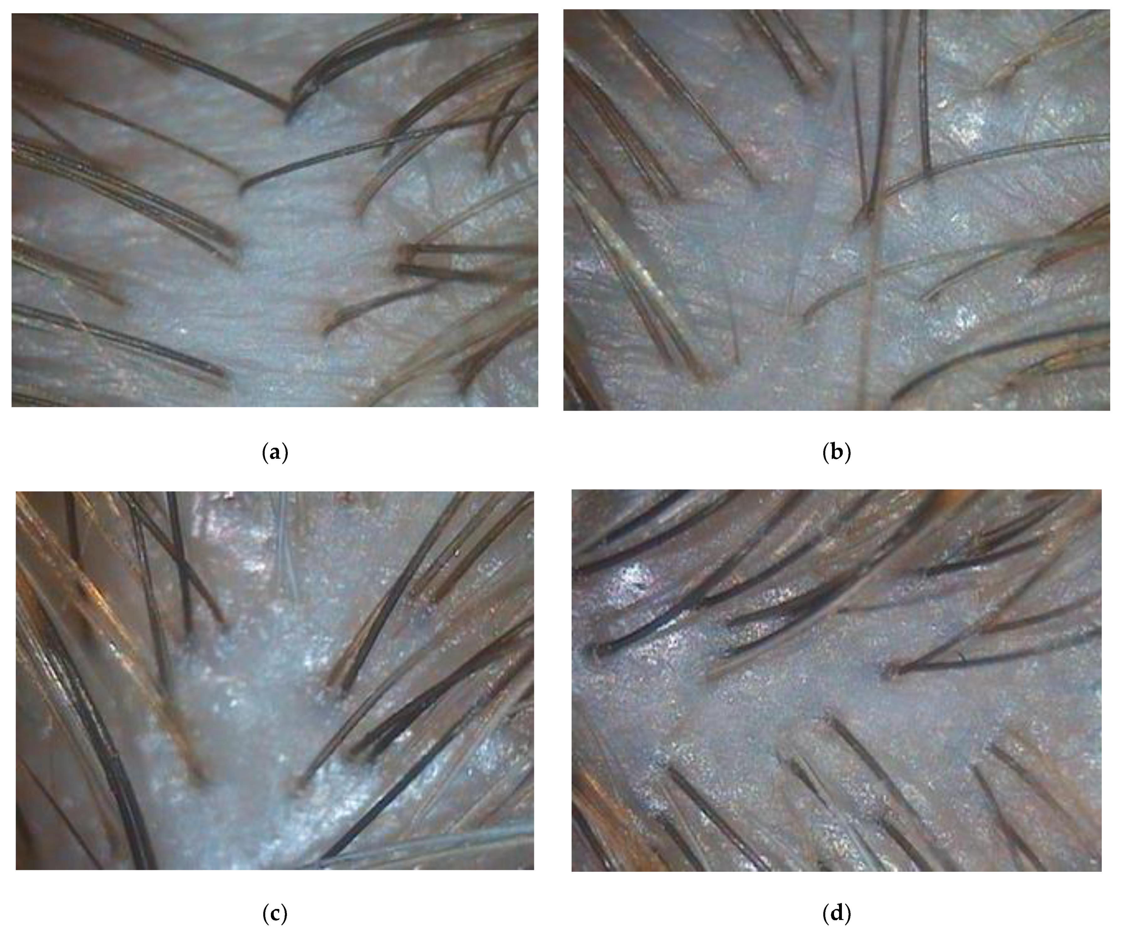
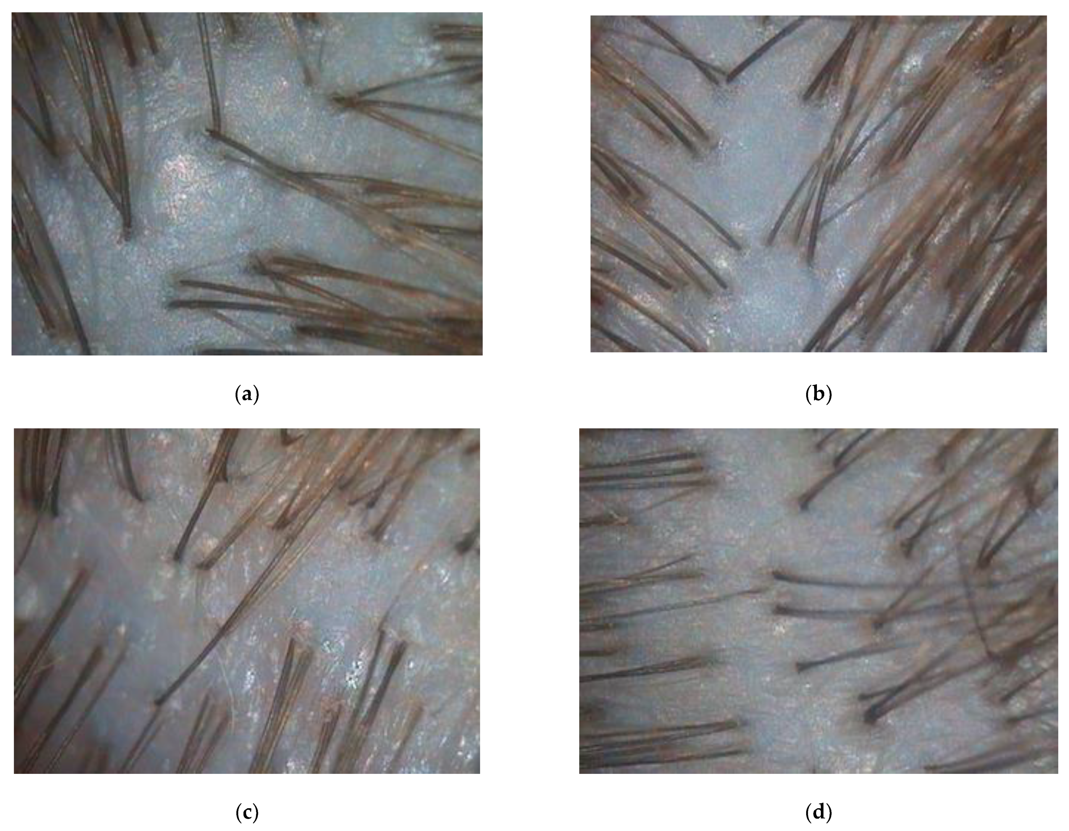
2.4. Instrumental Method of Hair Loss and Hair Quality Assessment
2.5. Lipid Peroxidation Assay
2.6. ATP Assay in Hair Follicles
2.7. Determination of SH-Group Content and Protein Leakage from Hair Shaft
2.8. Scalp Skin Microbiota Determination
2.9. Statistical Analysis
3. Results
3.1. Assessment of Clinical Efficacy of the Hair Care Cosmetics
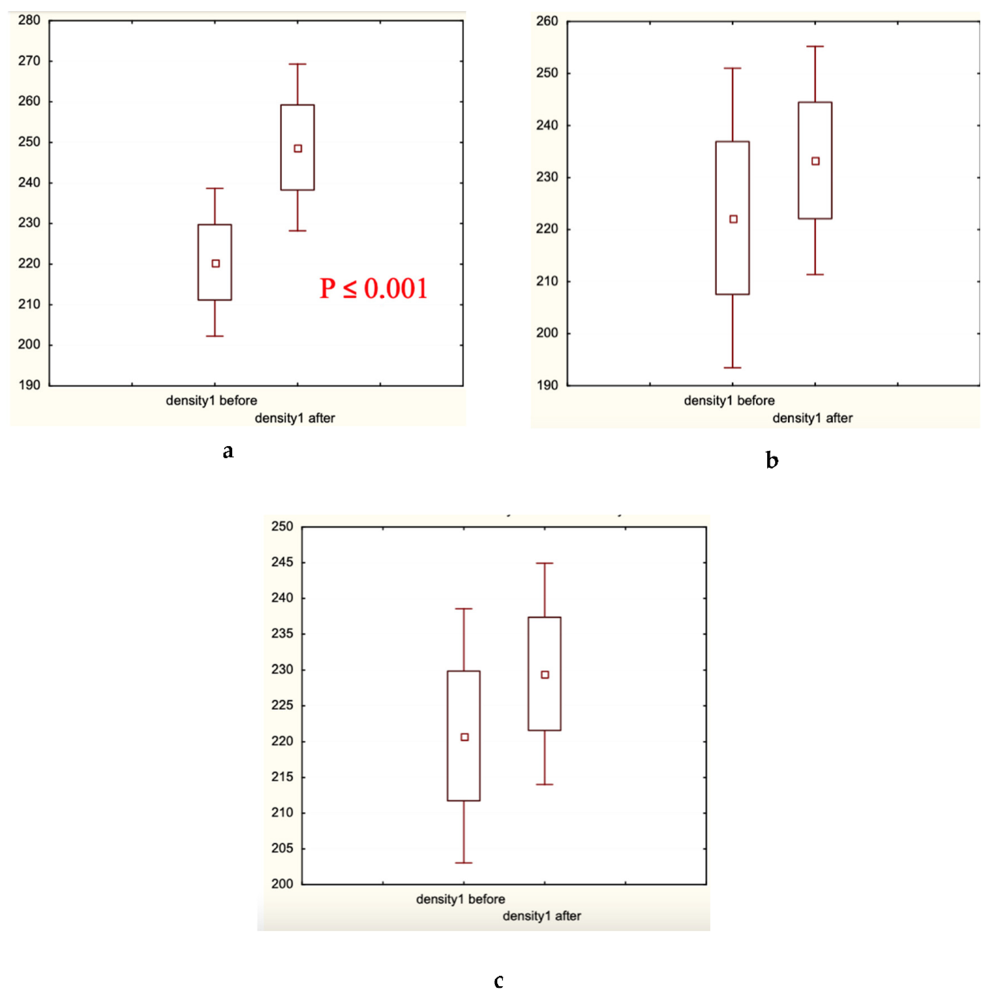
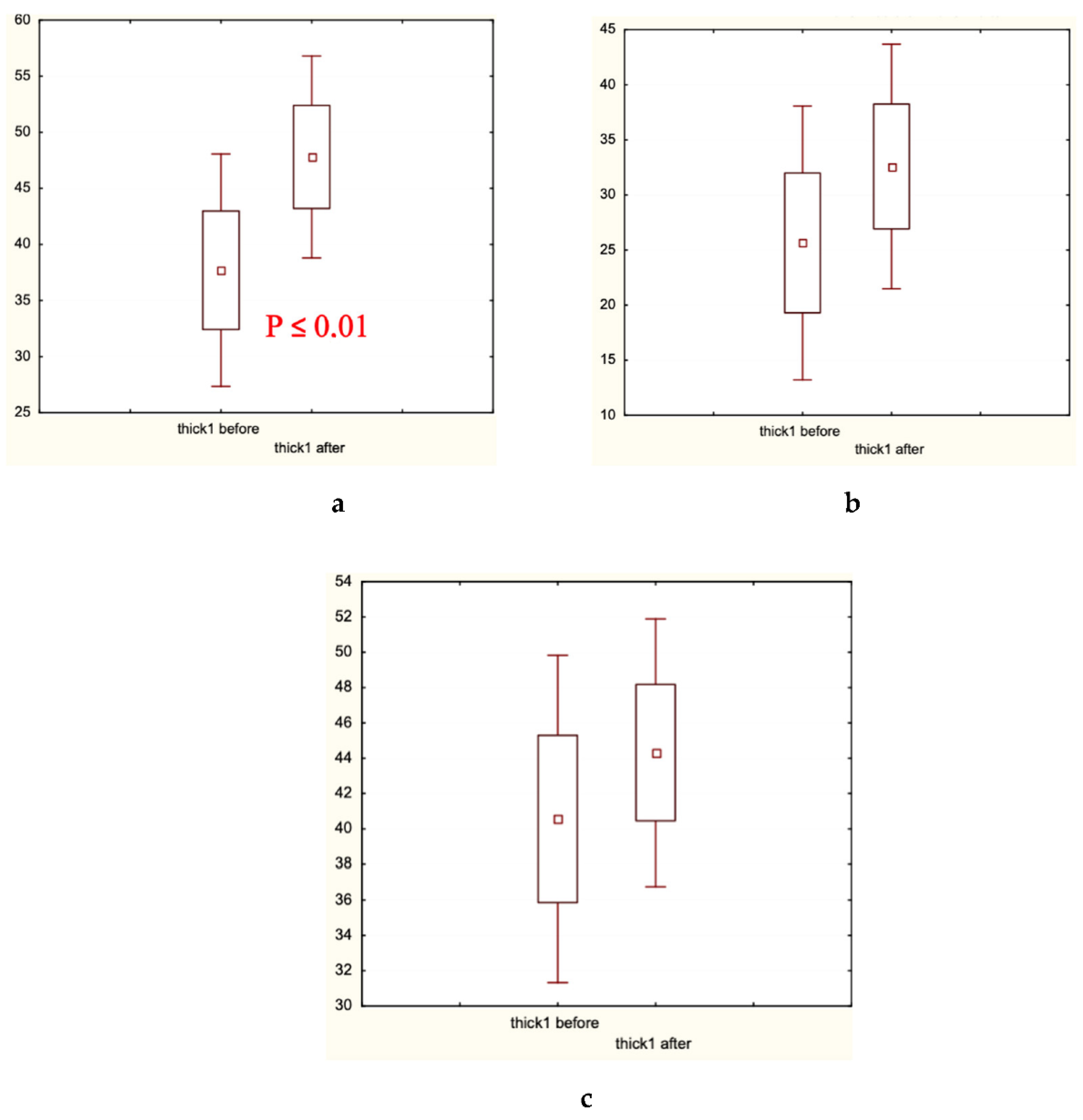
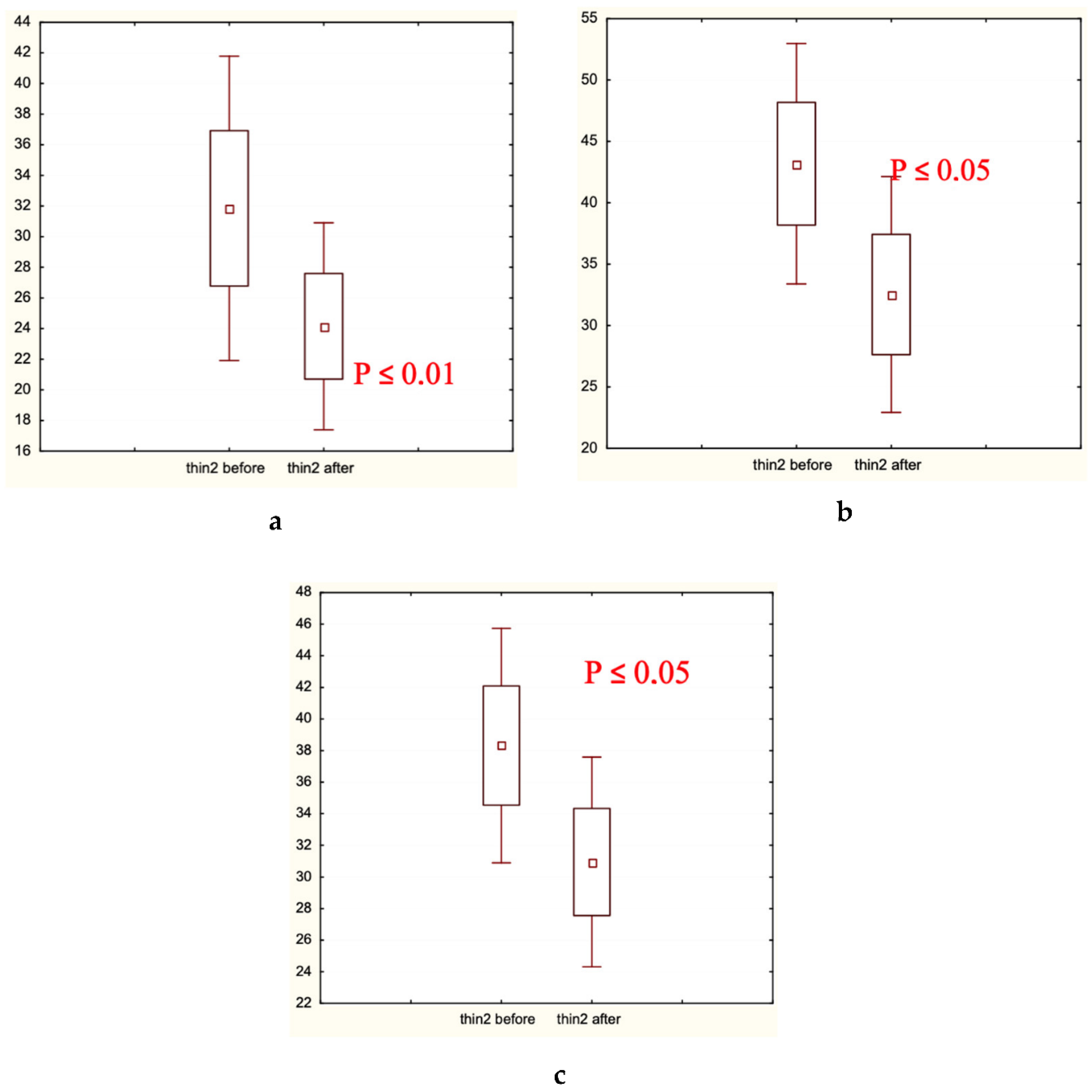
3.2. Instrumental Assessment of Hair Loss Prevention and Hair Quality Improvement Effects of the Hair Care Cosmetics
3.3. Effects of the Hair Care Products on the Intensity of Lipid Peroxidation in the Hair and Scalp Skin Lipids
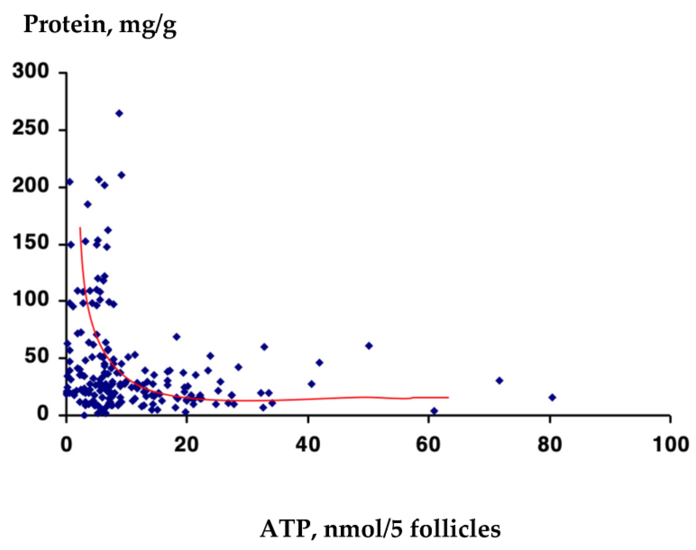
3.4. Comparative ATP Content (nmol/5 Follicles) in the Hair Follicles of Participants with Alopecia Androgenetica and Diffuse Hair Loss and Healthy Donors
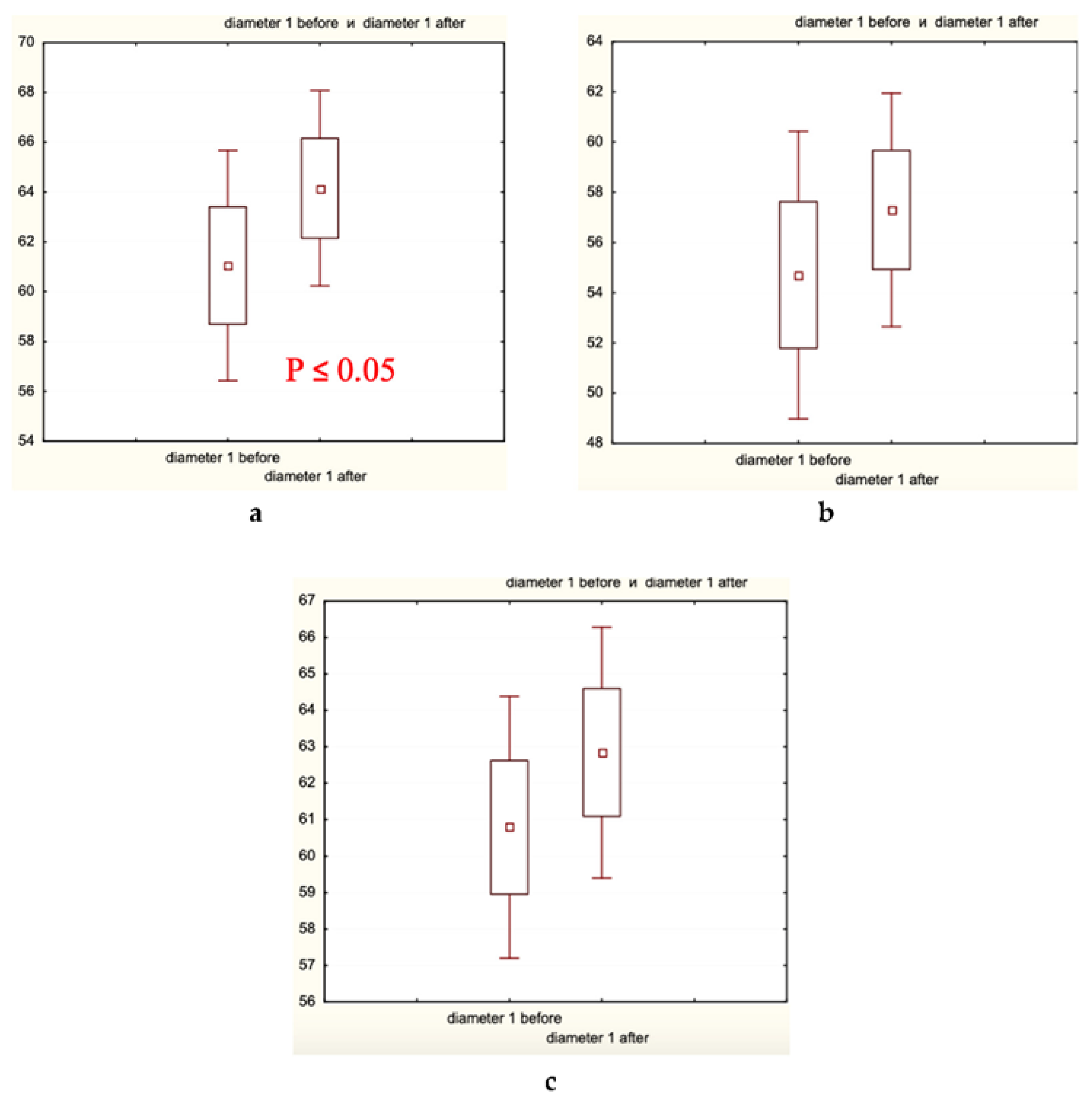
3.5. Effects of the Hair Care Products on the Hair Root/Follicle Diameter (μm) and Content of ATP (nmol/5 Follicles) in the Hair Follicles
3.6. Effects of the Hair Care Products on the Protein Leakage from the Hair Shaft and the SH-Group Content in the Hair Shaft
3.7. Effects of the Experimental Hair Care Products on the Microbiota Spectrum on the Scalp Skin
4. Discussion
Author Contributions
Funding
Institutional Review Board Statement
Informed Consent Statement
Data Availability Statement
Acknowledgments
Conflicts of Interest
Appendix A


References
- Welzel, J.; Wolff, H.H.; Gehring, W. Reduction of telogen rate and increase of hair density in androgenetic alopecia by a cosmetic product: Results of a randomized, prospective, vehicle-controlled double-blind study in men. J. Cosmet. Dermatol. 2022, 21, 1057–1064. [Google Scholar] [CrossRef] [PubMed]
- Han, J.H.; Kwon, O.S.; Chung, J.H.; Cho, K.H.; Eun, H.C.; Kim, K.H. Effect of minoxidil on proliferation and apoptosis in dermal papilla cells of human hair follicle. J. Dermatol. Sci. 2004, 34, 91–98. [Google Scholar] [CrossRef] [PubMed]
- Oaku, Y.; Abe, A.; Sasano, Y.; Sasaki, F.; Kubota, C.; Yamamoto, N.; Nagahama, T.; Nagai, N. Minoxidil nanoparticles targeting hair follicles enhance hair growth in C57BL/6 mice. Pharmaceutics 2022, 14, 947. [Google Scholar] [CrossRef]
- Herman, A.; Herman, A.P. Topically used herbal products for the treatment of hair loss: Preclinical and clinical studies. Arch. Dermatol. Res. 2017, 309, 595–610. [Google Scholar] [CrossRef] [PubMed]
- Arif, T.; Dorjay, K.; Adil, M.; Sami, M. Dutasteride in androgenetic alopecia: An update. Clin. Pharmacol. 2017, 12, 31–35. [Google Scholar] [CrossRef]
- Gupta, A.K.; Mays, R.R.; Dotzert, M.S.; Versteed, S.G.; Shear, N.H.; Piguet, V. Efficacy of non-surgical treatments for androgenetic alopecia: A systematic review and network meta-analysis. J. Dermatol. Venereol. 2018, 32, 2112–2125. [Google Scholar] [CrossRef]
- Gupta, A.K.; Talukder, M.; Bamimore, M.A. Natural products for male androgenetic alopecia. Dermatol. Ther. 2022, 35, e15323. [Google Scholar] [CrossRef]
- Suchonwanit, P.; Srisuwanwattana, P.; Chalermroj, N.; Khunkhet, S. A randomized, double-blind controlled study of the efficacy and safety of topical solution of 0.25% finasteride admixed with 3% minoxidil vs. 3% minoxidil solution in the treatment of male androgenetic alopecia. Eur. Acad. Dermatol. Venereol. 2018, 32, 2257–2263. [Google Scholar] [CrossRef]
- Suchonwanit, P.; Iamsumang, W.; Rojhirunsakool, S. Efficacy of topical combination of 0.25% finasteride and 3% minoxidil versus minoxidil solution in female pattern hair loss: A randomized, double-blind, controlled study. Am. J. Clin. Dermatol. 2019, 20, 147–153. [Google Scholar] [CrossRef]
- Shen, Y.-L.; Li, X.-Q.; Pan, R.-R.; Yue, W.; Zhang, L.-J.; Zhang, H. Medicinal plants for the treatment of heir loss and the suggested mechanisms. Curr. Pharm. Des. 2018, 24, 3090–3100. [Google Scholar] [CrossRef]
- Adil, A.; Godwin, M. The effectiveness of treatments for androgenetic alopecia: A systemic review and meta-analysis. J. Am. Acad. Dermatol. 2017, 77, 136–141. [Google Scholar] [CrossRef] [PubMed]
- Shorter, K.; Farjo, N.P.; Picksley, S.M.; Randall, V.A. Human hair follicles contain two forms of ATP-sensitive potassium channels, only one of which is sensitive to minoxidil. FASEB J. 2008, 22, 1725–1736. [Google Scholar] [CrossRef]
- Dou, J.; Zhang, Z.; Xu, X.; Zhang, X. Exploring the effects of Chinese herbal ingredients on the signaling pathway of alopecia and the screening of effective Chinese herbal compounds. J. Ethnopharmacol. 2022, 294, 115320. [Google Scholar] [CrossRef] [PubMed]
- Wall, D.; Meah, N.; Fagan, N.; York, K.; Sinclair, R. Advances in hair growth. Fac. Rev. 2022, 11, 1. [Google Scholar] [CrossRef]
- Leirõs, G.J.; Attorresi, A.I.; Balañá, M.E. Hair follicle stem cell differentiation is inhibited through cross-talk between Wnt/β-catenin and androgen signalling in dermal papilla cells from patients with androgenetic alopecia. Br. J. Dermatol. 2012, 166, 1035–1042. [Google Scholar] [CrossRef]
- Lee, S.-H.; Yoon, J.; Shin, S.H.; Zadoor, M.; Kim, H.J.; Park, P.J.; Park, W.-S.; Min, D.S.; Kim, H.-K.; Choi, K.-C. Valproic acid induces hair regeneration in murine model and activates alkaline phosphatase activity in human dermal papilla cells. PLoS ONE 2012, 7, e34152. [Google Scholar] [CrossRef]
- Riccio, G.; Sommella, E.; Badolati, N.; Salviati, E.; Bottone, S.; Campiglia, P.; Dentice, M.; Tenore, G.C.; Stornaiuolo, M.; Novellino, E. Annurca apple polyphenols protect murine hair follicles from taxane induced dystrophy and hijacks of polyunsaturated fatty acid metabolism toward β-oxidation. Nutrients 2018, 10, 1808. [Google Scholar] [CrossRef]
- Munkhbayar, S.; Jang, S.; Cho, A.R.; Choi, S.J.; Shin, C.Y.; Eun, H.C.; Kim, K.H.; Kwon, O. Role of arachidonic acid in promoting hair growth. Ann. Dermatol. 2016, 28, 55–64. [Google Scholar] [CrossRef] [PubMed]
- Blume-Peytavi, U.; Lönnfors, S.; Hillmann, K.; Garcia Bartels, N. A randomized double-blind placebo-controlled pilot study to assess the efficacy of a 24-week topical treatment by latanoprost 0.1% on hair growth and pigmentation in healthy volunteers with androgenetic alopecia. J. Am. Acad. Dermatol. 2012, 66, 794–800. [Google Scholar] [CrossRef]
- Lourith, N.; Kanlayavattanakul, M. Hair loss and herbs for treatment. Cosmet. Dermatol. 2013, 12, 210–222. [Google Scholar] [CrossRef] [PubMed]
- Chi, E.C.; Kim, K. A conprehensive review of biochemical factors in herbs and their constituent compounds in experimental studies on alopecia. J. Ethnopharmacol. 2020, 258, 112907. [Google Scholar] [CrossRef]
- Evron, E.; Juhasz, M.; Babajouni, A.; Mesinkovska, N.A. Natural hair supplement: Friend or foe? Saw palmetto: A systematic review in alopecia. Skin. Appendage Disord. 2020, 6, 329–337. [Google Scholar] [CrossRef] [PubMed]
- Zhu, H.L.; Gao, Y.H.; Yang, J.Q.; Li, J.B.; Gao, J. Serenoa repens extracts promote hair regeneration and repair of hair loss mouse models by activating TGF-beta and mitochondrial signaling pathway. Eur. Rev. Med. Pharm. Sci. 2018, 22, 4000–4008. [Google Scholar]
- Zhang, H.; Su, Y.; Wang, J.; Gao, Y.; Yang, F.; Li, G.; Shi, Q. Ginsenoside Rb1 promotes the growth of mink hair follicle via PI3K/AKT/GSK-3β signaling pathway. Life Sci. 2019, 229, 210–218. [Google Scholar] [CrossRef] [PubMed]
- Perna, S.; Spadaccini, D.; Botteri, L.; Girometta, C.; Riva, A.; Allegrini, P.; Petrangolini, G.; Infantino, V.; Rondanelli, M. Efficacy of bergamot: From anti-inflammatory and anti-oxidative mechanisms to clinical applications as preventive agent for cardiovascular morbidity, skin diseases, and mood apterations. Food Sci. Nutr. 2019, 7, 369–384. [Google Scholar] [CrossRef] [PubMed]
- Her, Y.; Lee, T.-K.; Sim, H.; Lee, J.-C.; Kim, D.W.; Choi, S.Y.; Hong, J.K.; Lee, J.-W.; Kim, J.-D.; Won, M.-H.; et al. Pinus thunbergii bark extract rich in flavonoids promotes hair growth in dorsal skin by regulating inflammatory cytokines and increasing growth factors in mice. Mol. Med. Rep. 2022, 25, 100. [Google Scholar] [CrossRef]
- Shin, H.-S.; Lee, J.-M.; Park, S.-Y.; Yang, J.-E.; Kim, J.-H.; Yi, T.-H. Hair growth activity of Crataegus pinnatifida on C57BL/6 mouse model. Phytother. Res. 2013, 27, 1352–1357. [Google Scholar] [CrossRef]
- Park, P.J.; Moon, B.S.; Lee, S.H.; Kim, S.N.; Kim, A.R.; Kim, H.J.; Park, W.S.; Choi, K.Y.; Cho, E.G.; Lee, T.R. Hair growth-promoting effect of Aconiti Ciliare Tuber extract mediated by the activation of Wnt/β-catenin signaling. Life Sci. 2012, 91, 935–943. [Google Scholar] [CrossRef]
- Kim, Y.E.; Choi, H.C.; Lee, I.C.; Yuk, D.Y.; Lee, H.; Choi, B.Y. 3-Deoxysappanchalcone Promotes Proliferation of Human Hair Follicle Dermal Papilla Cells and Hair Growth in C57BL/6 Mice by Modulating WNT/beta-Catenin and STAT Signaling. Biomol. Ther. 2016, 24, 572–580. [Google Scholar] [CrossRef]
- Shin, H.; Cho, A.-R.; Kim, D.Y.; Munkhbayer, S.; Choi, S.-J.; Jang, S.; Kim, S.H.; Shin, H.-C.; Kwon, O. Enhancement of human hair growth using Ecklonia cava polyphenols. Ann. Dermatol. 2016, 28, 15–21. [Google Scholar] [CrossRef]
- Park, S.; Lee, J. Modulation of hair growth promoting effect by natural products. Pharmaceutics 2021, 13, 2163. [Google Scholar] [CrossRef] [PubMed]
- Bassino, E.; Gasparri, F.; Munaron, L. Protective role of nutritional plants containing flavonoids in hair follicle disruption: A review. Int. J. Mol. Sci. 2020, 21, 523. [Google Scholar] [CrossRef]
- Tenore, G.C.; Caruso, D.; Buonomo, G.; D’Avino, M.; Irace, C.; Piccolo, M.; Maisto, M.; Novellino, E. Nutraceutical formulation enhances keratin expression in a human model of skin and promotes hair growth and tropism in a randomized clinical trial. J. Med. Food 2018, 21, 90–103. [Google Scholar] [CrossRef]
- Piccolo, M.; Ferraro, M.G.; Maione, F.; Maisto, M.; Stornaiuolo, M.; Tenore, G.C.; Santamaria, R.; Irace, C.; Novellino, E. Induction of hair keratins expression by an annurca apple-based nutraceutical formulation in human follicular cells. Nutrients 2019, 11, 3041. [Google Scholar] [CrossRef] [PubMed]
- Takahashi, T.; Kamimura, A.; Kagoura, M.; Toyoda, M.; Morohashi, M. Investigation of the topical application of procyanidine oligomers from apples to identify their potential use as a hair-growing agent. J. Cosmet. Dermatol. 2005, 4, 245–249. [Google Scholar] [CrossRef]
- Badolati, N.; Sommella, E.; Riccio, G.; Salviati, E.; Heintz, D.; Bottone, S.; Di Cicco, E.; Dentice, M.; Tenore, G.; Campiglia, P.; et al. Annurca Apple Polyphenols Ignite Keratin Production in Hair Follicles by Inhibiting the Pentose Phosphate Pathway and Amino Acid Oxidation. Nutrients 2018, 10, 1406. [Google Scholar] [CrossRef] [PubMed]
- Leem, J.; Jung, W.; Park, H.-J.; Kim, K. A network pharmacology-based approach to explore mechanism of action of medicinal herbs for alopecia treatment. Sci. Rep. 2022, 12, 2852. [Google Scholar] [CrossRef]
- Lin, Y.; Shao, R.; Xiao, T.; Sun, S. Promotion of hair regrowth by transdermal dissolvable microneedles loaded with rapamycin and epigallocatechin gallate nanoparticles. Pharmaceutics 2022, 14, 1404. [Google Scholar] [CrossRef]
- Herman, A.; Herman, A.P. Caffeine’s mechanisms of action and its cosmetic use. Skin. Pharmacol. Physiol. 2013, 26, 8–14. [Google Scholar] [CrossRef]
- Fischer, T.W.; Herczeg-Listes, E.; Funk, W.; Zillikens, D.; Biro’, T.; Paus, R. Differential effects of caffeine on hair shaft elongation, matrix and outer root sheath keratinocyte proliferation, and transforming growth factor-β2/insulin-like growth factor-1-mediated regulation of the hair cycle in male and female human hair follicles in vitro. Br. J. Dermatol. 2014, 171, 1031–1043. [Google Scholar] [CrossRef]
- Fischer, T.W.; Hipler, U.C.; Elsner, P. Effect of caffeine and testosterone on the proliferation of human hair follicles in vitro. Int. J. Dermatol. 2007, 46, 27–35. [Google Scholar] [CrossRef]
- Marotta, J.C.; Patel, G.; Carvalho, M.; Blakeney, S. Clinical efficacy of a topical compounded formulation in male androgenetic alopecia: Minoxidil 10%, finasteride 0.1%, biotin 0.2%, and caffeine citrate 0.05% hydroalcoholic solution. Int. J. Pharm. Compd. 2020, 24, 69–76. [Google Scholar] [PubMed]
- Arck, P.; Handjiski, B.; Hagen, E.; Pincus, M.; Bruenahl, C.; Bienenstock, J.; Paus, R. Is there a gut-brain-skin axis? Exp. Dermatol. 2010, 19, 401–405. [Google Scholar] [CrossRef] [PubMed]
- Hai, Z.; Ren, Y.; Hu, J.; Wang, H.; Qin, Q.; Chen, T. Evaluation of the treatment effect of Aloe vera fermentation in burn injury healing using a rat model. Mediat. Inflamm. 2019, 2019, 2020858. [Google Scholar] [CrossRef]
- Park, D.W.; Lee, H.S.; Shim, M.-S.; Yum, K.J.; Seo, J.T. Do kimchi and Cheonggukjang probiotics as a functional food improve androgenetic alopecia? A clinical pilot study. World J. Mens. Health 2020, 38, 95–102. [Google Scholar] [CrossRef]
- Yoon, Y.C.; Ahn, B.H.; Min, J.W.; Lee, K.R.; Park, S.H.; Kang, H.C. Stimulatory effects of extracellular vesicles derived from Leuconostoc holzapfelii that exists in human scalp on hair growth in human follicle dermal papilla cells. Curr. Issues Mol. Biol. 2022, 44, 845–866. [Google Scholar] [CrossRef]
- Hill, D.; Surgue, I.; Arendt, E.; Hill, C.; Stanton, C.; Ross, R.P. Recent advances in microbial fermentation for diary and health. F1000Research 2017, 6, 751. [Google Scholar] [CrossRef] [PubMed]
- Raman, M.; Ambalam, P.; Kondepudi, K.K.; Pithva, S.; Kothari, C.; Patel, A.T.; Purama, R.K.; Dave, J.M.; Vyas, B.R. Potential of probiotics, prebiotics and synbiotics for management of colorectal cancer. Gut Microbes 2013, 4, 181–192. [Google Scholar] [CrossRef]
- Swain, M.R.; Anandharaj, M.; Ray, R.C.; Rani, R.P. Fermented fruits and vegetables of Asia: A potential source of probiotics. Biotechnol. Res. Intern. 2014, 2014, 250424. [Google Scholar] [CrossRef]
- Kharaeva, Z.; Shokarova, A.; Shomakhova, Z.; Ibragimova, G.; Trakhtman, P.; Trakhtman, I.; Chung, J.; Mayer, W.; De Luca, C.; Korkina, L. Fermented Carica papaya and Morinda citrifolia as perspective food supplements for the treatment of post-COVID symptoms: Randomized placebo-controlled clinical laboratory study. Nutrients 2022, 14, 2203. [Google Scholar] [CrossRef]
- Mikhalchik, E.V.; Ivanova, A.V.; Anurov, M.V.; Titkova, S.M.; Pen’kov, L.Y.; Kharaeva, Z.F.; Korkina, L.G. Wound-healing effect of papaya-based preparation in experimental thermal trauma. Bull. Exp. Biol. Med. 2004, 137, 560–562. [Google Scholar] [CrossRef] [PubMed]
- Kharaeva, Z.; Trakhtman, P.; Trakhtman, I.; De Luca, C.; Mayer, W.; Chung, J.; Ibragimova, G.; Korkina, L. Fermented mangosteen (Garcinia mangostana L.) supplementation in the prevention of the HPV-induced cervical cancer: From mechanisms to clinical outcomes. Cancers 2022, 14, 4707. [Google Scholar] [CrossRef] [PubMed]
- Kharaeva, Z.F.; Zhanimova, L.R.; Mustafaev, M.S.; De Luca, C.; Mayer, W.; Chung, J.; Tiew, R.; Korkina, L.G. Effects of standardised fermented papaya gel on clinical symptoms, inflammatory cytokines, and nitric oxide metabolites in patients with chronic periodontitis: An open randomised clinical study. Med. Inflam. 2016, 2016, 9379840. [Google Scholar] [CrossRef] [PubMed]
- Kharaeva, Z.; Shogenova, A.; Barokova, E.; Trakhtman, I.; Chung, J.; Tiew, R.; Mayer, W.; De Luca, C.; Ibragimova, G.; Korkina, L. Post-COVID periodontitis: Clinical efficacy of fermented Carica papaya L. and possible mechanisms. Internat J. Dent. Ora Hea 2022, 8, 43–61. [Google Scholar]
- Kharaeva, Z.; Hokonova, T.; Elmurzaeva, J.; Dzamihova, I.; Mayer, W.; De Luca, C.; Trakhtman, I.; Korkina, L. Effects of heavy isotopes (2H1 and 18O16) depleted water consumption on physical recovery and metabolic and immunological parameters of healthy volunteers under regular fitness load. Sports 2021, 9, 110. [Google Scholar] [CrossRef]
- Reed, D.J.; Babson, J.R.; Beatty, P.W.; Brodie, A.E.; Ellis, W.W.; Potter, D.W. High performance liquid chromatography analysis of nanomole levels of glutathione disulphide and related thiols and disulphides. Anal. Biochem. 1980, 106, 55–62. [Google Scholar] [CrossRef] [PubMed]
- Bradford, M.M. A rapid and sensitive method for the quantitation of microgram quantities of protein utilizing the principle of protein-dye binding. Anal. Biochem. 1976, 72, 248–254. [Google Scholar] [CrossRef]
- Chen, Y.; Fischbach, M.; Balkaid, Y. Skin microbiota-host interactions. Nature 2018, 553, 427–436. [Google Scholar] [CrossRef]
- Osipov, G.A.; Turova, E.S. Studying species composition of microbial communities with the use of gas chromatography-mass spectrometry. Microbial community of kaolin. FEMS Microbiol. Rev. 1997, 20, 437–446. [Google Scholar] [CrossRef]
- Davis, M.G.; Piliang, M.P.; Bergfeld, W.F.; Caterino, T.L.; Fisher, B.K.; Sacha, J.P.; Carr, G.J.; Moulton, L.T.; Whittenbarger, D.J.; Schwartz, J.R. Scalp application of antioxidants improves scalp condition and reduces hair shedding in a 24-week randomized, double-blind, placebo-controlled clinical trial. Int. J. Cosmet. Sci. 2021, 43 (Suppl. S1), S14–S25. [Google Scholar] [CrossRef]
- Essendoubi, M.; Meunier, M.; Scandolera, A.; Gobinet, C.; Manfait, M.; Lambert, C.; Auriol, D.; Reynaud, R.; Piot, O. Conformation changes in human hair keratin observed using confocal Raman spectroscopy after active ingredient application. Internat J. Cosmet. Sci. 2019, 41, 203–212. [Google Scholar] [CrossRef] [PubMed]
- Fedorkova, M.V.; Brandt, N.N.; Chikishev, A.Y.; Smolina, N.V.; Balabushevich, N.G.; Gusev, S.A.; Lipatova, V.A.; Botchey, V.M.; Dobretsov, G.E.; Mikhalchik, E.V. Photoinduced formation of thiols in human hair. J. Photochem. Photobiol. B 2016, 164, 43–48. [Google Scholar] [CrossRef] [PubMed]
- Ashour, A.; Amen, Y.; Nakagawa, T.; Niwa, Y.; Mira, A.; Ohnuki, K.; Murakami, S.; Imao, M.; Shimizu, K. A new aliphatic ester of hydroxysalicylic acid from fermented Carica papaya L. preparation with a potential hair growth stimulating activity. Prod. Res. 2020, 34, 1750–1755. [Google Scholar] [CrossRef] [PubMed]


| Symptom | Group 1 (n = 100) | Group 2 (n = 29) | Group 3 (n = 25) | |||
|---|---|---|---|---|---|---|
| Before | After | Before | After | Before | After | |
| Hair shaft conditions | 3.0 ± 0.5 | 4.3 ± 0.3 ** | 3.1 ± 0.4 | 3.3 ± 0.7 | 3.1 ± 0.5 | 3.4 ± 0.5 |
| Scalp skin dryness | 1.7 ± 0.2 | 1.6 ± 0.1 | 1.6 ± 0.2 | 1.5 ± 0.2 | 1.5 ± 0.3 | 1.7 ± 0.2 |
| Scalp skin redness | 1.2 ± 0.1 | 1.0 ± 0.2 | 1.0 ± 0.1 | 1.0 ± 0.1 | 1.2 ± 0.1 | 1.0 ± 0.0 |
| Scalp skin/hair fatness | 1.8 ± 0.2 | 1.0 ± 0.1 ** | 2.0 ± 0.3 | 1.4 ± 0.1 * | 2.1 ± 0.2 | 1.4 ± 0.1 ** |
| Scalp skin irritation | 1.2 ± 0.1 | 1.0 ± 0.0 | 1.0 ± 0.1 | 1.0 ± 0.1 | 1.0 ± 0.2 | 1.0 ± 0.0 |
| Hair loss intensity | 2.4 ± 0.2 | 1.8 ± 0.2 ** | 2.5 ± 0.3 | 2.3 ± 0.2 | 2.2 ± 0.2 | 2.1 ± 0.3 |
| Group | Content of MDA, μmol/cm2 | |
|---|---|---|
| Before | After 14 Weeks | |
| Group 1, Experimental (lotion/shampoo containing fermented fruits and caffein) | 23.8 ± 1.6 | 20.0 ± 1.5 * |
| Group 2, Placebo Control (lotion/shampoo without actives) | 24.0 ± 1.3 | 23.9 ± 2.1 |
| Group 3, Caffeine Control (lotion/shampoo with caffein only) | 24.2 ± 1.2 | 24.5 ± 1.5 |
| Group | ATP Content (nmol/5 Follicles) | |
|---|---|---|
| Frontal Area | Occipital Area | |
| Healthy donors | 80.6 ± 9.1 ** | 74.8 ± 7.4 ** |
| Alopecia androgenetica | 19.9 ± 3.2 | 17.8 ± 2.7 |
| Diffuse hair loss | 24.8 ± 4.0 | 21.1 ± 1.8 |
| Group | Hair Root/Follicle Diameter (μm) | ATP Content (nmol/5 Follicles) | ||
|---|---|---|---|---|
| Before | After | Before | After | |
| Group 1 Experimental (lotion/shampoo containing fermented fruits and caffein) | 98 ± 13 | 74 ± 12 * | 20.5 ± 2.9 | 71.5 ± 3.1 **, # |
| Group 2 Placebo Control (lotion/shampoo without actives) | 96 ± 15 | 92 ± 11 | 17.7 ± 3.2 | 18.9 ± 2.8 |
| Group 3 Caffeine Control (lotion with caffein/placebo shampoo) | 97 ± 10 | 104 ± 16 | 18.5 ± 3.0 | 40.2 ± 4.8 *, # |
| Group | Labile Protein Leakage from Hair Shaft (μg/g Hair) | |
|---|---|---|
| Frontal Area | Occipital Area | |
| Healthy donors | 15.0 ± 0.9 ** | 15.3 ± 1.2 ** |
| Alopecia androgenetica | 49.2 ± 9.8 | 48.5 ± 7.0 |
| Diffuse hair loss | 47.9 ± 8.8 | 48.1 ± 9.6 |
| Group | Labile Protein Leakage from Hair Shaft (μg/g Hair) | Nonbinding SH-Groups in Hair Shaft (ng/mg Protein) | ||
|---|---|---|---|---|
| Before | After | Before | After | |
| Group 1 Experimental (lotion/shampoo containing fermented fruits and caffein) | 46.5 ± 7.2 | 21.3 ± 4.5 ** | 35.7 ± 5.0 | 12.3 ± 2.5 **, # |
| Group 2 Placebo Control (lotion/shampoo without actives) | 47.1 ± 9.0 | 28.9 ± 5.1 * | 41.1 ± 6.7 | 53.7 ± 7.6 |
| Group 3 Caffeine Control (lotion with caffein/placebo shampoo) | 45.9 ± 8.4 | 20.8 ± 4.4 ** | 33.2 ± 2.5 | 24.1 ± 1.9 *, # |
| Microorganisms | Normal Values, 105 Cells/g | Clinical Trial Data, 105 Cells/g | p between before and after | |
|---|---|---|---|---|
| Before | After | |||
| Streptococcus spp. | 270–350 | 355 ± 15 | 300 ± 15 | p < 0.05 |
| Staphylococcus epidermidis | 180–240 | 235 ± 15 | 190 ± 15 | p < 0.05 |
| Propionibacterium acnes | 0–42 | 35 ± 5 | 25 ± 3 | p < 0.05 |
| Cutibacterium acne | 0–50 | 55 ± 10 | 35 ± 4 | p < 0.05 |
| Malassezia restructa | 0–42 | 25 ± 5 | 10 ± 3 | p < 0.01 |
| Actinomyces spp. | 52–92 | 75 ± 10 | 77 ± 20 | p > 0.05 |
| Corynebacterium spp. | 575–635 | 600 ± 50 | 590 ± 30 | p > 0.05 |
| Malassezia globosa | 0–52 | 20 ± 5 | 25 ± 3 | p > 0.05 |
Disclaimer/Publisher’s Note: The statements, opinions and data contained in all publications are solely those of the individual author(s) and contributor(s) and not of MDPI and/or the editor(s). MDPI and/or the editor(s) disclaim responsibility for any injury to people or property resulting from any ideas, methods, instructions or products referred to in the content. |
© 2023 by the authors. Licensee MDPI, Basel, Switzerland. This article is an open access article distributed under the terms and conditions of the Creative Commons Attribution (CC BY) license (https://creativecommons.org/licenses/by/4.0/).
Share and Cite
Mayer, W.; Weibel, M.; De Luca, C.; Ibragimova, G.; Trakhtman, I.; Kharaeva, Z.; Chandler, D.L.; Korkina, L. Biomolecules of Fermented Tropical Fruits and Fermenting Microbes as Regulators of Human Hair Loss, Hair Quality, and Scalp Microbiota. Biomolecules 2023, 13, 699. https://doi.org/10.3390/biom13040699
Mayer W, Weibel M, De Luca C, Ibragimova G, Trakhtman I, Kharaeva Z, Chandler DL, Korkina L. Biomolecules of Fermented Tropical Fruits and Fermenting Microbes as Regulators of Human Hair Loss, Hair Quality, and Scalp Microbiota. Biomolecules. 2023; 13(4):699. https://doi.org/10.3390/biom13040699
Chicago/Turabian StyleMayer, Wolfgang, Michaela Weibel, Chiara De Luca, Galina Ibragimova, Ilya Trakhtman, Zaira Kharaeva, Danny L. Chandler, and Liudmila Korkina. 2023. "Biomolecules of Fermented Tropical Fruits and Fermenting Microbes as Regulators of Human Hair Loss, Hair Quality, and Scalp Microbiota" Biomolecules 13, no. 4: 699. https://doi.org/10.3390/biom13040699