Review

HSP70 and Primary Arterial Hypertension

by
Bernardo Rodriguez-Iturbe
1,*,
Richard J. Johnson
2,
Laura Gabriela Sanchez-Lozada
3 and
Hector Pons
4
1
Department of Nephrology, Instituto Nacional de Ciencias Médicas y Nutrición “Salvador Zubirán”, Mexico City 14080, Mexico
2
Division of Renal Diseases and Hypertension, University of Colorado Anschutz Medical Campus, Aurora, CO 80045, USA
3
Department of Cardio-Renal Physiopathology, National Institute of Cardiology Ignacio Chávez, Mexico City 14080, Mexico
4
Facultad de Medicina, Universidad del Zulia, Maracaibo 4011, Venezuela
*
Author to whom correspondence should be addressed.
Biomolecules 2023, 13(2), 272; https://doi.org/10.3390/biom13020272
Submission received: 16 December 2022 / Revised: 14 January 2023 / Accepted: 18 January 2023 / Published: 1 February 2023
(This article belongs to the Special Issue HSP70: From Signaling Mechanisms to Therapeutics)

Abstract

:
Heat shock protein 70 (HSP70) production is a stress-generated cellular response with high interspecies homology. HSP70 has both chaperone and cytokine functions and may induce, depending on the context, tolerogenic anti-inflammatory reactivity or immunogenic and autoimmune reactivity. Intracellular (chaperoning transit of antigens to MHC in antigen-presenting cells) and extracellular HSP70-related effects are associated with hypertension, which is an inflammatory condition recognized as the most important risk factor for cardiovascular disease mortality. Here, we review (a) the relationship between HSP70, inflammation and immune reactivity, (b) clinical evidence relating to stress, HSP70 and anti-HSP70 reactivity with primary hypertension and (c) experimental data showing that salt-sensitive hypertension is associated with delayed hypersensitivity to HSP70. This is a consequence of anti-HSP70 reactivity in the kidneys and may be prevented and corrected by the T-cell-driven inhibition of kidney inflammation triggered by specific epitopes of HSP70. Finally, we discuss our postulate that lifelong stress signals and danger-associated molecular patterns stimulate HSP-70 and individual genetic and epigenetic characteristics determine whether the HSP70 response would drive inflammatory immune reactivity causing hypertension or, alternatively, would drive immunomodulatory responses that protect against hypertension.

1. Introduction

Heat shock proteins (HSPs) are evolutionarily preserved immunodominant proteins that are constitutively expressed and rapidly increase to represent as much as 15% of cellular protein content in response to stressful stimuli [1]. Stress-induced overexpression aims to prevent the cellular aggregation of misfolded proteins by chaperoning their refolding or their degradation if they are irreparably damaged [2]. The adaptive reactivity generated by HSP activation includes antioxidant responses that prevent oxidative stress, suppress inflammation and inhibit apoptosis by binding proapoptotic factors and blocking Bax mitochondrial translocation [3]. The overall results of these actions are the protection of biological pathways critical for cell function and the prevention of DNA fragmentation [4].
In addition to their cytoprotective functions, HSPs are immunodominant molecules that are important in the recognition of infectious agents and drive antigens to the mixed histocompatibility complex (MHC) in antigen-presenting cells (APC) [5]. Hence, HSP-driven modifications in the course of diseases depend on the specific cell type and the pathogenic dysregulation involved. HSP responses inducing anti-inflammatory cytokine responses and regulatory T-cell expression are beneficial to downregulate uncontrolled autoimmune reactivity, but the suppression of inflammation may be deleterious when inflammation constitutes a defense mechanism. For instance, HSP expression in synovial tissues is anti-inflammatory and protective in rheumatoid arthritis [6], while the stimulation of antigen-presenting cells by HSP70 may be responsible for acute asthmatic attacks [7] and the HSP70-mediated suppression of immune reactivity and apoptosis may represent a survival factor for cancer cells [8].

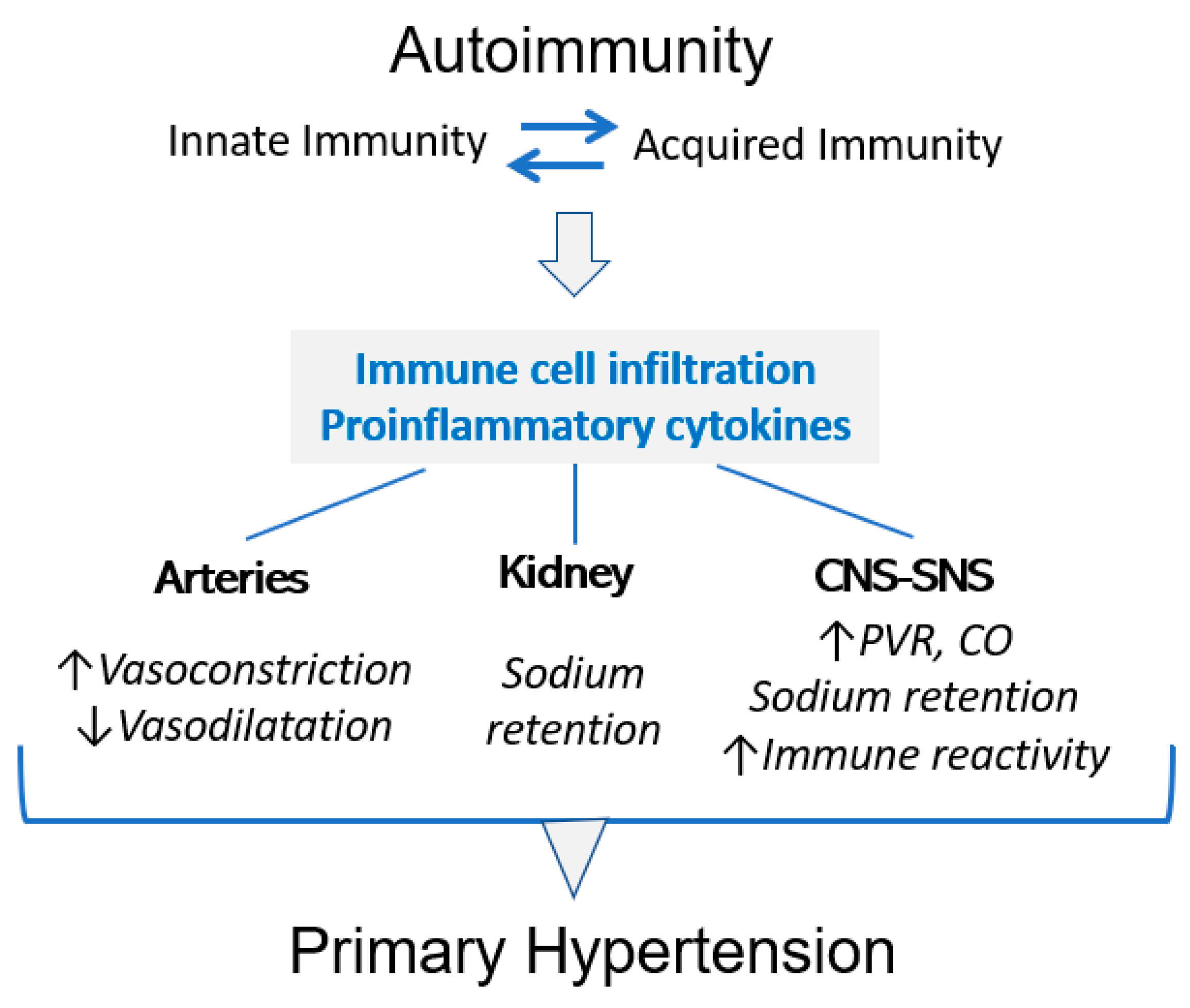
2. HSP70, Inflammation and Immune Reactivity

HSP proteins have a highly preserved interspecies homology and molecular mimicry makes self-HSPs potential targets for disease-related T-cell responses. HSP70 is the most extensively studied HSP and in the human is comprised of a family of 13 members located in cytosol, nuclei, mitochondria, and endoplasmic reticula. The structure of HSP70 family members includes an N-terminal nucleotide-binding domain, a C-terminal substrate-binding domain and a C-terminal tail that binds with co-chaperones. Functionally, HSP70 proteins are characterized by substrate recognition, adaptive responses imposed by cellular and extracellular localizations, and interactions with other chaperones that control the presentation of client proteins to HSP70 and their refolding after HSP70 activity [9,10].
HspA1A and HspA1B are the major stress-inducible HSP70 proteins of the HSP70 family. HSPs are intracellular proteins and extracellular locations of HSP70 (complexed with antigenic peptides, in exosomes and in free soluble form) may be a consequence of cell necrosis or result from release in exosomes, extrusion in cholesterol microdomains or lysosomal secretion [10].
HSP70 proteins induce intricate and contrasting immunomodulatory responses that drive the development of anti-inflammatory or proinflammatory immune reactivity. Collaborations between HSP70 and other co-chaperones, most notably HSP90 and HSP40 [11,12,13], are part of the HSP70 chaperone reaction cycle [10] and constitute a central characteristic of the heat shock response (HSR) that contributes to its pleiomorphic manifestations. HSP70 and HSP40 share a J-domain, DnaJ, necessary but not sufficient for the ATPase activity. A HSP40–HSP70 partnership is required for ATP hydrolysis and for substrate identification and, therefore, it is essential for HSP70-induced protein disaggregation, which is a preliminary step for subsequent refolding. The collaboration between HSP70 and HSP40 in the protection of proteins has been extensively reviewed [11]. HSP90 is another co-chaperone that has a large diversity of substrates and acts downstream of HSP70. While HSP70 binds to short hydrophobic segments, HSP90 interacts with extended binding domains of 50 to 100 aminoacids, driving the folding of proteins that restores their native condition. The absence of HSP90 results in the degradation of many substrates, most notably the receptors of steroid hormones. Interactions between HSP70 and HSP90 have been comprehensively reviewed [12,13].
The immune reactivity induced by HSP70 depends, at least in part, on the location of HSP70 activity: intracellular HSP70 is anti-inflammatory, while extracellular HSP70 may induce anti-inflammatory and proinflammatory responses [14,15,16] (Figure 1).
Martine et al. [18] have shown that HSP70 is a negative regulator of NLRP3 inflammasome in vivo and in vitro. Their studies focused on inducible HSP70 and showed that HSP70 deficiency enhances caspase -1 activation and IL-1β production in activated macrophages and overexpression of HSP70 and heat shock inhibits NLRP3 inflammasome activation and reduces caspase and IL-1β production.
Tolerogenic responses are not exclusive of intracellular HSP70 and may also be induced by extracellular HSP70 delivered to or present in cellular membranes or in ectosomes [19]. In vitro treatment of peripheral blood mononuclear cells of patients with rheumatoid arthritis with HSP70 induce the production of IL-10 and Mycobacterium tuberculosis HSP70 (mtHS70) blocks the maturation of bone marrow precursors that fail to express MHC class II and CD86. Using an innovative method of immersing graft tissue with mtHSP70, Borges et al. [20] showed that soluble human HSP70 induces a regulatory phenotype in monocyte-derived dendritic cells and inhibits acute allograph rejection. The internalization of extracellular HSP70 likely depends on scavenger receptors, such as LOX-1 (lectin-like oxidized low-density lipoprotein receptor 1), SREC-1 (scavenger receptor-1) and FEEl-1 (scavenger receptor expressed by endothelial cells-1). Intracellular signaling is apparently mediated by the activation of MAPK/ERK (mitogen-activated protein kinase / extracellular signal-regulated kinase) and STAT3 (Signal transducer and activator of transcription 3) pathways that stimulate Il-10 production and tolerance [19]. The clinical significance of the immune suppressive response that HSPs may induce was shown in seminal studies conducted by van Eden et al. [21,22,23]. Van Eden et al. showed that Mycobacterium TBC contained an epitope that was cross-reactive with self-antigens present in the joint cartilage of rats with adjuvant arthritis and in T lymphocytes of patients with rheumatoid arthritis. Screening mycobacteria antigens showed that T-cell clones of adjuvant arthritis recognized the aminoacid sequence in position 180–188 of Mycobacterium bovis BCG antigen and further showed that administration of this antigen induced resistance to adjuvant arthritis in rats [21]. In subsequent studies, the authors identified specific peptide sequences of mycobacterial HSP that stimulated a protective self-HSP cross-reactivity, inducing IL-10 mediated suppression of autoimmune colitis and autoimmune encephalitis [22,23].
Tolerogenic responses are not exclusive of intracellular HSP70 and may also be induced by extracellular HSP70 delivered to or present in cellular membranes or in ectosomes (reviewed in [19]). The in vitro treatment of peripheral blood mononuclear cells of patients with rheumatoid arthritis with HSP70 induces the production of IL-10, and Mycobacterium tuberculosis HSP70 (mtHS70) blocks the maturation of bone marrow precursors that fail to express MHC class II and CD86. Soluble human HSP70 induces a regulatory phenotype in monocyte-derived dendritic cells and soluble mtHSP70 inhibits acute allograph rejection [20]. The internalization of extracellular HSP70 likely depends on scavenger receptors, and signaling is transmitted through the TLR2 and ERK pathways [19]. The clinical significance of the immune-suppressive response that HSPs may induce was shown in seminal studies by van Eden et al. [21,22,23], who used specific peptide sequences of mycobacterial HSP to stimulate protective self-HSP cross-reactivity to induce the IL-10-mediated suppression of adjuvant arthritis, colitis and autoimmune encephalitis.
Proinflammatory effects of HSP70 were studied by Asea et al. [24], who demonstrated that the treatment of monocytes with exogenous HSP70 resulted in the phosphorylation of inhibitory I-κBα subunit at Ser3 and the activation of NFkB. HSP70 activates monocytes by inducing intracellular Ca++ flux and additional pathways, independent and dependent of CD14, that generate the release of tumor necrosis factor (TNF)-α, interleukin (IL)-1β and IL-6. Some studies of the proinflammatory stimulation induced by HSP70 were controversial and attributed to contamination with endotoxin [25], but the extracellular expression of HSP70 and the existence of anti-HSP70 serum antibodies are beyond dispute, as is the role that HSP70 plays in acquired immunity chaperoning intracellular and extracellular antigens to MHC I and II in APC in canonical and cross-presentation path (reviewed in [26]).
The potential of HSP70 to induce autoimmunity in vivo was demonstrated by Millar et al. [27] in the model of autoimmune diabetes. Their studies showed that the tolerance induced by the immunodominant gp33 epitope was changed to a rapid autoimmunity response, resulting in diabetes with the administration of HSP70. Furthermore, HSP70 may also determine the selection of the epitope chosen for the presentation of autoreactive T cells [28]. In human autoimmunity diseases, the existence of anti-HSP70 antibodies is well-recognized (reviewed in [29]).
A major insight into the contrasting proinflammatory and anti-inflammatory mechanisms activated by HSP70 was obtained through the studies of Fong et al. [30], who investigated the relationship between HSP70 and Ig-related lectins that recognize sialic acid (Siglecs). Sialic acids are self-associated molecular patterns (“SAMP”) essential to self-recognition by the immune system and Siglec-5 and Siglec-14 are paired receptors that recognize identical ligands but send opposing immune reactivity signals. Siglec-5 and Siglec-14 were identified as ligands of HSP70, and their response was anti-inflammatory and proinflammatory, respectively, suggesting a likely explanation for the contradictory results reported in the extracellular HSP70 activation [30].
Of relevance to the relation between HSP70 and blood pressure is the recognition that stress-induced HSP70 response may suppress as well as stimulate inflammation. The contrasting HSP70 response could generate and aggravate or prevent and ameliorate hypertensive cardiovascular disease.

3. HSP70 and High Blood Pressure

Hypertension is an inflammatory condition in which immune reactivity, innate and acquired, plays a critical pathogenic role [31,32,33,34,35]. Autoimmune reactivity triggered by protein adducts generated by isoketals (gamma ketoaldehydes) resulting from lipid peroxidation has been reported in experimental models of hypertension and plasma F2-isoprostanes, which are formed in concert with isoketals, were found to be elevated in [36]. Our group has suggested that HSP70 is a critical element in the pathogenesis of hypertension by either generating anti-HSP70 immune reactivity and/or driving autoantigens to the MHC in antigen-presenting cells [26,31,37].
Hypertension has been mechanistically associated with inflammation resulting from innate and acquired autoimmunity originating from danger-associated molecular patterns (DAMPs) generated by metabolic stress. Metabolic stress during a life span is impossible to determine, but is likely comparable in the vast majority of hypertensive and normotensive individuals. We suggest that the individual HSP70 response to stress, proinflammatory or anti-inflammatory, is the determining factor in the development of or protection from hypertension.

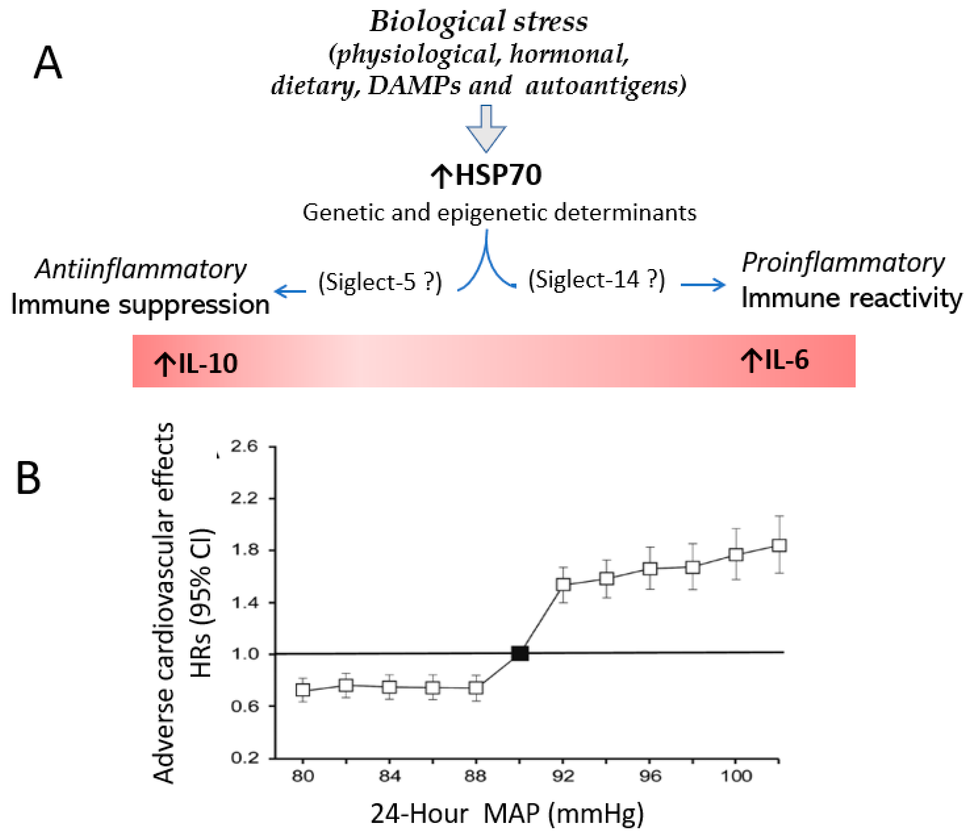
4. Stress, HSP70 and Hypertension

The association of environmental stress with an increase in blood pressure was reported more than 60 years ago in studies that showed that, in the general population, blood pressure was higher during the winter [38]. This finding was attributed to increased sympathetic activity in cold weather. However, the seasonal variability of blood pressure, while evident in hypertensive patients, was not confirmed in normotensive individuals, and changes in plasma and urinary catecholamines in the winter were similar in normotensives and hypertensives [39]. Comprehensive studies by the group of Pavel Hamet in Montreal confirmed an inverse relationship between blood pressure and daily temperature in hypertensive patients [40], and subsequent investigations in experimental models of hypertension demonstrated the increased sensitivity of blood pressure to various stresses. For instance, McMurthy et al. [41] showed that exposure to 50 °C for 1 min or to ether vapors raised blood pressure in spontaneously hypertensive rats (SHRs) more than in normotensive rats. The hypertensive response was preceded by hormonal changes, particularly in aldosterone and corticosterone blood levels.
Recognizing that heat shock protein activation is an evolution-preserved response to stress, several investigations showed that imposing thermal, painful and restrained conditions induced exaggerated increments in HSP70 mRNA in SHRs, hypertensive mice strains [42] and hypertensive humans [43]. These studies prompted the hypothesis that genetic characteristics in hypertensives could be responsible for the overexpression of HSP70 due to environmental stress. This hypothesis was supported by studies that localized the HSP70 gene near the MHC in SHRs [44].
While stressful stimuli induce an overexpression in HSP70 mRNA in hypertensive rodents and hypertensive humans, this exaggerated response is transient and may be a protective response. Chen et al. [45] showed that raising the body temperature to 42 °C for 45 min a day before the administration of angiotensin II suppressed the expression of NFkB in the aorta and reduced hypertension.
The increase in blood pressure by itself is a stress factor that stimulates HSP70. Xu et al. [46] showed that phenylephrine, dopamine, vasopressin, angiotensin II, and endothelin-1 increased HSP70 mRNA in cells harvested from the aorta 60 min after a blood pressure increase. Furthermore, they found a correlation between the increase in blood pressure and HSP70 expression and suggested that the findings indicated a protective response against hypertension.
Significantly, the immediate and short-term HSP70 increment resulting from stress differs from the lifelong increment in blood pressure characteristic of hypertensive cardiovascular disease. In fact, quite the opposite is suggested by studies that show that the same stress, if applied repeatedly, reduces baseline blood pressure. Kvetnansky et al. [47] induced repeated restrain episodes in SHRs and found that, contrary to expectations, the acute increment in blood pressure observed initially was attenuated, and the baseline blood pressure was normalized despite increased baseline levels of norepinephrine. Similar results were reported by Malo et al. [48], who showed that daily exposure to 50 °C for 5 min progressively lowered the basal blood pressure of hypertensive mice to levels similar to that of normotensive mice. This phenomenon was associated with a faster declination of HSP70 mRNA and protein and is relevant to reinforce that repeated episodic stress, by itself, does not play a role in chronic increments in blood pressure.

5. Anti-HSP70 Antibodies in Primary Hypertension

Table 1 summarizes the studies that showed an association between hypertension and HSP70 expression [31,43,49,50,51,52,53,54,55,56,57,58,59].
Evidence against the possibility that HSP70 overexpression is a protective response to hypertension was obtained in young prehypertensive SHRs. The overexpression of HSP72 was observed in 4-week-old SHRs, anteceding the development of hypertension [31]. Wu et al. [50] studied 764 steel mill workers from 6 work sites and showed that harsh working conditions (noise, dust and heat) were associated with an incidence of anti-HSP70 antibodies in 40.6% of hypertensive patients and only 18.6% of normotensive individuals; furthermore, statistical correlations between anti-HSP70 levels and hypertension were more significant in severely (>160/95 mmHg) hypertensive patients. Studies of circulating lymphocytes showed that lymphocytes harvested from hypertensive patients have a more intense HSP70mRNA expression than lymphocytes of normotensive individuals [43]. Bravo et al. [49] showed that HSP70 and vimentin are overexpressed in the cortical proximal tubular cells of the kidney in experimental modes of salt-sensitive hypertension. The constitutively expressed levels of HSP70 and their relationship with inflammatory markers were studied by Srivastava et al. [51] in 132 hypertensives and 132 normotensive individuals; they found that HSP70 mRNA in whole blood and HSP70 expression in plasma were increased in hypertensive patients. In addition, IL-6 and TNFα in hypertensive patients, but not in controls, were significantly correlated with HSP70 gene expression. Pockley et al. [52] studied serum samples obtained in the European Lacidipine Study on Atherosclerosis (ELSA) and the Swedish population-screening program from 111 hypertensive patients and 75 normotensive controls and found increased levels of antiHSP70 and anti-HSP65 antibodies in hypertensive patients. Similar results were found by our group [53]. Pons et al. [59] found that peripheral blood lymphocytes of patients with primary hypertension, in contrast to lymphocytes of normotensive individuals, develop a proliferative response when challenged with a specific HSP70 peptide sequence.
Genetic studies were done by Li et al. [54] focusing on polymorphisms of the HSP70 family and the risk of hypertension in the Uygur population in China that have a high homogeneity and unique life styles. HSP70 polymorphisms were associated with 3 to 5-fold increased risk of hypertension. Zheng et al. [55] also focused in HSP70 polymorphisms in oven workers in China and found a 2 to 4-fold increased relative risk with hypertension.

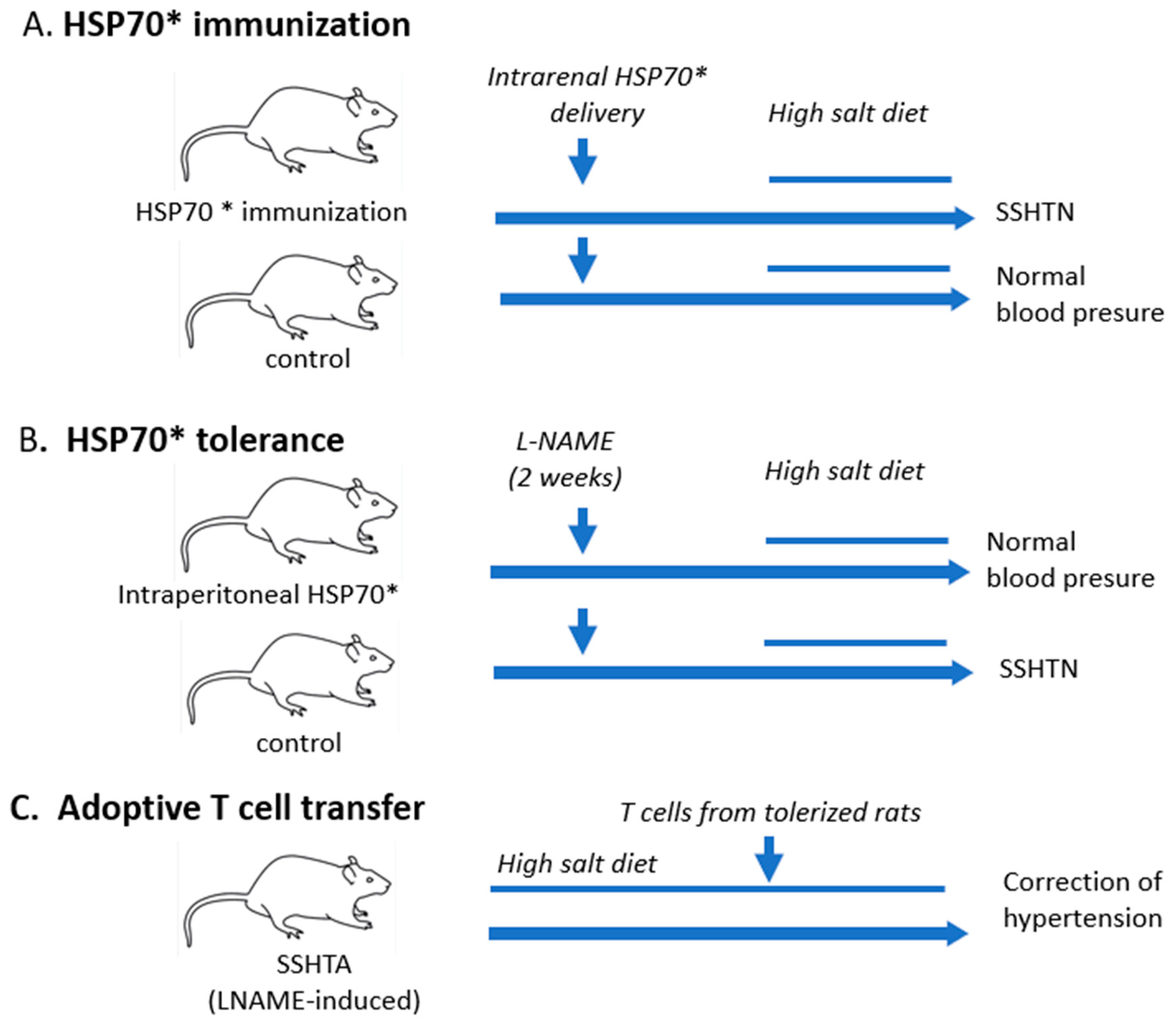
6. Hypertension Is Induced by Immune Reactivity to HSP70

The association between immune reactivity and hypertension is well-established. A large number of studies in genetic and induced experimental hypertension have demonstrated that hypertension is corrected or prevented by immunosuppressive anti-inflammatory treatment (reviewed in [31,37]). Autoimmune reactivity induces hypertension that, as a consequence of inflammatory infiltration in the adventitia of arteries and tubulointerstitial areas of the kidney, increases sympathetic nervous system activity (Figure 2). Barhoumi et al. [56], studying angiotensin-induced hypertension, showed that macrophage and T-cell infiltration of the aorta were prevented by the adoptive transfer of T-reg cells, which ameliorated hypertension. The activation of sodium reabsorption in the kidney induced by immune reactivity was demonstrated by Norlander et al. [57], who found that experimental hypertension was associated with T-cell production of IL-17 and that hypertension was blunted in the IL-17 -/- mice. IL-17 prohypertensive effects include the activation of sodium chloride co-transporter and epithelial sodium channel in the distal tubule. Increase sympathetic activity not only results in vasoconstriction and increased sodium reabsorption, but stimulates immune cell release from the spleen and supports immune responses [31,58].
The role played by the HSP70 response in hypertension was investigated in the experimental model of salt-sensitive hypertension (SSHTN) induced by the transient (3-week) administration of Nw-nitro-L-arginine methyl ester (LNAME) [60]. In these studies, we used a specific amino acid sequence of Mycobacterium tuberculosis HSP70 (mtHSP70) shown by Wendling et al. [61] and Praken et al. [62]) to be immunogenic and to induce the generation of T cells that were cross-reactive with the homologous rat HSP70 sequence. In their experiments [60,61], these peptide-induced T cells and T cells generated by the whole HSP70 produced IL-10 and, if administered intranasally, inhibited adjuvant arthritis. Rats with SSHTN were shown to have delayed hypersensitivity to mtHSP70 and their splenocytes had a clonal CD4 response when challenged with mtHSP70.
Immunization in the foot pads with mtHSP70 followed by the expression of HSP70 induced in the kidney via plasmid injection generated a tubulointerstitial inflammatory infiltrate of macrophages and lymphocytes and the development of SSHTN [59] (Figure 3A).

7. Immune Tolerance to HSP70 Corrects Hypertension

The participation of immune reactivity to HSP70 in the pathogenesis of SSHTN was further investigated by the induction of immune tolerance to HSP70 by the intraperitoneal administration of mtHSP70. The effectiveness of tolerization was confirmed by the suppression of delayed hypersensitivity to HSP70, the induction of T regulatory cells, the reduction in proinflammatory cytokines (IL-6) and the increase in IL-10 in the kidneys of rats with SSHTN. Furthermore, tolerization resulted in the inhibition of tubulointerstitial inflammation, which is the hallmark of SSHTN. Immune tolerance to HSP70 prevented the development of SSHTN (Figure 3B). In addition, the adoptive transfer of T cells from tolerized rats to rats with SSHTN corrected hypertension (Figure 3C) [59].

8. HSP70-Induced Immune Reactivity Modulates Inflammation in Hypertension

We contend that the characteristics of HSP70-induced immune reactivity are a central determinant of blood pressure. Overall biological stress is likely comparable in the majority of hypertensive and normotensive individuals, and increased or normal blood pressure is not a consequence of stress signals but instead the result of the proinflammatory or anti-inflammatory HSP70 response to those signals. It should be recognized that hypertension is a quantitative, rather than qualitative, deviation of the norm, and the division between normal and increased blood pressure is consensual and based on the incidence of associated complications [63,64]. As discussed earlier, stress-activated HSP70 overexpression may result in the stimulation or suppression of inflammation that, when present in the kidneys, the arterial walls and the central and sympathetic nervous system, determines primary hypertension.
In addition to stress-related HSP70 activity, autoimmunity may be induced by extracellular HSP70 antigenic determinants [16]; furthermore, HSP70 may play a supporting role in the antigenicity of other antigens identified in hypertensive patients [36].
As shown in Figure 4, we posit that the immune reactivity resulting from HSP70 stimulation is responsible for the prevalence of high blood pressure (>130/80 mmHg) in 51% of males and 39.7% of females 18 years of age and older in the United States [65].
In contrast with the stimulation of both innate and adaptive immunity in patients that develop severe hypertension, the remaining normotensive population responds to metabolic stress with a protective downregulation of inflammatory reactivity, which, as discussed earlier, is also a recognized HSP70 response to stress. The identification of genetic and epigenetic characteristics that determine the specific type of HSP70 response [67] and, perhaps, the differential engagement of Siglec-5 and Siglec-14, is critical in developing strategies for controlling hypertension.

9. Conclusions

There is considerable clinical evidence that associates stress, HSP70 and primary hypertension. Experimental data indicate that tubulointerstitial inflammation of the kidneys and salt-sensitive hypertension may be driven by immune reactivity to HSP70 and may be prevented and corrected by T-cell immunoregulatory responses that HSP70 may drive. The data on hypertension resemble the findings regarding other autoimmune diseases such as adjuvant arthritis in which disease activity is modulated by the induction of autoreactive HSPs [23]. We suggest that genetic and epigenetic factors determine whether HSP70-driven autoimmune reactivity results in prohypertensive inflammation or a protective anti-inflammatory response that prevents the development of hypertensive cardiovascular disease. The possibility of intervening in the HSP70 response should be considered a potential therapeutic strategy in primary hypertension.

Author Contributions

B.R.-I. wrote the article. R.J.J., L.G.S.-L. and H.P. contributed and edited the paper and approved the final form. All authors have read and agreed to the published version of the manuscript.

Funding

This research received no external funding.

Institutional Review Board Statement

Not applicable.

Informed Consent Statement

Not applicable.

Data Availability Statement

Not applicable.

Conflicts of Interest

R.J.J. and L.G.L. have equity with Colorado Research Partners LLC, R.J.J. also has stock with XORTX therapeutics, and R.J.J. has also received honoraria from Horizon Pharma. All other authors have no conflict of interest.

References

  1. Welch, W.J. Mammalian stress response: Cell physiology, structure/function of stress proteins, and implications for medicine and disease. Physiol. Rev. 1992, 72, 1063–1081. [Google Scholar] [CrossRef]
  2. Kurop, M.K.; Huyen, C.M.; Kelly, J.H.; Blagg, B.S. The heat shock response and small molecule regulators. Eur. J. Med. Chem. 2021, 226, 113846. [Google Scholar] [CrossRef] [PubMed]
  3. Beere, H.M. The stress of dying’: The role of heat shock proteins in the regulation of apoptosis. J. Cell Sci. 2004, 117, 1641–1651. [Google Scholar] [CrossRef]
  4. Lang, B.J.; Guerrero, M.E.; Prince, T.L.; Okusha, Y.; Bonorino, C.; Calderwood, S.K. The functions and regulation of heat shock proteins; key orchestrators of proteostasis and the heat shock response. Arch. Toxicol. 2021, 95, 1943–1970. [Google Scholar] [CrossRef] [PubMed]
  5. Bolhassani, A.; Agi, E. Heat shock proteins in infection. Clin. Chim. Acta 2019, 498, 90–100. [Google Scholar] [CrossRef] [PubMed]
  6. Schett, G.; Redlich, K.; Xu, Q.; Bizan, P.; Gröger, M.; Tohidast-Akrad, M.; Kiener, H.; Smolen, J.; Steiner, G. Enhanced expression of heat shock protein 70 (hsp70) and heat shock factor 1 (HSF1) activation in rheumatoid arthritis synovial tissue. Differential regulation of hsp70 expression and hsf1 activation in synovial fibroblasts by proinflammatory cytokines, shear stress, and antiinflammatory drugs. J. Clin. Investig. 1998, 102, 302–311. [Google Scholar] [CrossRef] [PubMed]
  7. Bertorelli, G.; Bocchino, V.; Zhuo, X.; Chetta, A.; Donno, D.; Foresi, A.; Testi, R.; Olivieri, D. Heat shock protein 70 upregulation is related to HLA-DR expression in bronchial asthma. Effects of inhaled glucocorticoids. Clin. Exp. Allergy 1998, 28, 551–560. [Google Scholar] [CrossRef] [PubMed]
  8. Wu, J.; Liu, T.; Rios, Z.; Mei, Q.; Lin, X.; Cao, S. Heat Shock Proteins and Cancer. Trends Pharmacol. Sci. 2017, 38, 226–256. [Google Scholar] [CrossRef]
  9. Rosenzweig, R.; Nillegoda, N.B.; Mayer, M.P.; Bukau, B. The Hsp70 chaperone network. Nat. Rev. Mol. Cell Biol. 2019, 20, 665–680. [Google Scholar] [CrossRef]
  10. Radons, J. The human HSP70 family of chaperones: Where do we stand? Cell Stress Chaperones 2016, 21, 379–404. [Google Scholar] [CrossRef] [Green Version]
  11. Liu, Q.; Liang, C.; Zhou, L. Structural and functional analysis of the Hsp70/Hsp40 chaperone system. Protein Sci. 2020, 29, 378–390. [Google Scholar] [CrossRef] [PubMed]
  12. Luengo, T.M.; Mayer, M.P.; Rüdiger, S.G. The Hsp70–Hsp90 Chaperone Cascade in Protein Folding. Trends Cell Biol. 2019, 29, 164–177. [Google Scholar] [CrossRef] [PubMed]
  13. Radli, M.; Rüdoger, S.G.D. Dancing with the Diva: Hsp90–Client Interactions. J. Mol Biol. 2018, 430, 3029–3040. [Google Scholar] [CrossRef]
  14. Chen, Y.; Voegeli, T.S.; Liu, P.P.; Noble, E.G.; Currie, R.W. Heat Shock Paradox and a New Role of Heat Shock Proteins and their Receptors as Anti-Inflammation Targets. Inflamm. Allergy-Drug Targets 2007, 6, 91–100. [Google Scholar] [CrossRef] [PubMed]
  15. Martine, P.; Rébé, C. Heat Shock Proteins and Inflammasomes. Int. J. Mol. Sci. 2019, 20, 4508. [Google Scholar] [CrossRef] [PubMed]
  16. Calderwood, S.K.; Gong, J.; Murshid, A. Extracellular HSPs: The Complicated Roles of Extracellular HSPs in Immunity. Front. Immunol. 2016, 7, 159. [Google Scholar] [CrossRef] [PubMed]
  17. Stocki, P.; Wang, X.N.; Dickinson, A.M. Inducible Heat Shock Protein 70 Reduces T Cell Responses and Stimulatory Capacity of Monocyte-derived Dendritic Cells. J. Biol. Chem. 2012, 287, 12387–12394. [Google Scholar] [CrossRef]
  18. Martine, P.; Chevriaux, A.; Derangère, V.; Apetoh, L.; Garrido, C.; Ghiringhelli, F.; Rébé, C. HSP70 is a negative regulator of NLRP3 inflammasome activation. Cell Death Dis. 2019, 10, 256. [Google Scholar] [CrossRef]
  19. Borges, T.J.; Wieten, L.; van Herwijnen, M.J.C.; Broere, F.; van der Zee, R.; Bonorino, C.; van Eden, W. The anti-inflammatory mechanisms of Hsp70. Front. Immunol. 2012, 3, 95. [Google Scholar] [CrossRef]
  20. Borges, T.J.; Porto, B.N.; Teixeira, C.A.; Rodrigues, M.; Machado, F.D.; Ornaghi, A.P.; de Souza, A.P.D.; Maito, F.; Pavanelli, W.R.; Silva, J.S.; et al. Prolonged Survival of Allografts Induced by Mycobacterial Hsp70 Is Dependent on CD4+CD25+ Regulatory T Cells. PLoS ONE 2010, 5, e14264. [Google Scholar] [CrossRef] [Green Version]
  21. Van Eden, W.; Tholet, J.E.R.; Van Der Zee, R.; Noordzij, A.; Van Embden, J.D.A.; Hensen, E.J.; Cohen, I.R. Cloning of the mycobacterial epitope recognized by T lymphocytes in adjuvant arthritis. Nature 1988, 331, 171–173. [Google Scholar] [CrossRef] [PubMed]
  22. Anderton, S.M.; van der Zee, R.; Noordzij, A.; van Eden, W. Differential mycobacterial 65-kDa heat shock protein T cell epitope recognition after adjuvant arthritis-inducing or protective immunization protocols. J. Immunol. 1994, 152, 3656–3664. [Google Scholar] [CrossRef] [PubMed]
  23. van Eden, W.; van der Zee, R.; Prakken, B. Heat-shock proteins induce T-cell regulation of chronic inflammation. Nat. Rev. Immunol. 2005, 5, 318–330. [Google Scholar] [CrossRef] [PubMed]
  24. Asea, A.; Kraeft, S.-K.; Kurt-Jones, E.A.; Stevenson, M.A.; Chen, L.B.; Finberg, R.W.; Koo, G.C.; Calderwood, S.K. HSP70 stimulates cytokine production through a CD14-dependant pathway, demonstrating its dual role as a chaperone and cytokine. Nat. Med. 2000, 6, 435–442. [Google Scholar] [CrossRef]
  25. Gao, B.; Tsan, M.-F. Endotoxin Contamination in Recombinant Human Heat Shock Protein 70 (Hsp70) Preparation Is Responsible for the Induction of Tumor Necrosis Factor α Release by Murine Macrophages. J. Biol. Chem. 2003, 278, 174–179. [Google Scholar] [CrossRef]
  26. Rodriguez-Iturbe, B.; Lanaspa, M.A.; Johnson, R.J. The role of autoimmune reactivity induced by heat shock protein 70 in the pathogenesis of essential hypertension. Br. J. Pharmacol. 2019, 176, 1829–1838. [Google Scholar] [CrossRef]
  27. Millar, D.; Garza, K.M.; Odermatt, B.; Elford, A.R.; Ono, N.; Li, Z.; Ohashi, P.S. Hsp70 promotes antigen-presenting cell function and converts T-cell tolerance to autoimmunity in vivo. Nat. Med. 2003, 9, 1469–1476. [Google Scholar] [CrossRef]
  28. Kolb, H.; Burkart, V. Chaperones may cause the focus of diabetes autoimmunity on distinct (pro)insulin peptides. J. Autoimmun. 2019, 105, 102304. [Google Scholar] [CrossRef]
  29. Androvitsanea, A.; Stylianou, K.; Drosataki, E.; Petrakis, I. The Pathophysiological Role of Heat Shock Response in Autoimmunity: A Literature Review. Cells 2021, 10, 2626. [Google Scholar] [CrossRef]
  30. Fong, J.J.; Sreedhara, K.; Deng, L.; Varki, N.M.; Angata, T.; Liu, Q.; Nizet, V.; Varki, A. Immunomodulatory activity of extracellular Hsp70 mediated via paired receptors Siglec-5 and Siglec-14. EMBO J. 2015, 34, 2775–2788. [Google Scholar] [CrossRef]
  31. Rodriguez-Iturbe, B.; Pons, H.; Johnson, R.J. Role of the Immune System in Hypertension. Physiol. Rev. 2017, 97, 1127–1164. [Google Scholar] [CrossRef]
  32. Trott, D.W.; Thabet, S.R.; Kirabo, A.; Saleh, M.A.; Itani, H.; Norlander, A.E.; Wu, J.; Goldstein, A.; Arendshorst, W.J.; Madhur, M.S.; et al. Oligoclonal cd8+ t cells play a critical role in the development of hypertension. Hypertension 2014, 64, 1108–1115. [Google Scholar] [CrossRef]
  33. Abais-Battad, J.M.; Rudemiller, N.P.; Mattson, D. L Hypertension and immunity: Mechanisms of T cell activation and pathways of hypertension. Curr. Opin. Nephrol. Hypertens. 2015, 24, 470–474. [Google Scholar] [CrossRef]
  34. Mikolajczyk, T.P.; Szczepaniak, P.; Vidler, F.; Maffia, P.; Graham, G.J.; Guzik, T.J. Role of inflammatory chemokines in hypertension. Pharmacol. Ther. 2021, 223, 107799. [Google Scholar] [CrossRef]
  35. Guzik, T.J.; Hoch, N.E.; Brown, K.A.; McCann, L.A.; Rahman, A.; Dikalov, S.; Goronzy, J.; Weyand, C.; Harrison, D.G. Role of the T cell in the genesis of angiotensin II induced hypertension and vascular dysfunction. J. Exp. Med. 2007, 204, 2449–2460. [Google Scholar] [CrossRef]
  36. Kirabo, A.; Fontana, V.; de Faria, A.P.; Loperena, R.; Galindo, C.L.; Wu, J.; Bikineyeva, A.T.; Dikalov, S.; Xiao, L.; Chen, W.; et al. DC isoketal-modified proteins activate T cells and promote hypertension. J. Clin. Investig. 2014, 124, 4642–4656. [Google Scholar] [CrossRef] [PubMed]
  37. Rodríguez-Iturbe, B.; Pons, H.; Quiroz, Y.; Johnson, R.J. The immunological basis of hypertension. Am. J. Hypertens. 2019, 32, 113–122. [Google Scholar] [CrossRef] [PubMed]
  38. Rose, G. Seasonal Variation in Blood Pressure in Man. Nature 1961, 189, 235. [Google Scholar] [CrossRef] [PubMed]
  39. Hata, T.; Ogihara, T.; Maruyama, A.; Mikami, H.; Nakamaru, M.; Naka, T.; Kumahara, Y.; Nugent, C.A. The Seasonal Variation of Blood Pressure in Patients with Essential Hypertension. Clin. Exp. Hypertens. A 1982, 4, 341–354. [Google Scholar] [CrossRef] [PubMed]
  40. Kunes, J.; Tremblay, J.; Bellavance, F.; Hamet, P. Influence of Environmental Temperature on the Blood Pressure of Hypertensive Patients in Montreal. Am. J. Hypertens. 1991, 4, 422–426. [Google Scholar] [CrossRef] [PubMed]
  41. McMurtry, J.P.; Wexler, B.C. Hypersensitivity of Spontaneously Hypertensive Rats to Heat and Ether before the Onset of High Blood Pressure. Endocrinology 1983, 112, 166–171. [Google Scholar] [CrossRef]
  42. Hamet, P.; Malo, D.; Tremblay, J. Increased transcription of a major stress gene in spontaneously hypertensive mice. Hypertension 1990, 15, 904–908. [Google Scholar] [CrossRef] [PubMed]
  43. Kunes, J.; Poirier, M.; Tremblay, J.; Hamet, P. Expression of hsp70 gene in lymphocytes from normotensive and hypertensive Humans. Acta Physiol. Scand. 1992, 146, 307–311. [Google Scholar] [CrossRef] [PubMed]
  44. Hamet, P.; Kong, D.; Pravenec, M.; Kunes, J.; Kren, V.; Klir, P.; Sun, Y.L.; Tremblay, J. Restriction fragment length polymorphism of hsp70 gene, localized in the RT1 complex, is associated with hypertension in spontaneously hypertensive rats. Hypertension 1992, 19, 611–614. [Google Scholar] [CrossRef] [PubMed]
  45. Chen, Y.; Ross, B.M.; Currie, R.W. Heat shock treatment protects against angiotensin II–induced hypertension and inflammation in aorta. Cell Stress Chaperones 2004, 9, 99–107. [Google Scholar] [CrossRef] [PubMed]
  46. Xu, Q.; Li, D.-G.; Holbrook, N.J.; Udelsman, R. Acute Hypertension Induces Heat-Shock Protein 70 Gene Expression in Rat Aorta. Circulation 1995, 92, 1223–1229. [Google Scholar] [CrossRef]
  47. Kvetnansky, R.; Mccarty, R.; Thoa, N.B.; Lake, C.R.; Kopin, I.J. Sympatho-adrenal responses of spontaneously hypertensive rats to immobilization stress. Am. J. Physiol. 1979, 236, H457–H462. [Google Scholar] [CrossRef]
  48. Malo, D.; Pang, S.C.; Schlager, G.; Tremblay, J.; Hamet, P. Decrease of Blood Pressure in Spontaneously Hypertensive Mice by Heat Treatment. Am. J. Hypertens. 1990, 3, 400–404. [Google Scholar] [CrossRef]
  49. Bravo, J.; Quiroz, Y.; Pons, H.; Parra, G.; Herrera-Acosta, J.; Johnson, R.J.; Rodríguez-Iturbe, B. Vimentin and heat shock protein expression are induced in the kidney by angiotensin and by nitric oxide inhibition. Kidney Int. 2003, 86, S46–S51. [Google Scholar] [CrossRef]
  50. Wu, T.; Ma, J.; Chen, S.; Sun, Y.; Xiao, C.; Gao, Y.; Wang, R.; Poudrier, J.; Dargis, M.; Currie, W.; et al. Association of plasma antibodies against the inducible Hsp70 with hypertension and harsh working conditions. Cell Stress Chaperones 2001, 6, 394. [Google Scholar] [CrossRef]
  51. Srivastava, K.; Narang, R.; Bhatia, J.; Saluja, D. Expression of Heat Shock Protein 70 Gene and Its Correlation with Inflammatory Markers in Essential Hypertension. PLoS ONE 2016, 11, e0151060. [Google Scholar] [CrossRef]
  52. Pockley, A.; De Faire, U.; Kiessling, R.; Lemne, C.; Thulin, T.; Frostegard, J. Circulating heat shock protein and heat shock protein antibody levels in established hypertension. J. Hypertens. 2002, 20, 1815–1820. [Google Scholar] [CrossRef]
  53. Rodriguez-Iturbe, B.; Johnson, R.J. The role of renal microvascular disease and interstitial inflammation in salt-sensitive hypertension. Hypertens. Res. 2010, 33, 975–980. [Google Scholar] [CrossRef] [PubMed]
  54. Li, J.-X.; Tang, B.-P.; Sun, H.-P.; Feng, M.; Cheng, Z.-H.; Niu, W.-Q. Interacting contribution of the five polymorphisms in three genes of Hsp70 family to essential hypertension in Uygur ethnicity. Cell Stress Chaperones 2009, 14, 355–362. [Google Scholar] [CrossRef] [PubMed]
  55. Zheng, J.; Ping, J.P.; Lyu., Y.; Li, R.; Tian, F.-J.; Mu, J.-B. Occup. Environ. Med. Epub ahead of print. [CrossRef]
  56. Barhoumi, T.; Kasal, D.A.; Li, M.W.; Shbat, L.; Laurant, P.; Neves, M.F.; Paradis, P.; Schiffrin, E.L. T Regulatory Lymphocytes Prevent Angiotensin II–Induced Hypertension and Vascular Injury. Hypertension 2011, 57, 469–476. [Google Scholar] [CrossRef] [PubMed]
  57. Castañeda-Bueno, M.; Ellison, D.H. Blood pressure effects of sodium transport along the distal nephron. Kidney Int. 2022, 102, 1247–1258. [Google Scholar] [CrossRef] [PubMed]
  58. Carnevale, D.; Pallante, F.; Fardella, V.; Fardella, S.; Iacobucci, R.; Federici, M.; Cifelli, G.; De Lucia, M.; Lembo, G. The Angiogenic Factor PlGF Mediates a Neuroimmune Interaction in the Spleen to Allow the Onset of Hypertension. Immunity 2014, 41, 737–752. [Google Scholar] [CrossRef] [PubMed]
  59. Pons, H.; Ferrebuz, A.; Quiroz, Y.; Romero-Vasquez, F.; Parra, G.; Johnson, R.J.; Rodriguez-Iturbe, B. Immune reactivity to heat shock protein 70 expressed in the kidney is cause of salt-sensitive hypertension. Am. J. Physiol. Physiol. 2013, 304, F289–F299. [Google Scholar] [CrossRef]
  60. Quiroz, Y.; Pons, H.; Gordon, K.L.; Rincón, J.; Chávez, M.; Parra, G.; Herrera-Acosta, J.; Gómez-Garre, D.; Largo, R.; Egido, J.; et al. Mycophenolate mofetil prevents salt-sensitive hypertension resulting from nitric oxide synthesis inhibition. Am. J. Physiol. Physiol. 2001, 281, F38–F47. [Google Scholar] [CrossRef] [Green Version]
  61. Wendling, U.; Paul, L.; van der Zee, R.; Prakken, B.; Singh, M.; van Eden, W. A Conserved Mycobacterial Heat Shock Protein (hsp) 70 Sequence Prevents Adjuvant Arthritis upon Nasal Administration and Induces IL-10-Producing T Cells That Cross-React with the Mammalian Self-hsp70 Homologue. J. Immunol. 2000, 164, 2711–2717. [Google Scholar] [CrossRef]
  62. Prakken, B.J.; Wendling, U.; van der Zee, R.; Rutten, V.P.; Kuis, W.; Van Eden, W. Induction of IL-10 and Inhibition of Experimental Arthritis Are Specific Features of Microbial Heat Shock Proteins That Are Absent for Other Evolutionarily Conserved Immunodominant Proteins. J. Immunol. 2001, 167, 4147–4153. [Google Scholar] [CrossRef]
  63. Oldham, P.; Pickering, G.; Roberts, J.F.; Sowry, G. The nature of essential hypertension. Lancet 1960, 1, 1085–1093. [Google Scholar] [CrossRef] [PubMed]
  64. Pater, C. The Blood Pressure “Uncertainty Range”—A pragmatic approach to overcome current diagnostic uncertainties (II). Curr. Control. Trials Cardiovasc. Med. 2005, 6, 5–16. [Google Scholar] [CrossRef] [PubMed]
  65. Ostchega, Y.; Fryar, C.D.; Nwankwo, T.; Nguyen, D.T. Hypertension Prevalence among Adults Aged 18 and Over: United States, 2017–2018; NCHS Data Brief, No. 364; National Center for Health Statistics: Hyattsville, MD, USA, 2020. Available online: https://www.cdc.gov/nchs/products/in,dex.htm (accessed on 16 December 2022).
  66. Melgarejo, J.D.; Yang, W.Y.; Thijs, L.; Li, Y.; Asayama, K.; Hansen, T.W.; Wei, F.-F.; Kikuya, M.; Ohkubo, T.; Dolan, E.; et al. For the International Database on Ambulatory Blood Pressure in Relation to Cardiovascular Outcome Investigators. Association of Fatal and Nonfatal Cardiovascular Outcomes with 24-Hour Mean Arterial Pressure. Hypertension 2021, 77, 39–48. [Google Scholar] [CrossRef]
  67. Ban, H.S.; Han, T.-S.; Hur, K.; Cho, H.-S. Epigenetic Alterations of Heat Shock Proteins (HSPs) in Cancer. Int. J. Mol. Sci. 2019, 20, 4758. [Google Scholar] [CrossRef] [PubMed] [Green Version]
Figure 1. Immune reactivity induced by HSP70. HSP70 response to stress may induce tolerogenic anti-inflammatory responses as well as immune reactivity. Intracellular HSP70 activation suppresses NLRP3 inflammasome activation. In addition, specific polypeptide sequences of HSP70 induce a tolerogenic responses characterized by IL-10 immunomodulatory secretion. In contrast, extracellular HSP70 induce immune reactivity that includes anti-HSP70 antibody formation. The chaperoning function of HSP70 include the protection and transfer of exogenous and endogenous antigenic peptides to the MHC I and II and the subsequent adaptive immune reactivity. Tolerogenic responses to intracellular HSP70 in dendritic cells and macrophages include reduction of TNFα and INFα and production of IL-10 by T cells. Suppression of T cell-induced inflammatory responses is independent of its effects on monocyte-derived immature dendritic cells [17].
Biomolecules 13 00272 g001
Figure 2. Autoimmunity in primary hypertension. Autoimmune reactivity is a central characteristic of primary hypertension. Prohypertensive immune reactivity has been demonstrated in the arterial adventitial layers, in the tubulointerstitial areas of the kidney and in central and sympathetic nervous systems. PVR: éripheral vascular resistance; CO: cardiac output.
Biomolecules 13 00272 g002
Figure 3. HSP70 immune reactivity and hypertension. (A) Immunization with a specific amino acid sequence * (59) of Mycobacterium tuberculosis HSP70 in the foot pads followed by intrarenal plasmid-delivered administration of HSP70 results in tubulointerstitial inflammation and salt-sensitive hypertension (SSHTN). (B) The induction of immune tolerance with the intraperitoneal administration of the antigenic sequence used to induce immune reactivity results in prtection against SSHTN. (C) Adoptive transfer of T cells obtained from rats tolerized to HSP70 corrects hypertension. (Data from Reference [59]).
Biomolecules 13 00272 g003
Figure 4. Stress-induced HSP70 overexpression and blood pressure. (A) We posit that biological stress induces overexpression of HSP70 that, depending on genetic and epigenetic determinants, induces or suppresses immune reactivity that drives or prevents hypertension. Adverse effects of hypertension are associated with inflammation. The potential role of interaction between HSP70 and Siglec-5 and Siglec-14 is discussed in the text. (B) Increased incidence of fatal and non-fatal cardiovascular outcomes is associated with 24 h mean arterial pressure of 92 mmHg or higher (Figure 4B was published as Figure 1A in the open access paper of Melgarejo JD et al. [66]).
Biomolecules 13 00272 g004
Table 1. Association between HSP70 and hypertension.
StudyExperimental/ClinicalResults References
HSP70 expressionSalt-sensitive hypertensionOverexpression in proximal tubular cells of the kidney [49]
HSP70 expression in relation to development of hypertension Spontaneously hypertensive ratsHSP72 overexpression precedes hypertension [31]
Working conditions and anti-HSP antibodiesHarsh working conditionsAnti-HSP70 antibodies in 40.2% of hypertensives and 18.6% of normotensive individuals [50]
Heat-induced HSP70 mRNA in lymphocytes 15 min at 42 °C in peripheral blood lymphocytes Increased HSP70 mRNA in lymphocytes from hypertensive patients [43]
Constitutive HSP70 mRNA and expression and inflammation 132 hypertensives and 132 normotensive individuals6-fold increase in HSP70 mRNA in hypertensives. Positive correlation with IL6 and TNFα levels [51]
Constitutive anti-HSP antibodies Serum samples from European Lacidipine Study on Atherosclerosis (ELSA) and from Swedish population-screening programmeAnti-HSP70 and anti-HSP65 levels increased in hypertensive patients [52,53]
Peripheral blood lymphocytes challenged with a mycobacterial HSP70 peptide sequence10 patients with primary hypertension, 12 normotensive patientsProliferative response (x2 proliferation index) in patients with primary hypertension [59]
Genetic HSP70 polymorphismsUygur ethnicity 5 polymorphisms of HSP70 associated with increased risk of hypertension [54]
HSP70 polymorphisms and working conditionsCoke oven workersIncreased hypertension risk: HSP70-1 GC, CC and C+CCgenotype
Reduced hypertension risk: HSP70-2 AG, GG and AG+GGgenotype
[55]
Disclaimer/Publisher’s Note: The statements, opinions and data contained in all publications are solely those of the individual author(s) and contributor(s) and not of MDPI and/or the editor(s). MDPI and/or the editor(s) disclaim responsibility for any injury to people or property resulting from any ideas, methods, instructions or products referred to in the content.

