

Targeting Apoptotic Pathway of Cancer Cells with Phytochemicals and Plant-Based Nanomaterials

 , , ,
, , ,  , ,
, ,  ,
,  and
and 
Abstract
:1. Introduction
2. Apoptosis: Molecular and Cellular Perspective
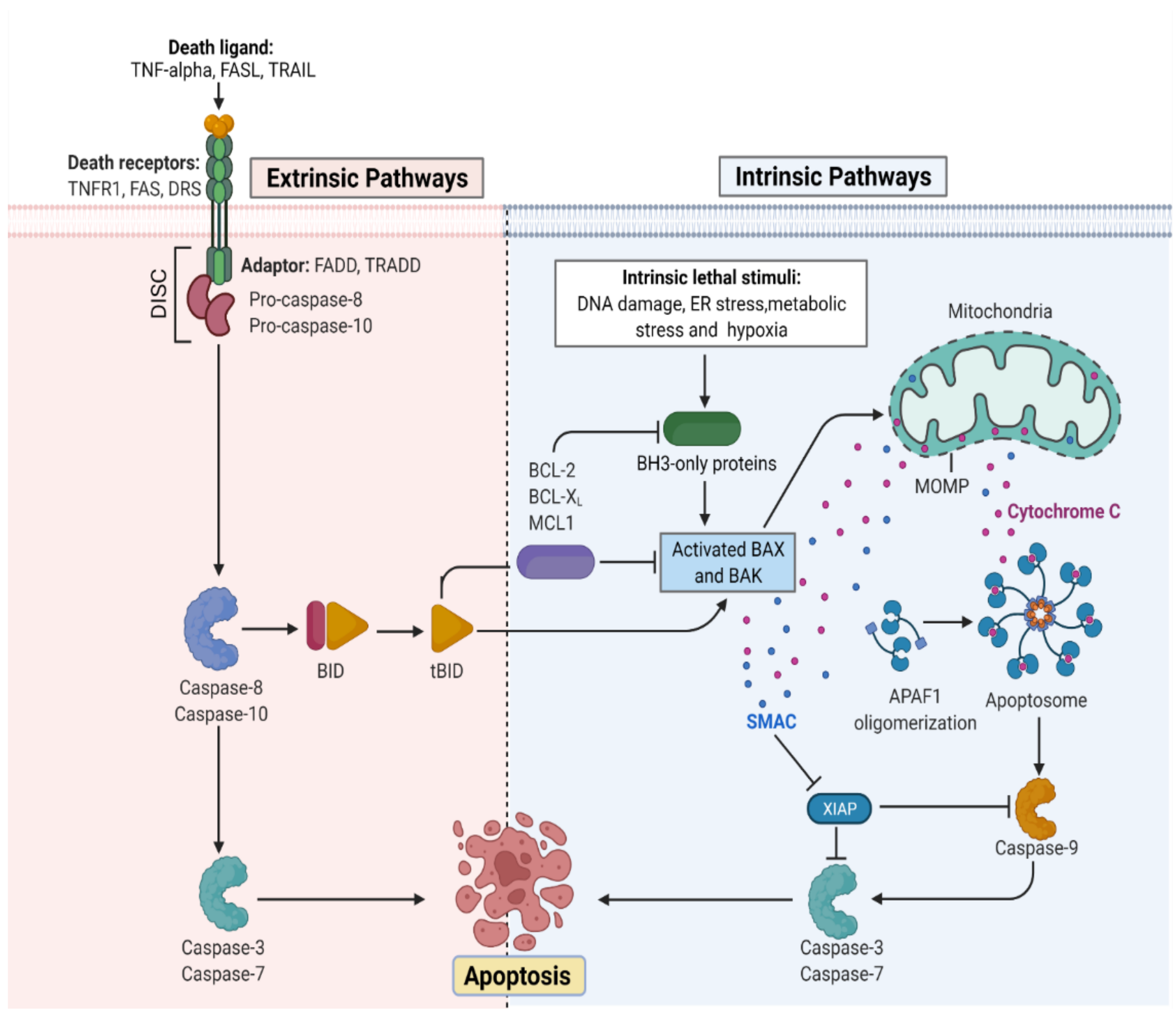
3. Apoptosis Activation: Mechanism
3.1. Mitochondria Pathway of Cellular Apoptosis
3.2. Death Receptor Pathway of Cellular Apoptosis
4. Hallmarks of Apoptosis
4.1. Morphological Indicators
4.2. Biochemical Indicators
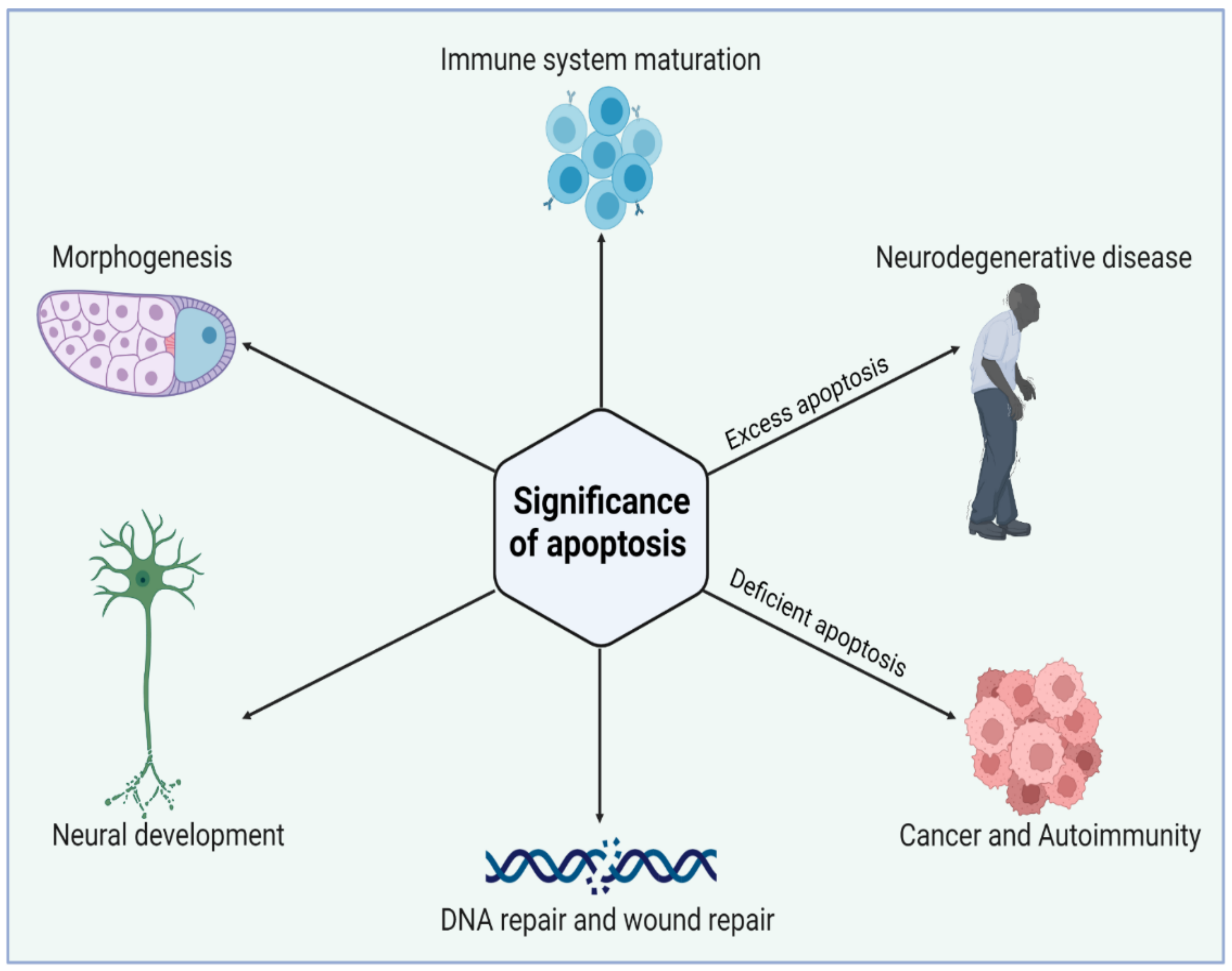
5. Significance of Apoptosis
5.1. Apoptosis: A Disease Paragon
5.2. Apoptosis: A Cancer Cell Hallmark
5.3. Apoptosis: Role in Degenerative Diseases
6. Phytochemicals (PCs) as Strong PCD Inducers
6.1. Vinca Alkaloids
6.2. Epipodophyllotoxins
6.3. Taxanes
6.4. Camptothecin
7. Combinatorial Use of PCs and Common Drugs as Pro-Apoptotic Agents
8. Hinderances in Using PCs as Pro-Apoptotic Agents in Cancerous Cells
9. Plant-Based Nanomaterials Targeting Apoptosis and Other Cancer-Associated Pathways
9.1. Plant-Based Silver NPs
9.2. Plant-Based Gold NPs
9.3. Plant-Based Zinc Oxide NPs
9.4. Plant-Based Copper NPs
9.5. Plant-Based Platinum and Titanium dioxide NPs
9.6. Plant-Based Iron Oxide NPs
9.7. Plant-Based Quantum Dots

| Type of NPs | Plant Name | Plant Part Used | Size and Shape of NPs | Cell Line | IC50 | Ref. |
|---|---|---|---|---|---|---|
| AgNPs | Litchi chinensis | Leaf | 41–55 nm (spherical) | MCF-7 | 40.9 μg/mL | [222] |
| AgNPs | Putranjiva roxburgi | Seed | 8 nm (spherical) | MDA-MB-231, HCT-116, and PANC-1 | 260, 540, 0.25 μg/mL | [223] |
| AgNPs | Alternanthera sessilis | Leaf | 30–50 nm (spherical) | PC3 | 6.8 μg/mL | [224] |
| AgNPs | Euprenolepis procera | Leaf | 60 nm (spherical) | MCF-7 | 9.63 μM | [225] |
| AgNPs | Solanum trilobatum | Fruit | 41.90 nm (spherical, polygonal) | MCF-7 | 30 μg/mL | [226] |
| AgNPs | Zingiber officinale | Leaf | 18.93 nm (spherical) | AsPC-1, PANC-1, and MIA PaCa-2 | 295, 312, 220 µg/mL | [227] |
| AgNPs | Punica granatum | Leaf | 46.1 nm | HeLa | 100 μg/mL | [228] |
| AgNPs | Ganoderma neo-japonicum | Whole | 5–8 nm (spherical) | MDA-MB-231 | 6.0 μg/mL | [229] |
| AgNPs | Derris trifoliate | Seed | 16.92 nm (spherical) | A549 | 86.23 μg/mL | [230] |
| AgNPs | Detarium microcarpum | Leaf | 81 nm (cubic) | HeLA, PANC-1 | 31.5, 84 μg/mL | [231] |
| AuNPs | Gelidium pusillum. | Whole | 12 ± 4.2 nm (spherical) | MDA-MB-231 | 43.09 ± 1.6 μg/mL | [232] |
| AuPs | Artocarpus hirsutus | Leaf | 5–40 nm (spherical) | HeLa, RKO, and A549 | 210, 210, 205 μg/mL | [233] |
| AuNPs conjugated with activated folic acid (FA, receptor) and chlorambucil | Artocarpus hirsutus | Leaf | 5–40 nm (spherical) | HeLa, RKO, and A549 | 130, 132, 133 μg/mL | [233] |
| AuNPs | Musa paradisiaca | Peel | 50 nm (triangular) | A549 | 58 μg/mL | [234] |
| AuNPs | Ferula persica | Gum | 37.05 nm (spherical) | CT26 | 2.4 μg/mL | [235] |
| AuNPs | Nigella sativa | Essential oil from seed | 15.6–28.4 nm (spherical) | A549 | 28.37 μg/mL | [236] |
| AuNPs | Cajanus cajan | Seed coat | 9–41 nm (spherical) | HepG2 | 6 μg/mL | [237] |
| AuNPs | Rhus chinensis | Plant gall | 20–40 nm (oval and spherical) | Hep3B | 150 μg/mL | [238] |
| AuNPs | Commelina nudiflora | NA | 24–150 nm (spherical, triangular) | HCT116 | 200 μg/mL | [239] |
| AuNPs | Butea monosperma | Bark | 35 nm (spherical) | MCF7 | 0.024 μg/mL | [240] |
| CuNPs | Olea europaea | NA | 20–50 nm (spherical) | SKOV-3 | 2.27 μg/mL | [186] |
| CuNPs | Ficus religiosa | Leaf | 577 nm (spherical) | A549 | 200 μg/mL | [187] |
| CuNPs | Olea europaea | 20–50 nm (spherical) | AMJ-13 | 1.47 μg/mL | [186] | |
| CuNPs | Azadirachta indica, Hibiscus rosa-sinensi, Murraya koenigii, Moringa oleifera, and Tamarindus indica | Leaf | 12 nm (spherical) | HeLa | 26.3, 21.63, 23.22, 30.08, 20.32, μg/mL | [241] |
| Leaf | 12 nm (spherical) | MCF-7 | 25.55, 22.45, 25.32, 26.1, 29.1 μg/mL | [241] | ||
| Leaf | 12 nm (spherical) | A549 | 26.03, 20.15, 25.05, 34.3, 18.11 μg/mL | [241] | ||
| Leaf | 12 nm (spherical) | Hep-2 | 28.59, 22.59, 25.59, 29.58, 21.66 μg/mL | [241] | ||
| ZnO NPs | Geranium wallichianum | Leaf | 18 nm (hexagonal) | HepG2 | 39.26 μg/mL | [242] |
| ZnO NPs | Raphanus sativus | Leaf | 209 nm (spherical and hexagonal) | A549 | 40 μg/mL | [243] |
| ZnO NPs | Rubia tinctorum | Leaf | 40 nm (spherical) | MCF-7 | 40 μg/mL | [244] |
| ZnO NPs | Pongamia pinnata | Seed | 30.2 nm (face centered, cubic) | MCF-7 | 32.8 μg/mL | [245] |
| ZnO NPs | Withania somnifera | Root | 32 nm (hexagonal wurtzite) | MCF-7 | 6.84 μg/mL | [246] |
| ZnO NPs | Abutilon indicum | Leaf | 50–500 nm (spherical) | Hela | 45.82 μg/mL | [247] |
| Fe2O3 NPs | Albizia adianthifolia | Leaf | 32–100 nm (spherical) | AMJ-13, MCF-7 | 1.8, 7.7 μg/mL | [206] |
| Fe2O3 NPs | Couroupita guianensis | Fruit | 17 nm (spherical) | HepG2 | 44.51 μg/mL | [248] |
| Fe2O3 NPs | Sargassum muticum | Whole | 18 nm (cubic) | Jurkat, MCF-7, HeLa, and HepG2 | 6.4, 18.5, 12.5, 23.83 μg/mL | [249] |
| PtNPs | Dioscorea bulbifera | Tuber | 2–5 nm (spherical) | HeLa | 10 μg/mL | [250] |
| Palladium (Pd) NPs | Dioscorea bulbifera | Tuber | 10–20 nm (spherical and blunt ended) | HeLa | 10 μg/mL | [250] |
| Pt-Pd | Dioscorea bulbifera | Tuber | 20–25 nm (irregular) | HeLa | 10 μg/mL | [250] |
| Tin oxide (SnO2) NPs | Annona squamosa | Peel | 2.5 nm (spherical) | HepG2 | 1–500 μg/mL | [251] |
10. Conclusions
Author Contributions
Funding
Data Availability Statement
Conflicts of Interest
References
- Siegel, R.L.; Miller, K.D.; Jemal, A. Cancer Statistics, 2019. CA Cancer J. Clin. 2019, 69, 7–34. [Google Scholar] [CrossRef] [PubMed] [Green Version]
- Siegel, R.L.; Miller, K.D.; Jemal, A. Cancer Statistics, 2020. CA Cancer J. Clin. 2020, 70, 7–30. [Google Scholar] [CrossRef] [PubMed]
- Siegel, R.L.; Miller, K.D.; Fuchs, H.E.; Jemal, A. Cancer Statistics, 2022. CA Cancer J. Clin. 2022, 72, 7–33. [Google Scholar] [CrossRef]
- Jemal, A.; Bray, F.; Center, M.M.; Ferlay, J.; Ward, E.; Forman, D. Global Cancer Statistics. CA Cancer J. Clin. 2011, 61, 69–90. [Google Scholar] [CrossRef] [PubMed] [Green Version]
- Miller, K.D.; Fidler-Benaoudia, M.; Keegan, T.H.; Hipp, H.S.; Jemal, A.; Siegel, R.L. Cancer Statistics for Adolescents and Young Adults, 2020. CA Cancer J. Clin. 2020, 70, 443–459. [Google Scholar] [CrossRef]
- Fares, J.; Fares, M.Y.; Khachfe, H.H.; Salhab, H.A.; Fares, Y. Molecular Principles of Metastasis: A Hallmark of Cancer Revisited. Signal Transduct. Target. Ther. 2020, 5, 28. [Google Scholar] [CrossRef] [Green Version]
- Ide, T.; Kitajima, Y.; Miyoshi, A.; Ohtsuka, T.; Mitsuno, M.; Ohtaka, K.; Miyazaki, K. The Hypoxic Environment in Tumor-Stromal Cells Accelerates Pancreatic Cancer Progression via the Activation of Paracrine Hepatocyte Growth Factor/c-Met Signaling. Ann. Surg. Oncol. 2007, 14, 2600–2607. [Google Scholar] [CrossRef]
- Hanahan, D.; Weinberg, R.A. Hallmarks of Cancer: The next Generation. Cell 2011, 144, 646–674. [Google Scholar] [CrossRef] [Green Version]
- Rojo de la Vega, M.; Chapman, E.; Zhang, D.D. NRF2 and the Hallmarks of Cancer. Cancer Cell 2018, 34, 21–43. [Google Scholar] [CrossRef]
- Papaliagkas, V.; Anogianaki, A.; Anogianakis, G.; Ilonidis, G. The Proteins and the Mechanisms of Apoptosis: A Mini-Review of the Fundamentals. Hippokratia 2007, 11, 108–113. [Google Scholar]
- Pucci, C.; Martinelli, C.; Ciofani, G. Innovative Approaches for Cancer Treatment: Current Perspectives and New Challenges. Ecancermedicalscience 2019, 13, 961. [Google Scholar] [CrossRef] [PubMed]
- Aziz, M.A.; Khan, A.H.; Adnan, M.; Ullah, H. Traditional Uses of Medicinal Plants Used by Indigenous Communities for Veterinary Practices at Bajaur Agency, Pakistan. J. Ethnobiol. Ethnomedicine 2018, 14, 11. [Google Scholar] [CrossRef] [PubMed] [Green Version]
- Akhtar, N.; Wani, A.K.; Mir, T.-U.G.; Kumar, N.; Mannan, M.A.-U. Sapindus Mukorossi: Ethnomedicinal uses, phytochemistry, and pharmacological activities. Plant Cell Biotechnol. Mol. Biol. 2021, 22, 300–319. [Google Scholar]
- Wani, A.K.; Akhtar, N.; Sharma, A.; El-Zahaby, S.A. Fighting Carcinogenesis with Plant Metabolites by Weakening Proliferative Signaling and Disabling Replicative Immortality Networks of Rapidly Dividing and Invading Cancerous Cells. Curr. Drug Deliv. 2022. [Google Scholar] [CrossRef]
- Mir, T.u.G.; Wani, A.K.; Singh, J.; Shukla, S. Therapeutic Application and Toxicity Associated with Crocus Sativus (Saffron) and Its Phytochemicals. Pharmacol. Res. Mod. Chin. Med. 2022, 4, 100136. [Google Scholar] [CrossRef]
- Falzone, L.; Salomone, S.; Libra, M. Evolution of Cancer Pharmacological Treatments at the Turn of the Third Millennium. Front. Pharmacol. 2018, 9, 1300. [Google Scholar] [CrossRef] [Green Version]
- Cheon, C. Synergistic Effects of Herbal Medicines and Anticancer Drugs. Medicine 2021, 100, e27918. [Google Scholar] [CrossRef]
- Cheon, C.; Ko, S.-G. Synergistic Effects of Natural Products in Combination with Anticancer Agents in Prostate Cancer: A Scoping Review. Front. Pharmacol. 2022, 13, 963317. [Google Scholar] [CrossRef]
- Jabeen, S.; Qureshi, R.; Munazir, M.; Maqsood, M.; Munir, M.; Shah, S.S.H.; Rahim, B.Z. Application of Green Synthesized Silver Nanoparticles in Cancer Treatment—A Critical Review. Mater. Res. Express 2021, 8, 092001. [Google Scholar] [CrossRef]
- Ghosh, S.; Dutta, S.; Sarkar, A.; Kundu, M.; Sil, P.C. Targeted Delivery of Curcumin in Breast Cancer Cells via Hyaluronic Acid Modified Mesoporous Silica Nanoparticle to Enhance Anticancer Efficiency. Colloids Surf. B Biointerfaces 2021, 197, 111404. [Google Scholar] [CrossRef]
- Fridlender, M.; Kapulnik, Y.; Koltai, H. Plant Derived Substances with Anti-Cancer Activity: From Folklore to Practice. Front. Plant Sci. 2015, 6, 799. [Google Scholar] [CrossRef] [PubMed]
- Fatima, N.; Baqri, S.S.R.; Alsulimani, A.; Fagoonee, S.; Slama, P.; Kesari, K.K.; Roychoudhury, S.; Haque, S. Phytochemicals from Indian Ethnomedicines: Promising Prospects for the Management of Oxidative Stress and Cancer. Antioxidants 2021, 10, 1606. [Google Scholar] [CrossRef] [PubMed]
- Choudhari, A.S.; Mandave, P.C.; Deshpande, M.; Ranjekar, P.; Prakash, O. Phytochemicals in Cancer Treatment: From Preclinical Studies to Clinical Practice. Front. Pharmacol. 2020, 10, 1614. [Google Scholar] [CrossRef] [PubMed] [Green Version]
- Morana, O.; Wood, W.; Gregory, C.D. The Apoptosis Paradox in Cancer. Int. J. Mol. Sci. 2022, 23, 1328. [Google Scholar] [CrossRef]
- Keeble, J.A.; Gilmore, A.P. Apoptosis Commitment—Translating Survival Signals into Decisions on Mitochondria. Cell Res. 2007, 17, 976–984. [Google Scholar] [CrossRef] [Green Version]
- Wallach, D.; Kang, T.-B. Programmed Cell Death in Immune Defense: Knowledge and Presumptions. Immunity 2018, 49, 19–32. [Google Scholar] [CrossRef] [Green Version]
- Favaloro, B.; Allocati, N.; Graziano, V.; Di Ilio, C.; De Laurenzi, V. Role of Apoptosis in Disease. Aging 2012, 4, 330–349. [Google Scholar] [CrossRef]
- Fulda, S.; Gorman, A.M.; Hori, O.; Samali, A. Cellular Stress Responses: Cell Survival and Cell Death. Int. J. Cell Biol. 2010, 2010, e214074. [Google Scholar] [CrossRef] [Green Version]
- Portt, L.; Norman, G.; Clapp, C.; Greenwood, M.; Greenwood, M.T. Anti-Apoptosis and Cell Survival: A Review. Biochim. Biophys. Acta BBA Mol. Cell Res. 2011, 1813, 238–259. [Google Scholar] [CrossRef] [Green Version]
- Hongmei, Z. Extrinsic and Intrinsic Apoptosis Signal Pathway Review. Apoptosis Med. 2012. [Google Scholar] [CrossRef] [Green Version]
- Webster, K.A. Mitochondrial Membrane Permeabilization and Cell Death during Myocardial Infarction: Roles of Calcium and Reactive Oxygen Species. Future Cardiol. 2012, 8, 863–884. [Google Scholar] [CrossRef] [PubMed] [Green Version]
- Kale, J.; Osterlund, E.J.; Andrews, D.W. BCL-2 Family Proteins: Changing Partners in the Dance towards Death. Cell Death Differ. 2018, 25, 65–80. [Google Scholar] [CrossRef] [PubMed]
- Lomonosova, E.; Chinnadurai, G. BH3-Only Proteins in Apoptosis and beyond: An Overview. Oncogene 2008, 27, S2–S19. [Google Scholar] [CrossRef] [PubMed] [Green Version]
- Tzifi, F.; Economopoulou, C.; Gourgiotis, D.; Ardavanis, A.; Papageorgiou, S.; Scorilas, A. The Role of BCL2 Family of Apoptosis Regulator Proteins in Acute and Chronic Leukemias. Adv. Hematol. 2011, 2012, e524308. [Google Scholar] [CrossRef] [PubMed] [Green Version]
- Bratton, S.B.; Salvesen, G.S. Regulation of the Apaf-1–Caspase-9 Apoptosome. J. Cell Sci. 2010, 123, 3209–3214. [Google Scholar] [CrossRef] [Green Version]
- Singh, R.; Letai, A.; Sarosiek, K. Regulation of Apoptosis in Health and Disease: The Balancing Act of BCL-2 Family Proteins. Nat. Rev. Mol. Cell Biol. 2019, 20, 175–193. [Google Scholar] [CrossRef]
- Vringer, E.; Tait, S.W.G. Mitochondria and Inflammation: Cell Death Heats Up. Front. Cell Dev. Biol. 2019, 7, 100. [Google Scholar] [CrossRef]
- Wang, F.; Ogasawara, M.A.; Huang, P. Small Mitochondria-Targeting Molecules as Anti-Cancer Agents. Mol. Aspects Med. 2010, 31, 75–92. [Google Scholar] [CrossRef] [Green Version]
- Marchi, S.; Pinton, P. Mitochondria in the Line of Fire. Cell Death Differ. 2022, 29, 1301–1303. [Google Scholar] [CrossRef]
- Parameswaran, N.; Patial, S. Tumor Necrosis Factor-α Signaling in Macrophages. Crit. Rev. Eukaryot. Gene Expr. 2010, 20, 87–103. [Google Scholar] [CrossRef]
- Wajant, H.; Siegmund, D. TNFR1 and TNFR2 in the Control of the Life and Death Balance of Macrophages. Front. Cell Dev. Biol. 2019, 7, 91. [Google Scholar] [CrossRef] [PubMed] [Green Version]
- Xie, P. TRAF Molecules in Cell Signaling and in Human Diseases. J. Mol. Signal. 2013, 8, 7. [Google Scholar] [CrossRef] [PubMed]
- MacFarlane, M.; Merrison, W.; Dinsdale, D.; Cohen, G.M. Active Caspases and Cleaved Cytokeratins Are Sequestered into Cytoplasmic Inclusions in Trail-Induced Apoptosis. J. Cell Biol. 2000, 148, 1239–1254. [Google Scholar] [CrossRef] [PubMed] [Green Version]
- Tummers, B.; Green, D.R. Caspase-8; Regulating Life and Death. Immunol. Rev. 2017, 277, 76–89. [Google Scholar] [CrossRef] [Green Version]
- Toné, S.; Sugimoto, K.; Tanda, K.; Suda, T.; Uehira, K.; Kanouchi, H.; Samejima, K.; Minatogawa, Y.; Earnshaw, W.C. Three Distinct Stages of Apoptotic Nuclear Condensation Revealed by Time-Lapse Imaging, Biochemical and Electron Microscopy Analysis of Cell-Free Apoptosis. Exp. Cell Res. 2007, 313, 3635–3644. [Google Scholar] [CrossRef] [Green Version]
- Sherstnev, V.V.; Yurasov, V.V.; Storozheva, Z.I.; Gruden, M.A.; Yakovleva, N.E. Biochemical Markers of Apoptosis in Different Parts of the Brain during Learning. Neurosci. Behav. Physiol. 2006, 36, 915–919. [Google Scholar] [CrossRef]
- Abaza, A.; Vasavada, A.M.; Sadhu, A.; Valencia, C.; Fatima, H.; Nwankwo, I.; Anam, M.; Maharjan, S.; Amjad, Z.; Khan, S. A Systematic Review of Apoptosis in Correlation With Cancer: Should Apoptosis Be the Ultimate Target for Cancer Treatment? Cureus 2022, 14, e28496. [Google Scholar] [CrossRef]
- Conradt, B.; Wu, Y.-C.; Xue, D. Programmed Cell Death During Caenorhabditis Elegans Development. Genetics 2016, 203, 1533–1562. [Google Scholar] [CrossRef] [Green Version]
- Gilbert, S.F. Cell Death and the Formation of Digits and Joints, 6th ed.; Sinauer Associates: Sunderland, MA, USA, 2000. [Google Scholar]
- Yang, X.; Zhou, J.; He, J.; Liu, J.; Wang, H.; Liu, Y.; Jiang, T.; Zhang, Q.; Fu, X.; Xu, Y. An Immune System-Modified Rat Model for Human Stem Cell Transplantation Research. Stem Cell Rep. 2018, 11, 514–521. [Google Scholar] [CrossRef] [Green Version]
- Sangiuliano, B.; Pérez, N.M.; Moreira, D.F.; Belizário, J.E. Cell Death-Associated Molecular-Pattern Molecules: Inflammatory Signaling and Control. Mediat. Inflamm. 2014, 2014, 821043. [Google Scholar] [CrossRef] [Green Version]
- Pfeffer, C.M.; Singh, A.T.K. Apoptosis: A Target for Anticancer Therapy. Int. J. Mol. Sci. 2018, 19, 448. [Google Scholar] [CrossRef] [PubMed] [Green Version]
- Young, A.I.; Timpson, P.; Gallego-Ortega, D.; Ormandy, C.J.; Oakes, S.R. Myeloid Cell Leukemia 1 (MCL-1), an Unexpected Modulator of Protein Kinase Signaling during Invasion. Cell Adhes. Migr. 2017, 12, 513–523. [Google Scholar] [CrossRef] [PubMed]
- Vo, T.-T.; Letai, A. BH3-Only Proteins and Their Effects on Cancer. Adv. Exp. Med. Biol. 2010, 687, 49–63. [Google Scholar] [PubMed] [Green Version]
- Bourgo, R.J.; Braden, W.A.; Wells, S.I.; Knudsen, E.S. Activation of the Retinoblastoma Tumor Suppressor Mediates Cell Cycle Inhibition and Cell Death in Specific Cervical Cancer Cell Lines. Mol. Carcinog. 2009, 48, 45–55. [Google Scholar] [CrossRef] [Green Version]
- Mattson, M.P.; Gleichmann, M.; Cheng, A. Mitochondria in Neuroplasticity and Neurological Disorders. Neuron 2008, 60, 748–766. [Google Scholar] [CrossRef] [Green Version]
- Chandrasekar, A.P.; Cummins, N.W.; Badley, A.D. The Role of the BCL-2 Family of Proteins in HIV-1 Pathogenesis and Persistence. Clin. Microbiol. Rev. 2019, 33, e00107-19. [Google Scholar] [CrossRef]
- Ramakrishna, W.; Kumari, A.; Rahman, N.; Mandave, P. Anticancer Activities of Plant Secondary Metabolites: Rice Callus Suspension Culture as a New Paradigm. Rice Sci. 2021, 28, 13–30. [Google Scholar] [CrossRef]
- Bailon-Moscoso, N.; Cevallos-Solorzano, G.; Romero-Benavides, J.C.; Orellana, M.I.R. Natural Compounds as Modulators of Cell Cycle Arrest: Application for Anticancer Chemotherapies. Curr. Genom. 2017, 18, 106–131. [Google Scholar] [CrossRef] [Green Version]
- Sarkar, S.; Rajput, S.; Tripathi, A.K.; Mandal, M. Targeted Therapy against EGFR and VEGFR Using ZD6474 Enhances the Therapeutic Potential of UV-B Phototherapy in Breast Cancer Cells. Mol. Cancer 2013, 12, 122. [Google Scholar] [CrossRef] [Green Version]
- Mavrogiannis, A.V.; Kokkinopoulou, I.; Kontos, C.K.; Sideris, D.C. Effect of Vinca Alkaloids on the Expression Levels of MicroRNAs Targeting Apoptosis-Related Genes in Breast Cancer Cell Lines. Curr. Pharm. Biotechnol. 2018, 19, 1076–1086. [Google Scholar] [CrossRef]
- Dhyani, P.; Quispe, C.; Sharma, E.; Bahukhandi, A.; Sati, P.; Attri, D.C.; Szopa, A.; Sharifi-Rad, J.; Docea, A.O.; Mardare, I.; et al. Anticancer Potential of Alkaloids: A Key Emphasis to Colchicine, Vinblastine, Vincristine, Vindesine, Vinorelbine and Vincamine. Cancer Cell Int. 2022, 22, 206. [Google Scholar] [CrossRef] [PubMed]
- Groninger, E.; Meeuwsen-De Boer, G.J.; De Graaf, S.S.N.; Kamps, W.A.; De Bont, E.S.J.M. Vincristine Induced Apoptosis in Acute Lymphoblastic Leukaemia Cells: A Mitochondrial Controlled Pathway Regulated by Reactive Oxygen Species? Int. J. Oncol. 2002, 21, 1339–1345. [Google Scholar] [CrossRef] [PubMed]
- Shinwari, Z.; Manogaran, P.S.; Alrokayan, S.A.; Al-Hussein, K.A.; Aboussekhra, A. Vincristine and Lomustine Induce Apoptosis and P21(WAF1) up-Regulation in Medulloblastoma and Normal Human Epithelial and Fibroblast Cells. J. Neurooncol. 2008, 87, 123–132. [Google Scholar] [CrossRef]
- Denny, W.A. Anti Cancer: DNA Topoisomerase Inhibitors. In Reference Module in Chemistry, Molecular Sciences and Chemical Engineering; Elsevier: Amsterdam, The Netherlands, 2013; ISBN 978-0-12-409547-2. [Google Scholar]
- McClendon, A.K.; Osheroff, N. DNA Topoisomerase II, Genotoxicity, and Cancer. Mutat. Res. 2007, 623, 83–97. [Google Scholar] [CrossRef] [PubMed] [Green Version]
- Miele, M.; Mumot, A.M.; Zappa, A.; Romano, P.; Ottaggio, L. Hazel and Other Sources of Paclitaxel and Related Compounds. Phytochem. Rev. 2012, 11, 211–225. [Google Scholar] [CrossRef]
- Mukhtar, E.; Adhami, V.M.; Mukhtar, H. Targeting Microtubules by Natural Agents for Cancer Therapy. Mol. Cancer Ther. 2014, 13, 275–284. [Google Scholar] [CrossRef] [Green Version]
- Li, F.; Jiang, T.; Li, Q.; Ling, X. Camptothecin (CPT) and Its Derivatives Are Known to Target Topoisomerase I (Top1) as Their Mechanism of Action: Did We Miss Something in CPT Analogue Molecular Targets for Treating Human Disease Such as Cancer? Am. J. Cancer Res. 2017, 7, 2350–2394. [Google Scholar]
- Venditto, V.J.; Simanek, E.E. Cancer Therapies Utilizing the Camptothecins: A Review of in Vivo Literature. Mol. Pharm. 2010, 7, 307–349. [Google Scholar] [CrossRef] [Green Version]
- Kim, S.-H.; Ryu, H.G.; Lee, J.; Shin, J.; Harikishore, A.; Jung, H.-Y.; Kim, Y.S.; Lyu, H.-N.; Oh, E.; Baek, N.-I.; et al. Ursolic Acid Exerts Anti-Cancer Activity by Suppressing Vaccinia-Related Kinase 1-Mediated Damage Repair in Lung Cancer Cells. Sci. Rep. 2015, 5, 14570. [Google Scholar] [CrossRef] [Green Version]
- Ahmadiankia, N. Molecular Targets of Pomegranate (Punica Granatum) in Preventing Cancer Metastasis. Iran. J. Basic Med. Sci. 2019, 22, 977–988. [Google Scholar] [CrossRef]
- Ravisankar, S.; Agah, S.; Kim, H.; Talcott, S.; Wu, C.; Awika, J. Combined Cereal and Pulse Flavonoids Show Enhanced Bioavailability by Downregulating Phase II Metabolism and ABC Membrane Transporter Function in Caco-2 Model. Food Chem. 2019, 279, 88–97. [Google Scholar] [CrossRef] [PubMed]
- Rauf, A.; Imran, M.; Khan, I.A.; Ur-Rehman, M.; Gilani, S.A.; Mehmood, Z.; Mubarak, M.S. Anticancer Potential of Quercetin: A Comprehensive Review. Phytother. Res. 2018, 32, 2109–2130. [Google Scholar] [CrossRef] [PubMed]
- Luo, H.; Rankin, G.O.; Liu, L.; Daddysman, M.K.; Jiang, B.-H.; Chen, Y.C. Kaempferol Inhibits Angiogenesis and VEGF Expression Through Both HIF Dependent and Independent Pathways in Human Ovarian Cancer Cells. Nutr. Cancer 2009, 61, 554–563. [Google Scholar] [CrossRef]
- Lee, K.H.; Hong, H.S.; Lee, C.H.; Kim, C.H. Induction of Apoptosis in Human Leukaemic Cell Lines K562, HL60 and U937 by Diethylhexylphthalate Isolated from Aloe Vera Linne. J. Pharm. Pharmacol. 2000, 52, 1037–1041. [Google Scholar] [CrossRef]
- Parida, S.; Pal, I.; Parekh, A.; Thakur, B.; Bharti, R.; Das, S.; Mandal, M. GW627368X Inhibits Proliferation and Induces Apoptosis in Cervical Cancer by Interfering with EP4/EGFR Interactive Signaling. Cell Death Dis. 2016, 7, e2154. [Google Scholar] [CrossRef] [Green Version]
- Imran, M.; Rauf, A.; Abu-Izneid, T.; Nadeem, M.; Shariati, M.A.; Khan, I.A.; Imran, A.; Orhan, I.E.; Rizwan, M.; Atif, M.; et al. Luteolin, a Flavonoid, as an Anticancer Agent: A Review. Biomed. Pharmacother. 2019, 112, 108612. [Google Scholar] [CrossRef] [PubMed]
- Tor, Y.S.; Yazan, L.S.; Foo, J.B.; Wibowo, A.; Ismail, N.; Cheah, Y.K.; Abdullah, R.; Ismail, M.; Ismail, I.S.; Yeap, S.K. Induction of Apoptosis in MCF-7 Cells via Oxidative Stress Generation, Mitochondria-Dependent and Caspase-Independent Pathway by Ethyl Acetate Extract of Dillenia Suffruticosa and Its Chemical Profile. PLoS ONE 2015, 10, e0127441. [Google Scholar] [CrossRef] [Green Version]
- Chang, M.-Y.; Shieh, D.-E.; Chen, C.-C.; Yeh, C.-S.; Dong, H.-P. Linalool Induces Cell Cycle Arrest and Apoptosis in Leukemia Cells and Cervical Cancer Cells through CDKIs. Int. J. Mol. Sci. 2015, 16, 28169–28179. [Google Scholar] [CrossRef] [Green Version]
- Chen, B.; Wang, H.-T.; Yu, B.; Zhang, J.-D.; Feng, Y. Carthamin Yellow Inhibits Matrix Degradation and Inflammation Induced by LPS in the Intervertebral Disc via Suppression of MAPK Pathway Activation. Exp. Ther. Med. 2017, 14, 1614–1620. [Google Scholar] [CrossRef] [Green Version]
- Vanamala, J.; Reddivari, L.; Radhakrishnan, S.; Tarver, C. Resveratrol Suppresses IGF-1 Induced Human Colon Cancer Cell Proliferation and Elevates Apoptosis via Suppression of IGF-1R/Wnt and Activation of P53 Signaling Pathways. BMC Cancer 2010, 10, 238. [Google Scholar] [CrossRef] [Green Version]
- Lu, X.; Jung, J.I.; Cho, H.J.; Lim, D.Y.; Lee, H.S.; Chun, H.S.; Kwon, D.Y.; Park, J.H.Y. Fisetin Inhibits the Activities of Cyclin-Dependent Kinases Leading to Cell Cycle Arrest in HT-29 Human Colon Cancer Cells. J. Nutr. 2005, 135, 2884–2890. [Google Scholar] [CrossRef] [PubMed] [Green Version]
- Choi, B.Y.; Kim, B.-W. Withaferin-A Inhibits Colon Cancer Cell Growth by Blocking STAT3 Transcriptional Activity. J. Cancer Prev. 2015, 20, 185–192. [Google Scholar] [CrossRef] [PubMed] [Green Version]
- Mallikarjuna, G.U.; Dhanalakshmi, S.; Raisuddin, S.; Ramesha Rao, A. Chemomodulatory Influence of Ferula Asafoetida on Mammary Epithelial Differentiation, Hepatic Drug Metabolizing Enzymes, Antioxidant Profiles and N-Methyl-N-Nitrosourea-Induced Mammary Carcinogenesis in Rats. Breast Cancer Res. Treat. 2003, 81, 1–10. [Google Scholar] [CrossRef]
- Samarghandian, S.; Borji, A. Anticarcinogenic Effect of Saffron (Crocus sativus L.) and Its Ingredients. Pharmacogn. Res. 2014, 6, 99–107. [Google Scholar] [CrossRef] [PubMed] [Green Version]
- Yang, H.; Dou, Q.P. Targeting Apoptosis Pathway with Natural Terpenoids: Implications for Treatment of Breast and Prostate Cancer. Curr. Drug Targets 2010, 11, 733–744. [Google Scholar] [CrossRef] [PubMed] [Green Version]
- Meng, B.; Ii, H.; Qu, W.; Yuan, H. Anticancer Effects of Gingerol in Retinoblastoma Cancer Cells (RB355 Cell Line) Are Mediated via Apoptosis Induction, Cell Cycle Arrest and Upregulation of PI3K/Akt Signaling Pathway. Med. Sci. Monit. Int. Med. J. Exp. Clin. Res. 2018, 24, 1980–1987. [Google Scholar] [CrossRef] [Green Version]
- Zhou, Q.; Yang, T.; Qiao, Y.; Guo, S.; Zhu, L.; Wu, H. Preparation of Poly(β-L-Malic Acid)-Based Charge-Conversional Nanoconjugates for Tumor-Specific Uptake and Cellular Delivery. Int. J. Nanomed. 2015, 10, 1941–1952. [Google Scholar] [CrossRef] [Green Version]
- Zhang, W.; Liu, C.; Li, J.; Liu, R.; Zhuang, J.; Feng, F.; Yao, Y.; Sun, C. Target Analysis and Mechanism of Podophyllotoxin in the Treatment of Triple-Negative Breast Cancer. Front. Pharmacol. 2020, 11, 1211. [Google Scholar] [CrossRef]
- Habtemariam, S. Recent Advances in Berberine Inspired Anticancer Approaches: From Drug Combination to Novel Formulation Technology and Derivatization. Molecules 2020, 25, 1426. [Google Scholar] [CrossRef] [Green Version]
- Karatoprak, G.Ş.; Küpeli Akkol, E.; Genç, Y.; Bardakci, H.; Yücel, Ç.; Sobarzo-Sánchez, E. Combretastatins: An Overview of Structure, Probable Mechanisms of Action and Potential Applications. Molecules 2020, 25, E2560. [Google Scholar] [CrossRef]
- Bhukta, S.; Gopinath, P.; Dandela, R. Target Identification of Anticancer Natural Products Using a Chemical Proteomics Approach. RSC Adv. 2021, 11, 27950–27964. [Google Scholar] [CrossRef] [PubMed]
- Kim, H.J.; Nam, Y.R.; Woo, J.; Kim, W.K.; Nam, J.H. Gardenia Jasminoides Extract and Its Constituent, Genipin, Inhibit Activation of CD3/CD28 Co-Stimulated CD4+ T Cells via ORAI1 Channel. Korean J. Physiol. Pharmacol. 2020, 24, 363–372. [Google Scholar] [CrossRef] [PubMed]
- Veisi, A.; Akbari, G.; Mard, S.A.; Badfar, G.; Zarezade, V.; Mirshekar, M.A. Role of Crocin in Several Cancer Cell Lines: An Updated Review. Iran. J. Basic Med. Sci. 2020, 23, 3–12. [Google Scholar] [CrossRef] [PubMed]
- Li, M.; Li, B.; Xia, Z.-M.; Tian, Y.; Zhang, D.; Rui, W.-J.; Dong, J.-X.; Xiao, F.-J. Anticancer Effects of Five Biflavonoids from Ginkgo biloba L. Male Flowers In Vitro. Molecules 2019, 24, 1496. [Google Scholar] [CrossRef] [Green Version]
- Zhang, J.; Li, A.; Sun, H.; Xiong, X.; Qin, S.; Wang, P.; Dai, L.; Zhang, Z.; Li, X.; Liu, Z. Amentoflavone Triggers Cell Cycle G2/M Arrest by Interfering with Microtubule Dynamics and Inducing DNA Damage in SKOV3 Cells. Oncol. Lett. 2020, 20, 168. [Google Scholar] [CrossRef] [PubMed]
- Xie, J.; Lai, Z.; Zheng, X.; Liao, H.; Xian, Y.; Li, Q.; Wu, J.; Ip, S.; Xie, Y.; Chen, J.; et al. Apoptotic Activities of Brusatol in Human Non-Small Cell Lung Cancer Cells: Involvement of ROS-Mediated Mitochondrial-Dependent Pathway and Inhibition of Nrf2-Mediated Antioxidant Response. Toxicology 2021, 451, 152680. [Google Scholar] [CrossRef]
- Wang, T.; Dou, Y.; Lin, G.; Li, Q.; Nie, J.; Chen, B.; Xie, J.; Su, Z.; Zeng, H.; Chen, J.; et al. The Anti-Hepatocellular Carcinoma Effect of Brucea Javanica Oil in Ascitic Tumor-Bearing Mice: The Detection of Brusatol and Its Role. Biomed. Pharmacother. 2021, 134, 111122. [Google Scholar] [CrossRef]
- Zhang, Z.; Luo, D.; Xie, J.; Lin, G.; Zhou, J.; Liu, W.; Li, H.; Yi, T.; Su, Z.; Chen, J. Octahydrocurcumin, a Final Hydrogenated Metabolite of Curcumin, Possesses Superior Anti-Tumor Activity through Induction of Cellular Apoptosis. Food Funct. 2018, 9, 2005–2014. [Google Scholar] [CrossRef] [PubMed]
- Pezzani, R.; Salehi, B.; Vitalini, S.; Iriti, M.; Zuñiga, F.A.; Sharifi-Rad, J.; Martorell, M.; Martins, N. Synergistic Effects of Plant Derivatives and Conventional Chemotherapeutic Agents: An Update on the Cancer Perspective. Medicina 2019, 55, 110. [Google Scholar] [CrossRef] [Green Version]
- Tan, B.L.; Norhaizan, M.E. Curcumin Combination Chemotherapy: The Implication and Efficacy in Cancer. Molecules 2019, 24, 2527. [Google Scholar] [CrossRef] [Green Version]
- Peng, S.; Wang, J.; Lu, C.; Xu, Z.; Chai, J.-J.; Ke, Q.; Deng, X.-Z. Emodin Enhances Cisplatin Sensitivity in Non-Small Cell Lung Cancer through Pgp Downregulation. Oncol. Lett. 2021, 21, 230. [Google Scholar] [CrossRef] [PubMed]
- Min, J.; Shen, H.; Xi, W.; Wang, Q.; Yin, L.; Zhang, Y.; Yu, Y.; Yang, Q.; Wang, Z.-N. Synergistic Anticancer Activity of Combined Use of Caffeic Acid with Paclitaxel Enhances Apoptosis of Non-Small-Cell Lung Cancer H1299 Cells in Vivo and in Vitro. Cell. Physiol. Biochem. 2018, 48, 1433–1442. [Google Scholar] [CrossRef] [PubMed]
- Chen, X.; Wu, Q.; Chen, Y.; Zhang, J.; Li, H.; Yang, Z.; Yang, Y.; Deng, Y.; Zhang, L.; Liu, B. Diosmetin Induces Apoptosis and Enhances the Chemotherapeutic Efficacy of Paclitaxel in Non-Small Cell Lung Cancer Cells via Nrf2 Inhibition. Br. J. Pharmacol. 2019, 176, 2079–2094. [Google Scholar] [CrossRef] [PubMed]
- Liu, P.; Ying, Q.; Liu, H.; Yu, S.-Q.; Bu, L.-P.; Shao, L.; Li, X.-Y. Curcumin Enhances Anti-cancer Efficacy of Either Gemcitabine or Docetaxel on Pancreatic Cancer Cells. Oncol. Rep. 2020, 44, 1393–1402. [Google Scholar] [CrossRef] [PubMed]
- Liu, P.; Feng, J.; Sun, M.; Yuan, W.; Xiao, R.; Xiong, J.; Huang, X.; Xiong, M.; Chen, W.; Yu, X.; et al. Synergistic Effects of Baicalein with Gemcitabine or Docetaxel on the Proliferation, Migration and Apoptosis of Pancreatic Cancer Cells. Int. J. Oncol. 2017, 51, 1878–1886. [Google Scholar] [CrossRef] [Green Version]
- Banerjee, S.; Zhang, Y.; Ali, S.; Bhuiyan, M.; Wang, Z.; Chiao, P.J.; Philip, P.A.; Abbruzzese, J.; Sarkar, F.H. Molecular Evidence for Increased Antitumor Activity of Gemcitabine by Genistein in Vitro and in Vivo Using an Orthotopic Model of Pancreatic Cancer. Cancer Res. 2005, 65, 9064–9072. [Google Scholar] [CrossRef] [PubMed] [Green Version]
- Zhao, Y.; Jing, Z.; Li, Y.; Mao, W. Berberine in Combination with Cisplatin Suppresses Breast Cancer Cell Growth through Induction of DNA Breaks and Caspase-3-Dependent Apoptosis. Oncol. Rep. 2016, 36, 567–572. [Google Scholar] [CrossRef] [Green Version]
- Liu, L.; Fan, J.; Ai, G.; Liu, J.; Luo, N.; Li, C.; Cheng, Z. Berberine in Combination with Cisplatin Induces Necroptosis and Apoptosis in Ovarian Cancer Cells. Biol. Res. 2019, 52, 37. [Google Scholar] [CrossRef] [Green Version]
- Gao, X.; Zhang, C.; Wang, Y.; Zhang, P.; Zhang, J.; Hong, T. Berberine and Cisplatin Exhibit Synergistic Anticancer Effects on Osteosarcoma MG-63 Cells by Inhibiting the MAPK Pathway. Molecules 2021, 26, 1666. [Google Scholar] [CrossRef]
- Ma, S.; Tan, W.; Du, B.; Liu, W.; Li, W.; Che, D.; Zhang, G. Oridonin Effectively Reverses Cisplatin Drug Resistance in Human Ovarian Cancer Cells via Induction of Cell Apoptosis and Inhibition of Matrix Metalloproteinase Expression. Mol. Med. Rep. 2016, 13, 3342–3348. [Google Scholar] [CrossRef] [Green Version]
- Lichota, A.; Gwozdzinski, K. Anticancer Activity of Natural Compounds from Plant and Marine Environment. Int. J. Mol. Sci. 2018, 19, 3533. [Google Scholar] [CrossRef] [PubMed] [Green Version]
- Karthikeyan, A.; Senthil, N.; Min, T. Nanocurcumin: A Promising Candidate for Therapeutic Applications. Front. Pharmacol. 2020, 11, 487. [Google Scholar] [CrossRef] [PubMed]
- Martínez-Ballesta, M.; Gil-Izquierdo, Á.; García-Viguera, C.; Domínguez-Perles, R. Nanoparticles and Controlled Delivery for Bioactive Compounds: Outlining Challenges for New “Smart-Foods” for Health. Foods 2018, 7, 72. [Google Scholar] [CrossRef]
- Annaji, M.; Poudel, I.; Boddu, S.H.S.; Arnold, R.D.; Tiwari, A.K.; Babu, R.J. Resveratrol-loaded Nanomedicines for Cancer Applications. Cancer Rep. 2021, 4, e1353. [Google Scholar] [CrossRef] [PubMed]
- Chen, B.; Wang, X.; Lin, D.; Xu, D.; Li, S.; Huang, J.; Weng, S.; Lin, Z.; Zheng, Y.; Yao, H.; et al. Proliposomes for Oral Delivery of Total Biflavonoids Extract from Selaginella Doederleinii: Formulation Development, Optimization, and in Vitro–in Vivo Characterization. Int. J. Nanomed. 2019, 14, 6691–6706. [Google Scholar] [CrossRef] [Green Version]
- Stark, W.J.; Stoessel, P.R.; Wohlleben, W.; Hafner, A. Industrial Applications of Nanoparticles. Chem. Soc. Rev. 2015, 44, 5793–5805. [Google Scholar] [CrossRef] [Green Version]
- Kumbhakar, P.; Ray, S.S.; Stepanov, A.L. Optical Properties of Nanoparticles and Nanocomposites. J. Nanomater. 2014, 2014, e181365. [Google Scholar] [CrossRef] [Green Version]
- Yetisen, A.K.; Qu, H.; Manbachi, A.; Butt, H.; Dokmeci, M.R.; Hinestroza, J.P.; Skorobogatiy, M.; Khademhosseini, A.; Yun, S.H. Nanotechnology in Textiles. ACS Nano 2016, 10, 3042–3068. [Google Scholar] [CrossRef]
- De Jong, W.H.; Borm, P.J. Drug Delivery and Nanoparticles: Applications and Hazards. Int. J. Nanomed. 2008, 3, 133–149. [Google Scholar] [CrossRef] [Green Version]
- Ghaffari, M.; Dolatabadi, J.E.N. Chapter 17—Nanotechnology for Pharmaceuticals. In Industrial Applications of Nanomaterials; Thomas, S., Grohens, Y., Pottathara, Y.B., Eds.; Micro and Nano Technologies; Elsevier: Amsterdam, The Netherlands, 2019; pp. 475–502. ISBN 978-0-12-815749-7. [Google Scholar]
- Malik, A.Q.; Mir, T.u.G.; Amin, O.; Sathish, M.; Kumar, D. Synthesis, Characterization, Photocatalytic Effect of CuS-ZnO Nanocomposite on Photodegradation of Congo Red and Phenol Pollutant. Inorg. Chem. Commun. 2022, 143, 109797. [Google Scholar] [CrossRef]
- Guerra, F.D.; Attia, M.F.; Whitehead, D.C.; Alexis, F. Nanotechnology for Environmental Remediation: Materials and Applications. Mol. J. Synth. Chem. Nat. Prod. Chem. 2018, 23, 1760. [Google Scholar] [CrossRef] [PubMed] [Green Version]
- Singh, S.; Kumar, V.; Romero, R.; Sharma, K.; Singh, J. Applications of Nanoparticles in Wastewater Treatment. In Nanobiotechnology in Bioformulations; Prasad, R., Kumar, V., Kumar, M., Choudhary, D., Eds.; Nanotechnology in the Life Sciences; Springer International Publishing: Cham, Switzerland, 2019; pp. 395–418. ISBN 978-3-030-17061-5. [Google Scholar]
- Lu, H.; Wang, J.; Stoller, M.; Wang, T.; Bao, Y.; Hao, H. An Overview of Nanomaterials for Water and Wastewater Treatment. Adv. Mater. Sci. Eng. 2016, 2016, e4964828. [Google Scholar] [CrossRef] [Green Version]
- Gun’ko, Y.K. Nanoparticles in Bioimaging. Nanomaterials 2016, 6, 105. [Google Scholar] [CrossRef] [PubMed]
- Pietro, P.D.; Strano, G.; Zuccarello, L.; Satriano, C. Gold and Silver Nanoparticles for Applications in Theranostics. Curr. Top. Med. Chem. 2016, 16, 3069–3102. [Google Scholar] [CrossRef]
- Alarcon, E.I.; Udekwu, K.I.; Noel, C.W.; Gagnon, L.B.-P.; Taylor, P.K.; Vulesevic, B.; Simpson, M.J.; Gkotzis, S.; Islam, M.M.; Lee, C.-J.; et al. Safety and Efficacy of Composite Collagen–Silver Nanoparticle Hydrogels as Tissue Engineering Scaffolds. Nanoscale 2015, 7, 18789–18798. [Google Scholar] [CrossRef] [PubMed] [Green Version]
- Benyettou, F.; Rezgui, R.; Ravaux, F.; Jaber, T.; Blumer, K.; Jouiad, M.; Motte, L.; Olsen, J.-C.; Platas-Iglesias, C.; Magzoub, M.; et al. Synthesis of Silver Nanoparticles for the Dual Delivery of Doxorubicin and Alendronate to Cancer Cells. J. Mater. Chem. B 2015, 3, 7237–7245. [Google Scholar] [CrossRef] [PubMed]
- Karmous, I.; Pandey, A.; Haj, K.B.; Chaoui, A. Efficiency of the Green Synthesized Nanoparticles as New Tools in Cancer Therapy: Insights on Plant-Based Bioengineered Nanoparticles, Biophysical Properties, and Anticancer Roles. Biol. Trace Elem. Res. 2020, 196, 330–342. [Google Scholar] [CrossRef]
- Ahmed, S.; Ahmad, M.; Swami, B.L.; Ikram, S. A Review on Plants Extract Mediated Synthesis of Silver Nanoparticles for Antimicrobial Applications: A Green Expertise. J. Adv. Res. 2016, 7, 17–28. [Google Scholar] [CrossRef] [Green Version]
- Kuppusamy, P.; Yusoff, M.M.; Maniam, G.P.; Govindan, N. Biosynthesis of Metallic Nanoparticles Using Plant Derivatives and Their New Avenues in Pharmacological Applications—An Updated Report. Saudi Pharm. J. 2016, 24, 473–484. [Google Scholar] [CrossRef] [PubMed]
- Peralta-Videa, J.R.; Huang, Y.; Parsons, J.G.; Zhao, L.; Lopez-Moreno, L.; Hernandez-Viezcas, J.A.; Gardea-Torresdey, J.L. Plant-Based Green Synthesis of Metallic Nanoparticles: Scientific Curiosity or a Realistic Alternative to Chemical Synthesis? Nanotechnol. Environ. Eng. 2016, 1, 4. [Google Scholar] [CrossRef] [Green Version]
- Patra, J.K.; Das, G.; Fraceto, L.F.; Campos, E.V.R.; Rodriguez-Torres, M.d.P.; Acosta-Torres, L.S.; Diaz-Torres, L.A.; Grillo, R.; Swamy, M.K.; Sharma, S.; et al. Nano Based Drug Delivery Systems: Recent Developments and Future Prospects. J. Nanobiotechnology 2018, 16, 71. [Google Scholar] [CrossRef] [PubMed] [Green Version]
- Singh, J.; Dutta, T.; Kim, K.-H.; Rawat, M.; Samddar, P.; Kumar, P. ‘Green’ Synthesis of Metals and Their Oxide Nanoparticles: Applications for Environmental Remediation. J. Nanobiotechnology 2018, 16, 84. [Google Scholar] [CrossRef] [PubMed]
- Thapa, R.K.; Kim, J.H.; Jeong, J.-H.; Shin, B.S.; Choi, H.-G.; Yong, C.S.; Kim, J.O. Silver Nanoparticle-Embedded Graphene Oxide-Methotrexate for Targeted Cancer Treatment. Colloids Surf. B Biointerfaces 2017, 153, 95–103. [Google Scholar] [CrossRef]
- Wang, M.; Liang, Y.; Zhang, Z.; Ren, G.; Liu, Y.; Wu, S.; Shen, J. Ag@Fe3O4@C Nanoparticles for Multi-Modal Imaging-Guided Chemo-Photothermal Synergistic Targeting for Cancer Therapy. Anal. Chim. Acta 2019, 1086, 122–132. [Google Scholar] [CrossRef]
- Venugopal, K.; Rather, H.A.; Rajagopal, K.; Shanthi, M.P.; Sheriff, K.; Illiyas, M.; Rather, R.A.; Manikandan, E.; Uvarajan, S.; Bhaskar, M.; et al. Synthesis of Silver Nanoparticles (Ag NPs) for Anticancer Activities (MCF 7 Breast and A549 Lung Cell Lines) of the Crude Extract of Syzygium Aromaticum. J. Photochem. Photobiol. B 2017, 167, 282–289. [Google Scholar] [CrossRef]
- Al-Sheddi, E.S.; Farshori, N.N.; Al-Oqail, M.M.; Al-Massarani, S.M.; Saquib, Q.; Wahab, R.; Musarrat, J.; Al-Khedhairy, A.A.; Siddiqui, M.A. Anticancer Potential of Green Synthesized Silver Nanoparticles Using Extract of Nepeta Deflersiana against Human Cervical Cancer Cells (HeLA). Bioinorg. Chem. Appl. 2018, 2018, e9390784. [Google Scholar] [CrossRef] [PubMed] [Green Version]
- Mortazavi-Derazkola, S.; Ebrahimzadeh, M.A.; Amiri, O.; Goli, H.R.; Rafiei, A.; Kardan, M.; Salavati-Niasari, M. Facile Green Synthesis and Characterization of Crataegus Microphylla Extract-Capped Silver Nanoparticles (CME@Ag-NPs) and Its Potential Antibacterial and Anticancer Activities against AGS and MCF-7 Human Cancer Cells. J. Alloys Compd. 2020, 820, 153186. [Google Scholar] [CrossRef]
- Ullah, I.; Khalil, A.T.; Ali, M.; Iqbal, J.; Ali, W.; Alarifi, S.; Shinwari, Z.K. Green-Synthesized Silver Nanoparticles Induced Apoptotic Cell Death in MCF-7 Breast Cancer Cells by Generating Reactive Oxygen Species and Activating Caspase 3 and 9 Enzyme Activities. Oxid. Med. Cell. Longev. 2020, 2020, e1215395. [Google Scholar] [CrossRef] [PubMed]
- Bin-Jumah, M.; AL-Abdan, M.; Albasher, G.; Alarifi, S. Effects of Green Silver Nanoparticles on Apoptosis and Oxidative Stress in Normal and Cancerous Human Hepatic Cells in Vitro. Int. J. Nanomed. 2020, 15, 1537–1548. [Google Scholar] [CrossRef] [Green Version]
- Xu, Z.; Feng, Q.; Wang, M.; Zhao, H.; Lin, Y.; Zhou, S. Green Biosynthesized Silver Nanoparticles With Aqueous Extracts of Ginkgo Biloba Induce Apoptosis via Mitochondrial Pathway in Cervical Cancer Cells. Front. Oncol. 2020, 10, 575415. [Google Scholar] [CrossRef]
- Govindaraju, K.; Krishnamoorthy, K.; Alsagaby, S.A.; Singaravelu, G.; Premanathan, M. Green Synthesis of Silver Nanoparticles for Selective Toxicity towards Cancer Cells. IET Nanobiotechnol. 2015, 9, 325–330. [Google Scholar] [CrossRef] [PubMed]
- Gomathi, A.C.; Xavier Rajarathinam, S.R.; Mohammed Sadiq, A.; Rajeshkumar, S. Anticancer Activity of Silver Nanoparticles Synthesized Using Aqueous Fruit Shell Extract of Tamarindus Indica on MCF-7 Human Breast Cancer Cell Line. J. Drug Deliv. Sci. Technol. 2020, 55, 101376. [Google Scholar] [CrossRef]
- Zhang, X.-D.; Wu, H.-Y.; Wu, D.; Wang, Y.-Y.; Chang, J.-H.; Zhai, Z.-B.; Meng, A.-M.; Liu, P.-X.; Zhang, L.-A.; Fan, F.-Y. Toxicologic Effects of Gold Nanoparticles in Vivo by Different Administration Routes. Int. J. Nanomed. 2010, 5, 771. [Google Scholar] [CrossRef] [PubMed]
- Bracamonte, M.V.; Bollo, S.; Labbé, P.; Rivas, G.A.; Ferreyra, N.F. Quaternized Chitosan as Support for the Assembly of Gold Nanoparticles and Glucose Oxidase: Physicochemical Characterization of the Platform and Evaluation of Its Biocatalytic Activity. Electrochim. Acta 2011, 56, 1316–1322. [Google Scholar] [CrossRef]
- Jennings, T.; Strouse, G. Past, Present, and Future of Gold Nanoparticles. In Bio-Applications of Nanoparticles; Chan, W.C.W., Ed.; Advances in Experimental Medicine and Biology; Springer: New York, NY, USA, 2007; pp. 34–47. ISBN 978-0-387-76713-0. [Google Scholar]
- Gao, Q.; Zhang, J.; Gao, J.; Zhang, Z.; Zhu, H.; Wang, D. Gold Nanoparticles in Cancer Theranostics. Front. Bioeng. Biotechnol. 2021, 9, 647905. [Google Scholar] [CrossRef] [PubMed]
- Jain, S.; Hirst, D.G.; O’Sullivan, J.M. Gold Nanoparticles as Novel Agents for Cancer Therapy. Br. J. Radiol. 2012, 85, 101. [Google Scholar] [CrossRef] [Green Version]
- Sani, A.; Cao, C.; Cui, D. Toxicity of Gold Nanoparticles (AuNPs): A Review. Biochem. Biophys. Rep. 2021, 26, 100991. [Google Scholar] [CrossRef]
- Baharara, J.; Ramezani, T.; Divsalar, A.; Mousavi, M.; Seyedarabi, A. Induction of Apoptosis by Green Synthesized Gold Nanoparticles Through Activation of Caspase-3 and 9 in Human Cervical Cancer Cells. Avicenna J. Med. Biotechnol. 2016, 8, 75–83. [Google Scholar]
- Liu, R.; Pei, Q.; Shou, T.; Zhang, W.; Hu, J.; Li, W. Apoptotic Effect of Green Synthesized Gold Nanoparticles from Curcuma Wenyujin Extract against Human Renal Cell Carcinoma A498 Cells. Int. J. Nanomed. 2019, 14, 4091–4103. [Google Scholar] [CrossRef] [Green Version]
- Zhang, X.; Tan, Z.; Jia, K.; Zhang, W.; Dang, M. Rabdosia Rubescens Linn: Green Synthesis of Gold Nanoparticles and Their Anticancer Effects against Human Lung Cancer Cells A549. Artif. Cells Nanomed. Biotechnol. 2019, 47, 2171–2178. [Google Scholar] [CrossRef] [Green Version]
- Sun, B.; Hu, N.; Han, L.; Pi, Y.; Gao, Y.; Chen, K. Anticancer Activity of Green Synthesised Gold Nanoparticles from Marsdenia Tenacissima Inhibits A549 Cell Proliferation through the Apoptotic Pathway. Artif. Cells Nanomed. Biotechnol. 2019, 47, 4012–4019. [Google Scholar] [CrossRef] [Green Version]
- Parida, U.K.; Biswal, S.K.; Bindhani, B.K. Green Synthesis and Characterization of Gold Nanoparticles: Study of Its Biological Mechanism in Human SUDHL-4 Cell Line. Adv. Biol. Chem. 2014, 4, 360. [Google Scholar] [CrossRef] [Green Version]
- Qian, L.; Su, W.; Wang, Y.; Dang, M.; Zhang, W.; Wang, C. Synthesis and Characterization of Gold Nanoparticles from Aqueous Leaf Extract of Alternanthera Sessilis and Its Anticancer Activity on Cervical Cancer Cells (HeLa). Artif. Cells Nanomed. Biotechnol. 2019, 47, 1173–1180. [Google Scholar] [CrossRef] [PubMed] [Green Version]
- Wu, F.; Zhu, J.; Li, G.; Wang, J.; Veeraraghavan, V.P.; Krishna Mohan, S.; Zhang, Q. Biologically Synthesized Green Gold Nanoparticles from Siberian Ginseng Induce Growth-Inhibitory Effect on Melanoma Cells (B16). Artif. Cells Nanomed. Biotechnol. 2019, 47, 3297–3305. [Google Scholar] [CrossRef] [PubMed]
- Jiang, J.; Pi, J.; Cai, J. The Advancing of Zinc Oxide Nanoparticles for Biomedical Applications. Bioinorg. Chem. Appl. 2018, 2018, e1062562. [Google Scholar] [CrossRef]
- Mishra, P.K.; Mishra, H.; Ekielski, A.; Talegaonkar, S.; Vaidya, B. Zinc Oxide Nanoparticles: A Promising Nanomaterial for Biomedical Applications. Drug Discov. Today 2017, 22, 1825–1834. [Google Scholar] [CrossRef]
- Hara, T.; Takeda, T.; Takagishi, T.; Fukue, K.; Kambe, T.; Fukada, T. Physiological Roles of Zinc Transporters: Molecular and Genetic Importance in Zinc Homeostasis. J. Physiol. Sci. 2017, 67, 283–301. [Google Scholar] [CrossRef]
- Tapiero, H.; Tew, K.D. Trace Elements in Human Physiology and Pathology: Zinc and Metallothioneins. Biomed. Pharmacother. 2003, 57, 399–411. [Google Scholar] [CrossRef]
- Anjum, S.; Hashim, M.; Malik, S.A.; Khan, M.; Lorenzo, J.M.; Abbasi, B.H.; Hano, C. Recent Advances in Zinc Oxide Nanoparticles (ZnO NPs) for Cancer Diagnosis, Target Drug Delivery, and Treatment. Cancers 2021, 13, 4570. [Google Scholar] [CrossRef]
- Umrani, R.D.; Paknikar, K.M. Zinc Oxide Nanoparticles Show Antidiabetic Activity in Streptozotocin-Induced Type 1 and 2 Diabetic Rats. Nanomedicine 2014, 9, 89–104. [Google Scholar] [CrossRef]
- Ruenraroengsak, P.; Kiryushko, D.; Theodorou, I.G.; Klosowski, M.M.; Taylor, E.R.; Niriella, T.; Palmieri, C.; Yagüe, E.; Ryan, M.P.; Coombes, R.C.; et al. Frizzled-7-Targeted Delivery of Zinc Oxide Nanoparticles to Drug-Resistant Breast Cancer Cells. Nanoscale 2019, 11, 12858–12870. [Google Scholar] [CrossRef] [PubMed]
- Rathinavel, T.; Ammashi, S.; Marimuthu, S. Optimization of Zinc Oxide Nanoparticles Biosynthesis from Crateva Adansonii Using Box-Behnken Design and Its Antimicrobial Activity. Chem. Data Collect. 2020, 30, 100581. [Google Scholar] [CrossRef]
- Kim, M.-H. Biological Effects of Zinc Oxide Nanoparticles on Inflammation. Cellmed 2016, 6, 23.1–23.6. [Google Scholar] [CrossRef] [Green Version]
- Agarwal, H.; Shanmugam, V. A Review on Anti-Inflammatory Activity of Green Synthesized Zinc Oxide Nanoparticle: Mechanism-Based Approach. Bioorganic Chem. 2020, 94, 103423. [Google Scholar] [CrossRef]
- Chung, I.-M.; Rahuman, A.A.; Marimuthu, S.; Kirthi, A.V.; Anbarasan, K.; Rajakumar, G. An Investigation of the Cytotoxicity and Caspase-Mediated Apoptotic Effect of Green Synthesized Zinc Oxide Nanoparticles Using Eclipta Prostrata on Human Liver Carcinoma Cells. Nanomaterials 2015, 5, 1317–1330. [Google Scholar] [CrossRef] [PubMed]
- Cheng, J.; Wang, X.; Qiu, L.; Li, Y.; Marraiki, N.; Elgorban, A.M.; Xue, L. Green Synthesized Zinc Oxide Nanoparticles Regulates the Apoptotic Expression in Bone Cancer Cells MG-63 Cells. J. Photochem. Photobiol. B 2020, 202, 111644. [Google Scholar] [CrossRef]
- Vimala, K.; Sundarraj, S.; Paulpandi, M.; Vengatesan, S.; Kannan, S. Green Synthesized Doxorubicin Loaded Zinc Oxide Nanoparticles Regulates the Bax and Bcl-2 Expression in Breast and Colon Carcinoma. Process Biochem. 2014, 49, 160–172. [Google Scholar] [CrossRef]
- Wang, Y.; Zhang, Y.; Guo, Y.; Lu, J.; Veeraraghavan, V.P.; Mohan, S.K.; Wang, C.; Yu, X. Synthesis of Zinc Oxide Nanoparticles from Marsdenia Tenacissima Inhibits the Cell Proliferation and Induces Apoptosis in Laryngeal Cancer Cells (Hep-2). J. Photochem. Photobiol. B 2019, 201, 111624. [Google Scholar] [CrossRef]
- Shanmugam, K.; Sellappan, S.; Alahmadi, T.A.; Almoallim, H.S.; Natarajan, N.; Veeraraghavan, V.P. Green Synthesized Zinc Oxide Nanoparticles from Cinnamomum Verum Bark Extract Inhibited Cell Growth and Induced Caspase-Mediated Apoptosis in Oral Cancer KB Cells. J. Drug Deliv. Sci. Technol. 2022, 74, 103577. [Google Scholar] [CrossRef]
- Thomas, S.; Gunasangkaran, G.; Arumugam, V.A.; Muthukrishnan, S. Synthesis and Characterization of Zinc Oxide Nanoparticles of Solanum Nigrum and Its Anticancer Activity via the Induction of Apoptosis in Cervical Cancer. Biol. Trace Elem. Res. 2022, 200, 2684–2697. [Google Scholar] [CrossRef]
- Tang, Q.; Xia, H.; Liang, W.; Huo, X.; Wei, X. Synthesis and Characterization of Zinc Oxide Nanoparticles from Morus Nigra and Its Anticancer Activity of AGS Gastric Cancer Cells. J. Photochem. Photobiol. B 2020, 202, 111698. [Google Scholar] [CrossRef] [PubMed]
- Gawande, M.B.; Goswami, A.; Felpin, F.-X.; Asefa, T.; Huang, X.; Silva, R.; Zou, X.; Zboril, R.; Varma, R.S. Cu and Cu-Based Nanoparticles: Synthesis and Applications in Catalysis. Chem. Rev. 2016, 116, 3722–3811. [Google Scholar] [CrossRef] [PubMed] [Green Version]
- Adewale Akintelu, S.; Kolawole Oyebamiji, A.; Charles Olugbeko, S.; Felix Latona, D. Green Chemistry Approach towards the Synthesis of Copper Nanoparticles and Its Potential Applications as Therapeutic Agents and Environmental Control. Curr. Res. Green Sustain. Chem. 2021, 4, 100176. [Google Scholar] [CrossRef]
- Al-Hakkani, M.F. Biogenic Copper Nanoparticles and Their Applications: A Review. SN Appl. Sci. 2020, 2, 505. [Google Scholar] [CrossRef]
- Sharma, P.; Pant, S.; Dave, V.; Tak, K.; Sadhu, V.; Reddy, K.R. Green Synthesis and Characterization of Copper Nanoparticles by Tinospora Cardifolia to Produce Nature-Friendly Copper Nano-Coated Fabric and Their Antimicrobial Evaluation. J. Microbiol. Methods 2019, 160, 107–116. [Google Scholar] [CrossRef]
- Letchumanan, D.; Sok, S.P.M.; Ibrahim, S.; Nagoor, N.H.; Arshad, N.M. Plant-Based Biosynthesis of Copper/Copper Oxide Nanoparticles: An Update on Their Applications in Biomedicine, Mechanisms, and Toxicity. Biomolecules 2021, 11, 564. [Google Scholar] [CrossRef]
- Bhagat, M.; Anand, R.; Sharma, P.; Rajput, P.; Sharma, N.; Singh, K. Review—Multifunctional Copper Nanoparticles: Synthesis and Applications. ECS J. Solid State Sci. Technol. 2021, 10, 063011. [Google Scholar] [CrossRef]
- Dong, C.; Cai, H.; Zhang, X.; Cao, C. Synthesis and Characterization of Monodisperse Copper Nanoparticles Using Gum Acacia. Phys. E Low-Dimens. Syst. Nanostructures 2014, 57, 12–20. [Google Scholar] [CrossRef]
- Mukhopadhyay, R.; Kazi, J.; Debnath, M.C. Synthesis and Characterization of Copper Nanoparticles Stabilized with Quisqualis Indica Extract: Evaluation of Its Cytotoxicity and Apoptosis in B16F10 Melanoma Cells. Biomed. Pharmacother. 2018, 97, 1373–1385. [Google Scholar] [CrossRef]
- Nagajyothi, P.C.; Muthuraman, P.; Sreekanth, T.V.M.; Kim, D.H.; Shim, J. Green Synthesis: In-Vitro Anticancer Activity of Copper Oxide Nanoparticles against Human Cervical Carcinoma Cells. Arab. J. Chem. 2017, 10, 215–225. [Google Scholar] [CrossRef] [Green Version]
- Sulaiman, G.M.; Tawfeeq, A.T.; Jaaffer, M.D. Biogenic Synthesis of Copper Oxide Nanoparticles Using Olea Europaea Leaf Extract and Evaluation of Their Toxicity Activities: An in Vivo and in Vitro Study. Biotechnol. Prog. 2018, 34, 218–230. [Google Scholar] [CrossRef] [PubMed]
- Sankar, R.; Maheswari, R.; Karthik, S.; Shivashangari, K.S.; Ravikumar, V. Anticancer Activity of Ficus Religiosa Engineered Copper Oxide Nanoparticles. Mater. Sci. Eng. C 2014, 44, 234–239. [Google Scholar] [CrossRef] [PubMed]
- Dey, A.; Manna, S.; Chattopadhyay, S.; Mondal, D.; Chattopadhyay, D.; Raj, A.; Das, S.; Bag, B.G.; Roy, S. Azadirachta Indica Leaves Mediated Green Synthesized Copper Oxide Nanoparticles Induce Apoptosis through Activation of TNF-α and Caspases Signaling Pathway against Cancer Cells. J. Saudi Chem. Soc. 2019, 23, 222–238. [Google Scholar] [CrossRef]
- Li, X.; Tang, G.; Guo, X.; Men, T. Characterization and Apoptotic Effect of Copper Nanoparticles Biosynthesized from Ziziphus Zizyphus Leaf on Human Renal Cell Carcinoma A498 Cells. Appl. Nanosci. 2021, 11, 139–148. [Google Scholar] [CrossRef]
- Şahin, B.; Aygün, A.; Gündüz, H.; Şahin, K.; Demir, E.; Akocak, S.; Şen, F. Cytotoxic Effects of Platinum Nanoparticles Obtained from Pomegranate Extract by the Green Synthesis Method on the MCF-7 Cell Line. Colloids Surf. B Biointerfaces 2018, 163, 119–124. [Google Scholar] [CrossRef]
- Selvi, A.M.; Palanisamy, S.; Jeyanthi, S.; Vinosha, M.; Mohandoss, S.; Tabarsa, M.; You, S.; Kannapiran, E.; Prabhu, N.M. Synthesis of Tragia Involucrata Mediated Platinum Nanoparticles for Comprehensive Therapeutic Applications: Antioxidant, Antibacterial and Mitochondria-Associated Apoptosis in HeLa Cells. Process Biochem. 2020, 98, 21–33. [Google Scholar] [CrossRef]
- Almeer, R.S.; Ali, D.; Alarifi, S.; Alkahtani, S.; Almansour, M. Green Platinum Nanoparticles Interaction With HEK293 Cells: Cellular Toxicity, Apoptosis, and Genetic Damage. Dose-Response 2018, 16, 1559325818807382. [Google Scholar] [CrossRef] [Green Version]
- Rokade, S.S.; Joshi, K.A.; Mahajan, K.; Patil, S.; Tomar, G.; Dubal, D.S.; Parihar, V.S.; Kitture, R.; Bellare, J.R.; Ghosh, S. Gloriosa Superba Mediated Synthesis of Platinum and Palladium Nanoparticles for Induction of Apoptosis in Breast Cancer. Bioinorg. Chem. Appl. 2018, 2018, e4924186. [Google Scholar] [CrossRef] [Green Version]
- Ziental, D.; Czarczynska-Goslinska, B.; Mlynarczyk, D.T.; Glowacka-Sobotta, A.; Stanisz, B.; Goslinski, T.; Sobotta, L. Titanium Dioxide Nanoparticles: Prospects and Applications in Medicine. Nanomaterials 2020, 10, 387. [Google Scholar] [CrossRef] [Green Version]
- Sagadevan, S.; Imteyaz, S.; Murugan, B.; Lett, J.A.; Sridewi, N.; Weldegebrieal, G.K.; Fatimah, I.; Oh, W.-C. A Comprehensive Review on Green Synthesis of Titanium Dioxide Nanoparticles and Their Diverse Biomedical Applications. Green Process. Synth. 2022, 11, 44–63. [Google Scholar] [CrossRef]
- Iqbal, H.; Razzaq, A.; Uzair, B.; Ul Ain, N.; Sajjad, S.; Althobaiti, N.A.; Albalawi, A.E.; Menaa, B.; Haroon, M.; Khan, M.; et al. Breast Cancer Inhibition by Biosynthesized Titanium Dioxide Nanoparticles Is Comparable to Free Doxorubicin but Appeared Safer in BALB/c Mice. Materials 2021, 14, 3155. [Google Scholar] [CrossRef] [PubMed]
- Ramasamy, K.; Dhavamani, S.; Natesan, G.; Sengodan, K.; Sengottayan, S.-N.; Tiwari, M.; Shivendra Vikram, S.; Perumal, V. A Potential Role of Green Engineered TiO2 Nanocatalyst towards Enhanced Photocatalytic and Biomedical Applications. Environ. Sci. Pollut. Res. 2021, 28, 41207–41223. [Google Scholar] [CrossRef] [PubMed]
- Burtea, C.; Laurent, S.; Roch, A.; Elst, L.V.; Muller, R.N. C-MALISA (Cellular Magnetic-Linked Immunosorbent Assay), a New Application of Cellular ELISA for MRI. J. Inorg. Biochem. 2005, 99, 1135–1144. [Google Scholar] [CrossRef] [PubMed]
- Chourpa, I.; Douziech-Eyrolles, L.; Ngaboni-Okassa, L.; Fouquenet, J.-F.; Cohen-Jonathan, S.; Soucé, M.; Marchais, H.; Dubois, P. Molecular Composition of Iron Oxide Nanoparticles, Precursors for Magnetic Drug Targeting, as Characterized by Confocal Raman Microspectroscopy. Analyst 2005, 130, 1395–1403. [Google Scholar] [CrossRef]
- Gupta, A.K.; Gupta, M. Synthesis and Surface Engineering of Iron Oxide Nanoparticles for Biomedical Applications. Biomaterials 2005, 26, 3995–4021. [Google Scholar] [CrossRef] [PubMed]
- Pardoe, H.; Clark, P.R.; St. Pierre, T.G.; Moroz, P.; Jones, S.K. A Magnetic Resonance Imaging Based Method for Measurement of Tissue Iron Concentration in Liver Arterially Embolized with Ferrimagnetic Particles Designed for Magnetic Hyperthermia Treatment of Tumors. Magn. Reson. Imaging 2003, 21, 483–488. [Google Scholar] [CrossRef]
- Nagajyothi, P.C.; Pandurangan, M.; Kim, D.H.; Sreekanth, T.V.M.; Shim, J. Green Synthesis of Iron Oxide Nanoparticles and Their Catalytic and In Vitro Anticancer Activities. J. Clust. Sci. 2017, 28, 245–257. [Google Scholar] [CrossRef]
- Ali, A.; Zafar, H.; Zia, M.; ul Haq, I.; Phull, A.R.; Ali, J.S.; Hussain, A. Synthesis, Characterization, Applications, and Challenges of Iron Oxide Nanoparticles. Nanotechnol. Sci. Appl. 2016, 9, 49–67. [Google Scholar] [CrossRef] [Green Version]
- Wu, W.; He, Q.; Jiang, C. Magnetic Iron Oxide Nanoparticles: Synthesis and Surface Functionalization Strategies. Nanoscale Res. Lett. 2008, 3, 397–415. [Google Scholar] [CrossRef] [Green Version]
- Bharathi, D.; Preethi, S.; Abarna, K.; Nithyasri, M.; Kishore, P.; Deepika, K. Bio-Inspired Synthesis of Flower Shaped Iron Oxide Nanoparticles (FeONPs) Using Phytochemicals of Solanum Lycopersicum Leaf Extract for Biomedical Applications. Biocatal. Agric. Biotechnol. 2020, 27, 101698. [Google Scholar] [CrossRef]
- Sulaiman, G.M.; Tawfeeq, A.T.; Naji, A.S. Biosynthesis, Characterization of Magnetic Iron Oxide Nanoparticles and Evaluations of the Cytotoxicity and DNA Damage of Human Breast Carcinoma Cell Lines. Artif. Cells Nanomed. Biotechnol. 2018, 46, 1215–1229. [Google Scholar] [CrossRef] [PubMed]
- Naz, S.; Islam, M.; Tabassum, S.; Fernandes, N.F.; Carcache de Blanco, E.J.; Zia, M. Green Synthesis of Hematite (α-Fe2O3) Nanoparticles Using Rhus Punjabensis Extract and Their Biomedical Prospect in Pathogenic Diseases and Cancer. J. Mol. Struct. 2019, 1185, 1–7. [Google Scholar] [CrossRef]
- Moacă, E.-A.; Watz, C.G.; Flondor (Ionescu), D.; Păcurariu, C.; Tudoran, L.B.; Ianoș, R.; Socoliuc, V.; Drăghici, G.-A.; Iftode, A.; Liga, S.; et al. Biosynthesis of Iron Oxide Nanoparticles: Physico-Chemical Characterization and Their In Vitro Cytotoxicity on Healthy and Tumorigenic Cell Lines. Nanomaterials 2022, 12, 2012. [Google Scholar] [CrossRef] [PubMed]
- Zhan, Q.; Tang, M. Research Advances on Apoptosis Caused by Quantum Dots. Biol. Trace Elem. Res. 2014, 161, 3–12. [Google Scholar] [CrossRef]
- Winnik, F.M.; Maysinger, D. Quantum Dot Cytotoxicity and Ways To Reduce It. Acc. Chem. Res. 2013, 46, 672–680. [Google Scholar] [CrossRef] [Green Version]
- Ul Gani Mir, T.; Malik, A.Q.; Singh, J.; Shukla, S.; Kumar, D. An Overview of Molecularly Imprinted Polymers Embedded with Quantum Dots and Their Implementation as an Alternative Approach for Extraction and Detection of Crocin. ChemistrySelect 2022, 7, e202200829. [Google Scholar] [CrossRef]
- Nikazar, S.; Sivasankarapillai, V.S.; Rahdar, A.; Gasmi, S.; Anumol, P.S.; Shanavas, M.S. Revisiting the Cytotoxicity of Quantum Dots: An in-Depth Overview. Biophys. Rev. 2020, 12, 703–718. [Google Scholar] [CrossRef]
- Liu, N.; Tang, M. Toxicity of Different Types of Quantum Dots to Mammalian Cells in Vitro: An Update Review. J. Hazard. Mater. 2020, 399, 122606. [Google Scholar] [CrossRef]
- Wu, J.; Wang, Z.M.; Dorogan, V.G.; Mazur, Y.I.; Li, S.; Salamo, G.J. Insight into Optical Properties of Strain-Free Quantum Dot Pairs. J. Nanoparticle Res. 2011, 13, 947–952. [Google Scholar] [CrossRef]
- Vibin, M.; Vinayakan, R.; John, A.; Raji, V.; Rejiya, C.S.; Abraham, A. Biokinetics and In Vivo Distribution Behaviours of Silica-Coated Cadmium Selenide Quantum Dots. Biol. Trace Elem. Res. 2011, 142, 213–222. [Google Scholar] [CrossRef]
- Rzigalinski, B.A.; Strobl, J.S. Cadmium-Containing Nanoparticles: Perspectives on Pharmacology and Toxicology of Quantum Dots. Toxicol. Appl. Pharmacol. 2009, 238, 280–288. [Google Scholar] [CrossRef] [PubMed] [Green Version]
- Driscoll, K.E.; Maurer, J.K.; Poynter, J.; Higgins, J.; Asquith, T.; Miller, N.S. Stimulation of Rat Alveolar Macrophage Fibronectin Release in a Cadmium Chloride Model of Lung Injury and Fibrosis. Toxicol. Appl. Pharmacol. 1992, 116, 30–37. [Google Scholar] [CrossRef] [PubMed]
- Shivaji, K.; Mani, S.; Ponmurugan, P.; De Castro, C.S.; Lloyd Davies, M.; Balasubramanian, M.G.; Pitchaimuthu, S. Green-Synthesis-Derived CdS Quantum Dots Using Tea Leaf Extract: Antimicrobial, Bioimaging, and Therapeutic Applications in Lung Cancer Cells. ACS Appl. Nano Mater. 2018, 1, 1683–1693. [Google Scholar] [CrossRef] [Green Version]
- Arkan, E.; Barati, A.; Rahmanpanah, M.; Hosseinzadeh, L.; Moradi, S.; Hajialyani, M. Green Synthesis of Carbon Dots Derived from Walnut Oil and an Investigation of Their Cytotoxic and Apoptogenic Activities toward Cancer Cells. Adv. Pharm. Bull. 2018, 8, 149–155. [Google Scholar] [CrossRef] [PubMed]
- Gholami, Z.; Dadmehr, M.; Jelodar, N.B.; Hosseini, M.; Oroojalian, F.; Parizi, A.P. One-Pot Biosynthesis of CdS Quantum Dots through in Vitro Regeneration of Hairy Roots of Rhaphanus sativus L. And Their Apoptosis Effect on MCF-7 and AGS Cancerous Human Cell Lines. Mater. Res. Express 2020, 7, 015056. [Google Scholar] [CrossRef]
- Li, C.-L.; Ou, C.-M.; Huang, C.-C.; Wu, W.-C.; Chen, Y.-P.; Lin, T.-E.; Ho, L.-C.; Wang, C.-W.; Shih, C.-C.; Zhou, H.-C.; et al. Carbon Dots Prepared from Ginger Exhibiting Efficient Inhibition of Human Hepatocellular Carcinoma Cells. J. Mater. Chem. B 2014, 2, 4564–4571. [Google Scholar] [CrossRef] [PubMed]
- Iqbal, M.J.; Ali, S.; Rashid, U.; Kamran, M.; Malik, M.F.; Sughra, K.; Zeeshan, N.; Afroz, A.; Saleem, J.; Saghir, M. Biosynthesis of Silver Nanoparticles from Leaf Extract of Litchi Chinensis and Its Dynamic Biological Impact on Microbial Cells and Human Cancer Cell Lines. Cell. Mol. Biol. 2018, 64, 42–47. [Google Scholar] [CrossRef] [PubMed]
- Balkrishna, A.; Sharma, V.K.; Das, S.K.; Mishra, N.; Bisht, L.; Joshi, A.; Sharma, N. Characterization and Anti-Cancerous Effect of Putranjiva roxburghii Seed Extract Mediated Silver Nanoparticles on Human Colon (HCT-116), Pancreatic (PANC-1) and Breast (MDA-MB 231) Cancer Cell Lines: A Comparative Study. Int. J. Nanomed. 2020, 15, 573–585. [Google Scholar] [CrossRef] [Green Version]
- Firdhouse, M.J.; Lalitha, P. Biosynthesis of Silver Nanoparticles Using the Extract of Alternanthera Sessilis—Antiproliferative Effect against Prostate Cancer Cells. Cancer Nanotechnol. 2013, 4, 137–143. [Google Scholar] [CrossRef] [PubMed] [Green Version]
- Elgamouz, A.; Idriss, H.; Nassab, C.; Bihi, A.; Bajou, K.; Hasan, K.; Abu Haija, M.; Patole, S.P. Green Synthesis, Characterization, Antimicrobial, Anti-Cancer, and Optimization of Colorimetric Sensing of Hydrogen Peroxide of Algae Extract Capped Silver Nanoparticles. Nanomaterials 2020, 10, 1861. [Google Scholar] [CrossRef] [PubMed]
- Ramar, M.; Manikandan, B.; Marimuthu, P.N.; Raman, T.; Mahalingam, A.; Subramanian, P.; Karthick, S.; Munusamy, A. Synthesis of Silver Nanoparticles Using Solanum Trilobatum Fruits Extract and Its Antibacterial, Cytotoxic Activity against Human Breast Cancer Cell Line MCF 7. Spectrochim. Acta A. Mol. Biomol. Spectrosc. 2015, 140, 223–228. [Google Scholar] [CrossRef] [PubMed]
- Wang, Y.; Chinnathambi, A.; Nasif, O.; Alharbi, S.A. Green Synthesis and Chemical Characterization of a Novel Anti-Human Pancreatic Cancer Supplement by Silver Nanoparticles Containing Zingiber Officinale Leaf Aqueous Extract. Arab. J. Chem. 2021, 14, 103081. [Google Scholar] [CrossRef]
- Sarkar, S.; Kotteeswaran, V. Green Synthesis of Silver Nanoparticles from Aqueous Leaf Extract of Pomegranate (Punica granatum) and Their Anticancer Activity on Human Cervical Cancer Cells. Adv. Nat. Sci. Nanosci. Nanotechnol. 2018, 9, 025014. [Google Scholar] [CrossRef]
- Gurunathan, S.; Raman, J.; Abd Malek, S.N.; John, P.A.; Vikineswary, S. Green Synthesis of Silver Nanoparticles Using Ganoderma Neo-Japonicum Imazeki: A Potential Cytotoxic Agent against Breast Cancer Cells. Int. J. Nanomed. 2013, 8, 4399. [Google Scholar]
- Cyril, N.; George, J.B.; Joseph, L.; Raghavamenon, A.C. Assessment of Antioxidant, Antibacterial and Anti-Proliferative (Lung Cancer Cell Line A549) Activities of Green Synthesized Silver Nanoparticles from Derris trifoliata. Toxicol. Res. 2019, 8, 297–308. [Google Scholar] [CrossRef]
- Adebayo, I.A.; Arsad, H.; Gagman, H.A.; Ismail, N.Z.; Samian, M.R. Inhibitory Effect of Eco-Friendly Naturally Synthesized Silver Nanoparticles from the Leaf Extract of Medicinal Detarium Microcarpum Plant on Pancreatic and Cervical Cancer Cells. Asian Pac. J. Cancer Prev. 2020, 21, 1247–1252. [Google Scholar] [CrossRef]
- Jeyarani, S.; Vinita, N.M.; Puja, P.; Senthamilselvi, S.; Devan, U.; Velangani, A.J.; Biruntha, M.; Pugazhendhi, A.; Kumar, P. Biomimetic Gold Nanoparticles for Its Cytotoxicity and Biocompatibility Evidenced by Fluorescence-Based Assays in Cancer (MDA-MB-231) and Non-Cancerous (HEK-293) Cells. J. Photochem. Photobiol. B 2020, 202, 111715. [Google Scholar] [CrossRef] [PubMed]
- Vijayashree, I.S.; Niranjana, P.; Prabhu, G.; Sureshbabu, V.V.; Manjanna, J. Conjugation of Au Nanoparticles with Chlorambucil for Improved Anticancer Activity. J. Clust. Sci. 2017, 28, 133–148. [Google Scholar] [CrossRef] [Green Version]
- Vijayakumar, S.; Vaseeharan, B.; Malaikozhundan, B.; Gopi, N.; Ekambaram, P.; Pachaiappan, R.; Velusamy, P.; Murugan, K.; Benelli, G.; Suresh Kumar, R.; et al. Therapeutic Effects of Gold Nanoparticles Synthesized Using Musa Paradisiaca Peel Extract against Multiple Antibiotic Resistant Enterococcus Faecalis Biofilms and Human Lung Cancer Cells (A549). Microb. Pathog. 2017, 102, 173–183. [Google Scholar] [CrossRef] [PubMed]
- Hosseinzadeh, N.; Shomali, T.; Hosseinzadeh, S.; Raouf Fard, F.; Pourmontaseri, M.; Fazeli, M. Green Synthesis of Gold Nanoparticles by Using Ferula Persica Willd. Gum Essential Oil: Production, Characterization and in Vitro Anti-Cancer Effects. J. Pharm. Pharmacol. 2020, 72, 1013–1025. [Google Scholar] [CrossRef]
- Manju, S.; Malaikozhundan, B.; Vijayakumar, S.; Shanthi, S.; Jaishabanu, A.; Ekambaram, P.; Vaseeharan, B. Antibacterial, Antibiofilm and Cytotoxic Effects of Nigella sativa Essential Oil Coated Gold Nanoparticles. Microb. Pathog. 2016, 91, 129–135. [Google Scholar] [CrossRef] [PubMed]
- Ashokkumar, T.; Prabhu, D.; Geetha, R.; Govindaraju, K.; Manikandan, R.; Arulvasu, C.; Singaravelu, G. Apoptosis in Liver Cancer (HepG2) Cells Induced by Functionalized Gold Nanoparticles. Colloids Surf. B Biointerfaces 2014, 123, 549–556. [Google Scholar] [CrossRef]
- Patil, M.P.; Ngabire, D.; Thi, H.H.P.; Kim, M.-D.; Kim, G.-D. Eco-Friendly Synthesis of Gold Nanoparticles and Evaluation of Their Cytotoxic Activity on Cancer Cells. J. Clust. Sci. 2017, 28, 119–132. [Google Scholar] [CrossRef]
- Kuppusamy, P.; Ichwan, S.J.A.; Al-Zikri, P.N.H.; Suriyah, W.H.; Soundharrajan, I.; Govindan, N.; Maniam, G.P.; Yusoff, M.M. In Vitro Anticancer Activity of Au, Ag Nanoparticles Synthesized Using Commelina nudiflora L. Aqueous Extract Against HCT-116 Colon Cancer Cells. Biol. Trace Elem. Res. 2016, 173, 297–305. [Google Scholar] [CrossRef] [Green Version]
- Pattanayak, S.; Mollick, M.M.R.; Maity, D.; Chakraborty, S.; Dash, S.K.; Chattopadhyay, S.; Roy, S.; Chattopadhyay, D.; Chakraborty, M. Butea Monosperma Bark Extract Mediated Green Synthesis of Silver Nanoparticles: Characterization and Biomedical Applications. J. Saudi Chem. Soc. 2017, 21, 673–684. [Google Scholar] [CrossRef]
- Rehana, D.; Mahendiran, D.; Kumar, R.S.; Rahiman, A.K. Evaluation of Antioxidant and Anticancer Activity of Copper Oxide Nanoparticles Synthesized Using Medicinally Important Plant Extracts. Biomed. Pharmacother. 2017, 89, 1067–1077. [Google Scholar] [CrossRef] [PubMed]
- Abbasi, B.A.; Iqbal, J.; Ahmad, R.; Zia, L.; Kanwal, S.; Mahmood, T.; Wang, C.; Chen, J.-T. Bioactivities of Geranium Wallichianum Leaf Extracts Conjugated with Zinc Oxide Nanoparticles. Biomolecules 2020, 10, 38. [Google Scholar] [CrossRef] [PubMed] [Green Version]
- Umamaheswari, A.; Prabu, S.L.; John, S.A.; Puratchikody, A. Green Synthesis of Zinc Oxide Nanoparticles Using Leaf Extracts of Raphanus sativus var. Longipinnatus and Evaluation of Their Anticancer Property in A549 Cell Lines. Biotechnol. Rep. 2021, 29, e00595. [Google Scholar] [CrossRef]
- Shamasi, Z.; Es-haghi, A.; Taghavizadeh Yazdi, M.E.; Amiri, M.S.; Homayouni-Tabrizi, M. Role of Rubia Tinctorum in the Synthesis of Zinc Oxide Nanoparticles and Apoptosis Induction in Breast Cancer Cell Line. Nanomed. J. 2021, 8, 65–72. [Google Scholar]
- Malaikozhundan, B.; Vaseeharan, B.; Vijayakumar, S.; Pandiselvi, K.; Kalanjiam, M.A.R.; Murugan, K.; Benelli, G. Biological Therapeutics of Pongamia Pinnata Coated Zinc Oxide Nanoparticles against Clinically Important Pathogenic Bacteria, Fungi and MCF-7 Breast Cancer Cells. Microb. Pathog. 2017, 104, 268–277. [Google Scholar] [CrossRef]
- Prasad, K.S.; Prasad, S.K.; Veerapur, R.; Lamraoui, G.; Prasad, A.; Prasad, M.N.N.; Singh, S.K.; Marraiki, N.; Syed, A.; Shivamallu, C. Antitumor Potential of Green Synthesized ZnONPs Using Root Extract of Withania somnifera against Human Breast Cancer Cell Line. Separations 2021, 8, 8. [Google Scholar] [CrossRef]
- Kulkarni, B.; Sultana, S.; Bora, M.; Dutta, I.; Paarakh, P.; Basappa, V. In Vitro Cytotoxicity Studies of Zn (Zinc) Nanoparticles Synthesized from Abutilon indicum L. against Human Cervical Cancer (HeLa) Cell Lines. Pharmacogn. J. 2016, 8, 127–131. [Google Scholar] [CrossRef] [Green Version]
- Sathishkumar, G.; Logeshwaran, V.; Sarathbabu, S.; Jha, P.K.; Jeyaraj, M.; Rajkuberan, C.; Senthilkumar, N.; Sivaramakrishnan, S. Green Synthesis of Magnetic Fe3O4 Nanoparticles Using Couroupita Guianensis Aubl. Fruit Extract for Their Antibacterial and Cytotoxicity Activities. Artif. Cells Nanomed. Biotechnol. 2018, 46, 589–598. [Google Scholar] [CrossRef] [Green Version]
- Namvar, F.; Rahman, H.S.; Mohamad, R.; Baharara, J.; Mahdavi, M.; Amini, E.; Chartrand, M.S.; Yeap, S.K. Cytotoxic Effect of Magnetic Iron Oxide Nanoparticles Synthesized via Seaweed Aqueous Extract. Int. J. Nanomed. 2014, 9, 2479–2488. [Google Scholar] [CrossRef] [PubMed] [Green Version]
- Ghosh, S.; Nitnavare, R.; Dewle, A.; Tomar, G.B.; Chippalkatti, R.; More, P.; Kitture, R.; Kale, S.; Bellare, J.; Chopade, B.A. Novel Platinum-Palladium Bimetallic Nanoparticles Synthesized by Dioscorea bulbifera: Anticancer and Antioxidant Activities. Int. J. Nanomed. 2015, 10, 7477–7490. [Google Scholar] [CrossRef] [Green Version]
- Roopan, S.M.; Kumar, S.H.S.; Madhumitha, G.; Suthindhiran, K. Biogenic-Production of SnO2 Nanoparticles and Its Cytotoxic Effect Against Hepatocellular Carcinoma Cell Line (HepG2). Appl. Biochem. Biotechnol. 2015, 175, 1567–1575. [Google Scholar] [CrossRef]
- Wani, A.K.; Roy, P.; Kumar, V.; Mir, T.U.G. Metagenomics and Artificial Intelligence in the Context of Human Health. Infect. Genet. Evol. 2022, 100, 105267. [Google Scholar] [CrossRef]
- Mir, T.U.G.; Wani, A.K.; Akhtar, N.; Shukla, S. CRISPR/Cas9: Regulations and Challenges for Law Enforcement to Combat Its Dual-Use. Forensic Sci. Int. 2022, 334, 111274. [Google Scholar] [CrossRef] [PubMed]
- Wani, A.K.; Akhtar, N.; Singh, R.; Prakash, A.; Raza, S.H.A.; Cavalu, S.; Chopra, C.; Madkour, M.; Elolimy, A.; Hashem, N.M. Genome Centric Engineering Using ZFNs, TALENs and CRISPR-Cas9 Systems for Trait Improvement and Disease Control in Animals. Vet. Res. Commun. 2022, 1–16. [Google Scholar] [CrossRef]


| Phytochemicals (Pcs) or Derivatives | Source | Cancer Type | Mode of Action | Ref. |
|---|---|---|---|---|
Ursolic acid | Oldenlandia diffusa | Lung cancer | Mitotic kinase activity inhibition | [71] |
| Punica granatum | Breast cancer | Anti-cancer property via Akt/mTOR signaling | [72] | |
Apigenin | Sorghum bicolor | Colon cancer | Inhibiting ATP-binding cassette (ABC) transporter expression | [73] |
Quercetin | Vigna unguiculat | Colon cancer | Inhibiting multidrug resistance gene 1 (MDR1) expression | [74] |
Kaempferol | Alangium salvifolium | Ascitic lymphoma | Inhibition of dihydrofolate inhibition, thereby damaging DNA of a cancerous cell | [75] |
Lupeol | Dillenia indica | Leukemia | Induces PCD | [76] |
Aurantio-obtusin | Cassia tora | Cervical cancer | Inhibits proliferation and induces apoptosis | [77] |
Luteolin | Eclipta alba | Glioma | Down regulating NF-κBleads to DNA damage and ultimately apoptosis. | [78] |
Paniculatine | Celastrus paniculatus | Breast cancer | Growth inhibition followed by apoptosis mediated by p53 and mitogen activated protein (MAP) kinases | [79] |
Linalool | Cinnamomum cassia | Cervical carcinoma | Reduced expression of Her-2 oncoprotein | [80] |
Carthamin | Carthamus tinctorius | Colon cancer | Caspase-3, -7, and -9 upregulation and Bcl-2 downregulation | [81] |
Resveratrol | Vitis vinifera | Colorectal cancer | Regulation of p53 and inhibition of IGF-IR pathway | [82] |
Fisetin | Fragaria ananassa | Hepatic cancer | Modulated mitogen activated protein kinases (MAPK) and CDK5 signaling | [83] |
Withaferin A | Withania somnifer | Colon cancer | Inhibition of (signal transducer and activator of transcription 3) STAT3 pathway | [84] |
Ferulic acid | Ferula asafetida | Mammary carcinogenesis | Reduction in cytochrome p450 levels | [85] |
Safranal | Crocus sativus | Cervical cancer | Inhibition of malignant cell line proliferation and induces apoptosis | [86] |
Limonene | Citrus sinensis | Breast cancer | Induction of apoptosis through upregulation of pro-apoptotic factors and down-regulation of anti-apoptotic factors | [87] |
Gingerol | Zingiber officinale | Colon cancer | G2/M cell cycle arrest, activation of PI3K/Akt pathway, and apoptosis induction | [88] |
Malic acid | Actinidia deliciosa | Brain tumor | Activates pro-apoptotic genes | [89] |
Podophyllotoxin | Podophyllum spp. | Testicular and breast cancer | Cell division blockage at metaphase of mitosis | [90] |
Berberine | Berberis vulgaris | Colon cancer | Cell cycle arrest and synergistic effect with drugs | [91] |
Combretastatin | Combretum caffrum | Leukemia | Destabilization of microtubules | [92] |
Ingenol mebutate | Euphorbia peplus | Prostate cancer | Suppression of P13/Akt pathway | [93] |
Genipin | Gardenia jasminoides | Medullobalstome | Increase in Bax level | [94] |
| Crocin | Crocus sativus | Prostate cancer | G2/M Cell cycle arrest | [95] |
 | ||||
Bilobetin | Ginkgo biloba | Leukemia | Cell cycle arrest at G2/M | [96] |
Amentoflavone | Selaginella tamariscina | Ovarian cancer | Inducing DNA damage by interfering in microtubule dynamics | [97] |
Brusatol | Brucea javanica | Lung cancer | Stimulation of ROS; G0-G1 cell cycle arrest and decrease in glutathione levels | [98] |
| Brucea javanica oil | Brucea javanica | Hepatocellular carcinoma | Upregulation of miRNA-29b and p53 expression levels | [99] |
Octahydrocurcumin | Curcuma longa | Hepatocellular carcinoma | Downregulation of murine double minute 2 (MDM2) expression levels | [100] |
| Phytochemicals (PCs) | Chemotherapeutic Drug | Cancer Cell Target | Combinatorial Effect | Ref. |
|---|---|---|---|---|
Emodin | Cisplatin | Non-small cell lung cancer | Enhances cisplatin sensitivity through p-glycoprotein downregulation | [103] |
Caffeic acid | Paclitaxel | Lung cancer | Increase in the activities of caspase-3 and -9; activation of Bax and Bid | [104] |
Diosmetin | Paclitaxel | Lung cancer | Increases therapeutic efficacy inducing ROS production, thereby disrupting PI3K/Akt pathway. | [105] |
Curcumin | Docetaxel and gemcitabine | Pancreatic cancer | Triggers apoptosis through caspase/PARP signaling pathway | [106] |
Baicalein | Gemcitabine and docetaxel | Pancreatic cancer | Cell cycle arrest in S phase | [107] |
Genistein | Gemcitabine | Pancreatic cancer | Inactivation of NF-kappaB signaling pathway | [108] |
Berberine | Cisplatin | Breast cancer | Induction of DNA breaks and upregulation of Bcl-2 | [109] |
| Ovarian cancer | G0/G1 cell cycle arrest | [110] | ||
| Osteosarcoma | Inhibition of MAPK pathway | [111] | ||
Oridonin | Cisplatin | Ovarian cancer | Reverses cisplatin drug resistance and thereby induces apoptosis | [112] |
Disclaimer/Publisher’s Note: The statements, opinions and data contained in all publications are solely those of the individual author(s) and contributor(s) and not of MDPI and/or the editor(s). MDPI and/or the editor(s) disclaim responsibility for any injury to people or property resulting from any ideas, methods, instructions or products referred to in the content. |
© 2023 by the authors. Licensee MDPI, Basel, Switzerland. This article is an open access article distributed under the terms and conditions of the Creative Commons Attribution (CC BY) license (https://creativecommons.org/licenses/by/4.0/).
Share and Cite
Wani, A.K.; Akhtar, N.; Mir, T.u.G.; Singh, R.; Jha, P.K.; Mallik, S.K.; Sinha, S.; Tripathi, S.K.; Jain, A.; Jha, A.; et al. Targeting Apoptotic Pathway of Cancer Cells with Phytochemicals and Plant-Based Nanomaterials. Biomolecules 2023, 13, 194. https://doi.org/10.3390/biom13020194
Wani AK, Akhtar N, Mir TuG, Singh R, Jha PK, Mallik SK, Sinha S, Tripathi SK, Jain A, Jha A, et al. Targeting Apoptotic Pathway of Cancer Cells with Phytochemicals and Plant-Based Nanomaterials. Biomolecules. 2023; 13(2):194. https://doi.org/10.3390/biom13020194
Chicago/Turabian StyleWani, Atif Khurshid, Nahid Akhtar, Tahir ul Gani Mir, Rattandeep Singh, Prakash Kumar Jha, Shyam Kumar Mallik, Shruti Sinha, Surya Kant Tripathi, Abha Jain, Aprajita Jha, and et al. 2023. "Targeting Apoptotic Pathway of Cancer Cells with Phytochemicals and Plant-Based Nanomaterials" Biomolecules 13, no. 2: 194. https://doi.org/10.3390/biom13020194