
A Review of Ganoderma Triterpenoids and Their Bioactivities

 , , ,
, , ,  , and
, and 
Abstract
:1. Introduction
Taxonomic Studies of Ganoderma
2. Triterpenoids
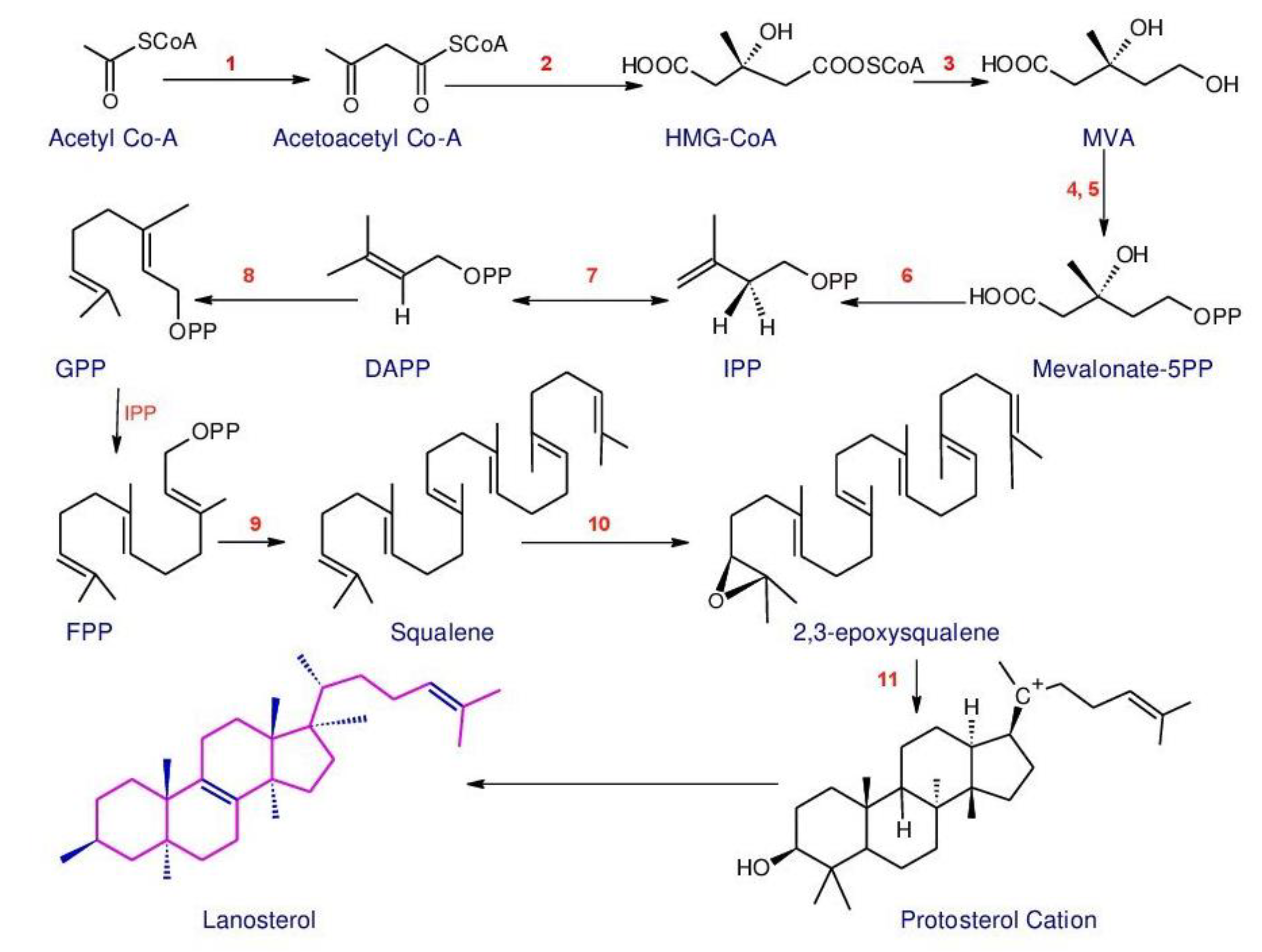
2.1. Biosynthesis of Triterpenoids
2.2. Structures and Bioactivities of Triterpenoids
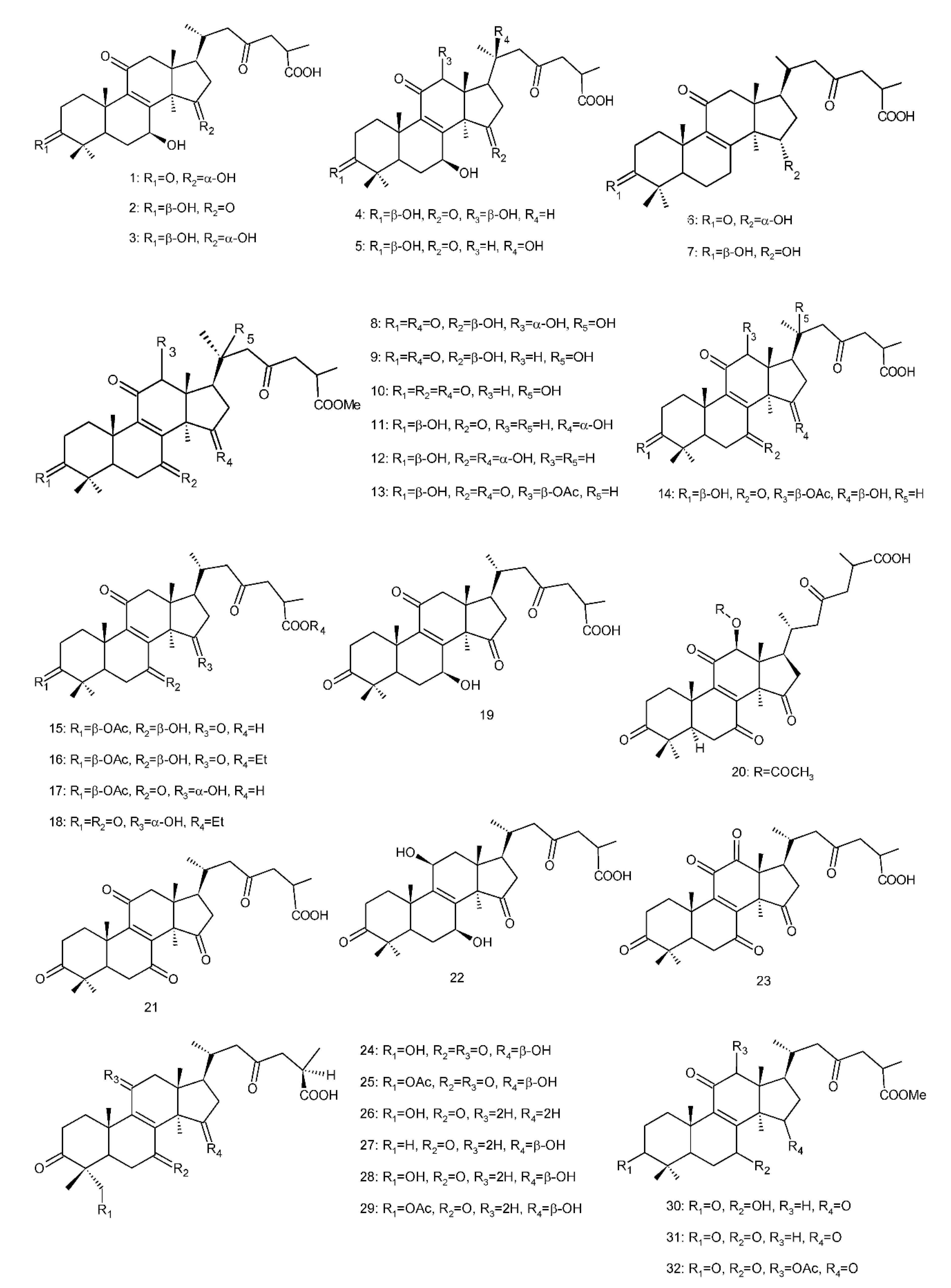
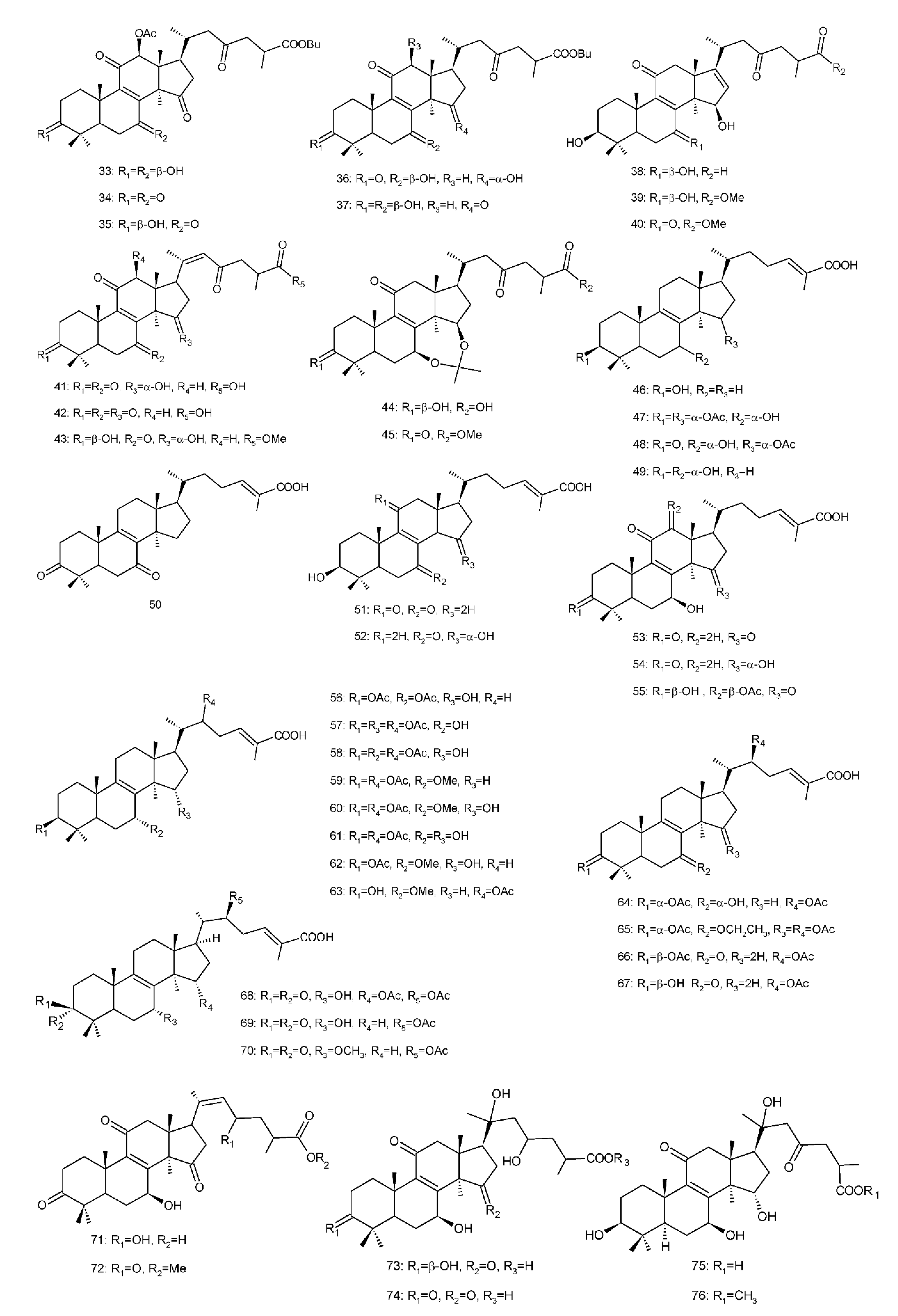
2.2.1. C30 Triterpenoids
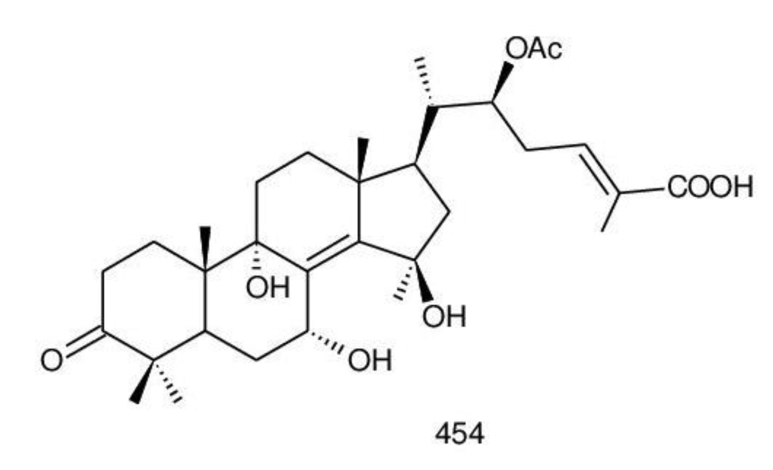
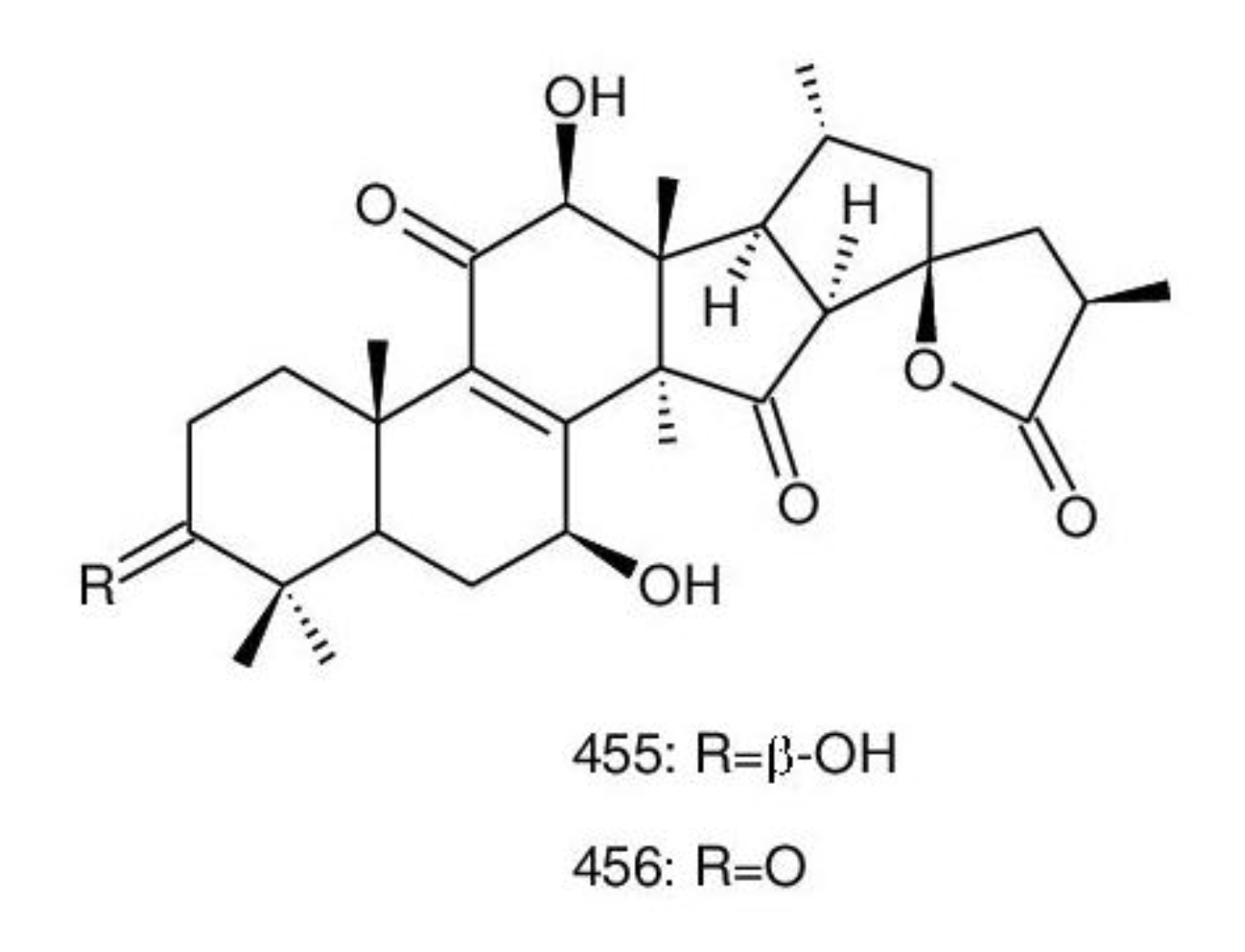
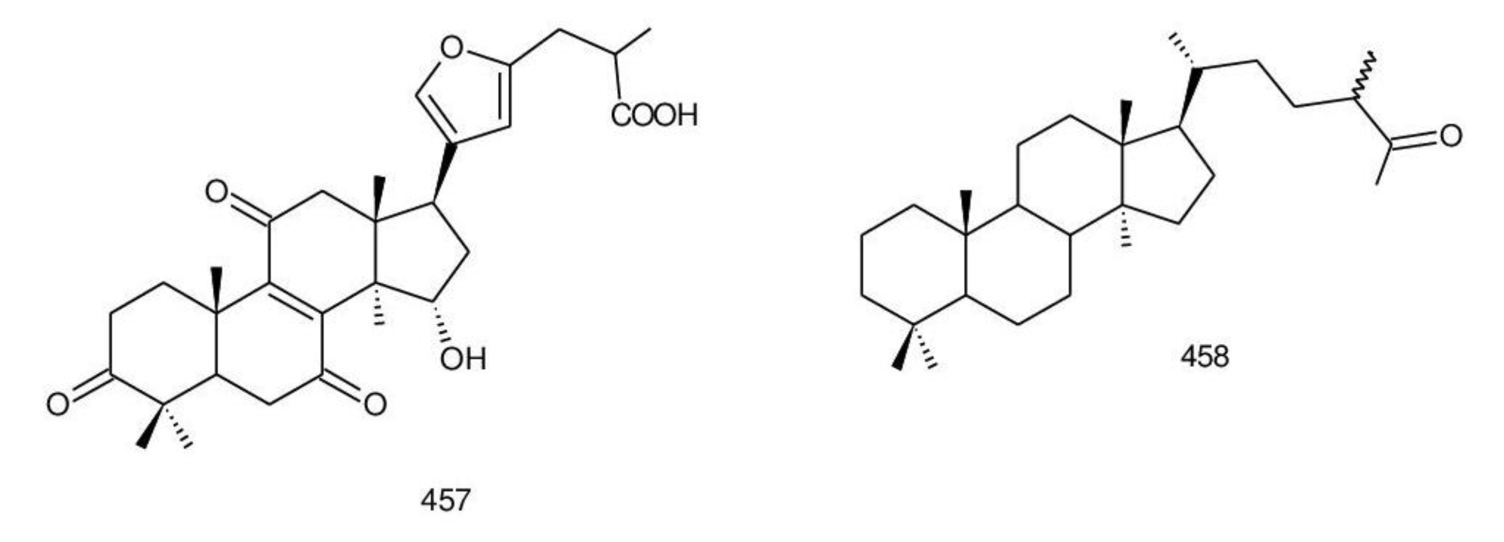
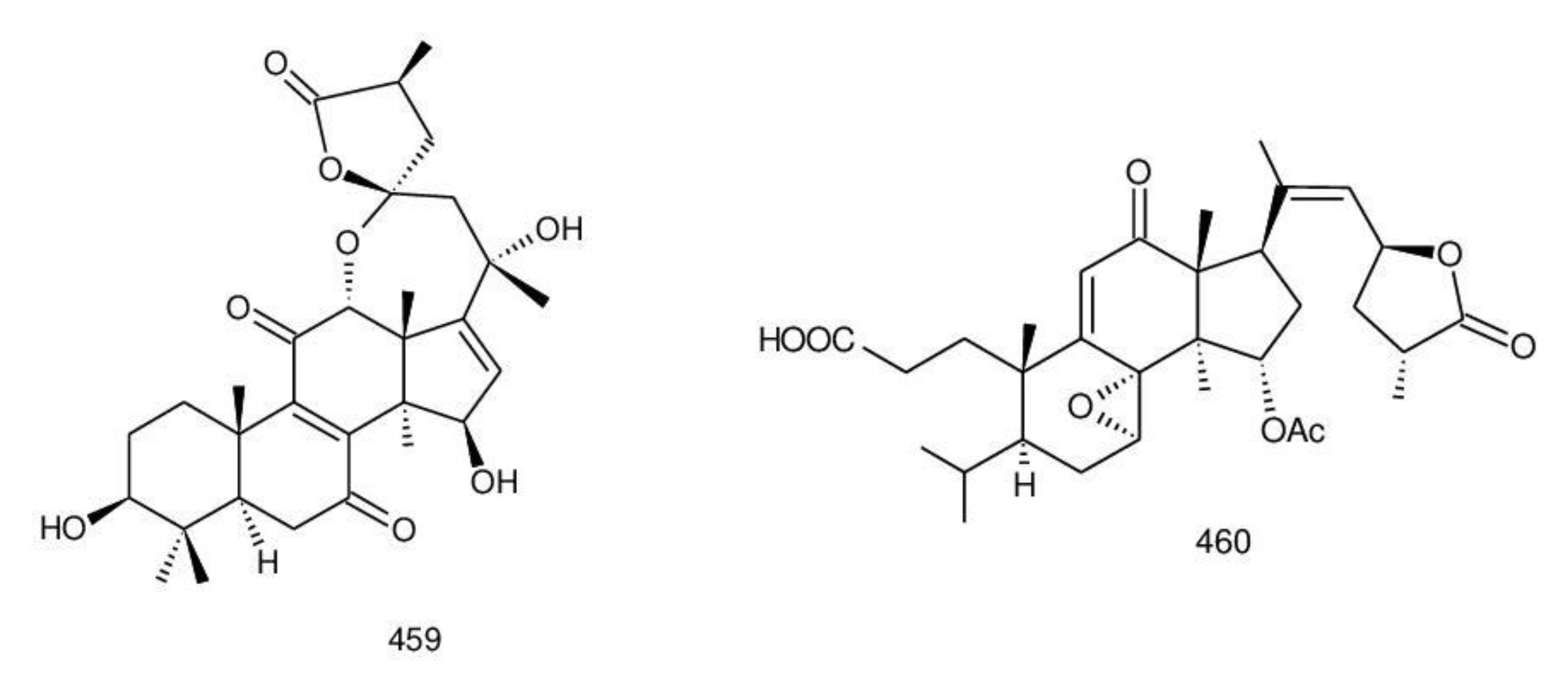
8,9-ene-triterpenoids

| No. | Trivial Names | Bioactivities (IC50/MIC or ED50) | Sources Ganoderma Species | References |
|---|---|---|---|---|
| 1. | Ganoderic acid A | Promising anticancer agent (via potent inhibitory effect on JAK/STAT3 pathway) | G. lucidum, G. tsugae | [165,166,167] |
| 2. | Ganoderic acid B | Moderately active inhibitor against HIV-1 PR (0.17 mM) | G. lucidum, G. tsugae | [165,166,168] |
| 3. | Ganoderic acid C | Suppressed LPS-induced TNF-α (IC50 = 24.5 µg/mL) production through down-regulating MAPK, NF-kappa B and AP-1 signaling pathways in macrophages | G. lucidum, G. tsugae | [165,169] |
| 4. | Ganoderic acid G | Antinociceptive effect | G. lucidum | [170,171] |
| 5. | Ganoderic acid I | Cytotoxicity against Hep G2 cells (IC50 = 0.26 mg/mL), HeLa cells (IC50 = 0.33 mg/mL), Caco-2 cells (IC50 = 0.39 mg/mL) | G. lucidum | [170,172] |
| 6. | Ganolucidic acid A | - | G. lucidum | [173] |
| 7. | Ganolucidic acid B | - | G. lucidum | [174] |
| 8. | Methyl ganoderate M | - | G. lucidum | [175] |
| 9. | Methyl ganoderate N | - | G. lucidum | [175] |
| 10. | Methyl ganoderate O | - | G. lucidum | [175] |
| 11. | Methyl ganoderate K | - | G. lucidum | [175] |
| 12. | Compound B9 | - | G. lucidum | [175] |
| 13. | Methyl ganoderate H | - | G. lucidum | [175] |
| 14. | Ganoderic acid α | Anti-HIV protease (0.19 mM) | G. lucidum | [168] |
| 15. | 3-O-acetylganoderic acid B | - | G. lucidum | [176] |
| 16. | Ethyl 3-O-acetylganoderate B | - | G. lucidum | [176] |
| 17. | 3-O-Acetylganoderic acid K | - | G. lucidum | [176] |
| 18. | Ethyl ganoderate J | - | G. lucidum | [176] |
| 19. | Ganoderic acid D | Cytotoxicity against HeLa cells (17.3 μM), 5α-reductase inhibition—NE | G. lucidum, G. applanatum, G. tsugae | [156,165, 177,178,179] |
| 20. | Ganoderic acid F | Cytotoxicity against HeLa cells (19.5 μM) | G. lucidum | [177,179] |
| 21. | Ganoderic acid E | Cytotoxicity against tumor cell lines [Hep G2 (1.44 × 10⁻⁴ μM), HepG2,2,15 (1.05 × 10⁻⁴ μM), κB—NE, CCM2 (31.25 μM), p388 (5.02 μΜ)] | G. lucidum, G. tsugae | [165,177, 180] |
| 22. | Ganoderic acid Df | Human aldose reductase inhibitory activity (22.8 μM/mL) | G. lucidum | [162] |
| 23. | Ganosporeric acid A | - | G. lucidum | [181] |
| 24. | Ganohainanic acid A | Cytotoxicity—NE | G. hainanense | [62] |
| 25. | Acetyl ganohainanic acid A | Cytotoxicity—NE | G. hainanense | [62] |
| 26. | Ganohainanic acid B | Cytotoxicity—NE | G. hainanense | [62] |
| 27. | Ganohainanic acid C | Cytotoxicity—NE | G. hainanense | [62] |
| 28. | Ganohainanic acid D | Cytotoxicity—NE | G. hainanense | [62] |
| 29. | Acetyl ganohainanic acid D | Cytotoxicity—NE | G. hainanense | [62] |
| 30. | Methyl ganoderate D | - | G. lucidum | [182,183] |
| 31. | Methyl ganoderate E | - | G. lucidum | [184] |
| 32. | Methyl ganoderate F | Inhibitory effects on EBV-EA induction (289 mol ratio/32 pmol TPA) | G. lucidum | [184,185] |
| 33. | 12β-Acetoxy-3β,7β-dihydroxy-11,15,23-trioxolanost-8-en-26-oic acid butyl ester | Antimicrobial [Staphylococcus aureus ATCC 6538 (68.5 μM) and Bacillus subtilis ATCC6633 (123.8 μM)] | G. lucidum | [186] |
| 34. | 12β-acetoxy-3,7,11,15,23-pentaoxolanost-8-en-26-oic acid butyl ester | Antimicrobial—NE | G. lucidum | [186] |
| 35. | n-Butyl ganoderate H | Selective cholinesterase inhibition | G. lucidum | [154] |
| 36. | Butyl ganoderate A | Cytotoxicity against 3T3-L1 cells —NE | G. lucidum | [183] |
| 37. | Butyl ganoderate B | Cytotoxicity against 3T3-L1 cells —NE | G. lucidum | [183] |
| 38. | 3β,7β,15β-Trihydroxy-11,23-dioxo-lanost- 8,16-dien-26-oic acid | Anti-AChE—NE | G. tropicum | [187] |
| 39. | 3β,7β,15β-Trihydroxy- 11,23-dioxo-lanost-8,16- dien-26-oic acid methyl ester | Anti-AChE (15.72%) | G. tropicum | [187] |
| 40. | 3β,15β-Dihydroxy- 7,11,23-trioxo-lanost- 8,16-dien-26-oic acid methyl ester | Anti-AChE—NE | G. tropicum | [187] |
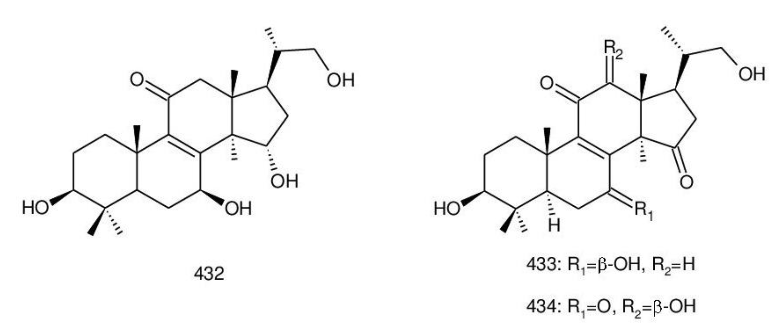
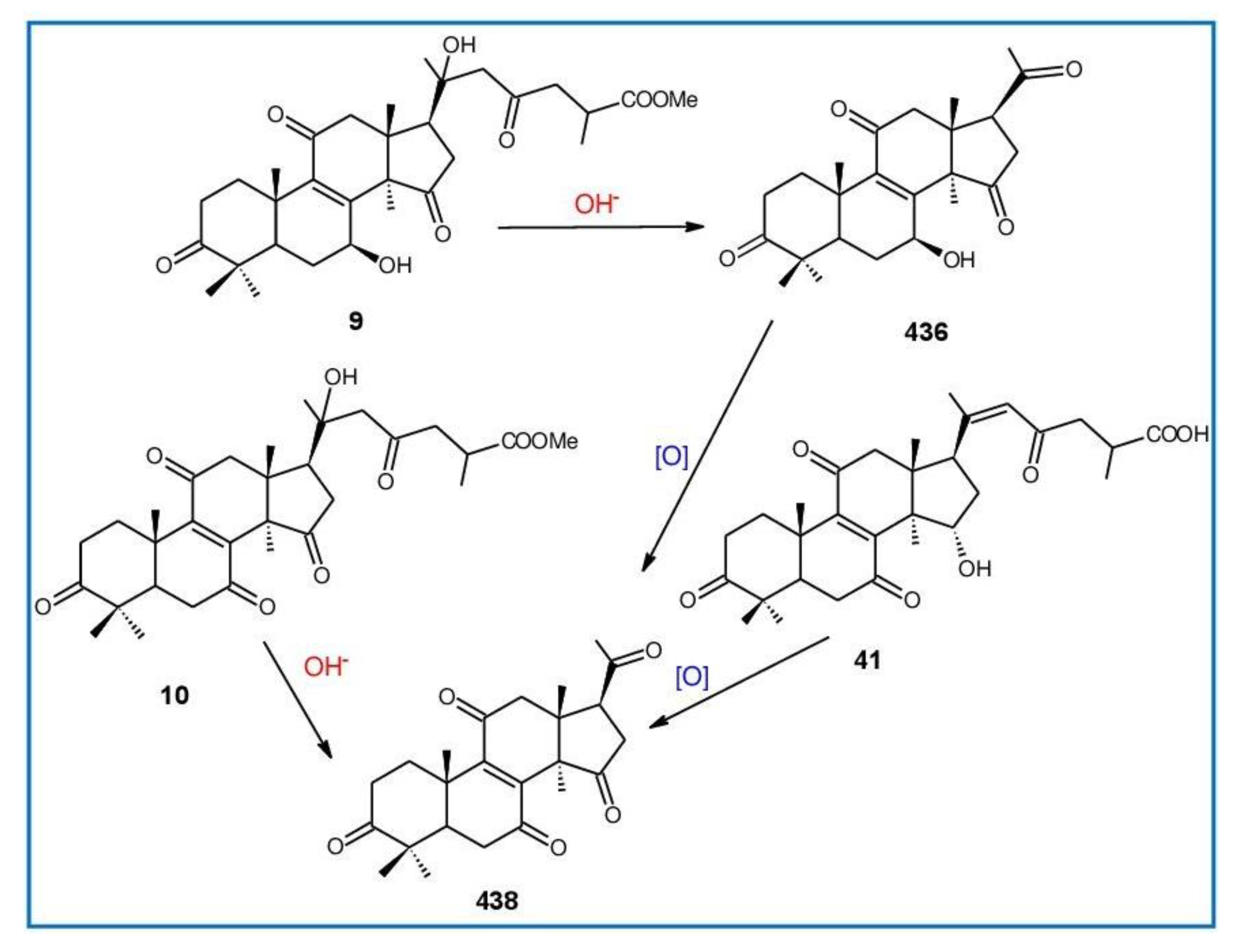
| 41. | Ganoderenic acid G | - | G. applanatum | [188] |
| 42. | Ganoderenic acid F | - | G. applanatum | [188] |
| 43. | Methyl ganoderate I | - | G. applanatum | [188] |
| 44. | Ganodermacetal | Toxic activity against brine shrimp larvae | G. amboinense | [153] |
| 45. | Methyl ganoderate A acetonide | Anti-AChE (18.35 μM), anti-BChE—NE | G. lucidum | [154] |
| 46. | Ganoderic acid Z | Cytotoxicity | G. lucidum | [189] |
| 47. | Ganoderic acid W | Cytotoxicity | G. lucidum | [189] |
| 48. | Ganoderic acid V | Cytotoxicity | G. lucidum | [189] |
| 49. | Ganoderic acid U | - | G. lucidum | [190] |
| 50. | Ganoderic acid DM | 5α-Reductase inhibition (10.6 μM), anti-androgen and anti-proliferative activities, osteoclastogenesis inhibitor, inhibits prostate cancer cell growth, inhibits breast cancer cell growth | G. lucidum, G. sinense | [107,157, 158,160, 191,192] |
| 51. | 7-Oxo-ganoderic acid Z2 | - | G. resinaceum | [193] |
| 52. | 7-Oxo-ganoderic acid Z3 | - | G. resinaceum | [193] |
| 53. | Ganoderic acid GS-1 | Anti-HIV protease (58 µM) | G. sinense | [192] |
| 54. | Ganoderic acid GS-2 | Anti-HIV protease (30 μM) | G. sinense | [192] |
| 55. | Ganoderic acid GS-3 | Anti-HIV protease—NE | G. sinense | [192] |
| 56. | Ganoderic acid Ma | - | G. lucidum | [194] |
| 57. | Ganoderic acid Mb | - | G. lucidum | [194] |
| 58. | Ganoderic acid Mc | - | G. lucidum | [194] |
| 59. | Ganoderic acid Md | - | G. lucidum | [194] |
| 60. | Ganoderic acid Mg | - | G. lucidum | [190] |
| 61. | Ganoderic acid Mh | - | G. lucidum | [190] |
| 62. | Ganoderic acid Mi | - | G. lucidum | [190] |
| 63. | Ganoderic acid Mj | - | G. lucidum | [190] |
| 64. | 3α,22β-Diacetoxy-7α-hydroxyl-5α-lanost-8,24E-dien-26-oic acid | Cytotoxicity against 95D (IC50 = 23 μM/mL), HeLa human tumor cell lines (IC50 = 14.7 μM/mL) | G. lucidum (mycelia) | [195] |
| 65. | 7-O-Ethyl ganoderic acid O | Cytotoxicity against 95D (46.7 μM), HeLa cells (59.1 μM) | G. lucidum | [196] |
| 66. | Ganorbiformin B | - | G. orbiforme | [36] |
| 67. | Ganorbiformin C | - | G. orbiforme | [36] |
| 68. | Ganorbiformin D | Cytotoxicity (against NCIH187, MCF-7, and κB—NE), nonmalignant Vero cells, antimalarial, antitubercular—NE | G. orbiforme | [36] |
| 69. | Ganorbiformin E | Cytotoxicity against NCIH187 (70 µM), MCF-7, κB—NE, nonmalignant Vero cells, antimalarial, antitubercular—NE | G. orbiforme | [36] |
| 70. | Ganorbiformin F | Cytotoxicity against NCIH187 (44 µM), MCF-7—NE and κB (63 µM), nonmalignant Vero cells (36 µM), antimalarial, antitubercular—NE | G. orbiforme | [36] |
| 71. | 7β,23ξ-Dihydroxy-3,11,15-trioxolanosta-8,20E(22)-dien-26-oic acid | - | G. applanatum | [38] |
| 72. | Methyl ganoderenate D | - | G. applanatum | [38] |
| 73. | 3β,7β,20,23ξ-Tetrahydroxy-11,15- dioxolanosta-8-en-26-oic acid | - | G. applanatum | [38] |
| 74. | 7β,20,23ξ-Trihydroxy-3,11,15- trioxolanosta-8-en-26-oic acid | - | G. applanatum | [38] |
| 75. | Ganoderic acid L | - | G. lucidum | [197] |
| 76. | Methyl ganoderate L | - | G. lucidum | [197] |
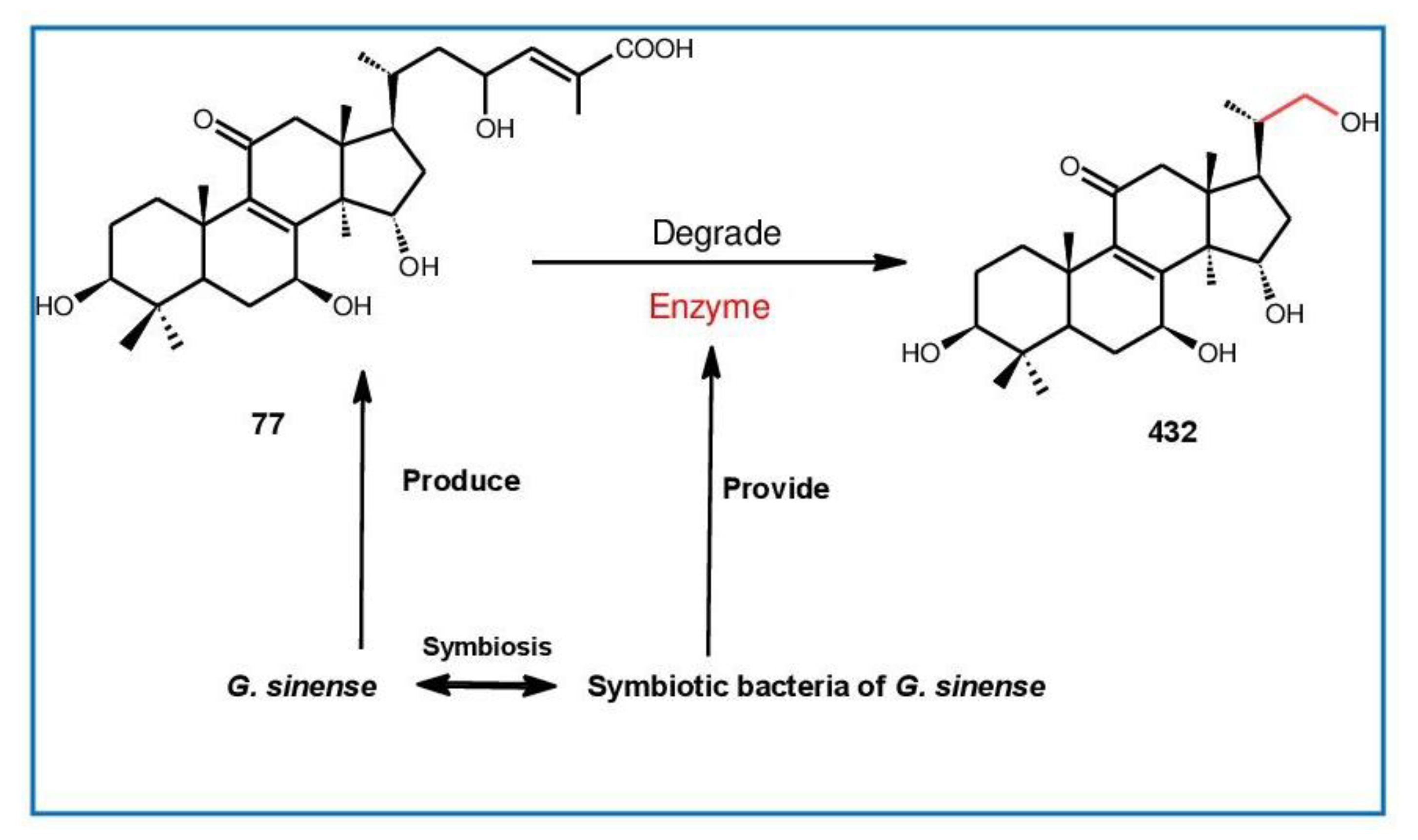
| 77. | Ganolucidic acid γa | PXR-mediated CYP3A4 expression—NE | G. sinense | [198] |
| 78. | Ganolucidate F | PXR-mediated CYP3A4 expression | G. sinense | [198] |
| 79. | Ganolucidic acid D | Cytotoxicity on tumor growth cells—NE | G. lucidum | [197,199] |
| 80. | Methyl ganolucidate D | - | G. lucidum | [197] |
| 81. | Hainanic acid A | Cytotoxicity—NE | G. hainanense | [62] |
| 82. | Hainanic acid B | Cytotoxicity—NE | G. hainanense | [62] |
| 83. | Ganoderic acid γ | Cytotoxicity against tumor cell growth Meth-A (ED50 = 15.6 μg/mL), LLC—NE | G. lucidum | [199] |
| 84. | Ganoderic acid δ | Cytotoxicity against tumor cell growth Meth-A and LLC—NE | G. lucidum | [199] |
| 85. | Ganoderic acid ε | Cytotoxicity against tumor cell growth Meth-A (ED50 = 12.2 μg/mL), LLC—NE | G. lucidum | [199] |
| 86. | Ganoderic acid ζ | Cytotoxicity against tumor cell growth Meth-A and LLC—NE | G. lucidum | [199] |
| 87. | Ganoderic acid η | Cytotoxicity against tumor cell growth Meth-A and LLC—NE | G. lucidum | [199] |
| 88. | Ganoderic acid θ | Cytotoxicity against tumor cell growth Meth-A (ED50 = 5.7 μg/mL), LLC (ED50 = 15.2 μg/mL) | G. lucidum | [199] |
| 89. | Ganoderiol G | - | G. lucidum | [200] |
| 90. | Ganoderiol H | - | G. lucidum | [200] |
| 91. | Ganoderiol I | - | G. lucidum | [200] |
| 92. | 24S,25R-Dihydroxy-3,7-dioxo-8-en-5α-lanost-26-ol | Cytotoxicity—NE | G. hainanense | [62] |
| 93. | Ganoderone A | Antiviral: influenza A—NE, HSV (0.3 μg/mL) | G. pfeifferi | [46] |
| 94. | Ganoderone C | Antiviral—NE | G. pfeifferi | [46] |
| 95. | 3,7,24-Trioxo-5α-lanost- 8,25-dien-26-ol | Cytotoxicity against HL-60 (15.70 µM), SMMC-7721 (15.52 µM), A-549 (15.81 µM), MCF-7 (20.08 µM), SW480—NE | G. hainanense | [62] |
| 96. | Hainanaldehyde A | Cytotoxicity—NE | G. hainanense | [62] |
| 97. | 21-Hydroxy-3,7-dioxo-5α-lanost-8,24E-dien-26-ol | Cytotoxicity—NE | G. hainanense | [62] |
| 98. | 3β,11α-Dihydroxy-7-oxo-5α-lanost-8,24E-dien-26-ol | Cytotoxicity—NE | G. hainanense | [62] |
| 99. | Lucialdehyde D | - | G. lucidum, G. pfeifferi | [46,201, 202] |
| 100. | Ganoderiol J | - | G. sinense | [198] |
| 101. | 16α,26-Dihydroxylanosta-8,24-dien-3-one | Cytotoxicity against K-562 cells (13.3 μg/mL) | G. hainanense | [75] |
| 102. | Lucidadiol | Antiviral: influenza virus type A (ED50 = 0.22 mmol/L), HSV—NE | G. lucidum, G. pfeifferi | [203,204] |
| 103. | Sinensoic acid | - | G. sinense | [205] |
| 104. | Tsugaric acid C | Cytotoxicity—NE | G. tsugae | [206] |
| 105. | Colossolactone A | Moderate cytotoxicity against L-929, K-562, HeLa cells—NE, anti-inflammatory properties | G. colossum | [207] |
| 106. | Ganoderenicfy A | Promoting angiogenesis activities | G. applanatum | [208] |
| 107. | Colossolactone I | Moderate cytotoxicity against HCT-116 colorectal cancer cells, Antimalarial: Plasmodium falciparum—NE | G. colossum | [92,209, 210] |
| 108. | Colossolactone II | Low cytotoxicity against HCT-116 colorectal cancer cells | G. colossum | [92,209] |
| 109. | Ganodermalactone E | Antimalarial: Plasmodium falciparum—NE | G. colossum | [210] |
| 110. | Colossolactone B | Moderate cytotoxicity against L-929, K-562, and HeLa cells, antimicrobial—NE, antibacterial—NE | G. colossum | [207,210, 211] |
| 111. | Methyl ganolucidate A | - | G. lucidum | [170,174] |
| 112. | Methyl ganolucidate B | - | G. lucidum | [170,174] |
| 113. | Methyl ganoderate A | - | G. lucidum | [182] |
| 114. | Methyl ganoderate B | - | G. lucidum | [182] |
| 115. | Methyl ganoderate C | - | G. lucidum | [182] |
| 116. | Methyl ganoderate C2 | - | G. lucidum (dried fruit bodies) | [212] |
| 117. | Compound B8 | - | G. lucidum (dried fruit bodies) | [212] |
| 118. | 3β-Oxo-formyl-7β,12β-dihydroxy-5α-lanost-11,15,23-trioxo-8-en(E)-26-oic acid | - | G. lucidum (fruit bodies) | [213] |
| 119. | Ganoderic acid B8 | Cytotoxicity against LLC—NE, T47-D—NE, S-180—NE, Meth-A—NE | G. lucidum (fruit bodies) | [214] |
| 120. | Ganoderic acid C1 | Inhibitory activity against HIV-PR (0.18 mM) | G. lucidum (fruit bodies) | [168,214] |
| 121. | 12β-Acetoxy-3,7,11,15,23-pentaoxo-5α-lanosta-8-en-26-oic acid ethyl ester | Cytotoxicity against human HeLa cervical cancer cell lines (63 μM) | G. lucidum | [215] |
| 122. | 3β,7β-Dihydroxy-12β-acetoxy-11,15,23-trioxo-5α-lanosta-8-en-26-oic acid methyl ester | - | G. lucidum | [32] |
| 123. | 3β-Hydroxy-7,11,12,15,23-pentaoxolanost-8-en-26-oic acid | Cytotoxic against p388 cell (9.85 μM), HeLa cell (17.10 μM), BEL-7402 cell (51.00 μM), SGC-7901 cells (42.00 μM) | G. lucidum (fruit bodies) | [216] |
| 124. | Ganoderic acid H | Inhibitory activity against HIV-PR (0.20 mM) | G. lucidum (fruit bodies) | [177,217] |
| 125. | Ganoderic acid K | Cytotoxicity against p388 cell (13. 8 μM), HeLa cell (8.23 μM); BEL-7402 cell (16.5 μM), SGC-7901cell (21.0 μM) | G. lucidum (fruit bodies) | [218] |
| 126. | Ganoderic acid AM1 | Cytotoxicity against p388 cell (13. 2 μM), HeLa cell (9.75 μM), BEL-7402 cell (20.9 μM), SGC-7901 cell (23.0 μM) | G. lucidum (fruit bodies) | [218] |
| 127. | Ganoderic acid J | Cytotoxicity against p388 cell (15. 8 μM), HeLa cell (12.2 μM), BEL-7402 cell (25.2 μM), SGC-7901 cell (20.2 μM) | G. lucidum (fruit bodies) | [218] |
| 128. | Ganoderic acid AP2 | - | G. applanatum (fruit bodies) | [178] |
| 129. | 23S-Hydroxy-3,7,11,15-tetraoxolanost-8,24E-diene-26-oic acid | Cytotoxicity against p388 cell (15.7 μM), HeLa cell (9.72 μM), BEL-7402 cell (25.6 μM), SGC-7901 cell (23.1 μM) | G. lucidum (fruit bodies) | [218] |
| 130. | 7-Oxoganoderic acid Z | Inhibitory activities against the HMG-CoA reductase (22.3 μM), acyl CoA acyltransferase (5.5 μM) | G. lucidum (fruit bodies) | [219] |
| 131. | Ganoderic acid LM2 | Potent enhancement of ConA-induced mice splenocytes proliferation in vitro | G. lucidum (fruit bodies) | [220] |
| 132. | Lucialdehyde B | Cytotoxic effect on tested tumor cells | G. lucidum (fruit bodies) | [214] |
| 133. | Lucialdehyde C | Cytotoxicity against LLC, T-47D (10.7 µg/mL), Sarcoma 180 (4.7 µg/mL), Meth-A tumor cells (3.8 µg/mL) | G. lucidum (fruit bodies) | [214] |
| 134. | Ganoderic acid β | HIV-I protease inhibitory activity (20 µM) | G. lucidum (spores) | [221] |
| 135. | Ganolucidic acid E | - | G. lucidum (fruit bodies) | [200] |
| 136. | Ganoderal B | - | G. lucidum | [222] |
| 137. | 11α-Hydroxy-3,7-dioxo-5α-lanosta-8,24(E)-dien-26-oic acid | Cytotoxicity against human HeLa cervical cancer cell lines (123 μM) | G. lucidum | [215] |
| 138. | 11β-Hydroxy-3,7-dioxo-5α-lanosta-8,24(E)-dien-26-oic acid | Cytotoxicity against human HeLa cervical cancer cell lines (51 μM) | G. lucidum | [215] |
| 139. | Lucidal | - | G. lucidum (cultured fruit bodies) | [203] |
| 140. | Lucialdehyde E | Cytotoxic activity against esophageal tumor EC109 cell line (18.7 mg/mL) | G. lucidum (spores) | [202] |
| 141. | 3α,22β-Diacetoxy-7α-hydroxyl-5α-lanost-8,24E-dien-26-oic acid | Cytotoxicity against HeLa cell lines (14.7 μM), 95D cell lines (23.01 μM) | G. lucidum (mycelial mat) | [195] |
| 142. | Ganoderic acid O | - | G. lucidum (cultured mycelium) | [223] |
| 143. | 7-O-Methylganoderic acid O | - | G. lucidum (cultured mycelium) | [223] |
| 144. | 12β-Acetoxy-3β-hydroxy-7,11,15,23- tetraoxo-lanost-8,20E-diene-26-oic acid | Cytotoxicity against human cancer cell p388 (12.7 µM), HeLa cell (8.72 µM), BEL-7402 (24.2 µM), SGC-7901 (18.7 µM) | G. lucidum (fruit bodies) | [218] |
| 145. | 23-Dihydroganoderenic acid D | - | G. applanatum (fruit bodies) | [38] |
| 146. | Ganoderenic acid A | - | G. lucidum (dried fruit bodies) | [177] |
| 147. | Ganoderenic acid B | - | G. lucidum (dried fruit bodies) | [177] |
| 148. | Ganoderenic acid C | - | G. lucidum (dried fruit bodies) | [177] |
| 149. | Ganoderenic acid D | - | G. lucidum (dried fruit bodies) | [177] |
| 150. | 12β-Acetoxy-7β-hydroxy-3,11,15,23- tetraoxo-5α-lanosta-8,20-dien-26-oic acid | Cytotoxicity against human HeLa cervical cancer cell lines—NE | G. lucidum | [215] |
| 151. | Methy ganoderenate H | - | G. applanatum (fruit bodies) | [188] |
| 152. | Methyl ganoderenate I | - | G. applanatum (fruit bodies) | [188] |
| 153. | 12β-Acetoxy-3β,7β-dihydroxy-11,15,23-trioxo-5α-lanosta-8,20-dien-26-oic acid | - | G. lucidum | [32] |
| 154. | Methyl ganoderate G | - | G. lucidum | [170] |
| 155. | Compound C5 | - | G. lucidum (fruit bodies) | [217] |
| 156. | Compound C6 | - | G. lucidum (fruit bodies) | [217] |
| 157. | Ganoderic acid AP3 | - | G. applanatum (fruit bodies) | [178] |
| 158. | 23-Dihydroganoderic acid I | - | G. applanatum (fruit bodies) | [38] |
| 159. | 23-Dihydroganoderic acid N | - | G. applanatum (fruit bodies) | [38] |
| 160. | 20-Hydroxylganoderic acid G | - | G. lucidum (fruit bodies) | [224] |
| 161. | Lucidumol A | HIV-I protease inhibitory activity—NE | G. lucidum (spores) | [221] |
| 162. | Ganoderiol C | - | G. lucidum (fruit bodies) | [200] |
| 163. | Ganoderiol D | - | G. lucidum (fruit bodies) | [200] |
| 164. | Ganoderitriol M | - | G. lucidum (fruit bodies) | [225] |
| 165. | Tsugaric acid A | - | G. tsugae | [226] |
| 166. | Tsugarioside A | Cytotoxicity against PLC/PRF/5 (ED50 = 6.5 μg/mL), T-24 (ED50 = 8.6 μg/mL), HT-3 (ED50 = 7.2 μg/mL), SiHa (ED50 = 9.5 μg/mL) | G. tsugae (fruit bodies) | [206] |
| 167. | 3-Oxo-5α-lanosta-8,24-dien-21-oic acid | Cytotoxicity—NE | G. resinaceum (fruit bodies) | [227] |
| 168. | 3β-Hydroxy-5α-lanosta-8,24-dien-21-oic acid | Cytotoxicity against T-24 (ED50 = 4.4 μg/mL), HT-3 (ED50 = 3.5 μg/mL), SiHa (ED50 = 5.5 μg/mL), CaSKi (ED50 = 6.2 μg/mL) | G. tsugae (fruit bodies) | [206] |
| 169. | 3β,7β-Dihydroxy-11,15,23-trioxolanost-8,16-dien-26-oic acid | - | G. lucidum (fruit bodies) | [203] |
| 170. | 3β,7β-Dihydroxy-11,15,23-trioxolanost-8,16-dien-26-oic acid methyl ester | - | G. lucidum (fruit bodies) | [203] |
| 171. | 12β-Acetoxy-3β,7β-dihydroxy-11,15,23-trioxolanost-8,16-dien-26-oic acid | - | G. lucidum (fruit bodies) | [228] |
| 172. | Methyl ganoderate AP | - | G. applanatum (fruit bodies) | [188] |
| 173. | Ganoderiol E | - | G. lucidum (fruit bodies) | [200] |
| 174. | Epoxyganoderiol A | - | G. lucidum | [222] |
| 175. | 3α-Carboxyacetoxy-24-methylene-23-oxolanost-8-en-26-oic acid | Cytotoxicity—NE | G. applanatum (fruit bodies) | [229] |
| 176. | 3α-Carboxyacetoxy-24-methyl-23- oxolanost-8-en-26-oic acid | Cytotoxicity—NE | G. applanatum (fruit bodies) | [229] |
| 177. | 3-Epipachymic acid | - | G. resinaceum (fruit bodies) | [227] |
| 178. | 3β,15α-Diacetoxylanosta-8,24-dien-26-oic acid | - | G. lucidum (mycelia) | [230] |
| 179. | Ganoderic acid V1 | - | G. lucidum | [231] |
| 180. | Tsugaric acid B | - | G. tsugae | [226] |
| 181. | Methyl ganoderenate E | - | G. lucidum (fruit bodies) | [175] |
| 182. | Lucidumol D | Selective anti-proliferative and cytotoxic effects | G. lingzhi | [232] |
| 183. | Lucidumol C | Selective anti-proliferative and cytotoxic effects | G. lingzhi | [232] |
| 184. | Leucocontextin A | - | G. leucocontextum | [233] |
| 185. | Leucocontextin B | - | G. leucocontextum | [233] |
| 186. | Leucocontextin C | - | G. leucocontextum | [233] |
| 187. | Leucocontextin D | - | G. leucocontextum | [233] |
| 188. | Leucocontextin E | - | G. leucocontextum | [233] |
| 189. | Leucocontextin F | - | G. leucocontextum | [233] |
| 190. | Leucocontextin G | - | G. leucocontextum | [233] |
| 191. | Leucocontextin H | - | G. leucocontextum | [233] |
| 192. | Leucocontextin I | - | G. leucocontextum | [233] |
| 193. | Leucocontextin R | Cytotoxicity against K562 and MCF-7 cell lines (IC50 = 20–30 μM) | G. leucocontextum | [233] |
| 194. | Ganoleuconin A | Cytotoxicity against K562 (17.8 μM), PC-3 cell lines—NE | G. leucocontextum | [34] |
| 195. | Ganoleuconin B | Cytotoxicity against K562 (19.7 μM), PC-3 cell lines—NE | G. leucocontextum | [34] |
| 196. | Ganoleuconin E | Cytotoxicity against K562 and PC-3 cell lines—NE | G. leucocontextum | [34] |
| 197. | Ganoleuconin G | Cytotoxicity against K562 (11.4 μM), PC-3 cell lines (132.4 μM) | G. leucocontextum | [34] |
| 198. | Ganoleuconin H | Cytotoxicity against K562 (115.4 μM), PC-3 cell lines (24.2 μM) | G. leucocontextum | [34] |
| 199. | Ganoleuconin I | Cytotoxicity against K562 and PC-3 cell lines—NE | G. leucocontextum | [34] |
| 200. | Ganoderenicfy B | Promoting angiogenesis activities | G. applanatum | [208] |
| 201. | (24E)-15α,26-Dihydroxy-3-oxo-lanosta-8,24-diene | Antimycobacteria (50 μg/mL), cytotoxicity (5.9 μg/mL) | G. casuarinicola | [234] |
| 202. | (24E)-7α,26-Dihydroxy-3-oxo-lanosta-8,24-diene. | Antimalarial activity (IC50 = 9.7 μg/mL) | G. casuarinicola | [234] |
| 203. | (24E)-3-Oxo-7α,15α,26-trihy-droxylanosta-8,24-diene | Antimalarial activity (IC50 = 9.2 μg/mL) | G. casuarinicola | [234] |
| 204. | (24E)-3β-Acetoxy-15α,26-dihydroxylanosta-8,24-diene | Antimycobacteria (25 μg/mL), cytotoxicity (6 μg/mL) | G. casuarinicola | [234] |
| 205. | (24E)-3β-Acetoxy-7α,15α,26- trihydroxylanosta-8,24-diene | Antimycobacteria (25 μg/mL), cytotoxicity (9 μg/mL) | G. casuarinicola | [234] |
| 206. | (24E)-3β,7α,15α,26-Tetra-hydroxylanosta-8,24-diene | - | G. casuarinicola | [234] |
| 207. | 7β,15α,20-Trihydroxy-3,11,23-trioxo-5α-lanosta-8-en-26-oic acid | - | G. lucidum | [235] |
| 208. | Ganoderic acid XL3 | - | G. theaecolum | [236] |
| 209. | Ganoderic acid XL4 | - | G. theaecolum | [236] |
| 210. | Ganodecalone A | Cytotoxicity against K562 (17.22 µM) | G. calidophilum | [53] |
| 211. | Ganoderic acid C6 | - | G. lucidum (fruit bodies) | [65] |
| 212. | Ganoderic acid D1 | - | G. lucidum (fruit bodies) | [65] |
| 213. | Ganoderic acid M | - | G. lucidum (fruit bodies) | [65] |
| 214. | Ganoderic acid N | - | G. lucidum (fruit bodies) | [65] |
| 215. | 12-Hydroxylganoderic acid C2 | - | G. lucidum (fruit bodies) | [65] |
| 216. | 3-Acetylganoderic acid H | - | G. lucidum (fruit bodies) | [65] |
| 217. | 12-Acetoxyganoderic acid D | - | G. lucidum (fruit bodies) | [65] |
| 218. | 12-Hydroxyganoderic acid D | - | G. lucidum (fruit bodies) | [65] |
| 219. | 12-Acetoxyganoderic acid F | - | G. lucidum (fruit bodies) | [65] |
| 220. | 12β-Hydroxy-3,7,11,15,23-pentaoxo-5α-lanosta-8-en-26-oic acid | - | G. lucidum (fruit bodies) | [65] |
| 221. | 12-Hydroxy-3,7,11,15,23-pentaoxo-lanost-8-en-26-oic acid | - | G. lucidum (fruit bodies) | [65] |
| 222. | 12,15-Bis(acetyloxy)-3-hydroxy-7,11,23-trioxo-lanost-8-en-26-oic acid | - | G. lucidum (fruit bodies) | [65] |
| 223. | Methyl ganoderate J | - | G. lucidum (fruit bodies) | [65] |
| 224. | Methyl-O-acetylganoderate C | - | G. lucidum (fruit bodies) | [65] |
| 225. | Methyl ganoderate C1 | - | G. lucidum (fruit bodies) | [65] |
| 226. | Methyl ganoderate AM | - | G. lucidum (fruit bodies) | [65] |
| 227. | Ganoderic aldehyde A | - | G. lucidum (fruit bodies) | [65] |
| 228. | Ganoderenic acid K | - | G. lucidum (fruit bodies) | [65] |
| 229. | Ganoderenic acid E | - | G. lucidum (fruit bodies) | [65] |
| 230. | Elfvingic acid A | - | G. lucidum (fruit bodies) | [65] |
| 231. | 12β-Acetoxy-3β,7β-dihydroxy-11,15,23-trioxo-5α-lanosta-8,20-dien-26-oic acid | - | G. lucidum (fruit bodies) | [65] |
| 232. | Methyl ganolucidate C | - | G. lucidum (fruit bodies) | [65] |
| 233. | Ganolucidic acid C | - | G. lucidum (fruit bodies) | [65] |
| 234. | Ganoderic acid C2 | - | G. lucidum (fruit bodies /spore) | [65] |
| 235. | Ganodrol A | Moderately inhibits FAAH (Inhibition rate in between 50–60%) | G. lucidum | [128] |
| 236. | Ganodrol C | Moderately inhibits FAAH (Inhibition rate in between 50–60%) | G. lucidum | [128] |
| 237. | Ganodrol D | Moderately inhibits FAAH (Inhibition rate in between 30–40%) | G. lucidum | [128] |
| 238. | Ganoderic acid XL5 | Cytotoxicity against human tumor cell lines—NE | G. theaecolum | [236] |
| 239. | Methyl gibbosate M | Anti-adipogenesis activity—NE | G. applanatum | [51] |
| 240. | Methyl ganoapplate E | Anti-adipogenesis activity—NE | G. applanatum | [51] |
| 241. | Applandiketone A | - | G. applanatum | [237] |
| 242. | Applandiketone B | Significant inhibitory effect against NO production in LPS-induced RAW264.7 cells (IC50 = 20.65 µM) | G. applanatum | [237] |
| 243. | 15α-Acetoxy-3α-hydroxylanota- 8,24-dien-26-oic | - | G. capense | [238] |
| 244. | Ganoderterpene A | Strongly suppressed NO generation in BV-2 microglial cells treated with lipopolysaccharide (LPS) (IC50 = 7.15 µM), significantly suppressed the activation of MAPK and TLR-4/NF-κB signaling pathways, effectively improved the LPS-induced mitochondrial membrane potential and apoptosis | G. lucidum | [239] |
| 245. | Ganodeweberiol G | Significant α-glucosidase inhibitory activity (IC50 = 165.9 µM) | G. weberianum | [77] |
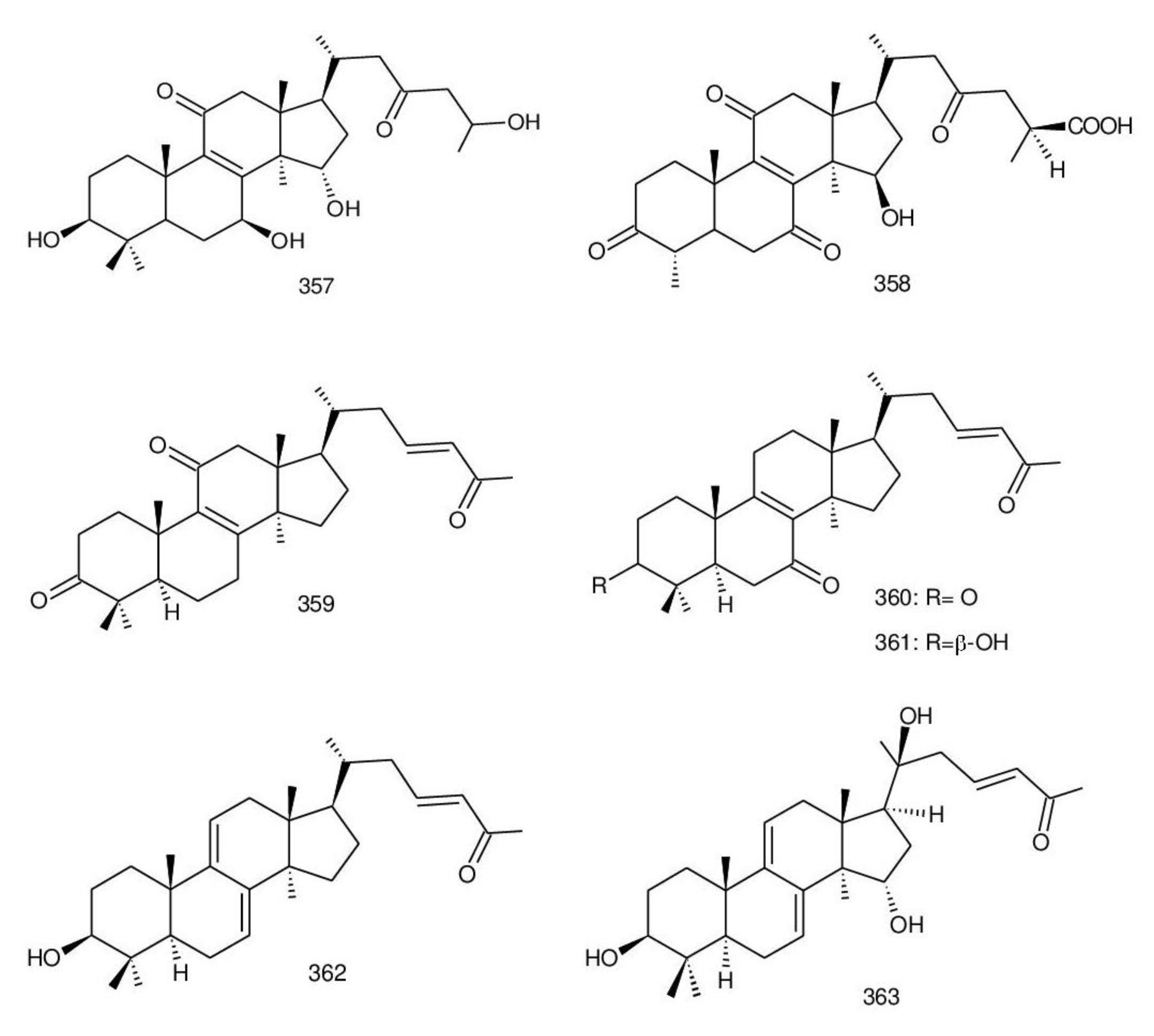
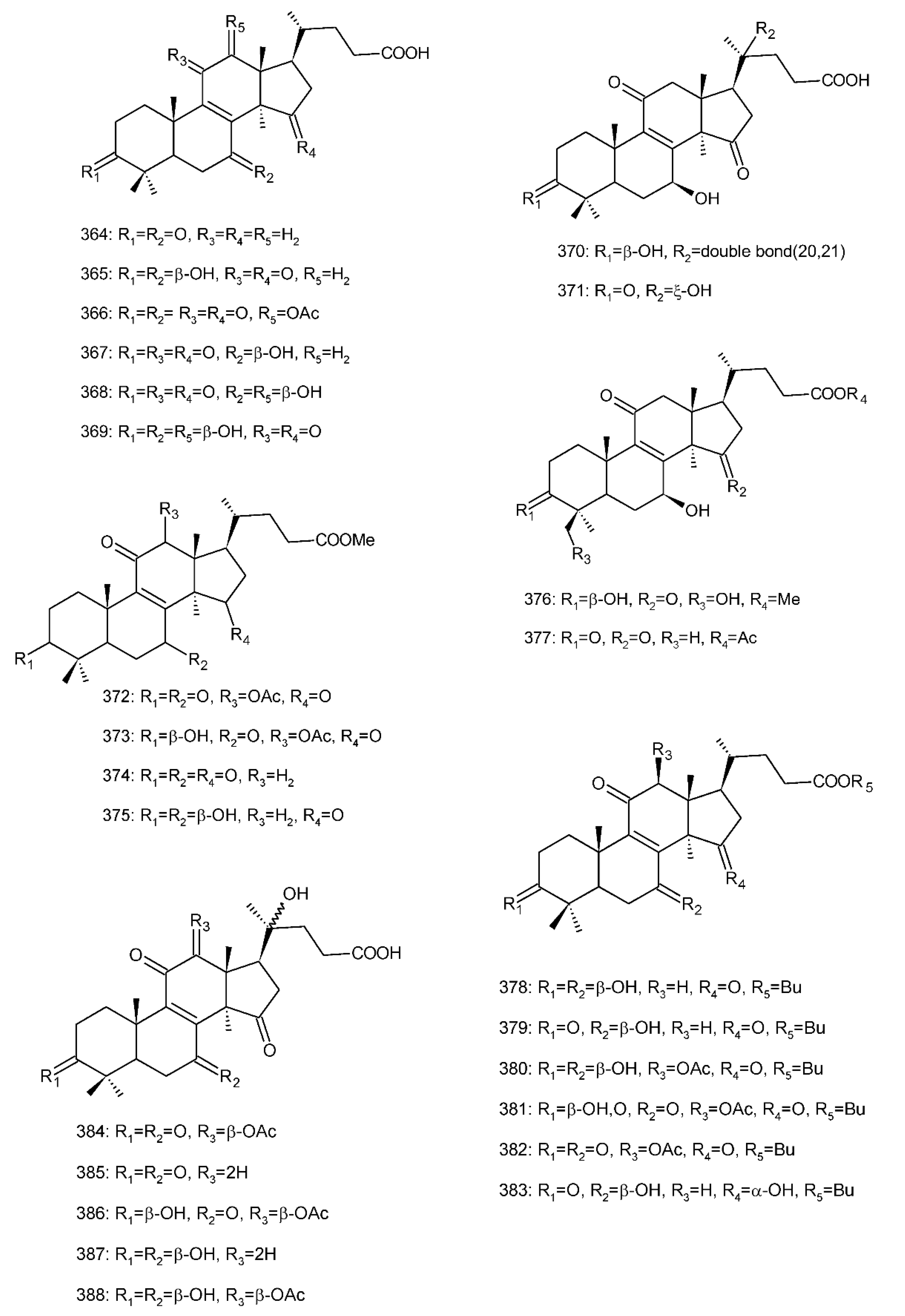
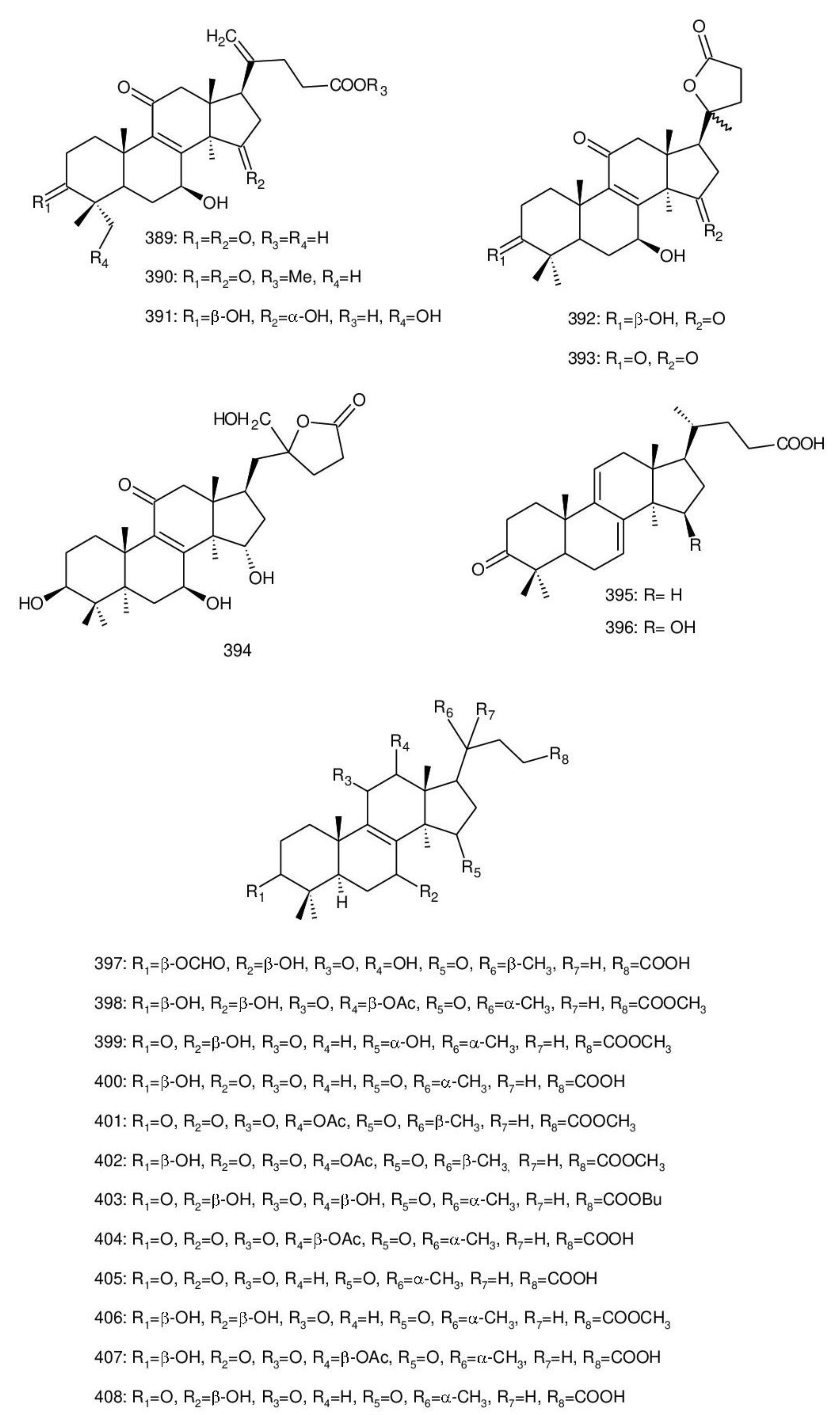
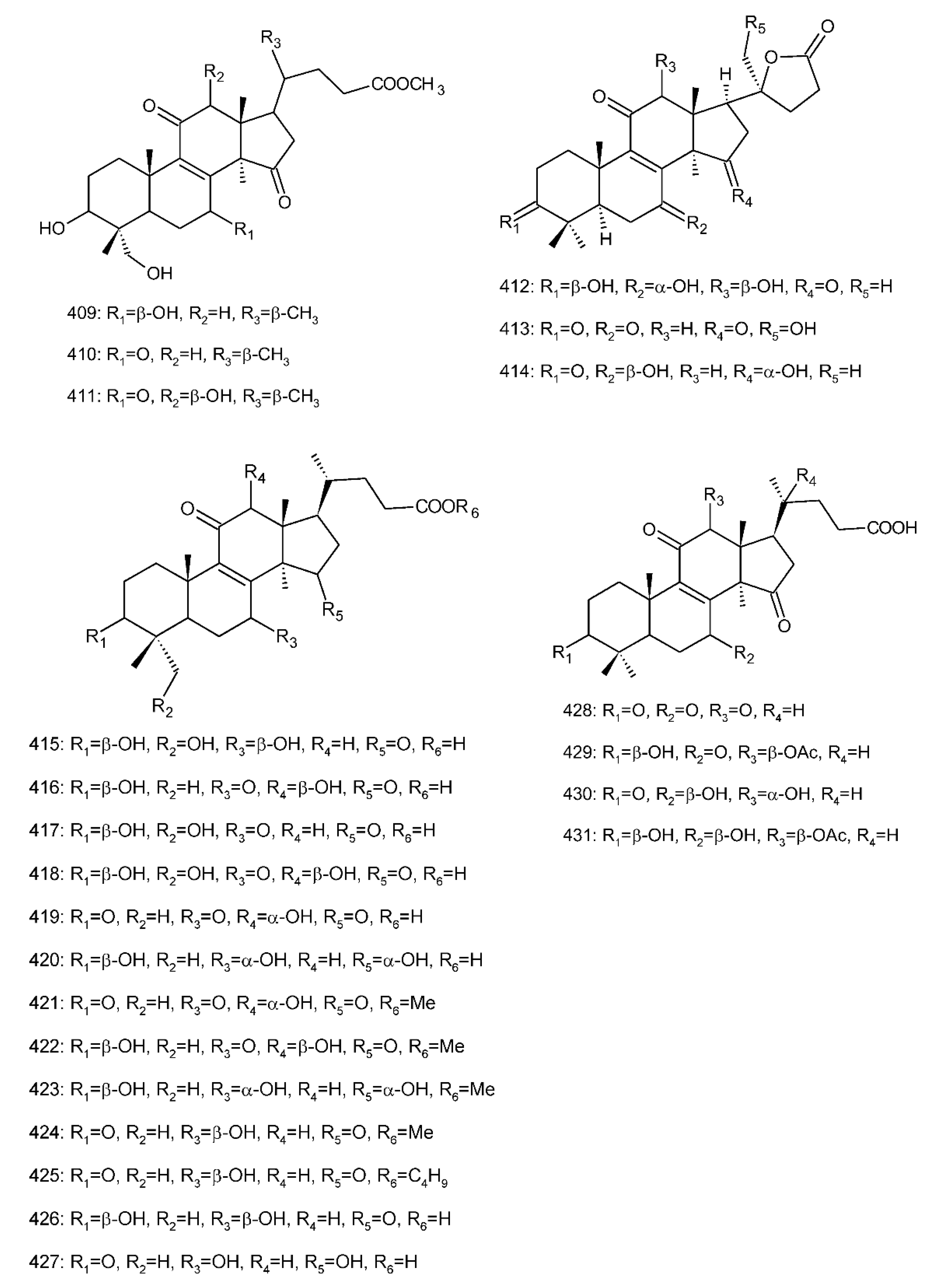
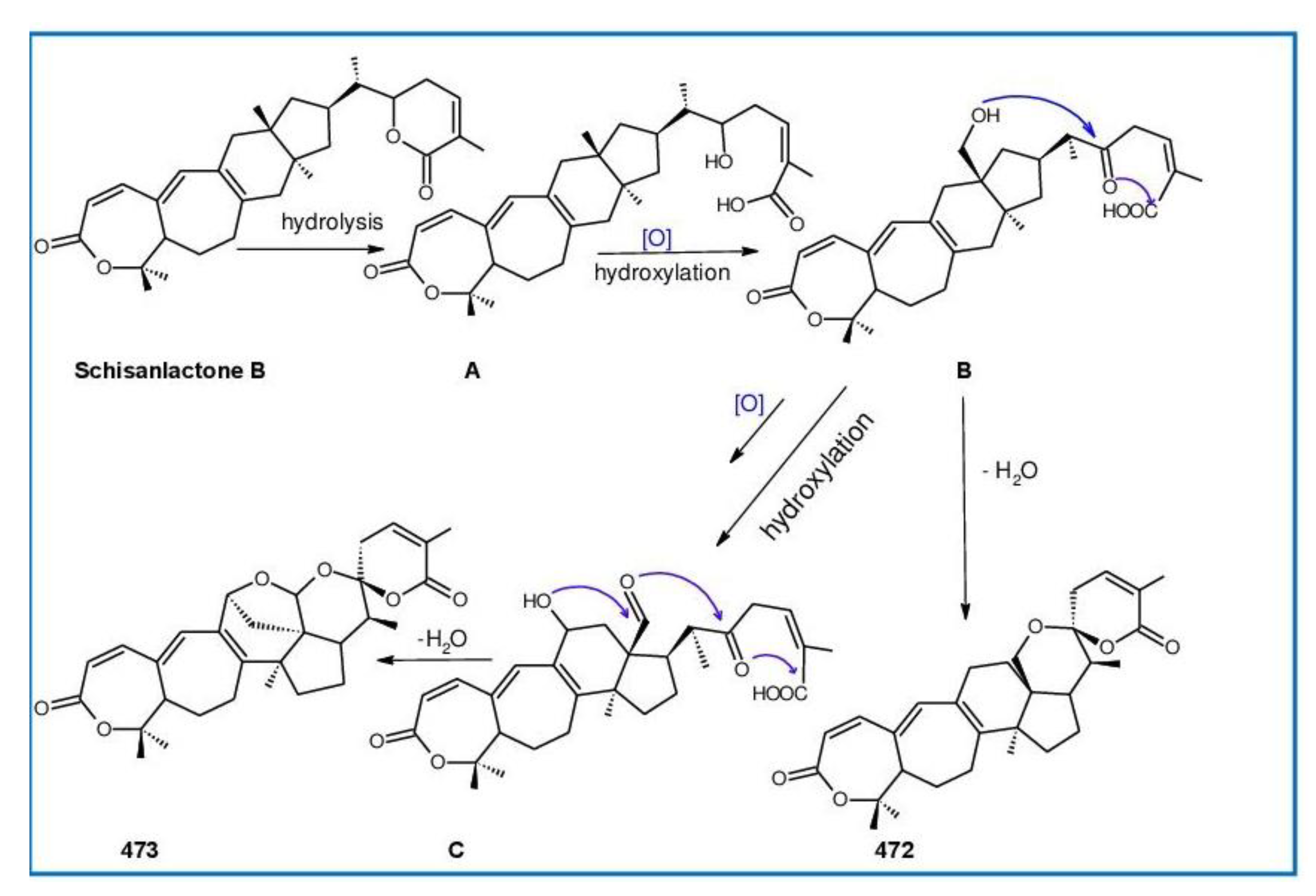
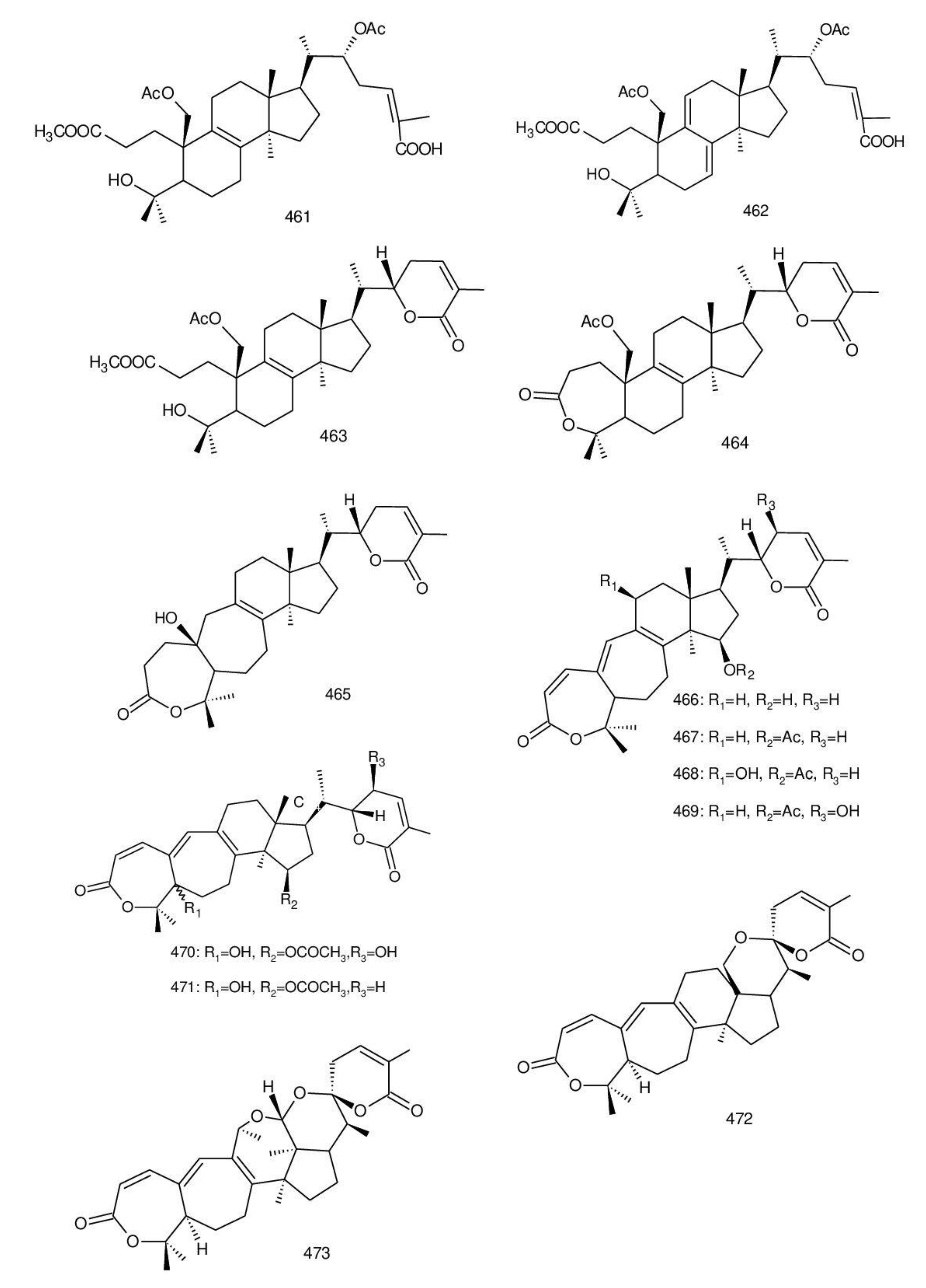
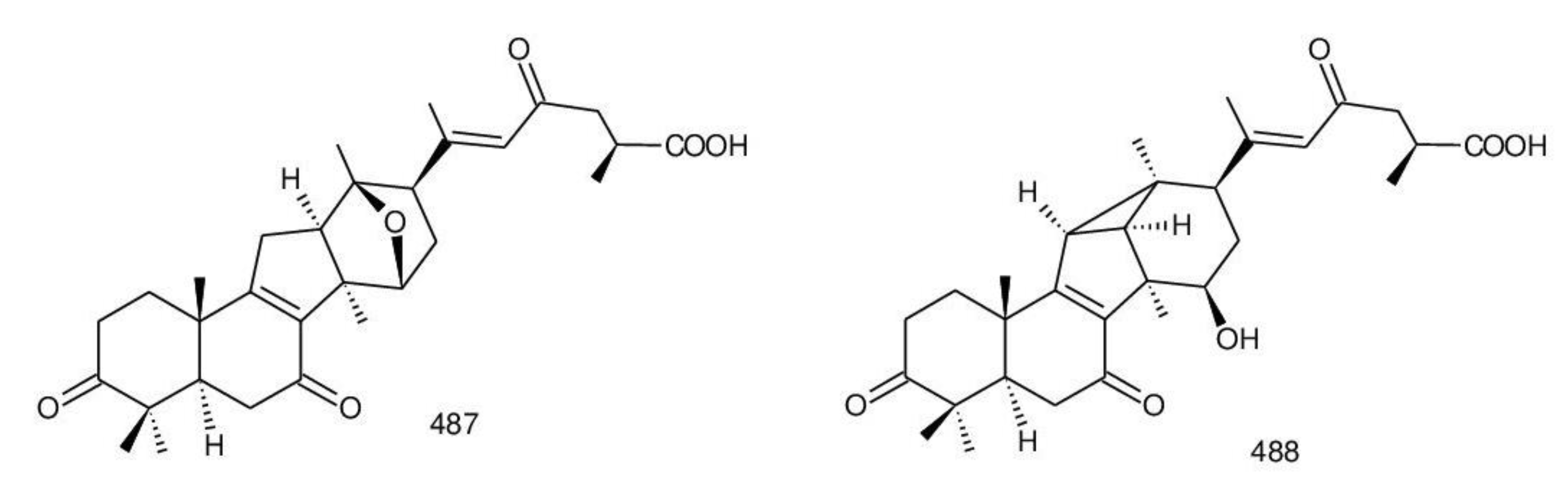
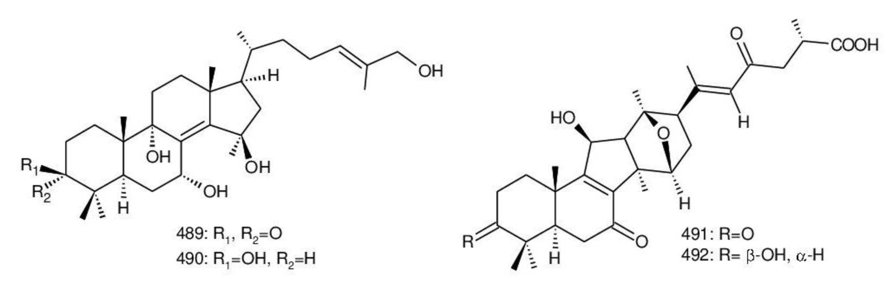
8,9-dihydro-triterpenoids
8,9-epoxy-triterpenoids
7(8),9(11)-diene-triterpenoids
| No. | Trivial Names | Bioactivities (IC50/MIC or ED50) | Sources Ganoderma Species | References |
|---|---|---|---|---|
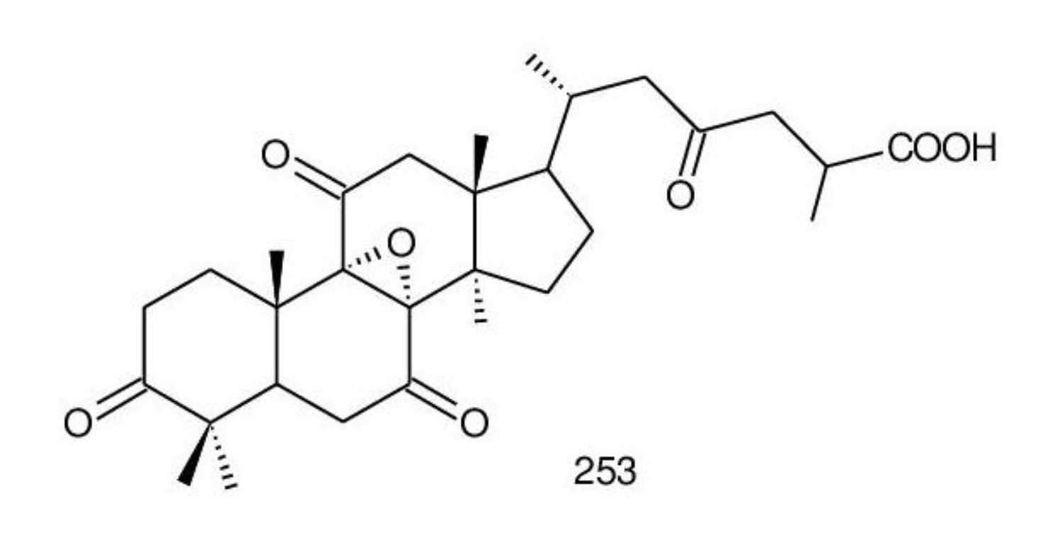
| 254. | Ganoderic acid Y | Mitogenesis, inhibition (0.180 mM), cholesterol production inhibition (1.40 µM), HMG-CoA reductase inhibition (8.60 µM), AChE inhibition (21.1 µM) | G. lucidum | [100,253] |
| 255. | Ganoderic acid X | DNA Topoisomerase I/II inhibition | G. lucidum | [100,260] |
| 256. | Ganodermic acid R | - | G. lucidum | [261,262] |
| 257. | Ganodermic acid S | Induces aggregation of human platelet, inhibition of human platelet function, inhibits thromboxane A2-dependent pathway in human platelets response to collagen, differential effect on the thromboxane A2-signaling pathways in human platelets, potentiation on prostaglandin E1-induced cyclic AMP elevation in human platelets | G. lucidum | [262,263] |
| 258. | Ganodermic acid T-O | - | G. lucidum | [264] |
| 259. | Ganodermic acid T-Q | - | G. lucidum | [264] |
| 260. | Ganoderic acid Jc | Cytotoxicity against HL-60 cells (8.30 μM) | G. sinense | [198] |
| 261. | Ganoderic acid T | Anti-tumor (induce P53) | G. lucidum | [247,265] |
| 262. | Ganoderic acid R | Strongly anti-hepatotoxic, multidrug resistance tumor cell line (κB-A1/Dox) and a sensitive tumor cell line (κB-A1) | G. lucidum | [247,266] |
| 263. | Ganoderic acid Mf | - | G. lucidum | [194] |
| 264. | Ganoderic acid P | - | G. lucidum | [223] |
| 265. | Ganoderic acid Q | - | G. lucidum | [223] |
| 266. | 3α,15α,22α-Trihydroxylanosta-7,9(11),24-trien-26-oic acid | - | G. lucidum | [230] |
| 267. | 3β,15α,22β-Trihydroxylanosta-7,9(11),24-trien- 26-oic acid | - | G. lucidum | [230] |
| 268. | 3β,15α-Diacetoxy-22α-hydroxylanosta-7,9(11),24-trien-26-oic acid | - | G. lucidum | [230] |
| 269. | 3α,15α-Diacetoxy-22α-hydroxylanosta-7,9(11),24-trien-26-oic acid | - | G. lucidum | [230] |
| 270. | 22β-Acetoxy-3α,15α-dihydroxylanosta-7,9(11),24-trien-26-oic acid | - | G. lucidum | [230] |
| 271. | 22β-Acetoxy-3β,15α-dihydroxylanosta-7,9(11),24-trien-26-oic acid | - | G. lucidum | [230] |
| 272. | Ganorbiformin G | Cytotoxicity against NCI-H187 (65.0 μM), MCF-7—NE, κB (65.0 μM), Vero (35.0 μM), antimalarial—NE, anti-TB—NE | G. orbiforme | [36] |
| 273. | Ganoderic acid S | Cytotoxicity against NCI-H187 (39 μM), MCF-7—NE, κB (53.0 μM), Vero—NE, antimalarial—NE, anti-TB—NE, Strongly anti-hepatotoxic | G. lucidum, G. orbiforme | [36,247] |
| 274. | Ganodermic acid P2 | - | G. lucidum | [261] |
| 275. | Ganoderic acid SZ | - | G. lucidum | [246] |
| 276. | 26,27-Dihydroxy-5α-lanosta-7,9(11),24-triene-3,22-dione | Induced NAD(P)H:quinone oxidoreductase (QR) in cultured hepalcic7 murine hepatoma cells (20.0 μg/mL) | G. lucidum | [267] |
| 277. | 26-Hydroxy-5α-lanosta-7,9(11),24-triene-3,22-dione | Induced NAD(P)H:quinone oxidoreductase (QR) in cultured hepalcic7 murine hepatoma cells (3 μg/mL) | G. lucidum | [267] |
| 278. | 3α,16α-Dihydroxylanosta-7,9(11),24-trien-21-oic acid | Cytotoxicity—NE | G. applanatum | [229] |
| 279. | 3α,16α,26-Trihydroxylanosta-7,9(11),24-trien-21-oic acid | Cytotoxicity—NE | G. applanatum | [229] |
| 280. | 16α-Hydroxy-3-oxolanosta-7,9(11),24-trien-21-oic acid | Cytotoxicity against P388 murine leukemia cells (111 μg/mL) | G. applanatum | [229] |
| 281. | Ganodermenonol | Cytotoxicity against LLC—NE, T-47D (4.8 μg/mL), S-180 (10.0 μg/mL), Meth-A (2.8 μg/mL) | G. lucidum | [214,268] |
| 282. | Ganodermadiol | Cytotoxicity against LLC—NE, T- 47D—NE, S-180—NE, Meth-A—10.3 μg/mL, protects Vero cells against HSV type 1 infection (ED50 = 0.068 mmol/L), protects MDCK cells against influenza virus type A infection (ED50 > 0.22 mmol/L) | G. lucidum, G. pfeifferi | [204,268] |
| 283. | Ganodermatriol | Inhibition of 5α-reductase activity (%) at 667 μM (39%) | G. lucidum | [156,268] |
| 284. | Ganoderiol F | Inhibition on cell growth in thepresence of testosterone or DHT, potential CDK4/CDK6 inhibitor for breast cancer therapy; anticomplementary activity (4.8 μM), inhibition of 5α-reductase—NE | G. lucidum, G. leucocontextum | [156,257, 269,270] |
| 285. | Ganodermatetraol | Induction ability of hPXR-mediated CYP3A4 expression | G. sinense | [198] |
| 286. | 5α-Lanosta-7,9(11),24-triene- 15α-26-dihydroxy-3-one | Induces apoptosis in human promyelocytic leukemia HL-60 cells, inhibition of 5α-reductase activity (41.9 μM), antiviral (enterovirus 71) | G. concinna, G. lucidum | [156,253, 271] |
| 287. | 5α-Lanosta-7,9(11),24-triene- 3β-hydroxy-26-al | Induces apoptosis in human promyelocytic leukemia HL-60 cells | G. concinna | [271] |
| 288. | Ganoderiol A | Suppresses migration and adhesion of MDA-MB-231 cells and minimal impact on cell invasion in MDA-MB-231 cells, 5α-reductase inhibitory activity—NE | G. lucidum | [156,272, 273] |
| 289. | Ganoderiol B | Moderately active inhibitor against HIV-1 PR (0.17 mM), inhibition of 5α-reductase—NE | G. lucidum | [156,168, 272] |
| 290. | 3β,24,26-Triacetoxy-5α-lanosta-7,9(11)-dien-25-ol | - | G. sinense | [274] |
| 291. | Ganodermanontriol | Anti-HIV-1 agent—(7.8 mg/mL), anti HIV protease; anticomplement activities, inhibition of 5α-reductase—NE | G. lucidum | [156,168, 248] |
| 292. | Lanosta-7,9(11),24-trien-3β,21-diol | - | G. australe | [275] |
| 293. | Ganodercochlearin C | - | G. cochlear | [57] |
| 294. | Ganodercochlearin A | - | G. cochlear | [57] |
| 295. | Ganodercochlearin B | - | G. cochlear | [57] |
| 296. | Ganodecochlearin B diacetate | - | G. cochlear | [57] |
| 297. | 3β,22S-Dihydroxylanosta-7,9(11),24-triene | - | G. cochlear | [57] |
| 298. | Applanoxidic acid A | Inhibitory effect on EBV-EA activation, antifungal activity against the growth of Microsporum cannis (1000 μg/mL), Trichophyton mentagrophytes (500 μg/mL), cytotoxicity against HL-60 cell line (132.0 μM) | G. applanatum, G. australe, G. annulare | [48,251, 252,276] |
| 299. | Applanoxidic acid B | Remarkable inhibitory effect on EBV-EA activation | G. applanatum | [251,252] |
| 300. | Applanoxidic acid E | Inhibitory effect on EBV-EA activation | G. applanatum | [252] |
| 301. | Applanoxidic acid F | Inhibitory effect on EBV-EA activation, antifungal activity against the growth of Microsporum cannis (1000 μg/mL), Trichophyton mentagrophytes (1000 μg/mL), cytotoxicity against HL-60 cells (315.0 μM) | G. applanatum, G. annular, G. australe | [48,252, 276] |
| 302. | Applanoxidic acid C | Inhibitory effect on EBV-EA activation, antifungal activity against the growth of Microsporum cannis (1000 μg/mL), Trichophyton mentagrophytes (1000 μg/mL), cytotoxicity against HL-60 cells (334.0 μM) | G. applanatum, G. annulare, G. australe | [48,251,252,276] |
| 303. | Applanoxidic acid D | Inhibitory effect on EBV-EA activation | G. applanatum | [251,252] |
| 304. | Applanoxidic acid G | Inhibitory effect on EBV-EA activation, antifungal—NE, antiviral—NE, cytotoxicity inhibits the viability and growth of the HL-60 cells (404.0 μM) | G.applanatum, G. annulare, G. pfeifferi, G. australe | [48,204, 252,276] |
| 305. | Applanoxidic acid H | Inhibitory effect on EBV-EA activation | G. applanatum | [252] |
| 306. | Ganoderic acid Jb | Inhibitory activities against the HMG-CoA reductase and acyl CoA acyltransferase | G. lucidum (fruit bodies) | [65,219] |
| 307. | 3α,16α-Dihydroxylanosta-7,9(11),24-trien-21-oic acid | Cytotoxicity against the P388 murine leukemia cell line—NE | G. applanatum (fruit bodies) | [137] |
| 308. | 3α,16α,26-Trihydroxylanosta-7,9(11),24-trien-21-oic acid | Cytotoxicity against the P388 murine leukemia cell line—NE | G. applanatum (fruit bodies) | [137] |
| 309. | Ganoderic acid Me | - | G. lucidum (cultured mycelial mat) | [194] |
| 310. | 26,27-Dihydroxylanosta-7,9(11),24-trien-3,16-dione | - | G. carnosum (fruit bodies) | [55] |
| 311. | Ganoderol B | - | G. lucidum | [277] |
| 312. | Ganoderic acid Mk | - | G. lucidum (mycelial mat) | [190] |
| 313. | Lanosta-7,9(11),24-trien-3β,15α, 22β-triacetoxy-26-oic acid | - | G. lucidum | [278] |
| 314. | Lanosta-7,9(11),24-trien-15α-acetoxy-3α-hydroxy-23-oxo-26-oic acid | - | G. lucidum | [278] |
| 315. | Lanosta-7,9(11),24-trien-3α,l5α-diacetoxy-23-oxo-26-oic acid | - | G. lucidum | [278] |
| 316. | Lanosta-7,9(11),24-trien-3α,15α-hydroxy-23-oxo-26-oic acid | - | G. lucidum | [32] |
| 317. | Lanosta-7,9(11),24-trien-3α-acetoxy-15α,22β-dihydroxy-26-oic acid | - | G. lucidum | [278] |
| 318. | Ganodermic acid T-N | - | G. lucidum (mycelia) | [264] |
| 319. | Compound 10 | - | G. orbiforme | [36] |
| 320. | 5α-Lanosta-7,9(11),24-triene- 3β-hydroxy-26-al | Concentration of 30 μM induced apoptosis in 15% of the human promyelocytic leukemia HL-60 cell (after treatment for 24 h) | G. concinna | [271] |
| 321. | Ganodermic acid P1 | - | G. lucidum (mycelia) | [261] |
| 322. | Lanosta-7,9(11),24-trien-3β,15α, 22-triacetoxy-26-oic acid | Concentration of 10 µg/mL showed toxicity towards the brine shrimp larvae (after treatment for 24 h) | G. amboinense (fruit bodies) | [153] |
| 323. | Lucialdehyde A | Cytotoxicity against Meth-A (10.4 µg/mL) | G. lucidum (fruit bodies) | [214] |
| 324. | Ganoderiol A triacetate | - | G. sinense (fruit bodies) | [274] |
| 325. | Ganoderal A | ACE inhibitory activity (10−5 M) | G. lucidum | [277] |
| 326. | Ganoderol A | ACE inhibitory activity (10−5M) | G. lucidum | [277] |
| 327. | Lucidumol B | HIV-I Protease inhibitory activity (50 µM) | G. lucidum (spores) | [221] |
| 328. | Ganodermanontiol | - | G. lucidum (spores) | [279] |
| 329. | Ganodermanondiol | - | G. lucidum (fruit bodies) | [280] |
| 330. | Ganoderic acid TR | Inhibitory effect on 5α-reductase (8.6 µM) | G. lucidum | [156] |
| 331. | Ganodermic acid Ja | - | G. lucidum (mycelia) | [261] |
| 332. | Ganodermic acid Jb | - | G. lucidum (mycelia) | [261] |
| 333. | 15α-Hydroxy-3-oxo-5α-lanosta-7,9,24(E)-triene-26-oic acid | Cytotoxicity against human HeLa cervical cancer cell lines (58 µM) | G. lucidum | [215] |
| 334. | 15α,26-Dihydroxy-5α-lanosta-7,9,24(E)-trien-3-one | Cytotoxicity against human HeLa cervical cancer cell lines (1 µM) | G. lucidum | [215] |
| 335. | 3β-Hydroxy-5α-lanosta-7,9,24(E)-trien-26-oic acid | Cytotoxicity against human HeLa cervical cancer cell lines (59 µM) | G. lucidum | [215] |
| 336. | Epoxyganoderiol B | - | G. lucidum | [222] |
| 337. | Epoxyganoderiol C | - | G. lucidum | [222] |
| 338. | Ganoapplic acid F | Inhibitory effects for the proliferation of hepatic stellate cells (HSCs) induced through transforming growth factor-β1 (TGF-β1) in vitro | G. applanatum | [50,51] |
| 339. | Ganoderic aldehyde TR | - | G. lucidum | [65] |
| 340. | Ganoderic acid TR1 | - | G. lucidum | [65] |
| 341. | 23-Hydroxy ganoderic acid S | - | G. lucidum | [65] |
| 342. | Ganoellipsic acid A | - | G. ellipsoideum | [129] |
| 343. | Ganoellipsic acid B | - | G. ellipsoideum | [129] |
| 344. | Ganoellipsic acid C | - | G. ellipsoideum | [129] |
| 345. | 26-Methy-15α,22β-diacetoxy-7,9 (11),24-trien-26-oic ester | Moderate cytotoxic activity against the human cancer cell line NCI-H1650 (IC50 = 22.3 μM) | G. capense | [238] |
| 346. | Methyl gibbosate L | Anti-adipogenesis activity—NE | G. applanatum | [41] |
| 347. | Methyl ganoapplate F | Anti-adipogenesis activity—NE | G. applanatum | [41] |
| 348. | Ganodeweberiol A | Cytotoxicity against HeLa cell line (IC50 = 31.6 μM) | G. weberianum | [77] |
| 349. | Ganodeweberiol B | Significant α-glucosidase inhibitory activity | G. weberianum | [77] |
| 350. | Ganodeweberiol C | Inhibits glucagon-inducedhepatic glucose production, inhibits hepatic glucose output through suppression hepatic cAMP accumulation, cytotoxicity against HeLa cell line (IC50 = 17.0 μM) | G. weberianum | [77] |
| 351. | Ganodeweberiol D | - | G. weberianum | [77] |
| 352. | Ganodeweberiol E | - | G. weberianum | [77] |
| 353. | Ganodeweberiol F | Inhibits glucagon-inducedhepatic glucose production, inhibits hepatic glucose output through suppression hepatic cAMP accumulation | G. weberianum | [77] |
Triterpenoid Saponins
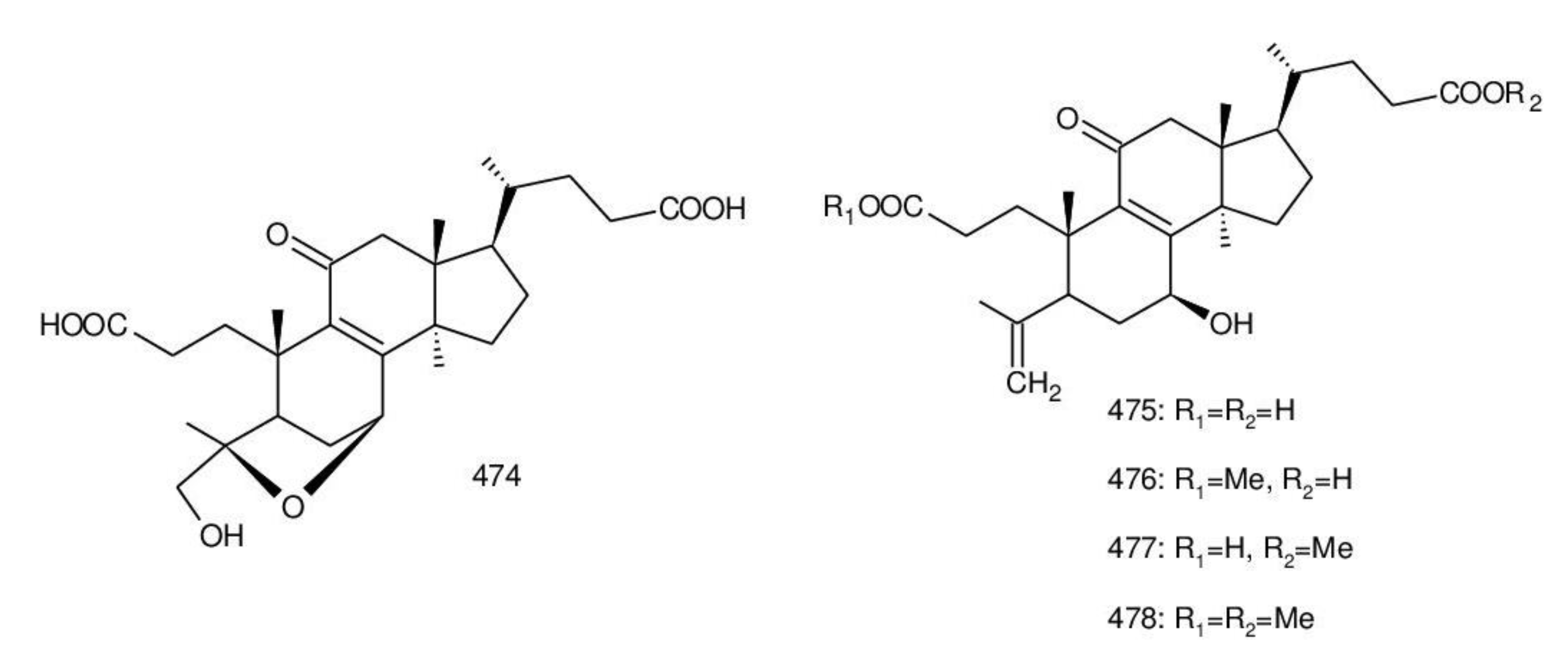
2.2.2. C29 Triterpenoids
2.2.3. C27 Triterpenoids
2.2.4. C25 Triterpenoids
2.2.5. C24 Triterpenoids
2.2.6. C31 Triterpenoids
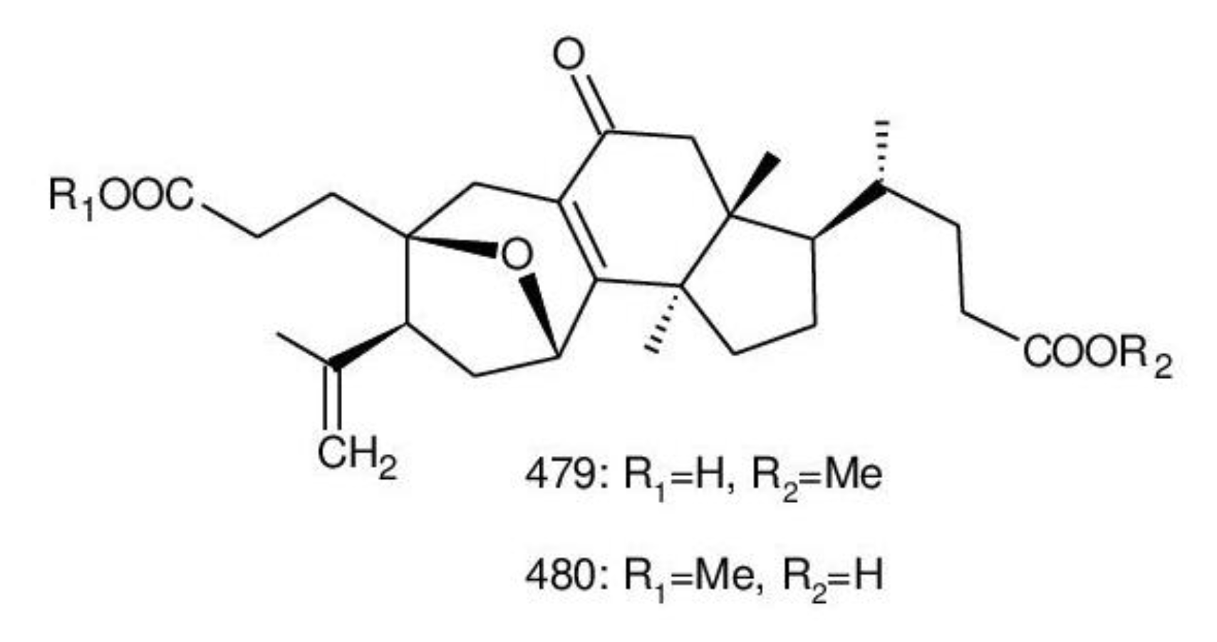
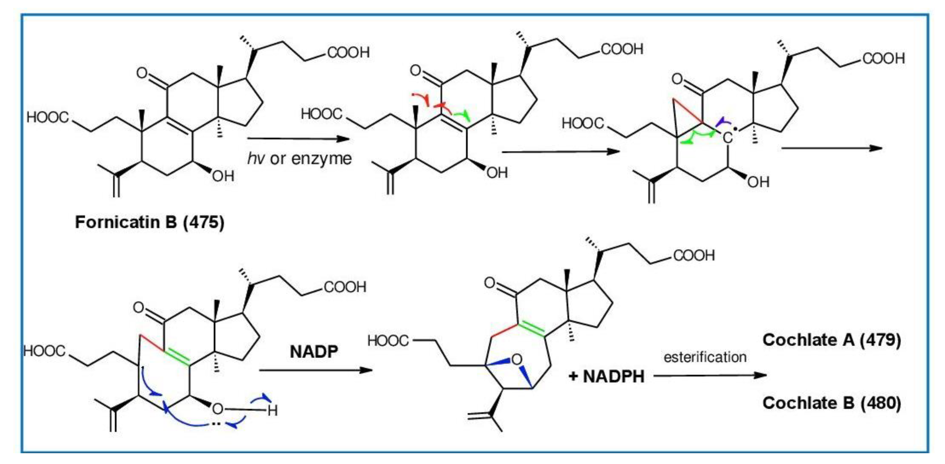
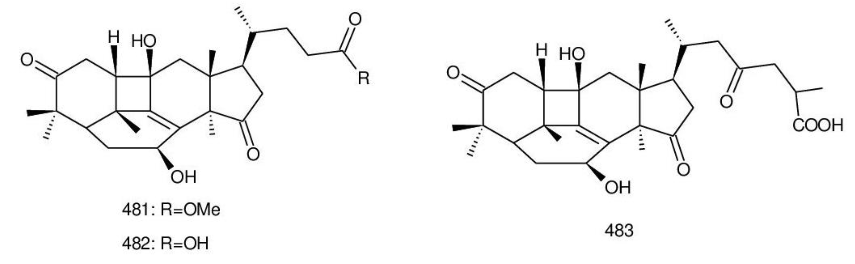
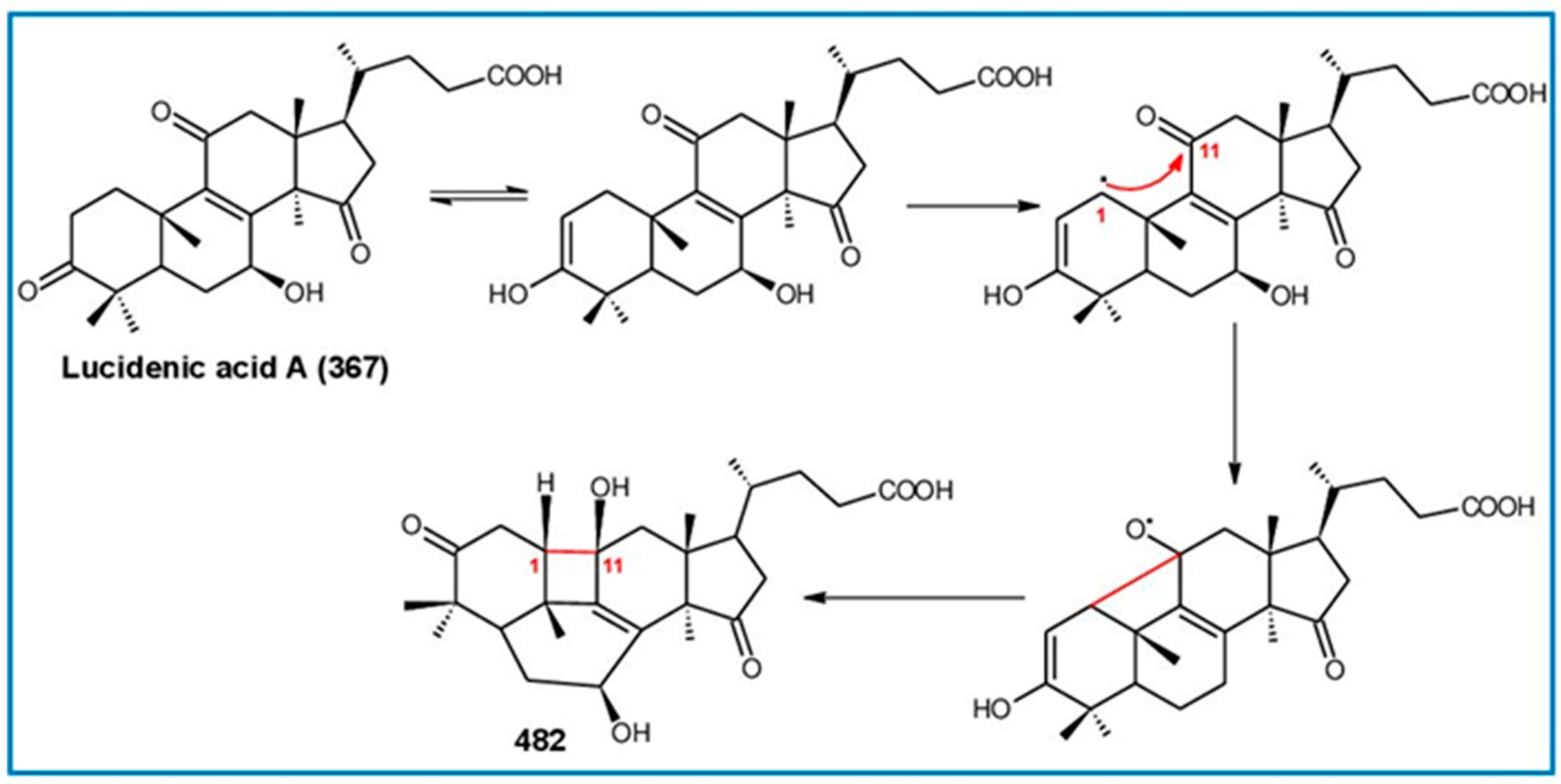
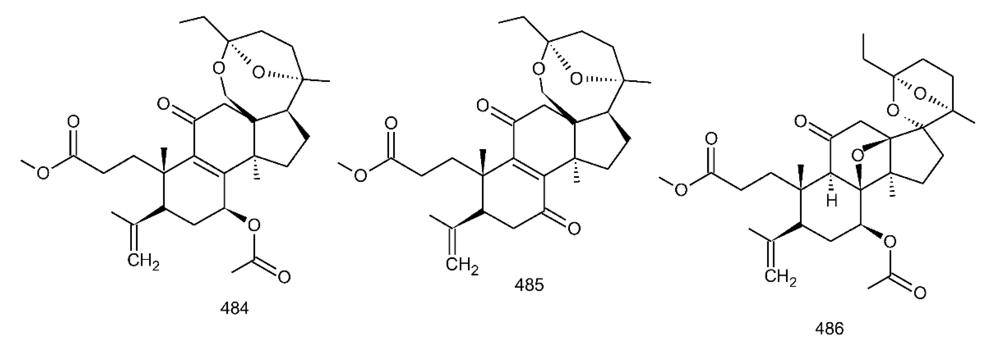
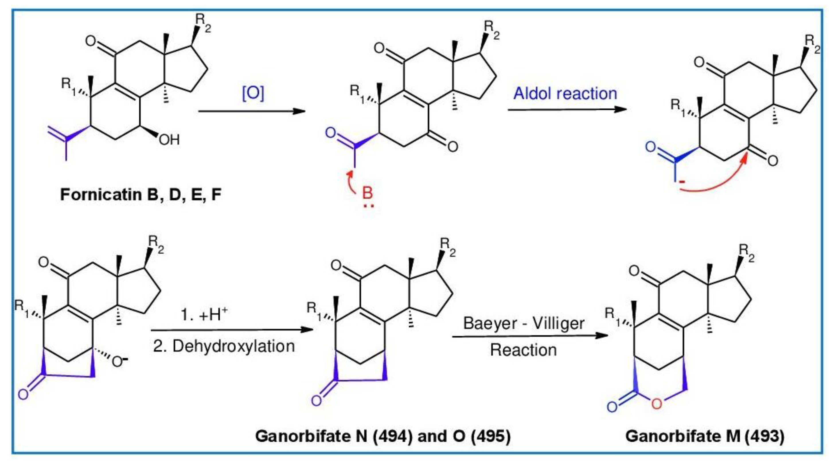
2.2.7. Rearranged Novel Triterpenoids
3. Conclusions
Author Contributions
Funding
Institutional Review Board Statement
Informed Consent Statement
Data Availability Statement
Acknowledgments
Conflicts of Interest
Abbreviations
| AChE | Acetylcholinesterase |
| ALT | Alanine aminotransferase |
| AST | Aspartate aminotransferase |
| EV71 | Enterovirus 71 |
| FAAH | Fatty Acid Amide Hydrolase |
| FPPs | Farnesyl diphosphate synthase |
| GA | Ganoderic Acid |
| GAS | Ganodermic acid S |
| GTs | Ganoderma Triterpenoids |
| HSCs | Hepatic stellate cells |
| HMGR | 3-Hydroxy-3-methylglutaryl-CoA reductase |
| HMGS | 3-Hydroxy-3-methylglutaryl-CoA synthase |
| IC50 | Half-maximal inhibitory concentration |
| ID50 | Inhibitory dose-50 |
| LS | Lanosterol synthase |
| MIC | Minimum Inhibitory Concentration |
| MVA | Mevalonate pathway |
| MVD | Phosphomevalonate decarboxylase |
| NOESY | Nuclear Overhauser Effect Spectroscopy |
| OSC | 2,3-Oxidosqualene lanosterol cyclase |
| PDC | Pyridinium dichromate |
| SA | Salicylic Acid |
| SQS | Squalene synthase |
| TPA | 12-O-Tetradecanoylphorbol 13-acetate |
| TGF-β1 | Transforming growth factor-β1 |
| uPA | Urokinase-plasminogen activator |
References
- Karsten, P.A. Enumeratio boletinearum et poly-porearum fennicarum. Systemate novo dispositarum. Rev. Mycol. 1881, 3, 16–19. [Google Scholar]
- Upadhyay, M.; Shrivastava, B.; Jain, A.; Kidwai, M.; Kumar, S.; Gomes, J.; Goswami, D.G.; Panda, A.K.; Kuhad, R.C. Production of ganoderic acid by Ganoderma lucidum RCKB-2010 and its therapeutic potential. Ann. Microbiol. 2014, 64, 839–846. [Google Scholar] [CrossRef]
- He, M.-Q.; Zhao, R.-L.; Hyde, K.D.; Begerow, D.; Kemler, M.; Yurkov, A.; McKenzie, E.H.; Raspé, O.; Kakishima, M.; Sánchez-Ramírez, S.; et al. Notes, outline and divergence times of basidiomycota. Fungal Divers. 2019, 99, 105–367. [Google Scholar] [CrossRef] [Green Version]
- Badalyan, S.M.; Gharibyan, N.G.; Iotti, M.; Zambonelli, A. Morphological and ecological screening of different collections of medicinal white-rot bracket fungus Ganoderma adspersum (Schulzer) Donk (Agaricomycetes, Polyporales). Ital. J. Mycol. 2019, 48, 1–15. [Google Scholar]
- Gryzenhout, M.; Ghosh, S.; Tchotet Tchoumi, J.M.; Vermeulen, M.; Kinge, T.R. Ganoderma: Diversity, ecological significances, and potential applications in industry and allied sectors. In Industrially Important Fungi for Sustainable Development, Fungal Biology; Abdel-Azeem, A.M., Yadav, A.N., Yadav, N., Usmani, Z., Eds.; Springer Nature: Cham, Switzerland, 2021; Volume 1, pp. 295–334. [Google Scholar] [CrossRef]
- Moncalvo, J.M.; Ryvarden, L. A Nomenclatural Study of the Ganodermataceae Donk, Synopsis Fungorum 11; Fungiflora: Oslo, Norway, 1997; pp. 1–114. [Google Scholar]
- Ryvarden, L.; Johansen, I. A Preliminary Polypore Flora of East Africa; Fungiflora: Oslo, Norway, 1980; pp. 1–636. [Google Scholar]
- Gilbertson, R.L.; Ryvarden, L. North American polypores. Abortiporus-Lindtneria; Fungiflora A/S: Oslo, Norway, 1986; Volume 1, pp. 1–433. [Google Scholar]
- Ryvarden, L.; Gilbertson, R.L. European Polypores 1, Synop Fungorum 6; Fungiflora A/S: Oslo, Norway, 1993; pp. 1–387. [Google Scholar]
- Quanten, E. The Polypores (Polyporaceae s.l.) of Papua New Guinea: A Preliminary Conspectus, Opera Botanica Belgica 11; National Botanic Garden of Belgium: Meise, Belgium, 1997; pp. 1–352. [Google Scholar]
- Nunez, M.; Ryvarden, L. East Asian Polypores 1. Ganodermataceae and Hymenochaetaceae, Synop Fungorum 13; Fungiflora A/S: Oslo, Norway, 2000; pp. 1–168. [Google Scholar]
- Welti, S.; Courtecuisse, R. The Ganodermataceae in the French West Indies (Guadeloupe and Martinique). Fungal Divers. 2010, 43, 103–126. [Google Scholar] [CrossRef]
- Arenas, M.C.; Tadiosa, E.R.; Reyes, R.G. Taxonomic inventory based on physical distribution of macrofungi in Mt. Maculot, Cuenca, Batangas, Philippines. Int. J. Biol. Pharm. Allied Sci. 2018, 7, 672–687. [Google Scholar] [CrossRef]
- Luangharn, T.; Karunarathna, S.C.; Dutta, A.K.; Paloi, S.; Promputtha, I.; Hyde, K.D.; Xu, J.; Mortimer, P.E. Ganoderma (Ganodermataceae, Basidiomycota) species from the Greater Mekong Subregion. J. Fungi 2021, 7, 819. [Google Scholar] [CrossRef]
- Morera, G.; Lupo, S.; Alaniz, S.; Robledo, G. Diversity of the Ganoderma species in Uruguay. Neotrop. Biodivers. 2021, 7, 570–585. [Google Scholar] [CrossRef]
- Runnel, K.; Miettinen, O.; Lõhmus, A. Polypore fungi as a flagship group to indicate changes in biodiversity—A test case from Estonia. IMA Fungus 2021, 12, 2. [Google Scholar] [CrossRef]
- Ueitele, I.S.E.; Horn, L.N.; Kadhila, N.P. Ganoderma research activities and development in Namibia. AJOM 2021, 4, 29–39. [Google Scholar]
- He, J.; Han, X.; Luo, Z.-L.; Li, E.-X.; Tang, S.-M.; Luo, H.-M.; Niu, K.-Y.; Su, X.-J.; Li, S.-H. Species diversity of Ganoderma (Ganodermataceae, Polyporales) with three new species and a key to Ganoderma in Yunnan province, China. Front. Microbiol. 2022, 13, 1035434. [Google Scholar] [CrossRef]
- Ying, C.-C.; Wang, Y.-C.; Tang, H. Icones of Medicinal Fungi from China; Science Press: Beijing, China, 1987; p. 575. [Google Scholar]
- Dai, Y.C.; Yang, Z.L.; Cui, B.K.; Yu, C.J.; Zhou, L.W. Species diversity and utilization of medicinal mushrooms and fungi in China (review). Int. J. Med. Mushrooms 2009, 3, 287–302. [Google Scholar] [CrossRef]
- Wu, F.; Zhou, L.-W.; Yang, Z.-L.; Bau, T.; Li, T.-H.; Dai, Y.-C. Resource diversity of Chinese macrofungi: Edible, medicinal and poisonous species. Fungal Divers. 2019, 98, 1–76. [Google Scholar] [CrossRef]
- El Sheikha, A.F.E. Nutritional profile and health benefits of Ganoderma lucidum “Lingzhi, Reishi, or Mannentake” as functional foods: Current scenario and future perspectives. Foods 2022, 11, 1030. [Google Scholar] [CrossRef]
- Li, Y.; Zhu, Z.; Yao, W.; Chen, R. Research status and progress of the triterpenoids in Ganoderma lucidum. Med. Plant 2012, 3, 75–81. [Google Scholar]
- Liu, H.; Guo, L.-J.; Li, S.-L.; Fan, L. Ganoderma shanxiense, a new species from northern China based on morphological and molecular evidence. Phytotaxa 2019, 406, 129–136. [Google Scholar] [CrossRef]
- Li, L.F.; Liu, H.B.; Zhang, Q.W.; Li, Z.P.; Wong, T.L.; Fung, H.Y.; Zhang, J.X.; Bai, S.P.; Lu, A.P.; Han, Q.B. Comprehensive comparison of polysaccharides from Ganoderma lucidum and G. sinense: Chemical, anti-tumor, immunomodulating and gut-microbiota modulatory properties. Sci. Rep. 2018, 8, 6172. [Google Scholar] [CrossRef] [Green Version]
- Ngai, P.H.K.; Ng, T.B. A mushroom (Ganoderma capense) lectin with spectacular thermostability, potent mitogenic activity on splenocytes, and antiproliferative activity toward tumor cells. Biochem. Bioph. Res. Commun. 2004, 314, 988–993. [Google Scholar] [CrossRef]
- Lee, S.; Shim, S.H.; Kim, J.S.; Shin, K.H.; Kang, S.S. Aldose reductase inhibitors from the fruiting bodies of Ganoderma applanatum. Biol. Pharm. Bull. 2005, 28, 1103–1105. [Google Scholar] [CrossRef] [Green Version]
- Gurunathan, S.; Raman, J.; Malek, S.N.A.; John, P.A.; Vikineswary, S. Green synthesis of silver nanoparticles using Ganoderma neo-japonicum Imazeki: A potential cytotoxic agent against breast cancer cells. Int. J. Nanomed. 2013, 8, 4399–4413. [Google Scholar]
- Wang, L.; Li, J.-Q.; Zhang, J.; Li, Z.-M.; Liu, H.-G.; Wang, Y.-Z. Traditional uses, chemical components and pharmacological activities of the genus Ganoderma P. Karst: A review. RSC Adv. 2020, 10, 42084–42097. [Google Scholar] [CrossRef] [PubMed]
- Shankar, A.; Sharma, K.K. Fungal secondary metabolites in food and pharmaceuticals in the era of multiomics. Appl. Microbiol. Biotechnol. 2022, 106, 3465–3488. [Google Scholar] [CrossRef] [PubMed]
- Chen, Y.; Lan, P. Total syntheses and biological evaluation of the Ganoderma lucidum alkaloids lucidimines B and C. ACS Omega 2018, 3, 3471–3481. [Google Scholar] [CrossRef] [PubMed] [Green Version]
- Xia, Q.; Zhang, H.; Sun, X.; Zhao, H.; Wu, L.; Zhu, D.; Yang, G.; Shao, Y.; Zhang, X.; Mao, X.; et al. A comprehensive review of the structure elucidation and biological activity of triterpenoids from Ganoderma spp. Molecules 2014, 19, 17478–17535. [Google Scholar] [CrossRef]
- Baby, S.; Johnson, A.J.; Govindan, B. Secondary metabolites from Ganoderma. Phytochemistry 2015, 114, 66–101. [Google Scholar] [CrossRef]
- Wang, K.; Bao, L.; Xiong, W.; Ma, K.; Han, J.; Wang, W.; Yin, W.; Liu, H. Lanostane triterpenes from the Tibetan medicinal mushroom Ganoderma leucocontextum and their inhibitory effects on HMG-CoA reductase and α-glucosidase. J. Nat. Prod. 2015, 78, 1977–1989. [Google Scholar] [CrossRef]
- Angulo-Sanchez, L.T.; López-Peña, D.; Torres-Moreno, H.; Gutiérrez, A.; Gaitán-Hernández, R.; Esquedaa, M. Biosynthesis, gene expression, and pharmacological properties of triterpenoids of Ganoderma species (Agaricomycetes): A review. Int. J. Med. Mushrooms 2022, 24, 1–17. [Google Scholar] [CrossRef]
- Isaka, M.; Chinthanom, P.; Kongthong, S.; Srichomthong, K.; Choeyklin, R. Lanostane triterpenes from cultures of the basidiomycete Ganoderma orbiforme BCC 22324. Phytochemistry 2013, 87, 133–139. [Google Scholar] [CrossRef]
- Martínez-Montemayor, M.M.; Ling, T.; Suárez-Arroyo, I.J.; Ortiz-Soto, G.; Santiago-Negrón, C.L.; Lacourt-Ventura, M.Y.; Valentín-Acevedo, A.; Lang, W.H.; Rivas, F. Identification of biologically active Ganoderma lucidum compounds and synthesis of improved derivatives that confer anti-cancer activities in vitro. Front. Pharmacol. 2019, 10, 115. [Google Scholar] [CrossRef] [Green Version]
- Shim, S.H.; Ryu, J.; Kim, J.S.; Kang, S.S.; Xu, Y.; Jung, S.H.; Lee, Y.S.; Lee, S.; Shin, K.H. New lanostane-type triterpenoids from Ganoderma applanatum. J. Nat. Prod. 2004, 67, 1110–1113. [Google Scholar] [CrossRef]
- Paterson, R.R. Ganoderma—A therapeutic fungal biofactory. Phytochemistry 2006, 67, 1985–2001. [Google Scholar] [CrossRef] [Green Version]
- Cilerdzic, J.; Vukojevic, J.; Stajic, M.; Stanojkovic, T.; Glamoclija, J. Biological activity of Ganoderma lucidum basidiocarps cultivated on alternative and commercial substrate. J. Ethnopharmacol. 2014, 155, 312–329. [Google Scholar] [CrossRef]
- Peng, X.R.; Li, L.; Dong, J.R.; Lu, S.Y.; Lu, J.; Li, X.N.; Zhou, L.; Qiu, M.H. Lanostane-type triterpenoids from the fruiting bodies of Ganoderma applanatum. Phytochemistry 2019, 157, 103–110. [Google Scholar] [CrossRef]
- Shi, J.-X.; Chen, G.-Y.; Sun, Q.; Meng, S.-Y.; Chi, W.-Q. Antimicrobial lanostane triterpenoids from the fruiting bodies of Ganoderma applanatum. J. Asian Nat. Prod. Res. 2021, 157, 1001–1007. [Google Scholar] [CrossRef]
- Muhsin, T.M.; Al-Duboon, A.-H.A.; Khalaf, K.T. Bioactive compounds from a polypore fungus Ganoderma applanatum (Pers. ex Wallr.) Pat. Jordan J. Biol. Sci. 2011, 4, 205–212. [Google Scholar]
- Kozarski, M.; Klaus, A.; Nikšić, M.; Vrvić, M.M.; Todorović, N.; Jakovljević, D.; Van Griensven, L.J.L.D. Antioxidative activities and chemical characterization of polysaccharide extracts from the widely used mushrooms Ganoderma applanatum, Ganoderma lucidum, Lentinus edodes and Trametes versicolor. J. Food Compost. Anal. 2012, 26, 144–153. [Google Scholar] [CrossRef]
- Al-Fatimi, M.; Wurster, M.; Kreisel, H.; Lindequist, U. Antimicrobial, cytotoxic and antioxidant activity of selected basidiomycetes from Yemen. Pharmazie 2005, 60, 776–780. [Google Scholar]
- Niedermeyer, T.H.; Lindequist, U.; Mentel, R.; Gordes, D.; Schmidt, E.; Thurow, K.; Lalk, M. Antiviral terpenoid constituents of Ganoderma pfeifferi. J. Nat. Prod. 2005, 68, 1728–1731. [Google Scholar] [CrossRef]
- Hsu, C.L.; Yu, Y.S.; Yen, G.C. Lucidenic acid B induces apoptosis in human leukemia cells via a mitochondria-mediated pathway. J. Agric. Food Chem. 2008, 56, 3973–3980. [Google Scholar] [CrossRef]
- Smania, E.F.A.; Delle Monache, F.; Smania, A.; Yunes, R.A.; Cuneo, R.S. Antifungal activity of sterols and triterpenes isolated from Ganoderma annulare. Fitoterapia 2003, 74, 375–377. [Google Scholar] [CrossRef]
- Rosecke, J.; Konig, W.A. Constituents of various wood-rotting basidiomycetes. Phytochemistry 2000, 54, 603–610. [Google Scholar] [CrossRef] [PubMed]
- Li, L.; Peng, X.R.; Dong, J.R.; Lu, S.Y.; Li, X.N.; Zhou, L.; Qiu, M.H. Rearranged lanostane-type triterpenoids with anti-hepatic fibrosis activities from Ganoderma applanatum. RSC Adv. 2018, 8, 31287–31295. [Google Scholar] [CrossRef] [PubMed] [Green Version]
- Peng, X.-R.; Wang, Q.; Su, H.-G.; Zhou, L.; Xiong, W.-Y.; Qiu, M.-H. Anti-adipogenic lanostane-type triterpenoids from the edible and medicinal mushroom Ganoderma applanatum. J. Fungi 2022, 8, 331. [Google Scholar] [CrossRef] [PubMed]
- Ma, K.; Ren, J.; Han, J.; Bao, L.; Li, L.; Yao, Y.; Sun, C.; Zhou, B.; Liu, H. Ganoboninketals A–C, antiplasmodial 3,4-seco-27-norlanostane triterpenes from Ganoderma boninense Pat. J. Nat. Prod. 2014, 77, 1847–1852. [Google Scholar] [CrossRef] [PubMed]
- Huang, S.-Z.; Ma, Q.-Y.; Kong, F.-D.; Guo, Z.-K.; Cai, C.-H.; Hu, L.-L.; Zhou, L.-M.; Wang, Q.; Dai, H.-F.; Mei, W.-L.; et al. Lanostane-type triterpenoids from the fruiting body of Ganoderma calidophilum. Phytochemistry 2017, 143, 104–110. [Google Scholar] [CrossRef]
- Li, N.; Yan, C.; Hua, D.; Zhang, D. Isolation, purification, and structural characterization of a novel polysaccharide from Ganoderma capense. Int. J. Biol. Macromol. 2013, 57, 285–290. [Google Scholar] [CrossRef]
- Keller, A.C.; Keller, J.; Maillard, M.P.; Hostettmann, K. A lanostane-type steroid from the fungus Ganoderma carnosum. Phytochemistry 1997, 46, 963–965. [Google Scholar] [CrossRef]
- Peng, X.-R.; Liu, J.-Q.; Wan, L.-S.; Li, X.-N.; Yan, Y.-X.; Qiu, M.-H. Four new polycyclic meroterpenoids from Ganoderma cochlear. Org. Lett. 2014, 16, 5262–5265. [Google Scholar] [CrossRef]
- Peng, X.-R.; Liu, J.-Q.; Wang, C.-F.; Li, X.-Y.; Shu, Y.; Zhou, L.; Qiu, M.-H. Hepatoprotective effects of triterpenoids from Ganoderma cochlear. J. Nat. Prod. 2014, 77, 737–743. [Google Scholar] [CrossRef]
- Yu, C.; Cao, C.Y.; Shi, P.D.; Yang, A.A.; Yang, Y.X.; Huang, D.S.; Chen, X.; Chen, Z.M.; Gao, J.M.; Yin, X. Highly oxygenated chemical constitutes and rearranged derivatives with neurotrophic activity from Ganoderma cochlear. J. Ethnopharmacol. 2022, 295, 115393. [Google Scholar] [CrossRef]
- Chen, S.-Y.; Chang, C.-L.; Chen, T.-H.; Chang, Y.-W.; Lin, S.-B. Colossolactone H, a new Ganoderma triterpenoid exhibits cytotoxicity and potentiates drug efficacy of gefitinib in lung cancer. Fitoterapia 2016, 114, 81–91. [Google Scholar] [CrossRef]
- Jo, W.-S.; Park, H.-N.; Cho, D.-H.; Yoo, Y.-B.; Park, S.-C. Detection of extracellular enzyme activities in Ganoderma neo-japonicum. Mycobiology 2011, 39, 118–120. [Google Scholar] [CrossRef] [Green Version]
- Qiao, Y.; Zhang, X.-M.; Dong, X.-C.; Qiu, M.-H. A new 18(13 → 12β)-abeo-lanostadiene triterpenoid from Ganoderma fornicatum. Helv. Chim. Acta 2006, 89, 1038–1041. [Google Scholar] [CrossRef]
- Peng, X.; Liu, J.; Xia, J.; Wang, C.; Li, X.; Deng, Y.; Bao, N.; Zhang, Z.; Qiu, M. Lanostane triterpenoids from Ganoderma hainanense J. D. Zhao. Phytochemistry 2015, 114, 137–145. [Google Scholar] [CrossRef]
- Li, T.-H.; Hu, H.-P.; Deng, W.-Q.; Wu, S.-H.; Wang, D.-M.; Tsering, T. Ganoderma leucocontextum, a new member of the G. lucidum complex from southwestern China. Mycoscience 2015, 56, 81–85. [Google Scholar] [CrossRef]
- Wei, J.-C.; Wang, Y.-X.; Dai, R.; Tian, X.-G.; Sun, C.-P.; Ma, X.-C.; Jia, J.-M.; Zhang, B.-J.; Huo, X.-K.; Wang, C. C27-Nor lanostane triterpenoids of the fungus Ganoderma lucidum and their inhibitory effects on acetylcholinesterase. Phytochem. Lett. 2017, 20, 263–268. [Google Scholar] [CrossRef]
- Yang, Y.; Zhang, H.; Zuo, J.; Gong, X.; Yi, F.; Zhu, W.; Li, L. Advances in research on the active constituents and physiological effects of Ganoderma lucidum. Biomed. Dermatol. 2019, 3, 6. [Google Scholar] [CrossRef]
- Li, D.; Leng, Y.; Liao, Z.; Hu, J.; Sun, Y.; Deng, S.; Wang, C.; Tian, X.; Zhou, J.; Wang, R. Nor-triterpenoids from the fruiting bodies of Ganoderma lucidum and their inhibitory activity against FAAH. Nat. Prod. Res. 2022, 158, 105161. [Google Scholar] [CrossRef]
- Su, H.; Peng, X.; Shi, Q.; Huang, Y.; Zhou, L.; Qiu, M. Lanostane triterpenoids with anti-inflammatory activities from Ganoderma lucidum. Phytochemistry 2020, 173, 112256. [Google Scholar] [CrossRef]
- Hirotani, M.; Ino, C.; Hatano, A.; Takayanagi, H.; Furuya, T. Ganomastenols A, B, C and D, cadinene sesquiterpenes, from Ganoderma mastoporum. Phytochemistry 1995, 40, 161–165. [Google Scholar] [CrossRef]
- Yangchum, A.; Fujii, R.; Choowong, W.; Rachtawee, P.; Pobkwamsuk, M.; Boonpratuang, T.; Mori, S.; Isaka, M. Lanostane triterpenoids from cultivated fruiting bodies of basidiomycete Ganoderma mbrekobenum. Phytochemistry 2022, 196, 113075. [Google Scholar] [CrossRef] [PubMed]
- Chen, X.Q.; Chen, L.X.; Zhao, J.; Tang, Y.P.; Li, S.P. Nortriterpenoids from the fruiting bodies of the mushroom Ganoderma resinaceum. Molecules 2017, 22, 1073. [Google Scholar] [CrossRef] [PubMed]
- Liu, C.; Zhao, F.; Chen, R. A novel alkaloid from the fruiting bodies of Ganoderma sinense Zhao, Xu et Zhang. Chin. Chem. Lett. 2010, 21, 197–199. [Google Scholar] [CrossRef]
- Liu, J.-Q.; Wang, C.-F.; Peng, X.-R.; Qiu, M.-H. New alkaloids from the fruiting bodies of Ganoderma sinense. Nat. Prod. Bioprospect. 2011, 1, 93–96. [Google Scholar] [CrossRef] [Green Version]
- Da, J.; Wu, W.-Y.; Hou, J.-J.; Long, H.-L.; Yao, S.; Yang, Z.; Cai, L.-Y.; Yang, M.; Jiang, B.-H.; Liu, X.; et al. Comparison of two officinal Chinese pharmacopoeia species of Ganoderma based on chemical research with multiple technologies and chemometrics analysis. J. Chromatogr. A 2012, 1222, 59–70. [Google Scholar] [CrossRef]
- Liu, L.-Y.; Chen, H.; Liu, C.; Wang, H.-Q.; Kang, J.; Li, Y.; Chen, R.-Y. Triterpenoids of Ganoderma theaecolum and their hepatoprotective activities. Fitoterapia 2014, 98, 254–259. [Google Scholar] [CrossRef]
- Ma, Q.-Y.; Luo, Y.; Huang, S.-Z.; Guo, Z.-K.; Dai, H.-F.; Zhao, Y.-X. Lanostane triterpenoids with cytotoxic activities from the fruiting bodies of Ganoderma hainanense. J. Asian Nat. Prod. Res. 2013, 15, 1214–1219. [Google Scholar] [CrossRef]
- Chien, R.-C.; Tsai, S.-Y.; Lai, E.Y.-C.; Mau, J.-L. Antiproliferative activities of hot water extracts from culinary-medicinal mushrooms, Ganoderma tsugae and Agrocybe cylindracea (Higher Basidiomycetes) on cancer cells. Int. J. Med. Mushrooms 2015, 17, 453–462. [Google Scholar] [CrossRef]
- Yang, L.; Kong, D.-X.; Xiao, N.; Ma, Q.-Y.; Xie, Q.-Y.; Guo, J.-C.; Ying Deng, C.; Ma, H.-X.; Hua, Y.; Dai, H.-F.; et al. Antidiabetic lanostane triterpenoids from the fruiting bodies of Ganoderma weberianum. Bioorg. Chem. 2022, 127, 106025. [Google Scholar] [CrossRef]
- Zhang, J.; Liu, Y.; Tang, Q.; Zhou, S.; Feng, J.; Chen, H. Polysaccharide of Ganoderma and its bioactivities. In Ganoderma and Health: Advances in Experimental Medicine and Biology; Lin, Z., Yang, B., Eds.; Springer: Singapore, 2019; Volume 1181, pp. 107–134. [Google Scholar] [CrossRef]
- Chan, J.S.; Asatiani, M.D.; Sharvit, L.E.; Trabelcy, B.; Barseghyan, G.S.; Wasser, S.P. Chemical composition and medicinal value of the new Ganoderma tsugae var. jannieae CBS-120304 medicinal higher basidiomycete mushroom. Int. J. Med. Mushrooms 2015, 17, 735–747. [Google Scholar] [CrossRef]
- Ahmad, R.; Riaz, M.; Khan, A.; Aljamea, A.; Algheryafi, M.; Sewaket, D.; Alqathama, A. Ganoderma lucidum (Reishi) an edible mushroom; a comprehensive and critical review of its nutritional, cosmeceutical, mycochemical, pharmacological, clinical, and toxicological properties. Phytother. Res. 2021, 35, 6030–6062. [Google Scholar] [CrossRef]
- Kolniak-Ostek, J.; Oszmianski, J.; Szyjka, A.; Moreira, H.; Barg, E. Anticancer and antioxidant activities in Ganoderma lucidum wild mushrooms in Poland, as well as their phenolic and triterpenoid compounds. Int. J. Mol. Sci. 2022, 23, 9359. [Google Scholar] [CrossRef]
- Bhat, Z.A.; Wani, A.H.; War, J.M.; Bhat, M.Y. Major bioactive properties of Ganoderma polysaccharides: A review. Asian J. Pharm. Clin. Res. 2021, 14, 11–24. [Google Scholar] [CrossRef]
- Gallo, A.L.; Soler, F.; Pellizas, C.; Vélez, M.L. Polysaccharide extracts from mycelia of Ganoderma australe: Effect on dendritic cell immunomodulation and antioxidant activity. J. Food Meas. Charact. 2022, 16, 3251–3262. [Google Scholar] [CrossRef]
- Veena, R.K.; Janardhanan, K.K. Polysaccharide-protein complex isolated from fruiting bodies and cultured mycelia of Lingzhi or reishi medicinal mushroom, Ganoderma lucidum (agaricomycetes), attenuates doxorubicin-induced oxidative stress and myocardial injury in rats. Int. J. Med. Mushrooms 2022, 24, 31–40. [Google Scholar] [CrossRef]
- Cragg, G.M.; Kingston, D.G.I.; Newman, D.J. Anticancer Agents from Natural Products, 1st ed.; CRC Press: Boca Raton, FL, USA, 2005; pp. 1–600. [Google Scholar]
- Mfopa, A.; Mediesse, F.K.; Mvongo, C.; Nkoubatchoundjwen, S.; Lum, A.A.; Sobngwi, E.; Kamgang, R.; Boudjeko, T. Antidyslipidemic potential of water-soluble polysaccharides of Ganoderma applanatum in MACAPOS-2-induced obese rats. Evid. Based Complement. Altern. Med. 2021, 2021, 2452057. [Google Scholar] [CrossRef]
- Seweryn, E.; Ziała, A.; Gamian, A. Health-promoting of polysaccharides extracted from Ganoderma lucidum. Nutrients 2021, 13, 2725. [Google Scholar] [CrossRef]
- Bleha, R.; Třešnáková, L.; Sushytskyi, L.; Capek, P.; Čopíková, J.; Klouček, P.; Jablonský, I.; Synytsya, A. Polysaccharides from basidiocarps of the polypore fungus Ganoderma resinaceum: Isolation and structure. Polymers 2022, 14, 255. [Google Scholar] [CrossRef]
- Su, Z.-Y.; Sheen, L.-Y. An evidence-based perspective of Ganoderma lucidum (Lucid Ganoderma) for cancer patients. In Evidence-Based Anticancer Materia Medica: Evidence-based Anticancer Complementary and Alternative Medicine; Cho, W., Ed.; Springer: Dordrecht, The Netherlands, 2011; pp. 245–263. [Google Scholar] [CrossRef]
- Jin, X.; Ruiz Beguerie, J.; Sze, D.M.Y.; Chan, G.C.F. Ganoderma lucidum (Reishi mushroom) for cancer treatment. Cochrane Database Syst. Rev. 2012, 6, CD007731. [Google Scholar] [CrossRef]
- Cao, Y.; Xu, X.; Liu, S.; Huang, L.; Gu, J. Ganoderma: A cancer immunotherapy review. Front. Pharmacol. 2018, 9, 1217. [Google Scholar] [CrossRef] [Green Version]
- Algehani, R.A.; Abou Khouzam, R.; Hegazy, G.A.; Alamoudi, A.A.; El-Halawany, A.M.; El Dine, R.S.; Ajabnoor, G.A.; Al-Abbasi, F.A.; Baghdadi, M.A.; Elsayed, I.; et al. Colossolactone-G synergizes the anticancer properties of 5-fluorouracil and gemcitabine against colorectal cancer cells. Biomed. Pharmacother. 2021, 140, 111730. [Google Scholar] [CrossRef] [PubMed]
- Rashid, N.; Bhat, R.A.; Mushtaq, N.; Ashraf, I. Exploitation of revered potent medicinal mushroom Ganoderma lucidum with particular accent on oncotherapeutics. In Medicinal and Aromatic Plants: Expanding Their Horizons through Omics, 1st ed.; Aftab, T., Hakeem, K.R., Eds.; Academic Press: Cambridge, MA, USA, 2021; pp. 397–431. [Google Scholar] [CrossRef]
- Bhambri, A.; Srivastava, M.; Mahale, V.G.; Mahale, S.; Karn, S.K. Mushrooms as potential sources of active metabolites and medicines. Front. Microbiol. 2022, 13, 837266. [Google Scholar] [CrossRef] [PubMed]
- Cao, L.; Jin, H.; Liang, Q.; Yang, H.; Li, S.; Liu, Z.; Yuan, Z. A new anti-tumor cytotoxic triterpene from Ganoderma lucidum. Nat. Prod. Res. 2022, 36, 4125–4131. [Google Scholar] [CrossRef] [PubMed]
- Wu, M.; Shen, C.E.; Lin, Q.F.; Zhong, J.Y.; Zhou, Y.F.; Liu, B.C.; Xu, J.H.; Zhang, Z.Q.; Li, P. Sterols and triterpenoids from Ganoderma lucidum and their reversal activities of tumor multidrug resistance. Nat. Prod. Res. 2022, 36, 1396–1399. [Google Scholar] [CrossRef] [PubMed]
- Zhang, X.; Gao, X.; Long, G.; Yang, Y.; Chen, G.; Hou, G.; Huo, X.; Jia, J.; Wang, A.; Hu, G. Lanostane-type triterpenoids from the mycelial mat of Ganoderma lucidum and their hepatoprotective activities. Phytochemistry 2022, 198, 113131. [Google Scholar] [CrossRef]
- Yuen, J.W.M.; Gohel, M.D.I. Anticancer effects of Ganoderma lucidum: A review of scientific evidence. Nutr. Cancer 2005, 53, 11–17. [Google Scholar] [CrossRef]
- Sohretoglu, D.; Huang, S. Ganoderma lucidum polysaccharides as an anti-cancer agent. Anti Cancer Agents Med. Chem. 2018, 18, 667–674. [Google Scholar] [CrossRef]
- Liang, C.; Tian, D.; Liu, Y.; Li, H.; Zhu, J.; Li, M.; Xin, M.; Xia, J. Review of the molecular mechanisms of Ganoderma lucidum triterpenoids: Ganoderic acids A, C2, D, F, DM, X and Y. Eur. J. Med. Chem. 2019, 174, 130–141. [Google Scholar] [CrossRef]
- Loyd, A.L.; Barnes, C.W.; Held, B.W.; Schink, M.J.; Smith, M.E.; Smith, J.A.; Blanchette, R.A. Elucidating “lucidum”: Distinguishing the diverse laccate Ganoderma species of the United States. PLoS ONE 2018, 13, e0199738. [Google Scholar] [CrossRef]
- Jargalmaa, S.; Eimes, J.A.; Park, M.S.; Park, J.Y.; Oh, S.-Y.; Lim, Y.W. Taxonomic evaluation of selected Ganoderma species and database sequence validation. PeerJ 2017, 5, e3596. [Google Scholar] [CrossRef] [Green Version]
- Tchotet Tchoumi, J.M.; Coetzee, M.P.; Rajchenberg, M.; Roux, J. Taxonomy and species diversity of Ganoderma species in the Garden Route National Park of South Africa inferred from morphology and multilocus phylogenies. Mycologia 2019, 111, 730–747. [Google Scholar] [CrossRef]
- Hou, D. A new species of Ganoderma from Taiwan. Quat. J. Taiwan Mus. 1950, 3, 101–105. [Google Scholar]
- Zhao, J.D.; Xu, L.W.; Zhang, X.Q. Taxonomic studies on the family Ganodermataceae of China II. Acta Mycol. Sin. 1983, 2, 159–167. [Google Scholar]
- Cao, Y.; Wu, S.H.; Dai, Y.C. Species clarification of the prize medicinal Ganoderma mushroom “Lingzhi”. Fungal Divers. 2012, 56, 49–62. [Google Scholar] [CrossRef]
- Wu, G.-S.; Lu, J.J.; Guo, J.-J.; Li, Y.-B.; Tan, W.; Dang, Y.-Y.; Zhong, Z.-F.; Xu, Z.-T.; Chen, X.-P.; Wang, Y.-T. Ganoderic acid DM, a natural triterpenoid, induces DNA damage, G1 cell cycle arrest and apoptosis in human breast cancer cells. Fitoterapia 2012, 83, 408–414. [Google Scholar] [CrossRef]
- Patouillard, N.T. Le genre Ganoderma. Bull. Soc. Mycol. Fr. 1889, 5, 64–80. [Google Scholar]
- Murrill, W.A. The polyporaceae of North America. I. The genus Ganoderma. Bull. Torrey Bot. Club 1902, 29, 599–608. [Google Scholar] [CrossRef]
- Murrill, W.A. Polyporaceae. North American Flora; The New York Botanical Garden: New York, NY, USA, 1908; Volume 9, pp. 73–131. [Google Scholar]
- Zhou, L.-W.; Cao, Y.; Wu, S.-H.; Vlasák, J.; Li, D.-W.; Li, M.-J.; Dai, Y.-C. Global diversity of the Ganoderma lucidum complex (Ganodermataceae, Polyporales) inferred from morphology and multilocus phylogeny. Phytochemistry 2015, 114, 7–15. [Google Scholar] [CrossRef]
- Du, Z.; Dong, C.-H.; Wang, K.; Yao, Y.-J. Classification, biological characteristics and cultivations of Ganoderma. In Ganoderma and Health: Advances in Experimental Medicine and Biology; Lin, Z., Yang, B., Eds.; Springer: Singapore, 2019; Volume 1181, pp. 15–58. [Google Scholar] [CrossRef]
- Zhang, X.; Xu, Z.; Pei, H.; Chen, Z.; Tan, X.; Hu, J.; Yang, B.; Sun, J. Intraspecific variation and phylogenetic relationships are revealed by ITS1 secondary structure analysis and single-nucleotide polymorphism in Ganoderma lucidum. PLoS ONE 2017, 12, e0169042. [Google Scholar] [CrossRef] [Green Version]
- Loyd, A.L.; Smith, J.A.; Richter, B.S.; Blanchette, R.A.; Smith, M.E. The laccate Ganoderma of the South-Eastern United States: A cosmopolitan and important genus of wood decay fungi. EDIS 2017, 2017, 6. [Google Scholar] [CrossRef]
- Anonymous. Shen Nong Materia Medica; Beijing People’s Hygiene Press: Beijing, China, 1955. [Google Scholar]
- Moncalvo, J.M.; Wang, H.F.; Hseu, R.S. Gene phylogeny of the Ganoderma lucidum complex based on ribosomal DNA sequences. Comparison with traditional taxonomic characters. Mycol. Res. 1995, 99, 1489–1499. [Google Scholar] [CrossRef]
- Hapuarachchi, K.K.; Wen, T.C.; Deng, C.Y.; Kang, J.C.; Hyde, K.D. Mycosphere Essays 1: Taxonomic confusion in the Ganoderma lucidum species complex. Mycosphere 2015, 6, 542–559. [Google Scholar] [CrossRef]
- Haddow, W.R. Studies in Ganoderma. J. Arnold Arbor. 1931, 12, 25–46. [Google Scholar] [CrossRef]
- Overholts, L.O. The Polyporaceae of the United States, Alaska and Canada; University of Michigan Press: Ann Arbor, MI, USA, 1953; pp. 1–466. [Google Scholar]
- Steyaert, R.L. Basidiospores of two Ganoderma Species and others of two related genera under the scanning electron microscope. Kew Bull. 1977, 31, 437–442. [Google Scholar] [CrossRef] [Green Version]
- Nobles, M.K. Identification of cultures of wood–inhabiting Hymenomycetes. Can. J. Bot. 1965, 43, 1097–1139. [Google Scholar] [CrossRef]
- Wang, D.M.; Wu, S.H.; Su, C.H.; Peng, J.T.; Shih, Y.H. Ganoderma multipileum, the correct name for “G. lucidum” in tropical Asia. Bot. Stud. 2009, 50, 451–458. [Google Scholar]
- Wang, X.-C.; Xi, R.-J.; Li, Y.; Wang, D.-M.; Yao, Y.-J. The species identity of the widely cultivated Ganoderma, ‘G. lucidum’ (Ling-zhi), in China. PLoS ONE 2012, 7, e40857. [Google Scholar] [CrossRef] [Green Version]
- Yang, L.Z.; Feng, B. What is the Chinese “Lingzhi”?—A taxonomic mini–review. Mycology 2013, 4, 1–4. [Google Scholar] [CrossRef]
- Talapatra, S.K.; Talapatra, B. Triterpenes (C30). In Chemistry of Plant Natural Products: Stereochemistry, Conformation, Synthesis, Biology, and Medicine, 1st ed.; Talapatra, S.K., Talapatra, B., Eds.; Springer: Berlin/Heidelberg, Germany, 2014; pp. 517–552. [Google Scholar]
- Wang, Q.; Cao, R.; Zhang, Y. Biosynthesis and regulation of terpenoids from basidiomycetes: Exploration of new research. AMB Expr. 2021, 11, 150. [Google Scholar] [CrossRef]
- Yang, M.; Wang, X.; Guan, S.; Xia, J.; Sun, J.; Guo, H.; Guo, D.A. Analysis of triterpenoids in Ganoderma lucidum using liquid chromatography coupled with electrospray ionization mass spectrometry. J. Am. Soc. Mass Spectr. 2007, 18, 927–939. [Google Scholar] [CrossRef] [Green Version]
- Lin, Y.-X.; Sun, J.-T.; Liao, Z.-Z.; Sun, Y.; Tian, X.-G.; Jin, L.-L.; Wang, C.; Leng, A.-J.; Zhou, J.; Li, D.-W. Triterpenoids from the fruiting bodies of Ganoderma lucidum and their inhibitory activity against FAAH. Fitoterapia 2022, 158, 105161. [Google Scholar] [CrossRef]
- Sappan, M.; Rachtawee, P.; Srichomthong, K.; Boonpratuang, T.; Choeyklin, R.; Feng, T.; Liu, J.-K.; Isaka, M. Ganoellipsic acids A–C, lanostane triterpenoids from artificially cultivated fruiting bodies of Ganoderma ellipsoideum. Phytochem. Lett. 2022, 49, 27–31. [Google Scholar] [CrossRef]
- Hirotani, M.; Asaka, I.; Furuya, T. Investigation of the biosynthesis of 3α-hydroxy triterpenoids, ganoderic acids T and S, by application of a feeding experiment using [1,2-13C2]acetate. J. Chem. Soc. Perkin Trans. 1990, 1, 2751–2754. [Google Scholar] [CrossRef]
- Noushahi, H.A.; Khan, A.H.; Noushahi, U.F.; Hussain, M.; Javed, T.; Zafar, M.; Batool, M.; Ahmed, U.; Liu, K.; Harrison, M.T.; et al. Biosynthetic pathways of triterpenoids and strategies to improve their biosynthetic efficiency. Plant Growth Regul. 2022, 97, 439–454. [Google Scholar] [CrossRef]
- Vranová, E.; Coman, D.; Gruissem, W. Network analysis of the MVA and MEP pathways for isoprenoid synthesis. Annu. Rev. Plant Biol. 2013, 64, 665–700. [Google Scholar] [CrossRef]
- McGarvey, D.J.; Croteau, R. Terpenoid metabolism. Plant Cell 1995, 7, 1015–1026. [Google Scholar] [CrossRef]
- Shi, L.; Ren, A.; Mu, D.; Zhao, M. Current progress in the study on biosynthesis and regulation of ganoderic acids. Appl. Microbiol. Biot. 2010, 88, 1243–1251. [Google Scholar] [CrossRef]
- Liu, D.; Gong, J.; Dai, W.; Kang, X.; Huang, Z.; Zhang, H.M.; Liu, W.; Liu, L.; Ma, J.; Xia, Z.; et al. The genome of Ganoderma lucidum provides insights into triterpense biosynthesis and wood degradation. PLoS ONE 2012, 7, e36146. [Google Scholar] [CrossRef] [Green Version]
- Shang, C.-H.; Zhu, F.; Li, N.; Ou-Yang, X.; Shi, L.; Zhao, M.-W.; Li, Y.-X. Cloning and characterization of a gene encoding HMG-CoA reductase from Ganoderma lucidum and its functional identification in yeast. Biosci. Biotechnol. Biochem. 2008, 72, 1333–1339. [Google Scholar] [CrossRef] [Green Version]
- Ding, Y.-X.; Ou-Yang, X.; Shang, C.-H.; Ren, A.; Shi, L.; Li, Y.-X.; Zhao, M.-W. Molecular cloning, characterization, and differential expression of a farnesyl-diphosphate synthase gene from the basidiomycetous fungus Ganoderma lucidum. Biosci. Biotechnol. Biochem. 2008, 72, 1571–1579. [Google Scholar] [CrossRef] [Green Version]
- Zhao, M.-W.; Liang, W.-Q.; Zhang, D.-B.; Wang, N.; Wang, C.-G.; Pan, Y.-J. Cloning and characterization of Squalene Synthase (SQS) gene from Ganoderma lucidum. J. Microbiol. Biotechnol. 2007, 17, 1106–1112. [Google Scholar]
- Ye, L.; Liu, S.; Xie, F.; Zhao, L.; Wu, X. Enhanced production of polysaccharides and triterpenoids in Ganoderma lucidum fruit bodies on induction with signal transduction during the fruiting stage. PLoS ONE 2018, 13, e0196287. [Google Scholar] [CrossRef] [PubMed] [Green Version]
- Fei, Y.; Li, N.; Zhang, D.H.; Xu, J.W. Increased production of ganoderic acids by overexpression of homologous farnesyl diphosphate synthase and kinetic modeling of ganoderic acid production in Ganoderma lucidum. Microb. Cell Fact. 2019, 18, 115. [Google Scholar] [CrossRef] [PubMed]
- Shi, L.; Qin, L.; Xu, Y.; Ren, A.; Fang, X.; Mu, D.; Tan, Q.; Zhao, M. Molecular cloning, characterization, and function analysis of a mevalonate pyrophosphate decarboxylase gene from Ganoderma lucidum. Mol. Biol. Rep. 2012, 39, 6149–6159. [Google Scholar] [CrossRef] [PubMed]
- Zhang, D.H.; Jiang, L.X.; Li, N.; Yu, X.; Zhao, P.; Li, T.; Xu, J.W. Overexpression of the squalene epoxidase gene alone and in combination with the 3-hydroxy-3-methylglutaryl coenzyme A gene increases ganoderic acid production in Ganoderma lingzhi. J. Agric. Food Chem. 2017, 65, 4683–4690. [Google Scholar] [CrossRef]
- Zhang, D.H.; Li, N.; Yu, X.Y.; Zhao, P.; Li, T.; Xu, J.W. Overexpression of the homologous lanosterol synthase gene in ganoderic acid biosynthesis in Ganoderma lingzhi. Phytochemistry 2017, 134, 46–53. [Google Scholar] [CrossRef]
- Howes, M.R. Phytochemicals as anti-inflammatory nutraceuticals and phytopharmaceuticals. In Immunity and Inflammation in Health and Disease: Emerging Roles of Nutraceuticals and Functional Foods in Immune Support; Chatterjee, S., Jungraithmayr, W., Bagchi, D., Eds.; Academic Press: Cambridge, MA, USA, 2018; pp. 363–388. [Google Scholar]
- Ludwiczuk, A.; Skalicka-Woźniak, K.; Georgiev, M. Terpenoids. In Pharmacognosy: Fundamentals, Applications and Strategies, 1st ed.; Badal, S., Delgoda, R., Eds.; Academic Press: London, UK, 2017; pp. 233–266. [Google Scholar]
- Muffler, K.; Leipold, D.; Scheller, M.C.; Haas, C.; Steingroewer, J.; Bley, T.; Neuhaus, H.E.; Mirata, M.A.; Schrader, J.; Ulber, R. Biotransformation of triterpenes. Process Biochem. 2011, 46, 1–15. [Google Scholar] [CrossRef]
- Blundell, R.; Azzopardi, J.; Briffa, J.; Rasul, A.; Vargas-de la Cruz, C.; Shah, M.A. Analysis of pentaterpenoids. In Recent Advances in Natural Products Analysis, 1st ed.; Nabavi, S.M., Saeedi, M., Nabavi, S.F., Silva, A.S., Eds.; Elsevier: Amsterdam, The Netherlands, 2020; pp. 457–475. [Google Scholar]
- Gong, T.; Yan, R.; Kang, J.; Chen, R. Chemical components of Ganoderma. In Ganoderma and Health: Advances in Experimental Medicine and Biology; Lin, Z., Yang, B., Eds.; Springer: Singapore, 2019; Volume 1181, pp. 59–106. [Google Scholar] [CrossRef]
- Chen, R.; Kang, J. Quantitative analysis of components in Ganoderma. In Ganoderma and Health: Advances in Experimental Medicine and Biology; Lin, Z., Yang, B., Eds.; Springer: Singapore, 2019; Volume 1181, pp. 135–155. [Google Scholar] [CrossRef]
- Du, Q.; Cao, Y.; Liu, C. Lingzhi; an overview. In The Lingzhi Mushroom Genome. Compendium of Plant Genomes, 1st ed.; Liu, C., Ed.; Springer: Cham, Switzerland, 2021; pp. 1–25. [Google Scholar] [CrossRef]
- Ma, B.; Ren, W.; Zhou, Y.; Ma, J.; Ruan, Y.; Wen, C.N. Triterpenoids from the spores of Ganoderma lucidum. N. Am. J. Med. Sci. 2011, 3, 495–498. [Google Scholar] [CrossRef]
- Liu, R.-M.; Li, Y.-B.; Liang, X.-F.; Liu, H.-Z.; Xiao, J.-H.; Zhong, J.-J. Structurally related ganoderic acids induce apoptosis in human cervical cancer HeLa cells: Involvement of oxidative stress and antioxidant protective system. Chem. Biol. Interact. 2015, 240, 134–144. [Google Scholar] [CrossRef]
- Yang, S.-X.; Yu, Z.-C.; Lu, Q.-Q.; Shi, W.-Q.; Laatsch, H.; Gao, J.-M. Toxic lanostane triterpenes from the basidiomycete Ganoderma amboinense. Phytochem. Lett. 2012, 5, 576–580. [Google Scholar] [CrossRef]
- Lee, I.S.; Ahn, B.R.; Choi, J.S.; Hattori, M.; Min, B.S.; Bae, K.H. Selective cholinesterase inhibition by lanostane triterpenes from fruiting bodies of Ganoderma lucidum. Bioorg. Med. Chem. Lett. 2011, 21, 6603–6607. [Google Scholar] [CrossRef]
- Liu, J.; Kurashiki, K.; Shimizu, K.; Kondo, R. 5α-Reductase inhibitory effect of triterpenoids isolated from Ganoderma lucidum. Biol. Pharm. Bull. 2006, 29, 392–395. [Google Scholar] [CrossRef] [Green Version]
- Liu, J.; Kurashiki, K.; Shimizu, K.; Kondo, R. Structure-activity relationship for inhibition of alpha-reductase by triterpenoids isolated from Ganoderma lucidum. Bioorg. Med. Chem. 2006, 14, 8654–8660. [Google Scholar] [CrossRef]
- Liu, J.; Shiono, J.; Shimizu, K.; Kukita, A.; Kukita, T.; Kondo, R. Ganoderic acid DM: Anti-androgenic osteoclastogenesis inhibitor. Bioorg. Med. Chem. Lett. 2009, 19, 2154–2157. [Google Scholar] [CrossRef]
- Liu, J.; Shiono, J.; Tsuji, Y.; Shimizu, K.; Kondo, R. Methyl ganoderic acid DM: A selective potent osteoclastogenesis inhibitor. Open Bioact. Compd. J. 2009, 2, 37–42. [Google Scholar] [CrossRef]
- Miyamoto, I.; Liu, J.; Shimizu, K.; Sato, M.; Kukita, A.; Kukita, T.; Kondo, R. Regulation of osteoclastogenesis by ganoderic acid DM isolated from Ganoderma lucidum. Eur. J. Pharmacol. 2009, 602, 1–7. [Google Scholar] [CrossRef]
- Johnson, B.M.; Doonan, B.P.; Radwan, F.F.; Haque, A. Ganoderic acid DM: An alternative agent for the treatment of advanced prostate cancer. Open Prostate Cancer J. 2010, 3, 78–85. [Google Scholar] [CrossRef] [Green Version]
- Liu, L.; Shimizu, K.; Tanaka, A.; Shinobu, W.; Ohnuki, K.; Nakamura, T.; Kondo, R. Target proteins of ganoderic acid DM provides clues to various pharmacological mechanisms. Sci. Rep. 2012, 2, 905. [Google Scholar] [CrossRef] [Green Version]
- Fatmawati, S.; Shimizu, K.; Kondo, R. Ganoderic acid Df, a new triterpenoid with aldose reductase inhibitory activity from the fruiting body of Ganoderma lucidum. Fitoterapia 2010, 81, 1033–1036. [Google Scholar] [CrossRef]
- Fatmawati, S.; Shimizu, K.; Kondo, R. Inhibition of aldose reductase in vitro by constituents of Ganoderma lucidum. Planta Med. 2010, 76, 1691–1693. [Google Scholar] [CrossRef] [Green Version]
- Fatmawati, S.; Kondo, R.; Shimizu, K. Structure–activity relationships of lanostane-type triterpenoids from Ganoderma lingzhi as α-glucosidase inhibitors. Bioorg. Med. Chem. Lett. 2013, 23, 5900–5903. [Google Scholar] [CrossRef] [PubMed]
- Chen, D.-H.; Chen, W.K.-D. Determination of ganoderic acids in triterpenoid constituents of Ganoderma tsugae. J. Food Drug Anal. 2003, 11, 195–201. [Google Scholar] [CrossRef]
- Kubota, T.; Asaka, Y.; Miura, I.; Mori, H. Structures of ganoderic acid A and B, two new lanostane type bitter triterpenes from Ganoderma lucidum (FR.) Karst. Helv. Chim. Acta 1982, 65, 611–619. [Google Scholar] [CrossRef]
- Yao, X.; Li, G.; Xu, H.; Lu, C. Inhibition of the JAK-STAT3 signaling pathway by ganoderic acid A enhances chemosensitivity of HepG2 cells to cisplatin. Planta Med. 2012, 78, 1740–1748. [Google Scholar] [CrossRef] [PubMed]
- El-Mekkawy, S.; Meselhy, M.R.; Nakamura, N.; Tezuka, Y.; Hattori, M.; Kakiuchi, N.; Shimotohno, K.; Kawahata, T.; Otake, T. Anti-HIV-1 and anti-HIV-1-protease substances from Ganoderma lucidum. Phytochemistry 1998, 49, 1651–1657. [Google Scholar] [CrossRef]
- Liu, C.; Yang, N.; Folder, W.; Cohn, J.; Wang, R.; Li, X. Ganoderic acid C isolated from Ganoderma lucidum suppress Lps-induced macrophage Tnf-α production by down-regulating Mapk, Nf-kappab and Ap-1 signaling pathways. J. Allergy. Clin. Immunol. 2012, 129, AB127. [Google Scholar] [CrossRef]
- Kikuchi, T.; Matsuda, S.; Kadota, S.; Ogita, Z. Ganoderic acid G and I and ganolucidic acid A and B, new triterpenoids from Ganoderma lucidum. Chem. Pharm. Bull. 1985, 33, 2628–2631. [Google Scholar] [CrossRef] [Green Version]
- Koyama, K.; Imaizumi, T.; Akiba, M.; Kinoshita, K.; Takahashi, K.; Suzuki, A.; Yano, S.; Horie, S.; Watanabe, K.; Naoi, Y. Antinociceptive components of Ganoderma lucidum. Planta Med. 1997, 63, 224–227. [Google Scholar] [CrossRef]
- Ruan, W.; Lim, A.H.H.; Huang, L.G.; Popovich, D.G. Extraction optimisation and isolation of triterpenoids from Ganoderma lucidum and their effect on human carcinoma cell growth. Nat. Prod. Res. 2014, 28, 2264–2272. [Google Scholar] [CrossRef]
- Hennicke, F.; Cheikh-Ali, Z.; Liebisch, T.; Maciá-Vicente, J.G.; Bode, H.B.; Piepenbring, M. Distinguishing commercially grown Ganoderma lucidum from Ganoderma lingzhi from Europe and East Asia on the basis of morphology, molecular phylogeny, and triterpenic acid profiles. Phytochemistry 2016, 127, 29–37. [Google Scholar] [CrossRef] [Green Version]
- Kikuchi, T.; Kanomi, S.; Murai, Y.; Kadota, S.; Tsubono, K.; Ogita, Z.I. Constituents of the fungus Ganoderma lucidum (FR.) Karst. III. Structures of ganolucidic acids A and B, new lanostane-type triterpenoids. Chem. Pharm. Bull. 1986, 34, 4030–4036. [Google Scholar] [CrossRef] [Green Version]
- Nishitoba, T.; Sato, H.; Sakamura, S. Triterpenoids from the fungus Ganoderma lucidum. Phytochemistry 1987, 26, 1777–1784. [Google Scholar] [CrossRef]
- Li, Y.-Y.; Mi, Z.-Y.; Tang, Y.; Wang, G.; Li, D.-S.; Tang, Y.-J. Lanostanoids isolated from Ganoderma lucidum mycelium cultured by submerged fermentation. Helv. Chim. Acta 2009, 92, 1586–1593. [Google Scholar] [CrossRef]
- Komoda, Y.; Nakamura, H.; Ishihara, S.; Uchida, M.; Kohda, H.; Yamasaki, K. Structures of new terpenoid constituents of Ganoderma lucidum (Fr.) Karst (Polyporaceae). Chem. Pharm. Bull. 1985, 33, 4829–4835. [Google Scholar] [CrossRef]
- Wang, F.; Liu, J.K. Highly oxygenated lanostane triterpenoids from the fungus Ganoderma applanatum. Chem. Pharm. Bull. 2008, 56, 1035–1037. [Google Scholar] [CrossRef] [Green Version]
- Yue, Q.-X.; Song, X.-Y.; Ma, C.; Feng, L.-X.; Guan, S.-H.; Wu, W.-Y.; Yang, M.; Jiang, B.-H.; Liu, X.; Cui, Y.-J.; et al. Effects of triterpenes from Ganoderma lucidum on protein expression profile of HeLa cells. Phytomedicine 2010, 17, 606–613. [Google Scholar] [CrossRef]
- Wu, T.-S.; Shi, L.-S.; Kuo, S.-C. Cytotoxicity of Ganoderma lucidum triterpenes. J. Nat. Prod. 2001, 64, 1121–1122. [Google Scholar] [CrossRef]
- Chen, R.-Y.; Yu, D.-Q. Studies on the triterpenoid constituents of the spores of Ganoderma lucidum (Curt.: Fr.) P. Karst. (Aphyllophoromycetideae). Int. J. Med. Mushrooms 1999, 1, 147–152. [Google Scholar] [CrossRef]
- Kohda, H.; Tokumoto, W.; Sakamoto, K.; Fujii, M.; Hirai, Y.; Yamasaki, K.; Komoda, Y.; Nakamura, H.; Ishihara, S. The biologically active constituents of Ganoderma lucidum (FR.) Karst. Histamine release-inhibitory triterpenes. Chem. Pharm. Bull. 1985, 33, 1367–1374. [Google Scholar] [CrossRef] [Green Version]
- Lee, I.S.; Seo, J.J.; Kim, J.P.; Kim, H.J.; Youn, U.J.; Lee, J.S.; Jung, H.J.; Na, M.K.; Hattori, M.; Min, B.S.; et al. Lanostane triterpenes from the fruiting bodies of Ganoderma lucidum and their inhibitory effects on adipocyte differentiation in 3T3-L1 cells. J. Nat. Prod. 2010, 73, 172–176. [Google Scholar] [CrossRef]
- Kikuchi, T.; Matsuda, S.; Kadota, S.; Murai, Y.; Ogita, Z. Ganoderic acid D, E, F, and H and lucidenic acid D, E, and F, new triterpenoids from Ganoderma lucidum. Chem. Pharm. Bull. 1985, 33, 2624–2627. [Google Scholar] [CrossRef] [Green Version]
- Iwatsuki, K.; Akihisa, T.; Tokuda, H.; Ukiya, M.; Oshikubo, M.; Kimura, Y.; Asano, T.; Nomura, A.; Nishino, H. Lucidenic acids P and Q, methyl lucidenate P, and other triterpenoids from the fungus Ganoderma lucidum and their inhibitory effects on epstein-barr virus activation. J. Nat. Prod. 2003, 66, 1582–1585. [Google Scholar] [CrossRef] [PubMed]
- Liu, D.-Z.; Zhu, Y.-Q.; Li, X.-F.; Shan, W.-G.; Gao, P.-F. New triterpenoids from the fruiting bodies of Ganoderma lucidum and their bioactivities. Chem. Biodivers. 2014, 11, 982–986. [Google Scholar] [CrossRef] [PubMed]
- Hu, L.-L.; Ma, Q.-Y.; Huang, S.-Z.; Guo, Z.-K.; Ma, H.-X.; Guo, J.-C.; Dai, H.-F.; Zhao, Y.-X. Three new lanostanoid triterpenes from the fruiting bodies of Ganoderma tropicum. J. Asian Nat. Prod. Res. 2013, 15, 357–362. [Google Scholar] [CrossRef]
- Nishitoba, T.; Goto, S.; Sato, H.; Sakamura, S. Bitter triterpenoids from the fungus Ganoderma applanatum. Phytochemistry 1989, 28, 193–197. [Google Scholar] [CrossRef]
- Toth, J.O.; Luu, B.; Ourisson, G. Les acides ganoderiques tàz: Triterpenes cytotoxiques de Ganoderma lucidum (Polyporacée). Tetrahedron Lett. 1983, 24, 1081–1084. [Google Scholar] [CrossRef]
- Nishitoba, T.; Sato, H.; Sakamura, S. Novel mycelial components, ganoderic acid Mg, Mh, Mi, Mj, and Mk, from the fungus Ganoderma lucidum. Agric. Biol. Chem. 1987, 51, 1149–1153. [Google Scholar] [CrossRef] [Green Version]
- Cai, H.; Wang, F.S.; Yang, J.S.; Zhang, Y.M. Structure of ganoderic acid DM, a new triterpenoid from the fruiting body of Ganoderma lucidum. Chin. Chem. Lett. 1995, 6, 1051–1052. [Google Scholar]
- Sato, N.; Zhang, Q.; Ma, C.-M.; Hattori, M. Anti-human immunodeficiency virus-1 protease activity of new lanostane-type triterpenoids from Ganoderma sinense. Chem. Pharm. Bull. 2009, 57, 1076–1080. [Google Scholar] [CrossRef] [Green Version]
- Peng, X.-R.; Liu, J.-Q.; Han, Z.-H.; Yuan, X.-X.; Luo, H.-R.; Qiu, M.-H. Protective effects of triterpenoids from Ganoderma resinaceum on H2O2-induced toxicity in HepG2 cells. Food Chem. 2013, 141, 920–926. [Google Scholar] [CrossRef]
- Nishitoba, T.; Sato, H.; Shirasu, S.; Sakamura, S. Novel triterpenoids from the mycelial mat at the previous stage of fruiting of Ganoderma lucidum. Agric. Biol. Chem. 1987, 51, 619–622. [Google Scholar] [CrossRef]
- Li, Y.-B.; Liu, R.-M.; Zhong, J.-J. A new ganoderic acid from Ganoderma lucidum mycelia and its stability. Fitoterapia 2013, 84, 115–122. [Google Scholar] [CrossRef]
- Wang, J.-L.; Li, Y.-B.; Liu, R.-M.; Zhong, J.-J. A new ganoderic acid from Ganoderma lucidum mycelia. J. Asian Nat. Prod. Res. 2010, 12, 727–730. [Google Scholar] [CrossRef]
- Nishitoba, T.; Sato, H.; Sakamura, S. New terpenoids, ganolucidic acid D, ganoderic acid L, lucidone C and lucidenic acid G, from the fungus Ganoderma lucidum. Agric. Biol. Chem. 1986, 50, 809–811. [Google Scholar] [CrossRef] [Green Version]
- Liu, J.-Q.; Wang, C.-F.; Li, Y.; Luo, H.-R.; Qiu, M.-H. Isolation and bioactivity evaluation of terpenoids from the medicinal fungus Ganoderma sinense. Planta Med. 2012, 78, 368–376. [Google Scholar] [CrossRef]
- Min, B.-S.; Gao, J.-J.; Nakamura, N.; Hattori, M. Triterpenes from the spores of Ganoderma lucidum and their cytotoxicity against Meth-A and LLC tumor cells. Chem. Pharm. Bull. 2000, 48, 1026–1033. [Google Scholar] [CrossRef] [Green Version]
- Nishitoba, T.; Oda, K.; Sato, H.; Sakamura, S. Novel triterpeniods from the fungus Ganoderma lucidum. Agric. Biol. Chem. 1988, 52, 367–372. [Google Scholar] [CrossRef]
- Hapuarachchi, K.K.; Cheng, C.R.; Wen, T.C.; Jeewon, R.; Kakumyan, P. Mycosphere Essays 20: Therapeutic potential of Ganoderma species: Insights into its use as traditional medicine. Mycosphere 2017, 8, 1653–1694. [Google Scholar] [CrossRef]
- Ma, B.-J.; Zhou, Y.; Ruan, Y.; Ma, J.-C.; Ren, W.; Wen, C.-N. Lanostane-type triterpenes from the sporoderm-broken spores of Ganoderma lucidum. J. Antibiot. 2012, 65, 165–167. [Google Scholar] [CrossRef] [Green Version]
- Gonzalez, A.G.; Leon, F.; Rivera, A.; Munoz, C.M.; Bermejo, J. Lanostanoid triterpenes from Ganoderma lucidum. J. Nat. Prod. 1999, 62, 1700–1701. [Google Scholar] [CrossRef]
- Mothana, R.A.A.; Awadh Ali, N.A.; Jansen, R.; Wegner, U.; Mentel, R.; Lindequist, U. Antiviral lanostanoid triterpenes from the fungus Ganoderma pfeifferi. Fitoterapia 2003, 74, 177–180. [Google Scholar] [CrossRef] [PubMed]
- Liu, C.; Chen, R. A new triterpene from fungal fruiting bodies of Ganoderma sinense. Zhongcaoyao 2010, 41, 8–11. [Google Scholar]
- Su, H.-J.; Fann, Y.-F.; Chung, M.-I.; Won, S.-J.; Lin, C.-N. New lanostanoids of Ganoderma tsugae. J. Nat. Prod. 2000, 63, 514–516. [Google Scholar] [CrossRef] [PubMed]
- Kleinwaechter, P.; Anh, N.; Kiet, T.T.; Schlegel, B.; Dahse, H.-M.; Haertl, A.; Graefe, U. Colossolactones, new triterpenoid metabolites from a Vietnamese mushroom Ganoderma colossum. J. Nat. Prod. 2001, 64, 236–239. [Google Scholar] [CrossRef] [PubMed]
- Jiang, C.; Ji, J.; Li, P.; Liu, W.; Yu, H.; Yang, X.; Xu, L.; Guo, L.; Fan, Y. New lanostane-type triterpenoids with proangiogenic activity from the fruiting body of Ganoderma applanatum. Nat. Prod. Res. 2022, 36, 1529–1535. [Google Scholar] [CrossRef]
- El Dine, R.S.; El Halawany, A.M.; Nakamura, N.; Ma, C.-M.; Hattori, M. New lanostane triterpene lactones from the Vietnamese mushroom Ganoderma colossum (FR.) C. F. Baker. Chem. Pharm. Bull. 2008, 56, 642–646. [Google Scholar] [CrossRef]
- Lakornwong, W.; Kanokmedhakul, K.; Kanokmedhakul, S.; Kongsaeree, P.; Prabpai, S.; Sibounnavong, P.; Soytong, K. Triterpene lactones from cultures of Ganoderma sp. KM01. J. Nat. Prod. 2014, 77, 1545–1553. [Google Scholar] [CrossRef]
- Ofodile, L.N.; Uma, N.; Grayer, R.J.; Ogundipe, O.T.; Simmonds, M.S.J. Antibacterial compounds from the mushroom Ganoderma colossum from Nigeria. Phytother. Res. 2012, 26, 748–751. [Google Scholar] [CrossRef]
- Kikuchi, T.; Matsuda, S.; Kadota, S.; Murai, Y.; Tsubono, K.; Ogita, Z. Constituents of the fungus Ganoderma lucidum (FR.) Karst. I. Structure of ganoderic acids of C2, E, I, and K, lucidenic acid F and related compounds. Chem. Pharm. Bull. 1986, 34, 3695–3712. [Google Scholar] [CrossRef] [Green Version]
- Luo, J.; Lin, Z.B. Structure identification of triterpenes from fruiting bodies of Ganoderma lucidum by NMR spectra and X-ray diffraction analysis. Chin. Tradit. Herb. Drugs 2002, 33, 197–200. [Google Scholar]
- Gao, J.-J.; Min, B.-S.; Ahn, E.-M.; Nakamura, N.; Lee, H.-K.; Hattori, M. New triterpene aldehydes, lucialdehydes A-C, from Ganoderma lucidum and their cytotoxicity against murine and human tumor cells. Chem. Pharm. Bull. 2002, 50, 837–840. [Google Scholar] [CrossRef] [Green Version]
- Cheng, C.-R.; Yue, Q.-X.; Wu, Z.-Y.; Song, X.-Y.; Tao, S.-J.; Wu, X.-H.; Xu, P.-P.; Liu, X.; Guan, S.-H.; Guo, D.-A. Cytotoxic triterpenoids from Ganoderma lucidum. Phytochemistry 2010, 71, 1579–1585. [Google Scholar] [CrossRef]
- Guan, S.H.; Yang, M.; Liu, X.; Xia, J.M.; Wang, X.M.; Jin, H.; Guo, D.A. Two new lanostanoid triterpenes from fruit body of Ganoderma lucidum-The major component of SunRecome®. Nat. Prod. Commun. 2006, 1, 177–181. [Google Scholar] [CrossRef]
- Kikuchi, T.; Kanomi, S.; Murai, Y.; Kadota, S.; Tsubono, K.; Ogita, Z.I. Constituents of the fungus Ganoderma lucidum (FR.) Karst. II. Structure of ganoderic acids F, G, and H, lucidenic acids D2 and E2, and related compounds. Chem. Pharm. Bull. 1986, 34, 4018–4029. [Google Scholar] [CrossRef] [Green Version]
- Guan, S.H.; Xia, J.M.; Yang, M.; Wang, X.M.; Liu, X.; Guo, D.A. Cytotoxic lanostanoid triterpenes from Ganoderma lucidum. J. Asian Nat. Prod. Res. 2008, 10, 695–700. [Google Scholar] [CrossRef]
- Li, C.J.; Li, Y.M.; Sun, H.H. New ganoderic acids, bioactive triterpenoid metabolites from the mushroom Ganoderma lucidum. Nat. Prod. Res. 2006, 20, 985–991. [Google Scholar] [CrossRef]
- Luo, J.; Zhao, Y.Y.; Lin, Z.B. A new lanostane-type triterpene from the fruiting bodies of Ganoderma lucidum. J. Asian Nat. Prod. Res. 2002, 4, 129–134. [Google Scholar] [CrossRef]
- Min, B.S.; Nakamura, N.; Miyashiro, H.; Bae, K.W.; Hattori, M. Triterpenes from the spores of Ganoderma lucidum and their inhibitory activity against HIV-1 protease. Chem. Pharm. Bull. 1998, 46, 1607–1612. [Google Scholar] [CrossRef] [Green Version]
- Nishitoba, T.; Sato, H.; Oda, K.; Sakamura, S. Novel triterpeniods and a steroid from the fungus Ganoderma lucidum. Agric. Biol. Chem. 1988, 52, 211–216. [Google Scholar] [CrossRef]
- Hirotani, M.; Asaka, I.; Ino, C.; Furuya, T.; Shiro, M. Ganoderic acid derivatives and ergosta-4,7,22-triene-3,6-dione from Ganoderma lucidum. Phytochemistry 1987, 26, 2797–2803. [Google Scholar] [CrossRef]
- Ma, J.; Ye, Q.; Hua, Y.; Zhang, D.; Cooper, R.; Chang, M.N.; Chang, J.Y.; Sun, H.H. New lanostanoids from the mushroom Ganoderma lucidum. J. Nat. Prod. 2002, 65, 72–75. [Google Scholar] [CrossRef] [PubMed]
- Chen, M.; Zhang, M.; Sun, S.; Xia, B.; Zhang, H.Q. A new triterpene from the fruiting bodies of Ganoderma lucidum. Acta Pharm. Sin. 2009, 44, 768–770. [Google Scholar]
- Lin, C.N.; Fann, Y.F.; Chung, M.I. Steroids of formosan Ganoderma tsugae. Phytochemistry 1997, 46, 1143–1146. [Google Scholar] [CrossRef]
- Niu, X.-M.; Li, S.-H.; Xiao, W.-L.; Sun, H.-D.; Che, C.-T. Two new lanostanoids from Ganoderma resinaceum. J. Asian Nat. Prod. Res. 2007, 9, 659–664. [Google Scholar] [CrossRef]
- Guan, S.H.; Yang, M.; Wang, X.M.; Xia, J.M.; Zhang, Z.M.; Liu, X.; Guo, D.A. Structure elucidation and complete NMR spectral assignments of three new lanostanoid triterpenes with unprecedented ∆16,17 double bond from Ganoderma lucidum. Magn. Reson. Chem. 2007, 45, 789–791. [Google Scholar] [CrossRef]
- De Silva, E.D.; van der Sar, S.A.; Santha, R.G.L.; Wijesundera, R.L.C.; Cole, A.L.J.; Blunt, J.W.; Munro, M.H.G. Lanostane triterpenoids from the Sri Lankan basidiomycete Ganoderma applanatum. J. Nat. Prod. 2006, 69, 1245–1248. [Google Scholar] [CrossRef]
- Lin, L.J.; Shiao, M.S.; Yeh, S.F. Seven new triterpenes from Ganoderma lucidum. J. Nat. Prod. 1988, 51, 918–924. [Google Scholar] [CrossRef]
- Hirotani, M.; Ino, C.; Furuya, T. Comparative study on the strain-specific triterpenoid components of Ganoderma lucidum. Phytochemistry 1993, 33, 379–382. [Google Scholar] [CrossRef]
- Satria, D.; Amen, Y.; Niwa, Y.; Ashour, A.; Allam, A.E.; Shimizu, K. Lucidumol D, a new lanostane-type triterpene from fruiting bodies of Reishi (Ganoderma lingzhi). Nat. Prod. Res. 2019, 33, 189–195. [Google Scholar] [CrossRef]
- Zhao, Z.-Z.; Chen, H.-P.; Li, Z.-H.; Dong, Z.-J.; Bai, X.; Zhou, Z.-Y.; Feng, T.; Liu, J.-K. Leucocontextins A–R, lanostane-type triterpenoids from Ganoderma leucocontextum. Fitoterapia 2016, 109, 91–98. [Google Scholar] [CrossRef]
- Isaka, M.; Chinthanom, P.; Rachtawee, P.; Choowong, W.; Choeyklin, R.; Thummarukcharoen, T. Lanostane triterpenoids from cultivated fruiting bodies of the wood-rot basidiomycete Ganoderma casuarinicola. Phytochemistry 2020, 170, 112225. [Google Scholar] [CrossRef]
- Wu, Y.; Han, F.; Luan, S.; Ai, R.; Zhang, P.; Li, H.; Chen, L. Triterpenoids from Ganoderma lucidum and their potential anti-inflammatory effects. J. Agric. Food Chem. 2019, 67, 5147–5158. [Google Scholar] [CrossRef]
- Liu, L.; Yan, Z.; Kang, J.; Chen, R.; Yu, D. Three new triterpenoids from Ganoderma theaecolum. J. Asian Nat. Prod. Res. 2017, 19, 847–853. [Google Scholar] [CrossRef]
- Gao, J.; Chen, Y.; Liu, W.; Liu, Y.; Li, M.; Chen, G.; Yuan, T. Applanhydrides A and B, lanostane triterpenoids with unprecedented seven-membered cyclo-anhydride in ring C from Ganoderma applanatum. Tetrahedron 2021, 79, 131839. [Google Scholar] [CrossRef]
- Tan, Z.; Zhao, J.-L.; Liu, J.-M.; Zhang, M.; Chen, R.-D.; Xie, K.-B.; Chen, D.-W.; Dai, J.-G. Lanostane triterpenoids and ergostane-type steroids from the cultured mycelia of Ganoderma capense. J. Asian Nat. Prod. Res. 2017, 20, 844–851. [Google Scholar] [CrossRef]
- Kou, R.-W.; Gao, Y.-Q.; Xia, B.; Wang, J.-Y.; Liu, X.-N.; Tang, J.-J.; Yin, X.; Gao, J.-M. Ganoderterpene A, a new triterpenoid from Ganoderma lucidum, attenuates LPS-induced inflammation and apoptosis via suppressing MAPK and TLR-4/NF-ΚB pathways in BV-2 cells. J. Agric. Food Chem. 2021, 69, 12730–12740. [Google Scholar] [CrossRef]
- Joseph, S.; Janardhanan, K.K.; George, V.; Baby, S. A new epoxidic ganoderic acid and other phytoconstituents from Ganoderma lucidum. Phytochem. Lett. 2011, 4, 386–388. [Google Scholar] [CrossRef]
- Bhat, Z.A.; Wani, A.H.; Bhat, M.Y.; Malik, A.R. Major bioactive triterpenoids from Ganoderma species and their therapeutic activity: A Review. Asian J. Pharm. Clin. Res. 2019, 12, 22–30. [Google Scholar] [CrossRef]
- Li, P.; Deng, Y.-P.; Wei, X.-X.; Xu, J.-H. Triterpenoids from Ganoderma lucidum and their cytotoxic activities. Nat. Prod. Res. 2013, 27, 17–22. [Google Scholar] [CrossRef]
- Wang, J.-L.; Li, Y.-B.; Qin, H.-L.; Zhong, J.-J. Kinetic study of 7-O-ethyl ganoderic acid O stability and its importance in the preparative isolation. Biochem. Eng. J. 2011, 53, 182–186. [Google Scholar] [CrossRef]
- Shiao, M.-S. Natural products of the medicinal fungus Ganoderma lucidum: Occurrence, biological activities, and pharmacological functions. Chem. Rec. 2003, 3, 172–180. [Google Scholar] [CrossRef] [PubMed]
- Quang, D.N.; Hashimoto, T.; Tanaka, M.; Asakawa, Y. Tyromycic acids F and G: Two new triterpenoids from the mushroom Tyromyces fissilis. Chem. Pharm. Bull. 2003, 51, 1441–1443. [Google Scholar] [CrossRef] [PubMed] [Green Version]
- Li, C.; Yin, J.; Guo, F.; Zhang, D.; Sun, H.H. Ganoderic acid Sz, a new lanostanoid from the mushroom Ganoderma lucidum. Nat. Prod. Res. 2005, 19, 461–465. [Google Scholar] [CrossRef] [PubMed]
- Hirotani, M.; Ino, C.; Furuya, T.; Shiro, M. Ganoderic acids T, S and R, new triterpenoids from the cultured mycelia of Ganoderma lucidum. Chem. Pharm. Bull. 1986, 34, 2282–2285. [Google Scholar] [CrossRef] [Green Version]
- Gao, J.-J.; Nakamura, N.; Min, B.-S.; Hirakawa, A.; Zuo, F.; Hattori, M. Quantitative determination of bitter principles in specimens of Ganoderma lucidum using high-performance liquid chromatography and its application to the evaluation of Ganoderma products. Chem. Pharm. Bull. 2004, 52, 688–695. [Google Scholar] [CrossRef] [Green Version]
- Kennedy, E.M.; P’Pool, S.J.; Jiang, J.; Sliva, D.; Minto, R.E. Semisynthesis and biological evaluation of ganodermanontriol and its stereoisomeric triols. J. Nat. Prod. 2011, 74, 2332–2337. [Google Scholar] [CrossRef]
- Nakata, T.; Yamada, T.; Taji, S.; Ohishi, H.; Wada, S.-I.; Tokuda, H.; Sakuma, K.; Tanaka, R. Structure determination of inonotsuoxides A and B and in vivo anti-tumor promoting activity of inotodiol from the sclerotia of Inonotus obliquus. Bioorg. Med. Chem. 2007, 15, 257–264. [Google Scholar] [CrossRef]
- Chairul; Tokuyama, T.; Hayashi, Y.; Nishizawa, M.; Tokuda, H.; Chairul, S.M.; Hayashi, Y. Applanoxidic acids A, B, C and D, biologically active tetracyclic triterpenes from Ganoderma applanatum. Phytochemistry 1991, 30, 4105–4109. [Google Scholar] [CrossRef]
- Chairul; Chairul, S.M.; Hayashi, Y. Lanostanoid triterpenes from Ganoderma applanatum. Phytochemistry 1994, 35, 1305–1308. [Google Scholar] [CrossRef]
- Zhang, W.; Tao, J.; Yang, X.; Yang, Z.; Zhang, L.; Liu, H.; Wu, K.; Wu, J. Antiviral effects of two Ganoderma lucidum triterpenoids against enterovirus 71 infection. Biochem. Biophys. Res. Commun. 2014, 449, 307–312. [Google Scholar] [CrossRef]
- Su, C.-Y.; Shiao, M.-S.; Wang, C.-T. Differential effects of ganodermic acid S on the thromboxane A2-signaling pathways in human platelets. Biochem. Pharmacol. 1999, 58, 587–595. [Google Scholar] [CrossRef]
- Su, C.-Y.; Shiao, M.-S.; Wang, C.-T. Predominant inhibition of ganodermic acid S on the thromboxane A2-dependent pathway in human platelets response to collagen. Biochim. Biophys. Acta. Mol. Cell Biol. Lipids 1999, 1437, 223–234. [Google Scholar] [CrossRef]
- Su, C.-Y.; Shiao, M.-S.; Wang, C.-T. Potentiation of ganodermic acid S on prostaglandin E1-induced cyclic AMP elevation in human platelets. Thromb. Res. 2000, 99, 135–145. [Google Scholar] [CrossRef]
- Liu, J.; Shimizu, K.; Kondo, R. The effects of ganoderma alcohols isolated from Ganoderma lucidum on the androgen receptor binding and the growth of LNCaP cells. Fitoterapia 2010, 81, 1067–1072. [Google Scholar] [CrossRef]
- Jiang, J.; Jedinak, A.; Sliva, D. Ganodermanontriol (GDNT) exerts its effect on growth and invasiveness of breast cancer cells through the down-regulation of CDC20 and uPA. Biochem. Biophys. Res. Commun. 2011, 415, 325–329. [Google Scholar] [CrossRef]
- Ha, D.T.; Oh, J.; Minh Khoi, N.; Dao, T.T.; Dung, L.V.; Do, T.N.Q.; Lee, S.M.; Jang, T.S.; Jeong, G.-S.; Na, M. In vitro and in vivo hepatoprotective effect of ganodermanontriol against t-BHP-induced oxidative stress. J. Ethnopharmacol. 2013, 150, 875–885. [Google Scholar] [CrossRef]
- Radwan, F.F.; Perez, J.M.; Haque, A. Apoptotic and immune restoration effects of ganoderic acids define a new prospective for complementary treatment of cancer. J. Clin. Cell Immunol. 2011, S3, 4. [Google Scholar] [CrossRef] [Green Version]
- Shiao, M.S.; Lin, L.J.; Yeh, S.F. Triterpenes in Ganoderma lucidum. Phytochemistry 1988, 27, 873–875. [Google Scholar] [CrossRef]
- Shiao, M.-S.; Lin, L.-J.; Yeh, S.-F.; Chou, C.-S. Two new triterpenes of the fungus Ganoderma lucidum. J. Nat. Prod. 1987, 50, 886–890. [Google Scholar] [CrossRef]
- Wang, C.N.; Chen, J.C.; Shiao, M.S.; Wang, C.T. The inhibition of human platelet function by ganodermic acids. Biochem. J. 1991, 277, 189–197. [Google Scholar] [CrossRef] [Green Version]
- Lin, L.J.; Shiao, M.S.; Yea, S.F. Triterpenes from Ganoderma lucidum. Phytochemistry 1988, 27, 2269–2271. [Google Scholar] [CrossRef]
- Chen, N.-H.; Zhong, J.-J. p53 is important for the anti-invasion of ganoderic acid T in human carcinoma cells. Phytomedicine 2011, 18, 719–725. [Google Scholar] [CrossRef] [PubMed]
- Ouyang, J.-J.; Wang, Y.-Q.; Wen, T. Ganoderic acid restores the sensitivity of multidrug resistance cancer cells to doxorubicin. Adv. Mater. Res. 2014, 834–836, 573–576. [Google Scholar] [CrossRef]
- Ha, T.B.; Gerhauser, C.; Zhang, W.D.; Ho-Chong-Line, N.; Fourasté, I. New lanostanoids from Ganoderma lucidum that induce NAD(P)H: Quinone oxidoreductase in cultured hepalcic7 murine hepatoma cells. Planta Med. 2000, 66, 681–684. [Google Scholar] [CrossRef] [PubMed]
- Arisawa, M.; Fujita, A.; Saga, M.; Fukumura, H.; Hayashi, T.; Shimizu, M.; Morita, N. Three new lanostanoids from Ganoderma lucidum. J. Nat. Prod. 1986, 49, 621–625. [Google Scholar] [CrossRef] [PubMed]
- Li, X.; Xie, Y.; Peng, J.; Hu, H.; Wu, Q.; Yang, B.B. Ganoderiol F purified from Ganoderma leucocontextum retards cell cycle progression by inhibiting CDK4/CDK6. Cell Cycle 2019, 18, 3030–3043. [Google Scholar] [CrossRef]
- Min, B.S.; Gao, J.J.; Hattori, M.; Lee, H.K.; Kim, Y.H. Anticomplement activity of terpenoids from the spores of Ganoderma lucidum. Planta Med. 2001, 67, 811–814. [Google Scholar] [CrossRef]
- Gonzalez, A.G.; Leon, F.; Rivera, A.; Padron, J.I.; Gonzalez-Plata, J.; Zuluaga, J.C.; Quintana, J.; Estevez, F.; Bermejo, J. New lanostanoids from the fungus Ganoderma concinna. J. Nat. Prod. 2002, 65, 417–421. [Google Scholar] [CrossRef]
- Sato, H.; Nishitoba, T.; Shirasu, S.; Oda, K.; Sakamura, S. Ganoderiol A and B, new triterpenoids from the fungus Ganoderma lucidum (Reishi). Agric. Biol. Chem. 1986, 50, 2887–2890. [Google Scholar] [CrossRef]
- Wu, G.-S.; Song, Y.-L.; Yin, Z.-Q.; Guo, J.-J.; Wang, S.-P.; Zhao, W.-W.; Chen, X.-P.; Zhang, Q.-W.; Lu, J.-J.; Wang, Y.-T. Ganoderiol A-enriched extract suppresses migration and adhesion of MDA-MB-231 cells by inhibiting FAK-SRC-paxillin cascade pathway. PLoS ONE 2013, 8, e76620. [Google Scholar] [CrossRef] [Green Version]
- Qiao, Y.; Zhang, X.-M.; Qiu, M.-H. Two novel lanostane triterpenoids from Ganoderma sinense. Molecules 2007, 12, 2038–2046. [Google Scholar] [CrossRef]
- Jain, A.C.; Gupta, S.K. The isolation of lanosta-7,9(11),24-trien-3β,21-diol from the fungus Ganoderma australe. Phytochemistry 1984, 23, 686–687. [Google Scholar] [CrossRef]
- Leon, F.; Valencia, M.; Rivera, A.; Nieto, I.; Quintana, J.; Estevez, F.; Bermejo, J. Novel Cytostatic lanostanoid triterpenes from Ganoderma australe. Helv. Chim. Acta 2003, 86, 3088–3095. [Google Scholar] [CrossRef]
- Morigiwa, A.; Kitabatake, K.; Fujimoto, Y.; Ikekawa, N. Angiotensin converting enzyme-inhibitory triterpenes from Ganoderma lucidum. Chem. Pharm. Bull. 1986, 34, 3025–3028. [Google Scholar] [CrossRef] [Green Version]
- Shiao, M.S.; Lin, L.J.; Yeh, S.F. Triterpenes from Ganoderma lucidum. Phytochemistry 1988, 27, 2911–2914. [Google Scholar] [CrossRef]
- Chen, R.Y.; Yu, D.Q. Studies on the triterpenoid constituents of the spores from Ganoderma lucidum karst. J. Chin. Pharm. Sci. 1993, 2, 91–96. [Google Scholar]
- Fujita, A.; Arisawa, M.; Saga, M.; Hayashi, T.; Fukumura, H.; Morita, N. Two new lanostanoids from Ganoderma lucidum. J. Nat. Prod. 1986, 49, 1122–1125. [Google Scholar] [CrossRef]
- Yao, L.; Lu, J.; Wang, J.; Gao, W.Y. Advances in biosynthesis of triterpenoid saponins in medicinal plants. Chin. J. Nat. Med. 2020, 18, 417–424. [Google Scholar] [CrossRef]
- Chang, T.-S.; Chiang, C.-M.; Wang, T.-Y.; Tsai, Y.-L.; Wu, Y.-W.; Ting, H.-J.; Wu, J.-Y. One-pot bi-enzymatic cascade synthesis of novel Ganoderma triterpenoid saponins. Catalysts 2021, 11, 580. [Google Scholar] [CrossRef]
- Gan, K.-H.; Fann, Y.-F.; Hsu, S.-H.; Kuo, K.-W.; Lin, C.-N. Mediation of the cytotoxicity of lanostanoids and steroids of Ganoderma tsugae through apoptosis and cell cycle. J. Nat. Prod. 1998, 61, 485–487. [Google Scholar] [CrossRef]
- Hu, L.; Ma, Q.; Huang, S.; Guo, Z.; Ma, H.; Guo, J.; Dai, H.; Zhao, Y. A new nortriterpenoid from the fruiting bodies of Ganoderma tropicum. Phytochem. Lett. 2014, 7, 11–13. [Google Scholar] [CrossRef]
- Su, H.-G.; Zhou, Q.-M.; Guo, L.; Huang, Y.-J.; Peng, C.; Xiong, L. Lanostane triterpenoids from Ganoderma luteomarginatum and their cytotoxicity against four human cancer cell lines. Phytochemistry 2018, 156, 89–95. [Google Scholar] [CrossRef] [PubMed]
- Nishitoba, T.; Sato, H.; Kasai, T.; Kawagishi, H.; Sakamura, S. New bitter C27 and C30 terpenoids from the fungus Ganoderma lucidum (Reishi). Agric. Biol. Chem. 1985, 49, 1793–1798. [Google Scholar] [CrossRef]
- Wang, F.S.; Cai, H.; Jing, Z.X. A new triterpenoid from the fungus Ganoderma lucidum. Chin. Chem. Lett. 1995, 6, 1049–1050. [Google Scholar]
- Mizushina, Y.; Takahashi, N.; Hanashima, L.; Koshino, H.; Esumi, Y.; Uzawa, J.; Sugawara, F.; Sakaguchi, K. Lucidenic acid O and lactone, new terpene inhibitors of eukaryotic DNA polymerases from a basidiomycete, Ganoderma lucidum. Bioorg. Med. Chem. 1999, 7, 2047–2052. [Google Scholar] [CrossRef] [PubMed]
- Li, P.; Chen, S.; Shen, S.; Liu, L.; Xu, J.; Zhang, Z. Ethyl lucidenates A reverses P-glycoprotein mediated vincristine resistance in K562/A02 cells. Nat. Prod. Res. 2019, 33, 732–735. [Google Scholar] [CrossRef]
- Nishitoba, T.; Sato, S.; Sakamura, S. New Terpenoids from Ganoderma lucidum and their bitterness. Agric. Biol. Chem. 1985, 49, 1547–1549. [Google Scholar] [CrossRef]
- Weng, C.J.; Chau, C.F.; Chen, K.D.; Chen, D.H.; Yen, G.C. The anti-invasive effect of lucidenic acids isolated from a new Ganoderma lucidum strain. Mol. Nutr. Food Res. 2007, 51, 1472–1477. [Google Scholar] [CrossRef]
- Lee, I.; Kim, H.; Youn, U.; Kim, J.; Min, B.; Jung, H.; Na, M.; Hattori, M.; Bae, K. Effect of lanostane triterpenes from the fruiting bodies of Ganoderma lucidum on adipocyte differentiation in 3T3-L1 cells. Planta Med. 2010, 76, 1558–1563. [Google Scholar] [CrossRef]
- Tung, N.T.; Cuong, T.D.; Hung, T.M.; Lee, J.H.; Woo, M.H.; Choi, J.S.; Kim, J.; Ryu, S.H.; Min, B.S. Inhibitory effect on NO production of triterpenes from the fruiting bodies of Ganoderma lucidum. Bioorg. Med. Chem. Lett. 2013, 23, 1428–1432. [Google Scholar] [CrossRef]
- Akihisa, T.; Tagata, M.; Ukiya, M.; Tokuda, H.; Suzuki, T.; Kimura, Y. Oxygenated lanostane-type triterpenoids from the fungus Ganoderma lucidum. J. Nat. Prod. 2005, 68, 559–563. [Google Scholar] [CrossRef]
- Akihisa, T.; Nakamura, Y.; Tagata, M.; Tokuda, H.; Yasukawa, K.; Uchiyama, E.; Suzuki, T.; Kimura, Y. Anti-inflammatory and anti-tumor-promoting effects of triterpene acids and sterols from the fungus Ganoderma lucidum. Chem. Biodivers. 2007, 4, 224–231. [Google Scholar] [CrossRef]
- Zhang, X.-Q.; Ip, F.C.F.; Zhang, D.-M.; Chen, L.-X.; Zhang, W.; Li, Y.L.; Ip, N.Y.; Ye, W.-C. Triterpenoids with neurotrophic activity from Ganoderma lucidum. Nat. Prod. Res. 2011, 25, 1607–1613. [Google Scholar] [CrossRef]
- Grove, J.F. 23,24,25,26,27-Pentanorlanost-8-en-3β,22-diol from Verticillium lecanii. Phytochemistry 1984, 23, 1721–1723. [Google Scholar] [CrossRef]
- Nishitoba, T.; Sato, H.; Sakamura, S. Bitterness and structure relationship of the triterpenoids from Ganoderma lucidum (Reishi). Agric. Biol. Chem. 1988, 52, 1791–1795. [Google Scholar] [CrossRef] [Green Version]
- Chen, B.; Tian, J.; Zhang, J.; Wang, K.; Liu, L.; Yang, B.; Bao, L.; Liu, H. Triterpenes and meroterpenes from Ganoderma lucidum with inhibitory activity against HMGs reductase, aldose reductase and α-glucosidase. Fitoterapia 2017, 120, 6–16. [Google Scholar] [CrossRef]
- Chairul; Tokuyama, T.; Nishizawa, M.; Shiro, M.; Tokuda, H.; Hayashi, Y. Malonate half-esters of homolanostanoid from an Asian Ganoderma fungus. Phytochemistry 1990, 29, 923–928. [Google Scholar] [CrossRef]
- Podust, L.M.; Sherman, D.H. Diversity of P450 enzymes in the biosynthesis of natural products. Diversity of P450 enzymes in the biosynthesis of natural products. Nat. Prod. Rep. 2012, 29, 1251–1266. [Google Scholar] [CrossRef] [Green Version]
- Ortiz de Montellano, P.R. Hydrocarbon hydroxylation by cytochrome P450 enzymes. Chem. Rev. 2010, 110, 932–948. [Google Scholar] [CrossRef] [Green Version]
- Zhu, D.Q.; Seo, M.-J.; Ikeda, H.; Cane, D.E. Genome mining in streptomyces. Discovery of an unprecedented P450-catalyzed oxidative rearrangement that is the final step in the biosynthesis of pentalenolactone. J. Am. Chem. Soc. 2011, 133, 2128–2131. [Google Scholar] [CrossRef]
- Cryle, M.J.; De Voss, J.J. Carbon-carbon bond cleavage by cytochrome p450 (BioI)(CYP107H1). Chem. Commun. 2004, 7, 86–87. [Google Scholar] [CrossRef] [PubMed]
- Jiang, Y.; He, X.; Ortiz de Montellano, P.R. Radical intermediates in the catalytic oxidation of hydrocarbons by bacterial and human cytochrome P450 enzymes. Biochemistry 2006, 45, 533–542. [Google Scholar] [CrossRef] [PubMed] [Green Version]
- Haralampidis, K.; Trojanowska, M.; Osbourn, A.E. Biosynthesis of triterpenoid saponins in plants. In History and Trends in Bioprocessing and Biotransformation. Advances in Biochemical Engineering/Biotechnology; Scheper, T., Ed.; Springer: Berlin/Heidelberg, Germany, 2002; Volume 75, pp. 31–49. [Google Scholar]
- Chen, R.-Y.; Yu, D.-Q. Studies on the triterpenoid constituents of the spores from Ganoderma lucidum karst. Yaoxue Xuebao 1991, 26, 267–273. [Google Scholar]
- Gan, K.-H.; Kuo, S.H.; Lin, C.N. Steroidal constituents of Ganoderma applanatum and Ganoderma neo-japonicum. J. Nat. Prod. 1998, 61, 1421–1422. [Google Scholar] [CrossRef]
- Liu, J.-S.; Huang, M.-F.; Arnold, G.F.; Arnold, E.; Clardy, J.; Ayer, W.A. Schisanlactone A, a new type of triterpenoid from a Schisandra sp. Tetrahedron Lett. 1983, 24, 2351–2354. [Google Scholar] [CrossRef]
- Chen, Y.; Lin, Z.; Zhang, H.; Sun, H. A triterpenoid from Katsura heteroclita. Phytochemistry 1990, 29, 3358–3359. [Google Scholar] [CrossRef]
- Chen, D.-F.; Zhang, S.-X.; Wang, H.-K.; Zhang, S.-Y.; Sun, Q.-Z.; Cosentino, L.M.; Lee, K.-H. Novel anti-HIV lancilactone C and related triterpenes from Kadsura lancilimba. J. Nat. Prod. 1999, 62, 94–97. [Google Scholar] [CrossRef]
- El Dine, R.S.; El Halawany, A.M.; Ma, C.M.; Hattori, M. Anti-HIV-1 protease activity of lanostane triterpenes from the Vietnamese mushroom Ganoderma colossum. J. Nat. Prod. 2008, 71, 1022–1026. [Google Scholar] [CrossRef]
- Happi, G.M.; Meikeu, L.Z.; Sikam, K.G.; Dzouemo, L.C.; Wansi, J.D. Mushrooms (Basidiomycetes) as a significant source of biologically active compounds for malaria control. Natr. Resour. Hum. Health 2022, 2, 129–141. [Google Scholar] [CrossRef]
- Niu, X.; Qiu, M.; Li, Z.; Lu, Y.; Cao, P.; Zheng, Q. Two novel 3,4-seco-trinorlanostane triterpenoids isolated from Ganoderma fornicatum. Tetrahedron Lett. 2004, 45, 2989–2993. [Google Scholar] [CrossRef]
- Wang, C.-F.; Liu, J.-Q.; Yan, Y.-X.; Chen, J.-C.; Lu, Y.; Guo, Y.-H.; Qiu, M.-H. Three new triterpenoids containing four-membered ring from the fruiting body of Ganoderma sinense. Org. Lett. 2010, 12, 1656–1659. [Google Scholar] [CrossRef]
- Su, H.-G.; Wang, Q.; Zhou, L.; Peng, X.-R.; Xiong, W.-Y.; Qiu, M.-H. Functional triterpenoids from medicinal fungi Ganoderma applanatum: A continuous search for antiadipogenic agents. Bioorg. Chem. 2021, 112, 104977. [Google Scholar] [CrossRef]
- Mohd-Rakib, M.R.; Joseph, B.C.F.; Khairulmazmi, A.; Idris-Abu, S. Genetic and morphological diversity of Ganoderma sp. from upper and basal stem rot infected oil palms. Int. J. Agric. Biol. 2014, 16, 691–699. [Google Scholar]
- Meng, L.; Bai, X.; Zhang, S.; Zhang, M.; Zhou, S.; Mukhtar, I.; Wang, L.; Li, Z.; Wang, W. Enhanced ganoderic acids accumulation and transcriptional responses of biosynthetic genes in Ganoderma lucidum fruiting bodies by elicitation supplementation. Int. J. Mol. Sci. 2019, 20, 2830. [Google Scholar] [CrossRef] [Green Version]
- Beck, T.; Gáperová, S.; Gáper, J.; Náplavová, K.; Šebesta, M.; Kisková, J.; Pristaš, P. Genetic (non)-homogeneity of the bracket fungi of the genus Ganoderma (Basidiomycota) in Central Europe. Mycosphere 2020, 11, 225–238. [Google Scholar] [CrossRef]
- Sun, Y.-F.; Lebreton, A.; Xing, J.-H.; Fang, Y.-X.; Si, J.; Morin, E.; Miyauchi, S.; Drula, E.; Ahrendt, S.; Cobaugh, K.; et al. Phylogenomics and comparative genomics highlight specific genetic features in Ganoderma species. J. Fungi 2022, 8, 311. [Google Scholar] [CrossRef]
- Newman, D.J.; Cragg, G.M. Natural products as sources of new drugs over the 30 years from 1981 to 2010. J. Nat. Prod. 2012, 75, 311–335. [Google Scholar] [CrossRef] [Green Version]
- Zeng, P.; Guo, Z.; Zeng, X.; Hao, C.; Zhang, Y.; Zhang, M.; Liu, Y.; Li, H.; Li, J.; Zhang, L. Chemical, biochemical, preclinical and clinical studies of Ganoderma lucidum polysaccharide as an approved drug for treating myopathy and other diseases in China. J. Cell Mol. Med. 2018, 22, 3278–3297. [Google Scholar] [CrossRef] [Green Version]
- Badalyan, S.M.; Barkhudaryan, A.; Rapior, S. Recent progress in research on the pharmacological potential of mushrooms and prospects for their clinical application. In Medicinal Mushrooms; Agrawal, D., Dhanasekaran, M., Eds.; Springer: Singapore, 2019; pp. 34–39. [Google Scholar] [CrossRef]
- Vlassopoulou, M.; Yannakoulia, M.; Pletsa, V.; Zervakis, G.I.; Kyriacou, A. Effects of fungal beta-glucans on health—A systematic review of randomized controlled trials. Food Funct. 2021, 12, 3366–3380. [Google Scholar] [CrossRef]
- Bishop, K.S.; Kao, C.H.J.; Xu, Y.; Glucina, M.P.; Paterson, R.R.; Ferguson, L.R. From 2000 years of Ganoderma lucidum to recent developments in nutraceuticals. Phytochemistry 2015, 114, 56–65. [Google Scholar] [CrossRef] [Green Version]
- Hapuarachchi, K.K.; Elkhateeb, W.A.; Karunarathna, S.C.; Cheng, C.R.; Bandara, A.R.; Kakumyan, P.; Hyde, K.D.; Daba, G.M.; Wen, T.C. Current status of global Ganoderma cultivation, products, industry and market. Mycosphere 2018, 9, 1025–1052. [Google Scholar] [CrossRef]
- Raut, J.K.; Bade, A.; Khyaju, S.; Baral, K. Ganoderma industry in Nepal: Current status and future prospects. AJOM 2021, 5, 1–15. [Google Scholar]


| No. | Trivial Names | Bioactivities (IC50/MIC or ED50) | Sources Ganoderma Species | References |
|---|---|---|---|---|
| 364. | 4,4,14α-Trimethyl-3,7-dioxo-5α-chol-8-en-24-oic acid | Moderate cytotoxicity against HeLa cervical cells (48.0 μM) | G. lucidum | [215] |
| 365. | Lucidenic acid N | Cytotoxicity against HL-60 (64.5 μM), HepG2 (2.06 × 10⁻⁴ μM), HepG 2,2,5 (1.66 × 10⁻³ μM), p388 cells (1.20 × 10⁻² μM) | G. lucidum | [47,180] |
| 366. | Lucidenic acid D | - | G. lucidum | [290] |
| 367. | Lucidenic acid A | Cytotoxicity against HL-60 (61.0 μM), HepG2 (1.64 × 10⁻⁴ μM), HepG2,2,15 (2.05 × 10⁻⁴ μM), κB (16.9 μM), CCM2 (27.15 μM), p388 cells (1.70 × 10⁻² μM) | G. lucidum | [47,127, 180,286] |
| 368. | Lucidenic acid B | Cytotoxicity against HL-601 (9.3 μM), reduces PMA- induced MMP-9 activity and anti-invasive effects | G. lucidum | [47,286, 291] |
| 369. | Lucidenic acid C | Cytotoxicity against HL-60 (45.2 μM) | G. lucidum | [47,286, 291] |
| 370. | 20(21)-Dehydrolucidenic acid N | Anti-HIV protease (48.0 μM) | G. sinense | [192] |
| 371. | 20-Hydroxylucidenic acid A | Anti-HIV protease—NE | G. sinense | [192] |
| 372. | Methyl lucidenate D | - | G. lucidum | [184] |
| 373. | Methyl lucidenate E | - | G. lucidum | [184] |
| 374. | Methyl lucidenate F | - | G. lucidum | [184] |
| 375. | Methyl lucidenate N | Cytotoxicity (inhibition of triglyceride accumulation in between 45%–50% at 80 μM during the differentiation of 3T3-L1 preadipocytes) | G. lucidum | [292] |
| 376. | Methyl lucidenate Ha | - | G. sinense | [198] |
| 377. | Ethyl lucidenate A | Cytotoxicity against HL-60 (25.9 μM/mL), CA46 cells (20.4 μM/mL) | G. lucidum | [242] |
| 378. | Butyl lucidenate N | Inhibits adipocyte differentiation in 3T3-L1 cells: inhibition of lipid droplet formation (56% at 40 μg/mL) | G. lucidum | [183] |
| 379. | Butyl lucidenate A | Inhibits adipocyte differentiation in 3T3-L1 cells: inhibition of lipid droplet formation (46%–48% at 40 μg/mL) | G. lucidum | [183] |
| 380. | Butyl lucidenate P | Anti-inflammation (7.4 μM) | G. lucidum | [293] |
| 381. | Butyl lucidenate D2 | Anti-inflammation (35 μM) | G. lucidum | [293] |
| 382. | Butyl lucidenate E2 | Anti-inflammation (6.4 μM) | G. lucidum | [293] |
| 383. | Butyl lucidenate Q | Anti-inflammation (4.3 μM) | G. lucidum | [293] |
| 384. | 20-Hydroxylucidenic acid D2 | - | G. lucidum | [294] |
| 385. | 20-Hydroxylucidenic acid F | - | G. lucidum | [294] |
| 386. | 20-Hydroxylucidenic acid E2 | - | G. lucidum | [294] |
| 387. | 20-Hydroxylucidenic acid N | - | G. lucidum | [294] |
| 388. | 20-Hydroxylucidenic acid P | - | G. lucidum | [294] |
| 389. | 20(21)-Dehydrolucidenic acid A | - | G. lucidum | [294] |
| 390. | Methyl 20(21)-dehydrolucidenate A | - | G. lucidum | [294] |
| 391. | Lucidenic acid O | Inhibition (DNA polymerase α and rat DNA polymerase β), anti-HIV-1 | G. lucidum | [288] |
| 392. | Ganolactone B | - | G. sinense | [274] |
| 393. | Ganolactone A | Anti- inflammation—NE | G. lucidum | [295] |
| 394. | Lucidenic lactone | Inhibition (DNA polymerase α and rat DNA polymerase β), anti-HIV-1 | G. lucidum | [288] |
| 395. | 4,4,14-Trimethyl-5α-chol-7,9(11)-dien-3-oxo-24-oic Acid | Brain-derived neurotrophic factor-like neuronal survival-promoting activity | G. lucidum | [296] |
| 396. | Ganoderic acid Jd | Cytotoxicity—NE | G. sinense | [198] |
| 397. | 3β-Oxo-formyl-7β,12β-dihydroxy-4,4,14α-trimethyl-5α-chol-11,15- dioxo-8-en(E)-24-oic acid | - | G. lucidum | [213] |
| 398. | Methyl lucidenate P | - | G. lucidum (fruit bodies) | [185] |
| 399. | Methyl lucidenate Q | - | G. lucidum (fruit bodies) | [185] |
| 400. | 3β-Hydroxy-4,4,14-trimethyl-7,11,15-trioxochol-8-en-24-oic acid | Cytotoxicity against p388 cell (18.00 μM), HeLa cell (12.70 μM), BEL-7402 cell (22.00 μM), SGC-7901 cell (1.50 μM) | G. lucidum (fruit bodies) | [216] |
| 401. | Methyl lucidenate D2 | - | G. lucidum (fruit bodies) | [217] |
| 402. | Methyl lucidenate E2 | - | G. lucidum (fruit bodies) | [217] |
| 403. | t-Butyl lucidenate B | Inhibitory effect on adipocyte differentiation in 3T3-L1 cells | G. lucidum (fruit bodies) | [292] |
| 404. | Lucidenic acid D2 | - | G. lucidum (fruit bodies) | [185] |
| 405. | Lucidenic acid F | - | G. lucidum (fruit bodies) | [212] |
| 406. | Methyl lucidenate C | Cytotoxicity against human HeLa cervical cancer cell lines (101 uM) | G. lucidum | [215] |
| 407. | Lucidenic acid E2 | - | G. lucidum (fruit bodies) | [185] |
| 408. | Lucideric acid A | Cytotoxicity against human HeLa cervical cancer cell lines—NE | G. lucidum | [215] |
| 409. | Methyl lucidenate H | - | G. lucidum (fruit bodies) | [175] |
| 410. | Methyl lucidenate I | - | G. lucidum (fruit bodies) | [175] |
| 411. | Methyl lucidenate J | - | G. lucidum (fruit bodies) | [175] |
| 412. | 7-Oxo-ganoderlactone D | AChE inhibitory effect (91.2 μM) | G. lucidum | [64] |
| 413. | 21-Hydroxyganoderlactone D | AChE inhibitory effect (177.0 μM) | G. lucidum | [64] |
| 414. | Ganoderlactone F | AChE inhibitory effect—NE | G. lucidum | [64] |
| 415. | Lucidenic acid H | - | G. lucidum (fruit bodies) | [65] |
| 416. | Lucidenic acid L | - | G. lucidum (fruit bodies) | [65] |
| 417. | Lucidenic acid I | - | G. lucidum (fruit bodies) | [65] |
| 418. | Lucidenic acid J | - | G. lucidum (fruit bodies) | [65] |
| 419. | Lucidenic acid K | - | G. lucidum (fruit bodies) | [65] |
| 420. | Lucidenic acid M | - | G. lucidum (fruit bodies) | [65] |
| 421. | Methyl lucidenate K | - | G. lucidum (fruit bodies) | [65] |
| 422. | Methyl lucidenate L | - | G. lucidum (fruit bodies) | [65] |
| 423. | Methyl lucidenate M | - | G. lucidum (fruit bodies) | [65] |
| 424. | Methyl lucidenate A | - | G. lucidum (mycelia) | [65] |
| 425. | n-Butyl lucidenate A | - | G. lucidum (fruit bodies) | [65] |
| 426. | n-Butyl lucidenate N | - | G. lucidum (fruit bodies) | [65] |
| 427. | 7,15-Dihydroxy-4,4,14-trimethyl-3,11-dioxochol-8-en–24-oic acid | - | G. lucidum (fruit bodies) | [65] |
| 428. | Lucidenic acid D1 | - | G. lucidum (fruit bodies) | [65] |
| 429. | Lucidenic acid E | - | G. lucidum (fruit bodies) | [65] |
| 430. | Lucidenic acid E1 | - | G. lucidum (fruit bodies) | [65] |
| 431. | Lucidenic acid P | - | G. lucidum (fruit bodies) | [65] |
Disclaimer/Publisher’s Note: The statements, opinions and data contained in all publications are solely those of the individual author(s) and contributor(s) and not of MDPI and/or the editor(s). MDPI and/or the editor(s) disclaim responsibility for any injury to people or property resulting from any ideas, methods, instructions or products referred to in the content. |
© 2022 by the authors. Licensee MDPI, Basel, Switzerland. This article is an open access article distributed under the terms and conditions of the Creative Commons Attribution (CC BY) license (https://creativecommons.org/licenses/by/4.0/).
Share and Cite
Galappaththi, M.C.A.; Patabendige, N.M.; Premarathne, B.M.; Hapuarachchi, K.K.; Tibpromma, S.; Dai, D.-Q.; Suwannarach, N.; Rapior, S.; Karunarathna, S.C. A Review of Ganoderma Triterpenoids and Their Bioactivities. Biomolecules 2023, 13, 24. https://doi.org/10.3390/biom13010024
Galappaththi MCA, Patabendige NM, Premarathne BM, Hapuarachchi KK, Tibpromma S, Dai D-Q, Suwannarach N, Rapior S, Karunarathna SC. A Review of Ganoderma Triterpenoids and Their Bioactivities. Biomolecules. 2023; 13(1):24. https://doi.org/10.3390/biom13010024
Chicago/Turabian StyleGalappaththi, Mahesh C. A., Nimesha M. Patabendige, Bhagya M. Premarathne, Kalani K. Hapuarachchi, Saowaluck Tibpromma, Dong-Qin Dai, Nakarin Suwannarach, Sylvie Rapior, and Samantha C. Karunarathna. 2023. "A Review of Ganoderma Triterpenoids and Their Bioactivities" Biomolecules 13, no. 1: 24. https://doi.org/10.3390/biom13010024