Article

t6A and ms2t6A Modified Nucleosides in Serum and Urine as Strong Candidate Biomarkers of COVID-19 Infection and Severity

by
Yu Nagayoshi
1,2,
Kayo Nishiguchi
1,
Ryosuke Yamamura
1,2,
Takeshi Chujo
1,
Hiroyuki Oshiumi
3,
Hiroko Nagata
1,
Hitomi Kaneko
1,
Keiichi Yamamoto
4,
Hirotomo Nakata
5,
Korin Sakakida
1,6,
Akihiro Kunisawa
7,
Masataka Adachi
2,
Yutaka Kakizoe
2,
Takanori Mizobe
8,
Jun-ichi Kuratsu
9,
Shinya Shimada
10,
Yasushi Nakamori
11,
Masao Matsuoka
5,
Masashi Mukoyama
2,
Fan-Yan Wei
12 and
Kazuhito Tomizawa
1,*
add Show full author list remove Hide full author list
1
Department of Molecular Physiology, Faculty of Life Sciences, Kumamoto University, Kumamoto 860-8556, Japan
2
Department of Nephrology, Faculty of Life Sciences, Kumamoto University, Kumamoto 860-8556, Japan
3
Department of Immunology, Faculty of Life Sciences, Kumamoto University, Kumamoto 860-8556, Japan
4
Department of Laboratory Medicine, Kumamoto University Hospital, Kumamoto 860-8556, Japan
5
Department of Hematology, Rheumatology and Infectious Diseases, Faculty of Life Sciences, Kumamoto University, Kumamoto 860-8556, Japan
6
Department of Metabolic Medicine, Faculty of Life Sciences, Kumamoto University, Kumamoto 860-8556, Japan
7
Shimadzu Corporation, Kyoto 604-8442, Japan
8
Kumamoto Kenhoku Hospital, Kumamoto 865-8005, Japan
9
Sakurajyuji Hospital, Kumamoto 861-4173, Japan
10
JCHO Kumamoto General Hospital, Kumamoto 866-8660, Japan
11
Department of Emergency and Critical Care Medicine, Kansai Medical University General Medical Center, Osaka 570-8507, Japan
12
Department of Modomics Biology and Medicine, Institute of Development Aging and Cancer, Tohoku University, Sendai 980-8575, Japan
*
Author to whom correspondence should be addressed.
Biomolecules 2022, 12(9), 1233; https://doi.org/10.3390/biom12091233
Submission received: 8 August 2022 / Revised: 29 August 2022 / Accepted: 31 August 2022 / Published: 3 September 2022
(This article belongs to the Special Issue Immune-Related Biomarkers)

Abstract

:
SARS-CoV-2 infection alters cellular RNA content. Cellular RNAs are chemically modified and eventually degraded, depositing modified nucleosides into extracellular fluids such as serum and urine. Here we searched for COVID-19-specific changes in modified nucleoside levels contained in serum and urine of 308 COVID-19 patients using liquid chromatography-mass spectrometry (LC-MS). We found that two modified nucleosides, N6-threonylcarbamoyladenosine (t6A) and 2-methylthio-N6-threonylcarbamoyladenosine (ms2t6A), were elevated in serum and urine of COVID-19 patients. Moreover, these levels were associated with symptom severity and decreased upon recovery from COVID-19. In addition, the elevation of similarly modified nucleosides was observed regardless of COVID-19 variants. These findings illuminate specific modified RNA nucleosides in the extracellular fluids as biomarkers for COVID-19 infection and severity.

1. Introduction

Coronavirus disease 2019 (COVID-19) is a respiratory infectious disease caused by the severe acute respiratory syndrome coronavirus 2 (SARS-CoV-2) [1,2]. This disease spread quickly around the world, causing millions of deaths. For confirmed diagnosis of COVID-19 at the bedsides, RT-PCR test targeting viral genome RNA and antigen test against viral spike proteins are mainly used. However, there are multiple problems in these clinical examinations. One major problem is that these tests show only negative or positive results. Therefore, they are not suitable for determining or predicting the severity of this disease. Some serum proteins such as CCL17 and IFN-gamma3 were reported as the biomarker for COVID-19 severity, but the specificities of these biomarkers are not high because of elevation in other diseases [3,4]. Another problem is the risk of infection from clinical samples. Currently, RNA extracted from saliva or pharyngeal swabs is used in both tests, and handling the samples always exposes healthcare workers to infection risks. SARS-CoV-2 is undetectable in serum and urine [5,6]. Therefore, if an appropriate diagnosis method is devised, blood and urine are ideal samples for COVID-19 diagnosis.
SARS-CoV-2 is an RNA virus, having a single-stranded, ~30 kb-long RNA genome with 12 open reading frames (ORFs) [7]. In ORF1a and 1b, two RNA modification enzymes are encoded. One is guanine N7-methyltransferase catalyzing 5′ terminal cap modification which prevents recognition by the host immunity and promotes SARS-CoV-2 protein synthesis [8]. The other is 2′-O-methyltransferase whose modification also contributes to the formation of cap structure and suppresses recognition by the host innate immune system [8]. Moreover, highly modified regions were suggested to exist in SARS-CoV-2 genome RNA and its transcripts using nanopore direct RNA sequencing [7]. These reports suggest that RNA modifications in SARS-CoV-2 play important roles in viral replication and self-defense. However, the clinical implications are completely unclear.
Over 100 kinds of chemical modifications of RNA are reported in the three domains of life and they have a variety of biochemical functions [9]. For example, N6-threonylcarbamoyladenosine (t6A) modification exists at position 37 of tRNAs that decipher ANN codons, and t6A governs the accuracy and efficiency of protein synthesis in the cytosol [10]. t6A modification is introduced by a protein complex called the kinase, putative endopeptidase, and other proteins of small size (KEOPS) [11]. Due to the physiological importance of t6A, the deficits of KEOPS components cause nephrotic syndrome and primary microcephaly [12]. t6A in tRNALys UUU is further thiomethylated by CDKAL1 protein, resulting in 2-methylthio-N6-threonylcarbamoyladenosine (ms2t6A) modification [13,14]. ms2t6A modification is important for proinsulin synthesis, and the deficit of this modification causes the development of type 2 diabetes [14]. Due to the physiological importance of tRNA modifications, the deficits of various other tRNA modifications in mammals also cause various diseases including mitochondrial diseases and neurological disorders [15,16,17]. At the end of its life, modified RNA is degraded into single nucleosides, and modified nucleosides are excreted to extracellular spaces, circulated in serum, and discarded into the urine [18,19].
In this study, we have identified a characteristic elevation in specific modified nucleosides through infection experiments on cultured cells. These modified nucleosides were significantly elevated in serum and urines of COVID-19 patients and might be useful for novel biomarkers of COVID-19.

2. Materials and Methods

2.1. Cell Culture and Viral Infection

ACE2-overexpressing HEK293 cells were maintained in DMEM (low glucose) with 10% heat-inactivated fetal calf serum (FCS) and penicillin-streptomycin solution (P/S). SARS-CoV-2 JPN/TY/WK-521 strain was obtained from the National Institute of Infectious Diseases in Japan and amplified with VeroE6/TMPRSS2 cells. ACE2-overexpressing HEK293 cells were infected by SARS-CoV-2 particles at an MOI 1.0. RNA extraction by TRIZOL Reagent (Thermo Scientific, Waltham, MA, USA) was performed 18 h after the infection. The extracted RNAs were degraded into single nucleosides using nuclease P1 and alkaline phosphatase.

2.2. Sample Preparation and LC-MS Analysis of Modified RNA Nucleosides

Nucleosides from culture cells were desalted at 4 °C, 12,000 rpm, 30 min centrifugation with Nanosep 3K Omega (Pall Corporation, New York, USA). Modified nucleoside quantification was performed by a triple quadrupole mass spectrometry system (LCMS-8050, Shimadzu Corporation, Kyoto, Japan) equipped with an electrospray ionization (ESI) source and an ultra-high performance liquid chromatography system [19]. The nucleoside samples were injected into an Inertsil ODS-3 column (GL Science, Tokyo, Japan). The mobile phase consisted of two types of solutions. One is 5 mM ammonium acetate in water adjusted to pH 5.3, and the other is 60% (v/v) acetonitrile in water. The LC gradient was set as follows: 1–10 min: 1–22.1% B, 10–15 min: 22.1–63.1% B, 15–17 min: 63.1–100% B, 17–22 min: 100% B, and 22–23 min, 100–0.6% B. The flow rate was 0.4 mL/min, and the injection volume was 2 μL. Detection was performed in the MRM (multiple reaction monitoring) modes of LabSolutions System (Shimadzu Corporation). The MRM transitions for modified nucleosides in this method are described in Supplementary Table S1. Interface temperature was 300 °C, desolvation line temperature was 250 °C, and heat block temperature was 400 °C. Nitrogen gas was supplied from an N2 feeder Model T24FD (System Instruments, Tokyo, Japan) for nebulization and drying, and argon gas was used for collision-induced dissociation.

2.3. Automatic Sample Preparation and LC-MS Analysis for t6A and ms2t6A in Serum and Urine Samples

Serum and urine samples were desalted and deproteinized by a fully automated sample preparation module (CLAM-2030, Shimadzu Corporation) coupled to an LCMS-8050. Twenty microliters of the sample was automatically delivered to a polytetrafluoroethylene filter vial (0.45 μm pore size) which was pre-conditioned with 20 μL methanol. Eighty microliters of methanol and 20 μL of isopropanol was added to the filter vial and stirred for 60 s. The samples were filtrated and delivered to LC-MS/MS system with 20 μL water. t6A and ms2t6A quantification was performed by the same LCMS-8050 system described above. The serum samples were injected into a Mastro2 C18 column (Shimadzu GLC Ltd., Tokyo, Japan) from CLAM-2030 automatically. The mobile phase consisted of two types of solutions. One is 0.1% (v/v) formic acid in water (A), and the other is 0.1% (v/v) formic acid in acetonitrile (B). The LC gradient was set as follows: 1–1.2 min 10–20% B, 1.2–3.8 min 20–35% B, 3.8–4.5 min 35–90% B, 4.5–4.7 min 90–10% B. The flow rate was 0.3 mL/min and the injection volume was 2 μL. Detection was performed in the MRM (multiple reaction monitoring) modes of LabSolutionsSystem (Shimadzu Corporation, Kyoto, Japan). The MRM transitions for modified nucleosides in this method are described in Supplementary Table S1. Interface temperature was 270 °C, desolvation line temperature was 250 °C, and heat block temperature was 400 °C. Nitrogen gas was supplied from an N2 feeder Model T24FD for nebulization and drying, and argon gas was used for collision-induced dissociation.

2.4. Patients and Severity Assessment

We enrolled COVID-19 patients diagnosed by real-time reverse transcription-polymerase chain reaction (RT-PCR) using extracted RNAs from saliva or pharyngeal swabs (Table 1). The presence of mutations was examined using TaqMan SARS-CoV-2 Mutation Panel (Thermo Scientific, Waltham, MA, USA). The severity definitions of COVID-19 were based on the Spectrum of SARS-CoV-2 Infection from the “COVID-19 Treatment Guidelines” of NIH. We classified COVID-19 patients into two groups: asymptomatic/mild and moderate/severe. Moderate patients were classified as having pneumonia and requiring oxygen administration, and severe patients as requiring ventilator management and extracorporeal circulation. The mild patients had various signs and symptoms of COVID-19, for example, fever, cough, sore throat, malaise, headache, muscle pain, nausea, vomiting, diarrhea, loss of taste and smell but who do not have shortness of breath, dyspnea, or abnormal chest imaging by CT scan. Asymptomatic patients had no symptoms of COVID-19. Patients with other infectious diseases, including bacterial pneumonia and other viral infection, were diagnosed by clinical investigators with various examinations including blood culture tests, pneumococcal urinary antigen tests, and flu tests performed before the COVID-19 pandemic. The information from these patients is described in Supplementary Table S2. We collected serum from the same COVID-19 patients at the infection period and recovery period. A recovery period was defined by the resolution of fever and other symptoms.

2.5. Statistical Analysis
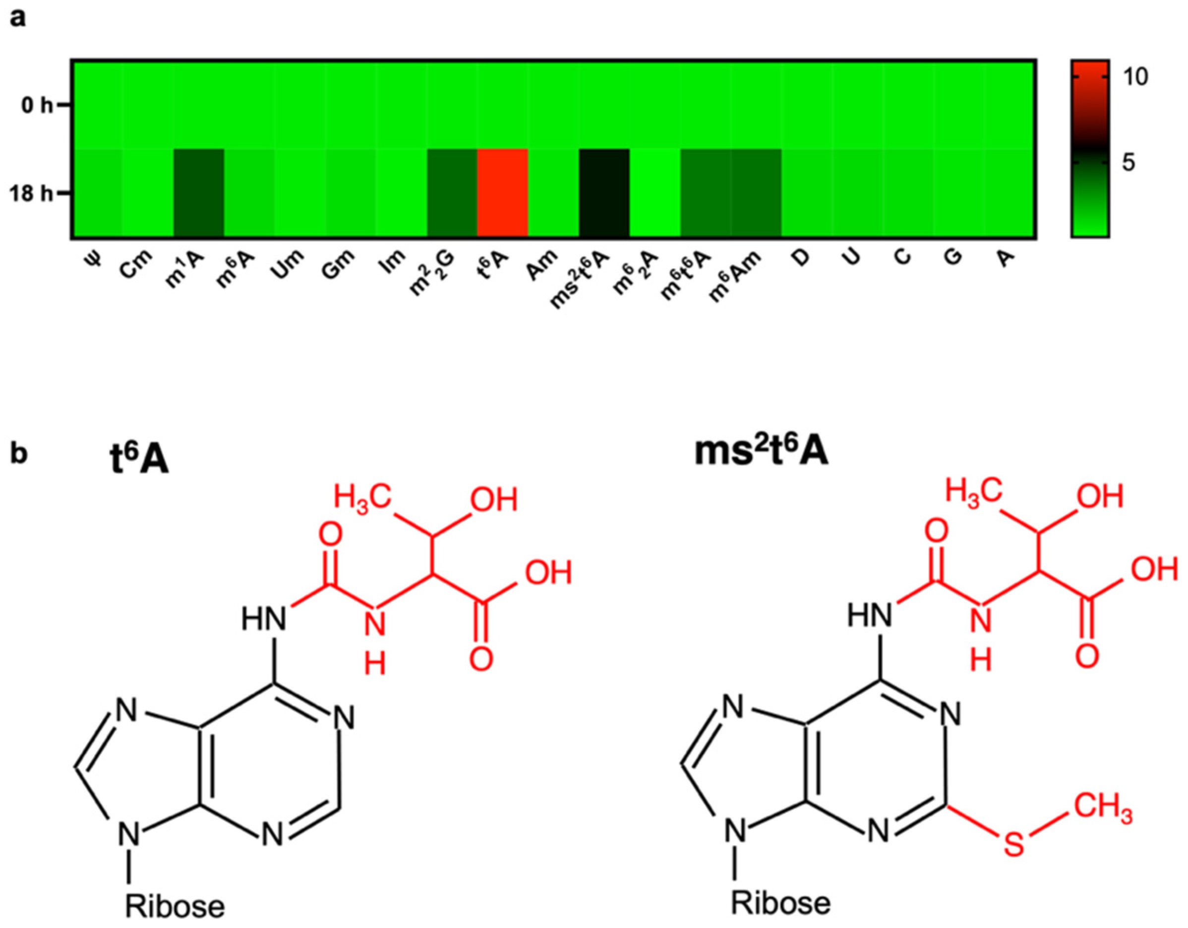
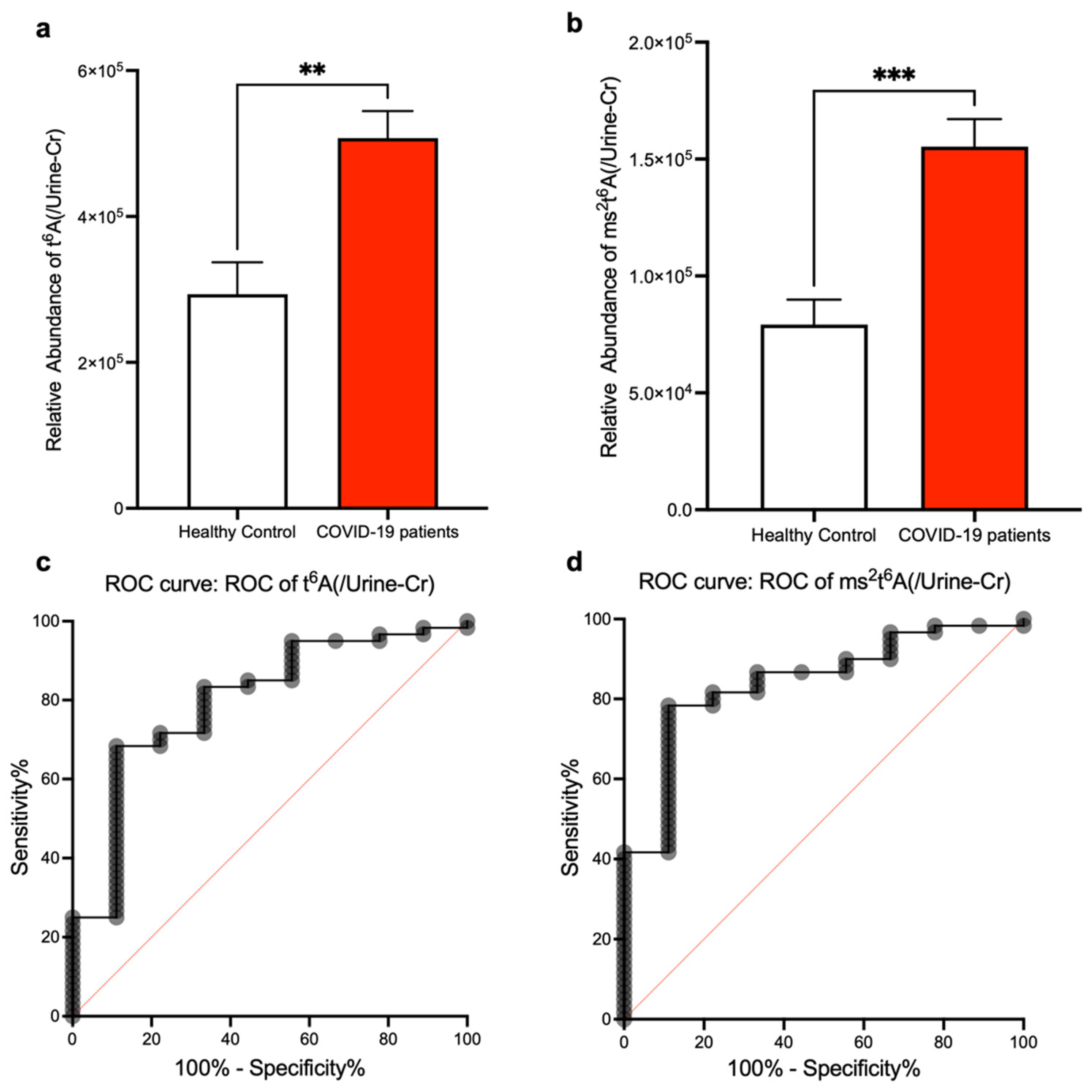
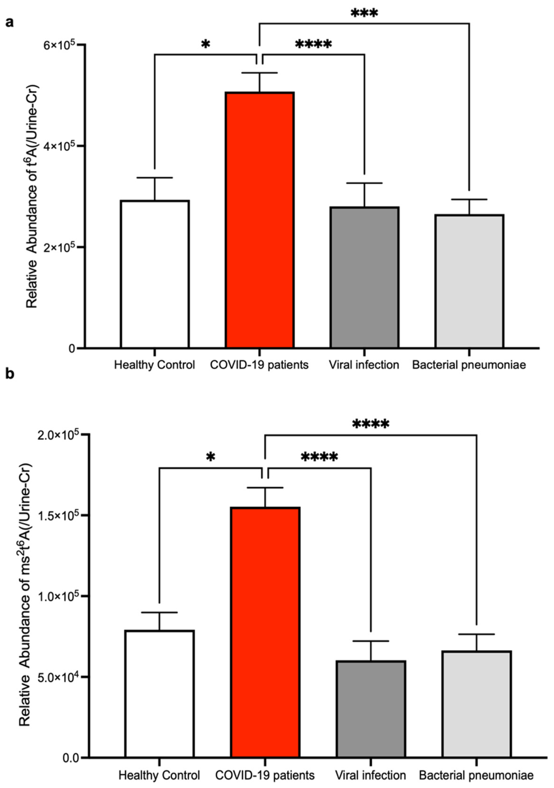
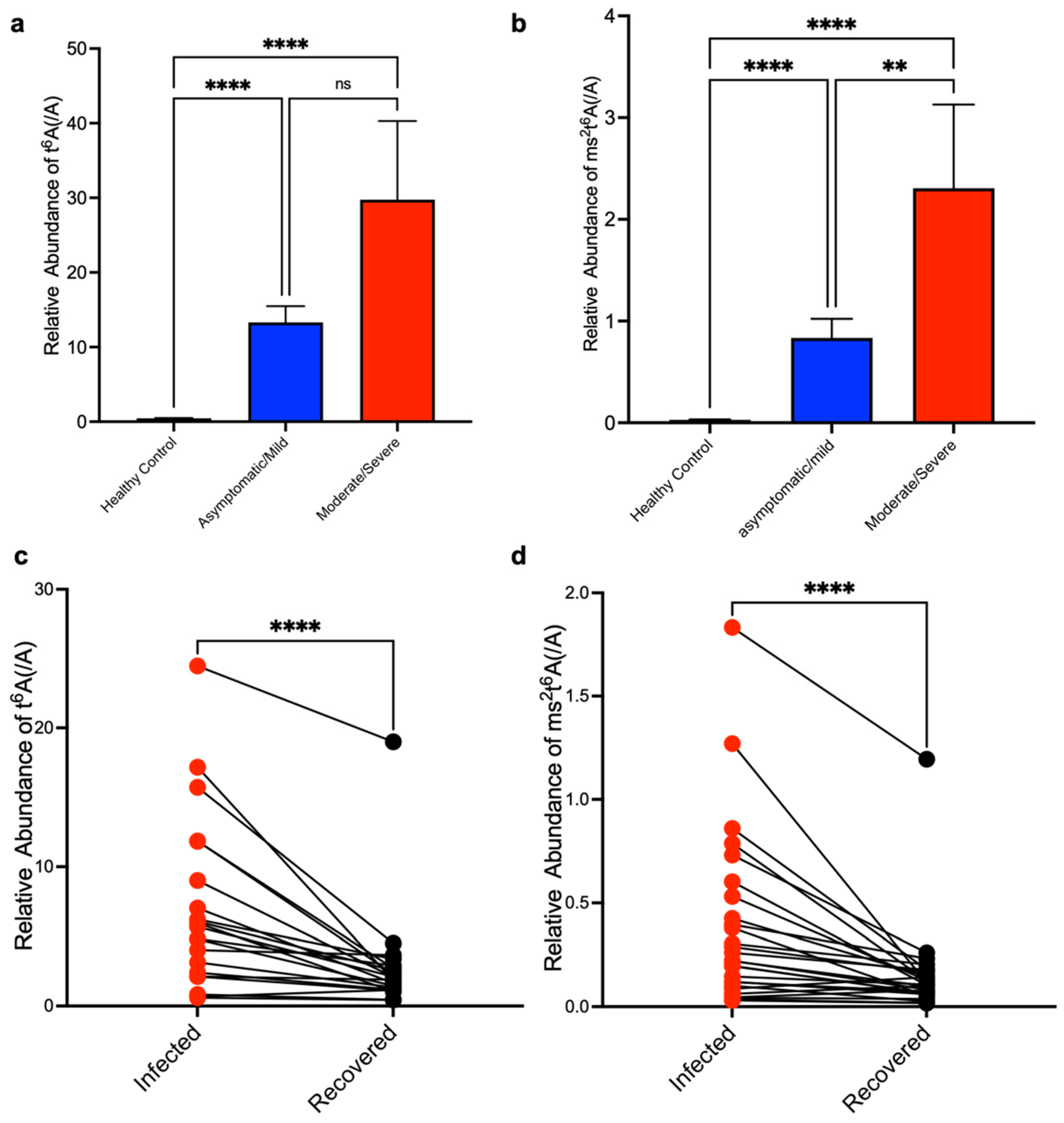
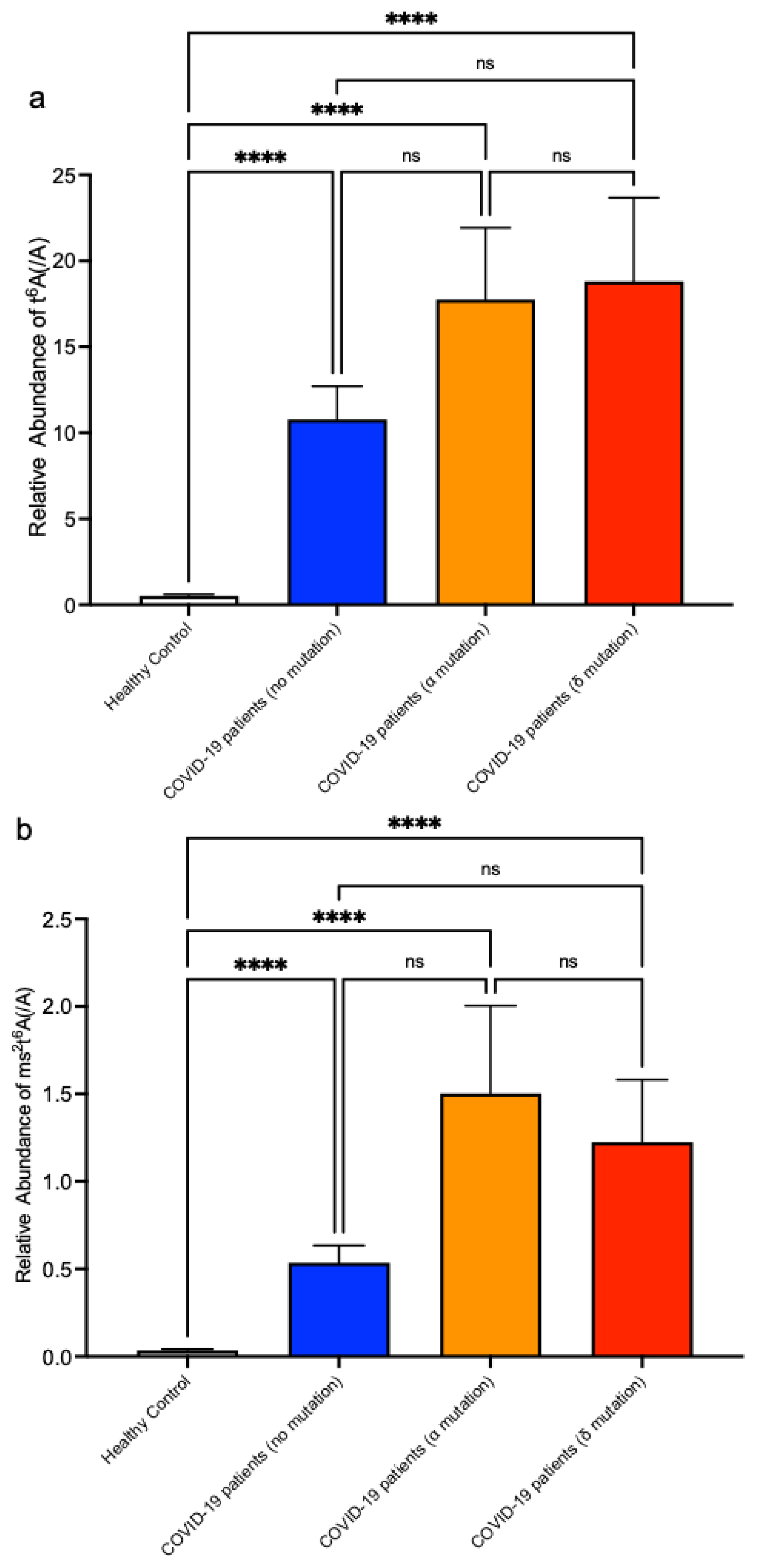
Data accorded with normal distribution and homogeneity of variance were expressed as the mean ± standard error of means (S.E.M) and compared by Mann–Whitney U tests. Categorical variables were compared by the Kruskal–Wallis test and Dunn’s multiple comparison tests. For calculation of sensitivity and specificity, we used receiver operating characteristic analysis to discriminate between healthy volunteers and COVID-19 patients. Statistical analyses were performed with the Prism 9 software (GraphPad, San Diego, CA, USA), and a p-value less than 0.05 was considered statistically significant.

3. Results

To identify modified nucleosides whose amount specifically changes in COVID-19, we first performed an infection experiment using angiotensin converting enzyme 2 (ACE2)-overexpressing human embryonic kidney (HEK) 293 cells. SARS-CoV-2 particles were infected at an MOI of 1. After 18 h of incubation, we extracted total RNA and degraded it into single nucleosides using nuclease P1 and alkaline phosphatase. We then quantified modified nucleosides by LC-MS. As a result, within the total RNA of SARS-CoV-2-infected cells, we observed elevation of six modified nucleosides, which are N1-methyladenosine (m1A), N2,N2-dimethylguanosine (m22G), N6-threonylcarbamoyladenosine (t6A), 2-methylthio-N6-threonylcarbamoyladenosine (ms2t6A), N6-methyl-N6-threonylcarbamoyladenosine (m6t6A), and N6,2′-O-dimethyladenosine (m6Am) (Figure 1a). Especially, t6A and ms2t6A (Figure 1b) were over 4 times elevated compared to control cells. From this result, t6A and ms2t6A were judged as good candidate biomarkers for SARS-CoV-2 infection.
Next, to investigate if t6A and ms2t6A within human urine can be used as SARS-CoV-2 infection biomarkers, we performed LC-MS analysis using the urine of patients with COVID-19. These patients were diagnosed by RT-PCR test against SARS-CoV-2 genome RNA from saliva or pharyngeal swabs (Table 1). Urine is highly susceptible to physiological conditions, and appropriate normalization is essential for the urine test. Generally, urine creatinine is the most commonly used normalization substance. Therefore, we analyzed t6A and ms2t6A in urine normalized by urine creatinine and these results were compared to healthy samples (Figure 2a,b). The t6A and ms2t6A levels in urine were significantly increased in COVID-19 patients. We also performed receiver-operating characteristic (ROC) analysis using data of t6A and ms2t6A normalized by urine creatinine. On t6A, setting the cutoff value to 344,420 resulted in a sensitivity of 71.7%, a specificity of 77.8%, and a likelihood ratio of 3.23 (Figure 2c). Regarding ms2t6A, setting the cutoff value to 76,878 resulted in a sensitivity of 86.6%, a specificity of 91.7%, and a likelihood ratio of 2.6 (Figure 2d).
To investigate if elevations of t6A and ms2t6A in urine are characteristic of COVID-19, we also compared the patient urine of COVID-19 with other infectious diseases including influenza and bacterial pneumonia. The elevation of t6A and ms2t6A in urine was observed only in the COVID-19 group (Figure 3a,b). From these results, measurements of t6A and ms2t6A in urine were observed to have the equivalent diagnostic ability to the RT-PCR test for COVID-19.
Next, to investigate if t6A and ms2t6A within human serum can be used as SARS-CoV-2 infection biomarkers, we measured t6A and ms2t6A in serum normalized by unmodified adenosine and compared them with healthy samples. The t6A and ms2t6A levels were significantly elevated in the serum of COVID-19 patients (Figure 4a,b). We also performed ROC analysis using data of t6A and ms2t6A. On t6A, setting the cutoff value to 1.039 resulted in a sensitivity of 98.4%, a specificity of 92.5%, and a likelihood ratio of 13.12 (Figure 4c). Regarding ms2t6A, setting the cutoff value to 0.1034 resulted in a sensitivity of 94.2%, a specificity of 92.5%, and a likelihood ratio of 12.55 (Figure 4d).
Next, to investigate if t6A and ms2t6A within human serum can be used as quantitative biomarkers to determine the severity of SARS-CoV-2 infection, we first examined the patients’ conditions from medical records and classified them by severity. Based on the Clinical Spectrum of SARS-CoV-2 Infection from “COVID-19 Treatment Guidelines” of NIH, we classified COVID-19 patients into two groups: asymptomatic/mild and moderate/severe. Then, we compared the measurements of t6A and ms2t6A in the serum of these two groups against a healthy group. As a result, as the severity of COVID-19 worsened, ms2t6A in serum also increased (Figure 5a,b). Next, we confirmed the relationships between the measurements t6A and ms2t6A in serum and clinical indicators related to COVID-19 severity (Table 1). Our results show the levels of lactate dehydrogenase (LDH), C-reactive proteins (CRP), and lymphocyte percentage in COVID-19 patients significantly correlated with t6A and ms2t6A levels (Supplementary Figure S1).
We also compared the changes in serum t6A and ms2t6A levels within the same COVID-19 moderate/severe patients at the infection period and recovered period. We found that t6A and ms2t6A in serum significantly decreased at the recovered period (Figure 5c,d). Based on these results, the measurement of t6A and ms2t6A in serum could be useful to determine the severity and the effect of treatment.
Since the end of 2020, patients with variants of SARS-CoV-2 have been reported from various regions, including the United Kingdom (B1.1.7), South Africa (B1.351), Brazil (P1), and India (B.1.617.2, AY.1, AY.2) [20]. These variants are often associated with enhanced transmissibility and evasion from host antibodies. We collected the serum of patients with B1.1.7 (α) and B.1.617.2 (δ) variants of SARS-CoV-2. Using the same LC-MS method, we measured t6A and ms2t6A in the serum of patients infected with these variants, and we found that t6A and ms2t6A were also elevated in the serum of patients infected with all monitored variants (Figure 6a,b). These results suggest that the diagnosis of COVID-19 by measuring t6A and ms2t6A in serum could be useful regardless of variants of SARS-CoV-2 spike protein.

4. Discussion

In this study, we first found characteristic elevations of specific modified nucleosides t6A and ms2t6A during SARS-CoV-2 infection experiments. These biomolecules were also elevated in the serum and urine of COVID-19 patients. Moreover, these elevations correlated with the severity and recovery of COVID-19. In the serum of patients infected with several mutant strains, these elevations were also observed.
To examine the presence of SARS-CoV-2, RT-PCR tests and antigen tests are easy and useful. However, clinical samples for these tests, which are saliva and nasopharyngeal swabs, often contain SARS-CoV-2, constantly exposing healthcare workers to the risk of infections during the collection and handling of these samples. Serum and urine contain very little of the SARS-CoV-2 virion [5,6]. Therefore, the establishment of COVID-19 diagnosis using modified nucleosides in serum and urine could provide more safety and less stress for healthcare workers. Considering the inaccessibility of mass spec machines in many facilities, we are currently trying to develop an easy and inexpensive t6A ELISA kit for COVID-19 detection using safer serum or urine samples rather than dangerous saliva and pharyngeal swabs.
In COVID-19 treatment, RT-PCR tests and antigen tests are not suitable for the proper assessment of COVID-19 severity. PCR tests and antigen tests for the SARS-CoV-2 viral genome from saliva or nasopharyngeal swab have no correlation with COVID-19 severity [21,22,23,24]. From our study, the elevations of t6A and ms2t6A in serum correlated with the severity and recovery of infection. The measurements of t6A and ms2t6A in serum could contribute to the appropriate assessment of severity and treatment effect, as well as to appropriately evaluate the efficacy of therapeutic agents during clinical trials. In this study, we examine only the elevation of t6A and ms2t6A in serum of patients with α and δ variants, and elevations in serum by infections with other variants should be checked.
From our study, the sources of t6A and ms2t6A are unclear, although there are some candidates. One is the result of cell damage to immune cells and/or tissue cells upon infection. When the host is infected by pathogens, large numbers of tissue cells and immune cells react and finally collapse. Our in vitro data using HEK293 cells indicate these elevations of modified nucleosides may be related to tissue cell damage. Moreover, we found that these elevations of modified nucleosides in serum correlated with LDH (Supplementary Figure S1). Upon destruction of tissue or immune cells, many modified nucleosides leak into the extracellular region and where they accumulate [18,19]. Therefore, the correlations of serum t6A and ms2t6A with COVID-19 severity might reflect the damage of tissue and/or immune cells upon SARS-CoV-2 infection. Another potential source of t6A and ms2t6A is the genome RNA of SARS-CoV-2. Within the viral RNA, chemically modified regions were detected using nanopore sequencing experiments, although the modification species are unidentified [7]. No obvious candidates for enzymes that modify t6A and ms2t6A are encoded in the genome RNA of SARS-CoV-2. Therefore, if the viral RNA contains t6A and ms2t6A, SARS-CoV-2 likely uses the host’s modifying enzymes, the KEOPS complex for t6A modification and CDKAL1 for ms2 modification [11,14,25]. Some RNA viruses, such as HIV-1, have been reported to use the host RNA modification enzyme to escape from host immunity [26]. In future studies, it will be necessary to monitor changes in the expression of these modifying enzymes upon viral infection, as well as the modification levels of the host tRNAs and other RNAs. Recently, many types of vaccinations, including mRNA vaccines, were certified and used in many countries to combat the COVID-19 pandemic. It will be important to investigate the changes of t6A and ms2t6A in vaccinated patient serum in future studies.

5. Conclusions

In summary, we discovered serum and urine t6A and ms2t6A nucleosides as effective biomarkers of COVID-19. Modified nucleosides are conceptually new metabolites to be measured in the clinical area, and our study is the first to monitor them in COVID-19. The most important merits of the modified nucleoside test over the RT-PCR test are: (1) correlation of serum t6A and ms2t6A levels with the severity and recovery and (2) accuracy of this test regardless of the mutation in the spike protein of SARS-CoV-2. This test is the first evidence for diagnosis using modified nucleosides for COVID-19 and could be useful for accurate assessment of COVID-19 severity and recovery.

Supplementary Materials

The following supporting information can be downloaded at: https://www.mdpi.com/article/10.3390/biom12091233/s1, Figure S1: Correlations between modified nucleosides in serum and clinical indicators of COVID-19, Table S1: MRM transition parameters for modified nucleosides, Table S2: Information of the patients with other infectious diseases.

Author Contributions

Y.N. (Yu Nagayoshi) conceived of this study, analyzed the data, and wrote the manuscript; K.N., R.Y., H.O., H.N. (Hiroko Nagata). and H.K. performed the experiments; T.C. supported the LC-MS measurements and manuscript preparation; K.S., M.A., Y.K. and M.M. (Masashi Mukoyama). supported the design of the clinical study; A.K. and F.-Y.W. supported the LC-MS measurements; K.Y., H.N. (Hirotomo Nakata)., T.M., J.-i.K., S.S., Y.N. (Yasushi Nakamori). and M.M. (Masao Matsuoka). collected the clinical samples and records; K.T. organized this study and supervised the manuscript preparation. All authors approved the final version of the paper before submission. The authors had full access to all the data in the study and had final responsibility for the decision to submit it for publication. All authors have read and agreed to the published version of the manuscript.

Funding

This study was supported by grants from the Cooperative Research Project Program of Joint Usage/Research Center at the Institute of Development, Aging and Cancer, Tohoku University, and AMABIE Research Project, Kumamoto University.

Institutional Review Board Statement

Written informed consent was obtained from all participants. This study was approved by the institutional ethics board of Kumamoto University (No. 2184) in accordance with the Helsinki Declaration of 1964 and its later amendments.

Informed Consent Statement

Written informed consent was obtained from all participants. This study was approved by the institutional ethics board of Kumamoto University (No. 2184).

Data Availability Statement

This dataset is available with the permission of the responsible author and the institutional ethics board of Kumamoto University.

Acknowledgments

The authors would like to thank Natalie DeWitt for the critical reading and language editing of the manuscript.

Conflicts of Interest

K.T. was funded by Shimadzu Corporation and AiSTI Science Corporation. Other authors have no competing interest.

References

  1. Zhu, N.; Zhang, D.; Wang, W.; Li, X.; Yang, B.; Song, J.; Zhao, X.; Huang, B.; Shi, W.; Lu, R.; et al. A novel coronavirus from patients with pneumonia in China, 2019. N. Engl. J. Med. 2020, 382, 727–733. [Google Scholar] [CrossRef] [PubMed]
  2. Zhou, P.; Yang, X.L.; Wang, X.G.; Hu, B.; Zhang, L.; Zhang, W.; Si, H.R.; Zhu, Y.; Li, B.; Huang, C.L.; et al. A pneumonia outbreak associated with a new coronavirus of probable bat origin. Nature 2020, 579, 270–273. [Google Scholar] [CrossRef] [PubMed]
  3. Sugiyama, M.; Kinoshita, N.; Ide, S.; Nomoto, H.; Nakamoto, T.; Saito, S.; Ishikane, M.; Kutsuna, S.; Hayakawa, K.; Hashimoto, M.; et al. Serum CCL17 level becomes a predictive marker to distinguish between mild/moderate and severe/critical disease in patients with COVID-19. Gene 2021, 766, 145145. [Google Scholar] [CrossRef] [PubMed]
  4. Renert-Yuval, Y.; Thyssen, J.P.; Bissonnette, R.; Bieber, T.; Kabashima, K.; Hijnen, D.; Guttman-Yassky, E. Biomarkers in atopic dermatitis-a review on behalf of the International Eczema Council. J. Allergy Clin. Immunol. 2021, 147, 1174–1190.e1171. [Google Scholar] [CrossRef]
  5. Ruan, Y.; Hu, B.; Liu, Z.; Liu, K.; Jiang, H.; Li, H.; Li, R.; Luan, Y.; Liu, X.; Yu, G.; et al. No detection of SARS-CoV-2 from urine, expressed prostatic secretions, and semen in 74 recovered COVID-19 male patients: A perspective and urogenital evaluation. Andrology 2021, 9, 99–106. [Google Scholar] [CrossRef] [PubMed]
  6. Jin, X.D.; Li, Y.; Song, Y.S.; Yang, Z.Z.; Wang, P.; Wei, T.T.; Fan, T.L. Progress in research on the detection of the novel coronavirus in human samples of different groups. Eur. Rev. Med. Pharmacol. Sci. 2020, 24, 10879–10884. [Google Scholar] [CrossRef]
  7. Kim, D.; Lee, J.Y.; Yang, J.S.; Kim, J.W.; Kim, V.N.; Chang, H. The Architecture of SARS-CoV-2 Transcriptome. Cell 2020, 181, 914–921.e910. [Google Scholar] [CrossRef]
  8. Nencka, R.; Silhan, J.; Klima, M.; Otava, T.; Kocek, H.; Krafcikova, P.; Boura, E. Coronaviral RNA-methyltransferases: Function, structure and inhibition. Nucleic Acids Res. 2022, 50, 635–650. [Google Scholar] [CrossRef]
  9. Boccaletto, P.; Machnicka, M.A.; Purta, E.; Piatkowski, P.; Baginski, B.; Wirecki, T.K.; de Crecy-Lagard, V.; Ross, R.; Limbach, P.A.; Kotter, A.; et al. MODOMICS: A database of RNA modification pathways. 2017 update. Nucleic Acids Res. 2018, 46, D303–D307. [Google Scholar] [CrossRef]
  10. Phelps, S.S.; Malkiewicz, A.; Agris, P.F.; Joseph, S. Modified nucleotides in tRNA(Lys) and tRNA(Val) are important for translocation. J. Mol. Biol. 2004, 338, 439–444. [Google Scholar] [CrossRef]
  11. Perrochia, L.; Guetta, D.; Hecker, A.; Forterre, P.; Basta, T. Functional assignment of KEOPS/EKC complex subunits in the biosynthesis of the universal t6A tRNA modification. Nucleic Acids Res. 2013, 41, 9484–9499. [Google Scholar] [CrossRef] [PubMed]
  12. Braun, D.A.; Rao, J.; Mollet, G.; Schapiro, D.; Daugeron, M.C.; Tan, W.; Gribouval, O.; Boyer, O.; Revy, P.; Jobst-Schwan, T.; et al. Mutations in KEOPS-complex genes cause nephrotic syndrome with primary microcephaly. Nat. Genet. 2017, 49, 1529–1538. [Google Scholar] [CrossRef] [PubMed]
  13. Arragain, S.; Handelman, S.K.; Forouhar, F.; Wei, F.Y.; Tomizawa, K.; Hunt, J.F.; Douki, T.; Fontecave, M.; Mulliez, E.; Atta, M. Identification of eukaryotic and prokaryotic methylthiotransferase for biosynthesis of 2-methylthio-N6-threonylcarbamoyladenosine in tRNA. J. Biol. Chem. 2010, 285, 28425–28433. [Google Scholar] [CrossRef] [PubMed]
  14. Wei, F.Y.; Suzuki, T.; Watanabe, S.; Kimura, S.; Kaitsuka, T.; Fujimura, A.; Matsui, H.; Atta, M.; Michiue, H.; Fontecave, M.; et al. Deficit of tRNA(Lys) modification by Cdkal1 causes the development of type 2 diabetes in mice. J. Clin. Investig. 2011, 121, 3598–3608. [Google Scholar] [CrossRef]
  15. Wei, F.Y.; Zhou, B.; Suzuki, T.; Miyata, K.; Ujihara, Y.; Horiguchi, H.; Takahashi, N.; Xie, P.; Michiue, H.; Fujimura, A.; et al. Cdk5rap1-mediated 2-methylthio modification of mitochondrial tRNAs governs protein translation and contributes to myopathy in mice and humans. Cell Metab. 2015, 21, 428–442. [Google Scholar] [CrossRef]
  16. Nagayoshi, Y.; Chujo, T.; Hirata, S.; Nakatsuka, H.; Chen, C.W.; Takakura, M.; Miyauchi, K.; Ikeuchi, Y.; Carlyle, B.C.; Kitchen, R.R.; et al. Loss of Ftsj1 perturbs codon-specific translation efficiency in the brain and is associated with X-linked intellectual disability. Sci. Adv. 2021, 7, eabf3072. [Google Scholar] [CrossRef]
  17. Chujo, T.; Tomizawa, K. Human transfer RNA modopathies: Diseases caused by aberrations in transfer RNA modifications. FEBS J. 2021, 288, 7096–7122. [Google Scholar] [CrossRef]
  18. Shi, S.L.; Fukuda, H.; Chujo, T.; Kouwaki, T.; Oshiumi, H.; Tomizawa, K.; Wei, F.Y. Export of RNA-derived modified nucleosides by equilibrative nucleoside transporters defines the magnitude of autophagy response and Zika virus replication. RNA Biol. 2021, 18, 478–495. [Google Scholar] [CrossRef]
  19. Ogawa, A.; Nagiri, C.; Shihoya, W.; Inoue, A.; Kawakami, K.; Hiratsuka, S.; Aoki, J.; Ito, Y.; Suzuki, T.; Suzuki, T.; et al. N6-methyladenosine (m6A) is an endogenous A3 adenosine receptor ligand. Mol. Cell 2021, 81, 659–674.e657. [Google Scholar] [CrossRef]
  20. Tao, K.; Tzou, P.L.; Nouhin, J.; Gupta, R.K.; de Oliveira, T.; Kosakovsky Pond, S.L.; Fera, D.; Shafer, R.W. The biological and clinical significance of emerging SARS-CoV-2 variants. Nat. Rev. Genet. 2021, 22, 757–773. [Google Scholar] [CrossRef]
  21. Fajnzylber, J.; Regan, J.; Coxen, K.; Corry, H.; Wong, C.; Rosenthal, A.; Worrall, D.; Giguel, F.; Piechocka-Trocha, A.; Atyeo, C.; et al. SARS-CoV-2 viral load is associated with increased disease severity and mortality. Nat. Commun. 2020, 11, 5493. [Google Scholar] [CrossRef] [PubMed]
  22. Singanayagam, A.; Patel, M.; Charlett, A.; Lopez Bernal, J.; Saliba, V.; Ellis, J.; Ladhani, S.; Zambon, M.; Gopal, R. Duration of infectiousness and correlation with RT-PCR cycle threshold values in cases of COVID-19, England, January to May 2020. Eurosurveill 2020, 25, 2001483. [Google Scholar] [CrossRef] [PubMed]
  23. Cantini, F.; Niccoli, L.; Matarrese, D.; Nicastri, E.; Stobbione, P.; Goletti, D. Baricitinib therapy in COVID-19: A pilot study on safety and clinical impact. J. Infect. 2020, 81, 318–356. [Google Scholar] [CrossRef] [PubMed]
  24. Yokoyama, R.; Kurano, M.; Nakano, Y.; Morita, Y.; Ohmiya, H.; Kishi, Y.; Okada, J.; Qian, C.; Xia, F.; He, F.; et al. Association of the Serum Levels of the Nucleocapsid Antigen of SARS-CoV-2 with the Diagnosis, Disease Severity, and Antibody Titers in Patients with COVID-19: A Retrospective Cross-Sectional Study. Front. Microbiol. 2021, 12, 791489. [Google Scholar] [CrossRef] [PubMed]
  25. de Crecy-Lagard, V.; Boccaletto, P.; Mangleburg, C.G.; Sharma, P.; Lowe, T.M.; Leidel, S.A.; Bujnicki, J.M. Matching tRNA modifications in humans to their known and predicted enzymes. Nucleic Acids Res. 2019, 47, 2143–2159. [Google Scholar] [CrossRef]
  26. Ringeard, M.; Marchand, V.; Decroly, E.; Motorin, Y.; Bennasser, Y. FTSJ3 is an RNA 2′-O-methyltransferase recruited by HIV to avoid innate immune sensing. Nature 2019, 565, 500–504. [Google Scholar] [CrossRef]
Figure 1. Screening of candidate modified nucleosides by SARS-CoV-2. (a) Heatmap analysis of modified nucleoside levels in extracted RNA from SARS-CoV-2-infected HEK293 cells. Color scale shows the auto-scaled relative mean of n = 3 biological replicates. ψ: pseudouridine; Cm: 2′-O-methylcytidine; m1A: N1-methyladenosine; m6A: N6-methyladenosine; Um: 2′-O-methyluridine; Gm: 2′-O-methylguanosine; Im: 2′-O-methylinosine; m22G: N2,N2-dimethylguanosine; t6A: N6-threonylcarbamoyladenosine; Am: 2′-O-methyladenosine; ms2t6A: 2-methylthio-N6-threonylcarbamoyladenosine; m6,2A: N6,N6-dimethyladenosine; m6t6A: N6-methyl-threonylcarbamoyladenosine; m6Am: N6,2′-O-dimethyladenosine; D: dihydrouridine; U: uridine; C: cytidine; G: guanosine; A: adenosine. (b) Chemical structures of t6A and ms2t6A. Modified residues are depicted in red.
Biomolecules 12 01233 g001
Figure 2. SARS-CoV-2 infection causes elevation of t6A and ms2t6A levels in urine. (a,b) Measurements of t6A (a) and ms2t6A (b) in urine and comparison with healthy volunteers. LC-MS peak areas of t6A and ms2t6A divided by urine creatinine levels are shown. ** p < 0.01, *** p < 0.001 by Mann–Whitney U test. (c,d) ROC analysis for measurements of t6A (c) and ms2t6A (d) in urine was performed for calculation of sensitivity and specificity.
Biomolecules 12 01233 g002
Figure 3. The comparison of t6A and ms2t6A levels in the urine of COVID-19 patients with other infectious diseases. (a,b) Comparison of t6A (a) and ms2t6A (b) levels in the urine of patients with viral infection or bacterial pneumoniae. LC-MS peak areas of t6A and ms2t6A divided by urine creatinine levels are shown. * p < 0.05, *** p < 0.001, **** p < 0.0001 by Kruskal–Wallis test and Dunn’s multiple comparison test.
Biomolecules 12 01233 g003
Figure 4. Elevation of t6A and ms2t6A in serum of COVID-19 patients. (a,b) Measurements of t6A (a) and ms2t6A (b) in serum of COVID-19 patients and comparison with healthy volunteers. LC-MS peak areas of t6A or ms2t6A divided by LC-MS peak areas of adenosine are shown. **** p < 0.0001 by Mann–Whitney U test. (c,d) ROC analysis for t6A (c) and ms2t6A (d) levels in serum was performed for calculation of sensitivity and specificity.
Biomolecules 12 01233 g004
Figure 5. Elevations of t6A and ms2t6A in serum of COVID-19 patients correlate with severity and recovery of COVID-19. (a,b) Elevation of t6A (a) and ms2t6A (b) in serum categorized by COVID-19 severity. LC-MS peak areas of t6A or ms2t6A divided by LC-MS peak areas of adenosine are shown. ** p < 0.01, **** p < 0.0001 by Kruskal–Wallis test and Dunn’s multiple comparison test. (c,d) Comparison of t6A (c) and ms2t6A (d) levels in serum of COVID-19 patients at the time of infection and recovery. A recovery period was defined by the resolution of fever and other symptoms. LC-MS peak areas of t6A or ms2t6A divided by LC-MS peak areas of adenosine are shown. **** p < 0.0001 by Wilcoxon rank-sum test.
Biomolecules 12 01233 g005
Figure 6. Elevation of t6A and ms2t6A levels in serum of patients infected by different SARS-CoV-2 strains. (a,b) Measurements of t6A (a) and ms2t6A (b) in serum of COVID-19 patients infected with α strain or δ strain compared with healthy volunteers. LC-MS peak areas of t6A or ms2t6A divided by LC-MS peak areas of adenosine are shown. **** p < 0.0001 by Kruskal–Wallis test and Dunn’s multiple comparison test. ns, not significant.
Biomolecules 12 01233 g006
Table 1. Patient characteristics of this study.
Healthy
Volunteers
(N = 40)
COVID-19
Patients
(N = 308)
Bacterial Infection
Patients
(N = 18)
Viral Infection
Patients
(N = 24)
Age at inclusion (year 95% CI)28.1
(31.3–24.9)
50.7
(52.9–48.5)
73.2
(77.9–68.4)
66.7
(71.9–61.4)
Sex
 Male (n, %)24 (60)178 (57.8)14 (77.8)13 (54.1)
 Female (n, %)16 (40)130 (42.2)4 (22.2)11 (45.8)
Serum collection40308--
Urine collection10601824
Race
 East Asian (%)100100100100
COVID-19 Severity
 Asymptomatic/mild (n, %)-235 (76.2)--
 Moderate/severe (n, %)-73 (23.8)--
Mutation of SARS-CoV-2
 No mutation (n, %)-51 (16.6)--
 α-mutation (n, %)-80 (26)--
 δ-mutation (n, %)-177 (57.4)--
CKD patients (eGFR < 60)
   (n, %)
-69
(20.6)
9
(50)
9
(37.5)
WBC (/μL)
   (95% CI)
-5675.1
(5318.7–6033.2)
10.375
(3570–26,980)
6314.1
(3110–9660)
Lymphocyte (%)
   (95% CI)
-22.8
(21.3–24.2)
LDH (U/L)
   (95% CI)
-247.8
(232.1–263.5)
--
CRP (mg/dL)
   (95% CI)
-2.979
(2.41–3.54)
9.17
(0.19–23.02)
1.17
(0.02–3.53)
Abbreviations: eGFR: estimated glomerular filtration rate. CKD: chronic kidney disease. WBC: white blood cells. LDH: lactate dehydrogenase. CRP: C-reactive protein.
Publisher’s Note: MDPI stays neutral with regard to jurisdictional claims in published maps and institutional affiliations.

