
Cutting-Edge AI Technologies Meet Precision Medicine to Improve Cancer Care


Abstract
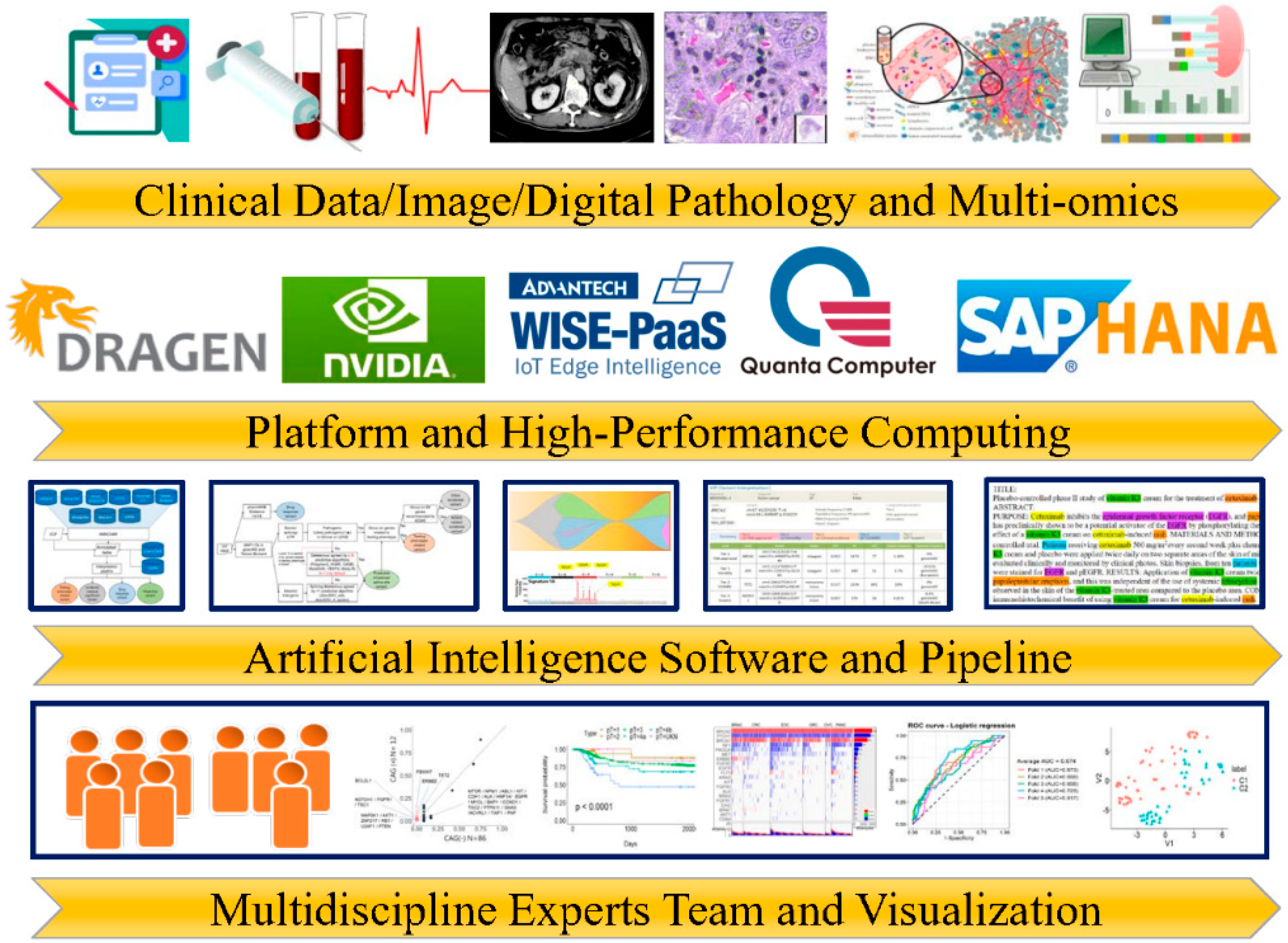
:1. Introduction
2. Computational Prediction of Pathogenic Variants of Cancer Susceptibility Genes
2.1. Sequencing-Based Prediction
2.2. Amino-Acid- or Protein-Based Prediction
2.3. AI Tools Based on ACMG/AMP and Functional Somatic Mutation
3. AI model for Mutational Analysis
3.1. Mutational Signatures and AI Tools
3.2. Cancer Evolution and AI Tools
3.3. Clinical Practice in Mutational Signature and Cancer Evolution
4. Single-Cell Genomics and Computational Biology
5. Text Mining for Identifying Genes Targets in Cancers
6. The NVIDIA GPUs, DRAGEN FPGAs Systems, and AI Medical Cloud Platforms in the Clinical NGS Lab
6.1. Using NVIDIA GPUs, DRAGEN FPGAs Systems in Bioinformatic Analysis
6.2. AI Medical Cloud Platforms for Cancer Care
7. Conclusions
Author Contributions
Funding
Institutional Review Board Statement
Informed Consent Statement
Data Availability Statement
Conflicts of Interest
References
- Bødker, J.S.; Sønderkær, M.; Vesteghem, C.; Schmitz, A.; Brøndum, R.F.; Sommer, M.; Rytter, A.S.; Nielsen, M.M.; Madsen, J.; Jensen, P.; et al. Development of a Precision Medicine Workflow in Hematological Cancers, Aalborg University Hospital, Denmark. Cancers 2020, 12, 312. [Google Scholar] [CrossRef] [PubMed]
- Conceição, S.I.R.; Couto, F.M. Text Mining for Building Biomedical Networks Using Cancer as a Case Study. Biomolecules 2021, 11, 1430. [Google Scholar] [CrossRef] [PubMed]
- Lin, P.C.; Yeh, Y.M.; Wu, P.Y.; Hsu, K.F.; Chang, J.Y.; Shen, M.R. Germline susceptibility variants impact clinical outcome and therapeutic strategies for stage III colorectal cancer. Sci. Rep. 2019, 9, 3931. [Google Scholar] [CrossRef] [PubMed]
- André, T.; Shiu, K.K.; Kim, T.W.; Jensen, B.V.; Jensen, L.H.; Punt, C.; Smith, D.; Garcia-Carbonero, R.; Benavides, M.; Gibbs, P.; et al. Pembrolizumab in Microsatellite-Instability-High Advanced Colorectal Cancer. N. Engl. J. Med. 2020, 383, 2207–2218. [Google Scholar] [CrossRef] [PubMed]
- Moore, K.; Colombo, N.; Scambia, G.; Kim, B.G.; Oaknin, A.; Friedlander, M.; Lisyanskaya, A.; Floquet, A.; Leary, A.; Sonke, G.S.; et al. Maintenance Olaparib in Patients with Newly Diagnosed Advanced Ovarian Cancer. N. Engl. J. Med. 2018, 379, 2495–2505. [Google Scholar] [CrossRef]
- Richards, S.; Aziz, N.; Bale, S.; Bick, D.; Das, S.; Gastier-Foster, J.; Grody, W.W.; Hegde, M.; Lyon, E.; Spector, E.; et al. Standards and guidelines for interpreting sequence variants: A joint consensus recommendation of the American College of Medical Genetics and Genomics and the Association for Molecular Pathology. Genet. Med. 2015, 17, 405–424. [Google Scholar] [CrossRef]
- Valenzuela-Palomo, A.; Bueno-Martínez, E.; Sanoguera-Miralles, L.; Lorca, V.; Fraile-Bethencourt, E.; Esteban-Sánchez, A.; Gómez-Barrero, S.; Carvalho, S.; Allen, J.; García-Álvarez, A.; et al. Splicing predictions, minigene analyses, and ACMG-AMP clinical classification of 42 germline PALB2 splice-site variants. J. Pathol. 2022, 256, 321–334. [Google Scholar] [CrossRef]
- Carter, H.; Douville, C.; Stenson, P.D.; Cooper, D.N.; Karchin, R. Identifying Mendelian disease genes with the variant effect scoring tool. BMC Genom. 2013, 14, S3. [Google Scholar] [CrossRef]
- Dong, C.; Wei, P.; Jian, X.; Gibbs, R.; Boerwinkle, E.; Wang, K.; Liu, X. Comparison and integration of deleteriousness prediction methods for nonsynonymous SNVs in whole exome sequencing studies. Hum. Mol. Genet. 2015, 24, 2125–2137. [Google Scholar] [CrossRef]
- Ioannidis, N.M.; Rothstein, J.H.; Pejaver, V.; Middha, S.; McDonnell, S.K.; Baheti, S.; Musolf, A.; Li, Q.; Holzinger, E.; Karyadi, D.; et al. REVEL: An Ensemble Method for Predicting the Pathogenicity of Rare Missense Variants. Am. J. Hum. Genet. 2016, 99, 877–885. [Google Scholar] [CrossRef]
- Sundaram, L.; Gao, H.; Padigepati, S.R.; McRae, J.F.; Li, Y.; Kosmicki, J.A.; Fritzilas, N.; Hakenberg, J.; Dutta, A.; Shon, J.; et al. Predicting the clinical impact of human mutation with deep neural networks. Nat. Genet. 2018, 50, 1161–1170. [Google Scholar] [CrossRef] [PubMed]
- Rentzsch, P.; Witten, D.; Cooper, G.M.; Shendure, J.; Kircher, M. CADD: Predicting the deleteriousness of variants throughout the human genome. Nucleic Acids Res. 2019, 47, D886–D894. [Google Scholar] [CrossRef] [PubMed]
- Jaganathan, K.; Kyriazopoulou Panagiotopoulou, S.; McRae, J.F.; Darbandi, S.F.; Knowles, D.; Li, Y.I.; Kosmicki, J.A.; Arbelaez, J.; Cui, W.; Schwartz, G.B.; et al. Predicting Splicing from Primary Sequence with Deep Learning. Cell. 2019, 176, 535–548. [Google Scholar] [CrossRef] [PubMed]
- Won, D.G.; Kim, D.W.; Woo, J.; Lee, K. 3Cnet: Pathogenicity prediction of human variants using multitask learning with evolutionary constraints. Bioinformatics 2021, 37, 4626–4634. [Google Scholar] [CrossRef] [PubMed]
- Abdollahi, S.; Lin, P.C.; Shen, M.R.; Chiang, J.H. Precise uncertain significance prediction using latent space matrix factorization models: Genomics variant and heterogeneous clinical data-driven approaches. Brief. Bioinform. 2021, 22, bbaa281. [Google Scholar] [CrossRef] [PubMed]
- Qi, H.; Zhang, H.; Zhao, Y.; Chen, C.; Long, J.J.; Chung, W.K.; Guan, Y.; Shen, Y. MVP predicts the pathogenicity of missense variants by deep learning. Nat. Commun. 2021, 12, 510. [Google Scholar] [CrossRef]
- Wu, Y.; Liu, H.; Li, R.; Sun, S.; Weile, J.; Roth, F.P. Improved pathogenicity prediction for rare human missense variants. Am. J. Hum. Genet. 2021, 108, 1891–1906. [Google Scholar] [CrossRef]
- Labes, S.; Stupp, D.; Wagner, N.; Bloch, I.; Lotem, M.; Lahad, E.L.; Polak, P.; Pupko, T.; Tabach, Y. Machine-learning of complex evolutionary signals improves classification of SNVs. NAR Genom. Bioinform. 2022, 4, lqac025. [Google Scholar] [CrossRef]
- Choi, Y.; Chan, A.P. PROVEAN /btv195.web server: A tool to predict the functional effect of amino acid substitutions and indels. Bioinformatics 2015, 31, 2745–2747. [Google Scholar] [CrossRef]
- Asgari, E.; Mofrad, M.R. Continuous Distributed Representation of Biological Sequences for Deep Proteomics and Genomics. PLoS ONE 2015, 10, e0141287. [Google Scholar] [CrossRef]
- Liu, B.; Gao, X.; Zhang, H. BioSeq-Analysis2.0: An updated platform for analyzing DNA, RNA and protein sequences at sequence level and residue level based on machine learning approaches. Nucleic Acids Res. 2019, 47, e127. [Google Scholar] [CrossRef] [PubMed]
- Ponzoni, L.; Peñaherrera, D.A.; Oltvai, Z.N.; Bahar, I. Rhapsody: Predicting the pathogenicity of human missense variants. Bioinformatics 2020, 36, 3084–3092. [Google Scholar] [CrossRef] [PubMed]
- Lai, J.; Yang, J.; Gamsiz Uzun, E.D.; Rubenstein, B.M.; Sarkar, I.N. LYRUS: A machine learning model for predicting the pathogenicity of missense variants. Bioinform. Adv. 2021, 2, vbab045. [Google Scholar] [CrossRef] [PubMed]
- Wu, T.H.; Lin, P.C.; Chou, H.H.; Shen, M.R.; Hsieh, S.Y. Pathogenicity Prediction of Single Amino Acid Variants with Machine Learning Model Based on Protein Structural Energies. IEEE/ACM Trans. Comput. Biol. Bioinform. 2021. [Google Scholar] [CrossRef] [PubMed]
- Tavtigian, S.V.; Greenblatt, M.S.; Harrison, S.M.; Nussbaum, R.L.; Prabhu, S.A.; Boucher, K.M.; Biesecker, L.G. Modeling the ACMG/AMP variant classification guidelines as a Bayesian classification framework. Genet. Med. 2018, 20, 1054–1060. [Google Scholar] [CrossRef]
- Scott, A.D.; Huang, K.L.; Weerasinghe, A.; Mashl, R.J.; Gao, Q.; Martins Rodrigues, F.; Wyczalkowski, M.A.; Ding, L. CharGer: Clinical Characterization of Germline variants. Bioinformatics 2019, 35, 865–867. [Google Scholar] [CrossRef]
- Kopanos, C.; Tsiolkas, V.; Kouris, A.; Chapple, C.E.; Aguilera, M.A.; Meyer, R.; Massouras, A. VarSome: The human genomic variant search engine. Bioinformatics 2019, 35, 1978–1980. [Google Scholar] [CrossRef]
- Nicora, G.; Zucca, S.; Limongelli, I.; Bellazzi, R.; Magni, P. A machine learning approach based on ACMG/AMP guidelines for genomic variant classification and prioritization. Sci. Rep. 2022, 12, 2517. [Google Scholar] [CrossRef]
- Mok, T.S.; Wu, Y.; Ahn, M.; Garassino, M.C.; Kim, H.R.; Ramalingam, S.S.; Shepherd, F.A.; He, Y.; Akamatsu, H.; Theelen, W.S.; et al. Osimertinib or platinum–pemetrexed in EGFR T790M–positive lung cancer. N. Engl. J. Med. 2017, 376, 629–640. [Google Scholar] [CrossRef]
- Garrett, A.; Durkie, M.; Callaway, A.; Burghel, G.J.; Robinson, R.; Drummond, J.; Torr, B.; Cubuk, C.; Berry, I.R.; Wallace, A.J.; et al. Combining evidence for and against pathogenicity for variants in cancer susceptibility genes: CanVIG-UK consensus recommendations. J. Med. Genet. 2021, 58, 297–304. [Google Scholar] [CrossRef]
- Landrum, M.J.; Lee, J.M.; Benson, M.; Brown, G.; Chao, C.; Chitipiralla, S.; Gu, B.; Hart, J.; Hoffman, D.; Hoover, J.; et al. ClinVar: Public archive of interpretations of clinically relevant variants. Nucleic Acids Res. 2016, 44, D862–D868. [Google Scholar] [CrossRef] [PubMed]
- Li, M.M.; Datto, M.; Duncavage, E.J.; Kulkarni, S.; Lindeman, N.I.; Roy, S.; Tsimberidou, A.M.; Vnencak-Jones, C.L.; Wolff, D.J.; Younes, A.; et al. Standards and Guidelines for the Interpretation and Reporting of Sequence Variants in Cancer: A Joint Consensus Recommendation of the Association for Molecular Pathology, American Society of Clinical Oncology, and College of American Pathologists. J. Mol. Diagn. 2017, 19, 4–23. [Google Scholar] [CrossRef] [PubMed]
- Tamborero, D.; Rubio-Perez, C.; Deu-Pons, J.; Schroeder, M.P.; Vivancos, A.; Rovira, A.; Tusquets, I.; Albanell, J.; Rodon, J.; Tabernero, J.; et al. Cancer Genome Interpreter annotates the biological and clinical relevance of tumor alterations. Genome Med. 2018, 10, 25. [Google Scholar] [CrossRef] [PubMed]
- Griffith, M.; Spies, N.C.; Krysiak, K.; McMichael, J.F.; Coffman, A.C.; Danos, A.M.; Ainscough, B.J.; Ramirez, C.A.; Rieke, D.T.; Kujan, L.; et al. CIViC is a community knowledgebase for expert crowdsourcing the clinical interpretation of variants in cancer. Nat. Genet. 2018, 49, 170–174. [Google Scholar] [CrossRef] [PubMed]
- Patterson, S.E.; Liu, R.; Statz, C.M.; Durkin, D.; Lakshminarayana, A.; Mockus, S.M. The clinical trial landscape in oncology and connectivity of somatic mutational profiles to targeted therapies. Hum. Genom. 2016, 10, 4. [Google Scholar] [CrossRef] [PubMed]
- Chakravarty, D.; Gao, J.; Phillips, S.M.; Kundra, R.; Zhang, H.; Wang, J.; Rudolph, J.E.; Yaeger, R.; Soumerai, T.; Nissan, M.H.; et al. OncoKB: A Precision Oncology Knowledge Base. JCO Precis. Oncol. 2017, 2017, PO.17.00011. [Google Scholar] [CrossRef]
- Huang, L.; Fernandes, H.; Zia, H.; Tavassoli, P.; Rennert, H.; Pisapia, D.; Imielinski, M.; Sboner, A.; Rubin, M.A.; Kluk, M.; et al. The cancer precision medicine knowledge base for structured clinical-grade mutations and interpretations. J. Am. Med. Inform. Assoc. 2017, 24, 513–519. [Google Scholar] [CrossRef]
- Fan, Y.; Xi, L.; Hughes, D.S.; Zhang, J.; Zhang, J.; Futreal, P.A.; Wheeler, D.A.; Wang, W. MuSE: Accounting for tumor heterogeneity using a sample-specific error model improves sensitivity and specificity in mutation calling from sequencing data. Genome Biol. 2016, 17, 178. [Google Scholar] [CrossRef]
- Ng, P.K.; Li, J.; Jeong, K.J.; Shao, S.; Chen, H.; Tsang, Y.H.; Sengupta, S.; Wang, Z.; Bhavana, V.H.; Tran, R.; et al. Systematic Functional Annotation of Somatic Mutations in Cancer. Cancer Cell 2018, 33, 450–462.e10. [Google Scholar] [CrossRef]
- Dhingra, P.; Fu, Y.; Gerstein, M.; Khurana, E. Using FunSeq2 for Coding and Non-Coding Variant Annotation and Prioritization. Curr. Protoc. Bioinform. 2017, 57, 15.11.1–15.11.17. [Google Scholar] [CrossRef]
- Koh, G.; Degasperi, A.; Zou, X.; Momen, S.; Nik-Zainal, S. Mutational signatures: Emerging concepts, caveats and clinical applications. Nat. Rev. Cancer 2021, 21, 619–637. [Google Scholar] [CrossRef] [PubMed]
- Alexandrov, L.B.; Kim, J.; Haradhvala, N.J.; Huang, M.N.; Tian Ng, A.W.; Wu, Y.; Boot, A.; Covington, K.R.; Gordenin, D.A.; Bergstrom, E.N.; et al. The repertoire of mutational signatures in human cancer. Nature 2020, 578, 94–101. [Google Scholar] [CrossRef] [PubMed]
- Rosenthal, R.; McGranahan, N.; Herrero, J.; Taylor, B.S.; Swanton, C. DeconstructSigs: Delineating mutational processes in single tumors distinguishes DNA repair deficiencies and patterns of carcinoma evolution. Genome Biol. 2016, 17, 31. [Google Scholar] [CrossRef] [PubMed]
- Bhagwate, A.V.; Liu, Y.; Winham, S.J.; McDonough, S.J.; Stallings-Mann, M.L.; Heinzen, E.P.; Davila, J.I.; Vierkant, R.A.; Hoskin, T.L.; Frost, M.; et al. Bioinformatics and DNA-extraction strategies to reliably detect genetic variants from FFPE breast tissue samples. BMC Genom. 2019, 20, 689. [Google Scholar] [CrossRef] [PubMed]
- Lal, A.; Liu, K.; Tibshirani, R.; Sidow, A.; Ramazzotti, D. De novo mutational signature discovery in tumor genomes using SparseSignatures. PLoS Comput. Biol. 2021, 17, e1009119. [Google Scholar] [CrossRef]
- Chevalier, A.; Yang, S.; Khurshid, Z.; Sahelijo, N.; Tong, T.; Huggins, J.H.; Yajima, M.; Campbell, J.D. The Mutational Signature Comprehensive Analysis Toolkit (musicatk) for the Discovery, Prediction, and Exploration of Mutational Signatures. Cancer Res. 2021, 81, 5813–5817. [Google Scholar] [CrossRef]
- Popic, V.; Salari, R.; Hajirasouliha, I.; Kashef-Haghighi, D.; West, R.B.; Batzoglou, S. Fast and scalable inference of multi-sample cancer lineages. Genome Biol. 2015, 16, 91. [Google Scholar] [CrossRef]
- Niknafs, N.; Beleva-Guthrie, V.; Naiman, D.Q.; Karchin, R. SubClonal Hierarchy Inference from Somatic Mutations: Automatic Reconstruction of Cancer Evolutionary Trees from Multi-region Next Generation Sequencing. PLoS Comput. Biol. 2015, 11, e1004416. [Google Scholar] [CrossRef]
- Jiang, Y.; Qiu, Y.; Minn, A.J.; Zhang, N.R. Assessing intratumor heterogeneity and tracking longitudinal and spatial clonal evolutionary history by next-generation sequencing. Proc. Natl. Acad. Sci. USA 2016, 113, E5528–E5537. [Google Scholar] [CrossRef]
- Dang, H.X.; White, B.S.; Foltz, S.M.; Miller, C.A.; Luo, J.; Fields, R.C.; Maher, C.A. ClonEvol: Clonal ordering and visualization in cancer sequencing. Ann. Oncol. 2017, 28, 3076–3082. [Google Scholar] [CrossRef]
- Sashittal, P.; Zaccaria, S.; El-Kebir, M. Parsimonious Clone Tree Integration in cancer. Algorithms Mol. Biol. 2022, 17, 3. [Google Scholar] [CrossRef] [PubMed]
- Satas, G.; Zaccaria, S.; El-Kebir, M.; Raphael, B.J. DeCiFering the elusive cancer cell fraction in tumor heterogeneity and evolution. Cell Syst. 2021, 12, 1004–1018.e10. [Google Scholar] [CrossRef] [PubMed]
- Miller, C.A.; White, B.S.; Dees, N.D.; Griffith, M.; Welch, J.S.; Griffith, O.L.; Vij, R.; Tomasson, M.H.; Graubert, T.A.; Walter, M.J.; et al. SciClone: Inferring Clonal Architecture and Tracking the Spatial and Temporal Patterns of Tumor Evolution. PLoS Comput. Biol. 2014, 10, e1003665. [Google Scholar] [CrossRef] [PubMed]
- Lin, P.C.; Yeh, Y.M.; Lin, B.W.; Lin, S.C.; Chan, R.H.; Chen, P.C.; Shen, M.R. Intratumor Heterogeneity of MYO18A and FBXW7 Variants Impact the Clinical Outcome of Stage III Colorectal Cancer. Front. Oncol. 2020, 10, 588557. [Google Scholar] [CrossRef] [PubMed]
- Argelaguet, R.; Arnol, D.; Bredikhin, D.; Deloro, Y.; Velten, B.; Marioni, J.C.; Stegle, O. MOFA+: A statistical framework for comprehensive integration of multi-modal single-cell data. Genome Biol. 2020, 21, 111. [Google Scholar] [CrossRef] [PubMed]
- Rodosthenous, T.; Shahrezaei, V.; Evangelou, M. Integrating multi-OMICS data through sparse Canonical Correlation Analysis for the prediction of complex traits: A comparison study. Bioinformatics 2020, 36, 4616–4625. [Google Scholar] [CrossRef] [PubMed]
- Cao, K.; Bai, X.; Hong, Y.; Wan, L. Unsupervised topological alignment for single-cell multi-omics integration. Bioinformatics 2020, 36, i48–i56. [Google Scholar] [CrossRef]
- Song, Q.; Su, J.; Zhang, W. scGCN: A graph convolutional networks algorithm for knowledge transfer in single cell Omics. bioRxiv. 2020. [Google Scholar] [CrossRef]
- Pierre-Jean, M.; Mauger, F.; Deleuze, J.F.; Le Floch, E. PIntMF: Penalized Integrative Matrix Factorization method for Multi-omics data. Bioinformatics 2021, 38, 900–907. [Google Scholar] [CrossRef]
- Ma, F.; Pellegrini, M. ACTINN: Automated identification of cell types in single cell RNA sequencing. Bioinformatics 2020, 36, 533–538. [Google Scholar] [CrossRef]
- Dohmen, J.; Baranovskii, A.; Ronen, J.; Uyar, B.; Franke, V.; Akalin, A. Identifying tumor cells at the single-cell level using machine learning. Genome Biol. 2022, 23, 123. [Google Scholar] [CrossRef] [PubMed]
- Lummertz da Rocha, E.; Rowe, R.G.; Lundin, V.; Malleshaiah, M.; Jha, D.K.; Rambo, C.R.; Li, H.; North, T.E.; Collins, J.J.; Daley, G.Q. Reconstruction of complex single-cell trajectories using CellRouter. Nat. Commun. 2018, 9, 892. [Google Scholar] [CrossRef] [PubMed]
- Chen, H.; Albergante, L.; Hsu, J.Y.; Lareau, C.A.; Lo Bosco, G.; Guan, J.; Zhou, S.; Gorban, A.N.; Bauer, D.E.; Aryee, M.J.; et al. Single-cell trajectories reconstruction, exploration and mapping of omics data with STREAM. Nat. Commun. 2019, 10, 1903. [Google Scholar] [CrossRef] [PubMed]
- Todorov, H.; Cannoodt, R.; Saelens, W.; Saeys, Y. TinGa: Fast and flexible trajectory inference with Growing Neural Gas. Bioinformatics 2020, 36, i66–i74. [Google Scholar] [CrossRef] [PubMed]
- Albergante, L.; Mirkes, E.; Bac, J.; Chen, H.; Martin, A.; Faure, L.; Barillot, E.; Pinello, L.; Gorban, A.; Zinovyev, A. Robust and Scalable Learning of Complex Intrinsic Dataset Geometry via ElPiGraph. Entropy 2020, 22, 296. [Google Scholar] [CrossRef]
- Zhao, C.; Xiu, W.; Hua, Y.; Zhang, N.; Zhang, Y. CStreet: A computed Cell State trajectory inference method for time-series single-cell RNA sequencing data. Bioinformatics 2021, 37, 3774–3780. [Google Scholar] [CrossRef]
- Tenha, L.; Song, M. Inference of trajectory presence by tree dimension and subset specificity by subtree cover. PLoS Comput. Biol. 2022, 18, e1009829. [Google Scholar] [CrossRef]
- Du, J.; Jia, P.; Dai, Y.; Tao, C.; Zhao, Z.; Zhi, D. Gene2vec: Distributed Representation of Genes Based on Co-expression. BMC Genom. 2019, 20, 82. [Google Scholar] [CrossRef]
- Erdogmus, M.; Sezerman, O.U. Application of Automatic Mutation- Gene Pair Extraction to Diseases. J. Bioinform. Comput. Biol. 2007, 5, 1261–1275. [Google Scholar] [CrossRef]
- Singhal, A.; Simmons, M.; Lu, Z. Text Mining for Precision Medicine: Automating Disease-Mutation Relationship Extraction from Biomedical Literature. J. Am. Med. Inform. Assoc. 2016, 23, 766–772. [Google Scholar] [CrossRef]
- Yeniterzi, S.; Sezerman, U. EnzyMiner: Automatic Identification of Protein Level Mutations and Their Impact on Target Enzymes from PubMed Abstracts. BMC Bioinform. 2009, 10, S2. [Google Scholar] [CrossRef] [PubMed]
- Wei, C.H.; Phan, L.; Feltz, J.; Maiti, R.; Hefferon, T.; Lu, Z. tmVar 2.0: Integrating genomic variant information from literature with dbSNP and ClinVar for precision medicine. Bioinformatics 2018, 34, 80–87. [Google Scholar] [CrossRef] [PubMed]
- Saberian, N.; Shafi, A.; Peyvandipour, A.; Draghici, S. MAGPEL: An autoMated Pipeline for Inferring vAriant-Driven Gene PanEls from the Full-Length Biomedical Literature. Sci. Rep. 2020, 10, 12365. [Google Scholar] [CrossRef] [PubMed]
- Chen, H.O.; Lin, P.C.; Liu, C.R.; Wang, C.S.; Chiang, J.H. Contextualizing Genes by Using Text-Mined Co-Occurrence Features for Cancer Gene Panel Discovery. Front. Genet. 2021, 12, 771435. [Google Scholar] [CrossRef] [PubMed]
- Wei, C.H.; Kao, H.Y.; Lu, Z. GNormPlus: An Integrative Approach for Tagging Genes, Gene Families, and Protein Domains. BioMed Res. Int. 2015, 2015, 918710. [Google Scholar] [CrossRef]
- Leaman, R.; Islamaj Dogan, R.; Lu, Z. DNorm: Disease name normalization with pairwise learning to rank. Bioinformatics 2013, 29, 2909–2917. [Google Scholar] [CrossRef] [PubMed]
- Haradhvala, N.J.; Kim, J.; Maruvka, Y.E.; Polak, P.; Rosebrock, D.; Livitz, D.; Hess, J.M.; Leshchiner, I.; Kamburov, A.; Mouw, K.W.; et al. Distinct mutational signatures characterize concurrent loss of polymerase proofreading and mismatch repair. Nat. Commun. 2018, 9, 1746. [Google Scholar] [CrossRef]
- Gorzynski, J.E.; Goenka, S.D.; Shafin, K.; Jensen, T.D.; Fisk, D.G.; Grove, M.E.; Spiteri, E.; Pesout, T.; Monlong, J.; Baid, G.; et al. Ultrarapid Nanopore Genome Sequencing in a Critical Care Setting. N. Engl. J. Med. 2022, 386, 700–702. [Google Scholar] [CrossRef]
- Wei, B.; Kang, J.; Kibukawa, M.; Arreaza, G.; Maguire, M.; Chen, L.; Qiu, P.; Lang, L.; Aurora-Garg, D.; Cristescu, R.; et al. Evaluation of the TruSight Oncology 500 Assay for Routine Clinical Testing of Tumor Mutational Burden and Clinical Utility for Predicting Response to Pembrolizumab. J. Mol. Diagn. 2022, 24, 600–608. [Google Scholar] [CrossRef]
- Pommergaard, H.C.; Yde, C.W.; Ahlborn, L.B.; Andersen, C.L.; Henriksen, T.V.; Hasselby, J.P.; Rostved, A.A.; Sørensen, C.L.; Rohrberg, K.S.; Nielsen, F.C.; et al. Personalized circulating tumor DNA in patients with hepatocellular carcinoma: A pilot study. Mol. Biol. Rep. 2022, 49, 1609–1616. [Google Scholar] [CrossRef]
- O’Connell, K.A.; Yosufzai, Z.B.; Campbell, R.A.; Lobb, C.J.; Engelken, H.T.; Gorrell, L.M.; Carlson, T.B.; Catana, J.J.; Mikdadi, D.; Bonazzi, V.R.; et al. Accelerating genomic workflows using NVIDIA Parabricks. bioRxiv 2022, 7, 498972. [Google Scholar] [CrossRef]
- Clark, M.M.; Hildreth, A.; Batalov, S.; Ding, Y.; Chowdhury, S.; Watkins, K.; Ellsworth, K.; Camp, B.; Kint, C.I.; Yacoubian, C.; et al. Diagnosis of genetic diseases in seriously ill children by rapid whole-genome sequencing and automated phenotyping and interpretation. Sci. Transl. Med. 2019, 11, eaat6177. [Google Scholar] [CrossRef] [PubMed]
- Huang, Y.-C.; Tsai, Y.-S.; Li, C.-I.; Chan, R.-H.; Yeh, Y.-M.; Chen, P.-C.; Shen, M.-R.; Lin, P.-C. Adjusted CT Image-Based Radiomic Features Combined with Immune Genomic Expression Achieve Accurate Prognostic Classification and Identification of Therapeutic Targets in Stage III Colorectal Cancer. Cancers 2022, 14, 1895. [Google Scholar] [CrossRef] [PubMed]
- Lin, P.C.; Chen, H.O.; Lee, C.J.; Yeh, Y.M.; Shen, M.R.; Chiang, J.H. Comprehensive assessments of germline deletion structural variants reveal the association between prognostic MUC4 and CEP72 deletions and immune response gene expression in colorectal cancer patients. Hum. Genom. 2021, 15, 3. [Google Scholar] [CrossRef] [PubMed]
- Lai, W.S.; Liu, I.T.; Tsai, J.H.; Su, P.F.; Chiu, P.H.; Huang, Y.T.; Chiu, G.L.; Chen, Y.Y.; Lin, P.C. Hospice delivery models and survival differences in the terminally ill: A large cohort study. BMJ Support. Palliat. Care 2021, 11. [Google Scholar] [CrossRef]
- Hoang, M.L.; Chen, C.H.; Sidorenko, V.S.; He, J.; Dickman, K.G.; Yun, B.H.; Moriya, M.; Niknafs, N.; Douville, C.; Karchin, R.; et al. Mutational signature of aristolochic acid exposure as revealed by whole-exome sequencing. Sci. Transl. Med. 2013, 5, 197ra102. [Google Scholar] [CrossRef]
- Knerr, S.; Guo, B.; Mittendorf, K.F.; Feigelson, H.S.; Gilmore, M.J.; Jarvik, G.P.; Kauffman, T.L.; Keast, E.; Lynch, F.L.; Muessig, K.R.; et al. Risk-reducing surgery in unaffected individuals receiving cancer genetic testing in an integrated health care system. Cancer 2022, 128, 3090–3098. [Google Scholar] [CrossRef]


| Methods | Categorical Prediction | Algorithms | Author |
|---|---|---|---|
| Sequencing-based prediction | |||
| VEST4 | Higher scores are more deleterious | RF | Carter et al., 2013 [8] |
| MetaSVM | Higher scores are more deleterious | Radial kernel SVM | Dong et al., 2015 [9] |
| REVEL | Higher scores are more deleterious | Ensemble methods/RF | Ioannidis et al., 2016 [10] |
| Primate AI | Higher scores are more deleterious | Convolutional neural network | Sundaram et al., 2018 [11] |
| CADD | Higher scores are more deleterious | Linear kernel SVM | Rentzsch et al., 2019 [12] |
| Splice AI | Higher scores are more deleterious | Deep neural network | Jaganathanet al., 2019 [13] |
| 3Cnet | Higher scores are more deleterious | Recurrent neural network | Won et al., 2021 [14] |
| CoLaSp | Higher scores are more deleterious | Latent space matrix factorization | Abdollahi et al., 2021 [15] |
| MVP | Higher scores are more deleterious | ResNets | Qi et al., 2021 [16] |
| VARITY | P: Pathogenicity; B: Benign | XGBoost | Wu et al., 2021 [17] |
| EvoDiagnostics | P: Pathogenicity; B: Benign | RF | Labes et al., 2022 [18] |
| Amino acid or protein-based prediction | |||
| PROVEAN | D: Deleterious; N: Neutral | Delta alignment score | Choi et al., 2015 [19] |
| ProtVec | P: Pathogenicity; B: Benign | NLP/SVM | Asgari et al., 2015 [20] |
| BioSeq-Analysis 2.0 | P: Pathogenicity; B: Benign | RF/SVM | Liu et al., 2019 [21] |
| Rhapsody | Pathogenicity probability | RF | Ponzoni et al., 2020 [22] |
| LYRUS | P: Pathogenicity; B: Benign | XGBoost | Lai et al., 2021 [23] |
| LightGBM | P: Pathogenicity; B: Benign | LightGBM | Wu et al., 2021 [24] |
| ACMG/AMP-based model | |||
| Modelling ACMG | P: Pathogenicity; B: Benign | Bayesian classification framework | Tavtigian et al., 2018 [25] |
| CharGer | Higher scores are more deleterious | Databases and criteria-based | Scott et al., 2019 [26] |
| VarSome | P: Pathogenicity; B: Benign | Databases and criteria-based | Kopanos et al., 2019 [27] |
| Clinvitae | P: Pathogenicity; B: Benign | Penalized logistic regression | Nicora et al., 2022 [28] |
| Methods | DATA | Algorithms | Author |
|---|---|---|---|
| Mutational signatures | |||
| COSMIC Signatures | SNV/indels | Non-negative matrix factorization | Alexandrov et al., 2020 [42] |
| DeconstructSigs | SNV/indels | Multiple linear regression model | Rosenthal et al., 2016 [43] |
| DeaminationSigs | SNV/indels | Non-negative matrix factorization | Bhagwate et al., 2019 [44] |
| SparseSignatures | SNV | Non-negative matrix factorization | Lal et al., 2021 [45] |
| Musicatk | SNV | Non-negative matrix factorization/LDA | Chevalier et al., 2021 [46] |
| Tumor evolution model | |||
| LICHeE | SNV/CNV | Directed acyclic graph | Popic et al., 2015 [47] |
| SCHISM | SNV/CNV | Directed acyclic graph | Niknafs et al., 2015 [48] |
| Canopy | SNV/CNV | Bayesian mixture models | Jiang et al., 2016 [49] |
| ClonEvol | SNV/CNV | Bootstrap resampling | Dang et al., 2017 [50] |
| PACTION | SNV/CNV | Mixed integer linear programming | Sashittal et al., 2022 [51] |
| DeCiFering | SNV | Descendant cell fraction | Satas et al., 2022 [52] |
| Methods | Goal | Algorithms | Author |
|---|---|---|---|
| Single-Cell Omics Data Integration | |||
| MOFA+ | Sparse data | Stochastic version of the algorithm | Argelaguet et al., 2020 [55] |
| sCCA | Sparse data | Sparse canonical correlation analysis (CCA) | Rodosthenous et al., 2020 [56] |
| Unicom | Distance matrix | Unsupervised topological alignment | Cao et al., 2020 [57] |
| sGCN | High-dimensional data | Graph convolutional networks | Song et al., 2020 [58] |
| PIntMF | Sparse data | Penalized integrative matrix factorization | Pierre-Jean et al., 2021 [59] |
| Cell type classification | |||
| ACTINN | Immune cell | Neural Networks | Ma et al., 2020 [60] |
| Ikarus | Tumor cell | Logistic regression/network propagation | Dohmen et al., 2022 [61] |
| Trajectory inference | |||
| CellRouter | Tree methods | Context likelihood of relatedness | Lummertz et al., 2018 [62] |
| STREAM | Graph methods | Gaussian process latent variable model | Chen et al., 2019 [63] |
| TinGA | Graph methods | Growing neural graph algorithm | Todorov et al., 2020 [64] |
| ELPIgraphy | Cyclic methods | Elastic energy functional and topological graph | Albergante et al., 2020 [65] |
| CStreet | Graph methods | k-nearest neighbors graph | Zhao et al., 2021 [66] |
| Tree methods | Euclidean minimum spanning tree | Tenha et al., 2022 [67] | |
| Relationships | Name | Algorithms | Author |
|---|---|---|---|
| Mutation–Gene | MuGeX | Naïve Bayes/Rocchio algorithm-TF-IDF | Erdogmus et al., 2007 [69] |
| Disease–Mutation | C4.5 decision tree | Singhal et al., 2016 [70] | |
| Protein–Mutation | EnzyMiner | Probabilistic indexing | Yeniterzi et al., 2009 [71] |
| Variants–Literature | tmVar 2.0 | Conditional random fields | Wei et al., 2018 [72] |
| Variant–Disease–Gene | MAGPEL | Sentence co-occurrence scoring | Saberian et al., 2020 [73] |
| Cancer–Genes | Hypergeometric test | Chen et al., 2021 [74] |
| Name | Computing System | Clinical Practice | Author |
|---|---|---|---|
| NVIDIA | GPUs | Mutational signature | Haradhvala et al., 2018 [77] |
| Critical care | Gorzynski et al., 2022 [78] | ||
| DRAGEN | FPGAs | TSO500 FFPE pipeline | Wei et al., 2022 [79] |
| TSO500 ctDNA pipeline | Pommergaard et al., 2022 [80] |
Publisher’s Note: MDPI stays neutral with regard to jurisdictional claims in published maps and institutional affiliations. |
© 2022 by the authors. Licensee MDPI, Basel, Switzerland. This article is an open access article distributed under the terms and conditions of the Creative Commons Attribution (CC BY) license (https://creativecommons.org/licenses/by/4.0/).
Share and Cite
Lin, P.-C.; Tsai, Y.-S.; Yeh, Y.-M.; Shen, M.-R. Cutting-Edge AI Technologies Meet Precision Medicine to Improve Cancer Care. Biomolecules 2022, 12, 1133. https://doi.org/10.3390/biom12081133
Lin P-C, Tsai Y-S, Yeh Y-M, Shen M-R. Cutting-Edge AI Technologies Meet Precision Medicine to Improve Cancer Care. Biomolecules. 2022; 12(8):1133. https://doi.org/10.3390/biom12081133
Chicago/Turabian StyleLin, Peng-Chan, Yi-Shan Tsai, Yu-Min Yeh, and Meng-Ru Shen. 2022. "Cutting-Edge AI Technologies Meet Precision Medicine to Improve Cancer Care" Biomolecules 12, no. 8: 1133. https://doi.org/10.3390/biom12081133