Next Issue
Volume 11, August
Previous Issue
Volume 11, June


Biomolecules, Volume 11, Issue 7 (July 2021) – 146 articles

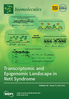
Cover Story (view full-size image): Rett syndrome (RTT) is a devastating neurodevelopmental disease—the pathogenetic mechanisms of which are still under investigation. RTT is caused by mutations in MeCP2, a master epigenetic modulator of transcription, involved in the organization of chromatin architecture. MeCP2 dysfunction leads to large‐scale alterations of the epigenome, including increased oxidation of mC, abnormal binding of chromatin-associated proteins, unbalanced deposition of epigenetic modifications, deregulated gene expression and altered acetylation of histone and transcription factors. Further examination of the epigenomic and transcriptomic alterations caused by MeCP2 dysfunction may help to develop focused therapeutic strategies for RTT. View this paper
  • Issues are regarded as officially published after their release is announced to the table of contents alert mailing list.
  • You may sign up for e-mail alerts to receive table of contents of newly released issues.
  • PDF is the official format for papers published in both, html and pdf forms. To view the papers in pdf format, click on the "PDF Full-text" link, and use the free Adobe Reader to open them.
Order results
Result details
Section
Select all
Export citation of selected articles as:
8 pages, 2567 KiB  
Case Report
Adventitial Microcirculation Is a Major Target of SARS-CoV-2-Mediated Vascular Inflammation
by Francesco Vasuri, Carmen Ciavarella, Salvatore Collura, Chiara Mascoli, Sabrina Valente, Alessio Degiovanni, Mauro Gargiulo, Miriam Capri and Gianandrea Pasquinelli
Biomolecules 2021, 11(7), 1063; https://doi.org/10.3390/biom11071063 - 20 Jul 2021
Cited by 10 | Viewed by 2320
Abstract
We report the case of a 77-year-old woman affected by coronavirus disease-19 (COVID-19) who developed an occlusive arterial disease of the lower limb requiring a left leg amputation. We studied the mechanisms of vascular damage by SARS-CoV-2 by means of a comprehensive multi-technique [...] Read more.
We report the case of a 77-year-old woman affected by coronavirus disease-19 (COVID-19) who developed an occlusive arterial disease of the lower limb requiring a left leg amputation. We studied the mechanisms of vascular damage by SARS-CoV-2 by means of a comprehensive multi-technique in situ analysis on the diseased popliteal arterial district, including immunohistochemistry (IHC), transmission electron microscopy (TEM) and miRNA analysis. At histological analyses, we observed a lymphocytic inflammatory infiltrate, oedema and endothelialitis of adventitial vasa vasorum while the media was normal and the intima had only minor changes. The vasa vasorum expressed the ACE2 receptor and factor VIII; compared with the controls, VEGFR2 staining was reduced. TEM analyses showed endothelial injury and numerous Weibel–Palade bodies in the cytoplasm. No coronavirus particle was seen. IL-6 protein and mRNA, together with miR-155-5p and miRs-27a-5p, which can target IL-6, were significantly increased compared with that in the controls. Our case report suggests an involvement of adventitial artery microcirculation by inflammation in the course of COVID-19. Without evident signs of current infection by SARS-CoV-2, endothelial cells show a spectrum of structural and functional alterations that can fuel the cardiovascular complications observed in people infected with SARS-CoV-2. Full article
Show Figures

Figure 1

10 pages, 2810 KiB  
Article
VEGF Maintains Maternal Vascular Space Homeostasis in the Mouse Placenta through Modulation of Trophoblast Giant Cell Functions
by Xiujun Fan, Shanmugam Muruganandan, Philemon D Shallie, Sabita Dhal, Matthew Petitt and Nihar R Nayak
Biomolecules 2021, 11(7), 1062; https://doi.org/10.3390/biom11071062 - 20 Jul 2021
Cited by 12 | Viewed by 3919
Abstract
Vascular endothelial growth factor (VEGF) is an angiogenic growth factor that acts primarily on endothelial cells, but numerous studies suggest that VEGF also acts on non-endothelial cells, including trophoblast cells. Inhibition of VEGF signaling by excess production of the endogenous soluble VEGF receptor [...] Read more.
Vascular endothelial growth factor (VEGF) is an angiogenic growth factor that acts primarily on endothelial cells, but numerous studies suggest that VEGF also acts on non-endothelial cells, including trophoblast cells. Inhibition of VEGF signaling by excess production of the endogenous soluble VEGF receptor sFlt1 in trophoblast cells has been implicated in several pregnancy complications. Our previous studies and other reports have shown that VEGF directly regulates placental vascular development and functions and that excess VEGF production adversely affects placental vascular development. Trophoblast giant cells (TGCs) line the maternal side of the placental vasculature in mice and function like endothelial cells. In this study, we specifically examined the effect of excess VEGF signaling on TGC development associated with defective placental vascular development using two mouse models an endometrial VEGF overexpression model and a placenta-specific sFlt1 knockdown model. Placentas of endometrial VEGF-overexpressing dams at embryonic days (E) 11.5 and 14.5 showed dramatic enlargement of the venous maternal spaces in junctional zones. The size and number of the parietal TGCs that line these venous spaces in the placenta were also significantly increased. Although junctional zone venous blood spaces from control and VEGF-overexpressing dams were not markedly different in size at E17.5, the number and size of P-TGCs were both significantly increased in the placentas from VEGF-overexpressing dams. In sFlt1 knockdown placentas, however, there was a significant increase in the size of the sinusoidal TGC-lined, alkaline phosphatase-positive maternal blood spaces in the labyrinth. These results suggest that VEGF signaling plays an important role in maintaining the homeostasis of the maternal vascular space in the mouse placenta through modulation of TGC development and differentiation, similar to the effect of VEGF on endothelial cells in other vascular beds. Full article
Show Figures

Figure 1

12 pages, 1646 KiB  
Article
Screening for Fatal Traumatic Brain Injuries in Cerebrospinal Fluid Using Blood-Validated CK and CK–MB Immunoassays
by Johann Zwirner, Sven Anders, Simone Bohnert, Ralph Burkhardt, Ugo Da Broi, Niels Hammer, Dirk Pohlers, Rexson Tse and Benjamin Ondruschka
Biomolecules 2021, 11(7), 1061; https://doi.org/10.3390/biom11071061 - 20 Jul 2021
Cited by 6 | Viewed by 4134
Abstract
A single, specific, sensitive biochemical biomarker that can reliably diagnose a traumatic brain injury (TBI) has not yet been found, but combining different biomarkers would be the most promising approach in clinical and postmortem settings. In addition, identifying new biomarkers and developing laboratory [...] Read more.
A single, specific, sensitive biochemical biomarker that can reliably diagnose a traumatic brain injury (TBI) has not yet been found, but combining different biomarkers would be the most promising approach in clinical and postmortem settings. In addition, identifying new biomarkers and developing laboratory tests can be time-consuming and economically challenging. As such, it would be efficient to use established clinical diagnostic assays for postmortem biochemistry. In this study, postmortem cerebrospinal fluid samples from 45 lethal TBI cases and 47 controls were analyzed using commercially available blood-validated assays for creatine kinase (CK) activity and its heart-type isoenzyme (CK–MB). TBI cases with a survival time of up to two hours showed an increase in both CK and CK–MB with moderate (CK–MB: AUC = 0.788, p < 0.001) to high (CK: AUC = 0.811, p < 0.001) diagnostic accuracy. This reflected the excessive increase of the brain-type CK isoenzyme (CK–BB) following a TBI. The results provide evidence that CK immunoassays can be used as an adjunct quantitative test aid in diagnosing acute TBI-related fatalities. Full article
(This article belongs to the Special Issue Postmortem Biochemistry-When Death Matters)
Show Figures

Figure 1

21 pages, 3307 KiB  
Review
Intrinsic Disorder and Phosphorylation in BRCA2 Facilitate Tight Regulation of Multiple Conserved Binding Events
by Manon Julien, Rania Ghouil, Ambre Petitalot, Sandrine M. Caputo, Aura Carreira and Sophie Zinn-Justin
Biomolecules 2021, 11(7), 1060; https://doi.org/10.3390/biom11071060 - 20 Jul 2021
Cited by 6 | Viewed by 3489
Abstract
The maintenance of genome integrity in the cell is an essential process for the accurate transmission of the genetic material. BRCA2 participates in this process at several levels, including DNA repair by homologous recombination, protection of stalled replication forks, and cell division. These [...] Read more.
The maintenance of genome integrity in the cell is an essential process for the accurate transmission of the genetic material. BRCA2 participates in this process at several levels, including DNA repair by homologous recombination, protection of stalled replication forks, and cell division. These activities are regulated and coordinated via cell-cycle dependent modifications. Pathogenic variants in BRCA2 cause genome instability and are associated with breast and/or ovarian cancers. BRCA2 is a very large protein of 3418 amino acids. Most well-characterized variants causing a strong predisposition to cancer are mutated in the C-terminal 700 residues DNA binding domain of BRCA2. The rest of the BRCA2 protein is predicted to be disordered. Interactions involving intrinsically disordered regions (IDRs) remain difficult to identify both using bioinformatics tools and performing experimental assays. However, the lack of well-structured binding sites provides unique functional opportunities for BRCA2 to bind to a large set of partners in a tightly regulated manner. We here summarize the predictive and experimental arguments that support the presence of disorder in BRCA2. We describe how BRCA2 IDRs mediate self-assembly and binding to partners during DNA double-strand break repair, mitosis, and meiosis. We highlight how phosphorylation by DNA repair and cell-cycle kinases regulate these interactions. We finally discuss the impact of cancer-associated variants on the function of BRCA2 IDRs and more generally on genome stability and cancer risk. Full article
Show Figures

Figure 1

12 pages, 1836 KiB  
Article
JAK2 Phosphorylation Signals and Their Associated Cytokines Involved in Chronic Rhinosinusitis with Nasal Polyps and Correlated with Disease Severity
by Yu-Tsai Lin, Wei-Chih Chen, Ming-Hsien Tsai, Jing-Ying Chen, Chih-Yen Chien and Shun-Chen Huang
Biomolecules 2021, 11(7), 1059; https://doi.org/10.3390/biom11071059 - 20 Jul 2021
Cited by 2 | Viewed by 2302
Abstract
Janus kinase 2 (JAK2) is a member of the JAK family that transduces cytokine-mediated signals via the JAKs/STATs (signal transducer and activator of transcription proteins) pathway, which plays an important role in many inflammatory diseases. This study investigates the association of p-JAK2 and [...] Read more.
Janus kinase 2 (JAK2) is a member of the JAK family that transduces cytokine-mediated signals via the JAKs/STATs (signal transducer and activator of transcription proteins) pathway, which plays an important role in many inflammatory diseases. This study investigates the association of p-JAK2 and JAK2-associated cytokines from nasal polyp (NP) tissue with disease severity, and evaluates the p-JAK2-mediated STATs in chronic rhinosinusitis (CRS) with NP. Sixty-one CRSwNP patients with nasal polyps undergoing endoscopic sinus surgery were enrolled, while the turbinate tissues from 26 nasal obstruction patients were examined as the control group. Elevated levels of p-JAK2 were detected in CRSwNP, and significantly correlated with scores of disease severity (LMK-CT, TPS, and SNOT-22). Expressions of the JAK2-associated cytokines, such as IL-5, IL-6, IL-13, G-CSF, and IFN-γ were significantly higher in CRSwNP than in the controls, while the levels of IL-5, IL-6, IL-13, or G-CSF had positive correlation with scores of disease severity. Moreover, markedly increased expression of p-STAT3 in CRSwNP was observed relative to the control. Taken together, these data showed that the JAK2-associated cytokines including IL-6 and G-CSF may stimulate JAK2 phosphorylation to activate p-STAT3, indicating an association with disease severity and supporting its development of JAK2 inhibitor as a potential therapeutic agent for CRS. Full article
Show Figures

Figure 1

20 pages, 1102 KiB  
Review
Oligodendrocytes and Microglia: Key Players in Myelin Development, Damage and Repair
by Ilias Kalafatakis and Domna Karagogeos
Biomolecules 2021, 11(7), 1058; https://doi.org/10.3390/biom11071058 - 20 Jul 2021
Cited by 35 | Viewed by 8484
Abstract
Oligodendrocytes, the myelin-making cells of the CNS, regulate the complex process of myelination under physiological and pathological conditions, significantly aided by other glial cell types such as microglia, the brain-resident, macrophage-like innate immune cells. In this review, we summarize how oligodendrocytes orchestrate myelination, [...] Read more.
Oligodendrocytes, the myelin-making cells of the CNS, regulate the complex process of myelination under physiological and pathological conditions, significantly aided by other glial cell types such as microglia, the brain-resident, macrophage-like innate immune cells. In this review, we summarize how oligodendrocytes orchestrate myelination, and especially myelin repair after damage, and present novel aspects of oligodendroglial functions. We emphasize the contribution of microglia in the generation and regeneration of myelin by discussing their beneficial and detrimental roles, especially in remyelination, underlining the cellular and molecular components involved. Finally, we present recent findings towards human stem cell-derived preclinical models for the study of microglia in human pathologies and on the role of microbiome on glial cell functions. Full article
(This article belongs to the Special Issue Molecular and Cellular Mechanisms of Neuroinflammation)
Show Figures

Figure 1

23 pages, 9050 KiB  
Article
Interaction Mode of the Novel Monobactam AIC499 Targeting Penicillin Binding Protein 3 of Gram-Negative Bacteria
by Stefan Freischem, Immanuel Grimm, Arancha López-Pérez, Dieter Willbold, Burkhard Klenke, Cuong Vuong, Andrew J. Dingley and Oliver H. Weiergräber
Biomolecules 2021, 11(7), 1057; https://doi.org/10.3390/biom11071057 - 19 Jul 2021
Cited by 9 | Viewed by 3301 | Correction
Abstract
Novel antimicrobial strategies are urgently required because of the rising threat of multi drug resistant bacterial strains and the infections caused by them. Among the available target structures, the so-called penicillin binding proteins are of particular interest, owing to their good accessibility in [...] Read more.
Novel antimicrobial strategies are urgently required because of the rising threat of multi drug resistant bacterial strains and the infections caused by them. Among the available target structures, the so-called penicillin binding proteins are of particular interest, owing to their good accessibility in the periplasmic space, and the lack of homologous proteins in humans, reducing the risk of side effects of potential drugs. In this report, we focus on the interaction of the innovative β-lactam antibiotic AIC499 with penicillin binding protein 3 (PBP3) from Escherichia coli and Pseudomonas aeruginosa. This recently developed monobactam displays broad antimicrobial activity, against Gram-negative strains, and improved resistance to most classes of β-lactamases. By analyzing crystal structures of the respective complexes, we were able to explore the binding mode of AIC499 to its target proteins. In addition, the apo structures determined for PBP3, from P. aeruginosa and the catalytic transpeptidase domain of the E. coli orthologue, provide new insights into the dynamics of these proteins and the impact of drug binding. Full article
Show Figures

Figure 1

17 pages, 4446 KiB  
Article
Can Glycosylation Mask the Detection of MHC Expressing p53 Peptides by T Cell Receptors?
by Thanh Binh Nguyen, David P. Lane and Chandra S. Verma
Biomolecules 2021, 11(7), 1056; https://doi.org/10.3390/biom11071056 - 19 Jul 2021
Cited by 1 | Viewed by 2612
Abstract
Proteins of the major histocompatibility complex (MHC) class I, or human leukocyte antigen (HLA) in humans interact with endogenous peptides and present them to T cell receptors (TCR), which in turn tune the immune system to recognize and discriminate between self and foreign [...] Read more.
Proteins of the major histocompatibility complex (MHC) class I, or human leukocyte antigen (HLA) in humans interact with endogenous peptides and present them to T cell receptors (TCR), which in turn tune the immune system to recognize and discriminate between self and foreign (non-self) peptides. Of especial importance are peptides derived from tumor-associated antigens. T cells recognizing these peptides are found in cancer patients, but not in cancer-free individuals. What stimulates this recognition, which is vital for the success of checkpoint based therapy? A peptide derived from the protein p53 (residues 161–169 or p161) was reported to show this behavior. T cells recognizing this unmodified peptide could be further stimulated in vitro to create effective cancer killing CTLs (cytotoxic T lymphocytes). We hypothesize that the underlying difference may arise from post-translational glycosylation of p161 in normal individuals, likely masking it against recognition by TCR. Defects in glycosylation in cancer cells may allow the presentation of the native peptide. We investigate the structural consequences of such peptide glycosylation by investigating the associated structural dynamics. Full article
(This article belongs to the Special Issue Protein–Protein Interactions: Methods and Applications)
Show Figures

Figure 1

26 pages, 1644 KiB  
Perspective
Wrestling and Wrapping: A Perspective on SUMO Proteins in Schwann Cells
by Iman F. Fergani and Luciana R. Frick
Biomolecules 2021, 11(7), 1055; https://doi.org/10.3390/biom11071055 - 19 Jul 2021
Cited by 1 | Viewed by 2981
Abstract
Schwann cell development and peripheral nerve myelination are finely orchestrated multistep processes; some of the underlying mechanisms are well described and others remain unknown. Many posttranslational modifications (PTMs) like phosphorylation and ubiquitination have been reported to play a role during the normal development [...] Read more.
Schwann cell development and peripheral nerve myelination are finely orchestrated multistep processes; some of the underlying mechanisms are well described and others remain unknown. Many posttranslational modifications (PTMs) like phosphorylation and ubiquitination have been reported to play a role during the normal development of the peripheral nervous system (PNS) and in demyelinating neuropathies. However, a relatively novel PTM, SUMOylation, has not been studied in these contexts. SUMOylation involves the covalent attachment of one or more small ubiquitin-like modifier (SUMO) proteins to a substrate, which affects the function, cellular localization, and further PTMs of the conjugated protein. SUMOylation also regulates other proteins indirectly by facilitating non-covalent protein–protein interaction via SUMO interaction motifs (SIM). This pathway has important consequences on diverse cellular processes, and dysregulation of this pathway has been reported in several diseases including neurological and degenerative conditions. In this article, we revise the scarce literature on SUMOylation in Schwann cells and the PNS, we propose putative substrate proteins, and we speculate on potential mechanisms underlying the possible involvement of this PTM in peripheral myelination and neuropathies. Full article
(This article belongs to the Special Issue Neuroimmune and Neuroglial Interactions in Health and Disease)
Show Figures

Figure 1

13 pages, 3314 KiB  
Article
Suppression of Inflammation-Associated Kidney Damage Post-Transplant Using the New PrC-210 Free Radical Scavenger in Rats
by Torsten R. Goesch, Nancy A. Wilson, Weifeng Zeng, Bret M. Verhoven, Weixiong Zhong, Maya M. Coumbe Gitter and William E. Fahl
Biomolecules 2021, 11(7), 1054; https://doi.org/10.3390/biom11071054 - 19 Jul 2021
Cited by 3 | Viewed by 2668
Abstract
Allograft kidney transplantation, which triggers host cellular- and antibody-mediated rejection of the kidney, is a major contributor to kidney damage during transplant. Here, we asked whether PrC-210 would suppress damage seen in allograft kidney transplant. Brown Norway (BN) rat kidneys were perfused in [...] Read more.
Allograft kidney transplantation, which triggers host cellular- and antibody-mediated rejection of the kidney, is a major contributor to kidney damage during transplant. Here, we asked whether PrC-210 would suppress damage seen in allograft kidney transplant. Brown Norway (BN) rat kidneys were perfused in situ (UW Solution) with or without added 30 mM PrC-210, and then immediately transplanted into Lewis (LEW) rats. 20 h later, the transplanted BN kidneys and LEW rat plasma were analyzed. Kidney histology, and kidney/serum levels of several inflammation-associated cytokines, were measured to assess mismatch-related kidney pathology, and PrC-210 protective efficacy. Twenty hours after the allograft transplants: (i) significant histologic kidney tubule damage and mononuclear inflammatory cell infiltration were seen in allograft kidneys; (ii) kidney function metrics (creatinine and BUN) were significantly elevated; (iii) significant changes in key cytokines, i.e., TIMP-1, TNF-alpha and MIP-3A/CCL20, and kidney activated caspase levels were seen. In PrC-210-treated kidneys and recipient rats, (i) kidney histologic damage (Banff Scores) and mononuclear infiltration were reduced to untreated background levels; (ii) creatinine and BUN were significantly reduced; and (iii) activated caspase and cytokine changes were significantly reduced, some to background. In conclusion, the results suggest that PrC-210 could provide broadly applicable organ protection for many allograft transplantation conditions; it could protect transplanted kidneys during and after all stages of the transplantation process—from organ donation, through transportation, re-implantation and the post-operative inflammation—to minimize acute and chronic rejection. Full article
(This article belongs to the Special Issue Redox Imbalance and Mitochondrial Abnormalities in Kidney Disease)
Show Figures

Figure 1

10 pages, 857 KiB  
Article
Sesquiterpene Lactones with the 12,8-Guaianolide Skeleton from Algerian Centaurea omphalotricha
by Sara Kebbi, Maria Letizia Ciavatta, Ali Mokhtar Mahmoud, Marianna Carbone, Alessia Ligresti, Ramdane Seghiri and Margherita Gavagnin
Biomolecules 2021, 11(7), 1053; https://doi.org/10.3390/biom11071053 - 18 Jul 2021
Cited by 7 | Viewed by 2370
Abstract
In continuing our investigation on the chemical diversity of Algerian plants, we examined Centaurea omphalotricha, whose chemical composition has been poorly studied. The present work was aimed at characterizing the secondary metabolite pattern of the CHCl3 extract of the aerial parts [...] Read more.
In continuing our investigation on the chemical diversity of Algerian plants, we examined Centaurea omphalotricha, whose chemical composition has been poorly studied. The present work was aimed at characterizing the secondary metabolite pattern of the CHCl3 extract of the aerial parts of this plant that displayed antiproliferative properties in a preliminary screening on HeLa cell line. The chemical analysis led us to characterize the bioactive oxygenated terpenoid fraction which includes, within major known metabolites, two new minor sesquiterpene lactones, centaurolide-A (1) and centaurolide-B (2). The structures of two compounds exhibiting the 12,8-guaianolide skeleton were determined by spectroscopic methods as well as by chemical correlation with inuviscolide (3), a well-known bioactive guaianolide isolated from Dittrichia (=Inula) viscosa. Centaurolides A and B represent the first report of 8,12-guaianolide skeleton in Centaurea genus. The effect of new compounds 1 and 2 and inuviscolide (3) on HeLa cell has also been evaluated. Full article
Show Figures

Graphical abstract

9 pages, 1410 KiB  
Article
Combined Beneficial Effect of Genistein and Atorvastatin on Adipogenesis in 3T3-L1 Adipocytes
by Dahae Lee, Ji-Youn Kim, Hae-Won Kim, Jeong-Eun Yoo and Ki Sung Kang
Biomolecules 2021, 11(7), 1052; https://doi.org/10.3390/biom11071052 - 18 Jul 2021
Cited by 4 | Viewed by 2644
Abstract
Genistein (4,5,7-trihydroxyisoflavone) is abundant in various dietary vegetables, especially soybeans, and is known to have not only an estrogenic effect but also an antiadipogenic effect. Atorvastatin (dihydroxy monocarboxylic acid) is a statin used to prevent heart disease. Although genistein and atorvastatin have been [...] Read more.
Genistein (4,5,7-trihydroxyisoflavone) is abundant in various dietary vegetables, especially soybeans, and is known to have not only an estrogenic effect but also an antiadipogenic effect. Atorvastatin (dihydroxy monocarboxylic acid) is a statin used to prevent heart disease. Although genistein and atorvastatin have been reported to possess antiadipogenic effects, their combined effects are still unclear. The aim of the current study was to explore whether the combination of genistein and atorvastatin at low concentrations significantly suppresses adipogenesis in a murine preadipocyte cell line (3T3-L1) compared to treatment with genistein or atorvastatin alone. Our results showed that cotreatment with 50 µM genistein and 50 nM atorvastatin significantly suppressed preadipocyte differentiation, whereas when each compound was used alone, there was no inhibitory effect. Additionally, cotreatment with genistein and atorvastatin significantly downregulated adipogenic marker proteins, including mitogen-activated protein kinases (MAPKs), peroxisome proliferator-activated receptor γ (PPARγ), CCAAT/enhancer-binding protein alpha (C/EBPα), glucocorticoid receptor (GR), and CCAAT/enhancer-binding protein β (C/EBPβ). This is the first evidence of the combined antiadipogenic effects of genistein and atorvastatin. Although additional experiments are required, combinational treatment with genistein and atorvastatin may be an alternative treatment for menopause-associated lipid metabolic disorders and obesity. Full article
Show Figures

Graphical abstract

21 pages, 10020 KiB  
Article
Exploration of the Activation Mechanism of the Epigenetic Regulator MLL3: A QM/MM Study
by Sebastián Miranda-Rojas, Kevin Blanco-Esperguez, Iñaki Tuñón, Johannes Kästner and Fernando Mendizábal
Biomolecules 2021, 11(7), 1051; https://doi.org/10.3390/biom11071051 - 17 Jul 2021
Cited by 3 | Viewed by 2227
Abstract
The mixed lineage leukemia 3 or MLL3 is the enzyme in charge of the writing of an epigenetic mark through the methylation of lysine 4 from the N-terminal domain of histone 3 and its deregulation has been related to several cancer lines. An [...] Read more.
The mixed lineage leukemia 3 or MLL3 is the enzyme in charge of the writing of an epigenetic mark through the methylation of lysine 4 from the N-terminal domain of histone 3 and its deregulation has been related to several cancer lines. An interesting feature of this enzyme comes from its regulation mechanism, which involves its binding to an activating dimer before it can be catalytically functional. Once the trimer is formed, the reaction mechanism proceeds through the deprotonation of the lysine followed by the methyl-transfer reaction. Here we present a detailed exploration of the activation mechanism through a QM/MM approach focusing on both steps of the reaction, aiming to provide new insights into the deprotonation process and the role of the catalytic machinery in the methyl-transfer reaction. Our finding suggests that the source of the activation mechanism comes from conformational restriction mediated by the formation of a network of salt-bridges between MLL3 and one of the activating subunits, which restricts and stabilizes the positioning of several residues relevant for the catalysis. New insights into the deprotonation mechanism of lysine are provided, identifying a valine residue as crucial in the positioning of the water molecule in charge of the process. Finally, a tyrosine residue was found to assist the methyl transfer from SAM to the target lysine. Full article
Show Figures

Figure 1

29 pages, 1174 KiB  
Review
Mitochondria and Antibiotics: For Good or for Evil?
by Juan M. Suárez-Rivero, Carmen J. Pastor-Maldonado, Suleva Povea-Cabello, Mónica Álvarez-Córdoba, Irene Villalón-García, Marta Talaverón-Rey, Alejandra Suárez-Carrillo, Manuel Munuera-Cabeza and José A. Sánchez-Alcázar
Biomolecules 2021, 11(7), 1050; https://doi.org/10.3390/biom11071050 - 17 Jul 2021
Cited by 21 | Viewed by 5736
Abstract
The discovery and application of antibiotics in the common clinical practice has undeniably been one of the major medical advances in our times. Their use meant a drastic drop in infectious diseases-related mortality and contributed to prolonging human life expectancy worldwide. Nevertheless, antibiotics [...] Read more.
The discovery and application of antibiotics in the common clinical practice has undeniably been one of the major medical advances in our times. Their use meant a drastic drop in infectious diseases-related mortality and contributed to prolonging human life expectancy worldwide. Nevertheless, antibiotics are considered by many a double-edged sword. Their extensive use in the past few years has given rise to a global problem: antibiotic resistance. This factor and the increasing evidence that a wide range of antibiotics can damage mammalian mitochondria, have driven a significant sector of the medical and scientific communities to advise against the use of antibiotics for purposes other to treating severe infections. Notwithstanding, a notorious number of recent studies support the use of these drugs to treat very diverse conditions, ranging from cancer to neurodegenerative or mitochondrial diseases. In this context, there is great controversy on whether the risks associated to antibiotics outweigh their promising beneficial features. The aim of this review is to provide insight in the topic, purpose for which the most relevant findings regarding antibiotic therapies have been discussed. Full article
Show Figures

Figure 1

19 pages, 2335 KiB  
Review
Celecoxib Analogues for Cancer Treatment: An Update on OSU-03012 and 2,5-Dimethyl-Celecoxib
by Cyril Sobolewski and Noémie Legrand
Biomolecules 2021, 11(7), 1049; https://doi.org/10.3390/biom11071049 - 16 Jul 2021
Cited by 8 | Viewed by 3336
Abstract
Cyclooxygenase-2 (COX-2) is an important enzyme involved in prostaglandins biosynthesis from arachidonic acid. COX-2 is frequently overexpressed in human cancers and plays a major tumor promoting function. Accordingly, many efforts have been devoted to efficiently target the catalytic site of this enzyme in [...] Read more.
Cyclooxygenase-2 (COX-2) is an important enzyme involved in prostaglandins biosynthesis from arachidonic acid. COX-2 is frequently overexpressed in human cancers and plays a major tumor promoting function. Accordingly, many efforts have been devoted to efficiently target the catalytic site of this enzyme in cancer cells, by using COX-2 specific inhibitors such as celecoxib. However, despite their potent anti-tumor properties, the myriad of detrimental effects associated to the chronic inhibition of COX-2 in healthy tissues, has considerably limited their use in clinic. In addition, increasing evidence indicate that these anti-cancerous properties are not strictly dependent on the inhibition of the catalytic site. These findings have led to the development of non-active COX-2 inhibitors analogues aiming at preserving the antitumor effects of COX-2 inhibitors without their side effects. Among them, two celecoxib derivatives, 2,5-Dimethyl-Celecoxib and OSU-03012, have been developed and suggested for the treatment of viral (e.g., recently SARS-CoV-2), inflammatory, metabolic diseases and cancers. These molecules display stronger anti-tumor properties than celecoxib and thus may represent promising anti-cancer molecules. In this review, we discuss the impact of these two analogues on cancerous processes but also their potential for cancer treatment alone or in combination with existing approaches. Full article
(This article belongs to the Special Issue Novel Molecules for Cancer Treatment)
Show Figures

Figure 1

17 pages, 22198 KiB  
Article
Interference of Polydatin/Resveratrol in the ACE2:Spike Recognition during COVID-19 Infection. A Focus on Their Potential Mechanism of Action through Computational and Biochemical Assays
by Fulvio Perrella, Federico Coppola, Alessio Petrone, Chiara Platella, Daniela Montesarchio, Annarita Stringaro, Giampietro Ravagnan, Maria Pia Fuggetta, Nadia Rega and Domenica Musumeci
Biomolecules 2021, 11(7), 1048; https://doi.org/10.3390/biom11071048 - 16 Jul 2021
Cited by 22 | Viewed by 4474
Abstract
In the search for new therapeutic strategies to contrast SARS-CoV-2, we here studied the interaction of polydatin (PD) and resveratrol (RESV)—two natural stilbene polyphenols with manifold, well known biological activities—with Spike, the viral protein essential for virus entry into host cells, and ACE2, [...] Read more.
In the search for new therapeutic strategies to contrast SARS-CoV-2, we here studied the interaction of polydatin (PD) and resveratrol (RESV)—two natural stilbene polyphenols with manifold, well known biological activities—with Spike, the viral protein essential for virus entry into host cells, and ACE2, the angiotensin-converting enzyme present on the surface of multiple cell types (including respiratory epithelial cells) which is the main host receptor for Spike binding. Molecular Docking simulations evidenced that both compounds can bind Spike, ACE2 and the ACE2:Spike complex with good affinity, although the interaction of PD appears stronger than that of RESV on all the investigated targets. Preliminary biochemical assays revealed a significant inhibitory activity of the ACE2:Spike recognition with a dose-response effect only in the case of PD. Full article
Show Figures

Graphical abstract

18 pages, 1270 KiB  
Review
Osteopontin in Cardiovascular Diseases
by Kohsuke Shirakawa and Motoaki Sano
Biomolecules 2021, 11(7), 1047; https://doi.org/10.3390/biom11071047 - 16 Jul 2021
Cited by 37 | Viewed by 5220
Abstract
Unprecedented advances in secondary prevention have greatly improved the prognosis of cardiovascular diseases (CVDs); however, CVDs remain a leading cause of death globally. These findings suggest the need to reconsider cardiovascular risk and optimal medical therapy. Numerous studies have shown that inflammation, pro-thrombotic [...] Read more.
Unprecedented advances in secondary prevention have greatly improved the prognosis of cardiovascular diseases (CVDs); however, CVDs remain a leading cause of death globally. These findings suggest the need to reconsider cardiovascular risk and optimal medical therapy. Numerous studies have shown that inflammation, pro-thrombotic factors, and gene mutations are focused not only on cardiovascular residual risk but also as the next therapeutic target for CVDs. Furthermore, recent clinical trials, such as the Canakinumab Anti-inflammatory Thrombosis Outcomes Study trial, showed the possibility of anti-inflammatory therapy for patients with CVDs. Osteopontin (OPN) is a matricellular protein that mediates diverse biological functions and is involved in a number of pathological states in CVDs. OPN has a two-faced phenotype that is dependent on the pathological state. Acute increases in OPN have protective roles, including wound healing, neovascularization, and amelioration of vascular calcification. By contrast, chronic increases in OPN predict poor prognosis of a major adverse cardiovascular event independent of conventional cardiovascular risk factors. Thus, OPN can be a therapeutic target for CVDs but is not clinically available. In this review, we discuss the role of OPN in the development of CVDs and its potential as a therapeutic target. Full article
(This article belongs to the Special Issue Molecular Biomarkers In Cardiology 2021)
Show Figures

Figure 1

15 pages, 3594 KiB  
Article
Bisphenol a Exposure and Kidney Diseases: Systematic Review, Meta-Analysis, and NHANES 03–16 Study
by Rafael Moreno-Gómez-Toledano, María I. Arenas, Esperanza Vélez-Vélez, Elisabeth Coll, Borja Quiroga, Jordi Bover and Ricardo J. Bosch
Biomolecules 2021, 11(7), 1046; https://doi.org/10.3390/biom11071046 - 16 Jul 2021
Cited by 23 | Viewed by 3499
Abstract
Bisphenol A (BPA) is a compound that is especially widespread in most commonly used objects due to its multiple uses in the plastic industry. However, several data support the need to restrict its use. In recent years, new implications of BPA on the [...] Read more.
Bisphenol A (BPA) is a compound that is especially widespread in most commonly used objects due to its multiple uses in the plastic industry. However, several data support the need to restrict its use. In recent years, new implications of BPA on the renal system have been discovered, which denotes the need to expand studies in patients. To this end, a systematic review and a meta-analysis was performed to explore existing literature that examines the BPA-kidney disease paradigm and to determine what and how future studies will need to be carried out. Our systematic review revealed that only few relevant publications have focused on the problem. However, the subsequent meta-analysis revealed that high blood concentrations of BPA could be a factor in developing kidney disease, at least in people with previous pathologies such as diabetes or hypertension. Furthermore, BPA could also represent a risk factor in healthy people whose urinary excretion is higher. Finally, the data analyzed from the NHANES 03-16 cohort provided new evidence on the possible involvement of BPA in kidney disease. Therefore, our results underline the need to carry out a thorough and methodologically homogeneous study, delving into the relationship between urinary and blood BPA, glomerular filtration rate, and urine albumin-to-creatinine ratio, preferably in population groups at risk, and subsequently in the general population, to solve this relevant conundrum with critical potential implications in Public Health. Full article
Show Figures

Figure 1

18 pages, 14341 KiB  
Review
Leptin-Activity Modulators and Their Potential Pharmaceutical Applications
by Marianna Greco, Marzia De Santo, Alessandra Comandè, Emilia Lucia Belsito, Sebastiano Andò, Angelo Liguori and Antonella Leggio
Biomolecules 2021, 11(7), 1045; https://doi.org/10.3390/biom11071045 - 16 Jul 2021
Cited by 13 | Viewed by 4466
Abstract
Leptin, a multifunctional hormone primarily, but not exclusively, secreted in adipose tissue, is implicated in a wide range of biological functions that control different processes, such as the regulation of body weight and energy expenditure, reproductive function, immune response, and bone metabolism. In [...] Read more.
Leptin, a multifunctional hormone primarily, but not exclusively, secreted in adipose tissue, is implicated in a wide range of biological functions that control different processes, such as the regulation of body weight and energy expenditure, reproductive function, immune response, and bone metabolism. In addition, leptin can exert angiogenic and mitogenic actions in peripheral organs. Leptin biological activities are greatly related to its interaction with the leptin receptor. Both leptin excess and leptin deficiency, as well as leptin resistance, are correlated with different human pathologies, such as autoimmune diseases and cancers, making leptin and leptin receptor important drug targets. The development of leptin signaling modulators represents a promising strategy for the treatment of cancers and other leptin-related diseases. In the present manuscript, we provide an update review about leptin-activity modulators, comprising leptin mutants, peptide-based leptin modulators, as well as leptin and leptin receptor specific monoclonal antibodies and nanobodies. Full article
(This article belongs to the Special Issue Leptin and Beyond: Actors in Cancer)
Show Figures

Figure 1

12 pages, 2294 KiB  
Article
Enzymatic and Chemical Syntheses of Vacor Analogs of Nicotinamide Riboside, NMN and NAD
by Lars Jansen Sverkeli, Faisal Hayat, Marie E. Migaud and Mathias Ziegler
Biomolecules 2021, 11(7), 1044; https://doi.org/10.3390/biom11071044 - 16 Jul 2021
Cited by 11 | Viewed by 3659
Abstract
It has recently been demonstrated that the rat poison vacor interferes with mammalian NAD metabolism, because it acts as a nicotinamide analog and is converted by enzymes of the NAD salvage pathway. Thereby, vacor is transformed into the NAD analog vacor adenine dinucleotide [...] Read more.
It has recently been demonstrated that the rat poison vacor interferes with mammalian NAD metabolism, because it acts as a nicotinamide analog and is converted by enzymes of the NAD salvage pathway. Thereby, vacor is transformed into the NAD analog vacor adenine dinucleotide (VAD), a molecule that causes cell toxicity. Therefore, vacor may potentially be exploited to kill cancer cells. In this study, we have developed efficient enzymatic and chemical procedures to produce vacor analogs of NAD and nicotinamide riboside (NR). VAD was readily generated by a base-exchange reaction, replacing the nicotinamide moiety of NAD by vacor, catalyzed by Aplysia californica ADP ribosyl cyclase. Additionally, we present the chemical synthesis of the nucleoside version of vacor, vacor riboside (VR). Similar to the physiological NAD precursor, NR, VR was converted to the corresponding mononucleotide (VMN) by nicotinamide riboside kinases (NRKs). This conversion is quantitative and very efficient. Consequently, phosphorylation of VR by NRKs represents a valuable alternative to produce the vacor analog of NMN, compared to its generation from vacor by nicotinamide phosphoribosyltransferase (NamPT). Full article
Show Figures

Figure 1

22 pages, 11492 KiB  
Article
In Silico Analysis of the Enzymes Involved in Haloarchaeal Denitrification
by Eric Bernabeu, Jose María Miralles-Robledillo, Micaela Giani, Elena Valdés, Rosa María Martínez-Espinosa and Carmen Pire
Biomolecules 2021, 11(7), 1043; https://doi.org/10.3390/biom11071043 - 16 Jul 2021
Cited by 3 | Viewed by 3016
Abstract
During the last century, anthropogenic activities such as fertilization have led to an increase in pollution in many ecosystems by nitrogen compounds. Consequently, researchers aim to reduce nitrogen pollutants following different strategies. Some haloarchaea, owing to their denitrifier metabolism, have been proposed as [...] Read more.
During the last century, anthropogenic activities such as fertilization have led to an increase in pollution in many ecosystems by nitrogen compounds. Consequently, researchers aim to reduce nitrogen pollutants following different strategies. Some haloarchaea, owing to their denitrifier metabolism, have been proposed as good model organisms for the removal of not only nitrate, nitrite, and ammonium, but also (per)chlorates and bromate in brines and saline wastewater. Bacterial denitrification has been extensively described at the physiological, biochemical, and genetic levels. However, their haloarchaea counterparts remain poorly described. In previous work the model structure of nitric oxide reductase was analysed. In this study, a bioinformatic analysis of the sequences and the structural models of the nitrate, nitrite and nitrous oxide reductases has been described for the first time in the haloarchaeon model Haloferax mediterranei. The main residues involved in the catalytic mechanism and in the coordination of the metal centres have been explored to shed light on their structural characterization and classification. These results set the basis for understanding the molecular mechanism for haloarchaeal denitrification, necessary for the use and optimization of these microorganisms in bioremediation of saline environments among other potential applications including bioremediation of industrial waters. Full article
(This article belongs to the Special Issue Advances in Membrane Proteins 2021)
Show Figures

Figure 1

14 pages, 2844 KiB  
Article
Computational Probing the Methylation Sites Related to EGFR Inhibitor-Responsive Genes
by Rui Yuan, Shilong Chen and Yongcui Wang
Biomolecules 2021, 11(7), 1042; https://doi.org/10.3390/biom11071042 - 16 Jul 2021
Cited by 2 | Viewed by 2243
Abstract
The emergence of drug resistance is one of the main obstacles to the treatment of lung cancer patients with EGFR inhibitors. Here, to further understand the mechanism of EGFR inhibitors in lung cancer and offer novel therapeutic targets for anti-EGFR-inhibitor resistance via the [...] Read more.
The emergence of drug resistance is one of the main obstacles to the treatment of lung cancer patients with EGFR inhibitors. Here, to further understand the mechanism of EGFR inhibitors in lung cancer and offer novel therapeutic targets for anti-EGFR-inhibitor resistance via the deep mining of pharmacogenomics data, we associated DNA methylation with drug sensitivities for uncovering the methylation sites related to EGFR inhibitor sensitivity genes. Specifically, we first introduced a grouped regularized regression model (Group Least Absolute Shrinkage and Selection Operator, group lasso) to detect the genes that were closely related to EGFR inhibitor effectiveness. Then, we applied the classical regression model (lasso) to identify the methylation sites associated with the above drug sensitivity genes. The new model was validated on the well-known cancer genomics resource: CTRP. GeneHancer and Encyclopedia of DNA Elements (ENCODE) database searches indicated that the predicted methylation sites related to EGFR inhibitor sensitivity genes were related to regulatory elements. Moreover, the correlation analysis on sensitivity genes and predicted methylation sites suggested that the methylation sites located in the promoter region were more correlated with the expression of EGFR inhibitor sensitivity genes than those located in the enhancer region and the TFBS. Meanwhile, we performed differential expression analysis of genes and predicted methylation sites and found that changes in the methylation level of some sites may affect the expression of the corresponding EGFR inhibitor-responsive genes. Therefore, we supposed that the effectiveness of EGFR inhibitors in lung cancer may be improved by methylation modification in their sensitivity genes. Full article
(This article belongs to the Special Issue Novel Indications of Epigenetic Therapy in Cancer)
Show Figures

Figure 1

14 pages, 600 KiB  
Article
Whole-Body Cryotherapy Increases the Activity of Nitric Oxide Synthase in Older Men
by Magdalena Wiecek, Zbigniew Szygula, Joanna Gradek, Justyna Kusmierczyk and Jadwiga Szymura
Biomolecules 2021, 11(7), 1041; https://doi.org/10.3390/biom11071041 - 16 Jul 2021
Cited by 8 | Viewed by 2705
Abstract
Aging causes oxidative stress, endothelial dysfunction and a reduction in the bioavailability of nitric oxide. The study aim was to determine whether, as a result of repeated whole-body exposure to cryogenic temperature (3 min −130 °C), there is an increase of inducible nitric [...] Read more.
Aging causes oxidative stress, endothelial dysfunction and a reduction in the bioavailability of nitric oxide. The study aim was to determine whether, as a result of repeated whole-body exposure to cryogenic temperature (3 min −130 °C), there is an increase of inducible nitric oxide synthase (iNOS) concentration in senior subjects (59 ± 6 years), and if this effect is stronger in athletes. In 10 long-distance runners (RUN) and 10 untraining (UTR) men, 24 whole-body cryotherapy (WBC) procedures were performed. Prior to WBC, after 12th and 24th treatments and 7 days later, the concentration of iNOS, asymmetric dimethylarginine (ADMA), 3-nitrotyrosine (3-NTR), homocysteine (HCY), C-reactive protein (CRP) and interleukins such as: IL-6, IL-1β, IL-10 were measured. In the RUN and UTR groups, after 24 WBC, iNOS concentration was found to be comparable and significantly higher (F = 5.95, p < 0.01) (large clinical effect size) compared to before 1st WBC and after 12th WBC sessions. There were no changes in the concentration of the remaining markers as a result of WBC (p > 0.05). As a result of applying 24 WBC treatments, using the every-other-day model, iNOS concentration increased in the group of older men, regardless of their physical activity level. Along with this increase, there were no changes in nitro-oxidative stress or inflammation marker levels. Full article
Show Figures

Figure 1

17 pages, 43366 KiB  
Communication
Systematic Structure-Based Search for Ochratoxin-Degrading Enzymes in Proteomes from Filamentous Fungi
by Ana Lúcia Leitão and Francisco J. Enguita
Biomolecules 2021, 11(7), 1040; https://doi.org/10.3390/biom11071040 - 16 Jul 2021
Cited by 10 | Viewed by 2716
Abstract
(1) Background: ochratoxins are mycotoxins produced by filamentous fungi with important implications in the food manufacturing industry due to their toxicity. Decontamination by specific ochratoxin-degrading enzymes has become an interesting alternative for the treatment of contaminated food commodities. (2) Methods: using a structure-based [...] Read more.
(1) Background: ochratoxins are mycotoxins produced by filamentous fungi with important implications in the food manufacturing industry due to their toxicity. Decontamination by specific ochratoxin-degrading enzymes has become an interesting alternative for the treatment of contaminated food commodities. (2) Methods: using a structure-based approach based on homology modeling, blind molecular docking of substrates and characterization of low-frequency protein motions, we performed a proteome mining in filamentous fungi to characterize new enzymes with potential ochratoxinase activity. (3) Results: the proteome mining results demonstrated the ubiquitous presence of fungal binuclear zinc-dependent amido-hydrolases with a high degree of structural homology to the already characterized ochratoxinase from Aspergillus niger. Ochratoxinase-like enzymes from ochratoxin-producing fungi showed more favorable substrate-binding pockets to accommodate ochratoxins A and B. (4) Conclusions: filamentous fungi are an interesting and rich source of hydrolases potentially capable of degrading ochratoxins, and could be used for the detoxification of diverse food commodities. Full article
Show Figures

Graphical abstract

20 pages, 10595 KiB  
Article
Anti-Proliferative and Anti-Migratory Activities of Hispidulin on Human Melanoma A2058 Cells
by Chi-Jen Chang, Yen-Ling Hung, Ting-Chen Chen, Hsin-Ju Li, Yuan-Hsin Lo, Nan-Lin Wu, Der-Chen Chang and Chi-Feng Hung
Biomolecules 2021, 11(7), 1039; https://doi.org/10.3390/biom11071039 - 16 Jul 2021
Cited by 7 | Viewed by 2582
Abstract
Melanoma represents less than 5% of skin cancers, but is the most lethal, mainly because of its high-metastatic potential and resistance to various therapies. Therefore, it is important to develop effective treatments, especially chemotherapeutic drugs with cytotoxicity, anti-metastaticity, and few side effects. One [...] Read more.
Melanoma represents less than 5% of skin cancers, but is the most lethal, mainly because of its high-metastatic potential and resistance to various therapies. Therefore, it is important to develop effective treatments, especially chemotherapeutic drugs with cytotoxicity, anti-metastaticity, and few side effects. One such natural product is hispidulin, a flavone distributed in plants of the Asteraceae. Previous studies have demonstrated that hispidulin has various pharmacological benefits, such as anti-tumor, anti-inflammation, and anti-allergic effects. This study aims to explore the effects of hispidulin against melanoma in vitro and in vivo. Results revealed that hispidulin selectively decreased the cell viability of A2058 cells in a dose- and time-dependent manner. Hispidulin induced cells accumulated in the sub-G1 phase via activating caspase 8 and 9, increased cleaved caspase 3, and cleaved PARP expression. Hispidulin was able to decrease AKT and ERK phosphorylation, which facilitated cell growth and survival. Moreover, hispidulin promoted reactive oxygen species generation in cells and suppressed cell migration through downregulated matrix metalloproteinase-2 expression. Hispidulin significantly inhibited tumor growth in a xenograft model. Based on these results, hispidulin produces its anti-melanoma effects by inducing cancer cell apoptosis and reducing its migration. Therefore, we suggest hispidulin as a potent therapeutic candidate for melanoma treatment. Full article
Show Figures

Figure 1

15 pages, 1084 KiB  
Review
Epithelial Protein Lost in Neoplasm, EPLIN, the Cellular and Molecular Prospects in Cancers
by Jianyuan Zeng, Wen G. Jiang and Andrew J. Sanders
Biomolecules 2021, 11(7), 1038; https://doi.org/10.3390/biom11071038 - 16 Jul 2021
Cited by 10 | Viewed by 3000
Abstract
Epithelial Protein Lost In Neoplasm (EPLIN), also known as LIMA1 (LIM Domain And Actin Binding 1), was first discovered as a protein differentially expressed in normal and cancerous cell lines. It is now known to be key to the progression and metastasis of [...] Read more.
Epithelial Protein Lost In Neoplasm (EPLIN), also known as LIMA1 (LIM Domain And Actin Binding 1), was first discovered as a protein differentially expressed in normal and cancerous cell lines. It is now known to be key to the progression and metastasis of certain solid tumours. Despite a slow pace in understanding the biological role in cells and body systems, as well as its clinical implications in the early years since its discovery, recent years have witnessed a rapid progress in understanding the mechanisms of this protein in cells, diseases and indeed the body. EPLIN has drawn more attention over the past few years with its roles expanding from cell migration and cytoskeletal dynamics, to cell cycle, gene regulation, angiogenesis/lymphangiogenesis and lipid metabolism. This concise review summarises and discusses the recent progress in understanding EPLIN in biological processes and its implications in cancer. Full article
Show Figures

Figure 1

16 pages, 2130 KiB  
Article
Identification of Potential Kinase Inhibitors within the PI3K/AKT Pathway of Leishmania Species
by Rodrigo Ochoa, Amaya Ortega-Pajares, Florencia A. Castello, Federico Serral, Darío Fernández Do Porto, Janny A. Villa-Pulgarin, Rubén E. Varela-M and Carlos Muskus
Biomolecules 2021, 11(7), 1037; https://doi.org/10.3390/biom11071037 - 16 Jul 2021
Cited by 5 | Viewed by 3431
Abstract
Leishmaniasis is a public health disease that requires the development of more effective treatments and the identification of novel molecular targets. Since blocking the PI3K/AKT pathway has been successfully studied as an effective anticancer strategy for decades, we examined whether the same approach [...] Read more.
Leishmaniasis is a public health disease that requires the development of more effective treatments and the identification of novel molecular targets. Since blocking the PI3K/AKT pathway has been successfully studied as an effective anticancer strategy for decades, we examined whether the same approach would also be feasible in Leishmania due to their high amount and diverse set of annotated proteins. Here, we used a best reciprocal hits protocol to identify potential protein kinase homologues in an annotated human PI3K/AKT pathway. We calculated their ligandibility based on available bioactivity data of the reported homologues and modelled their 3D structures to estimate the druggability of their binding pockets. The models were used to run a virtual screening method with molecular docking. We found and studied five protein kinases in five different Leishmania species, which are AKT, CDK, AMPK, mTOR and GSK3 homologues from the studied pathways. The compounds found for different enzymes and species were analysed and suggested as starting point scaffolds for the design of inhibitors. We studied the kinases’ participation in protein–protein interaction networks, and the potential deleterious effects, if inhibited, were supported with the literature. In the case of Leishmania GSK3, an inhibitor of its human counterpart, prioritized by our method, was validated in vitro to test its anti-Leishmania activity and indirectly infer the presence of the enzyme in the parasite. The analysis contributes to improving the knowledge about the presence of similar signalling pathways in Leishmania, as well as the discovery of compounds acting against any of these kinases as potential molecular targets in the parasite. Full article
Show Figures

Figure 1

13 pages, 1811 KiB  
Article
Identification of the Active Ingredient and Beneficial Effects of Vitex rotundifolia Fruits on Menopausal Symptoms in Ovariectomized Rats
by Ji Hwan Lee, Sullim Lee, Quynh Nhu Nguyen, Hung Manh Phung, Myoung-Sook Shin, Jae-Yong Kim, Hyukjae Choi, Sang Hee Shim and Ki Sung Kang
Biomolecules 2021, 11(7), 1033; https://doi.org/10.3390/biom11071033 - 16 Jul 2021
Cited by 4 | Viewed by 3939
Abstract
Estrogen replacement therapy is a treatment to relieve the symptoms of menopause. Many studies suggest that natural bioactive ingredients from plants resemble estrogen in structure and biological functions and can relieve symptoms of menopause. The fruit of V. rotundifolia, called “Man [...] Read more.
Estrogen replacement therapy is a treatment to relieve the symptoms of menopause. Many studies suggest that natural bioactive ingredients from plants resemble estrogen in structure and biological functions and can relieve symptoms of menopause. The fruit of V. rotundifolia, called “Man HyungJa” in Korean, is a traditional medicine used to treat headache, migraine, eye pain, neuralgia, and premenstrual syndrome in Korea and China. The aim of the present study was to confirm that V. rotundifolia fruit extract (VFE) exerts biological functions similar to those of estrogen in menopausal syndrome. We investigated its in vitro effects on MCF-7 cells and in vivo estrogen-like effects on weight gain and uterine contraction in ovariectomized rats. Using the polar extract, the active constituents of VFE (artemetin, vitexicarpin, hesperidin, luteolin, vitexin, and vanillic acid) with estrogen-like activity were identified in MCF-7 cells. In animal experiments, the efficacy of VFE in ameliorating body weight gain was similar to that of estrogen, as evidenced from improvements in uterine atrophy. Vitexin and vitexicarpin are suggested as the active constituents of V. rotundifolia fruits. Full article
(This article belongs to the Special Issue Phytohormones 2021)
Show Figures

Figure 1

16 pages, 2002 KiB  
Review
Fibrinogen and a Triad of Thrombosis, Inflammation, and the Renin-Angiotensin System in Premature Coronary Artery Disease in Women: A New Insight into Sex-Related Differences in the Pathogenesis of the Disease
by Karolina E. Kryczka, Mariusz Kruk, Marcin Demkow and Barbara Lubiszewska
Biomolecules 2021, 11(7), 1036; https://doi.org/10.3390/biom11071036 - 15 Jul 2021
Cited by 16 | Viewed by 4332
Abstract
Coronary artery disease (CAD) is the leading cause of morbidity and mortality in women worldwide. Its social impact in the case of premature CAD is particularly devastating. Many differences in the presentation of the disease in women as compared to men, including atypical [...] Read more.
Coronary artery disease (CAD) is the leading cause of morbidity and mortality in women worldwide. Its social impact in the case of premature CAD is particularly devastating. Many differences in the presentation of the disease in women as compared to men, including atypical symptoms, microvascular involvement, and differences in pathology of plaque formation or progression, make CAD diagnosis in women a challenge. The contribution of different risk factors, such as smoking, diabetes, hyperlipidemia, or obesity, may vary between women and men. Certain pathological pathways may have different sex-related magnitudes on CAD formation and progression. In spite of the already known differences, we lack sufficiently powered studies, both clinical and experimental, that assess the multipathogenic differences in CAD formation and progression related to sex in different age periods. A growing quantity of data that are presented in this article suggest that thrombosis with fibrinogen is of more concern in the case of premature CAD in women than are other coagulation factors, such as factors VII and VIII, tissue-type plasminogen activator, and plasminogen inhibitor-1. The rise in fibrinogen levels in inflammation is mainly affected by interleukin-6 (IL-6). The renin–angiotensin (RA) system affects the inflammatory process by increasing the IL-6 level. Unlike in men, in young women, the hypertensive arm of the RA system is naturally downregulated by estrogens. At the same time, estrogens promote the fibrinolytic path of the RA system. In young women, the promoted fibrinolytic process upregulates IL-6 release from leukocytes via fibrin degradation products. Moreover, fibrinogen, whose higher levels are observed in women, increases IL-6 synthesis and exacerbates inflammation, contributing to CAD. Therefore, the synergistic interplay between thrombosis, inflammation, and the RA system appears to have a more significant influence on the underlying CAD atherosclerotic plaque formation in young women than in men. This issue is further discussed in this review. Fibrinogen is the biomolecule that is central to these three pathways. In this review, fibrinogen is shown as the biomolecule that possesses a different impact on CAD formation, progression, and destabilization in women to that observed in men, being more pathogenic in women at the early stages of the disease than in men. Fibrinogen is a three-chain glycoprotein involved in thrombosis. Although the role of thrombosis is of great magnitude in acute coronary events, fibrinogen also induces atherosclerosis formation by accumulating in the arterial wall and enabling low-density lipoprotein cholesterol aggregation. Its level rises during inflammation and is associated with most cardiovascular risk factors, particularly smoking and diabetes. It was noted that fibrinogen levels were higher in women than in men as well as in the case of premature CAD in women. The causes of this phenomenon are not well understood. The higher fibrinogen levels were found to be associated with a greater extent of coronary atherosclerosis in women with CAD but not in men. Moreover, the lysability of a fibrin clot, which is dependent on fibrinogen properties, was reduced in women with subclinical CAD compared to men at the same stage of the disease, as well as in comparison to women without coronary artery atherosclerosis. These findings suggest that the magnitude of the pathological pathways contributing to premature CAD differs in women and men, and they are discussed in this review. While many gaps in both experimental and clinical studies on sex-related differences in premature CAD exist, further studies on pathological pathways are needed. Full article
(This article belongs to the Special Issue Biomolecules and Cardiovascular Disease in Women)
Show Figures

Figure 1

14 pages, 3415 KiB  
Article
Loss of the MAF Transcription Factor in Laryngeal Squamous Cell Carcinoma
by Joanna Janiszewska, Magdalena Bodnar, Julia Paczkowska, Adam Ustaszewski, Maciej J. Smialek, Lukasz Szylberg, Andrzej Marszalek, Katarzyna Kiwerska, Reidar Grenman, Krzysztof Szyfter, Malgorzata Wierzbicka, Maciej Giefing and Malgorzata Jarmuz-Szymczak
Biomolecules 2021, 11(7), 1035; https://doi.org/10.3390/biom11071035 - 15 Jul 2021
Cited by 4 | Viewed by 2777
Abstract
MAF is a transcription factor that may act either as a tumor suppressor or as an oncogene, depending on cell type. We have shown previously that the overexpressed miR-1290 influences MAF protein levels in LSCC (laryngeal squamous cell carcinoma) cell lines. In this [...] Read more.
MAF is a transcription factor that may act either as a tumor suppressor or as an oncogene, depending on cell type. We have shown previously that the overexpressed miR-1290 influences MAF protein levels in LSCC (laryngeal squamous cell carcinoma) cell lines. In this study, we shed further light on the interaction between miR-1290 and MAF, as well as on cellular MAF protein localization in LSCC. We confirmed the direct interaction between miR-1290 and MAF 3′UTR by a dual-luciferase reporter assay. In addition, we used immunohistochemistry staining to analyze MAF protein distribution and observed loss of MAF nuclear expression in 58% LSCC samples, of which 10% showed complete absence of MAF, compared to nuclear and cytoplasmatic expression in 100% normal mucosa. Using TCGA data, bisulfite pyrosequencing and CNV analysis, we excluded the possibility that loss-of-function mutations, promoter region DNA methylation or CNV are responsible for MAF loss in LSCC. Finally, we identified genes involved in the regulation of apoptosis harboring the MAF binding motif in their promoter region by applied FIMO and DAVID GO analysis. Our results highlight the role of miR-1290 in suppressing MAF expression in LSCC. Furthermore, MAF loss or mislocalization in FFPE LSCC tumor samples might suggest that MAF acts as a LSCC tumor suppressor by regulating apoptosis. Full article
(This article belongs to the Special Issue Genetics and Molecular Biology of Head and Neck Cancer)
Show Figures

Figure 1

Previous Issue
Next Issue
Back to TopTop