Next Issue
Volume 10, May
Previous Issue
Volume 10, March


Biomolecules, Volume 10, Issue 4 (April 2020) – 169 articles

Cover Story (view full-size image): 
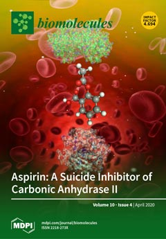
Carbonic Anhydrase II (CAII) is a ubiquitously expressed zinc-metalloenzyme highly expressed in red blood cells. This enzyme catalyzes the reversible hydration/dehydration of CO2/ HCO3-, while also having an innate carboxylesterase activity. Aspirin (acetyl-salicylic acid), one of the most commonly used drugs globally, has been shown to have a short half-life in the blood of ~15 minutes. Here, we report that CAII, and specifically its carboxylesterase activity, is responsible for aspirin’s short half-life. Furthermore, the esterase product, salicylic acid, acts as an inhibitor of CAII once formed, blocking the active site. Thus, CAII not only degrades aspirin but, in turn, aspirin also acts as a suicide inhibitor of CAII within red blood cells. View this paper.
  • Issues are regarded as officially published after their release is announced to the table of contents alert mailing list.
  • You may sign up for e-mail alerts to receive table of contents of newly released issues.
  • PDF is the official format for papers published in both, html and pdf forms. To view the papers in pdf format, click on the "PDF Full-text" link, and use the free Adobe Reader to open them.
Order results
Result details
Section
Select all
Export citation of selected articles as:
17 pages, 4233 KiB  
Article
Bioconversion of Biologically Active Indole Derivatives with Indole-3-Acetic Acid-Degrading Enzymes from Caballeronia glathei DSM50014
by Mikas Sadauskas, Roberta Statkevičiūtė, Justas Vaitekūnas and Rolandas Meškys
Biomolecules 2020, 10(4), 663; https://doi.org/10.3390/biom10040663 - 24 Apr 2020
Cited by 15 | Viewed by 4558
Abstract
A plant auxin hormone indole-3-acetic acid (IAA) can be assimilated by bacteria as an energy and carbon source, although no degradation has been reported for indole-3-propionic acid and indole-3-butyric acid. While significant efforts have been made to decipher the Iac (indole-3-acetic acid catabolism)-mediated [...] Read more.
A plant auxin hormone indole-3-acetic acid (IAA) can be assimilated by bacteria as an energy and carbon source, although no degradation has been reported for indole-3-propionic acid and indole-3-butyric acid. While significant efforts have been made to decipher the Iac (indole-3-acetic acid catabolism)-mediated IAA degradation pathway, a lot of questions remain regarding the mechanisms of individual reactions, involvement of specific Iac proteins, and the overall reaction scheme. This work was aimed at providing new experimental evidence regarding the biodegradation of IAA and its derivatives. Here, it was shown that Caballeronia glathei strain DSM50014 possesses a full iac gene cluster and is able to use IAA as a sole source of carbon and energy. Next, IacE was shown to be responsible for the conversion of 2-oxoindole-3-acetic acid (Ox-IAA) intermediate into the central intermediate 3-hydroxy-2-oxindole-3-acetic acid (DOAA) without the requirement for IacB. During this reaction, the oxygen atom incorporated into Ox-IAA was derived from water. Finally, IacA and IacE were shown to convert a wide range of indole derivatives, including indole-3-propionic acid and indole-3-butyric acid, into corresponding DOAA homologs. This work provides novel insights into Iac-mediated IAA degradation and demonstrates the versatility and substrate scope of IacA and IacE enzymes. Full article
Show Figures

Graphical abstract

10 pages, 715 KiB  
Article
Macrophage Cholesterol Efflux Downregulation Is Not Associated with Abdominal Aortic Aneurysm (AAA) Progression
by Marina Canyelles, Mireia Tondo, Jes S. Lindholt, David Santos, Irati Fernández-Alonso, David de Gonzalo-Calvo, Luis Miguel Blanco-Colio, Joan Carles Escolà-Gil, José Luís Martín-Ventura and Francisco Blanco-Vaca
Biomolecules 2020, 10(4), 662; https://doi.org/10.3390/biom10040662 - 24 Apr 2020
Cited by 2 | Viewed by 2600
Abstract
Recent studies have raised the possibility of a role for lipoproteins, including high-density lipoprotein cholesterol (HDLc), in abdominal aortic aneurysm (AAA). The study was conducted in plasmas from 39 large size AAA patients (aortic diameter > 50 mm), 81 small/medium size AAA patients [...] Read more.
Recent studies have raised the possibility of a role for lipoproteins, including high-density lipoprotein cholesterol (HDLc), in abdominal aortic aneurysm (AAA). The study was conducted in plasmas from 39 large size AAA patients (aortic diameter > 50 mm), 81 small/medium size AAA patients (aortic diameter between 30 and 50 mm) and 38 control subjects (aortic diameter < 30 mm). We evaluated the potential of HDL-mediated macrophage cholesterol efflux (MCE) to predict AAA growth and/or the need for surgery. MCE was impaired in the large aortic diameter AAA group as compared with that in the small/medium size AAA group and the control group. However, no significant difference in HDL-mediated MCE capacity was observed in 3 different progression subgroups (classified according to growth rate < 1 mm per year, between 1 and 5 mm per year or >5 mm per year) in patients with small/medium size AAA. Moreover, no correlation was found between MCE capacity and the aneurysm growth rate. A multivariate Cox regression analysis revealed a significant association between lower MCE capacity with the need for surgery in all AAA patients. Nevertheless, the significance was lost when only small/medium size AAA patients were included. Our results suggest that MCE, a major HDL functional activity, is not involved in AAA progression. Full article
Show Figures

Graphical abstract

21 pages, 3351 KiB  
Article
Discovering the RNA-Binding Proteome of Plant Leaves with an Improved RNA Interactome Capture Method
by Marcel Bach-Pages, Felix Homma, Jiorgos Kourelis, Farnusch Kaschani, Shabaz Mohammed, Markus Kaiser, Renier A. L. van der Hoorn, Alfredo Castello and Gail M. Preston
Biomolecules 2020, 10(4), 661; https://doi.org/10.3390/biom10040661 - 24 Apr 2020
Cited by 55 | Viewed by 14106
Abstract
RNA-binding proteins (RBPs) play a crucial role in regulating RNA function and fate. However, the full complement of RBPs has only recently begun to be uncovered through proteome-wide approaches such as RNA interactome capture (RIC). RIC has been applied to various cell lines [...] Read more.
RNA-binding proteins (RBPs) play a crucial role in regulating RNA function and fate. However, the full complement of RBPs has only recently begun to be uncovered through proteome-wide approaches such as RNA interactome capture (RIC). RIC has been applied to various cell lines and organisms, including plants, greatly expanding the repertoire of RBPs. However, several technical challenges have limited the efficacy of RIC when applied to plant tissues. Here, we report an improved version of RIC that overcomes the difficulties imposed by leaf tissue. Using this improved RIC method in Arabidopsis leaves, we identified 717 RBPs, generating a deep RNA-binding proteome for leaf tissues. While 75% of these RBPs can be linked to RNA biology, the remaining 25% were previously not known to interact with RNA. Interestingly, we observed that a large number of proteins related to photosynthesis associate with RNA in vivo, including proteins from the four major photosynthetic supercomplexes. As has previously been reported for mammals, a large proportion of leaf RBPs lack known RNA-binding domains, suggesting unconventional modes of RNA binding. We anticipate that this improved RIC method will provide critical insights into RNA metabolism in plants, including how cellular RBPs respond to environmental, physiological and pathological cues. Full article
(This article belongs to the Special Issue Ribonucleoprotein Particles (RNPs): From Structure to Function)
Show Figures

Graphical abstract

17 pages, 4392 KiB  
Article
Structure of the ALS Mutation Target Annexin A11 Reveals a Stabilising N-Terminal Segment
by Peder A. G. Lillebostad, Arne Raasakka, Silje J. Hjellbrekke, Sudarshan Patil, Trude Røstbø, Hanne Hollås, Siri A. Sakya, Peter D. Szigetvari, Anni Vedeler and Petri Kursula
Biomolecules 2020, 10(4), 660; https://doi.org/10.3390/biom10040660 - 24 Apr 2020
Cited by 8 | Viewed by 5082
Abstract
The functions of the annexin family of proteins involve binding to Ca2+, lipid membranes, other proteins, and RNA, and the annexins share a common folded core structure at the C terminus. Annexin A11 (AnxA11) has a long N-terminal region, which is [...] Read more.
The functions of the annexin family of proteins involve binding to Ca2+, lipid membranes, other proteins, and RNA, and the annexins share a common folded core structure at the C terminus. Annexin A11 (AnxA11) has a long N-terminal region, which is predicted to be disordered, binds RNA, and forms membraneless organelles involved in neuronal transport. Mutations in AnxA11 have been linked to amyotrophic lateral sclerosis (ALS). We studied the structure and stability of AnxA11 and identified a short stabilising segment in the N-terminal end of the folded core, which links domains I and IV. The crystal structure of the AnxA11 core highlights main-chain hydrogen bonding interactions formed through this bridging segment, which are likely conserved in most annexins. The structure was also used to study the currently known ALS mutations in AnxA11. Three of these mutations correspond to buried Arg residues highly conserved in the annexin family, indicating central roles in annexin folding. The structural data provide starting points for detailed structure–function studies of both full-length AnxA11 and the disease variants being identified in ALS. Full article
(This article belongs to the Special Issue Metal Binding Proteins 2020)
Show Figures

Figure 1

25 pages, 4910 KiB  
Article
P1′ Residue-Oriented Virtual Screening for Potent and Selective Phosphinic (Dehydro) Dipeptide Inhibitors of Metallo-Aminopeptidases
by Michał Talma and Artur Mucha
Biomolecules 2020, 10(4), 659; https://doi.org/10.3390/biom10040659 - 24 Apr 2020
Cited by 11 | Viewed by 3313
Abstract
Designing side chain substituents complementary to enzyme binding pockets is of great importance in the construction of potent and selective phosphinic dipeptide inhibitors of metallo-aminopeptidases. Proper structure selection makes inhibitor construction more economic, as the development process typically consists of multiple iterative preparation/bioassay [...] Read more.
Designing side chain substituents complementary to enzyme binding pockets is of great importance in the construction of potent and selective phosphinic dipeptide inhibitors of metallo-aminopeptidases. Proper structure selection makes inhibitor construction more economic, as the development process typically consists of multiple iterative preparation/bioassay steps. On the basis of these principles, using noncomplex computation and modeling methodologies, we comprehensively screened 900 commercial precursors of the P1′ residues of phosphinic dipeptide and dehydrodipeptide analogs to identify the most promising ligands of 52 metallo-dependent aminopeptidases with known crystal structures. The results revealed several nonproteinogenic residues with an improved energy of binding compared with the best known inhibitors. The data are discussed taking into account the selectivity and stereochemical implications of the enzymes. Using this approach, we were able to identify nontrivial structural elements substituting the recognized phosphinic peptidomimetic scaffold of metallo-aminopeptidase inhibitors. Full article
Show Figures

Figure 1

31 pages, 1465 KiB  
Review
Selenium and Selenoproteins in Adipose Tissue Physiology and Obesity
by Alexey A. Tinkov, Olga P. Ajsuvakova, Tommaso Filippini, Ji-Chang Zhou, Xin Gen Lei, Eugenia R. Gatiatulina, Bernhard Michalke, Margarita G. Skalnaya, Marco Vinceti, Michael Aschner and Anatoly V. Skalny
Biomolecules 2020, 10(4), 658; https://doi.org/10.3390/biom10040658 - 24 Apr 2020
Cited by 66 | Viewed by 7518
Abstract
Selenium (Se) homeostasis is tightly related to carbohydrate and lipid metabolism, but its possible roles in obesity development and in adipocyte metabolism are unclear. The objective of the present study is to review the current data on Se status in obesity and to [...] Read more.
Selenium (Se) homeostasis is tightly related to carbohydrate and lipid metabolism, but its possible roles in obesity development and in adipocyte metabolism are unclear. The objective of the present study is to review the current data on Se status in obesity and to discuss the interference between Se and selenoprotein metabolism in adipocyte physiology and obesity pathogenesis. The overview and meta-analysis of the studies on blood Se and selenoprotein P (SELENOP) levels, as well as glutathione peroxidase (GPX) activity in obese subjects, have yielded heterogenous and even conflicting results. Laboratory studies demonstrate that Se may modulate preadipocyte proliferation and adipogenic differentiation, and also interfere with insulin signaling, and regulate lipolysis. Knockout models have demonstrated that the selenoprotein machinery, including endoplasmic reticulum-resident selenoproteins together with GPXs and thioredoxin reductases (TXNRDs), are tightly related to adipocyte development and functioning. In conclusion, Se and selenoproteins appear to play an essential role in adipose tissue physiology, although human data are inconsistent. Taken together, these findings do not support the utility of Se supplementation to prevent or alleviate obesity in humans. Further human and laboratory studies are required to elucidate associations between Se metabolism and obesity. Full article
(This article belongs to the Special Issue Toxic and Essential Metals in Human Health and Disease)
Show Figures

Figure 1

14 pages, 2788 KiB  
Article
Antikinetoplastid Activity of Indolocarbazoles from Streptomyces sanyensis
by Luis Cartuche, Ines Sifaoui, Atteneri López-Arencibia, Carlos J. Bethencourt-Estrella, Desirée San Nicolás-Hernández, Jacob Lorenzo-Morales, José E. Piñero, Ana R. Díaz-Marrero and José J. Fernández
Biomolecules 2020, 10(4), 657; https://doi.org/10.3390/biom10040657 - 24 Apr 2020
Cited by 24 | Viewed by 3679
Abstract
Chagas disease and leishmaniasis are neglected tropical diseases caused by kinetoplastid parasites of Trypanosoma and Leishmania genera that affect poor and remote populations in developing countries. These parasites share similar complex life cycles and modes of infection. It has been demonstrated that the [...] Read more.
Chagas disease and leishmaniasis are neglected tropical diseases caused by kinetoplastid parasites of Trypanosoma and Leishmania genera that affect poor and remote populations in developing countries. These parasites share similar complex life cycles and modes of infection. It has been demonstrated that the particular group of phosphorylating enzymes, protein kinases (PKs), are essential for the infective mechanisms and for parasite survival. The natural indolocarbazole staurosporine (STS, 1) has been extensively used as a PKC inhibitor and its antiparasitic effects described. In this research, we analyze the antikinetoplastid activities of three indolocarbazole (ICZs) alkaloids of the family of staurosporine STS, 24, and the commercial ICZs rebeccamycin (5), K252a (6), K252b (7), K252c (8), and arcyriaflavin A (9) in order to establish a plausive approach to the mode of action and to provide a preliminary qualitative structure–activity analysis. The most active compound was 7-oxostaurosporine (7OSTS, 2) that showed IC50 values of 3.58 ± 1.10; 0.56 ± 0.06 and 1.58 ± 0.52 µM against L. amazonensis; L. donovani and T. cruzi, and a Selectivity Index (CC50/IC50) of 52 against amastigotes of L. amazonensis compared to the J774A.1 cell line of mouse macrophages. Full article
(This article belongs to the Special Issue Bioactives from Marine Products)
Show Figures

Figure 1

17 pages, 1739 KiB  
Article
Fluorescence Methods Applied to the Description of Urea-Dependent YME1L Protease Unfolding
by Sydney Moore, Alyssa Pickens, Jessica L. Rodriguez, Justin D. Marsee and Justin M. Miller
Biomolecules 2020, 10(4), 656; https://doi.org/10.3390/biom10040656 - 23 Apr 2020
Cited by 2 | Viewed by 2635
Abstract
ATP-dependent proteases are ubiquitous across all kingdoms of life and are critical to the maintenance of intracellular protein quality control. The enzymatic function of these enzymes requires structural stability under conditions that may drive instability and/or loss of function in potential protein substrates. [...] Read more.
ATP-dependent proteases are ubiquitous across all kingdoms of life and are critical to the maintenance of intracellular protein quality control. The enzymatic function of these enzymes requires structural stability under conditions that may drive instability and/or loss of function in potential protein substrates. Thus, these molecular machines must demonstrate greater stability than their substrates in order to ensure continued function in essential quality control networks. We report here a role for ATP in the stabilization of the inner membrane YME1L protease. Qualitative fluorescence data derived from protein unfolding experiments with urea reveal non-standard protein unfolding behavior that is dependent on [ATP]. Using multiple fluorophore systems, stopped-flow fluorescence experiments demonstrate a depletion of the native YME1L ensemble by urea-dependent unfolding and formation of a non-native conformation. Additional stopped-flow fluorescence experiments based on nucleotide binding and unfoldase activities predict that unfolding yields significant loss of active YME1L hexamers from the starting ensemble. Taken together, these data clearly define the stress limits of an important mitochondrial protease. Full article
(This article belongs to the Section Cellular Biochemistry)
Show Figures

Graphical abstract

32 pages, 2970 KiB  
Review
Diseases Caused by Mutations in Mitochondrial Carrier Genes SLC25: A Review
by Ferdinando Palmieri, Pasquale Scarcia and Magnus Monné
Biomolecules 2020, 10(4), 655; https://doi.org/10.3390/biom10040655 - 23 Apr 2020
Cited by 66 | Viewed by 9896
Abstract
In the 1980s, after the mitochondrial DNA (mtDNA) had been sequenced, several diseases resulting from mtDNA mutations emerged. Later, numerous disorders caused by mutations in the nuclear genes encoding mitochondrial proteins were found. A group of these diseases are due to defects of [...] Read more.
In the 1980s, after the mitochondrial DNA (mtDNA) had been sequenced, several diseases resulting from mtDNA mutations emerged. Later, numerous disorders caused by mutations in the nuclear genes encoding mitochondrial proteins were found. A group of these diseases are due to defects of mitochondrial carriers, a family of proteins named solute carrier family 25 (SLC25), that transport a variety of solutes such as the reagents of ATP synthase (ATP, ADP, and phosphate), tricarboxylic acid cycle intermediates, cofactors, amino acids, and carnitine esters of fatty acids. The disease-causing mutations disclosed in mitochondrial carriers range from point mutations, which are often localized in the substrate translocation pore of the carrier, to large deletions and insertions. The biochemical consequences of deficient transport are the compartmentalized accumulation of the substrates and dysfunctional mitochondrial and cellular metabolism, which frequently develop into various forms of myopathy, encephalopathy, or neuropathy. Examples of diseases, due to mitochondrial carrier mutations are: combined D-2- and L-2-hydroxyglutaric aciduria, carnitine-acylcarnitine carrier deficiency, hyperornithinemia-hyperammonemia-homocitrillinuria (HHH) syndrome, early infantile epileptic encephalopathy type 3, Amish microcephaly, aspartate/glutamate isoform 1 deficiency, congenital sideroblastic anemia, Fontaine progeroid syndrome, and citrullinemia type II. Here, we review all the mitochondrial carrier-related diseases known until now, focusing on the connections between the molecular basis, altered metabolism, and phenotypes of these inherited disorders. Full article
(This article belongs to the Special Issue Mitochondrial Transport Proteins)
Show Figures

Figure 1

16 pages, 4159 KiB  
Article
Recombinant Lectin from Tepary Bean (Phaseolus acutifolius) with Specific Recognition for Cancer-Associated Glycans: Production, Structural Characterization, and Target Identification
by Dania Martínez-Alarcón, Annabelle Varrot, Elaine Fitches, John A. Gatehouse, Min Cao, Prashant Pyati, Alejandro Blanco-Labra and Teresa Garcia-Gasca
Biomolecules 2020, 10(4), 654; https://doi.org/10.3390/biom10040654 - 23 Apr 2020
Cited by 6 | Viewed by 3554
Abstract
Herein, we report the production of a recombinant Tepary bean lectin (rTBL-1), its three-dimensional (3D) structure, and its differential recognition for cancer-type glycoconjugates. rTBL-1 was expressed in Pichia pastoris, yielding 316 mg per liter of culture, and was purified by [...] Read more.
Herein, we report the production of a recombinant Tepary bean lectin (rTBL-1), its three-dimensional (3D) structure, and its differential recognition for cancer-type glycoconjugates. rTBL-1 was expressed in Pichia pastoris, yielding 316 mg per liter of culture, and was purified by nickel affinity chromatography. Characterization of the protein showed that rTBL-1 is a stable 120 kDa homo-tetramer folded as a canonical leguminous lectin with two divalent cations (Ca2+ and Mn2+) attached to each subunit, confirmed in its 3D structure solved by X-ray diffraction at 1.9 Å resolution. Monomers also presented a ~2.5 kDa N-linked glycan located on the opposite face of the binding pocket. It does not participate in carbohydrate recognition but contributes to the stabilization of the interfaces between protomers. Screening for potential rTBL-1 targets by glycan array identified 14 positive binders, all of which correspond to β1-6 branched N-glycans’ characteristics of cancer cells. The presence of α1-6 core fucose, also tumor-associated, improved carbohydrate recognition. rTBL-1 affinity for a broad spectrum of mono- and disaccharides was evaluated by isothermal titration calorimetry (ITC); however, no interaction was detected, corroborating that carbohydrate recognition is highly specific and requires larger ligands for binding. This would explain the differential recognition between healthy and cancer cells by Tepary bean lectins. Full article
Show Figures

Graphical abstract

15 pages, 2041 KiB  
Article
Role of 3-Mercaptopyruvate Sulfurtransferase in the Regulation of Proliferation and Cellular Bioenergetics in Human Down Syndrome Fibroblasts
by Theodora Panagaki, Elisa B. Randi and Csaba Szabo
Biomolecules 2020, 10(4), 653; https://doi.org/10.3390/biom10040653 - 23 Apr 2020
Cited by 25 | Viewed by 4509
Abstract
Down syndrome (trisomy of human chromosome 21) is a common genetic disorder. Overproduction of the gaseous mediator hydrogen sulfide (H2S) has been implicated in the pathogenesis of neurological and metabolic deficits associated with Down syndrome. Several lines of data indicate that [...] Read more.
Down syndrome (trisomy of human chromosome 21) is a common genetic disorder. Overproduction of the gaseous mediator hydrogen sulfide (H2S) has been implicated in the pathogenesis of neurological and metabolic deficits associated with Down syndrome. Several lines of data indicate that an important enzyme responsible for H2S overproduction in Down syndrome is cystathionine-β-synthase (CBS), an enzyme localized on chromosome 21. The current study explored the possibility that a second H2S-producing enzyme, 3-mercaptopyruvate sulfurtransferase (3-MST), may also contribute to the development of functional deficits of Down syndrome cells. Western blotting analysis demonstrated a significantly higher level of 3-MST protein expression in human Down syndrome fibroblasts compared to cells from healthy control individuals; the excess 3-MST was mainly localized to the mitochondrial compartment. Pharmacological inhibition of 3-MST activity improved mitochondrial electron transport and oxidative phosphorylation parameters (but did not affect the suppressed glycolytic parameters) and enhanced cell proliferation in Down syndrome cells (but not in healthy control cells). The findings presented in the current report suggest that in addition to the indisputable role of CBS, H2S produced from 3-MST may also contribute to the development of mitochondrial metabolic and functional impairments in Down syndrome cells. Full article
Show Figures

Graphical abstract

15 pages, 1210 KiB  
Review
Antimicrobial and Antibiofilm Peptides
by Angela Di Somma, Antonio Moretta, Carolina Canè, Arianna Cirillo and Angela Duilio
Biomolecules 2020, 10(4), 652; https://doi.org/10.3390/biom10040652 - 23 Apr 2020
Cited by 144 | Viewed by 10151
Abstract
The increasing onset of multidrug-resistant bacteria has propelled microbiology research towards antimicrobial peptides as new possible antibiotics from natural sources. Antimicrobial peptides are short peptides endowed with a broad range of activity against both Gram-positive and Gram-negative bacteria and are less prone to [...] Read more.
The increasing onset of multidrug-resistant bacteria has propelled microbiology research towards antimicrobial peptides as new possible antibiotics from natural sources. Antimicrobial peptides are short peptides endowed with a broad range of activity against both Gram-positive and Gram-negative bacteria and are less prone to trigger resistance. Besides their activity against planktonic bacteria, many antimicrobial peptides also show antibiofilm activity. Biofilms are ubiquitous in nature, having the ability to adhere to virtually any surface, either biotic or abiotic, including medical devices, causing chronic infections that are difficult to eradicate. The biofilm matrix protects bacteria from hostile environments, thus contributing to the bacterial resistance to antimicrobial agents. Biofilms are very difficult to treat, with options restricted to the use of large doses of antibiotics or the removal of the infected device. Antimicrobial peptides could represent good candidates to develop new antibiofilm drugs as they can act at different stages of biofilm formation, on disparate molecular targets and with various mechanisms of action. These include inhibition of biofilm formation and adhesion, downregulation of quorum sensing factors, and disruption of the pre-formed biofilm. This review focuses on the proprieties of antimicrobial and antibiofilm peptides, with a particular emphasis on their mechanism of action, reporting several examples of peptides that over time have been shown to have activity against biofilm. Full article
Show Figures

Graphical abstract

10 pages, 2039 KiB  
Article
Connexin 43 Deficiency Is Associated with Reduced Myocardial Scar Size and Attenuated TGFβ1 Signaling after Transient Coronary Occlusion in Conditional Knock-Out Mice
by Laura Valls-Lacalle, Marta Consegal, Marisol Ruiz-Meana, Begoña Benito, Javier Inserte, Ignasi Barba, Ignacio Ferreira-González and Antonio Rodríguez-Sinovas
Biomolecules 2020, 10(4), 651; https://doi.org/10.3390/biom10040651 - 23 Apr 2020
Cited by 8 | Viewed by 2707
Abstract
Previous studies demonstrated a reduction in myocardial scar size in heterozygous Cx43+/- mice subjected to permanent coronary occlusion. However, patients presenting with ST segment elevation myocardial infarction often undergo rapid coronary revascularization leading to prompt restoration of coronary flow. Therefore, we aimed [...] Read more.
Previous studies demonstrated a reduction in myocardial scar size in heterozygous Cx43+/- mice subjected to permanent coronary occlusion. However, patients presenting with ST segment elevation myocardial infarction often undergo rapid coronary revascularization leading to prompt restoration of coronary flow. Therefore, we aimed to assess changes in scar size and left ventricular remodeling following transient myocardial ischemia (45 min) followed by 14 days of reperfusion using Cx43fl/fl (controls) and Cx43Cre-ER(T)/fl inducible knock-out (Cx43 content: 50%) mice treated with vehicle or 4-hydroxytamoxifen (4-OHT) to induce a Cre-ER(T)-mediated global deletion of the Cx43 floxed allele. The scar area (picrosirius red), measured 14 days after transient coronary occlusion, was similarly reduced in both vehicle and 4-OHT-treated Cx43Cre-ER(T)/fl mice, compared to Cx43fl/fl animals, having normal Cx43 levels (15.78% ± 3.42% and 16.54% ± 2.31% vs. 25.40% ± 3.14% and 22.43% ± 3.88% in vehicle and 4-OHT-treated mice, respectively, p = 0.027). Left ventricular dilatation was significantly attenuated in both Cx43-deficient groups (p = 0.037 for left ventricular end-diastolic diameter). These protective effects were correlated with an attenuated enhancement in pro-transforming growth factor beta 1 (TGFβ1) expression after reperfusion. In conclusion, our data demonstrate that Cx43 deficiency induces a protective effect on scar formation after transient coronary occlusion in mice, an effect associated with reduced left ventricular remodeling and attenuated enhancement in pro-TGFβ1 expression. Full article
(This article belongs to the Special Issue Connexins, Innexins, and Pannexins: From Biology to Clinical Targets)
Show Figures

Figure 1

22 pages, 681 KiB  
Review
Redox-Mediated Post-Translational Modifications of Proteolytic Enzymes and Their Role in Protease Functioning
by Anastasiia I. Petushkova and Andrey A. Zamyatnin, Jr.
Biomolecules 2020, 10(4), 650; https://doi.org/10.3390/biom10040650 - 23 Apr 2020
Cited by 18 | Viewed by 3674
Abstract
Proteolytic enzymes play a crucial role in metabolic processes, providing the cell with amino acids through the hydrolysis of multiple endogenous and exogenous proteins. In addition to this function, proteases are involved in numerous protein cascades to maintain cellular and extracellular homeostasis. The [...] Read more.
Proteolytic enzymes play a crucial role in metabolic processes, providing the cell with amino acids through the hydrolysis of multiple endogenous and exogenous proteins. In addition to this function, proteases are involved in numerous protein cascades to maintain cellular and extracellular homeostasis. The redox regulation of proteolysis provides a flexible dose-dependent mechanism for proteolytic activity control. The excessive reactive oxygen species (ROS) and reactive nitrogen species (RNS) in living organisms indicate pathological conditions, so redox-sensitive proteases can swiftly induce pro-survival responses or regulated cell death (RCD). At the same time, severe protein oxidation can lead to the dysregulation of proteolysis, which induces either protein aggregation or superfluous protein hydrolysis. Therefore, oxidative stress contributes to the onset of age-related dysfunction. In the present review, we consider the post-translational modifications (PTMs) of proteolytic enzymes and their impact on homeostasis. Full article
(This article belongs to the Special Issue Redox Regulation of Protein Functioning)
Show Figures

Figure 1

19 pages, 4216 KiB  
Article
Evolution of Angiotensin Peptides and Peptidomimetics as Angiotensin II Receptor Type 2 (AT2) Receptor Agonists
by Silvana Vasile, Anders Hallberg, Jessica Sallander, Mathias Hallberg, Johan Åqvist and Hugo Gutiérrez-de-Terán
Biomolecules 2020, 10(4), 649; https://doi.org/10.3390/biom10040649 - 23 Apr 2020
Cited by 13 | Viewed by 4583
Abstract
Angiotensin II receptor type 1 and 2 (AT1R and AT2R) are two G-protein coupled receptors that mediate most biological functions of the octapeptide Angiotensin II (Ang II). AT2R is upregulated upon tissue damage and its activation by selective AT2R agonists has become a [...] Read more.
Angiotensin II receptor type 1 and 2 (AT1R and AT2R) are two G-protein coupled receptors that mediate most biological functions of the octapeptide Angiotensin II (Ang II). AT2R is upregulated upon tissue damage and its activation by selective AT2R agonists has become a promising approach in the search for new classes of pharmaceutical agents. We herein analyzed the chemical evolution of AT2R agonists starting from octapeptides, through shorter peptides and peptidomimetics to the first drug-like AT2R-selective agonist, C21, which is in Phase II clinical trials and aimed for idiopathic pulmonary fibrosis. Based on the recent crystal structures of AT1R and AT2R in complex with sarile, we identified a common binding model for a series of 11 selected AT2R agonists, consisting of peptides and peptidomimetics of different length, affinity towards AT2R and selectivity versus AT1R. Subsequent molecular dynamics simulations and free energy perturbation (FEP) calculations of binding affinities allowed the identification of the bioactive conformation and common pharmacophoric points, responsible for the key interactions with the receptor, which are maintained by the drug-like agonists. The results of this study should be helpful and facilitate the search for improved and even more potent AT2R-selective drug-like agonists. Full article
Show Figures

Figure 1

16 pages, 4053 KiB  
Article
Src/NF-κB-Targeted Anti-Inflammatory Effects of Potentilla glabra var. Mandshurica (Maxim.) Hand.-Mazz. Ethanol Extract
by Haeyeop Kim, Kon Kuk Shin, Han Gyung Kim, Minkyeong Jo, Jin Kyeong Kim, Jong Sub Lee, Eui Su Choung, Wan Yi Li, Sang Woo Lee, Kyung-Hee Kim, Byong Chul Yoo and Jae Youl Cho
Biomolecules 2020, 10(4), 648; https://doi.org/10.3390/biom10040648 - 22 Apr 2020
Cited by 10 | Viewed by 2957
Abstract
Inflammation is a complex protective response of body tissues to harmful stimuli. Acute inflammation can progress to chronic inflammation, which can lead to severe disease. Therefore, this research focuses on the development of anti-inflammatory drugs, and natural extracts have been explored as potential [...] Read more.
Inflammation is a complex protective response of body tissues to harmful stimuli. Acute inflammation can progress to chronic inflammation, which can lead to severe disease. Therefore, this research focuses on the development of anti-inflammatory drugs, and natural extracts have been explored as potential agents. No study has yet examined the inflammation-associated pharmacological activity of Potentilla glabra Var. mandshurica (Maxim.) Hand.-Mazz ethanol extract (Pg-EE). To examine the mechanisms by which Pg-EE exerts anti-inflammatory effects, we studied its activities in lipopolysaccharide (LPS)-treated murine macrophage RAW264.7 cells and an HCl/EtOH-induced gastritis model. LPS-triggered nitric oxide (NO) release and mRNA levels of inducible nitric oxide synthase (iNOS), tumor necrosis factor-alpha (TNF-α), interleukin-6 (IL-6), and interleukin-1 beta (IL-1β) in RAW264.7 cells were suppressed by Pg-EE in a dose-dependent manner. Using a luciferase assay and western blot assay, we found that the NF-κB pathway was inhibited by Pg-EE, particularly by the decreased level of phosphorylated proteins of nuclear factor kappa-light-chain-enhancer of activated B cells (NF-κB) subunits (p65 and p50), inhibitor of kappa B alpha (IκBα), p85, and Src. Using an overexpression strategy, cellular thermal shift assay, and immunoprecipitation analysis, we determined that the anti-inflammatory effect of Pg-EE was mediated by the inhibition of Src. Pg-EE further showed anti-inflammatory effects in vivo in the HCl/EtOH-induced gastritis mouse model. In conclusion, Pg-EE exerts anti-inflammatory activities by targeting Src in the NF-κB pathway, and these results suggest that Pg-EE could be used as an anti-inflammatory herbal medicine. Full article
Show Figures

Figure 1

22 pages, 5626 KiB  
Article
The Cytosolic Phospholipase A2α N-Terminal C2 Domain Binds and Oligomerizes on Membranes with Positive Curvature
by Katherine E. Ward, Ranjan Sengupta, James P. Ropa, Souad Amiar and Robert V. Stahelin
Biomolecules 2020, 10(4), 647; https://doi.org/10.3390/biom10040647 - 22 Apr 2020
Cited by 6 | Viewed by 3752
Abstract
Group IV phospholipase A2α (cPLA2α) regulates the production of prostaglandins and leukotrienes via the formation of arachidonic acid from membrane phospholipids. The targeting and membrane binding of cPLA2α to the Golgi involves the N-terminal C2 domain, whereas [...] Read more.
Group IV phospholipase A2α (cPLA2α) regulates the production of prostaglandins and leukotrienes via the formation of arachidonic acid from membrane phospholipids. The targeting and membrane binding of cPLA2α to the Golgi involves the N-terminal C2 domain, whereas the catalytic domain produces arachidonic acid. Although most studies of cPLA2α concern its catalytic activity, it is also linked to homeostatic processes involving the generation of vesicles that traffic material from the Golgi to the plasma membrane. Here we investigated how membrane curvature influences the homeostatic role of cPLA2α in vesicular trafficking. The cPLA2α C2 domain is known to induce changes in positive membrane curvature, a process which is dependent on cPLA2α membrane penetration. We showed that cPLA2α undergoes C2 domain-dependent oligomerization on membranes in vitro and in cells. We found that the association of the cPLA2α C2 domain with membranes is limited to membranes with positive curvature, and enhanced C2 domain oligomerization was observed on vesicles ~50 nm in diameter. We demonstrated that the cPLA2α C2 domain localizes to cholesterol enriched Golgi-derived vesicles independently of cPLA2α catalytic activity. Moreover, we demonstrate the C2 domain selectively localizes to lipid droplets whereas the full-length enzyme to a much lesser extent. Our results therefore provide novel insight into the molecular forces that mediate C2 domain-dependent membrane localization in vitro and in cells. Full article
(This article belongs to the Special Issue 2020 Feature Papers by Biomolecules’ Editorial Board Members)
Show Figures

Figure 1

22 pages, 3420 KiB  
Article
3D Chitin Scaffolds from the Marine Demosponge Aplysina archeri as a Support for Laccase Immobilization and Its Use in the Removal of Pharmaceuticals
by Jakub Zdarta, Tomasz Machałowski, Oliwia Degórska, Karolina Bachosz, Andriy Fursov, Hermann Ehrlich, Viatcheslav N. Ivanenko and Teofil Jesionowski
Biomolecules 2020, 10(4), 646; https://doi.org/10.3390/biom10040646 - 22 Apr 2020
Cited by 25 | Viewed by 3345
Abstract
For the first time, 3D chitin scaffolds from the marine demosponge Aplysina archeri were used for adsorption and immobilization of laccase from Trametes versicolor. The resulting chitin–enzyme biocatalytic systems were applied in the removal of tetracycline. Effective enzyme immobilization was confirmed by [...] Read more.
For the first time, 3D chitin scaffolds from the marine demosponge Aplysina archeri were used for adsorption and immobilization of laccase from Trametes versicolor. The resulting chitin–enzyme biocatalytic systems were applied in the removal of tetracycline. Effective enzyme immobilization was confirmed by scanning electron microscopy. Immobilization yield and kinetic parameters were investigated in detail, in addition to the activity of the enzyme after immobilization. The designed systems were further used for the removal of tetracycline under various process conditions. Optimum process conditions, enabling total removal of tetracycline from solutions at concentrations up to 1 mg/L, were found to be pH 5, temperature between 25 and 35 °C, and 1 h process duration. Due to the protective effect of the chitinous scaffolds and stabilization of the enzyme by multipoint attachment, the storage stability and thermal stability of the immobilized biomolecules were significantly improved as compared to the free enzyme. The produced biocatalytic systems also exhibited good reusability, as after 10 repeated uses they removed over 90% of tetracycline from solution. Finally, the immobilized laccase was used in a packed bed reactor for continuous removal of tetracycline, and enabled the removal of over 80% of the antibiotic after 24 h of continuous use. Full article
(This article belongs to the Special Issue Marine-Derived Biomolecules)
Show Figures

Figure 1

20 pages, 291 KiB  
Review
High-Density Lipoprotein Cholesterol in Age-Related Ocular Diseases
by Bjorn Kaijun Betzler, Tyler Hyungtaek Rim, Charumathi Sabanayagam, Chui Ming Gemmy Cheung and Ching-Yu Cheng
Biomolecules 2020, 10(4), 645; https://doi.org/10.3390/biom10040645 - 22 Apr 2020
Cited by 15 | Viewed by 3663
Abstract
There is limited understanding of the specific role of high-density lipoprotein cholesterol (HDL-C) in the development of various age-related ocular diseases, despite it being a common measurable biomarker in lipid profiles. This literature review summarizes current knowledge of the role of HDL-C, if [...] Read more.
There is limited understanding of the specific role of high-density lipoprotein cholesterol (HDL-C) in the development of various age-related ocular diseases, despite it being a common measurable biomarker in lipid profiles. This literature review summarizes current knowledge of the role of HDL-C, if any, in pathogenesis and progression of four age-related ocular diseases, namely age-related macular degeneration (AMD), age-related cataract, glaucoma, and diabetic retinopathy (DR), and will primarily discuss epidemiological and genetic evidence. Full article
Show Figures

Graphical abstract

31 pages, 4412 KiB  
Review
Spermatogonial Stem Cells in Fish: Characterization, Isolation, Enrichment, and Recent Advances of In Vitro Culture Systems
by Xuan Xie, Rafael Nóbrega and Martin Pšenička
Biomolecules 2020, 10(4), 644; https://doi.org/10.3390/biom10040644 - 22 Apr 2020
Cited by 22 | Viewed by 6984
Abstract
Spermatogenesis is a continuous and dynamic developmental process, in which a single diploid spermatogonial stem cell (SSC) proliferates and differentiates to form a mature spermatozoon. Herein, we summarize the accumulated knowledge of SSCs and their distribution in the testes of teleosts. We also [...] Read more.
Spermatogenesis is a continuous and dynamic developmental process, in which a single diploid spermatogonial stem cell (SSC) proliferates and differentiates to form a mature spermatozoon. Herein, we summarize the accumulated knowledge of SSCs and their distribution in the testes of teleosts. We also reviewed the primary endocrine and paracrine influence on spermatogonium self-renewal vs. differentiation in fish. To provide insight into techniques and research related to SSCs, we review available protocols and advances in enriching undifferentiated spermatogonia based on their unique physiochemical and biochemical properties, such as size, density, and differential expression of specific surface markers. We summarize in vitro germ cell culture conditions developed to maintain proliferation and survival of spermatogonia in selected fish species. In traditional culture systems, sera and feeder cells were considered to be essential for SSC self-renewal, in contrast to recently developed systems with well-defined media and growth factors to induce either SSC self-renewal or differentiation in long-term cultures. The establishment of a germ cell culture contributes to efficient SSC propagation in rare, endangered, or commercially cultured fish species for use in biotechnological manipulation, such as cryopreservation and transplantation. Finally, we discuss organ culture and three-dimensional models for in vitro investigation of fish spermatogenesis. Full article
(This article belongs to the Special Issue Relevant Biomolecules for Germ Cells and Fertilization)
Show Figures

Figure 1

17 pages, 4404 KiB  
Article
Sequential Interferon β-Cisplatin Treatment Enhances the Surface Exposure of Calreticulin in Cancer Cells via an Interferon Regulatory Factor 1-Dependent Manner
by Pei-Ming Yang, Yao-Yu Hsieh, Jia-Ling Du, Shih-Chieh Yen and Chien-Fu Hung
Biomolecules 2020, 10(4), 643; https://doi.org/10.3390/biom10040643 - 21 Apr 2020
Cited by 20 | Viewed by 3244
Abstract
Immunogenic cell death (ICD) refers to a unique form of cell death that activates an adaptive immune response against dead-cell-associated antigens. Accumulating evidence indicates that the efficacy of conventional anticancer agents relies on not only their direct cytostatic/cytotoxic effects but also the activation [...] Read more.
Immunogenic cell death (ICD) refers to a unique form of cell death that activates an adaptive immune response against dead-cell-associated antigens. Accumulating evidence indicates that the efficacy of conventional anticancer agents relies on not only their direct cytostatic/cytotoxic effects but also the activation of antitumor ICD. Common anticancer ICD inducers include certain chemotherapeutic agents (such as anthracyclines, oxaliplatin, and bortezomib), radiotherapy, photodynamic therapy (PDT), and oncolytic virotherapies. However, most chemotherapeutic reagents are inefficient or fail to trigger ICD. Therefore, better understanding on the molecular determinants of chemotherapy-induced ICD will help in the development of more efficient combinational anticancer strategies through converting non- or relatively weak ICD inducers into bona fide ICD inducers. In this study, we found that sequential, but not concurrent, treatment of cancer cells with interferon β (IFNβ), a type I IFN, and cisplatin (an inefficient ICD inducer) can enhance the expression of ICD biomarkers in cancer cells, including surface translocation of an endoplasmic reticulum (ER) chaperone, calreticulin (CRT), and phosphorylation of the eukaryotic translation initiation factor alpha (eIF2α). These results suggest that exogenous IFNβ may activate molecular determinants that convert cisplatin into an ICD inducer. Further bioinformatics and in vitro experimental analyses found that interferon regulatory factor 1 (IRF1) acted as an essential mediator of surface CRT exposure by sequential IFNβ-cisplatin combination. Our findings not only help to design more effective combinational anticancer therapy using IFNβ and cisplatin, but also provide a novel insight into the role of IRF1 in connecting the type I IFN responses and ICD. Full article
(This article belongs to the Special Issue Cell Death in Cancer and Inflammation: From Pathogenesis to Treatment)
Show Figures

Figure 1

56 pages, 32130 KiB  
Review
The Role of Branch Cell Symmetry and Other Critical Nanoscale Design Parameters in the Determination of Dendrimer Encapsulation Properties
by Donald A. Tomalia, Linda S. Nixon and David M. Hedstrand
Biomolecules 2020, 10(4), 642; https://doi.org/10.3390/biom10040642 - 21 Apr 2020
Cited by 43 | Viewed by 4845
Abstract
This article reviews progress over the past three decades related to the role of dendrimer-based, branch cell symmetry in the development of advanced drug delivery systems, aqueous based compatibilizers/solubilizers/excipients and nano-metal cluster catalysts. Historically, it begins with early unreported work by the Tomalia [...] Read more.
This article reviews progress over the past three decades related to the role of dendrimer-based, branch cell symmetry in the development of advanced drug delivery systems, aqueous based compatibilizers/solubilizers/excipients and nano-metal cluster catalysts. Historically, it begins with early unreported work by the Tomalia Group (i.e., The Dow Chemical Co.) revealing that all known dendrimer family types may be divided into two major symmetry categories; namely: Category I: symmetrical branch cell dendrimers (e.g., Tomalia, Vögtle, Newkome-type dendrimers) possessing interior hollowness/porosity and Category II: asymmetrical branch cell dendrimers (e.g., Denkewalter-type) possessing no interior void space. These two branch cell symmetry features were shown to be pivotal in directing internal packing modes; thereby, differentiating key dendrimer properties such as densities, refractive indices and interior porosities. Furthermore, this discovery provided an explanation for unimolecular micelle encapsulation (UME) behavior observed exclusively for Category I, but not for Category II. This account surveys early experiments confirming the inextricable influence of dendrimer branch cell symmetry on interior packing properties, first examples of Category (I) based UME behavior, nuclear magnetic resonance (NMR) protocols for systematic encapsulation characterization, application of these principles to the solubilization of active approved drugs, engineering dendrimer critical nanoscale design parameters (CNDPs) for optimized properties and concluding with high optimism for the anticipated role of dendrimer-based solubilization principles in emerging new life science, drug delivery and nanomedical applications. Full article
Show Figures

Figure 1

41 pages, 3186 KiB  
Review
Phenolic Compounds Exerting Lipid-Regulatory, Anti-Inflammatory and Epigenetic Effects as Complementary Treatments in Cardiovascular Diseases
by Laura Toma, Gabriela Maria Sanda, Loredan Stefan Niculescu, Mariana Deleanu, Anca Volumnia Sima and Camelia Sorina Stancu
Biomolecules 2020, 10(4), 641; https://doi.org/10.3390/biom10040641 - 21 Apr 2020
Cited by 40 | Viewed by 6627
Abstract
Atherosclerosis is the main process behind cardiovascular diseases (CVD), maladies which continue to be responsible for up to 70% of death worldwide. Despite the ongoing development of new and potent drugs, their incomplete efficacy, partial intolerance and numerous side effects make the search [...] Read more.
Atherosclerosis is the main process behind cardiovascular diseases (CVD), maladies which continue to be responsible for up to 70% of death worldwide. Despite the ongoing development of new and potent drugs, their incomplete efficacy, partial intolerance and numerous side effects make the search for new alternatives worthwhile. The focus of the scientific world turned to the potential of natural active compounds to prevent and treat CVD. Essential for effective prevention or treatment based on phytochemicals is to know their mechanisms of action according to their bioavailability and dosage. The present review is focused on the latest data about phenolic compounds and aims to collect and correlate the reliable existing knowledge concerning their molecular mechanisms of action to counteract important risk factors that contribute to the initiation and development of atherosclerosis: dyslipidemia, and oxidative and inflammatory-stress. The selection of phenolic compounds was made to prove their multiple benefic effects and endorse them as CVD remedies, complementary to allopathic drugs. The review also highlights some aspects that still need clear scientific explanations and draws up some new molecular approaches to validate phenolic compounds for CVD complementary therapy in the near future. Full article
(This article belongs to the Special Issue Phytochemical Omics in Medicinal Plants)
Show Figures

Figure 1

8 pages, 276 KiB  
Editorial
H2S, Polysulfides, and Enzymes: Physiological and Pathological Aspects
by Noriyuki Nagahara and Maria Wróbel
Biomolecules 2020, 10(4), 640; https://doi.org/10.3390/biom10040640 - 21 Apr 2020
Cited by 8 | Viewed by 2554
Abstract
We have been studying the general aspects of the functions of H2S and polysulfides, and the enzymes involved in their biosynthesis, for more than 20 years. Our aim has been to elucidate novel physiological and pathological functions of H2S [...] Read more.
We have been studying the general aspects of the functions of H2S and polysulfides, and the enzymes involved in their biosynthesis, for more than 20 years. Our aim has been to elucidate novel physiological and pathological functions of H2S and polysulfides, and unravel the regulation of the enzymes involved in their biosynthesis, including cystathionine β-synthase (EC 4.2.1.22), cystathionine γ-lyase (EC 4.4.1.1), thiosulfate sulfurtransferase (rhodanese, EC 2.8.1.1), and 3-mercaptopyruvate sulfurtransferase (EC 2.8.1.2). Physiological and pathological functions, alternative biosynthetic processes, and additional functions of H2S and polysulfides have been reported. Further, the structure and reaction mechanisms of related enzymes have also been reported. We expect this issue to advance scientific knowledge regarding the detailed functions of H2S and polysulfides as well as the general properties and regulation of the enzymes involved in their metabolism. We would like to cover four topics: the physiological and pathological functions of H2S and polysulfides, the mechanisms of the biosynthesis of H2S and polysulfides, the properties of the biosynthetic enzymes, and the regulation of enzymatic activity. The knockout mouse technique is a useful tool to determine new physiological functions, especially those of H2S and polysulfides. In the future, we shall take a closer look at symptoms in the human congenital deficiency of each enzyme. Further studies on the regulation of enzymatic activity by in vivo substances may be the key to finding new functions of H2S and polysulfides. Full article
22 pages, 35471 KiB  
Article
Meridianins and Lignarenone B as Potential GSK3β Inhibitors and Inductors of Structural Neuronal Plasticity
by Laura Llorach-Pares, Ened Rodriguez-Urgelles, Alfons Nonell-Canals, Jordi Alberch, Conxita Avila, Melchor Sanchez-Martinez and Albert Giralt
Biomolecules 2020, 10(4), 639; https://doi.org/10.3390/biom10040639 - 21 Apr 2020
Cited by 17 | Viewed by 6559
Abstract
Glycogen Synthase Kinase 3 (GSK3) is an essential protein, with a relevant role in many diseases such as diabetes, cancer and neurodegenerative disorders. Particularly, the isoform GSK3β is related to pathologies such as Alzheimer’s disease (AD). This enzyme constitutes a very interesting target [...] Read more.
Glycogen Synthase Kinase 3 (GSK3) is an essential protein, with a relevant role in many diseases such as diabetes, cancer and neurodegenerative disorders. Particularly, the isoform GSK3β is related to pathologies such as Alzheimer’s disease (AD). This enzyme constitutes a very interesting target for the discovery and/or design of new therapeutic agents against AD due to its relation to the hyperphosphorylation of the microtubule-associated protein tau (MAPT), and therefore, its contribution to neurofibrillary tangles (NFT) formation. An in silico target profiling study identified two marine molecular families, the indole alkaloids meridianins from the tunicate genus Aplidium, and lignarenones, the secondary metabolites of the shelled cephalaspidean mollusc Scaphander lignarius, as possible GSK3β inhibitors. The analysis of the surface of GSK3β, aimed to find possible binding regions, and the subsequent in silico binding studies revealed that both marine molecular families can act over the ATP and/or substrate binding regions. The predicted inhibitory potential of the molecules from these two chemical families was experimentally validated in vitro by showing a ~50% of increased Ser9 phosphorylation levels of the GSK3β protein. Furthermore, we determined that molecules from both molecular families potentiate structural neuronal plasticity in vitro. These results allow us to suggest that meridianins and lignarenone B could be used as possible therapeutic candidates for the treatment of GSK3β involved pathologies, such as AD. Full article
(This article belongs to the Special Issue Marine-Derived Biomolecules)
Show Figures

Figure 1

12 pages, 1583 KiB  
Review
Possible Mechanisms of Eosinophil Accumulation in Eosinophilic Pneumonia
by Kazuyuki Nakagome and Makoto Nagata
Biomolecules 2020, 10(4), 638; https://doi.org/10.3390/biom10040638 - 21 Apr 2020
Cited by 19 | Viewed by 4614
Abstract
Eosinophilic pneumonia (EP), including acute EP and chronic EP, is characterized by the massive pulmonary infiltration of eosinophils into the lung. However, the mechanisms underlying the selective accumulation of eosinophils in EP have not yet been fully elucidated. We reported that bronchoalveolar lavage [...] Read more.
Eosinophilic pneumonia (EP), including acute EP and chronic EP, is characterized by the massive pulmonary infiltration of eosinophils into the lung. However, the mechanisms underlying the selective accumulation of eosinophils in EP have not yet been fully elucidated. We reported that bronchoalveolar lavage fluid (BALF) from EP patients induced the transmigration of eosinophils across endothelial cells in vitro. The concentrations of eotaxin-2 (CCL24) and monocyte chemotactic protein (MCP)-4 (CCL13), which are CC chemokine receptor (CCR) 3 ligands, were elevated in the BALF of EP patients, and anti-CCR3 monoclonal antibody inhibited the eosinophil transmigration induced by the BALF of EP patients. The concentration of macrophage inflammatory protein 1β (CCL4), a CCR5 ligand that induces eosinophil migration, was increased in the BALF of EP patients. Furthermore, the concentration of interleukin (IL) 5 was increased in the BALF of EP patients, and it has been reported that anti-IL-5 antibody treatment resulted in remission and the reduction of glucocorticoid use in some cases of chronic EP. The concentrations of lipid mediators, such as leukotriene (LT) B4, damage-associated molecular pattern molecules (DAMPs), such as uric acid, or extracellular matrix proteins, such as periostin, were also increased in the BALF of EP patients. These findings suggest that chemokines, such as CCR3/CCR5 ligands, cytokines, such as IL-5, lipid mediators, such as LTB4, DAMPs, and extracellular matrix proteins may play roles in the accumulation or activation of eosinophils in EP. Full article
(This article belongs to the Special Issue Eosinophilic Inflammation)
Show Figures

Figure 1

21 pages, 5633 KiB  
Article
New Therapeutic Strategies for Osteoarthritis by Targeting Sialic Acid Receptors
by Paula Carpintero-Fernandez, Marta Varela-Eirin, Alessandra Lacetera, Raquel Gago-Fuentes, Eduardo Fonseca, Sonsoles Martin-Santamaria and Maria D. Mayan
Biomolecules 2020, 10(4), 637; https://doi.org/10.3390/biom10040637 - 21 Apr 2020
Cited by 16 | Viewed by 3709
Abstract
Osteoarthritis (OA) is the most common degenerative joint disease characterized by articular cartilage degradation and joint degeneration. The articular cartilage is mainly formed by chondrocytes and a collagen-proteoglycan extracellular matrix that contains high levels of glycosylated proteins. It was reported that the shift [...] Read more.
Osteoarthritis (OA) is the most common degenerative joint disease characterized by articular cartilage degradation and joint degeneration. The articular cartilage is mainly formed by chondrocytes and a collagen-proteoglycan extracellular matrix that contains high levels of glycosylated proteins. It was reported that the shift from glycoproteins containing α-2,6-linked sialic acids to those that contain α-2,3 was associated with the onset of common types of arthritis. However, the pathophysiology of α-2,3-sialylation in cartilage has not been yet elucidated. We show that cartilage from osteoarthritic patients expresses high levels of the α-2,3-sialylated transmembrane mucin receptor, known as podoplanin (PDPN). Additionally, the Maackia amurensis seed lectin (MASL), that can be utilized to target PDPN, attenuates the inflammatory response mediated by NF-kB activation in primary chondrocytes and protects human cartilage breakdown ex vivo and in an animal model of arthritis. These findings reveal that specific lectins targeting α-2,3-sialylated receptors on chondrocytes might effectively inhibit cartilage breakdown. We also present a computational 3D molecular model for this interaction. These findings provide mechanistic information on how a specific lectin could be used as a novel therapy to treat degenerative joint diseases such as osteoarthritis. Full article
(This article belongs to the Section Chemical Biology)
Show Figures

Figure 1

14 pages, 8099 KiB  
Article
Structural and Quantitative Characterization of Mucin-Type O-Glycans and the Identification of O-Glycosylation Sites in Bovine Submaxillary Mucin
by Jihye Kim, Changsoo Ryu, Jongkwan Ha, Junmyoung Lee, Donghwi Kim, Minkyoo Ji, Chi Soo Park, Jaeryong Lee, Dae Kyong Kim and Ha Hyung Kim
Biomolecules 2020, 10(4), 636; https://doi.org/10.3390/biom10040636 - 20 Apr 2020
Cited by 11 | Viewed by 4533
Abstract
Bovine submaxillary mucin (BSM) is a gel-forming glycoprotein polymer, and Ser/Thr-linked glycans (O-glycans) are important in regulating BSM’s viscoelasticity and polymerization. However, details of O-glycosylation have not been reported. This study investigates the structural and quantitative characteristics of O-glycans [...] Read more.
Bovine submaxillary mucin (BSM) is a gel-forming glycoprotein polymer, and Ser/Thr-linked glycans (O-glycans) are important in regulating BSM’s viscoelasticity and polymerization. However, details of O-glycosylation have not been reported. This study investigates the structural and quantitative characteristics of O-glycans and identifies O-glycosylation sites in BSM using liquid chromatography–tandem mass spectrometry. The O-glycans (consisting of di- to octa-saccharides) and their quantities (%) relative to total O-glycans (100%; 1.1 pmol per 1 μg of BSM) were identified with 14 major (>1.0%), 12 minor (0.1%–1.0%), and eight trace (<0.1%) O-glycans, which were characterized based on their constituents (sialylation (14 O-glycans; 81.9%, sum of relative quantities of each glycan), non-sialylation (20; 18.1%), fucosylation (20; 17.5%), and terminal-galactosylation (6; 3.6%)) and six core structure types [Gal-GalNAc, Gal-(GlcNAc)GalNAc, GlcNAc-GalNAc, GlcNAc-(GlcNAc)GalNAc, and GalNAc-GalNAc]. O-glycosylation sites were identified using O-glycopeptides (bold underlined; 56SGETRTSVI, 259SHSSSGRSRTI, 272GSPSSVSSAEQI, 307RPSYGAL, 625QTLGPL, 728TMTTRTSVVV, and 1080RPEDNTAVA) obtained from proteolytic BSM; these sites are in the four domains of BSM. The gel-forming mucins share common domain structures and glycosylation patterns; these results could provide useful information on mucin-type O-glycans. This is the first study to characterize O-glycans and identify O-glycosylation sites in BSM. Full article
Show Figures

Figure 1

44 pages, 21451 KiB  
Review
Recent Synthetic Approaches towards Small Molecule Reactivators of p53
by Jerson L. Silva, Carolina G. S. Lima, Luciana P. Rangel, Giulia D. S. Ferretti, Fernanda P. Pauli, Ruan C. B. Ribeiro, Thais de B. da Silva, Fernando C. da Silva and Vitor F. Ferreira
Biomolecules 2020, 10(4), 635; https://doi.org/10.3390/biom10040635 - 20 Apr 2020
Cited by 20 | Viewed by 5205
Abstract
The tumor suppressor protein p53 is often called “the genome guardian” and controls the cell cycle and the integrity of DNA, as well as other important cellular functions. Its main function is to trigger the process of apoptosis in tumor cells, and approximately [...] Read more.
The tumor suppressor protein p53 is often called “the genome guardian” and controls the cell cycle and the integrity of DNA, as well as other important cellular functions. Its main function is to trigger the process of apoptosis in tumor cells, and approximately 50% of all cancers are related to the inactivation of the p53 protein through mutations in the TP53 gene. Due to the association of mutant p53 with cancer therapy resistance, different forms of restoration of p53 have been subject of intense research in recent years. In this sense, this review focus on the main currently adopted approaches for activation and reactivation of p53 tumor suppressor function, focusing on the synthetic approaches that are involved in the development and preparation of such small molecules. Full article
(This article belongs to the Special Issue Recent Advances in p53)
Show Figures

Figure 1

17 pages, 1398 KiB  
Review
Effects of Red and Fermented Ginseng and Ginsenosides on Allergic Disorders
by Myung Joo Han and Dong-Hyun Kim
Biomolecules 2020, 10(4), 634; https://doi.org/10.3390/biom10040634 - 20 Apr 2020
Cited by 28 | Viewed by 6252
Abstract
Both white ginseng (WG, dried root of Panax sp.) and red ginseng (RG, steamed and dried root of Panax sp.) are reported to exhibit a variety of pharmacological effects such as anticancer, antidiabetic, and neuroprotective activities. These ginsengs contain hydrophilic sugar-conjugated ginsenosides and [...] Read more.
Both white ginseng (WG, dried root of Panax sp.) and red ginseng (RG, steamed and dried root of Panax sp.) are reported to exhibit a variety of pharmacological effects such as anticancer, antidiabetic, and neuroprotective activities. These ginsengs contain hydrophilic sugar-conjugated ginsenosides and polysaccharides as the bioactive constituents. When taken orally, their hydrophilic constituents are metabolized into hydrophobic ginsenosides compound K, Rh1, and Rh2 that are absorbable into the blood. These metabolites exhibit the pharmacological effects more strongly than hydrophilic parental constituents. To enforce these metabolites, fermented WG and RG are developed. Moreover, natural products including ginseng are frequently used for the treatment of allergic disorders. Therefore, this review introduces the current knowledge related to the effectiveness of ginseng on allergic disorders including asthma, allergic rhinitis, atopic dermatitis, and pruritus. We discuss how ginseng, its constituents, and its metabolites regulate allergy-related immune responses. We also describe how ginseng controls allergic disorders. Full article
(This article belongs to the Special Issue Advances in Ginsenosides)
Show Figures

Figure 1

Previous Issue
Next Issue
Back to TopTop