


Metabolites, Volume 8, Issue 1 (March 2018) – 24 articles
Cover Story (view full-size image):
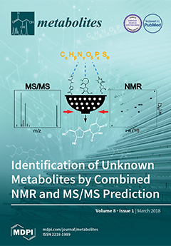
We introduce a cheminformatics approach that combines highly selective and orthogonal structure elucidation parameters, accurate mass, MS/MS (MS2), and NMR into a single analysis platform to accurately identify unknown metabolites in untargeted studies. The approach starts with an unknown LC-MS feature, and then combines the experimental MS/MS and NMR information of the unknown to effectively filter the false positive candidate structures based on their predicted MS/MS and NMR spectra. We demonstrate the approach on a model mixture and then we identify an uncatalogued secondary metabolite in Arabidopsis thaliana. The NMR/MS2 approach facilitates the identification of metabolites that are not present in experimental NMR and MS metabolomics databases. View the paper
- Issues are regarded as officially published after their release is announced to the table of contents alert mailing list.
- You may sign up for e-mail alerts to receive table of contents of newly released issues.
- PDF is the official format for papers published in both, html and pdf forms. To view the papers in pdf format, click on the "PDF Full-text" link, and use the free Adobe Reader to open them.
Previous Issue
Next Issue