Carbonic Anhydrase Inhibition and the Management of Hypoxic Tumors


Università degli Studi di Firenze, Dipartimento Neurofarba, Sezione di Scienze Farmaceutiche e Nutraceutiche, Via U. Schiff 6, 50019 Sesto Fiorentino, Florence, Italy
Metabolites 2017, 7(3), 48; https://doi.org/10.3390/metabo7030048
Submission received: 31 August 2017
/
Revised: 15 September 2017
/
Accepted: 15 September 2017
/
Published: 16 September 2017
(This article belongs to the Special Issue Carbonic Anhydrases and Metabolism)
Abstract
:Hypoxia and acidosis are salient features of many tumors, leading to a completely different metabolism compared to normal cells. Two of the simplest metabolic products, protons and bicarbonate, are generated by the catalytic activity of the metalloenzyme carbonic anhydrase (CA, EC 4.2.1.1), with at least two of its isoforms, CA IX and XII, mainly present in hypoxic tumors. Inhibition of tumor-associated CAs leads to an impaired growth of the primary tumors, metastases and reduces the population of cancer stem cells, leading thus to a complex and beneficial anticancer action for this class of enzyme inhibitors. In this review, I will present the state of the art on the development of CA inhibitors (CAIs) targeting the tumor-associated CA isoforms, which may have applications for the treatment and imaging of cancers expressing them. Small molecule inhibitors, one of which (SLC-0111) completed Phase I clinical trials, and antibodies (girentuximab, discontinued in Phase III clinical trials) will be discussed, together with the various approaches used to design anticancer agents with a new mechanism of action based on interference with these crucial metabolites, protons and bicarbonate.
Keywords:
tumor; metabolism; carbonic anhydrase; isoforms IX and XII; inhibitor; sulfonamide; antibody1. Introduction
A salient feature of many tumors is the fact that they are hypoxic and acidic compared to normal tissues of the same type. This has been known for many decades as the Warburg effect [1,2] but has been explained at the molecular level only recently, after the discovery of a transcription factor regulating these phenomena, the hypoxia inducible factor 1α, HIF-1α [3,4,5].
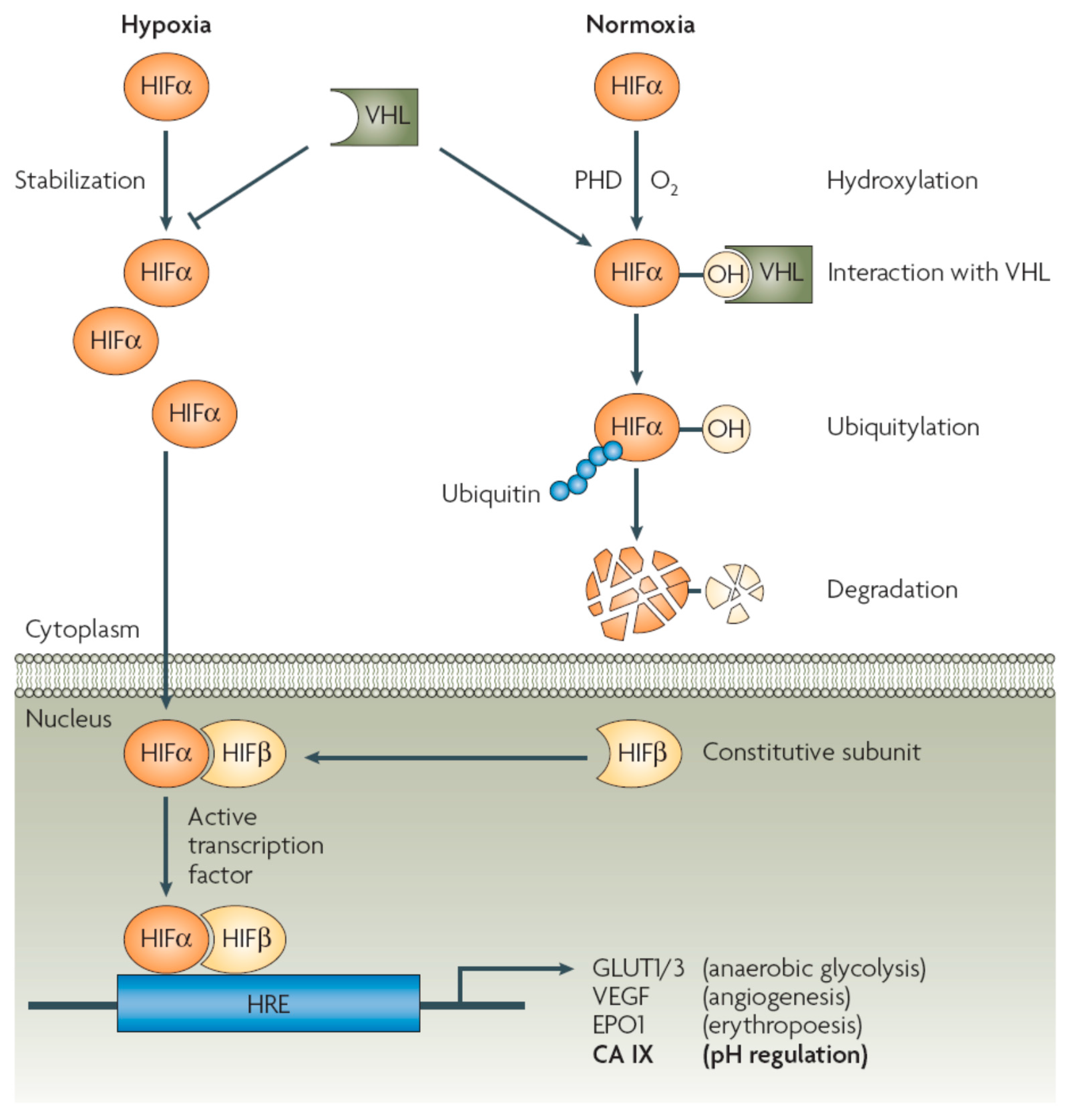
As seen from Figure 1, in normoxic conditions HIF-1α is unstable, being degraded rapidly by a well understood biochemical process: under the action of prolyl hydroxylases (PHD), a proline residue from the transcription factor is hydroxylated, being then recognized by a protein possessing ubiquitin ligase E3 activity, more precisely the von Hippel Lindau protein (VHL), which targets it to ubiquitylation and degradation within the proteosomes (Figure 1) [5,6,7,8].
However, in hypoxia, which as mentioned above is frequent in many tumor cells [1,2,3], an accumulation of HIF-1α occurs, followed by its translocation from the cytosol to the nucleus, where it forms a dimer with a constitutive subunit, HIF-1β, leading to an active transcription factor, which, by interaction with a hypoxia responsive element (HRE) found on different genes, leads to overexpression of proteins involved in aerobic glycolysis (such as, for example, the glucose transporters GLUT1-3), angiogenesis (such as, for example, the vascular endothelial growth factor, VEGF), erythropoesis (such as, for example, erythropoetin 1) and pH regulation (such as the tumor-associated enzymes CA IX and XII) [5,6,7,8,9,10,11].
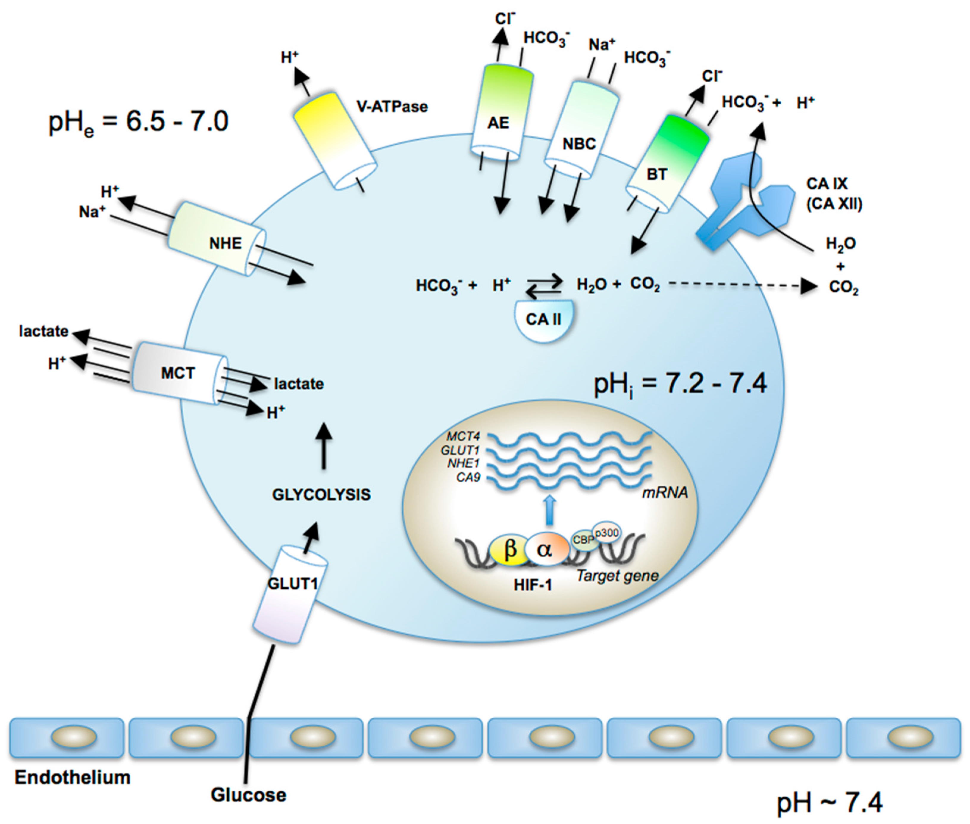
The overexpression of these proteins has profound effects on the metabolism of cancer cells, which on one hand are deprived of oxygen for the normal metabolism involving the oxidative phosphorylation [1,2], and on the other one, have an enhanced uptake of glucose (due to the overexpression of the glucose transporters GLUT1-GLUT3, which import the sugar within the cell), which cannot undergo the oxidative pathways for the generation of ATP [5,6,7,8]. Thus, an alternative pathway, the glycolytic one, occurs, with the formation of pyruvic (and lactic acids) from glucose, which generates less ATP (compared to the oxidative pathway), but which seems to be enough for the cancer cells to survive in hypoxic conditions [1,2,3,4]. The formed organic acids are extruded from the cells through the monocarboxylate transporters MCT1-MCT4 (some of which are overexpressed in tumors [4]), leading to an acidification of the extracellular milieu, up to pH values as low as 6.5 [4,5,6,7,8]. Additional perturbations of the extra- and intracellular pH equilibrium of the tumor cells are also furnished by other proteins which are involved in this process (Figure 2), among which the sodium-proton exchanger (Na+–H+ antiporter) NHE, which may import or export protons in exchange for sodium ions, the plasma membrane proton pump H+-ATPase (V-ATPase), the various isoforms of the anion exchangers (chloride-bicarbonate exchangers) AE1–AE3, the sodium bicarbonate channels NBCs, which transport sodium and bicarbonate out of the cell or import it within the cell, various other bicarbonate transporters BT, as well as several isoforms of the metalloenzyme CA, such as the cytosolic CA II, and the transmembrane CA IX/XII, which efficiently catalyze CO2 hydration to bicarbonate and protons [4,5,6,7,8,9,10,11]. By the coupling of all these effects, a slightly alkaline intracellular pH is achieved (of around 7.2) and an acidic extracellular pH of the tumor is formed, with values as low as 6.5 [4,5,6,7,8,9,10,11] (Figure 2). The extracellular acidosis (coupled with the hypoxia) is beneficial for the growth of the tumor cell and impairs the growth of the normal cells, leading thus to a massive proliferation, invasion and subsequently metastasis of the primary tumors [12,13,14,15].
Data of Figure 1 and Figure 2 show the multitude of proteins involved in these processes, which in the end lead to features of the tumor cells which are quite different from those of the normal ones, and could thus be exploited for designing novel anticancer therapies. Among those who proposed this approach for the first time was Pouysségur et al. [4] who initially considered the NHE inhibitors as the most interesting pharmacological agents for interfering with tumor hypoxia/acidosis [16]. However, the significant toxicity of this class of drugs, or the lack of isoform-selective ones for other proteins involved in these processes (such as the MCTs, AEs, V-ATPase, etc. [17,18]) led to most of the work being concentrated on the metalloenzyme involved in pH regulation, i.e., the carbonic anhydrase (CA, EC 4.2.1.1) [7,8,19,20]. It should be however mentioned that H+/K+-inhibitors of the omeprazole type were shown to possess, alone or in combination with CA inhibitors (CAIs) significant antitumor effects [21,22,23]. Here I shall review the field of the CAIs as theragnostic agents for the management of hypoxic, metastatic tumors, without considering the other valuable approaches found in the literature which target other of the many proteins involved in these processes, and which have been reviewed by other researchers [6,13,16].
2. Validation of CA IX/XII as Antitumor Drug Targets
CA IX was discovered by Pastorek et al. in 1994 [10] and CA XII by Tureci et al. in 1998 [11], and it became immediately obvious that they differ considerably from other members of this family of proteins, which includes 15 isoforms in humans, hCA I-hCA VA, hCA VB, hCA VI-hCA XIV [7,8,20]. The first unusual feature of CA IX and XII was that the two enzymes are transmembrane, multi-domain proteins incorporating a short intra-cytosolic tail, a transmembrane short domain, and an extracellular catalytic domain, rather homologous to the one found in the cytosolic, mitochondrial, secreted or membrane-anchored CA isoforms known at that time [10,11,24,25,26,27]. Furthermore, CA IX has an additional domain at its N-terminus, termed the proteoglycan (PG) domain, which seems to play important functions connected with the role of CA IX in tumorigenesis being present only in this CA isoform [28,29] (Figure 3). In fact, all domains of this molecule, the intracellular tail [30], the catalytic domain [25,29] and the PG domain play diverse functions in tumorigenesis, making CA IX one of the key proteins involved in such processes in hypoxic tumors [7,8,15,17,24,25,26,27,28,29,30]. It is also interesting to note that CA IX seems to be an even more complicated protein: recent proteomic/interactomic studies suggests that at a stage in the cell’s life CA IX possibly has a nuclear localization [31], interacting with proteins involved in nuclear/cytoplasmic transport processes, gene transcription, and protein stability, among which cullin-associated NEDD8-dissociated protein 1 (CAND1), which is itself involved in gene transcription and assembly of ubiquitin ligase complexes [32]. The precise role of these interactions of CA IX with this type of proteins is poorly understood at this moment but may lead to significant drug design developments in the future.
Returning to the main function of CA IX/XII, that of catalyzing the hydration of CO2 to bicarbonate and protons [7,8], the validation of these proteins as drug targets followed the usual steps that most drug targets experience. They are summarized below:
(1) recombinant CA IX and XII were shown to possess a significant catalytic activity (in vitro) for the physiologic reaction (hydration of carbon dioxide to bicarbonate and protons), being among the most effective catalysts known in nature, with the following kinetic parameters: for human (h) CA IX (full length): kcat of 1.1 × 10⁶ s−1, kcat/KM of 1.5 × 10⁸ M−1 s−1 [24], whereas for hCA XII (catalytic domain) these parameters are kcat of 4.2 × 10⁵ s−1, kcat/KM of 3.5 × 10⁷ M−1 s−1 [33].
(2) potent in vitro CAIs of the sulfonamide type have been identified for both hCA IX [34] and hCA XII [33], followed by a large number of drug design studies of such agents [35], which have been reviewed recently and will be not detailed here [36,37,38]. As a consequence of such studies a large number of sulfonamide, sulfamate and sulfamides showing effective hCA IX/XII inhibitory potency (in vitro) and sometimes also some selectivity for inhibiting these two isoforms over the cytosolic, off-target and widespread ones hCA I and II, became available for in vivo studies [33,34,35,36,37,38,39].
The drug design of CAIs targeting isoform IX were highly favored by the report of the X-ray crystal structure of the protein (its catalytic domain) by De Simone’s group in 2009 [25]. This 3D structure allowed the identification of similarities and differences between CA IX and the other members of the family, which led to the identification soon thereafter of highly isoform-selective inhibitors belonging to a variety of chemical classes, such as the sulfonamides, sulfamates, sulfamides, coumarins, polyeamines, etc. [36,37,38].
(3) Pastorekova’s group [29] demonstrated the role of CA IX in extracellular acidification of hypoxic tumors, and the possibility to reverse this effect by inhibiting the enzyme activity with sulfonamides. Furthermore, in the same studies it was observed that a fluorescent potent sulfonamide CA IX/II inhibitor accumulated only in the hypoxic cells, whereas it did not bind in cells expressing CA IX, but in normoxic conditions [29,39]. This effect has been explained as being due to the PG domain of the protein, which in normoxic conditions closes the active site. The opening of the active site is triggered by hypoxia, making it available for inhibitors to bind, but only in hypoxic conditions [39]. This makes CA IX an ideal drug target, as this phenomenon will lead to the inhibition of only the CA IX present in tumors, leading thus to drugs with fewer side effects compared to the classical chemotherapeutic agents [29,39].
(4) Dubois et al. [40,41] then published the proof-of-concept studies showing that in xenograft animal models of hypoxic tumors it is possible to image the hypoxic regions rich in CA IX/XII by using fluorescent sulfonamide CAIs possessing the same structural elements as the compounds used in the study of Pastorekova’s group, mentioned above [29].
(5) The first study showing an in vivo antitumor effect due to CA IX inhibition was from Neri’s group [42], followed shortly thereafter by similar studies from different laboratories, on diverse models and cancer types, which demonstrated that sulfonamide/sulfamate [42,43,44,45,46] or coumarin [47] CA IX/XII inhibitors have a profound effect in inhibiting the growth of the primary tumors and the metastases expressing CA IX/XII. Probably the most interesting studies are those from Dedhar’s group [44,45,47] who rigorously showed the involvement of CA IX/XII in the antitumor/antimetastatic effects of the inhibitors of the sulfonamide or coumarin types. In fact, as it will be shown shortly, one of the compounds described in such studies progressed to clinical trials and completed Phase I trials in 2016 [45].
(6) Dedhar’s group [48] also discovered another important phenomenon connected to CA IX/XII inhibition, i.e., the depletion of cancer stem cell population within the hypoxic tumors, which is considered to be a very positive feature of an antitumor agent, considering the fact that most such therapies lead to an increase of this stem cell population, hypothesized to be one of the reasons for the recurrence of some cancers [49]. The same group recently elucidated [50] the mechanism used by the hypoxic tumors for invasion, which reinforces the role played by CA IX in tumor progression and clinical outcome of cancer patients harboring CA IX-positive tumors. This relevant study demonstrated an association between CA IX and matrix metalloproteinase 14 (MMP14), with the first protein furnishing H+ ions used in the proteolytic cleavage of collagen mediated by MMP14, which leads to tissue degradation. This study showed that CA IX is one of the metabolic components of the cellular migration and invasion mechanisms in hypoxic tumors, and provides new mechanistic insights into the role played by this enzyme in tumor cell biology, with the possibility to design dual agents, targeting both these enzymes (CA IX and MMP14) as new antitumor drugs [50].
3. Small Molecule CA IX/XII Inhibitors as Antitumor Agents
Among the huge number of sulfonamide, sulfamate, sulfamide and coumarin CA IX/XII inhibitors reported to date [4,7,34,35,36,37,38], few compounds were investigated in detail in animal tumor models, and only one such derivatives, SLC-0111 (also known as WBI-5111) progressed to clinical trials [45,51].
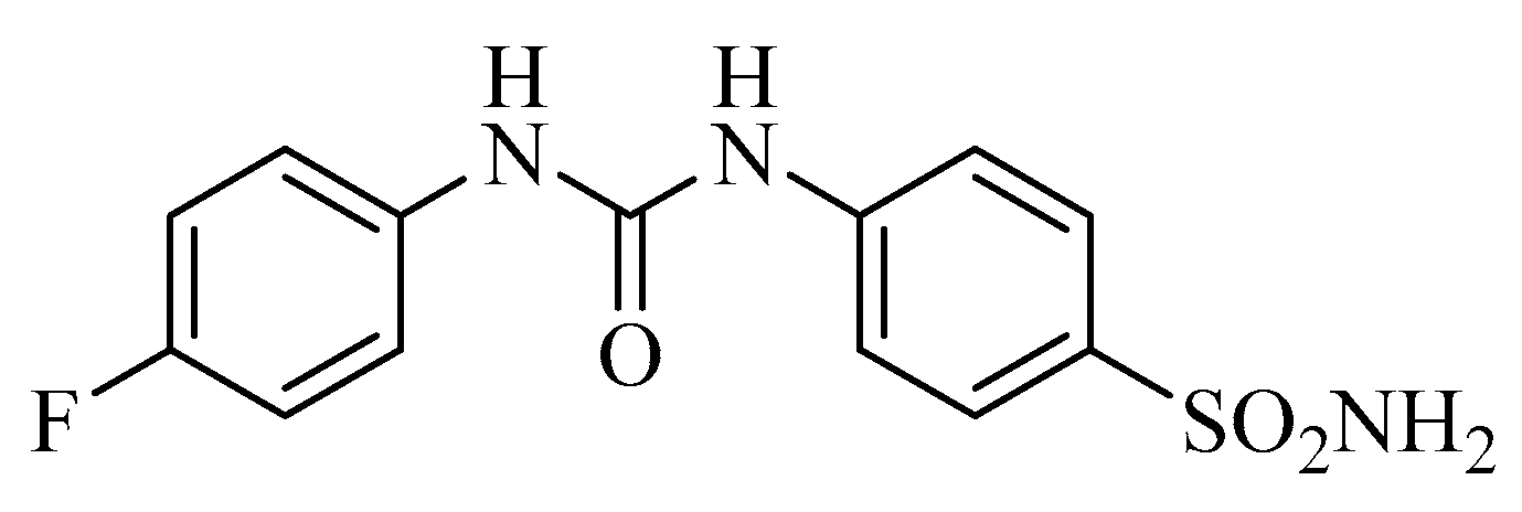
As seen from Figure 4, SLC-0111 is a simple, ureido-substituted benzenesulfonamide derivative which has significant hCA IX and XII inhibitory properties in vitro (KIs of 45 nM against hCA IX and of 4.5 nM against hCA XII), being much less effective as an inhibitor of hCA I and II, widespread cytosolic CAs in many organs [45]. The CA IX/XII-selective inhibitory properties of this sulfonamide and of some of its congeners were explained at the molecular level by using X-ray crystallography of enzyme-inhibitor adducts [52]. This study allowed to observe that the tail of the inhibitors (in the case of SLC-0111, the tail is a 4-fluorophenyl moiety) adopts very different conformations when the sulfonamide is bound within the enzyme active site cavity, and is orientated towards the exit of the cavity, which is the most variable part of the different CA isoforms present in mammals [52]. As a consequence, this class of sulfonamide CAIs show some of the highest selectivity ratios for inhibiting the tumor-associated over the cytosolic isoforms [52]. In vivo studies showed SLC-0111 to potently inhibit the growth of tumors harboring CA IX/XII, whereas tumors that did not express these enzymes were unaffected [44,45]. The metastases formation was also inhibited in the T4 murine breast cancer model [44], and important antitumor effects were observed also in combination with other anticancer agents used clinically, such as paclitaxel, doxorubicine, etc. [44,45]. As mentioned above, a notable depletion of the cancer stem cell population was also evident after the treatment with this compound. Although the results of the Phase I clinical trial are not yet published, the compound has been scheduled for Phase II trials which will start late in 2016 [51].
Although there are many other highly effective in vitro CA IX inhibitors reported so far, only a few of them were investigated in vivo in details. In one such study [53], important inhibition of growth of osteosarcoma was observed after inhibiting CA IX with positively charged pyridinium sulfonamides, suggesting their potential use for this refractory, difficult to treat tumor. In another study, [54], the CA IX and AP endonuclease-1/redox effector factor 1 (APE1/Ref-1) dual targeting was shown to be synergistic in pancreatic ductal adenocarcinomas (PDACs), another difficultly treatable tumor. A different and innovative approach has been used on the other hand by Neri’s group [55], who conjugated maytansinoid DM1, a cytotoxic natural product payload, to a sulfonamide, more precisely a derivative of acetazolamide (a clinically used CAI drug for decades [7,8]), as targeting ligand for CA IX recognition. This conjugate molecule exhibited a potent in vivo antitumor effect in SKRC52 renal cell carcinomas [55].
It is probable that many other small molecule CA IX/XII inhibitors may enter soon in clinical trials, but probably, most researchers/companies wait for results of the clinical trials of the first-in-the-class such compound (SLC-0111) to be released.
4. Antibodies Targeting CA IX and XII as Antitumor Agents
4.1. Anti-CA IX Antibodies
The renal cell carcinoma (RCC)-associated protein G250 was recognized by its discoverers to be an anti-CA IX monoclonal antibody (Mab) and proposed as a possible antitumor target for RCC [56]. Indeed, G250, formulated as chimeric IgG1 monoclonal antibody and denominated girentuximab, was the first CA IX inhibitor to enter clinical trials [57], being actually in Phase III, although it seems that its development has been interrupted due to lack of efficacy [58]. Thus, no other details will be discussed about this Mab, but Pastorekova’s group [59,60] proposed several interesting approaches based both on antibodies that inhibit the catalytic activity as well as those that target the PG domain of CA IX (and do not inhibit the CO2 hydrase activity of the enzyme). For example the mouse monoclonal antibody VII/20 was shown to bind to the catalytic domain of CA IX, leading to an efficient receptor-mediated internalization of the antibody-enzyme conjugate, which is the main process that regulates abundance and signaling of cell surface proteins [60]. This internalization has a considerable impact on immunotherapy and in this particular case elicited significant anticancer effects in a mouse xenograft model of colorectal cancer [60]. The same group [59] demonstrated that the monoclonal antibody M75 (targeting the PG domain of CA IX and widely used as a reagent in immune-histochemical studies [10,17]) can be encapsulated into alginate microbeads or microcapsules made of sodium alginate, cellulose sulfate, and poly(methylene-co-guanidine), which afforded a rapid M75 antibody release at pH 6.8 (characteristic of the acidic tumors) compared to pH 7.4 (the physiologic, normal pH) [59].
4.2. Anti-CA XII Antibodies
There are far fewer studies to target CA XII with Mabs compared to CA IX. The most significant one comes from Zeidler’s group [61] who discovered 6A10, the first monoclonal antibody that binds to the catalytic domain of CA XII and also acts as an inhibitor of the enzyme. 6A10 was shown to be a low nanomolar CA XII inhibitor and to inhibit the growth of tumor cells in spheroids and in vivo, in a mouse xenograft model of human cancer [61,62].
5. Imaging CA IX/XII Positive Tumors
The initial imaging strategy (after the fluorescent sulfonamides used for the proof-of-concept study mentioned above, which cannot be used to image human cancers [41]) was to incorporate 99mTc or 18F as positron-emitting isotopes in the molecules of sulfonamide or coumarin CAIs in order to obtain agents useful for positron emission tomography (PET) [63,64,65,66]. The initial sulfonamides or coumarins labeled with these isotopes were not highly efficient imaging agents, probably due to pharmacokinetic-related problems. For example the SLC-0111 analog labeled with 18F as well as a coumarin derivative incorporating the same isotope, although highly potent as in vitro CA IX inhibitors in vivo, in HT-29 (colorectal) xenografts in mice did not accumulate in the tumor, but were principally present in the blood, liver and nose of the animals, making them inappropriate as PET agents. However, the next generation inhibitors labeled with 18F (trimeric sulfonamides [67] or positively-charged sulfonamides [68]) or 68Ga-labelled sulfonamides (originally reported by Bénard’s group [69] and soon thereafter by Poulsen’s group [70]) showed that such 68Ga-polyaminocarboxylate chelator-conjugated sulfonamides do accumulate preferentially within the hypoxic tumor, making them excellent candidates for clinical studies [69]. In HT-29 colorectal xenograft tumors in mice, the gallium-containing sulfonamides showed an excellent and specific tumor accumulation, coupled with a low uptake in blood and clearing intact into the urine, making them of great interest for further development [69,70].
Antibodies were also proposed as imaging agents for CA IX-positive tumors, originally by Neri’s group [71]. By using the phage technology, high-affinity Mabs targeting hCA IX were generated (denominated A3 and CC7) which were used for imaging purposes in animal models of colorectal cancer (LS174T cell line). Such imaging studies with the two anti-hCA IX Mabs disclosed by this group closely matched the pimonidazole (an azole agent which accumulates in hypoxic regions of tumors) staining of these tumors, furnishing the proof-of-concept study that, in addition to the small molecule CA IX inhibitors, the antibodies can also be used for non-invasive imaging of hypoxic tumors. There are in fact many other similar imaging studies of the Mab in clinical trials mentioned above, girentuximab, which has been labeled with various isotopes for these purposes. For example, 111In- [72], 99mTc- [73] and 124I [74]—labeled girentuximab as well as dual-labeled Mab with a radionuclide and a fluorescence tag [75] have been developed and used for hypoxic tumor imaging with various degrees of success. However, antibodies have some problematic pharmacological aspects that must be considered attentively when used, and most probably small molecule CA IX/XII inhibitors may be more useful and easier to develop for a possible theragnostic agent targeting these enzymes.
6. Conclusions
Discovered at the beginning of the 90s, CA IX (and subsequently CA XII) were shown to possess crucial roles in tumorigenesis due to their involvement in the metabolism of hypoxic, acidic tumors. Overexpressed in tumor cells as a consequence of the HIF-1 cascade, these enzymes generate H+ and bicarbonate ions, the simplest metabolites known, from CO2 as substrate, being involved in many processes connected to tumorigenesis, from the regulation of the internal/external tumor cell pH, to migration, invasion, metastases formation as well as regulation of the cancer stem cell population. Many of these fascinating phenomena, discovered in the last decade, were shown to be useful for obtaining antitumor therapies/tumor imaging agents with a novel mechanism of action, by targeting these enzymes either with small molecule inhibitors or antibodies. The initial success of the clinical trials started with these agents, which is a continuing story, constitutes an excellent example of how fundamental research discoveries, thought to only explain some intricate biochemical/physiologic processes, may lead to innovative therapeutic strategies for fighting tumors.
Acknowledgments
Research from author’s group was financed by several European Union projects (EUROXY, METOXIA, DeZnIT and Dynano, in the period 2004–2014) and by Signal Life Sciences (in the period 2013–2015). No funds were received for covering the costs to publish in open access this paper.
Conflicts of Interest
The author declares conflict of interest, being one of the inventors of SLC-0111.
References
- Warburg, O. On respiratory impairment in cancer cells. Science 1956, 124, 269–270. [Google Scholar] [PubMed]
- Schwartz, L.; Supuran, C.T.; Alfarouk, K.O. The Warburg effect and the hallmarks of cancer. Anticancer Agents Med. Chem. 2017, 17, 164–170. [Google Scholar] [CrossRef] [PubMed]
- Semenza, G.L. Hypoxia-inducible factor 1: Oxygen homeostasis and disease pathophysiology. Trends Mol. Med. 2001, 7, 345–350. [Google Scholar] [CrossRef]
- Pouysségur, J.; Dayan, F.; Mazure, N.M. Hypoxia signalling in cancer and approaches to enforce tumour regression. Nature 2006, 441, 437–443. [Google Scholar] [CrossRef] [PubMed]
- Hockel, M.; Vaupel, P. Tumor hypoxia: Definitions and current clinical, biologic, and molecular aspects. J. Natl. Cancer Inst. 2001, 93, 266–276. [Google Scholar] [CrossRef] [PubMed]
- Kremer, G.; Pouysségur, J. Tumor cell metabolism: Cancer’s Achilles’ heel. Cancer Cell. 2008, 13, 472–482. [Google Scholar] [CrossRef] [PubMed]
- Supuran, C.T. Carbonic anhydrases: novel therapeutic applications for inhibitors and activators. Nat. Rev. Drug Discov. 2008, 7, 168–181. [Google Scholar] [CrossRef] [PubMed]
- Neri, D.; Supuran, C.T. Interfering with pH regulation in tumours as a therapeutic strategy. Nat. Rev. Drug Discov. 2011, 10, 767–777. [Google Scholar] [CrossRef] [PubMed]
- Wykoff, C.C.; Beasley, N.J.; Watson, P.H.; Turner, K.J.; Pastorek, J.; Sibtain, A.; Wilson, G.D.; Turley, H.; Talks, K.L.; Maxwell, P.H.; et al. Hypoxia-inducible expression of tumor-associated carbonic anhydrases. Cancer Res. 2000, 60, 7075–7083. [Google Scholar] [PubMed]
- Pastorek, J.; Pastoreková, S.; Callebaut, I.; Mornon, J.P.; Zelník, V.; Opavský, R.; Zat’ovicová, M.; Liao, S.; Portetelle, D.; Stanbridge, E.J. Cloning and characterization of MN, a human tumor-associated protein with a domain homologous to carbonic anhydrase and putative helix-loop-helix DNA binding segment. Oncogene 1994, 9, 2877–2888. [Google Scholar] [PubMed]
- Tureci, O.; Sahin, U.; Vollmar, E.; Siemer, S.; Göttert, E.; Seitz, G.; Parkkila, A.K.; Shah, G.N.; Grubb, J.H.; Pfreundschuh, M.; et al. Human carbonic anhydrase XII: cDNA cloning, expression, and chromosomal localization of a carbonic anhydrase gene that is overexpressed in some renal cell cancers. Proc. Natl. Acad. Sci. USA 1998, 95, 7608–7613. [Google Scholar] [CrossRef] [PubMed]
- Boron, W.F. Regulation of intracellular pH. Adv. Physiol. Educ. 2004, 28, 160–179. [Google Scholar] [CrossRef] [PubMed]
- Gatenby, R.A.; Gillies, R.J. A microenvironmental model of carcinogenesis. Nat. Rev. Cancer. 2008, 8, 56–61. [Google Scholar] [CrossRef] [PubMed]
- Gatenby, R.A.; Gillies, R.J. Why do cancers have high aerobic glycolysis? Nat. Rev. Cancer. 2004, 4, 891–899. [Google Scholar] [CrossRef] [PubMed]
- Fiaschi, T.; Giannoni, E.; Taddei, M.L.; Cirri, P.; Marini, A.; Pintus, G.; Nativi, C.; Richichi, B.; Scozzafava, A.; Carta, F.; et al. Carbonic anhydrase IX from cancer-associated fibroblasts drives epithelial-mesenchymal transition in prostate carcinoma cells. Cell Cycle 2013, 12, 1791–1801. [Google Scholar] [CrossRef] [PubMed]
- Parks, S.K.; Chiche, J.; Pouysségur, J. Disrupting proton dynamics and energy metabolism for cancer therapy. Nat. Rev. Cancer 2013, 13, 611–623. [Google Scholar] [CrossRef] [PubMed]
- Pettersen, E.O.; Ebbesen, P.; Gieling, R.G.; Williams, K.J.; Dubois, L.; Lambin, P.; Ward, C.; Meehan, J.; Kunkler, I.H.; Langdon, S.P.; et al. Targeting tumour hypoxia to prevent cancer metastasis. From biology, biosensing and technology to drug development: the METOXIA consortium. J. Enzyme Inhib. Med. Chem. 2015, 30, 689–721. [Google Scholar] [CrossRef] [PubMed]
- Perez-Sayans, M.; Garcia-Garcia, A.; Scozzafava, A.; Supuran, C.T. Inhibition of V-ATPase and carbonic anhydrases as interference strategy with tumor acidification processes. Curr. Pharm. Des. 2012, 18, 1407–1413. [Google Scholar] [CrossRef] [PubMed]
- Alterio, V.; Di Fiore, A.; D’Ambrosio, K.; Supuran, C.T.; De Simone, G. Multiple binding modes of inhibitors to carbonic anhydrases: How to design specific drugs targeting 15 different isoforms? Chem. Rev. 2012, 112, 4421–4468. [Google Scholar] [CrossRef] [PubMed]
- Supuran, C.T. Structure and function of carbonic anhydrases. Biochem. J. 2016, 473, 2023–2032. [Google Scholar] [CrossRef] [PubMed]
- De Milito, A.; Marino, M.L.; Fais, S. A rationale for the use of proton pump inhibitors as antineoplastic agents. Curr. Pharm. Des. 2012, 18, 1395–1406. [Google Scholar] [CrossRef] [PubMed]
- Lugini, L.; Federici, C.; Borghi, M.; Azzarito, T.; Marino, M.L.; Cesolini, A.; Spugnini, E.P.; Fais, S. Proton pump inhibitors while belonging to the same family of generic drugs show different anti-tumor effect. J. Enzyme Inhib. Med. Chem. 2016, 31, 538–545. [Google Scholar] [CrossRef] [PubMed]
- Federici, C.; Lugini, L.; Marino, M.L.; Carta, F.; Iessi, E.; Azzarito, T.; Supuran, C.T.; Fais, S. Lansoprazole and carbonic anhydrase IX inhibitors sinergize against human melanoma cells. J. Enzyme Inhib. Med. Chem. 2016, 31, 119–125. [Google Scholar] [CrossRef] [PubMed]
- Hilvo, M.; Baranauskiene, L.; Salzano, A.M.; Scaloni, A.; Matulis, D.; Innocenti, A.; Scozzafava, A.; Monti, S.M.; Di Fiore, A.; De Simone, G.; et al. Biochemical characterization of CA IX: One of the most active carbonic anhydrase isozymes. J. Biol. Chem. 2008, 283, 27799–27809. [Google Scholar] [CrossRef] [PubMed]
- Alterio, V.; Hilvo, M.; Di Fiore, A.; Supuran, C.T.; Pan, P.; Parkkila, S.; Scaloni, A.; Pastorek, J.; Pastorekova, S.; Pedone, C.; et al. Crystal structure of the extracellular catalytic domain of the tumor-associated human carbonic anhydrase IX. Proc. Natl. Acad. Sci. USA 2009, 106, 16233–16238. [Google Scholar] [CrossRef] [PubMed]
- Innocenti, A.; Pastorekova, S.; Pastorek, J.; Scozzafava, A.; De Simone, G.; Supuran, C.T. The proteoglycan region of the tumor-associated carbonic anhydrase isoform IX acts as an intrinsic buffer optimizing CO2 hydration at acidic pH values characteristic of solid tumors. Bioorg. Med. Chem. Lett. 2009, 19, 5825–5828. [Google Scholar] [CrossRef] [PubMed]
- Pastorek, J.; Pastorekova, S. Hypoxia-induced carbonic anhydrase IX as a target for cancer therapy: From biology to clinical use. Semin. Cancer Biol. 2015, 31, 52–64. [Google Scholar] [CrossRef] [PubMed]
- Svastová, E.; Zilka, N.; Zat’ovicová, M.; Gibadulinová, A.; Ciampor, F.; Pastorek, J.; Pastoreková, S. Carbonic anhydrase IX reduces E-cadherin-mediated adhesion of MDCK cells via interaction with beta-catenin. Exp. Cell Res. 2003, 290, 332–345. [Google Scholar] [CrossRef]
- Svastová, E.; Hulíková, A.; Rafajová, M.; Zat’ovicová, M.; Gibadulinová, A.; Casini, A.; Cecchi, A.; Scozzafava, A.; Supuran, C.T.; Pastorek, J.; et al. Hypoxia activates the capacity of tumor-associated carbonic anhydrase IX to acidify extracellular pH. FEBS Lett. 2004, 577, 439–445. [Google Scholar] [CrossRef] [PubMed]
- Ditte, P.; Dequiedt, F.; Svastova, E.; Hulikova, A.; Ohradanova-Repic, A.; Zatovicova, M.; Csaderova, L.; Kopacek, J.; Supuran, C.T.; Pastorekova, S.; et al. Phosphorylation of carbonic anhydrase IX controls its ability to mediate extracellular acidification in hypoxic tumors. Cancer Res. 2011, 71, 7558–7567. [Google Scholar] [CrossRef] [PubMed]
- Buanne, P.; Renzone, G.; Monteleone, F.; Vitale, M.; Monti, S.M.; Sandomenico, A.; Garbi, C.; Montanaro, D.; Accardo, M.; Troncone, G.; et al. Characterization of carbonic anhydrase IX interactome reveals proteins assisting its nuclear localization in hypoxic cells. J. Proteome Res. 2013, 12, 282–292. [Google Scholar] [CrossRef] [PubMed]
- Buonanno, M.; Langella, E.; Zambrano, N.; Succoio, M.; Sasso, E.; Alterio, V.; Di Fiore, A.; Sandomenico, A.; Supuran, C.T.; Scaloni, A.; et al. Disclosing the interaction of carbonic anhydrase IX with cullin-associated NEDD8-dissociated protein 1 by molecular modeling and integrated binding measurements. ACS Chem. Biol. 2017, 12, 1460–1465. [Google Scholar] [CrossRef] [PubMed]
- Vullo, D.; Innocenti, A.; Nishimori, I.; Pastorek, J.; Scozzafava, A.; Pastoreková, S.; Supuran, C.T. Carbonic anhydrase inhibitors. Inhibition of the transmembrane isozyme XII with sulfonamides-a new target for the design of antitumor and antiglaucoma drugs? Bioorg. Med. Chem. Lett. 2005, 15, 963–969. [Google Scholar] [CrossRef] [PubMed]
- Vullo, D.; Franchi, M.; Gallori, E.; Pastorek, J.; Scozzafava, A.; Pastorekova, S.; Supuran, C.T. Carbonic anhydrase inhibitors: Inhibition of the tumor-associated isozyme IX with aromatic and heterocyclic sulfonamides. Bioorg. Med. Chem. Lett. 2003, 13, 1005–1009. [Google Scholar] [CrossRef]
- Guler, O.O.; De Simone, G.; Supuran, C.T. Drug design studies of the novel antitumor targets carbonic anhydrase IX and XII. Curr. Med. Chem. 2010, 17, 1516–1526. [Google Scholar] [CrossRef] [PubMed]
- Supuran, C.T. How many carbonic anhydrase inhibition mechanisms exist? J. Enzyme Inhib. Med. Chem. 2016, 31, 345–360. [Google Scholar] [CrossRef] [PubMed]
- Supuran, C.T. Advances in structure-based drug discovery of carbonic anhydrase inhibitors. Expert Opin. Drug Discov. 2017, 12, 61–88. [Google Scholar] [CrossRef] [PubMed]
- Supuran, C.T. Structure-based drug discovery of carbonic anhydrase inhibitors. J. Enzyme Inhib. Med. Chem. 2012, 27, 759–772. [Google Scholar] [CrossRef] [PubMed]
- Cecchi, A.; Hulikova, A.; Pastorek, J.; Pastoreková, S.; Scozzafava, A.; Winum, J.Y.; Montero, J.L.; Supuran, C.T. Carbonic anhydrase inhibitors. Design of fluorescent sulfonamides as probes of tumor-associated carbonic anhydrase IX that inhibit isozyme IX-mediated acidification of hypoxic tumors. J. Med. Chem. 2005, 48, 4834–4841. [Google Scholar] [CrossRef] [PubMed]
- Dubois, L.; Douma, K.; Supuran, C.T.; Chiu, R.K.; van Zandvoort, M.A.; Pastoreková, S.; Scozzafava, A.; Wouters, B.G.; Lambin, P. Imaging the hypoxia surrogate marker CA IX requires expression and catalytic activity for binding fluorescent sulfonamide inhibitors. Radiother. Oncol. 2007, 83, 367–373. [Google Scholar] [CrossRef] [PubMed]
- Dubois, L.; Lieuwes, N.G.; Maresca, A.; Thiry, A.; Supuran, C.T.; Scozzafava, A.; Wouters, B.G.; Lambin, P. Imaging of CA IX with fluorescent labelled sulfonamides distinguishes hypoxic and (re)-oxygenated cells in a xenograft tumour model. Radiother. Oncol. 2009, 92, 423–428. [Google Scholar] [CrossRef] [PubMed]
- Ahlskog, J.K.; Dumelin, C.E.; Trüssel, S.; Mårlind, J.; Neri, D. In vivo targeting of tumor-associated carbonic anhydrases using acetazolamide derivatives. Bioorg. Med. Chem. Lett. 2009, 19, 4851–4856. [Google Scholar] [CrossRef] [PubMed]
- Dubois, L.; Peeters, S.; Lieuwes, N.G.; Geusens, N.; Thiry, A.; Wigfield, S.; Carta, F.; McIntyre, A.; Scozzafava, A.; Dogné, J.M.; et al. Specific inhibition of carbonic anhydrase IX activity enhances the in vivo therapeutic effect of tumor irradiation. Radiother. Oncol. 2011, 99, 424–431. [Google Scholar] [CrossRef] [PubMed]
- Lou, Y.; McDonald, P.C.; Oloumi, A.; Chia, S.; Ostlund, C.; Ahmadi, A.; Kyle, A.; Auf dem Keller, U.; Leung, S.; Huntsman, D.; et al. Targeting tumor hypoxia: Suppression of breast tumor growth and metastasis by novel carbonic anhydrase IX inhibitors. Cancer Res. 2011, 71, 3364–3376. [Google Scholar] [CrossRef] [PubMed]
- Pacchiano, F.; Carta, F.; McDonald, P.C.; Lou, Y.; Vullo, D.; Scozzafava, A.; Dedhar, S.; Supuran, C.T. Ureido-substituted benzenesulfonamides potently inhibit carbonic anhydrase IX and show antimetastatic activity in a model of breast cancer metastasis. J. Med. Chem. 2011, 54, 1896–1902. [Google Scholar] [CrossRef] [PubMed]
- Gieling, R.G.; Babur, M.; Mamnani, L.; Burrows, N.; Telfer, B.A.; Carta, F.; Winum, J.Y.; Scozzafava, A.; Supuran, C.T.; Williams, K.J. Antimetastatic effect of sulfamate carbonic anhydrase IX inhibitors in breast carcinoma xenografts. J. Med. Chem. 2012, 55, 5591–5600. [Google Scholar] [CrossRef] [PubMed]
- Touisni, N.; Maresca, A.; McDonald, P.C.; Lou, Y.; Scozzafava, A.; Dedhar, S.; Winum, J.Y.; Supuran, C.T. Glycosyl coumarin carbonic anhydrase IX and XII inhibitors strongly attenuate the growth of primary breast tumors. J. Med. Chem. 2011, 54, 8271–8277. [Google Scholar] [CrossRef] [PubMed]
- Lock, F.E.; McDonald, P.C.; Lou, Y.; Serrano, I.; Chafe, S.C.; Ostlund, C.; Aparicio, S.; Winum, J.Y.; Supuran, C.T.; Dedhar, S. Targeting carbonic anhydrase IX depletes breast cancer stem cells within the hypoxic niche. Oncogene. 2013, 32, 5210–5219. [Google Scholar] [CrossRef] [PubMed]
- McDonald, P.C.; Chafe, S.C.; Dedhar, S. Overcoming hypoxia-mediated tumor progression: combinatorial approaches targeting ph regulation, angiogenesis and immune dysfunction. Front. Cell Dev. Biol. 2016, 4, 27. [Google Scholar] [CrossRef] [PubMed]
- Swayampakula, M.; McDonald, P.C.; Vallejo, M.; Coyaud, E.; Chafe, S.C.; Westerback, A.; Venkateswaran, G.; Shankar, J.; Gao, G.; Laurent, E.M.N.; et al. The interactome of metabolic enzyme carbonic anhydrase IX reveals novel roles in tumor cell migration and invadopodia/MMP14-mediated invasion. Oncogene. 2017, in press. [Google Scholar] [CrossRef] [PubMed]
- A phase I, multi-center, open-label, study to investigate the safety, tolerability and pharmacokinetic of SLC-0111 in subjects with advanced solid tumours. 2016. Available online: https://clinicaltrials.gov/ct2/show/NCT02215850 (accessed on 15 September 2017).
- Pacchiano, F.; Aggarwal, M.; Avvaru, B.S.; Robbins, A.H.; Scozzafava, A.; McKenna, R.; Supuran, C.T. Selective hydrophobic pocket binding observed within the carbonic anhydrase II active site accommodate different 4-substituted-ureido-benzenesulfonamides and correlate to inhibitor potency. Chem. Commun. 2010, 46, 8371–8373. [Google Scholar] [CrossRef] [PubMed]
- Perut, F.; Carta, F.; Bonuccelli, G.; Grisendi, G.; Di Pompo, G.; Avnet, S.; Sbrana, F.V.; Hosogi, S.; Dominici, M.; Kusuzaki, K.; et al. Carbonic anhydrase IX inhibition is an effective strategy for osteosarcoma treatment. Expert Opin. Ther. Targets. 2015, 19, 1593–1605. [Google Scholar] [CrossRef] [PubMed]
- Logsdon, D.P.; Grimard, M.; Luo, M.; Shahda, S.; Jiang, Y.; Tong, Y.; Yu, Z.; Zyromski, N.; Schipani, E.; Carta, F.; et al. Regulation of HIF1α under hypoxia by APE1/Ref-1 impacts CA9 expression: Dual targeting in patient-derived 3D pancreatic cancer models. Mol. Cancer Ther. 2016, 15, 2722–2732. [Google Scholar] [CrossRef] [PubMed]
- Krall, N.; Pretto, F.; Decurtins, W.; Bernardes, G.J.; Supuran, C.T.; Neri, D. A small-molecule drug conjugate for the treatment of carbonic anhydrase IX expressing tumors. Angew. Chem. Int. Ed. Engl. 2014, 53, 4231–4235. [Google Scholar] [CrossRef] [PubMed]
- Grabmaier, K.; Vissers, J.L.; De Weijert, M.C.; Oosterwijk-Wakka, J.C.; Van Bokhoven, A.; Brakenhoff, R.H.; Noessner, E.; Mulders, P.A.; Merkx, G.; Figdor, C.G.; et al. Molecular cloning and immunogenicity of renal cell carcinoma-associated antigen G250. Int. J. Cancer 2000, 85, 865–870. [Google Scholar] [CrossRef]
- Siebels, M.; Rohrmann, K.; Oberneder, R.; Stahler, M.; Haseke, N.; Beck, J.; Hofmann, R.; Kindler, M.; Kloepfer, P.; Stief, C. A clinical phase I/II trial with the monoclonal antibody cG250 (RENCAREX®) and interferon-alpha-2a in metastatic renal cell carcinoma patients. World J. Urol. 2011, 29, 121–126. [Google Scholar] [CrossRef] [PubMed]
- A Randomized, Double Blind Phase III Study to Evaluate Adjuvant cG250 Treatment Versus Placebo in Patients with Clear Cell RCC and High Risk of Recurrence (ARISER). Available online: https://clinicaltrials.gov/ct2/show/NCT00087022?term=girentuximab&rank=6 (accessed on 15 September 2017).
- Takacova, M.; Hlouskova, G.; Zatovicova, M.; Benej, M.; Sedlakova, O.; Kopacek, J.; Pastorek, J.; Lacik, I.; Pastorekova, S. Encapsulation of anti-carbonic anhydrase IX antibody in hydrogel microspheres for tumor targeting. J. Enzyme Inhib. Med. Chem. 2016, 31, 110–118. [Google Scholar] [CrossRef] [PubMed]
- Zatovicova, M.; Jelenska, L.; Hulikova, A.; Csaderova, L.; Ditte, Z.; Ditte, P.; Goliasova, T.; Pastorek, J.; Pastorekova, S. Carbonic anhydrase IX as an anticancer therapy target: Preclinical evaluation of internalizing monoclonal antibody directed to catalytic domain. Curr. Pharm. Des. 2010, 16, 3255–3263. [Google Scholar] [CrossRef] [PubMed]
- Battke, C.; Kremmer, E.; Mysliwietz, J.; Gondi, G.; Dumitru, C.; Brandau, S.; Lang, S.; Vullo, D.; Supuran, C.; Zeidler, R. Generation and characterization of the first inhibitory antibody targeting tumour-associated carbonic anhydrase XII. Cancer Immunol. Immunother. 2011, 60, 649–658. [Google Scholar] [CrossRef] [PubMed]
- Gondi, G.; Mysliwietz, J.; Hulikova, A.; Jen, J.P.; Swietach, P.; Kremmer, E.; Zeidler, R. Antitumor efficacy of a monoclonal antibody that inhibits the activity of cancer-associated carbonic anhydrase XII. Cancer Res. 2013, 73, 6494–6503. [Google Scholar] [CrossRef] [PubMed]
- Akurathi, V.; Dubois, L.; Lieuwes, N.G.; Chitneni, S.K.; Cleynhens, B.J.; Vullo, D.; Supuran, C.T.; Verbruggen, A.M.; Lambin, P.; Bormans, G.M. Synthesis and biological evaluation of a 99mTc-labelled sulfonamide conjugate for in vivo visualization of carbonic anhydrase IX expression in tumor hypoxia. Nucl. Med. Biol. 2010, 37, 557–564. [Google Scholar] [CrossRef] [PubMed]
- Akurathi, V.; Dubois, L.; Celen, S.; Lieuwes, N.G.; Chitneni, S.K.; Cleynhens, B.J.; Innocenti, A.; Supuran, C.T.; Verbruggen, A.M.; Lambin, P.; et al. Development and biological evaluation of 99mTc-sulfonamide derivatives for in vivo visualization of CA IX as surrogate tumor hypoxia markers. Eur. J. Med. Chem. 2014, 71, 374–384. [Google Scholar] [CrossRef] [PubMed]
- Peeters, S.G.; Dubois, L.; Lieuwes, N.G.; Laan, D.; Mooijer, M.; Schuit, R.C.; Vullo, D.; Supuran, C.T.; Eriksson, J.; Windhorst, A.D.; et al. [18F]VM4–o37 MicroPET imaging and biodistribution of two in vivo CAIX-expressing tumor models. Mol. Imaging Biol. 2015, 17, 615–619. [Google Scholar] [CrossRef] [PubMed]
- Pan, J.; Lau, J.; Mesak, F.; Hundal, N.; Pourghiasian, M.; Liu, Z.; Bénard, F.; Dedhar, S.; Supuran, C.T.; Lin, K.S. Synthesis and evaluation of 18F-labeled carbonic anhydrase IX inhibitors for imaging with positron emission tomography. J. Enzyme Inhib. Med. Chem. 2014, 29, 249–255. [Google Scholar] [CrossRef] [PubMed]
- Lau, J.; Liu, Z.; Lin, K.S.; Pan, J.; Zhang, Z.; Vullo, D.; Supuran, C.T.; Perrin, D.M.; Bénard, F. Trimeric radiofluorinated sulfonamide derivatives to achieve in vivo selectivity for carbonic anhydrase IX-targeted PET imaging. J. Nucl. Med. 2015, 56, 1434–1440. [Google Scholar] [CrossRef] [PubMed]
- Zhang, Z.; Lau, J.; Zhang, C.; Colpo, N.; Nocentini, A.; Supuran, C.T.; Bénard, F.; Lin, K.S. Design, synthesis and evaluation of 18F-labeled cationic carbonic anhydrase IX inhibitors for PET imaging. J. Enzyme Inhib. Med. Chem. 2017, 32, 722–730. [Google Scholar] [CrossRef] [PubMed]
- Lau, J.; Zhang, Z.; Jenni, S.; Kuo, H.T.; Liu, Z.; Vullo, D.; Supuran, C.T.; Lin, K.S.; Bénard, F. PET imaging of carbonic anhydrase IX expression of HT-29 tumor xenograft mice with 68Ga-labeled benzenesulfonamides. Mol. Pharm. 2016, 13, 1137–1146. [Google Scholar] [CrossRef] [PubMed]
- Sneddon, D.; Niemans, R.; Bauwens, M.; Yaromina, A.; van Kuijk, S.J.; Lieuwes, N.G.; Biemans, R.; Pooters, I.; Pellegrini, P.A.; Lengkeek, N.A.; et al. Synthesis and in vivo biological evaluation of 68Ga-labeled carbonic anhydrase IX targeting small molecules for positron emission tomography. J. Med. Chem. 2016, 59, 6431–6443. [Google Scholar] [CrossRef] [PubMed]
- Ahlskog, J.K.; Schliemann, C.; Mårlind, J.; Qureshi, U.; Ammar, A.; Pedley, R.B.; Neri, D. Human monoclonal antibodies targeting carbonic anhydrase IX for the molecular imaging of hypoxic regions in solid tumours. Br. J. Cancer. 2009, 101, 645–657. [Google Scholar] [CrossRef] [PubMed]
- Huizing, F.J.; Hoeben, B.A.W.; Franssen, G.; Lok, J.; Heskamp, S.; Oosterwijk, E.; Boerman, O.C.; Bussink, J. Preclinical validation of 111In-girentuximab-F(ab′)2 as a tracer to image hypoxia related marker CA IX expression in head and neck cancer xenografts. Radiother Oncol. 2017, in press. [Google Scholar] [CrossRef]
- Honarvar, H.; Garousi, J.; Gunneriusson, E.; Höidén-Guthenberg, I.; Altai, M.; Widström, C.; Tolmachev, V.; Frejd, F.Y. Imaging of CAIX-expressing xenografts in vivo using 99mTc-HEHEHE-ZCAIX: 1 affibody molecule. Int. J. Oncol. 2015, 46, 513–520. [Google Scholar] [CrossRef] [PubMed]
- Khandani, A.H.; Rathmell, W.K.; Wallen, E.M.; Ivanovic, M. PET/CT with 124I-cG250: Great potential and some open questions. AJR Am. J. Roentgenol. 2014, 203, 261–262. [Google Scholar] [CrossRef] [PubMed]
- Muselaers, C.H.; Rijpkema, M.; Bos, D.L.; Langenhuijsen, J.F.; Oyen, W.J.; Mulders, P.F.; Oosterwijk, E.; Boerman, O.C. Radionuclide and fluorescence imaging of clear cell renal cell carcinoma using dual labeled anti-carbonic anhydrase IX antibody G250. J. Urol. 2015, 194, 532–538. [Google Scholar] [CrossRef] [PubMed]
Figure 1.
Mechanism by which the transcription factor HIF-1α (abbreviated as HIFα) orchestrates the overexpression of proteins involved in aerobic glycolysis, angiogenesis, erythropoesis and pH regulation in hypoxic tumors. In normoxia HIFα is hydroxylated at a Pro residue and targeted for degradation by the proteasome (PHD, prolyl-hydroxylase; VHL, von Hippel-Lindau factor, HRE, hypoxia responsive element). In hypoxia, its accumulation leads to overexpression of the proteins involved in tumorigenesis mentioned above [5,6,7,8].

Figure 2.
Proteins involved in pH regulation in tumors: GLUT1, the glucose transporter isoform 1; MCT, monocarboxylate transporter, which extrude lactic acid and other monocarboxylates formed by the glycolytic degradation of glucose; NHE, sodium-proton exchanger (Na+–H+ antiporter); V-ATPase, plasma membrane proton pump; AE, anion exchanger (chloride-bicarbonate exchanger); NBC, sodium bicarbonate channels; BT, bicarbonate transporter; CA II (cytosolic) and CA IX/XII, which catalyze CO2 hydration to bicarbonate and protons [4,5,6,7,8,9,10,11,12,13,14,15].

Figure 3.
CA IX X-ray crystal structure of the catalytic domain (in blue), the PG domain (cartoon in pink), plasma membrane (in red), the transmembrane domain in yellow (modeled) and the intracytosolic tail (modeled, in green) [29].
Figure 3.
CA IX X-ray crystal structure of the catalytic domain (in blue), the PG domain (cartoon in pink), plasma membrane (in red), the transmembrane domain in yellow (modeled) and the intracytosolic tail (modeled, in green) [29].

Figure 4.
Structure of SLC-0111 (WBI-5111), the sulfonamide CA IX/XII inhibitor in Phase I/II clinical trials.
Figure 4.
Structure of SLC-0111 (WBI-5111), the sulfonamide CA IX/XII inhibitor in Phase I/II clinical trials.

© 2017 by the author. Licensee MDPI, Basel, Switzerland. This article is an open access article distributed under the terms and conditions of the Creative Commons Attribution (CC BY) license (http://creativecommons.org/licenses/by/4.0/).
Share and Cite
MDPI and ACS Style
Supuran, C.T. Carbonic Anhydrase Inhibition and the Management of Hypoxic Tumors. Metabolites 2017, 7, 48. https://doi.org/10.3390/metabo7030048
AMA Style
Supuran CT. Carbonic Anhydrase Inhibition and the Management of Hypoxic Tumors. Metabolites. 2017; 7(3):48. https://doi.org/10.3390/metabo7030048
Chicago/Turabian StyleSupuran, Claudiu T. 2017. "Carbonic Anhydrase Inhibition and the Management of Hypoxic Tumors" Metabolites 7, no. 3: 48. https://doi.org/10.3390/metabo7030048
Note that from the first issue of 2016, this journal uses article numbers instead of page numbers. See further details here.