Next Issue
Volume 7, December
Previous Issue
Volume 7, June


Metabolites, Volume 7, Issue 3 (September 2017) – 18 articles

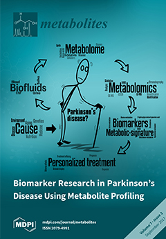
Cover Story (view full-size image): Parkinson’s disease (PD) is a multifactorial condition; optimal treatment is dependent on early and precise diagnosis, and accurate monitoring of disease activity and drug side effects. Metabolomics is a powerful tool to profile biofluid biomarkers that may stratify PD patients into different therapeutic regimens. In this review, we discuss the different analytical platforms and methodologies that are applicable to study alterations in metabolic pathways in clinical and experimental PD. Despite impressive progress, we conclude that the identification and quantification of compounds showing differences between controls and PD patients with or without L-DOPA treatment, is still a major challenge. View this paper
  • Issues are regarded as officially published after their release is announced to the table of contents alert mailing list.
  • You may sign up for e-mail alerts to receive table of contents of newly released issues.
  • PDF is the official format for papers published in both, html and pdf forms. To view the papers in pdf format, click on the "PDF Full-text" link, and use the free Adobe Reader to open them.
Order results
Result details
Section
Select all
Export citation of selected articles as:
3200 KiB  
Review
Carbonic Anhydrase Inhibition and the Management of Hypoxic Tumors
by Claudiu T. Supuran
Metabolites 2017, 7(3), 48; https://doi.org/10.3390/metabo7030048 - 16 Sep 2017
Cited by 211 | Viewed by 10518
Abstract
Hypoxia and acidosis are salient features of many tumors, leading to a completely different metabolism compared to normal cells. Two of the simplest metabolic products, protons and bicarbonate, are generated by the catalytic activity of the metalloenzyme carbonic anhydrase (CA, EC 4.2.1.1), with [...] Read more.
Hypoxia and acidosis are salient features of many tumors, leading to a completely different metabolism compared to normal cells. Two of the simplest metabolic products, protons and bicarbonate, are generated by the catalytic activity of the metalloenzyme carbonic anhydrase (CA, EC 4.2.1.1), with at least two of its isoforms, CA IX and XII, mainly present in hypoxic tumors. Inhibition of tumor-associated CAs leads to an impaired growth of the primary tumors, metastases and reduces the population of cancer stem cells, leading thus to a complex and beneficial anticancer action for this class of enzyme inhibitors. In this review, I will present the state of the art on the development of CA inhibitors (CAIs) targeting the tumor-associated CA isoforms, which may have applications for the treatment and imaging of cancers expressing them. Small molecule inhibitors, one of which (SLC-0111) completed Phase I clinical trials, and antibodies (girentuximab, discontinued in Phase III clinical trials) will be discussed, together with the various approaches used to design anticancer agents with a new mechanism of action based on interference with these crucial metabolites, protons and bicarbonate. Full article
(This article belongs to the Special Issue Carbonic Anhydrases and Metabolism)
Show Figures

Figure 1

848 KiB  
Article
A Protocol for Generating and Exchanging (Genome-Scale) Metabolic Resource Allocation Models
by Alexandra-M. Reimers, Henning Lindhorst and Steffen Waldherr
Metabolites 2017, 7(3), 47; https://doi.org/10.3390/metabo7030047 - 06 Sep 2017
Cited by 17 | Viewed by 6035
Abstract
In this article, we present a protocol for generating a complete (genome-scale) metabolic resource allocation model, as well as a proposal for how to represent such models in the systems biology markup language (SBML). Such models are used to investigate enzyme levels and [...] Read more.
In this article, we present a protocol for generating a complete (genome-scale) metabolic resource allocation model, as well as a proposal for how to represent such models in the systems biology markup language (SBML). Such models are used to investigate enzyme levels and achievable growth rates in large-scale metabolic networks. Although the idea of metabolic resource allocation studies has been present in the field of systems biology for some years, no guidelines for generating such a model have been published up to now. This paper presents step-by-step instructions for building a (dynamic) resource allocation model, starting with prerequisites such as a genome-scale metabolic reconstruction, through building protein and noncatalytic biomass synthesis reactions and assigning turnover rates for each reaction. In addition, we explain how one can use SBML level 3 in combination with the flux balance constraints and our resource allocation modeling annotation to represent such models. Full article
(This article belongs to the Special Issue Metabolism and Systems Biology Volume 2)
Show Figures

Graphical abstract

1108 KiB  
Article
Metabolomics Analysis of Urine Samples from Children after Acetaminophen Overdose
by Laura K. Schnackenberg, Jinchun Sun, Sudeepa Bhattacharyya, Pritmohinder Gill, Laura P. James and Richard D. Beger
Metabolites 2017, 7(3), 46; https://doi.org/10.3390/metabo7030046 - 06 Sep 2017
Cited by 13 | Viewed by 5883
Abstract
Acetaminophen (APAP), a commonly used over-the-counter analgesic, accounts for approximately fifty percent of the cases of acute liver failure (ALF) in the United States due to overdose, with over half of those unintentional. Current clinical approaches for assessing APAP overdose rely on identifying [...] Read more.
Acetaminophen (APAP), a commonly used over-the-counter analgesic, accounts for approximately fifty percent of the cases of acute liver failure (ALF) in the United States due to overdose, with over half of those unintentional. Current clinical approaches for assessing APAP overdose rely on identifying the precise time of overdose and quantitating acetaminophen alanine aminotransferase (ALT) levels in peripheral blood. Novel specific and sensitive biomarkers may provide additional information regarding patient status post overdose. Previous non-clinical metabolomics studies identified potential urinary biomarkers of APAP-induced hepatotoxicity and metabolites involved pathways of tricarboxylic acid cycle, ketone metabolism, and tryptophan metabolism. In this study, biomarkers identified in the previous non-clinical study were evaluated in urine samples collected from healthy subjects (N = 6, median age 14.08 years) and overdose patients (N = 13, median age 13.91 years) as part of an IRB-approved multicenter study of APAP toxicity in children. The clinical results identified metabolites from pathways previously noted, and pathway analysis indicated analogous pathways were significantly altered in both the rats and humans after APAP overdose. The results suggest a metabolomics approach may enable the discovery of specific, translational biomarkers of drug-induced hepatotoxicity that may aid in the assessment of patients. Full article
(This article belongs to the Special Issue Metabolomics and Its Application in Human Diseases)
Show Figures

Figure 1

1630 KiB  
Article
Recommendations for Improving Identification and Quantification in Non-Targeted, GC-MS-Based Metabolomic Profiling of Human Plasma
by Hanghang Wang, Michael J. Muehlbauer, Sara K. O’Neal, Christopher B. Newgard, Elizabeth R. Hauser, James R. Bain and Svati H. Shah
Metabolites 2017, 7(3), 45; https://doi.org/10.3390/metabo7030045 - 25 Aug 2017
Cited by 13 | Viewed by 4593
Abstract
The field of metabolomics as applied to human disease and health is rapidly expanding. In recent efforts of metabolomics research, greater emphasis has been placed on quality control and method validation. In this study, we report an experience with quality control and a [...] Read more.
The field of metabolomics as applied to human disease and health is rapidly expanding. In recent efforts of metabolomics research, greater emphasis has been placed on quality control and method validation. In this study, we report an experience with quality control and a practical application of method validation. Specifically, we sought to identify and modify steps in gas chromatography-mass spectrometry (GC-MS)-based, non-targeted metabolomic profiling of human plasma that could influence metabolite identification and quantification. Our experimental design included two studies: (1) a limiting-dilution study, which investigated the effects of dilution on analyte identification and quantification; and (2) a concentration-specific study, which compared the optimal plasma extract volume established in the first study with the volume used in the current institutional protocol. We confirmed that contaminants, concentration, repeatability and intermediate precision are major factors influencing metabolite identification and quantification. In addition, we established methods for improved metabolite identification and quantification, which were summarized to provide recommendations for experimental design of GC-MS-based non-targeted profiling of human plasma. Full article
Show Figures

Figure 1

3995 KiB  
Article
Impact of Soil Warming on the Plant Metabolome of Icelandic Grasslands
by Albert Gargallo-Garriga, Marta Ayala-Roque, Jordi Sardans, Mireia Bartrons, Victor Granda, Bjarni D. Sigurdsson, Niki I. W. Leblans, Michal Oravec, Otmar Urban, Ivan A. Janssens and Josep Peñuelas
Metabolites 2017, 7(3), 44; https://doi.org/10.3390/metabo7030044 - 23 Aug 2017
Cited by 14 | Viewed by 5049
Abstract
Climate change is stronger at high than at temperate and tropical latitudes. The natural geothermal conditions in southern Iceland provide an opportunity to study the impact of warming on plants, because of the geothermal bedrock channels that induce stable gradients of soil temperature. [...] Read more.
Climate change is stronger at high than at temperate and tropical latitudes. The natural geothermal conditions in southern Iceland provide an opportunity to study the impact of warming on plants, because of the geothermal bedrock channels that induce stable gradients of soil temperature. We studied two valleys, one where such gradients have been present for centuries (long-term treatment), and another where new gradients were created in 2008 after a shallow crustal earthquake (short-term treatment). We studied the impact of soil warming (0 to +15 °C) on the foliar metabolomes of two common plant species of high northern latitudes: Agrostis capillaris, a monocotyledon grass; and Ranunculus acris, a dicotyledonous herb, and evaluated the dependence of shifts in their metabolomes on the length of the warming treatment. The two species responded differently to warming, depending on the length of exposure. The grass metabolome clearly shifted at the site of long-term warming, but the herb metabolome did not. The main up-regulated compounds at the highest temperatures at the long-term site were saccharides and amino acids, both involved in heat-shock metabolic pathways. Moreover, some secondary metabolites, such as phenolic acids and terpenes, associated with a wide array of stresses, were also up-regulated. Most current climatic models predict an increase in annual average temperature between 2–8 °C over land masses in the Arctic towards the end of this century. The metabolomes of A. capillaris and R. acris shifted abruptly and nonlinearly to soil warming >5 °C above the control temperature for the coming decades. These results thus suggest that a slight warming increase may not imply substantial changes in plant function, but if the temperature rises more than 5 °C, warming may end up triggering metabolic pathways associated with heat stress in some plant species currently dominant in this region. Full article
(This article belongs to the Special Issue Environmental Metabolomics)
Show Figures

Figure 1

1010 KiB  
Review
Extracellular Microbial Metabolomics: The State of the Art
by Farhana R. Pinu and Silas G. Villas-Boas
Metabolites 2017, 7(3), 43; https://doi.org/10.3390/metabo7030043 - 22 Aug 2017
Cited by 91 | Viewed by 8979
Abstract
Microorganisms produce and secrete many primary and secondary metabolites to the surrounding environment during their growth. Therefore, extracellular metabolites provide important information about the changes in microbial metabolism due to different environmental cues. The determination of these metabolites is also comparatively easier than [...] Read more.
Microorganisms produce and secrete many primary and secondary metabolites to the surrounding environment during their growth. Therefore, extracellular metabolites provide important information about the changes in microbial metabolism due to different environmental cues. The determination of these metabolites is also comparatively easier than the extraction and analysis of intracellular metabolites as there is no need for cell rupture. Many analytical methods are already available and have been used for the analysis of extracellular metabolites from microorganisms over the last two decades. Here, we review the applications and benefits of extracellular metabolite analysis. We also discuss different sample preparation protocols available in the literature for both types (e.g., metabolites in solution and in gas) of extracellular microbial metabolites. Lastly, we evaluate the authenticity of using extracellular metabolomics data in the metabolic modelling of different industrially important microorganisms. Full article
(This article belongs to the Special Issue Microbial Metabolomics Volume 2)
Show Figures

Graphical abstract

729 KiB  
Review
Biomarker Research in Parkinson’s Disease Using Metabolite Profiling
by Jesper F. Havelund, Niels H. H. Heegaard, Nils J. K. Færgeman and Jan Bert Gramsbergen
Metabolites 2017, 7(3), 42; https://doi.org/10.3390/metabo7030042 - 11 Aug 2017
Cited by 96 | Viewed by 12277
Abstract
Biomarker research in Parkinson’s disease (PD) has long been dominated by measuring dopamine metabolites or alpha-synuclein in cerebrospinal fluid. However, these markers do not allow early detection, precise prognosis or monitoring of disease progression. Moreover, PD is now considered a multifactorial disease, which [...] Read more.
Biomarker research in Parkinson’s disease (PD) has long been dominated by measuring dopamine metabolites or alpha-synuclein in cerebrospinal fluid. However, these markers do not allow early detection, precise prognosis or monitoring of disease progression. Moreover, PD is now considered a multifactorial disease, which requires a more precise diagnosis and personalized medication to obtain optimal outcome. In recent years, advanced metabolite profiling of body fluids like serum/plasma, CSF or urine, known as “metabolomics”, has become a powerful and promising tool to identify novel biomarkers or “metabolic fingerprints” characteristic for PD at various stages of disease. In this review, we discuss metabolite profiling in clinical and experimental PD. We briefly review the use of different analytical platforms and methodologies and discuss the obtained results, the involved metabolic pathways, the potential as a biomarker and the significance of understanding the pathophysiology of PD. Many of the studies report alterations in alanine, branched-chain amino acids and fatty acid metabolism, all pointing to mitochondrial dysfunction in PD. Aromatic amino acids (phenylalanine, tyrosine, tryptophan) and purine metabolism (uric acid) are also altered in most metabolite profiling studies in PD. Full article
(This article belongs to the Special Issue Big Data in Metabolomics)
Show Figures

Figure 1

155 KiB  
Editorial
Special Issue: Cancer Metabolism
by Madhu Basetti
Metabolites 2017, 7(3), 41; https://doi.org/10.3390/metabo7030041 - 09 Aug 2017
Cited by 4 | Viewed by 4420
Abstract
This special issue is designed to present the latest research findings and developments in the field of cancer metabolism. Cancer is a complex disease and a common term used for more than 100 diseases, whereas metabolism describes a labyrinth of complex biochemical pathways [...] Read more.
This special issue is designed to present the latest research findings and developments in the field of cancer metabolism. Cancer is a complex disease and a common term used for more than 100 diseases, whereas metabolism describes a labyrinth of complex biochemical pathways in the cell. It is essential to understand metabolism in the context of cancer for the early detection of disease biomarkers and to find proper targets for potential treatments. The articles presented in this issue cover metabolic aspects of brain tumours, breast tumours, paraganglioma, and the metabolic activity of tumour suppressor gene p53. Full article
(This article belongs to the Special Issue Cancer Metabolism)
2998 KiB  
Article
Exercise-Induced Alterations in Skeletal Muscle, Heart, Liver, and Serum Metabolome Identified by Non-Targeted Metabolomics Analysis
by Joseph W. Starnes, Traci L. Parry, Sara K. O’Neal, James R. Bain, Michael J. Muehlbauer, Aubree Honcoop, Amro Ilaiwy, Peter M. Christopher, Cam Patterson and Monte S. Willis
Metabolites 2017, 7(3), 40; https://doi.org/10.3390/metabo7030040 - 08 Aug 2017
Cited by 33 | Viewed by 6676
Abstract
Background: The metabolic and physiologic responses to exercise are increasingly interesting, given that regular physical activity enhances antioxidant capacity, improves cardiac function, and protects against type 2 diabetes. The metabolic interactions between tissues and the heart illustrate a critical cross-talk we know little [...] Read more.
Background: The metabolic and physiologic responses to exercise are increasingly interesting, given that regular physical activity enhances antioxidant capacity, improves cardiac function, and protects against type 2 diabetes. The metabolic interactions between tissues and the heart illustrate a critical cross-talk we know little about. Methods: To better understand the metabolic changes induced by exercise, we investigated skeletal muscle (plantaris, soleus), liver, serum, and heart from exercise trained (or sedentary control) animals in an established rat model of exercise-induced aerobic training via non-targeted GC-MS metabolomics. Results: Exercise-induced alterations in metabolites varied across tissues, with the soleus and serum affected the least. The alterations in the plantaris muscle and liver were most alike, with two metabolites increased in each (citric acid/isocitric acid and linoleic acid). Exercise training additionally altered nine other metabolites in the plantaris (C13 hydrocarbon, inosine/adenosine, fructose-6-phosphate, glucose-6-phosphate, 2-aminoadipic acid, heptadecanoic acid, stearic acid, alpha-tocopherol, and oleic acid). In the serum, we identified significantly decreased alpha-tocopherol levels, paralleling the increases identified in plantaris muscle. Eleven unique metabolites were increased in the heart, which were not affected in the other compartments (malic acid, serine, aspartic acid, myoinositol, glutamine, gluconic acid-6-phosphate, glutamic acid, pyrophosphate, campesterol, phosphoric acid, creatinine). These findings complement prior studies using targeted metabolomics approaches to determine the metabolic changes in exercise-trained human skeletal muscle. Specifically, exercise trained vastus lateralus biopsies had significantly increased linoleic acid, oleic acid, and stearic acid compared to the inactive groups, which were significantly increased in plantaris muscle in the present study. Conclusions: While increases in alpha-tocopherol have not been identified in muscle after exercise to our knowledge, the benefits of vitamin E (alpha-tocopherol) supplementation in attenuating exercise-induced muscle damage has been studied extensively. Skeletal muscle, liver, and the heart have primarily different metabolic changes, with few similar alterations and rare complementary alterations (alpha-tocopherol), which may illustrate the complexity of understanding exercise at the organismal level. Full article
Show Figures

Figure 1

9505 KiB  
Review
Volatile Metabolites Emission by In Vivo Microalgae—An Overlooked Opportunity?
by Komandoor E. Achyuthan, Jason C. Harper, Ronald P. Manginell and Matthew W. Moorman
Metabolites 2017, 7(3), 39; https://doi.org/10.3390/metabo7030039 - 31 Jul 2017
Cited by 65 | Viewed by 13969
Abstract
Fragrances and malodors are ubiquitous in the environment, arising from natural and artificial processes, by the generation of volatile organic compounds (VOCs). Although VOCs constitute only a fraction of the metabolites produced by an organism, the detection of VOCs has a broad range [...] Read more.
Fragrances and malodors are ubiquitous in the environment, arising from natural and artificial processes, by the generation of volatile organic compounds (VOCs). Although VOCs constitute only a fraction of the metabolites produced by an organism, the detection of VOCs has a broad range of civilian, industrial, military, medical, and national security applications. The VOC metabolic profile of an organism has been referred to as its ‘volatilome’ (or ‘volatome’) and the study of volatilome/volatome is characterized as ‘volatilomics’, a relatively new category in the ‘omics’ arena. There is considerable literature on VOCs extracted destructively from microalgae for applications such as food, natural products chemistry, and biofuels. VOC emissions from living (in vivo) microalgae too are being increasingly appreciated as potential real-time indicators of the organism’s state of health (SoH) along with their contributions to the environment and ecology. This review summarizes VOC emissions from in vivo microalgae; tools and techniques for the collection, storage, transport, detection, and pattern analysis of VOC emissions; linking certain VOCs to biosynthetic/metabolic pathways; and the role of VOCs in microalgae growth, infochemical activities, predator-prey interactions, and general SoH. Full article
(This article belongs to the Special Issue Marine Metabolomics)
Show Figures

Graphical abstract

3853 KiB  
Article
Non-Targeted Metabolomics Analysis of Golden Retriever Muscular Dystrophy-Affected Muscles Reveals Alterations in Arginine and Proline Metabolism, and Elevations in Glutamic and Oleic Acid In Vivo
by Muhammad Abdullah, Joe N. Kornegay, Aubree Honcoop, Traci L. Parry, Cynthia J. Balog-Alvarez, Sara K. O’Neal, James R. Bain, Michael J. Muehlbauer, Christopher B. Newgard, Cam Patterson and Monte S. Willis
Metabolites 2017, 7(3), 38; https://doi.org/10.3390/metabo7030038 - 29 Jul 2017
Cited by 25 | Viewed by 7762
Abstract
Background: Like Duchenne muscular dystrophy (DMD), the Golden Retriever Muscular Dystrophy (GRMD) dog model of DMD is characterized by muscle necrosis, progressive paralysis, and pseudohypertrophy in specific skeletal muscles. This severe GRMD phenotype includes atrophy of the biceps femoris (BF) as compared to [...] Read more.
Background: Like Duchenne muscular dystrophy (DMD), the Golden Retriever Muscular Dystrophy (GRMD) dog model of DMD is characterized by muscle necrosis, progressive paralysis, and pseudohypertrophy in specific skeletal muscles. This severe GRMD phenotype includes atrophy of the biceps femoris (BF) as compared to unaffected normal dogs, while the long digital extensor (LDE), which functions to flex the tibiotarsal joint and serves as a digital extensor, undergoes the most pronounced atrophy. A recent microarray analysis of GRMD identified alterations in genes associated with lipid metabolism and energy production. Methods: We, therefore, undertook a non-targeted metabolomics analysis of the milder/earlier stage disease GRMD BF muscle versus the more severe/chronic LDE using GC-MS to identify underlying metabolic defects specific for affected GRMD skeletal muscle. Results: Untargeted metabolomics analysis of moderately-affected GRMD muscle (BF) identified eight significantly altered metabolites, including significantly decreased stearamide (0.23-fold of controls, p = 2.89 × 10⁻³), carnosine (0.40-fold of controls, p = 1.88 × 10⁻²), fumaric acid (0.40-fold of controls, p = 7.40 × 10⁻⁴), lactamide (0.33-fold of controls, p = 4.84 × 10⁻²), myoinositol-2-phosphate (0.45-fold of controls, p = 3.66 × 10⁻²), and significantly increased oleic acid (1.77-fold of controls, p = 9.27 × 10⁻²), glutamic acid (2.48-fold of controls, p = 2.63 × 10⁻²), and proline (1.73-fold of controls, p = 3.01 × 10⁻²). Pathway enrichment analysis identified significant enrichment for arginine/proline metabolism (p = 5.88 × 10⁻⁴, FDR 4.7 × 10⁻²), where alterations in L-glutamic acid, proline, and carnosine were found. Additionally, multiple Krebs cycle intermediates were significantly decreased (e.g., malic acid, fumaric acid, citric/isocitric acid, and succinic acid), suggesting that altered energy metabolism may be underlying the observed GRMD BF muscle dysfunction. In contrast, two pathways, inosine-5'-monophosphate (VIP Score 3.91) and 3-phosphoglyceric acid (VIP Score 3.08) mainly contributed to the LDE signature, with two metabolites (phosphoglyceric acid and inosine-5'-monophosphate) being significantly decreased. When the BF and LDE were compared, the most significant metabolite was phosphoric acid, which was significantly less in the GRMD BF compared to control and GRMD LDE groups. Conclusions: The identification of elevated BF oleic acid (a long-chain fatty acid) is consistent with recent microarray studies identifying altered lipid metabolism genes, while alterations in arginine and proline metabolism are consistent with recent studies identifying elevated L-arginine in DMD patient sera as a biomarker of disease. Together, these studies demonstrate muscle-specific alterations in GRMD-affected muscle, which illustrate previously unidentified metabolic changes. Full article
Show Figures

Figure 1

517 KiB  
Article
Rapid Quantification of Major Volatile Metabolites in Fermented Food and Beverages Using Gas Chromatography-Mass Spectrometry
by Farhana R. Pinu and Silas G. Villas-boas
Metabolites 2017, 7(3), 37; https://doi.org/10.3390/metabo7030037 - 26 Jul 2017
Cited by 34 | Viewed by 7357
Abstract
Here we present a method for the accurate quantification of major volatile metabolites found in different food and beverages, including ethanol, acetic acid and other aroma compounds, using gas chromatography coupled to mass spectrometry (GC-MS). The method is combined with a simple sample [...] Read more.
Here we present a method for the accurate quantification of major volatile metabolites found in different food and beverages, including ethanol, acetic acid and other aroma compounds, using gas chromatography coupled to mass spectrometry (GC-MS). The method is combined with a simple sample preparation procedure using sodium chloride and anhydrous ethyl acetate. The GC-MS analysis was accomplished within 4.75 min, and over 80 features were detected, of which 40 were positively identified using an in-house and a commercialmass spectrometry (MS) library. We determined different analytical parameters of these metabolites including the limit of detection (LOD), limit of quantitation (LOQ) and range of quantification. In order to validate the method, we also determined detailed analytical characteristics of five major fermentation end products including ethanol, acetic acid, isoamyl alcohol, ethyl-L-lactate and, acetoin. The method showed very low technical variability for the measurements of these metabolites in different matrices (<3%) with an excellent accuracy (100% ± 5%), recovery (100% ± 10%), reproducibility and repeatability [Coefficient of variation (CV) 1–10%)]. To demonstrate the applicability of the method, we analysed different fermented products including balsamic vinegars, sourdough, distilled (whisky) and non-distilled beverages (wine and beer). Full article
Show Figures

Graphical abstract

3383 KiB  
Article
The Effect of Season on the Metabolic Profile of the European Clam Ruditapes decussatus as Studied by 1H-NMR Spectroscopy
by Violetta Aru, Søren Balling Engelsen, Francesco Savorani, Jacopo Culurgioni, Giorgia Sarais, Giulia Atzori, Serenella Cabiddu and Flaminia Cesare Marincola
Metabolites 2017, 7(3), 36; https://doi.org/10.3390/metabo7030036 - 26 Jul 2017
Cited by 9 | Viewed by 4519
Abstract
In this study, the metabolome of Ruditapes decussatus, an economically and ecologically important marine bivalve species widely distributed in the Mediterranean region, was characterized by using proton Nuclear Magnetic Resonance (1H-NMR) spectroscopy. Significant seasonal variations in the content of carbohydrates and [...] Read more.
In this study, the metabolome of Ruditapes decussatus, an economically and ecologically important marine bivalve species widely distributed in the Mediterranean region, was characterized by using proton Nuclear Magnetic Resonance (1H-NMR) spectroscopy. Significant seasonal variations in the content of carbohydrates and free amino acids were observed. The relative amounts of alanine and glycine were found to exhibit the same seasonal pattern as the temperature and salinity at the harvesting site. Several putative sex-specific biomarkers were also discovered. Substantial differences were found for alanine and glycine, whose relative amounts were higher in males, while acetoacetate, choline and phosphocholine were more abundant in female clams. These findings reveal novel insights into the baseline metabolism of the European clam and represent a step forward towards a comprehensive metabolic characterization of the species. Besides providing a holistic view on the prominent nutritional components, the characterization of the metabolome of this bivalve represents an important prerequisite for elucidating the underlying metabolic pathways behind the environment-organism interactions. Full article
(This article belongs to the Special Issue Marine Metabolomics)
Show Figures

Figure 1

3535 KiB  
Article
Integrated Metabolomics Assessment of Human Dried Blood Spots and Urine Strips
by Jeremy Drolet, Vladimir Tolstikov, Brian A. Williams, Bennett P. Greenwood, Collin Hill, Vivek K. Vishnudas, Rangaprasad Sarangarajan, Niven R. Narain and Michael A. Kiebish
Metabolites 2017, 7(3), 35; https://doi.org/10.3390/metabo7030035 - 15 Jul 2017
Cited by 41 | Viewed by 8085
Abstract
(1) Background: Interest in the application of metabolomics toward clinical diagnostics development and population health monitoring has grown significantly in recent years. In spite of several advances in analytical and computational tools, obtaining a sufficient number of samples from patients remains an obstacle. [...] Read more.
(1) Background: Interest in the application of metabolomics toward clinical diagnostics development and population health monitoring has grown significantly in recent years. In spite of several advances in analytical and computational tools, obtaining a sufficient number of samples from patients remains an obstacle. The dried blood spot (DBS) and dried urine strip (DUS) methodologies are a minimally invasive sample collection method allowing for the relative simplicity of sample collection and minimal cost. (2) Methods: In the current report, we compared results of targeted metabolomics analyses of four types of human blood sample collection methods (with and without DBS) and two types of urine sample collection (DUS and urine) across several parameters including the metabolite coverage of each matrix and the sample stability for DBS/DUS using commercially available Whatman 903TM paper. The DBS/DUS metabolomics protocols were further applied to examine the temporal metabolite level fluctuations within hours and days of sample collection. (3) Results: Several hundred polar metabolites were monitored using DBS/DUS. Temporal analysis of the polar metabolites at various times of the day and across days identified several species that fluctuate as a function of day and time. In addition, a subset of metabolites were identified to be significantly altered across hours within a day and within successive days of the week. (4) Conclusion: A comprehensive DBS/DUS metabolomics protocol was developed for human blood and urine analyses. The described methodology demonstrates the potential for enabling patients to contribute to the expanding bioanalytical demands of precision medicine and population health studies. Full article
(This article belongs to the Special Issue Clinical Metabolomics)
Show Figures

Figure 1

2771 KiB  
Article
Natural Product Discovery Using Planes of Principal Component Analysis in R (PoPCAR)
by Shaurya Chanana, Chris S. Thomas, Doug R. Braun, Yanpeng Hou, Thomas P. Wyche and Tim S. Bugni
Metabolites 2017, 7(3), 34; https://doi.org/10.3390/metabo7030034 - 13 Jul 2017
Cited by 23 | Viewed by 8946
Abstract
Rediscovery of known natural products hinders the discovery of new, unique scaffolds. Efforts have mostly focused on streamlining the determination of what compounds are known vs. unknown (dereplication), but an alternative strategy is to focus on what is different. Utilizing statistics and assuming [...] Read more.
Rediscovery of known natural products hinders the discovery of new, unique scaffolds. Efforts have mostly focused on streamlining the determination of what compounds are known vs. unknown (dereplication), but an alternative strategy is to focus on what is different. Utilizing statistics and assuming that common actinobacterial metabolites are likely known, focus can be shifted away from dereplication and towards discovery. LC-MS-based principal component analysis (PCA) provides a perfect tool to distinguish unique vs. common metabolites, but the variability inherent within natural products leads to datasets that do not fit ideal standards. To simplify the analysis of PCA models, we developed a script that identifies only those masses or molecules that are unique to each strain within a group, thereby greatly reducing the number of data points to be inspected manually. Since the script is written in R, it facilitates integration with other metabolomics workflows and supports automated mass matching to databases such as Antibase. Full article
(This article belongs to the Special Issue Marine Metabolomics)
Show Figures

Figure 1

1673 KiB  
Article
NMR Profiling of Metabolites in Larval and Juvenile Blue Mussels (Mytilus edulis) under Ambient and Low Salinity Conditions
by Melissa A. May, Karl D. Bishop and Paul D. Rawson
Metabolites 2017, 7(3), 33; https://doi.org/10.3390/metabo7030033 - 06 Jul 2017
Cited by 19 | Viewed by 4278
Abstract
Blue mussels (Mytilus edulis) are ecologically and economically important marine invertebrates whose populations are at risk from climate change-associated variation in their environment, such as decreased coastal salinity. Blue mussels are osmoconfomers and use components of the metabolome (free amino acids) [...] Read more.
Blue mussels (Mytilus edulis) are ecologically and economically important marine invertebrates whose populations are at risk from climate change-associated variation in their environment, such as decreased coastal salinity. Blue mussels are osmoconfomers and use components of the metabolome (free amino acids) to help maintain osmotic balance and cellular function during low salinity exposure. However, little is known about the capacity of blue mussels during the planktonic larval stages to regulate metabolites during osmotic stress. Metabolite studies in species such as blue mussels can help improve our understanding of the species’ physiology, as well as their capacity to respond to environmental stress. We used 1D 1H nuclear magnetic resonance (NMR) and 2D total correlation spectroscopy (TOCSY) experiments to describe baseline metabolite pools in larval (veliger and pediveliger stages) and juvenile blue mussels (gill, mantle, and adductor tissues) under ambient conditions and to quantify changes in the abundance of common osmolytes in these stages during low salinity exposure. We found evidence for stage- and tissue-specific differences in the baseline metabolic profiles of blue mussels, which reflect variation in the function and morphology of each larval stage or tissue type of juveniles. These differences impacted the utilization of osmolytes during low salinity exposure, likely stemming from innate physiological variation. This study highlights the importance of foundational metabolomic studies that include multiple tissue types and developmental stages to adequately evaluate organismal responses to stress and better place these findings in a broader physiological context. Full article
(This article belongs to the Special Issue Marine Metabolomics)
Show Figures

Figure 1

1106 KiB  
Article
Development and Validation of a High-Throughput Mass Spectrometry Based Urine Metabolomic Test for the Detection of Colonic Adenomatous Polyps
by Lu Deng, David Chang, Rae R. Foshaug, Roman Eisner, Victor K. Tso, David S. Wishart and Richard N. Fedorak
Metabolites 2017, 7(3), 32; https://doi.org/10.3390/metabo7030032 - 22 Jun 2017
Cited by 28 | Viewed by 6074
Abstract
Background: Colorectal cancer is one of the leading causes of cancer deaths worldwide. The detection and removal of the precursors to colorectal cancer, adenomatous polyps, is the key for screening. The aim of this study was to develop a clinically scalable (high throughput, [...] Read more.
Background: Colorectal cancer is one of the leading causes of cancer deaths worldwide. The detection and removal of the precursors to colorectal cancer, adenomatous polyps, is the key for screening. The aim of this study was to develop a clinically scalable (high throughput, low cost, and high sensitivity) mass spectrometry (MS)-based urine metabolomic test for the detection of adenomatous polyps. Methods: Prospective urine and stool samples were collected from 685 participants enrolled in a colorectal cancer screening program to undergo colonoscopy examination. Statistical analysis was performed on 69 urine metabolites measured by one-dimensional nuclear magnetic resonance spectroscopy to identify key metabolites. A targeted MS assay was then developed to quantify the key metabolites in urine. A MS-based urine metabolomic diagnostic test for adenomatous polyps was established using 67% samples (un-blinded training set) and validated using the remaining 33% samples (blinded testing set). Results: The MS-based urine metabolomic test identifies patients with colonic adenomatous polyps with an AUC of 0.692, outperforming the NMR based predictor with an AUC of 0.670. Conclusion: Here we describe a clinically scalable MS-based urine metabolomic test that identifies patients with adenomatous polyps at a higher level of sensitivity (86%) over current fecal-based tests (<18%). Full article
(This article belongs to the Special Issue Clinical Metabolomics)
Show Figures

Figure 1

1688 KiB  
Article
Non-Targeted Metabolomics Analysis of the Effects of Tyrosine Kinase Inhibitors Sunitinib and Erlotinib on Heart, Muscle, Liver and Serum Metabolism In Vivo
by Brian C. Jensen, Traci L. Parry, Wei Huang, Amro Ilaiwy, James R. Bain, Michael J. Muehlbauer, Sara K. O’Neal, Cam Patterson, Gary L. Johnson and Monte S. Willis
Metabolites 2017, 7(3), 31; https://doi.org/10.3390/metabo7030031 - 22 Jun 2017
Cited by 17 | Viewed by 6589
Abstract
Background: More than 90 tyrosine kinases have been implicated in the pathogenesis of malignant transformation and tumor angiogenesis. Tyrosine kinase inhibitors (TKIs) have emerged as effective therapies in treating cancer by exploiting this kinase dependency. The TKI erlotinib targets the epidermal growth factor [...] Read more.
Background: More than 90 tyrosine kinases have been implicated in the pathogenesis of malignant transformation and tumor angiogenesis. Tyrosine kinase inhibitors (TKIs) have emerged as effective therapies in treating cancer by exploiting this kinase dependency. The TKI erlotinib targets the epidermal growth factor receptor (EGFR), whereas sunitinib targets primarily vascular endothelial growth factor receptor (VEGFR) and platelet-derived growth factor receptor (PDGFR).TKIs that impact the function of non-malignant cells and have on- and off-target toxicities, including cardiotoxicities. Cardiotoxicity is very rare in patients treated with erlotinib, but considerably more common after sunitinib treatment. We hypothesized that the deleterious effects of TKIs on the heart were related to their impact on cardiac metabolism. Methods: Female FVB/N mice (10/group) were treated with therapeutic doses of sunitinib (40 mg/kg), erlotinib (50 mg/kg), or vehicle daily for two weeks. Echocardiographic assessment of the heart in vivo was performed at baseline and on Day 14. Heart, skeletal muscle, liver and serum were flash frozen and prepped for non-targeted GC-MS metabolomics analysis. Results: Compared to vehicle-treated controls, sunitinib-treated mice had significant decreases in systolic function, whereas erlotinib-treated mice did not. Non-targeted metabolomics analysis of heart identified significant decreases in docosahexaenoic acid (DHA), arachidonic acid (AA)/ eicosapentaenoic acid (EPA), O-phosphocolamine, and 6-hydroxynicotinic acid after sunitinib treatment. DHA was significantly decreased in skeletal muscle (quadriceps femoris), while elevated cholesterol was identified in liver and elevated ethanolamine identified in serum. In contrast, erlotinib affected only one metabolite (spermidine significantly increased). Conclusions: Mice treated with sunitinib exhibited systolic dysfunction within two weeks, with significantly lower heart and skeletal muscle levels of long chain omega-3 fatty acids docosahexaenoic acid (DHA), arachidonic acid (AA)/eicosapentaenoic acid (EPA) and increased serum O-phosphocholine phospholipid. This is the first link between sunitinib-induced cardiotoxicity and depletion of the polyunsaturated fatty acids (PUFAs) and inflammatory mediators DHA and AA/EPA in the heart. These compounds have important roles in maintaining mitochondrial function, and their loss may contribute to cardiac dysfunction. Full article
(This article belongs to the Special Issue Clinical Metabolomics)
Show Figures

Figure 1

Previous Issue
Next Issue
Back to TopTop