Article

Diterpenoids from Euphorbia gedrosiaca as Potential Anti-Proliferative Agents against Breast Cancer Cells

by
Zeinab Yazdiniapour
1,
Mohammad Hossein Sohrabi
1,
Newsha Motinia
1,
Behzad Zolfaghari
1,
Pegah Mehdifar
1,
Mustafa Ghanadian
1,* and
Virginia Lanzotti
2,*
1
Department of Pharmacognosy, Isfahan University of Medical Sciences, Isfahan 81746-73461, Iran
2
Dipartimento di Agraria, Università di Napoli Federico II, 80055 Portici, Napoli, Italy
*
Authors to whom correspondence should be addressed.
Metabolites 2023, 13(2), 225; https://doi.org/10.3390/metabo13020225
Submission received: 25 December 2022 / Revised: 28 January 2023 / Accepted: 30 January 2023 / Published: 3 February 2023

Abstract

:
Isolated diterpenes from various species of Euphorbia are important compounds for drug discovery with a broad spectrum of structures and biological effects. In this study, Euphorbia gedrosiaca, one of the endemic species of Iran, was analyzed in terms of the presence and structural determination of diterpenoid compounds. They were extracted with dichloromethane/acetone (2:1) from aerial parts of this plant and purified by chromatographic methods such as MPLC and HPLC. Four premyrsinane compounds and one myrsinane diterpene were isolated from Euphorbia gedrosiaca. They were characterized by extensive 1D and 2D NMR and HRMS analyses. Additionally, their activities were evaluated against two breast cancer cell lines, MDA-MB-231 and MCF-7, by MTT proliferation assay. They exhibited cytotoxic effects in a dose-dependent manner with promising results, which can help to find possible therapeutic application of diterpenoids in breast cancer treatment.

1. Introduction

As the main genera of the Euphorbiaceae family, Euphorbia has more than 2000 species that grow in all temperate and tropical regions. They cover a wide range of habitats and are distinguished by their particular inflorescences, designated as Cyathia [1,2]. Medicinal species are traditionally used to treat warts, and as a paste to relieve pains [3]. From 2013 to 2019, 455 diterpenoids were isolated from 53 species of Euphorbia [4], most of which are effective in a broad range of biological activities with potential usage in health maintenance [5]. Cyclomyrsinane diterpenoids with 5/7/6/3-tetracyclic carbon framework, myrsinane with 5/7/6 tricyclic ring system (Figure 1A), and premyrsinane diterpenoids with 5/7/6/3-tetracyclic carbon (Figure 1B) belong to myrsinane-type diterpenoids [4]. Premyrsinane diterpenoids are obtained by the cyclization at C-12 and C-6 of lathyrane diterpenoids [4] and might be rearranged to myrsinane structures by breaking of C-9 and C-10 bond [4].
They have chemotaxonomic significance and were isolated only from Euphorbia species, including E. prolifera [6,7], E. sogdiana [8], E. dracunculoides [9,10], E. lathyris [11] E. sanctae-catharinae [12], E. macroclada [13], E. falcata [14,15], and E. aleppica [16] with antifungal effects against plant pathogenic fungi, cytotoxic activities, and modulating the drug resistance in cancer cells [7,8,15]
E. gedrosiaca, also identified by the synonym Tithymalus gedrosiacus, is closely related to E. erythradenia. It is a rare localized endemic plant in the Iranian plateau that grows mainly in the center and southeastern parts of Iran [17,18]. In the previous research on this plant, two new myrsinane and two known cyclomyrsinane-type diterpenes with moderate cytotoxicity and proapoptotic properties against B16F10 and A375 melanoma skin tumor cells were reported. In the cycle arrest analysis, a significant increase was reported in the G2/M phases for both myrsinane and cyclomyrsinane compounds [19]. In this paper, our investigation led to the isolation of four premyrsinane diterpenes and one myrsinane diterpene from this plant, along with the determination of their cytotoxic activities against MCF-7 and MDA-MB 231 breast cancer cells by standard MTT assay.

2. Materials and Methods

2.1. General Experimental Procedures

1H and 13C NMR spectra were recorded on Bruker 400 (1H at 400 MHz and 13C at 100 MHz) spectrometer (Bruker Corporation, Billerica, MA, USA) using CDCl3 (CDCl3: δH 7.26, δC 77.16) as a solvent. Mass spectra were recorded on an Agilent 1100 SL series LC mass spectrometer (Agilent Technologies, Santa Clara, CA, USA). Medium-pressure liquid chromatography (MPLC) was performed on Büchi 861 equipment (BÜCHI Labortechnik AG, Flawil, Switzerland) using silica gel (15–40 mesh, Merck) as the stationary phase. HPLC was performed in isocratic mode on a Waters 515 equipment with a refractive index detector (Waters 2414) and Dual λ Absorbance Detector (Waters 2487), using semipreparative YMC-Pack Sil 10 µm (20 × 250 mm i.d) and Waters Spherisorb 5 µm (10 × 250 mm i.d.) columns (Waters Corp, Milford, MA, USA). Thin-layer chromatography (TLC) was performed on Merck TLC silica gel plates (Merck & Co., Inc., Rahway, NJ, USA) with hexane-acetone (7:3) as a mobile phase and cerium sulfate in 2N sulfuric acid as a reagent for visualizing the spots.

2.2. Plant Materials

Aerial parts of E. gedrosiaca Rech.f., Aellen and Esfand., were collected from the South of Nehbandan, South Khorasan province (Iran) in May 2013 in its flowering time. It was identified by Amir Hossein Pahlevan, Department of Botany, Herbaceous Sciences Research Center at the Ferdowsi University of Mashhad, and a voucher specimen (Sam-3638) was kept at the Samsam-Shariat herbarium, Department of Pharmacognosy, Isfahan University of Medical Sciences, Iran.

2.3. Extraction Procedure

Plant material was air-dried in the shade (4 kg), and extracted with dichloromethane: acetone (2:1 v/v) to obtain 170 g of deep green crude extract. It was filtered on a C-18 cartridge using MeOH: H2O (6:4) to get a brown color extract free from unwanted chlorophylls, and fatty contents. The fraction eluted by MeOH: H2O (6:4 v/v, 57.1 g) was concentrated and resubmitted on MPLC silica gel (25–40 μm) column (49 × 46 mm) with hexane: EtOAc (1: 97:3; 2: 95:5; 3: 90:10; 4: 85:15; 5: 80:20; 6: 75:25; 7: 70:30; 8: 60:40). Based on a preliminary H-NMR, fractions 5-7 with characteristic peaks of diterpene polyesters were added together (7g), and chromatographed on silica gel MPLC column (15–25 μm; 26 × 460) with a linear gradient solvent system hexane: EtOAc (5 to 50%). Fr. 5 (3870 mg) eluted in hexane: EtOAc (8:2) was purified with hexane: EtOAc (85:15) and afforded 1 (1.4 mg), 2 (40 mg), and 3 (2.6 mg). Fr.9 (3.02 g) eluted in hexane: EtOAc (70:30) was purified on the same MPLC column using hexane: EtOAc (80:20) resulting in 5 (4.1 mg). Fr. 10 (2 g), eluted with 40% EtOAc in hexane, was chromatographed on a silica gel MPLC column (15–25 μm; 26 × 460) with hexane: EtOAc (10% to 35%). Fr.1 (70 mg) was further purified with hexane: EtOAc (90:10) to yield 4 (2.5 mg). To see HPLC chromatograms, refer to the Supplementary Materials, Figures S35–S37.

2.4. MTT Viability Assay

MDA MB-231 and MCF-7 (ATCCR HTB-26 and ATCCR HTB-22) breast cancer cells, originated from Pasteur Institute (IPI, Tehran, I.R. Iran), were cultured in RPMI1640 with streptomycin, penicillin, and fetal bovine serum (FBS) (100 µg/mL, 100 units/mL, 10%) with criteria of 5% carbon dioxide, 37 °C, and 95% humidity for a few days. Mouse normal fibroblast cell line (NCTC clone 929 from the same institute) was cultured in DMEM+10% FBS+ 1% penicillin/streptomycin in the same conditions. After incubation, cells were seeded at 7 × 10³ cells in each well of ninety-six well plates for 12 h. Then, the media were changed, and sample compounds in concentrations of 1, 10, and 100 µM were added. For L929 normal cell line, samples were added in the concentrations of 1, 10, 100, 250, and 500 µM. Placebo with the same amount of DMSO in each concentration used for compounds and Taxol in concentrations of 0.001, 0.01, 0.1, 1, and 10 µM were added as the negative and positive controls. After 24 h incubation time, the reduction agent (MTT, 5 mg/mL, 20 µL) was added to each well and incubated for 4 h to let the MTT be reduced into formazan derivative by the mitochondria of survived cells. The media was then changed with DMSO (200 µL) to solubilize the insoluble formazan crystals, and the absorbance was read by a Synergy ELISA reader (BioTek Instruments, Winooski, VT, USA) at 570 nm [20].

3. Results

3.1. Spectroscopic Data of the Isolated Compounds

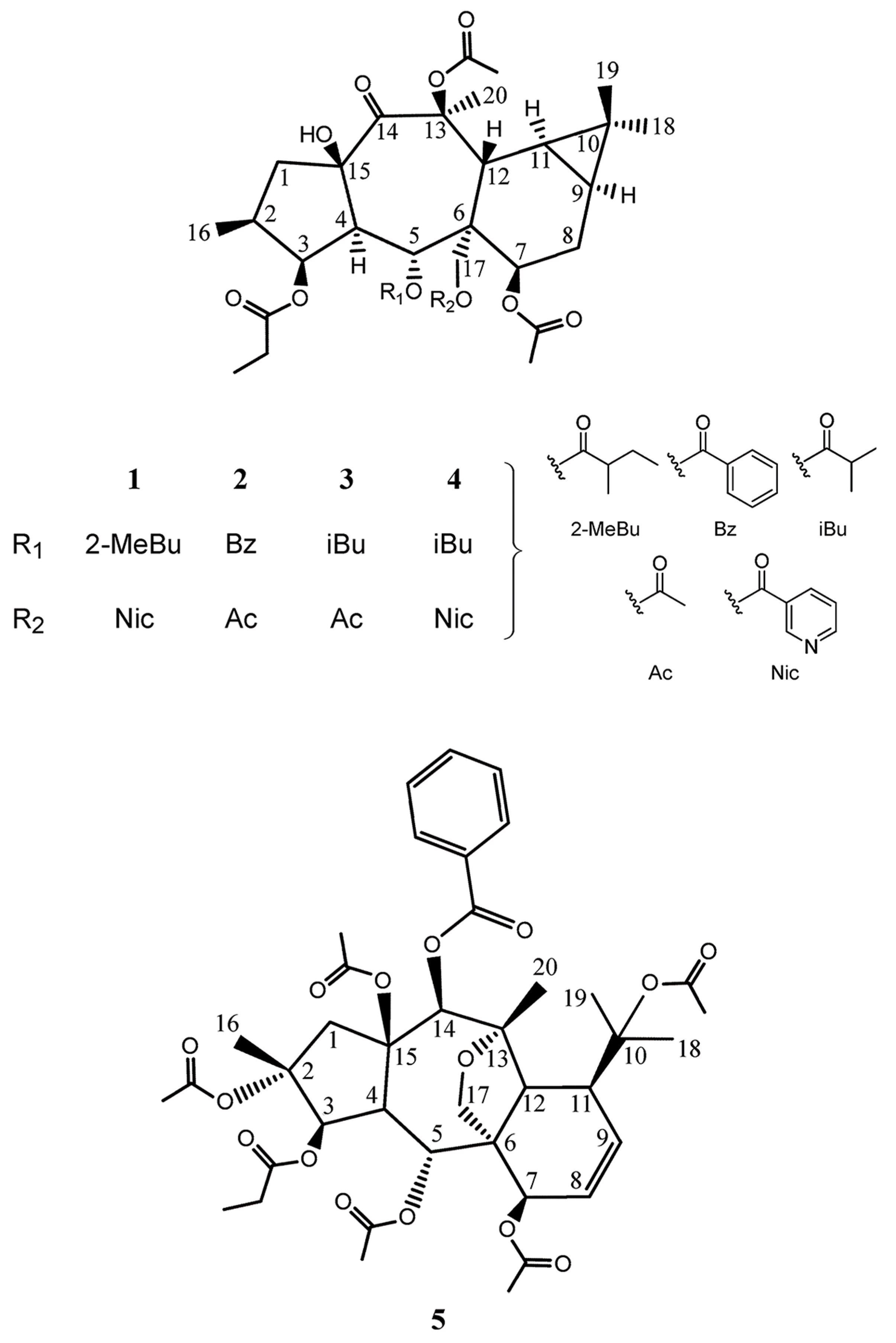
  • 13β-O-propanoyl-5α-O-methylbutanoyl-7α,13β-O-diacetyl-17α-O-nicotinoyl-14-oxopremyrsinane (1): colorless oil; +16.4 (c 0.14 EtOAc); IR (NaCl) νmax 3502, 2964, 2877, 1734, 1716, 1593, 1456, 1373, 1024, 968, 754 cm−1; for 1H and 13C NMR data, see Table 1; UV (EtOAc) λmax (log ε) 260.2 (2.51); HR-ESI-MS m/z 736.3311 [M+Na]+. For additional spectra, refer to the Supplementary Materials, Figures S1–S11.
  • 3β-O-propanoyl-5α-O-benzoyl-7α,13β, 17α -O-triacetyl-14-oxopremyrsinane (2): colorless oil; +70.0 (c 0.03 EtOAc); IR (NaCl) νmax 3498, 2954, 2929, 2856, 1739, 1456, 1261, 1038 cm−1; for 1H and 13C NMR data, see Table 1; UV (EtOAc) λmax (log ε) 254.1 (2.63); HR-ESI-MS m/z 693.2910 [M+Na]+. For additional spectra, refer to the Supplementary Materials, Figures S12–S18.
  • 3β-O-propanoyl-5α-O-isobutanoyl-7α,13β, 17α -O-triacetyl-14-oxopremyrsinane (3): colorless oil; +10.0 (c 0.13 EtOAc); IR (KBr) νmax 3492, 2974, 2941, 2879, 1734, 1716, 1456, 1371, 1342, 1232, 1192, 1142, 1038, 968, 758 cm−1; for 1H and 13C NMR data, see Table 1; UV (EtOAc) λmax (log ε) 260.5 (2.57); HR-ESI-MS m/z 659.3050 [M+K]+. For additional spectra, refer to the Supplementary Materials, Figures S19–S24.
  • 3β-O-propanoyl-5α-O-isobutanoyl-7α,13β-O-diacetyl-17α-O-nicotinoyl-14-oxopremyrsinane (4): colorless oil; +18.3 (c 0.34 EtOAc); IR (KBr) νmax 3494, 2968, 2941, 2879, 1732, 1593, 1464, 1423, 1373, 1271, 1234, 1115, 1084, 1026, 966, 756 cm−1; for 1H and 13C NMR data, see Table 1; UV (EtOAc) λmax (log ε) 261.8 (2.23); HR-ESI-MS m/z 700.3361[M+H]+, 722.3162 [M+Na]+. For additional spectra, refer to the Supplementary Materials, Figures S25–S28.
  • 2,5,7,10,15-O-pentaacetyl-3-O-propanoyl-14-O-benzoyl-13,17-epoxy-8-myrsinene (5): colorless oil; for 1H and 13C NMR data, see Table 2; HR-ESI-MS m/z 793.20 [M+Na]+. For additional spectra, refer to the Supplementary Materials, Figures S29–34.

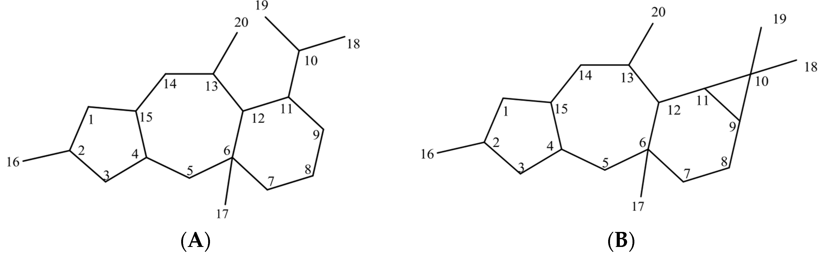
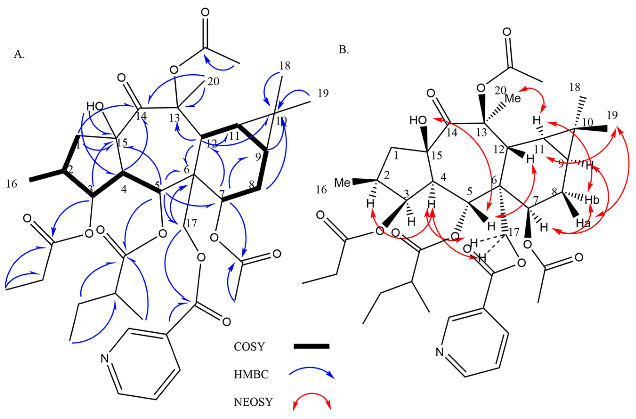
3.2. Structure Identification of Compounds

The chemical structure of each isolated compound had been adequately confirmed by specific spectroscopic methods such as NMR and HRMS (Figure 2).
Compound 1, isolated with optical rotation of +16.4 (c 0.14 EtOAc), was established as C38H51NO12 (HR-ESIMS m/z 736 [M+Na]+; calcd 736, Δ = 3.1 ppm). Resonances at δC 165.0, 128.1, 150.7 (δH: 9.16, d, J = 2.0 Hz), 136.9 (δH: 8.19, dt, J = 8.0, 2.0 Hz), 123.8 (δH: 7.44 (dd, J = 8.0, 5.0 Hz), 154.1 (δH: 8.82, bd, J = 5.2, 1.6 Hz) belong to the nicotinoyl group at C-15 and C-24. δC 174.6, 40.8 (δH: 1.95 m), 25.8 (δH: 1.09–1.19/1.50–1.57, m), 11.7 (δH: 0.63, t, J = 7.6 Hz), 14.3 (δH: 0.84, d, J = 6.4 Hz) were indicative of 2-methylbutanoyl C-24, δC 174.2, 27.8 (δH: 2.29, q, J = 7.6 Hz), 9.0 (δH: 1.07, t, J = 7.2 Hz) relative to propanoyl ester, and δC 170.1, 21.4 (δH: 2.11, s), as well as δC 170.8, 21.4 (δH: 2.11, s) belong to two acetyl groups [14,21,22]. Without esters, the core consists of twenty carbons, including four methyls, three methylenes (one is oxygenated), eight methines (three of them attached to oxygen), and five unprotonated carbons (one ketone, and two quaternary oxycarbons). In the 1H-NMR, one doublet methyl (δH: 0.87, d, J = 6.4 Hz, Me-16), three singlet methyls (δH: 0.95, s, Me-19/1.06, s, Me-18/1.73, s, Me-20), two methylenes (δH: 1.62, dd, J = 13.8, 12.0 Hz/3.17, dd, J = 13.8, 7.7 Hz, H-1a, b, and δH: 1.86-1.94/2.18-2.24, m, H-8a, b), one oxygenated methylene (δH: 4.48, d, J = 12.0 Hz/4.87, d, J = 12.0 Hz, H-17a, b), three methines including H-2 (δH: 1.81-1.88, m), H-4 (δH: 2.38, dd, J = 11.5, 3.6 Hz), and H-12 (δH: 3.47, d, J = 6.7 Hz), two unusual upfield methines at δH: 0.82-0.74, m/0.80-0.74, m related to H-9 and H-11 on a cyclopropane moiety, three oxymethins at δH: 5.22 (dd, J = 3.2,3.2 Hz, H-3), 6.25 (d, J = 11.5 Hz, H-5), and 4.69 (bd, J = 6.7 Hz, H-7) were seen. 1H-1H COSY couplings determined two spin systems A (H-1—H-2(H-16) —H-3—H-4—H-5): CH2—CH(CH3)—CHO—CH—CHO and B (H-7—H-8—H-9—H-11—H-12): CHO—CH2—CH—CH—CH. HMBC long-range correlations of spin components at H1-5, H1-7, H1-12, and H2-17 with C-6; H1-9, H1-11, H3-18, H3-19 with C-10; H2-1/C-15, C-14; H1-12/C-13, C-14; H1-4/C-14; H3-20/C-13, C-14; H1-3/C-15 indicative of a 14-oxopremyrsinane derivative. HMBC correlations located nicotinoyl at C-17, methylbutanoyl at C-5, propanoyl at C-3, and one acetyl ester at C-7 (Figure 3A). HMBC of δH: 4.44 (OH) with C-15, and C-4 located free hydroxy group at C-15, and suggested the last acetate group with no HMBC with at remaining quaternary oxycarbon C-13. For stereochemistry, taking H-4 in alpha, typical in premyrsinane diterpenes, NOESY of H-4/H-2, H-17b, as well as the small coupling constant of J3,4 = 3.2 Hz related to cis-relationship of H-4/H-3, determined alpha orientation of H-2, H-3, and H-17 [23,24]. A large coupling constant of J4,5 = 11.5 Hz indicative of trans relationship determined beta orientation of H-5 and alpha orientation of 5-O-methylbutanoyl [25,26]. NOEs of H-5/H-12, 15-OH; H-12/Me-19, and Me-19/H-8a determined the beta orientation of H-8a, H-12, 15-OH, and Me-19NOEs of H-9/H-7; H-11/H-8b, and Me-20 determined H-7, H-9, H-11, and Me-20 alpha orientation. Accordingly, compound 1 was suggested as 3β-O-propanoyl-5α-O-methylbutanoyl-7α,13β-O-diacetyl-17α-O-nicotinoyl-14-oxopremyrsinane (Figure 3B) similar to those reported by Hegazy et al. from Egyptian Plant Euphorbia Sanctae-Catharinae [21].
Compound 2 was isolated as a colorless oil with optical rotation +70.0 (c 0.03 EtOAc), with the molecular formula of C36H46O12 based on the exact ESI mass ion peak at m/z 693 ([M+Na]+, calc. 693, Δ 4.1 ppm). Resonances at δC 165.3, 130.0, 129.8 (δH: 7.88, dd, J = 8.4, 1.5 Hz), 128.4 (δH: 7.37, dd, J = 8.4, 7.6 Hz), 133.2 (δH: 7.44, bt, J = 7.6 Hz), belong to the benzoyl group [14,21,27]. δC 173.7, 27.7 (δH: 2.28, bq, J = 7.6 Hz), 8.9 (δH: 1.11, t, J = 7.2 Hz) relative to propanoyl ester, and δC 170.3, 21.5 (δH: 2.12, s), 170.8, 21.4 (δH: 2.15, s), and 170.9, 21.4 (δH: 1.49, s), belong to three acetate groups [14,21,22]. NMR data of polyol resembled 1 differed in 3-0-acetate instead of 3-O-nicotinoyl, and 5-O-benzoyl instead of 5-O-methylbutanoyl ester group based on HMBC correlations. In agreement with compound 4f isolated from Euphorbia pithyusa [28], this compound was identified as 3β-O-propanoyl-5α-O-benzoyl-7α,13β, 17α -O-triacetyl-14-oxopremyrsinane.
Compound 3 was isolated as a colorless oil with positive optical rotation +10.0 (c 0.13 EtOAc), with the molecular formula of C33H48O12 based on the exact ESI mass ion peak at m/z 659 ([M+Na]+, calc. 659, Δ 1.8 ppm). Resonances at δC 175.2, 34.2 (δH: 2.37, m), 18.7 (δH: 1.12, d, J = 8 Hz), 18.9 (δH: 1.09 d, J = 8 Hz), belong to the isobutanoyl group [12,14,21,27]. δC 174.3, 27.9 (δH: 2.31, q, J = 3.6 Hz), 9(δH: 1.10, t, J = 6.8 Hz) relative to propanoyl ester, and δC 170.1, 21.5 (δH: 2.07, s), δC 170.5, 21.4 (δH: 2.10, s), and 170.8, 21.3 (δH: 2.10, s), belong to three acetate groups at C-17, C-24 and C-25. NMR data of polyol resembled 2 differed in 5-O-isobutanoyl instead of the 5-O-benzoyl group [28]. Finally, compound 2 was assigned as 3β-O-propanoyl-5α-O-isobutanoyl-7α,13β, 17α -O-triacetyl-14-oxopremyrsinane.
Compound 4 as a colorless oil with positive optical rotation +18.3 (c 0.34 EtOAc), showed the molecular formula of C37H49NO12 based on the exact ESI mass ion peak at m/z 700 ([M+H]+, calc. 700, Δ 4.7 ppm). Resonances at δC 176.9, 34.0 (δH: 2.36, m), 18.3 (δH: 0.92, d, J = 6.8 Hz), 18.4 (δH: 0.44a), belong to the isobutanoyl group [12,14,21,27]. δC 165.3, 127.5, 150.8 (δH: 9.16, d, J = 2.0 Hz), 136.9 (δH: 8.20, dt, J = 8.0, 1.5 Hz), 123.8 (δH: 7.44 dd, J = 8.0, 4.8 Hz), 154.2 (δH: 8.82, bd, J = 4.8, 1.6 Hz) belong to the nicotinoyl group [12,21]. δC 174.4, 27.8 (δH: 2.27, q, J = 3.6 Hz), 8.9 (δH: 0.57, t, J = 7.2 Hz) relative to propanoyl ester, and δC 170.1, 21.4 (δH: 2.12, s) and 170.9, 21.4 (δH: 2.12, s) belong to two acetate groups [14,21,22]. NMR data resembled 1 differed only in 5-O-isobutanoyl instead of the 5-O-methylbutanoyl group [28], which was detected as 3β-O-propanoyl-5α-O-isobutanoyl-7α,13β-O-diacetyl-17α-O-nicotinoyl-14-oxopremyrsinane.
Compound 5, with a molecular weight of 770 g/mol was identified as C40H50O15 based on HR-ESIMS m/z 793 [M+Na] + and NMR spectral data. Analyzing the 1HNMR, 13CNMR, DEPT90, and DEPT135 determined that five methyl groups at δH 1.70, 1.99, 2.05, 2.10, and 2.14 are related to five signals at δC 169.6 (7-OAc), 170.6 (10-OAc), 169.4 (2-OAc), 168.7 (5-OAc), and 170.9 (15-OAc) belonging to acetate groups. Resonances in 1HNMR spectra along with their relative carbon signals indicated 3-O-propanoyl ester groups at δH 2.36 (α, β), and 1.17, 14-O-benzoyl ester groups at δH 8.08 (d, J = 8.2 Hz), 7.45 (t, J = 8.2 Hz), and 7.58 (t, J = 8.2 Hz). Compound 5 contains 40 carbons out of which 20 of the carbon signals were associated with side chains, confirming that the remaining twenty belong to the carbons present in the core myrsinane skeleton, including four methyls, two methylenes, nine methines, and five unprotonated carbons.
HSQC and HMBC correlations confirmed the myrsinane structure of this compound in addition to seven oxygens from ketone groups attached to C-2, C-3, C-5, C-7, C-10, C-14, and C-15. The locations of ester groups were determined with HMBC, in which H-3 (δH: 5.40, d, J = 5.4 Hz), H-5 (δH: 5.95, dd, J = 11.2–1.6 Hz), H-7 (δH: 4.85, d, J = 6.4 Hz), and H-14 (δH: 5.82, s) were, respectively correlated with δC 173.7 (OPro), 168.69 (OAc), 169.6 (OAc), and 165.97 (OBz). These signals indicated that the propanoyl ester was located at C-3, acetyl esters at C-5 and C-7, and benzoic ester at C-14. Three signals belonging to quaternary carbons at δC 87.1 (C-2), 86.0 (C-10), and 90.0 (C-15) determined that an acetate group was situated on each of these carbons.
Based on the data analysis and after comparison with those in the literature [29], the molecular formula of this myrsinane-type diterpene was determined to be 2,5,7,10,15-pentaacetyl-3-O-propanoyl-14-O-benzoyl-13,17-epoxy-8-myrsinene, also known as Euphorprolitherin B.

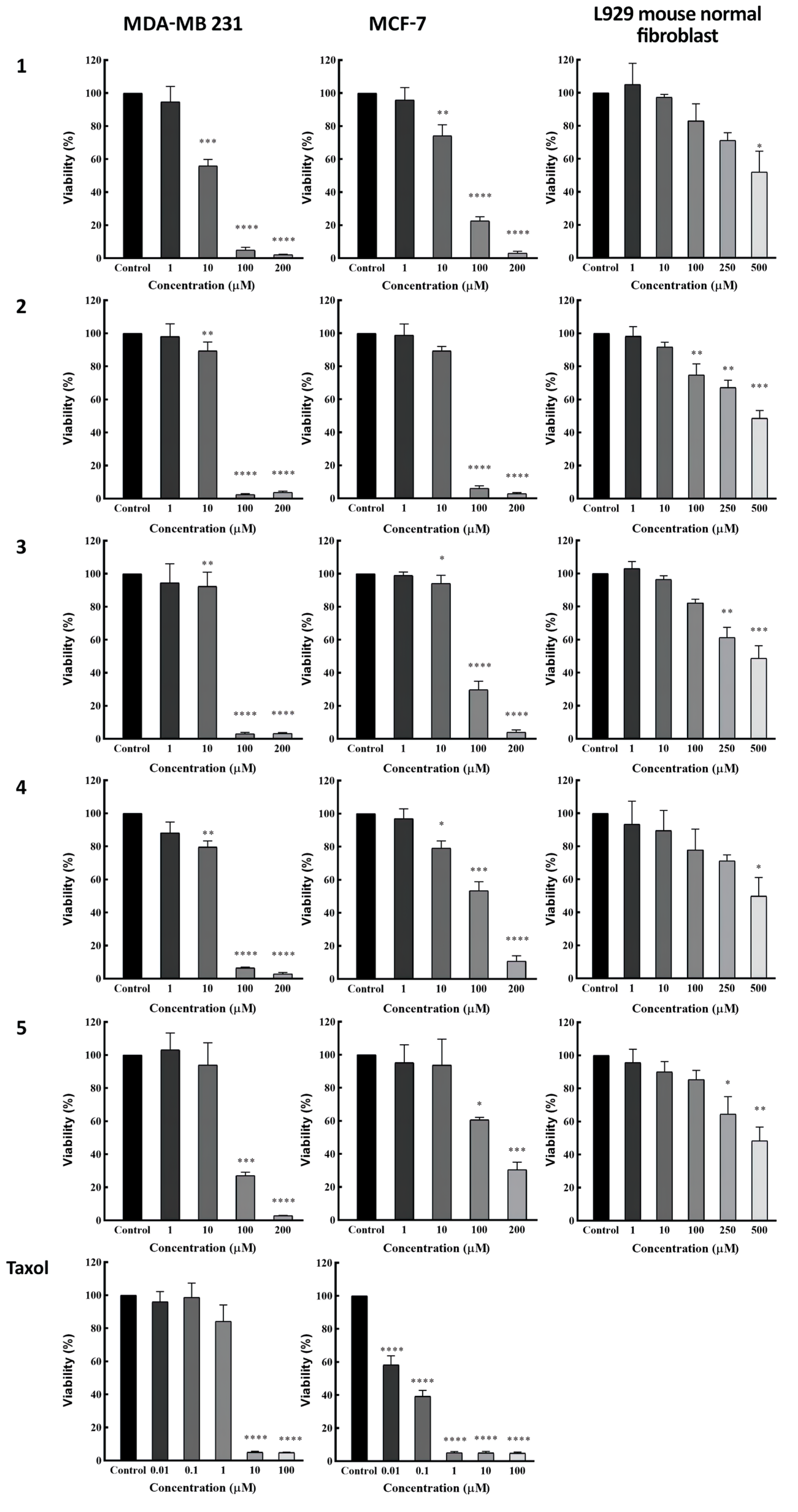
3.3. Determination of Cytotoxic Activity

Compounds 15 were tested by MTT assay to evaluate their cytotoxic activity (Table 3). All of the compounds exhibited cytotoxic effects in a dose-dependently manner with IC50 values of 10.8, 22.2, 24.5, 27.3, and 33.7 μM against the MDA-MB-231 (ATCCR HTB-26) breast cancer cells, and IC50 values of 22.2, 27.8, 62.6, 74.4, and 125.6 μM against the MCF-7 (ATCCR HTB-22) breast cancer cells, respectively (Figure 4).

4. Discussion

All of the diterpenoids presented in this study are isolated and introduced for the first time from Euphorbia gedrosiaca, and despite their rare occurrence, compounds 1, 3, and 4 were isolated during a study on an endemic species of Euphorbiaceae in Egypt called Euphorbia sanctae-catharinae and they showed moderate cytotoxicity against the proliferation of human lung (A549) and colon (Caco-2) tumor cells in different degrees [21]. Compounds 3 and 4 were also elucidated from Euphorbia pithyusa subsp. cupanii previously [28]. Compound 2 was elucidated from Euphorbia dracunculoides [9], and Euphorbia pithyusa [30] until now. Formerly, this compound was isolated from the roots of Euphorbia prolifera and showed acceptable lipid-lowering activities and TG inhibitory effects during an investigation on this plant [31]. Compound 5 has been reported in previous studies from Euphorbia species like Euphorbia prolifera [32], Euphorbia dracunculoides Lam [33], and Euphorbia nematocypha Hand.-Mazz [34].
Regarding the anti-proliferative activity of the premyrsinane diterpenes (1–4) in the current study, results from the MTT viability assay showed that amongst all compounds, 1 is the most effective against both cell lines, especially MDA-MB-231 with an IC50 value of 10.8 μM which is considerable. In general, all four diterpenes showed significant apoptotic effects against MDA-MB-231 based on the cell viability in 100 μM concentration of these bioactive agents (p < 0.0001). The evaluated IC50 from compounds 2, 3, and 4, respectively, as 22.2, 24.5, and 27.3 μM, exhibits that they are similarly effective against MDA-MB-231. Compound 2 is the second most effective one based on our findings, which had quite the same efficacy on both cell lines. Although the effectiveness of 1 against MCF-7 did not differ significantly from 2, both were reasonably potent in 100 μM concentration with P values less than 0.0001. Compounds 3 and 4 demonstrated nearly similar MTT test results with almost three times higher cytotoxicity against the MDA-MB-231 cell line compared to MCF-7, which can be considered a moderate activity and not as potent as other responses in this study. This comparison shows that this similarity is due to their chemical structural analogy, which only differs at C-17 by replacing an acetyl ester group with a nicotinoyl group. Compound 5 was also tested for its cytotoxicity against these two cell lines but showed less potency than the other four compounds. However, the efficacy is acceptable against the MDA-MB-231 with an IC50 value of 33.7 μM, and can be worthy of consideration, but the IC50 value of 125.6 μM against MCF-7 shows low activity. The comparison of MTT assay data also reveals that the myrsinane diterpenes have less efficacy as anti-proliferative agents against these two cancer cell lines. New findings about the structure–activity relationship for cytotoxic efficacy can be helpful to approach the best chemical structure in therapeutic applications.
All of the isolated compounds were also tested on mouse normal fibroblast cell lines to prove their safety, and the results were promising, while none of them demonstrated toxicity at concentrations up to 250 µM towards L929 normal cell line.
Former studies concerning the evaluation of cytotoxicity and possible therapeutic application of diterpenoids in breast cancer treatment have also revealed valuable results as well. In an identical study on Euphorbia aleppica, two new cytotoxic premyrsinane diterpenes were introduced and assessed for their anti-cancer effects on MCF-7 and MDA-MB-231 cell lines which determined their probable potency [16]. Similarly, another investigation on cyclomyrsinane diterpenes from Euphorbia species proved the potential activity of compounds that had structural similarities to the ones elucidated in the present research against the same breast tumor cell lines [35].

5. Conclusions

Five diterpenoids were determined from E.gedrosiaca and evaluated for their anti-proliferative properties against MCF-7 and MDA-MB231 cell lines. The MTT viability assay proves that compound 1 is the most effective against both cell lines, especially MDA-MB-231. We can conclude that these diterpenes show better apoptotic effects against MDA-MB-231 than MCF-7, and it is clear that the premyrsinane skeletal structure improves the cytotoxic activity of compounds against breast cancer cell lines. On the other hand, the results displayed the safety of the isolated compounds against normal cells, which can be beneficial in drug development.
Considering all of the above mentioned, in order to achieve valuable and novel discoveries as practical anti-breast cancer treatments, it is worthwhile to survey and precisely determine the most effective bioactive chemical structure of this type of diterpenoids and detect the most potent apoptotic agent by comparing the replacement of different chemical groups and assess their effects.

Supplementary Materials

The following supporting information can be downloaded at: https://www.mdpi.com/article/10.3390/metabo13020225/s1, Figure S1. 1H NMR spectrum of (1), Figure S2. 13C NMR spectrum of (1), Figure S3. 13C DEPT135 spectrum of (1), Figure S4. 13C DEPT90 spectrum of (1), Figure S5. HSQCGP spectrum of (1), Figure S6. HMBCGP spectrum of (1), Figure S7. DQF-COSY spectrum of (1), Figure S8. FT-IR spectrum of (1), Figure S9. (A) Selected HMBC and COSY correlations and (B) representation of key NOESY cross peaks of (1), Figure S10. NOESY spectrum of (1), Figure S11. HR-ESI-MS spectrum of (1), Figure S12. 1H NMR spectrum of (2), Figure S13. 13C NMR spectrum of (2), Figure S14. 13C DEPT135 spectrum of (2), Figure S15. 13C DEPT90 spectrum of (2), Figure S16. HMBCGP spectrum of (2), Figure S17. HR-ESI-MS spectrum of (2), Figure S18. FT-IR spectrum of (2), Figure S19. 1H NMR spectrum of (3), Figure S20. 13C NMR spectrum of (3), Figure S21. 13C DEPT135 spectrum of (3), Figure S22. 13C DEPT90 spectrum of (3), Figure S23. HR-ESI-MS spectrum of (3), Figure S24. FT-IR spectrum of (3), Figure S25. 1H NMR spectrum (4), Figure S26. 13C NMR spectrum of (4), Figure S27. HR-ESI-MS spectrum of (4), Figure S28. FT-IR spectrum of (4), Figure S29. 1H NMR spectrum of (5), Figure S30. 13C NMR spectrum of (5), Figure S31. 13C DEPT135 spectrum of (5), Figure S32. 13C DEPT90 spectrum of (5), Figure S33. HSQCGP spectrum of (5), Figure S34. HMBCGP spectrum of (5), Figure S35. HPLC chromatogram of fraction 5 corresponding to (1), (2) and (3), Figure S36. HPLC chromatogram of fraction 10-1 corresponding to (4), Figure S37. HPLC chromatogram of fraction 9 corresponding to (5).

Author Contributions

Conceptualization, B.Z.; methodology, M.G.; software, P.M.; validation, V.L.; formal analysis, Z.Y.; investigation, M.H.S.; resources, Z.Y.; data curation, Z.Y.; writing—original draft preparation, Z.Y.; writing—review and editing, N.M.; visualization, M.G.; supervision, V.L.; project administration, Z.Y.; funding acquisition, Z.Y. All authors have read and agreed to the published version of the manuscript.

Funding

This research was funded by the Iran National Science Foundation (INSF), grant number 92000554 and Isfahan University of Medical Sciences, project number 398385.

Institutional Review Board Statement

The study was conducted in accordance with the Declaration of Helsinki, and approved by the Ethics Committee of Isfahan University of Medical Sciences approved this research with the code of IR.MUI.RESEARCH.REC.1398.278.

Informed Consent Statement

Not applicable.

Data Availability Statement

The data presented in this study are available in the article.

Conflicts of Interest

The authors declare no conflict of interest.

Abbreviations

TermDefinition
COSYCorrelated Spectroscopy
DMSODimethyl sulfoxide
ELISAEnzyme-linked Immunosorbent Assay
EtOAcEthyl acetate
HMBCHeteronuclear Multiple Quantum Correlation
HPLCHigh Performance Liquid Chromatography
HRMSHigh-resolution Mass Spectrometry
HSQCHeteronuclear Single Quantum Correlation
MPLCMedium Pressure Liquid Chromatography
MTTMicroculture Tetrazolium Test
NMRNuclear Magnetic Resonance
NOESYNuclear Overhauser Enhancement Spectroscopy
TLCThin Layer Chromatography

References

  1. Gunawardana, M.; Hyde, E.R.; Lahmeyer, S.; Dorsey, B.L.; La Val, T.P.; Mullen, M.; Yoo, J.; Knight, R.; Baum, M.M. Euphorbia plant latex is inhabited by diverse microbial communities. Am. J. Bot. 2015, 102, 1966–1977. [Google Scholar] [CrossRef] [PubMed]
  2. Pascal, O.A.; Bertrand, A.E.V.; Esaïe, T.; Sylvie, H.-A.M.; Eloi, A.Y. A review of the ethnomedical uses, phytochemistry and pharmacology of the Euphorbia genus. J. Pharm. Innov. 2017, 6, 34. [Google Scholar]
  3. Kemboi, D.; Peter, X.; Langat, M.; Tembu, J. A review of the ethnomedicinal uses, biological activities, and triterpenoids of Euphorbia species. Molecules 2020, 25, 4019. [Google Scholar] [CrossRef]
  4. Xu, Y.; Tang, P.; Zhu, M.; Wang, Y.; Sun, D.; Li, H.; Chen, L. Diterpenoids from the genus Euphorbia: Structure and biological activity (2013–2019). Phytochemistry 2021, 190, 112846. [Google Scholar] [CrossRef] [PubMed]
  5. Salehi, B.; Iriti, M.; Vitalini, S.; Antolak, H.; Pawlikowska, E.; Kręgiel, D.; Sharifi-Rad, J.; Oyeleye, S.I.; Ademiluyi, A.O.; Czopek, K. Euphorbia-derived natural products with potential for use in health maintenance. Biomolecules 2019, 9, 337. [Google Scholar] [CrossRef]
  6. Xu, J.; Yang, B.; Fang, L.; Wang, S.; Guo, Y.; Yamakuni, T.; Ohizumi, Y. Four new myrsinol diterpenes from Euphorbia prolifera. J. Nat. Med. 2013, 67, 333–338. [Google Scholar] [CrossRef]
  7. Xu, J.; Kang, J.; Cao, X.; Sun, X.; Yu, S.; Zhang, X.; Sun, H.; Guo, Y. Characterization of diterpenes from Euphorbia prolifera and their antifungal activities against phytopathogenic fungi. J. Agric. Food Chem. 2015, 63, 5902–5910. [Google Scholar] [CrossRef]
  8. Zolfaghari, B.; Yazdiniapour, Z.; Ghanadian, M.; Lanzotti, V. Cyclomyrsinane and premyrsinane diterpenes from Euphorbia sogdiana Popov. Tetrahedron 2016, 72, 5394–5401. [Google Scholar] [CrossRef]
  9. Wang, L.; Ma, Y.-T.; Sun, Q.-Y.; Li, X.-N.; Yan, Y.; Yang, J.; Yang, F.-M.; Liu, F.-Y.; Zang, Z.; Wu, X.-H. Structurally diversified diterpenoids from Euphorbia dracunculoides. Tetrahedron 2015, 71, 5484–5493. [Google Scholar] [CrossRef]
  10. Wang, L.; Zang, Z.; Zhang, J.; Wu, X.; Huang, S.; Cao, P.; Zhao, Y. A new Premyrsinane-type diterpenoid polyester from Euphorbia dracunculoides Lam. Rec. Nat. Prod. 2015, 9, 374. [Google Scholar]
  11. Wang, Q.; Zhen, Y.Q.; Gao, F.; Huang, S.; Zhou, X.L. Five new diterpenoids from the seeds of Euphorbia lathyris. Chem. Biodivers. 2018, 15, e1800386. [Google Scholar] [CrossRef] [PubMed]
  12. Elshamy, A.I.; Mohamed, T.A.; Al-Rowaily, S.L.; Abd-ElGawad, A.M.; Dar, B.A.; Shahat, A.A.; Hegazy, M.-E.F. Euphosantianane E–G: Three new premyrsinane type diterpenoids from Euphorbia sanctae-catharinae with contribution to chemotaxonomy. Molecules 2019, 24, 2412. [Google Scholar] [CrossRef] [PubMed] [Green Version]
  13. Shokoohinia, Y.; Sajjadi, S.-E.; Zolfaghari, B.; Chianese, G.; Appendino, G.; Taglialatela-Scafati, O. Diterpenoid (poly) esters and a ring A-seco-phorboid from the aerial parts of Euphorbia macroclada Boiss. Fitoterapia 2010, 81, 884–890. [Google Scholar] [CrossRef]
  14. Sulyok, E.; Vasas, A.; Rédei, D.; Forgo, P.; Kele, Z.; Pinke, G.; Hohmann, J. New premyrsinane-type diterpene polyesters from Euphorbia falcata. Tetrahedron 2011, 67, 7289–7293. [Google Scholar] [CrossRef]
  15. Vasas, A.; Sulyok, E.; Martins, A.; Rédei, D.; Forgo, P.; Kele, Z.; Zupkó, I.; Molnár, J.; Pinke, G.; Hohmann, J. Cyclomyrsinane and premyrsinane diterpenes from Euphorbia falcata modulate resistance of cancer cells to doxorubicin. Tetrahedron 2012, 68, 1280–1285. [Google Scholar] [CrossRef]
  16. Zolfaghari, B.; Farahani, A.; Jannesari, A.; Aghaei, M.; Ghanadian, M. New Cytotoxic Premyrsinane-Type Diterpenes from Euphorbia aleppica Against Breast Cancer Cells. Iran. J. Pharm. Res. 2022, 21, e127028. [Google Scholar] [CrossRef] [PubMed]
  17. Pahlevani, A.H.; Liede-Schumann, S.; Akhani, H. Seed and capsule morphology of Iranian perennial species of Euphorbia (Euphorbiaceae) and its phylogenetic application. Bot. J. Linn. Soc. 2015, 177, 335–377. [Google Scholar] [CrossRef]
  18. Pahlevani, A.H.; Maroofi, H.; Joharchi, M.R. Notes on six endemic or rare species of Euphorbia subg. esula (Euphorbiaceae) in Iran. Willdenowia 2011, 41, 267–276. [Google Scholar] [CrossRef]
  19. Yazdiniapour, Z.; Mirian, M.; Zolfaghari, B.; Mehdifar, P.; Ghanadian, M.; Lanzotti, V. Myrsinane-type diterpenes from Euphorbia gedrosiaca with cell growth inhibitory activity and apoptotic effects on melanoma cancer cells. Fitoterapia 2022, 157, 105138. [Google Scholar] [CrossRef]
  20. Mohammadi Pour, P.; Yegdaneh, A.; Aghaei, M.; Ali, Z.; Khan, I.A.; Ghanadian, M. Novel 16, 17-epoxy-23-methylergostane derivative from Sinularia variabilis, a soft coral from the Persian Gulf, with apoptotic activities against breast cancer cell lines. Nat. Prod. Res. 2021, 1–10. [Google Scholar] [CrossRef]
  21. Hegazy, M.-E.F.; Hamed, A.R.; Ibrahim, M.A.; Talat, Z.; Reda, E.H.; Abdel-Azim, N.S.; Hammouda, F.M.; Nakamura, S.; Matsuda, H.; Haggag, E.G. Euphosantianane A–D: Antiproliferative premyrsinane diterpenoids from the endemic Egyptian plant Euphorbia sanctae-catharinae. Molecules 2018, 23, 2221. [Google Scholar] [CrossRef] [PubMed]
  22. Nothias-Esposito, M.; Nothias, L.F.; Da Silva, R.R.; Retailleau, P.; Zhang, Z.; Leyssen, P.; Roussi, F.; Touboul, D.; Paolini, J.; Dorrestein, P.C. Investigation of premyrsinane and myrsinane esters in Euphorbia cupanii and Euphobia pithyusa with MS2LDA and combinatorial molecular network annotation propagation. J. Nat. Prod. 2019, 82, 1459–1470. [Google Scholar] [CrossRef] [PubMed]
  23. Zarei, S.M.; Ayatollahi, A.M.; Ghanadian, M.; Aghaei, M.; Choudhary, M.I.; Fallahian, F. Unusual ingenoids from Euphorbia erythradenia Bioss. with pro-apoptotic effects. Fitoterapia 2013, 91, 87–94. [Google Scholar] [CrossRef]
  24. Sekine, T.; Kamiya, M.; Ikegami, F.; Qi, J.-F. Structure of Esulol A, a novel macrocyclic diterpene, from inner Mongolian Euphorbia esula. Nat. Prod. Lett 1998, 12, 237–239. [Google Scholar] [CrossRef]
  25. Shadi, S.; Saeidi, H.; Ghanadian, M.; Rahimnejad, M.R.; Aghaei, M.; Ayatollahi, S.M.; Iqbal Choudhary, M. New macrocyclic diterpenes from Euphorbia connata Boiss. with cytotoxic activities on human breast cancer cell lines. Nat. Prod. Res. 2015, 29, 607–614. [Google Scholar] [CrossRef] [PubMed]
  26. He, X.; Abulizi, X.; Li, X.; Ma, G.; Sun, Z.; Wei, H.; Xu, X.; Shi, L.; Zhang, J. Daphnane-Type Diterpenes from Stelleropsis tianschanica and Their Antitumor Activity. Molecules 2022, 27, 5701. [Google Scholar] [CrossRef] [PubMed]
  27. Flores-Giubi, E.; Geribaldi-Doldán, N.; Murillo-Carretero, M.; Castro, C.; Durán-Patrón, R.; Macías-Sánchez, A.J.; Hernández-Galán, R. Lathyrane, premyrsinane, and related diterpenes from Euphorbia boetica: Effect on in vitro neural progenitor cell proliferation. J. Nat. Prod. 2019, 82, 2517–2528. [Google Scholar] [CrossRef]
  28. Appendino, G.; Belloro, E.; Tron, G.C.; Jakupovic, J.; Ballero, M. Diterpenoids from Euphorbia pithyusa subsp. cupanii. J. Nat. Prod. 1999, 62, 1399–1404. [Google Scholar] [CrossRef]
  29. Zhang, W.J.; Chen, D.F.; Hou, A.J. New myrsinol diterpenes from Euphorbia prolifera. Chin. J. Chem. 2004, 22, 103–108. [Google Scholar] [CrossRef]
  30. Esposito, M.l.; Nothias, L.-F.l.; Retailleau, P.; Costa, J.; Roussi, F.; Neyts, J.; Leyssen, P.; Touboul, D.; Litaudon, M.; Paolini, J. Isolation of premyrsinane, myrsinane, and tigliane diterpenoids from Euphorbia pithyusa using a Chikungunya virus cell-based assay and analogue annotation by molecular networking. J. Nat. Prod. 2017, 80, 2051–2059. [Google Scholar] [CrossRef]
  31. Song, Q.-Q.; Rao, Y.; Tang, G.-H.; Sun, Z.-H.; Zhang, J.-S.; Huang, Z.-S.; Yin, S. Tigliane diterpenoids as a new type of antiadipogenic agents inhibit GRα-Dexras1 axis in adipocytes. J. Med. Chem. 2019, 62, 2060–2075. [Google Scholar] [CrossRef] [PubMed]
  32. Li, J.; Zhao, W.; Deng, L.; Li, X.-R. Components of myrsinane-type diterpenes from Euphorbia prolifera. J. Zhejiang Univ. Sci. 2011, 40, 380–383. [Google Scholar] [CrossRef]
  33. Wang, L.; Zang, Z.; Wang, Y.-F.; Huang, S.-X.; Cao, P.; Zhao, Y. Two new myrinsol diterpenoids from Euphorbia dracunculoides Lam. Chin. Chem. Lett. 2015, 26, 121–123. [Google Scholar] [CrossRef]
  34. Chen, R.; You, C.-X.; Wang, Y.; Zhang, W.-J.; Yang, K.; Geng, Z.-F.; Liu, Z.-L.; Deng, Z.-W.; Wang, Y.-Y.; Du, S.-S. Chemical constituents from the roots of Euphorbia nematocypha Hand.-Mazz. Biochem. Syst. Ecol. 2014, 57, 1–5. [Google Scholar] [CrossRef]
  35. Rabbani, F.; Yazdiniapour, Z.; Ghanadian, M.; Zolfaghari, B.; Maleki, M.; Shafiee, F. Cytotoxicity and apoptosis assay of novel cyclomyrsinol diterpenes against breast cancer cell lines. World J. Tradit. Chin. Med. 2022, 8, 273. [Google Scholar] [CrossRef]
Figure 1. (A) Skeletal structure of myrsinanes; (B) Skeletal structure of premyrsinanes.
Metabolites 13 00225 g001
Figure 2. Premyrsinanes (14), and myrsinane diterpene (5) from Euphorbia gedrosiaca.
Metabolites 13 00225 g002
Figure 3. (A) Selected HMBC and COSY correlations and (B) representation of key NOESY cross peaks of 1.
Metabolites 13 00225 g003
Figure 4. Cytotoxicity activity by using MTT viability assay against MCF-7, MDA-MB231, and L929 normal cell lines. These cells were treated with various concentrations of compounds 1–5 and Taxol as the positive control. Results are presented as the mean ± SD (n = 3). * p < 0.05, ** p < 0.01, *** p < 0.001, and **** p < 0.0001 versus control group.
Metabolites 13 00225 g004
Table 1. 1H (400 MHz) and 13C (100 MHz) NMR data of compounds 1–4 in CDCl3.
Position1234
δH (mult., J in Hz)δC (Type)δH (mult., J in Hz)δC (Type)δH (mult., J in Hz)δC (Type)δH (mult., J in Hz)δC (Type)
1a1.62 (dd, 13.8, 12.0)42.9 (CH2)1.65 (a 1)43.0 (CH2)1.62 (a)42.9 (CH2)1.63 (a)43.0 (CH2)
1b3.17 (dd, 13.8, 7.7)-3.16 (dd, 13.6, 7.6)-3.15 (dd, 13.6, 7.6)-3.18 (dd, 13.6, 7.6)-
21.81–1.88 (m)37.5 (CH)1.78–1.82 (m)37.4 (CH)1.72–1.79 (m)37.5 (CH)1.81–1.88 (m)37.5 (CH)
35.22 (t, 3.2)78.4 (CH)5.38 (t, 3.6)78.3 (CH)5.27 (t, 3.2)78.4 (CH)5.24 (t, 3.2)78.4 (CH)
42.38 (dd, 11.5, 3.6)50.6 (CH)2.39 (dd, 11.6, 3.6)50.4 (CH)2.40 (dd, 11.6, 3.2) 50.4 (CH)2.32 (dd, 11.6, 3.2)50.6 (CH)
56.25 (d, 11.5)69.1 (CH)6.38 (d, 11.6)70.0 (CH)6.19 (d, 11.6)68.9 (CH)6.23 (d, 11.6)69.1 (CH)
6-47.7 (C)-47.9 (C)-47.5 (C)-47.8 (C)
74.69 (bd, 6.7)70.8 (CH)4.80 (d, 6.8)70.8 (CH)4.51 (d, 6.8)70.7 (CH)4.71 (d, 6.8)70.8 (CH)
8a1.86–1.94 (m) 22.5 (CH2)1.83–1.90 (m)22.2 (CH2)1.78–1.86 (m)22.2 (CH2)1.88–1.96 (m)22.5 (CH2)
8b2.18–2.24 (m)-2.19–2.25 (m)-2.18–2.24 (m)-2.16–2.22 (m)-
90.74–0.82 (m)19.0 (CH)0.69–0.77 (m)19.1 (CH)0.71–0.80 (m)18.90 (CH)0.75-.083 (m)19.1 (CH)
10-18.4 (C)-18.4 (C)-18.2 (C)-18.56 (C)
110.74–0.80 (m)23.9 (CH)0.69–0.77 (m)24.0 (CH)0.71–0.80 (m)23.9 (CH)0.75–0.83 (m)24.0 (CH)
123.47 (d, 6.7)35.0 (CH)3.52 (d, 6.4)35.2 (CH)3.39 (d, 6.4)34.9 (CH)3.49 (d, 6.8)35.1 (CH)
13-85.8 (C)-85.9 (C)-86.0 (C)-85.9 (C)
14-204.4 (C)-204.4 (C)-204.5 (C)-204.8 (C)
15-84.2 (C)-84.3 (C)-84.1 (C)-84.2 (C)
160.87 (d, 6.4)14.7 (CH3)0.86 (d, 6.4) 14.0 (CH3)0.88 (d, 6.4) 14.2 (CH3)0.88 (d, 7.2)14.2 (CH3)
17a4.48 (d, 12.0)64.6 (CH2)4.31 (d, 11.6) 62.9 (CH2)4.35 (d, 11.6) 63.6 (CH2)4.51 (d, 11.6) 64.5(CH2)
17b4.87 (d, 12.0)-4.69 (d, 11.6) -4.42 (d, 11.6) -4.88 (d, 12.0) -
181.06 (s)29.6 (CH3)1.06 (s)29.6 (CH3)1.05 (s)29.6 (CH3)1.07 (s)29.6 (CH3)
190.95 (s)14.9 (CH3)0.95 (s)15.0 (CH3)0.91 (s)14.9 (CH3)0.95 (s)15.0 (CH3)
3-OPro-174.2-173.7-174.3-174.4
2.34 (q, 7.6) 27.82.30 (q, 7.2)27.72.31 (q, 7.2)27.92.32 (q, 7.6)27.8
0.63 (t, 7.6)9.00.98 (t, 7.2)8.90.74-0.79 (m)9.00.57 (t, 7.2)8.9
5-OMeBu-174.6------
1.95 (m)40.8------
1.09–1.19 (m)25.8------
1.50–1.57 (m)
0.63 (t, 7.6)11.7------
0.84 (d, 6.4)14.3------
5-OiBut-----175.2-176.9
----2.38 (m)34.22.36 (m)34.0
----1.12 (d, 7.8)18.70.92 (d, 6.8)18.3
----1.09 (d, 7.8)18.90.44 (a)18.4
5-OBz---129.8----
--7.87 (bd, 7.8)128.4----
--7.37 (t, 7.8)133.2----
- 7.51 (t, 7.6) - -
7-OAc-170.1-170.8-170.25-170.1
2.11 (s)21.42.15 (s)21.42.10 (s)21.42.12 (s)21.4
13-OAc-170.8-170.9-170.8-170.9
2.11 (s)21.41.49 (s)20.62.10 (s)21.32.12 (s)21.4
17-OAc-- 170.3 170.1 -
--2.12 (s)21.52.07 (s)21.5--
17-ONic-165.0--.---165.3
9.16 (bs)128.1----9.16 (d, 2.0)127.5
-150.7-----150.8
8.19 (bd, 8.0)136.9----8.20 (dt, 8.0, 1.5)136.9
7.44 (dd, 8.0, 5.0)123.8----7.44 (dd, 8.0, 4.8)123.8
8.82 (bd, 5.2)154.1----8.82 (bd, 4.8, 1.6)154.2
1 a: overlap.
Table 2. 1H (400 MHz) and 13C (100 MHz) NMR data of compound 5 in CDCl3.
PositionδH (mult., J in Hz)δC (Type)PositionδH (mult., J in Hz)δC (Type)
1a3.31 (d, 17.2)47.2 (CH2)201.23 (s)24.5 (CH3)
1b2.36 (d, 17.2)-2-OAc - 169.4
2-87.1 (C) 2.05 (s)22.7
35.40 (bd, 5.4)78.3 (CH)3-OPro-173.7
43.74 (dd, 11.2, 4)47.6 (CH) 2.36 (m)28.2
55.95 (dd, 11.2, 1.6)68.7 (CH) 1.17 (s)8.9
6-53.7 (C)5-OAc-168.7
74.85 (d, 6.4)63.1 (CH) 2.10 (s)21.4
86.19 (dd, 9.9, 6.6)126.0 (CH)7-OAc-169.6
96.23 (bd, 10.4)131.8 (CH) 1.70 (s)22.4
10-86.0 (C)10-OAc-170.6
113.18 (bd, 5)44.8 (CH) 1.99 (s)21.0
123.20 a37.2 (CH)14-OAc-166.0
13-90.2 (C) -130.1
145.82 (s)73.3 (CH) 8.08 (d, 8.2)130.3
15-90.0 (C) 7.45 (t, 8.2)128.6
161.31 (s)18.9 (CH3) 7.58 (t, 8.2)133.5
17a4.16 (d, 8.8)70.0 (CH2)15-OAc-170.9
17b3.52 (dd, 8.6, 1.6)- 2.14 (s)22.5
181.64 (s)25.4 (CH3)
191.54 (s)21.1 (CH3)
a: overlap.
Table 3. Cytotoxic activity [IC50 (μM)] of compounds 1–5.
Tested Cell Line 12345
MDA-MB-23110.8 ± 2.122.2 ± 4.024.5 ± 3.827.3 ± 3.233.7 ± 4.6
MCF-722.2 ± 2.427.8 ± 2.562.6 ± 4.974.4 ± 5.2125.6 ± 4.2
L929 (normal)528.3 ± 23.8553.1 ± 26.7433.1 ± 22.1716.4 ± 26.9596.3 ± 23.5
Tested cell lines were MDA-MB-231 (ATCCR HTB-26), and MCF-7 (ATCCR HTB-22) breast cancer cells. L929 mouse fibroblast cell line was used as a normal cell line. Results are presented as the mean ± SD (n = 3).
Disclaimer/Publisher’s Note: The statements, opinions and data contained in all publications are solely those of the individual author(s) and contributor(s) and not of MDPI and/or the editor(s). MDPI and/or the editor(s) disclaim responsibility for any injury to people or property resulting from any ideas, methods, instructions or products referred to in the content.

