
Alterations in Renin–Angiotensin System (RAS) Peptide Levels in Patients with HIV

 , , , and
Abstract
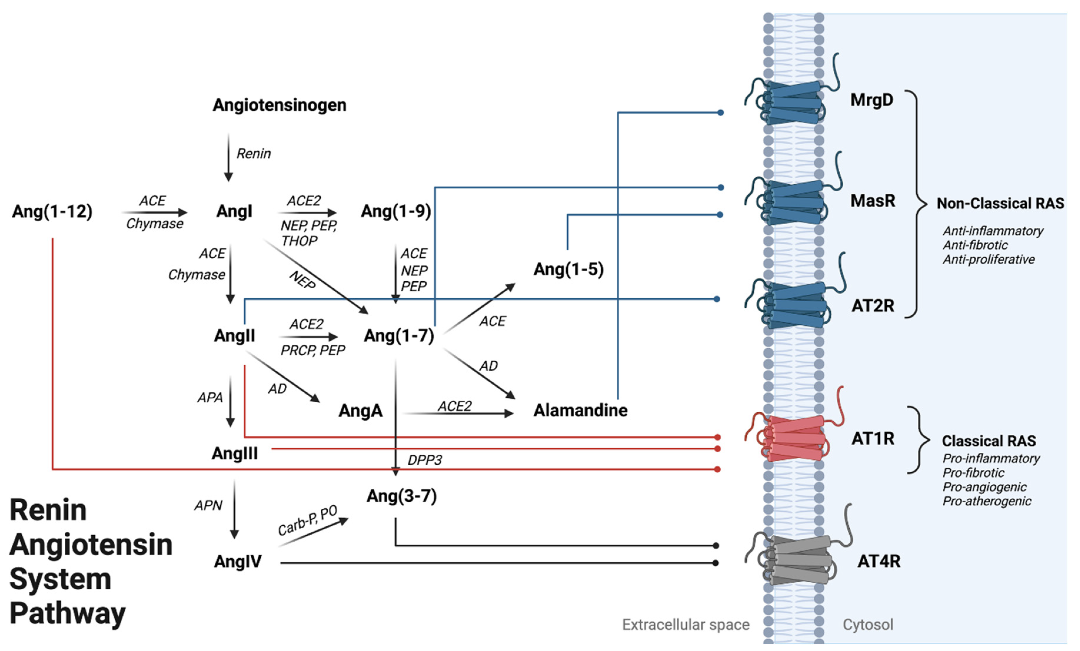
:1. Introduction
2. Methods
2.1. Study Participants
2.2. Clinical Assessment
2.3. RAS Peptide Metabolites
2.4. Plasma Biomarker Assessment
2.5. Carotid and Bifurcation Carotid Artery Intima-Media Thickness
2.6. Statistical Analyses
3. Results
3.1. Clinical Demographics
3.2. Systemic RAS Metabolites
3.3. Intimal Media Thickness Association
4. Discussion
5. Conclusions
Author Contributions
Funding
Institutional Review Board Statement
Informed Consent Statement
Data Availability Statement
Conflicts of Interest
Nonstandard Abbreviations and Acronyms
| RAS | renin–angiotensin system |
| RCCA | right common carotid artery |
| RBIF | right carotid bifurcation |
| CIMT | carotid intima-media thickness |
| AT1R | angiotensin II type 1 receptor |
| AT2R | angiotensin II type 2 receptor |
| ACE1 | angiotensin-converting enzyme 1 |
| ACE2 | angiotensin-converting enzyme 2 |
| NEP | neprilysin |
| PEP | prolyl endopeptidase |
| ANP | atrial natriuretic peptide |
References
- Jalbert, E.; Crawford, T.Q.; D’Antoni, M.L.; Keating, S.M.; Norris, P.J.; Nakamoto, B.K.; Seto, T.; Parikh, N.I.; Shikuma, C.M.; Ndhlovu, L.C.; et al. IL-1Β Enriched Monocytes Mount Massive IL-6 Responses to Common Inflammatory Triggers among Chronically HIV-1 Infected Adults on Stable Anti-Retroviral Therapy at Risk for Cardiovascular Disease. PLoS ONE 2013, 8, e75500. [Google Scholar] [CrossRef] [PubMed]
- Hsue, P.Y.; Waters, D.D. Time to Recognize HIV Infection as a Major Cardiovascular Risk Factor. Circulation 2018, 138, 1113–1115. [Google Scholar] [CrossRef] [PubMed] [Green Version]
- Shah, A.S.; Stelzle, D.; Lee, K.K.; Beck, E.J.; Alam, S.; Clifford, S.; Mills, N.L. Global Burden of Atherosclerotic Cardiovascular Disease in People Living With HIV: Systematic Review and Meta-Analysis. Circulation 2018, 138, 1100–1112. [Google Scholar] [CrossRef] [PubMed]
- Freiberg, M.S.; Chang, C.C.H.; Kuller, L.H.; Skanderson, M.; Lowy, E.; Kraemer, K.L.; Butt, A.A.; Goetz, M.B.; Leaf, D.; Oursler, K.A.; et al. HIV Infection and the Risk of Acute Myocardial Infarction. JAMA Intern. Med. 2013, 173, 614–622. [Google Scholar] [CrossRef] [PubMed]
- Alonso, A.; Barnes, A.E.; Guest, J.L.; Shah, A.; Shao, I.Y.; Marconi, V. HIV Infection and Incidence of Cardiovascular Diseases: An Analysis of a Large Healthcare Database. J. Am. Hear. Assoc. 2019, 8, e012241. [Google Scholar] [CrossRef] [Green Version]
- Hemkens, L.G.; Bucher, H.C. HIV infection and cardiovascular disease. Eur. Hear. J. 2014, 35, 1373–1381. [Google Scholar] [CrossRef] [Green Version]
- Longenecker, C.T.; Sullivan, C.; Baker, J.V. Immune activation and cardiovascular disease in chronic HIV infection. Curr. Opin. HIV AIDS 2016, 11, 216–225. [Google Scholar] [CrossRef]
- Weir, M.R.; Dzau, V.J. The renin-angiotensin-aldosterone system: A specific target for hypertension management. Am. J. Hypertens. 1999, 12 Pt 3, 205–213. [Google Scholar] [CrossRef]
- Navar, L.G.; Imig, J.; Zou, L.; Wang, C.T. Intrarenal production of angiotensin II. Semin. Nephrol. 1997, 17, 412–422. [Google Scholar]
- Yoo, T.-H.; Li, J.-J.; Kim, J.; Jung, D.-S.; Kwak, S.-J.; Ryu, D.-R.; Choi, H.Y.; Kim, H.; Han, S.H.; Lee, J.E.; et al. Activation of the renin–angiotensin system within podocytes in diabetes. Kidney Int. 2007, 71, 1019–1027. [Google Scholar] [CrossRef] [Green Version]
- Huang, Y.; Noble, N.A.; Zhang, J.; Xu, C.; Border, W.A. Renin-stimulated TGF-beta1 expression is regulated by a mitogen-activated protein kinase in mesangial cells. Kidney Int. 2007, 72, 45–52. [Google Scholar] [CrossRef] [PubMed]
- Nicholas, S.B.; Mauer, M.; Basgen, J.M.; Aguiniga, E.; Chon, Y. Effect of Angiotensin II on Glomerular Structure in Streptozotocin-Induced Diabetic Rats. Am. J. Nephrol. 2004, 24, 549–556. [Google Scholar] [CrossRef] [PubMed]
- Tone, A.; Shikata, K.; Ogawa, D.; Sasaki, S.; Nagase, R.; Sasaki, M.; Yozai, K.; Usui, H.K.; Okada, S.; Wada, J.; et al. Changes of gene expression profiles in macrophages stimulated by angiotensin II—Angiotensin II induces MCP-2 through AT1-receptor. J. Renin-Angiotensin-Aldosterone Syst. 2007, 8, 45–50. [Google Scholar] [CrossRef] [Green Version]
- Dasgupta, C.; Zhang, L. Angiotensin II receptors and drug discovery in cardiovascular disease. Drug Discov. Today 2011, 16, 22–34. [Google Scholar] [CrossRef] [PubMed] [Green Version]
- Ocaranza, M.P.; Riquelme, J.A.; García, L.; Jalil, J.E.; Chiong, M.; Santos, R.A.S.; Lavandero, S. Counter-regulatory renin–angiotensin system in cardiovascular disease. Nat. Rev. Cardiol. 2020, 17, 116–129. [Google Scholar] [CrossRef] [PubMed] [Green Version]
- Dandona, P.; Dhindsa, S.; Ghanim, H.; Chaudhuri, A. Angiotensin II and inflammation: The effect of angiotensin-converting enzyme inhibition and angiotensin II receptor blockade. J. Hum. Hypertens. 2007, 21, 20–27. [Google Scholar] [CrossRef] [PubMed] [Green Version]
- Paoletti, R.; Gotto, A.M., Jr.; Hajjar, D.P. Inflammation in Atherosclerosis and Implications for Therapy. Circulation 2004, 109, III-20–III-26. [Google Scholar] [CrossRef] [Green Version]
- Kaschina, E.; Steckelings, U.; Unger, T. Hypertension and the Renin–Angiotensin–Aldosterone System☆. In Encyclopedia of Endocrine Diseases, 2nd ed.; Huhtaniemi, I., Martini, L., Eds.; Academic Press: Oxford, UK, 2018; pp. 505–510. [Google Scholar]
- Yang, H.; Zeng, X.-J.; Wang, H.-X.; Zhang, L.-K.; Dong, X.-L.; Guo, S.; Du, J.; Li, H.-H.; Tang, C.-S. Angiotensin IV protects against angiotensin II-induced cardiac injury via AT4 receptor. Peptides 2011, 32, 2108–2115. [Google Scholar] [CrossRef]
- Lochard, N.; Thibault, G.; Silversides, D.W.; Touyz, R.M.; Reudelhuber, T.L. Chronic Production of Angiotensin IV in the Brain Leads to Hypertension That Is Reversible With an Angiotensin II AT1 Receptor Antagonist. Circ. Res. 2004, 94, 1451–1457. [Google Scholar] [CrossRef]
- D’Agostino Sr, R.B.; Vasan, R.S.; Pencina, M.J.; Wolf, P.A.; Cobain, M.; Massaro, J.M.; Kannel, W.B. General cardiovascular risk profile for use in primary care: The Framingham Heart Study. Circulation 2008, 117, 743–753. [Google Scholar] [CrossRef] [Green Version]
- Gavras, I.; Gavras, H. Angiotensin II as a cardiovascular risk factor. J. Hum. Hypertens. 2002, 16 (Suppl. S2), S2–S6. [Google Scholar] [CrossRef] [PubMed]
- Lévy, B.I. Can Angiotensin II Type 2 Receptors Have Deleterious Effects in Cardiovascular Disease? Circulation 2004, 109, 8–13. [Google Scholar] [CrossRef] [PubMed] [Green Version]
- Zhuo, J.L.; Li, X.C. Angiotensin III/AT<sub>2</sub> Receptor/NHE3 Signaling Pathway in the Proximal Tubules of the Kidney: A Novel Natriuretic and Antihypertensive Mechanism in Hypertension. J. Am. Heart Assoc. 2019, 8, e012644. [Google Scholar] [PubMed] [Green Version]
- Chan, K.H.; Chen, Y.H.; Zhang, Y.; Wong, Y.H.; Dun, N.J. Angiotensin-[1-12] interacts with angiotensin type I receptors. Neuropharmacology 2014, 81, 267–273. [Google Scholar] [CrossRef] [Green Version]
- Yu, L.; Yuan, K.; Phuong, H.T.A.; Park, B.M.; Kim, S.H. Angiotensin-(1-5), an active mediator of renin-angiotensin system, stimulates ANP secretion via Mas receptor. Peptides 2016, 86, 33–41. [Google Scholar] [CrossRef]
- Nagata, S.; Kato, J.; Sasaki, K.; Minamino, N.; Eto, T.; Kitamura, K. Isolation and identification of proangiotensin-12, a possible component of the renin-angiotensin system. Biochem. Biophys. Res. Commun. 2006, 350, 1026–1031. [Google Scholar] [CrossRef]
- Prosser, H.; Richards, A.; Forster, M.; Pemberton, C. Regional vascular response to ProAngiotensin-12 (PA12) through the rat arterial system. Peptides 2010, 31, 1540–1545. [Google Scholar] [CrossRef]
- Isa, K.; García-Espinosa, M.A.; Arnold, A.C.; Pirro, N.T.; Tommasi, E.N.; Ganten, D.; Chappell, M.C.; Ferrario, C.M.; Diz, D.I. Chronic immunoneutralization of brain angiotensin-(1-12) lowers blood pressure in transgenic (mRen2)27 hypertensive rats. Am. J. Physiol. Integr. Comp. Physiol. 2009, 297, R111–R115. [Google Scholar] [CrossRef] [Green Version]
- Sumners, C.; Silva, I.S.; Steckelings, U. Angiotensin receptors—Affinitiy and beyond. Clin. Sci. 2022, 136, 799–802. [Google Scholar] [CrossRef]
- Arakawa, K.; Urata, H. Hypothesis regarding the pathophysiological role of alternative pathways of angiotensin II formation in atherosclerosis. Hypertension 2000, 36, 638–641. [Google Scholar] [CrossRef] [Green Version]
- Jessup, J.A.; Trask, A.J.; Chappell, M.C.; Nagata, S.; Kato, J.; Kitamura, K.; Ferrario, C.M. Localization of the novel angiotensin peptide, angiotensin-(1-12), in heart and kidney of hypertensive and normotensive rats. Am. J. Physiol. Circ. Physiol. 2008, 294, H2614–H2618. [Google Scholar] [CrossRef] [PubMed]
- Ahmad, S.; Varagic, J.; Groban, L.; Dell’Italia, L.J.; Nagata, S.; Kon, N.D.; Ferrario, C.M. Angiotensin-(1-12): A Chymase-Mediated Cellular Angiotensin II Substrate. Curr. Hypertens. Rep. 2014, 16, 429. [Google Scholar] [CrossRef] [PubMed] [Green Version]
- Rice, G.I.; Thomas, D.A.; Grant, P.J.; Turner, A.J.; Hooper, N.M. Evaluation of angiotensin-converting enzyme (ACE), its homologue ACE2 and neprilysin in angiotensin peptide metabolism. Biochem. J. 2004, 383 Pt1, 45–51. [Google Scholar] [CrossRef] [Green Version]


| Variable | HIV Negative (n = 72) | HIV Positive (n = 71) | p Value |
|---|---|---|---|
| Age | 58.96 (9.29) | 59.36 (7.59) | 0.931 |
| Sex (M), n (%) | 61 (84.7) | 61 (84.7) | 1.000 |
| Ethnicity, White, n (%) | 37 (51.4) | 44 (61) | 0.278 |
| Height, in | 67.78 (3.62) | 68.45 (3.58) | 0.265 |
| Weight, lbs. | 193.10 (54.47) | 181.30 (36.94) | 0.130 |
| BMI (M), kg/m2 | 29.83 (7.52) | 26.94 (4.05) | 0.009 |
| BMI (F), kg/m2 | 26.89 (6.53) | 27.55 (5.28) | 0.804 |
| Systolic blood pressure, mmHg | 131.9 (17.47) | 125.2 (13.42) | 0.011 |
| Diastolic blood pressure, mmHg | 78.14 (10.88) | 77.18 (10.19) | 0.589 |
| Mean arterial pressure, mmHg | 96.05 (11.87) | 93.17 (10.02) | 0.121 |
| Hypertension, n (%) | 24 (33.3) | 31 (43.1) | 0.255 |
| Antihypertensive medication use, n (%) | 13 (44%) | 32 (18%) | <0.001 |
| High Cholesterol, n (%) | 25 (34.7) | 36 (50) | 0.074 |
| Diabetes, n (%) | 6 (8.3) | 9 (12.5) | 0.429 |
| Framingham risk score (%) | 13.5 (13.1) | 12.1 (9) | 0.460 |
| CD4 count cells/mm3 | - | 673 (311) | - |
| Presence of carotid plaque, n (%) | 25 (34.7) | 32 (45.1) | 0.260 |
| Current tobacco smoking, n (%) | 8 (11%) | 8 (11%) | 0.976 |
| Substance use, including alcohol, n (%) | 52 (72%) | 67 (93%) | 0.002 |
| Beta blocker use, n (%) | 7 (9.7) | 7 (8.9) | 0.978 |
| RAS Peptide | HIV Negative N = 72 | HIV Positive N = 71 | p-Value | RAS Peptide Median Ratios (HIV+/HIV−) |
|---|---|---|---|---|
| Median [IQR] [ng/mL] | Median [IQR] [ng/mL] | |||
| Ang(1-12) | 0.05 [0.01–0.05] | 0.05 [0.03–0.50] | <0.001 | 1.00 |
| AngI (Ang(1-10)) | 0.05 [0.01–0.40] | 0.67 [0.05–2.14] | <0.001 | 13.40 |
| Ang(1-9) | 0.34 [0.06–0.63] | 0.60 [0.19–1.04] | 0.002 | 1.76 |
| AngII (Ang(1-8)) | 0.11 [0.05–0.48] | 0.38 [0.05–0.74] | 0.223 | 3.45 |
| Ang(1-7) | 0.05 [0.02–0.26] | 0.09 [0.05–0.42] | 0.016 | 1.80 |
| Ang(1-5) | 0.33 [0.05–0.60] | 0.09 [0.05–0.37] | 0.003 | 0.27 |
| AA(1-7) | 0.05 [0.01–0.53] | 0.05 [0.01–0.33] | 0.755 | 1.00 |
| AngIII (Ang(2-8)) | 0.53 [0.29–0.64] | 0.61 [0.11–1.53] | 0.092 | 1.15 |
| AngIV (Ang(3-8)) | 0.10 [0.05–0.44] | 0.28 [0.05–0.67] | 0.019 | 2.80 |
| Ang(3-7) | 0.05 [0.02–0.10] | 0.10 [0.05–0.19] | 0.005 | 2.00 |
| RAS Peptide Ratio (Product/Reactant) | HIV Negative N = 72 | HIV Positive N = 71 | p-Value | Median Ratios (HIV+/HIV−) |
|---|---|---|---|---|
| Median [IQR] | Median [IQR] | |||
| AngI/Ang(1-12) | 2.0 [1.0–9.83] | 1.0 [0.70–7.69] | 0.102 | 2.00 |
| Ang(1-9)/AngI | 1.0 [0.49–19.61] | 1.30 [0.48–13.0] | 0.495 | 1.30 |
| AngII/AngI | 1.0 [0.15–11.96] | 0.56 [0.24–1.0] | 0.028 | 0.56 |
| Ang(1-7)/Ang(1-9) | 0.42 [0.05–0.89] | 0.22 [0.06–0.57] | 0.624 | 0.52 |
| Ang(1-7)/AngII | 0.76 [0.14–1.0] | 0.86 [0.56–1.0] | 0.034 | 1.13 |
| Ang(1-5)/Ang(1-7) | 1.87 [1.0–11.15] | 1.0 [0.28–5.67] | 0.001 | 0.54 |
| AA(1-7)/Ang(1-7) | 1.0 [0.26–7.99] | 1.0 [0.11–2.31] | 0.248 | 1.00 |
| Ang(3-7)/Ang(1-7) | 0.89 [0.20–1.0] | 0.66 [0.30–1.0] | 0.761 | 0.74 |
| AngIII/AngII | 1.89 [1.28–4.45] | 2.11 [1.23–8.72] | 0.368 | 1.12 |
| AngIV/AngIII | 0.40 [0.11–1.0] | 0.38 [0.10–1.0] | 0.905 | 0.95 |
| Ang(3-7)/AngIV | 0.84 [0.14–1.27] | 0.45 [0.29–1.07] | 0.679 | 0.54 |
| RAS Peptide | HIV Negative N = 58 | HIV Positive N = 40 | p-Value | Median Ratios (HIV+/HIV−) |
|---|---|---|---|---|
| Median [IQR] | Median [IQR] | |||
| Ang(1-12) | 0.05 [0.01–0.05] | 0.05 [0.01–0.88] | 0.013 | 1.00 |
| AngI (Ang(1-10)) | 0.05 [0.05–0.36] | 0.47 [0.05–1.89] | 0.004 | 9.40 |
| Ang(1-9) | 0.30 [0.05–0.63] | 0.59 [0.19–1.02] | 0.024 | 1.97 |
| AngII (Ang(1-8)) | 0.10 [0.05–0.49] | 0.43 [0.05–0.77] | 0.039 | 4.30 |
| Ang(1-7) | 0.05 [0.05–0.24] | 0.18 [0.05–0.47] | 0.013 | 3.60 |
| Ang(1-5) | 0.34 [0.05–0.64] | 0.11 [0.01–0.53] | 0.048 | 0.32 |
| AA1-7 | 0.05 [0.01–0.52] | 0.05 [0.01–0.50] | 0.451 | 1.00 |
| AngIV(Ang(3-8)) | 0.12 [0.05–0.46] | 0.30 [0.06–0.69] | 0.017 | 2.50 |
| Ang(3-7) | 0.05 [0.01–0.10] | 0.1 [0.05–0.22] | 0.003 | 2.00 |
| AngIII (Ang(2-8)) | 0.45 [0.07–0.63] | 0.62 [0.32–3.67] | 0.015 | 1.38 |
| Cytokine | HIV Negative N = 72 | HIV Positive N = 71 | p-Value |
|---|---|---|---|
| Median [IQR] | Median [IQR] | ||
| Neopterin | 7.18 [4.23–8.82] | 9.45 [7.49–11.68] | <0.001 |
| CD14 | 1.64 × 10⁶ [1.26 × 10⁶–2.26 × 10⁶] | 1.94 × 10⁶ [1.56 × 10⁶–2.47 × 10⁶] | 0.012 |
| CRP | 1.319 × 10⁶ [4.73 × 10⁵–3.18 × 10⁶] | 8.75 × 10⁵ [4.25 × 10⁵–2.35 × 10⁶] | 0.130 |
| TNF alpha | 1.27 [0.10–2.41] | 2.39 [1.57–3.29] | <0.001 |
| IL-6 | 0.75 [0.46–1.10] | 1.11 [0.82–1.66] | <0.001 |
| CCL2 | 85.13 [71.07–99.56] | 94.00 [78.04–114.70] | 0.022 |
| CD163 | 1.21 × 10⁵ [7.52 × 10⁴–1.61 × 10⁵] | 1.38 × 10⁵ [9.39 × 10⁴–2.21 × 10⁵] | 0.034 |
| D-dimer | 796.6 [597.6–628.2] | 967.9 [628.2–1650] | 0.204 |
| Dependent Variable | Model Variables | Beta Coefficient | Sig. (p-Value) |
|---|---|---|---|
| Right Common Carotid Artery Thickness (RCCA) | (Constant) | 0.432 | 0.005 |
| Age (years) | 0.003 | 0.030 | |
| HIV positive | −0.019 | 0.511 | |
| Gender (Female) | −0.004 | 0.906 | |
| MAP (mmHg) | 0.001 | 0.430 | |
| TNF alpha (pg/mL) | 0.022 | <0.001 | |
| AngII (ng/mL) | 0.208 | <0.001 | |
| AngIII (ng/mL) | −0.026 | <0.001 | |
| Right Carotid Bifurcation Thickness (RBIF) | (Constant) | 0.499 | 0.004 |
| Age (years) | 0.003 | 0.071 | |
| MAP (mmHg) | 0.002 | 0.171 | |
| Gender (Female) | −0.027 | 0.488 | |
| HIV positive | −0.009 | 0.789 | |
| AngI/Ang(1-12) | 0.001 | 0.005 | |
| TNF alpha (pg/mL) | 0.016 | 0.002 | |
| AngIII (ng/mL) | −0.014 | 0.029 |
Disclaimer/Publisher’s Note: The statements, opinions and data contained in all publications are solely those of the individual author(s) and contributor(s) and not of MDPI and/or the editor(s). MDPI and/or the editor(s) disclaim responsibility for any injury to people or property resulting from any ideas, methods, instructions or products referred to in the content. |
© 2022 by the authors. Licensee MDPI, Basel, Switzerland. This article is an open access article distributed under the terms and conditions of the Creative Commons Attribution (CC BY) license (https://creativecommons.org/licenses/by/4.0/).
Share and Cite
Asante, I.; Lu, A.; Mitchell, B.I.; Boisvert, W.A.; Shikuma, C.M.; Chow, D.C.; Louie, S.G. Alterations in Renin–Angiotensin System (RAS) Peptide Levels in Patients with HIV. Metabolites 2023, 13, 61. https://doi.org/10.3390/metabo13010061
Asante I, Lu A, Mitchell BI, Boisvert WA, Shikuma CM, Chow DC, Louie SG. Alterations in Renin–Angiotensin System (RAS) Peptide Levels in Patients with HIV. Metabolites. 2023; 13(1):61. https://doi.org/10.3390/metabo13010061
Chicago/Turabian StyleAsante, Isaac, Angela Lu, Brooks I. Mitchell, William A. Boisvert, Cecilia M. Shikuma, Dominic C. Chow, and Stan G. Louie. 2023. "Alterations in Renin–Angiotensin System (RAS) Peptide Levels in Patients with HIV" Metabolites 13, no. 1: 61. https://doi.org/10.3390/metabo13010061