
Metabolic Disposition and Elimination of Tritum-Labeled Sulfamethoxazole in Pigs, Chickens and Rats

 , ,
, ,
Abstract
:1. Introduction
2. Materials and Methods
2.1. Chemicals
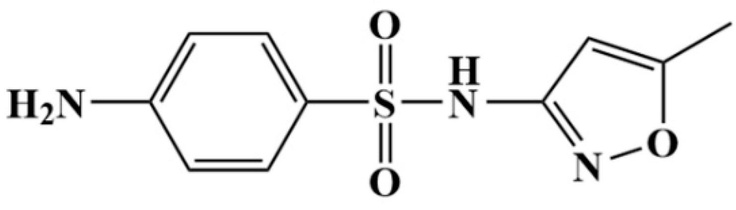
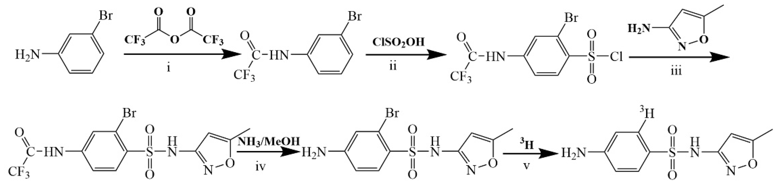
2.2. Tritum-Labeled SMZ
2.3. [3H]-SMZ Injection
2.4. [3H]-SMZ Oral Solution
2.5. Animals
2.6. Animal Experimental Design
2.6.1. Excretion and Metabolism Studies
2.6.2. Distribution and Residue Studies
2.6.3. Sample Analysis
2.7. Quantification and Structural Identification of Metabolites
2.7.1. Urine, Blood and Bile Preparation
2.7.2. Feces or Tissues Preparation
2.7.3. Validation of the Quantitation Methods
2.7.4. Data Analysis
3. Results
3.1. Radioactivity Excretion of SMZ in Animals
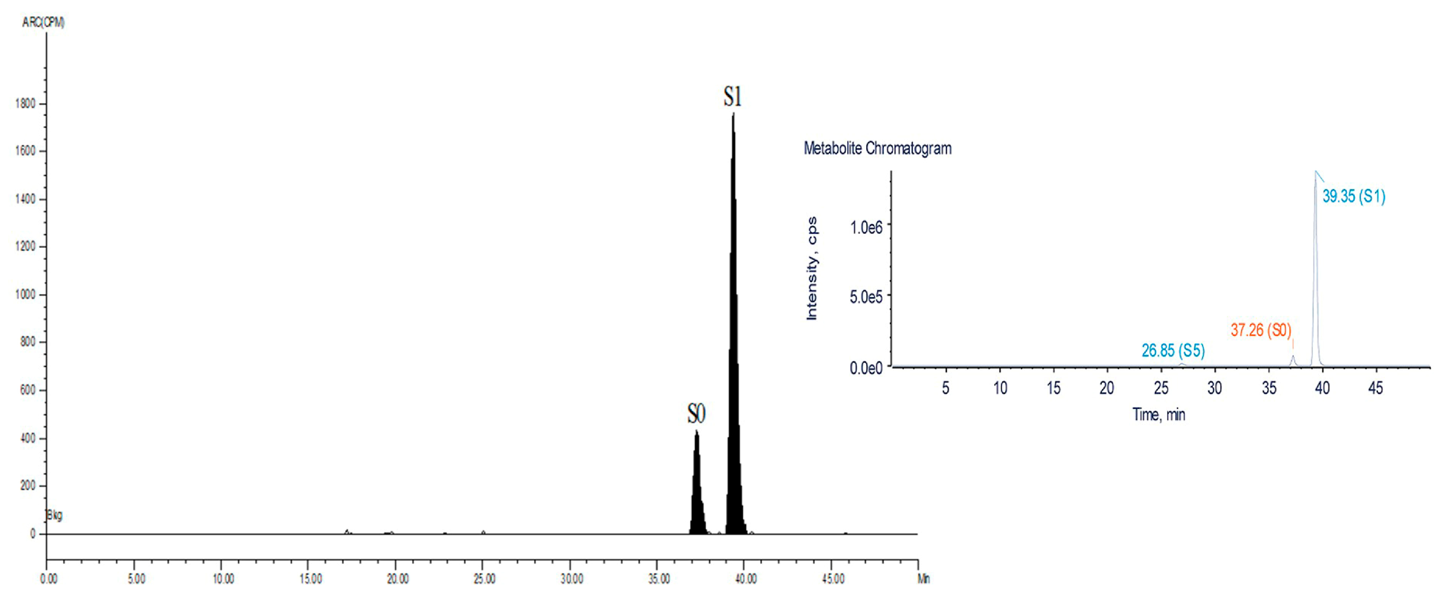
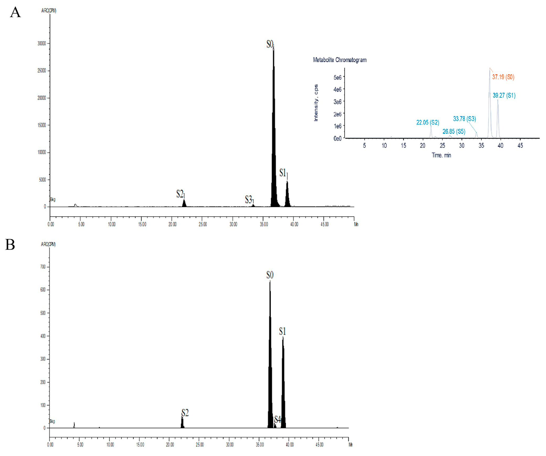
3.2. Metabolism of SMZ in Animals
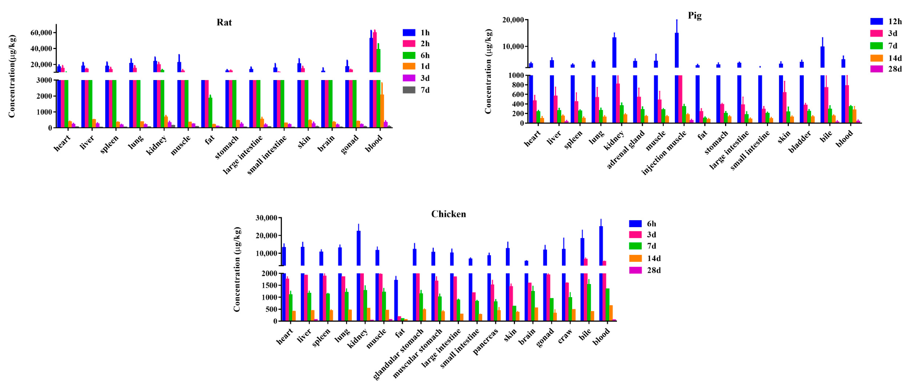
3.3. Distribution
3.4. Residue Depletion
4. Discussion
Author Contributions
Funding
Institutional Review Board Statement
Informed Consent Statement
Data Availability Statement
Conflicts of Interest
References
- Kantiani, L.; Llorca, M.; Sanchis, J.; Farre, M.; Barcelo, D. Emerging food contaminants: A review. Anal. Bioanal. Chem. 2010, 398, 2413–2427. [Google Scholar] [CrossRef] [PubMed]
- Boufas, W.; Dupont, N.; Berredjem, M.; Berrezag, K.; Becheker, I.; Berredjem, H.; Aouf, N.-E. Synthesis and antibacterial activity of sulfonamides. SAR and DFT studies. J. Mol. Struct. 2014, 1074, 180–185. [Google Scholar] [CrossRef]
- Rama, A.; Lucatello, L.; Benetti, C.; Galina, G.; Bajraktari, D. Assessment of antibacterial drug residues in milk for consumption in Kosovo. J. Food Drug Anal. 2017, 25, 525–553. [Google Scholar] [CrossRef] [PubMed] [Green Version]
- Baran, W.; Adamek, E.; Ziemianska, J.; Sobczak, A. Effects of the presence of sulfonamides in the environment and their influence on human health. J. Hazard Mater. 2011, 196, 1–15. [Google Scholar] [CrossRef]
- Supuran, C.T. Special Issue: Sulfonamides. Molecules 2017, 22, 1642. [Google Scholar] [CrossRef] [Green Version]
- Mainra, R.R.; Card, S.E. Trimethoprim-sulfamethoxazole-associated hepatotoxicity—Part of a hypersensitivity syndrome. Can. J. Clin. Pharmacol. 2003, 10, 175–178. [Google Scholar]
- Yang, J.J.; Huang, C.H.; Liu, C.E.; Tang, H.J.; Yang, C.J.; Lee, Y.C.; Lee, K.Y.; Tsai, M.S.; Lin, S.W.; Chen, Y.H.; et al. Multicenter Study of Trimethoprim/Sulfamethoxazole-Related Hepatotoxicity: Incidence and Associated Factors among HIV-Infected Patients Treated for Pneumocystis jirovecii Pneumonia. PLoS ONE 2014, 9, e106141. [Google Scholar] [CrossRef] [Green Version]
- Ovung, A.; Bhattacharyya, J. Sulfonamide drugs: Structure, antibacterial property, toxicity and biophysical interactions. Biophys. Rev. 2021, 13, 259–272. [Google Scholar] [CrossRef]
- Biošić, M.; Mitrevski, M.; Babić, S. Environmental behavior of sulfadiazine, sulfamethazine, and their metabolites. Environ. Sci. Pollut. Res. Int. 2017, 24, 9802–9812. [Google Scholar] [CrossRef]
- Aguilera-Luiz, M.M.; Martinez Vidal, J.L.; Romero-Gonzalez, R.; Garrido Frenich, A. Multiclass method for fast determination of veterinary drug residues in baby food by ultra-high-performance liquid chromatography-tandem mass spectrometry. Food Chem. 2012, 132, 2171–2180. [Google Scholar] [CrossRef]
- Vree, T.B.; van der Ven, A.J.; Verwey-van Wissen, C.P.; van Ewijk-Beneken Kolmer, E.W.; Swolfs, A.E.; van Galen, P.M.; Amatdjais-Groenen, H. Isolation, identification and determination of sulfamethoxazole and its known metabolites in human plasma and urine by high-performance liquid chromatography. J. Chromatogr. B Biomed. Appl. 1994, 658, 327–340. [Google Scholar] [CrossRef] [PubMed]
- Lehmann, D.F. The metabolic rationale for a lack of cross-reactivity between sulfonamide antimicrobials and other sulfonamide-containing drugs. Drug Metab. Lett. 2012, 6, 129–133. [Google Scholar] [CrossRef] [PubMed]
- Vree, T.; Reekers-Ketting, J.; Hekster, C.; Nouws, J.F.M. Acetylation and deacetylation of sulphonamides in dogs. J. Vet. Pharmacol. Ther. 1983, 6, 153–155. [Google Scholar] [CrossRef]
- vanderVen, A.; Vree, T.B.; Koopmans, P.P.; vanderMeer, J.W.M. Adverse reactions to co-trimoxazole in HIV infection: A reappraisal of the glutathione-hydroxylamine hypothesis. J. Antimicrob. Chemother. 1996, 37, 55–60. [Google Scholar] [CrossRef] [Green Version]
- Mengelers, M.J.B.; Gogh, E.; Kuiper, H.; Pijpers, A.; Verheijden, J.H.M.; Miert, A. Pharmacokinetics of sulfadimethoxine and sulfamethoxazole in combination with trimethoprim after intravenous administration to healthy and pneumonic pigs. J. Vet. Pharmacol. Ther. 1995, 18, 243–253. [Google Scholar] [CrossRef] [PubMed]
- Vanginneken, V.J.T.; Nouws, J.F.M.; Grondel, J.L.; Driessens, F.; Degen, M. Pharmacokinetics of sulphadimidine in carp (Cyprinus carpio L.) and rainbow trout (Salmo gairdneri Richardson) acclimated at two different temperature levels. Vet. Quart. 1991, 13, 88–96. [Google Scholar] [CrossRef]
- Kitakaze, T.; Ito, K.; Ogawa, Y. Absorption, distribution, excretion and metabolism of sulfamethoxazole in rats. Chemotherapy 1973, 21, 224–228. (In Japanese) [Google Scholar] [CrossRef]
- Queralt, J.; Castells, I. Pharmacokinetics of sulfamethoxazole and trimethoprim association in hens. Poult. Sci. 1985, 64, 2362–2367. [Google Scholar] [CrossRef]
- Ramamoorthy, A.; Bende, G.; Chow, E.C.Y.; Dimova, H.; Hartman, N.; Jean, D.; Pahwa, S.; Ren, Y.; Shukla, C.; Yang, Y.; et al. Human radiolabeled mass balance studies supporting the FDA approval of new drugs. Clin. Transl. Sci. 2022, 15, 2567–2575. [Google Scholar] [CrossRef]
- Dolovich, M.B. Radiolabeling Methods. J. Aerosol Med. Pulm. Drug Deliv. 2022, 35, 227–236. [Google Scholar] [CrossRef]
- Wang, L.; Huang, L.; Pan, Y.; Kuča, K.; Klímová, B.; Wu, Q.; Xie, S.; Ahmad, I.; Chen, D.; Tao, Y.; et al. Metabolism and Disposition of Aditoprim in Swine, Broilers, Carp and Rats. Sci. Rep. 2016, 6, 20370. [Google Scholar] [CrossRef] [PubMed]
- Lingli, H.; Ning, X.; Harnud, S.; Yuanhu, P.; Dongmei, C.; Yanfei, T.; Zhenli, L.; Zonghui, Y. Metabolic Disposition and Elimination of Cyadox in Pigs, Chickens, Carp, and Rats. J. Agric. Food Chem. 2015, 63, 5557–5569. [Google Scholar] [CrossRef] [PubMed]
- Nouws, J.F.M.; Mevius, D.; Vree, T.B.; Degen, M. Pharmacokinetics and renal clearance of sulphadimidine, sul phamerazine and sulphadiazine and their N4-acetyl and hydroxy metabolites in pigs. Vet. Quart. 1989, 11, 78–86. [Google Scholar] [CrossRef] [PubMed]
- Dalvie, D. Recent Advances in the Applications of Radioisotopes in Drug Metabolism, Toxicology and Pharmacokinetics. Curr. Pharm. Des. 2000, 6, 1009–1028. [Google Scholar] [CrossRef]
- Shah, B.H.; Mawaz, M.; Javed, I.; Gilani, A.U.H. Pharmacokinetics and renal excretion of sulfamethoxazole in sheep. Arch. Pharm. Res. 1989, 12, 154. [Google Scholar] [CrossRef]
- Vree, T.; Vree, J.; Nouws, J. Acetylation and hydroxyla-tion of sulfadimidine by the turtle Cuora amboniensis. J. Vet. Pharmacol. Ther. 1986, 9, 330–332. [Google Scholar] [CrossRef]
- Jesus Garcia-Galan, M.; Silvia Diaz-Cruz, M.; Barcelo, D. Identification and determination of metabolites and degradation products of sulfonamide antibiotics. TrAC Trend. Anal. Chem. 2008, 27, 1008–1022. [Google Scholar] [CrossRef]
- Cribb, A.E.; Miller, M.; Leeder, J.S.; Hill, J.; Spielberg, S.P. Reactions of the nitroso and hydroxylamine metabolites of sulfamethoxazole with reduced glutathione. Implications for idiosyncratic toxicity. Drug Metab. Dispos. 1991, 19, 900. [Google Scholar]
- Nakamura, H.; Uetrecht, J.; Cribb, A.E.; Miller, M.A.; Spielberg, S.P. In vitro formation, disposition and toxicity of N-acetoxy-sulfamethoxazole, a potential mediator of sulfamethoxazole toxicity. J. Pharmacol. Exp. Ther. 1995, 274, 1099–1104. [Google Scholar]
- Oikawa, H.; Nakamoto, K.; Hirota, K.; Katagiri, K. Clearance of sulfamethoxazole in eggs and tissues of chickens. Poult. Sci. 1977, 56, 813–821. [Google Scholar] [CrossRef]
- Mengelers, M.J.B.; Klingeren, B.; Miert, A. In vitro susceptibility of some porcine respiratory tract pathogens to aditoprim, trimethoprim, sulfadimethoxine, sulfamethoxazole and combinations of these agents. Am. J. Vet. Res. 1990, 51, 1860–1864. [Google Scholar] [PubMed]
- Nebbia, C. Biotransformation enzymes as determinants of xenobiotic toxicity in domestic animals. Vet. J. 2001, 161, 238–252. [Google Scholar] [CrossRef] [PubMed]


| Time (d) | Pigs | Chickens | Rats | ||||
|---|---|---|---|---|---|---|---|
| Male | Female | ||||||
| Urine | Feces | Excreta | Urine | Feces | Urine | Feces | |
| 0–0.5 | 60.02 ± 16.54 | 5.56 ± 0.42 | 60.65 ± 13.08 | 45.33 ± 4.79 | 17.3 ± 1.16 | 45.38 ± 6.09 | 14.31 ± 1.73 |
| 0.5–1 | 15.3 ± 8.73 | 2.69 ± 3.12 | 15.08 ± 6.87 | 19.31 ± 7.15 | 5.3 ± 1.43 | 19.41 ± 2.15 | 4.45 ± 1.87 |
| 1–3 | 4.52 ± 2.22 | 5.19 ± 3.36 | 11.91 ± 9.67 | 7.49 ± 2.04 | 0.35 ± 0.14 | 10.09 ± 3.14 | 1.02 ± 0.72 |
| 3–7 | 0.59 ± 0.17 | 0.82 ± 0.34 | 5.48 ± 1.89 | 2.7 ± 1.89 | 0.25 ± 0.14 | 3.97 ± 1.24 | 0.42 ± 0.28 |
| 7–14 | 0.16 ± 0.04 | 0.45 ± 0.3 | 0.26 ± 0.13 | 0.49 ± 0.3 | 0.05 ± 0.03 | 0.38 ± 0.17 | 0.05 ± 0.03 |
| 0–14 | 80.59 ± 5.72 | 14.72 ± 1.31 | 93.39 ± 2.28 | 75.32 ± 4.54 | 23.24 ± 1.79 | 77.9 ± 5.93 | 19.58 ± 2.09 |
| Total | 95.31 ± 4.41 | 93.39 ± 2.28 | 98.56 ± 2.82 | 97.3 ± 1.16 | |||
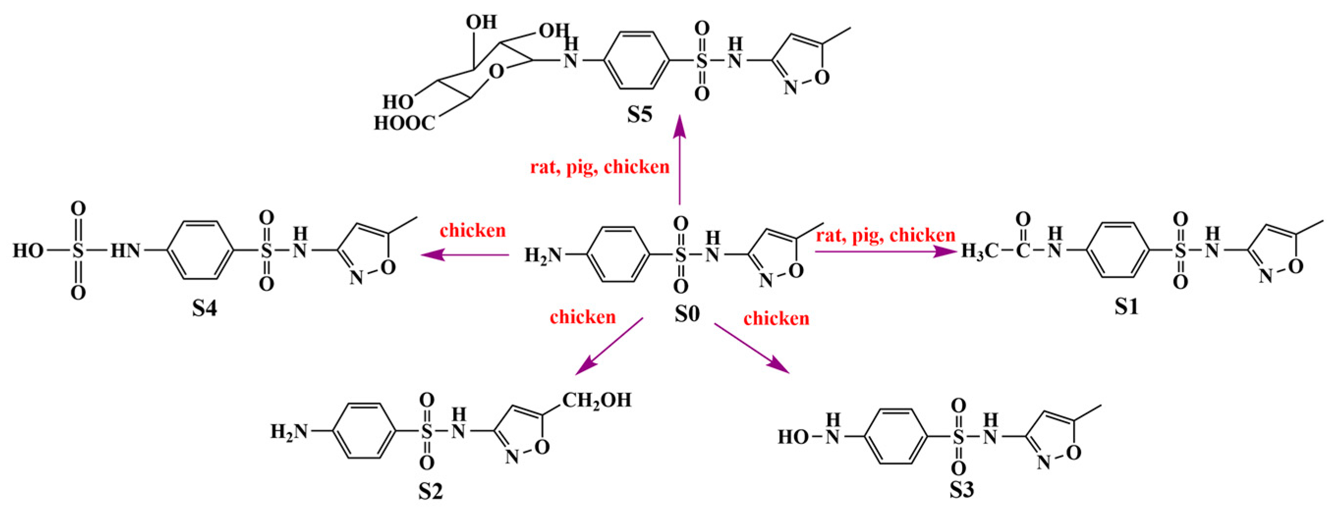
| Compound | Chemical Name | [M+H] (Da) | Formula | Major Product Ions |
|---|---|---|---|---|
| S0 | 4-Amino-N-(5-methyl-3-isoxazolyl) | 254.0521 | C10H11N3O3S | 156, 108, 92, 188, 99, 147 |
| S1 | N-Acetyl sulfamethoxazole | 296.0627 | C12H13N3O4S | 198, 134, 188 |
| S2 | 4-Amino-N-(5-hydroxymethyl-3-isoxazolyl) | 270.0564 | C10H11N3O4S | 252, 204, 156, 147, 108, 92 |
| S3 | 4-Hydroxyamino-N-(5-methyl-3-isoxazolyl) | 270.0564 | C10H11N3O4S | 204, 172, 108 |
| S4 | Sulfamethoxazole-N4-slufate | 334.0172 | C10H11N3O6S2 | 254, 156 |
| S5 | sulfamethoxazole- N4-glucuronide | 430.0945 | C16H19N3O9S | 332, 254, 156, 147, 99 |
| Compound | Rat | Pig | Chicken | |||||||
|---|---|---|---|---|---|---|---|---|---|---|
| Urine | Feces | Plasma | Urine | Feces | Plasma | Bile | Extra | Bile | Plasma | |
| S0 | 48.4 | 38.3 | 94.5 | 24.6 | 12.1 | 80.3 | 74.5 | 69.6 | 88.2 | 91.2 |
| S1 | 51.6 | 61.7 | 5.5 | 75.4 | 87.9 | 19.7 | 25.5 | 16.4 | 9.3 | 6.7 |
| S2 | ND | ND | ND | ND | ND | ND | ND | 9.6 | 2.5 | 1.2 |
| S3 | ND | ND | ND | ND | ND | ND | ND | 4.4 | ND | 0.9 |
| S4 | ND | ND | ND | ND | ND | ND | ND | ND | ND | ND |
| S5 | ND | ND | ND | ND | ND | ND | ND | ND | ND | ND |
| Tissue | Time (Days) | Concentration of the Metabolites (μg/kg) | Time (Days) | Concentration of the Metabolites (μg/kg) | ||
|---|---|---|---|---|---|---|
| Rat | Pig | |||||
| S0 | S1 | S0 | S1 | |||
| Liver | 0.25 | 7730 ± 658 | 840 ± 102 | 0.5 | 1270 ± 345 | 2420 ± 582 |
| 1 | 410 ± 76 | 72 ± 21 | 3 | 437 ± 58 | 124 ± 32 | |
| 3 | 210 ± 54 | ND | 7 | 200 ± 41 | 44 ± 17 | |
| 7 | 22 ± 8 | ND | 14 | 120 ± 33 | ND | |
| 28 | 22 ± 7 | ND | ||||
| Kidney | 0.25 | 7140 ± 988 | 4046 ± 584 | 0.5 | 4355 ± 564 | 8645 ± 987 |
| 1 | 379 ± 88 | 280 ± 79 | 3 | 503 ± 77 | 308 ± 56 | |
| 3 | 198 ± 65 | 70 ± 21 | 7 | 261 ± 43 | 93 ± 33 | |
| 7 | 118 ± 37 | ND | 14 | 125 ± 25 | 38 ± 11 | |
| Muscle | 0.25 | 3657 ± 753 | 1833 ± 202 | 0.5 | 2391 ± 437 | 1890 ± 388 |
| 1 | 204 ± 58 | 147 ± 54 | 3 | 365 ± 55 | 94 ± 33 | |
| 3 | 141 ± 36 | 80 ± 40 | 7 | 234 ± 48 | 30 ± 15 | |
| 7 | 58 ± 16 | ND | 14 | 90 ± 21 | ND | |
| Fat | 0.25 | 1046 ± 194 | 798 ± 201 | 0.5 | 745 ± 102 | 1034 ± 321 |
| 1 | 138 ± 32 | 52 ± 11 | 3 | 188 ± 54 | 65 ± 22 | |
| 3 | 75 ± 15 | ND | 7 | 98 ± 28 | ND | |
| 7 | 28 ± 9 | ND | 14 | 69 ± 16 | ND | |
| Tissue | Time (Days) | Concentration of the Metabolites (μg/kg) | ||||
|---|---|---|---|---|---|---|
| Chicken | ||||||
| S0 | S1 | S2 | S3 | S4 | ||
| Liver | 0.25 | 3622 ± 821 | 9159 ± 1148 | ND | ND | ND |
| 3 | 1521 ± 423 | 184 ± 32 | ND | ND | ND | |
| 7 | 1013 ± 221 | 35 ± 8 | ND | ND | ND | |
| 14 | 365 ± 105 | ND | ND | ND | ND | |
| 28 | 41 ± 12 | ND | ND | ND | ND | |
| Kidney | 0.25 | 20,951 ± 2198 | 234 ± 87 | 1058 ± 211 | 154 ± 45 | 108 ± 21 |
| 3 | 1898 ± 478 | 89 ± 34 | ND | ND | ND | |
| 7 | 1012 ± 399 | 38 ± 11 | ND | ND | ND | |
| 14 | 521 ± 87 | ND | ND | ND | ND | |
| 28 | 22 ± 9 | ND | ND | ND | ND | |
| Muscle | 0.25 | 5783 ± 1103 | 4209 ± 1897 | ND | ND | ND |
| 3 | 1131 ± 325 | 769 ± 184 | ND | ND | ND | |
| 7 | 751 ± 98 | 443 ± 75 | ND | ND | ND | |
| 14 | 374 ± 56 | 134 ± 42 | ND | ND | ND | |
| 28 | 39 ± 8 | ND | ND | ND | ND | |
| Fat | 0.25 | 480 ± 103 | 1120 ± 154 | ND | ND | ND |
| 3 | 102 ± 33 | 76 ± 21 | ND | ND | ND | |
| 7 | 75 ± 19 | ND | ND | ND | ND | |
| 14 | 37 ± 13 | ND | ND | ND | ND | |
| Species | Tissue | Compound | t1/2 (d) | Elimination Equation |
|---|---|---|---|---|
| Rat | Liver | Total Residues | 1.87 | C = 563·e−kt |
| S0 | 1.39 | C = 769·e−kt | ||
| S1 | 0.22 | C = 1648·e−kt | ||
| Kidney | Total Residues | 2.56 | C = 795·e−kt | |
| S0 | 3.74 | C = 769·e−kt | ||
| S1 | 0.53 | C = 2763·e−kt | ||
| Muscle | Total Residues | 2.41 | C = 457·e−kt | |
| S0 | 3.47 | C = 220·e-kt | ||
| S1 | 0.72 | C = 1094·e−kt | ||
| Fat | Total Residues | 2.36 | C = 240·e−kt | |
| S0 | 2.63 | C = 173·e−kt | ||
| Pig | Liver | Total Residues | 5.76 | C = 709·e−kt |
| S0 | 5.63 | C = 741·e−kt | ||
| S1 | 1.19 | C = 1820·e−kt | ||
| Kidney | Total Residues | 3.88 | C = 2296·e−kt | |
| S0 | 4.29 | C = 1477·e−kt | ||
| S1 | 3.78 | C = 445·e−kt | ||
| Muscle | Total Residues | 3.84 | C = 1183·e-kt | |
| S0 | 4.09 | C = 844·e−kt | ||
| S1 | 1.15 | C = 1449·e−kt | ||
| Fat | Total Residues | 5.80 | C = 339·e−kt | |
| S0 | 5.81 | C = 389·e−kt | ||
| Chicken | Liver | Total Residues | 5.16 | C = 3064·e−kt |
| S0 | 4.90 | C = 2697·e−kt | ||
| S1 | 0.87 | C = 5889·e−kt | ||
| Kidney | Total Residues | 4.59 | C = 3542·e−kt | |
| S0 | 4.63 | C = 3279·e−kt | ||
| S1 | 2.61 | C = 229·e−kt | ||
| Muscle | Total Residues | 5.16 | C = 3234·e−kt | |
| S0 | 5.32 | C = 1746·e−kt | ||
| S1 | 3.64 | C = 1478·e−kt | ||
| Fat | total residues | 5.36 | C = 251·e−kt | |
| S0 | 5.29 | C = 201·e−kt |
Disclaimer/Publisher’s Note: The statements, opinions and data contained in all publications are solely those of the individual author(s) and contributor(s) and not of MDPI and/or the editor(s). MDPI and/or the editor(s) disclaim responsibility for any injury to people or property resulting from any ideas, methods, instructions or products referred to in the content. |
© 2022 by the authors. Licensee MDPI, Basel, Switzerland. This article is an open access article distributed under the terms and conditions of the Creative Commons Attribution (CC BY) license (https://creativecommons.org/licenses/by/4.0/).
Share and Cite
Guo, J.; Sun, Y.; Zhao, Y.; Huang, L.; Peng, D.; Hao, H.; Tao, Y.; Chen, D.; Cheng, G.; Wang, X.; et al. Metabolic Disposition and Elimination of Tritum-Labeled Sulfamethoxazole in Pigs, Chickens and Rats. Metabolites 2023, 13, 57. https://doi.org/10.3390/metabo13010057
Guo J, Sun Y, Zhao Y, Huang L, Peng D, Hao H, Tao Y, Chen D, Cheng G, Wang X, et al. Metabolic Disposition and Elimination of Tritum-Labeled Sulfamethoxazole in Pigs, Chickens and Rats. Metabolites. 2023; 13(1):57. https://doi.org/10.3390/metabo13010057
Chicago/Turabian StyleGuo, Jingchao, Yaqi Sun, Yongxia Zhao, Lingli Huang, Dapeng Peng, Haihong Hao, Yanfei Tao, Dongmei Chen, Guyue Cheng, Xu Wang, and et al. 2023. "Metabolic Disposition and Elimination of Tritum-Labeled Sulfamethoxazole in Pigs, Chickens and Rats" Metabolites 13, no. 1: 57. https://doi.org/10.3390/metabo13010057