Review

In Vitro and Ex Vivo Models for Screening Topical Anti-Inflammatory Drugs

by
Juan Luis Pérez-Salas
1,
Martha Rocío Moreno-Jiménez
1,*,
Nuria Elizabeth Rocha-Guzmán
1,
Rubén Francisco González-Laredo
1,
Luis Medina-Torres
2 and
José Alberto Gallegos-Infante
1,*
1
TecNM/Instituto Tecnológico de Durango, UPIDET, Blvd Felipe Pescador 1830 Ote, Col Nueva Vizcaya, Durango 34080, Mexico
2
Facultad de Química, Universidad Nacional Autónoma de México, Ciudad de México 04510, Mexico
*
Authors to whom correspondence should be addressed.
Sci. Pharm. 2023, 91(2), 20; https://doi.org/10.3390/scipharm91020020
Submission received: 15 February 2023 / Revised: 27 March 2023 / Accepted: 10 April 2023 / Published: 17 April 2023
(This article belongs to the Special Issue Feature Papers in Scientia Pharmaceutica)

Abstract

:
Skin inflammation occurs as an immune response to various stimuli such as ultraviolet light, irritants, or any type of skin barrier injury. Finding safe and effective drugs to combat skin inflammation remains a research challenge. Ethical and legal considerations in animal testing encourage the development of in vitro and ex vivo models for the detection of skin inflammation. This report presents an updated review of non-animal study models available for screening drugs with anti-inflammatory potential. It includes a description of the basic methods used to inhibit protein denaturation and red blood cell membrane stability. Three in vitro inhibition assay methods for enzymes relevant to the skin inflammatory process are then described. The development of cell culture models is described: relatively simple and easy-to-produce two-dimensional (2D) skin cell cultures that allow assessment of response to a given stimulus, three-dimensional (3D) cell cultures that better mimic human skin physiology by more accurately replicating mechanical and chemical signals, and vascularized 3D skin models with dynamic perfusion and microfluidic devices known as skin on a chip. Finally, ex vivo skin models are presented that could more accurately represent human skin in terms of structure, cell signaling mechanisms, and absorption effects. Although the current development of models without the use of animals is promising, improvements and refinements are needed to make the models more suitable as screening platforms for topical anti-inflammatory drugs.

1. Introduction

The clinical features of inflammation were described in an Egyptian papyrus around 3000 BC, but Celso was the first to list the four cardinal signs of inflammation: redness, tumor, heat, and pain. A fifth clinical sign, loss of function, was added later by Virchow. In 1793, the Scottish surgeon John Hunter stated that inflammation is not a disease, but a non-specific reaction that has a curative effect on the host [1]. Inflammation can be acute, for example in response to tissue damage, or chronic, with pathological consequences such as arthritis [2].
Local inflammatory mediators include histamine, kinins, and arachidonic acid metabolites, which cause increased capillary permeability, tissue edema, and immune cell infiltration [3,4]. Tissue injury also promotes the release of histamine from mast cells and the production of bradykinin and kallidin, which in turn act directly on the vascular endothelium to transport cellular and protein elements of the immune system to the site of injury [5].
During an acute inflammatory response, cellular and molecular events reduce the likelihood of injury or infection. This process helps to restore tissue homeostasis and allows the acute inflammation to subside. However, uncontrolled acute inflammation can become chronic and contribute to the development of several chronic inflammatory diseases [6]. When this is the case, anti-inflammatory drugs are needed to control and limit the harmful effects of inflammation.
The administration of anti-inflammatory drugs remains a very active area of research. On the other hand, it is equally important to determine the potential or toxicological risk of topical application substances capable of inducing skin inflammation (pro-inflammatory agents).
Inflammation-related diseases affect the majority of the human population. Although several agents are available for the treatment of various inflammatory diseases, their prolonged use leads to serious adverse effects. Therefore, there is a need for robust and accurate methods to determine the modulation of skin inflammation by xenobiotics that have successfully penetrated the barrier function of the stratum corneum. Currently, study models are expected to reduce or eliminate the use of live animals [7].
The aim of this review is to present established and novel in vitro and ex vivo techniques as alternatives in the investigation of natural or synthetic molecules with potential topical anti-inflammatory properties.

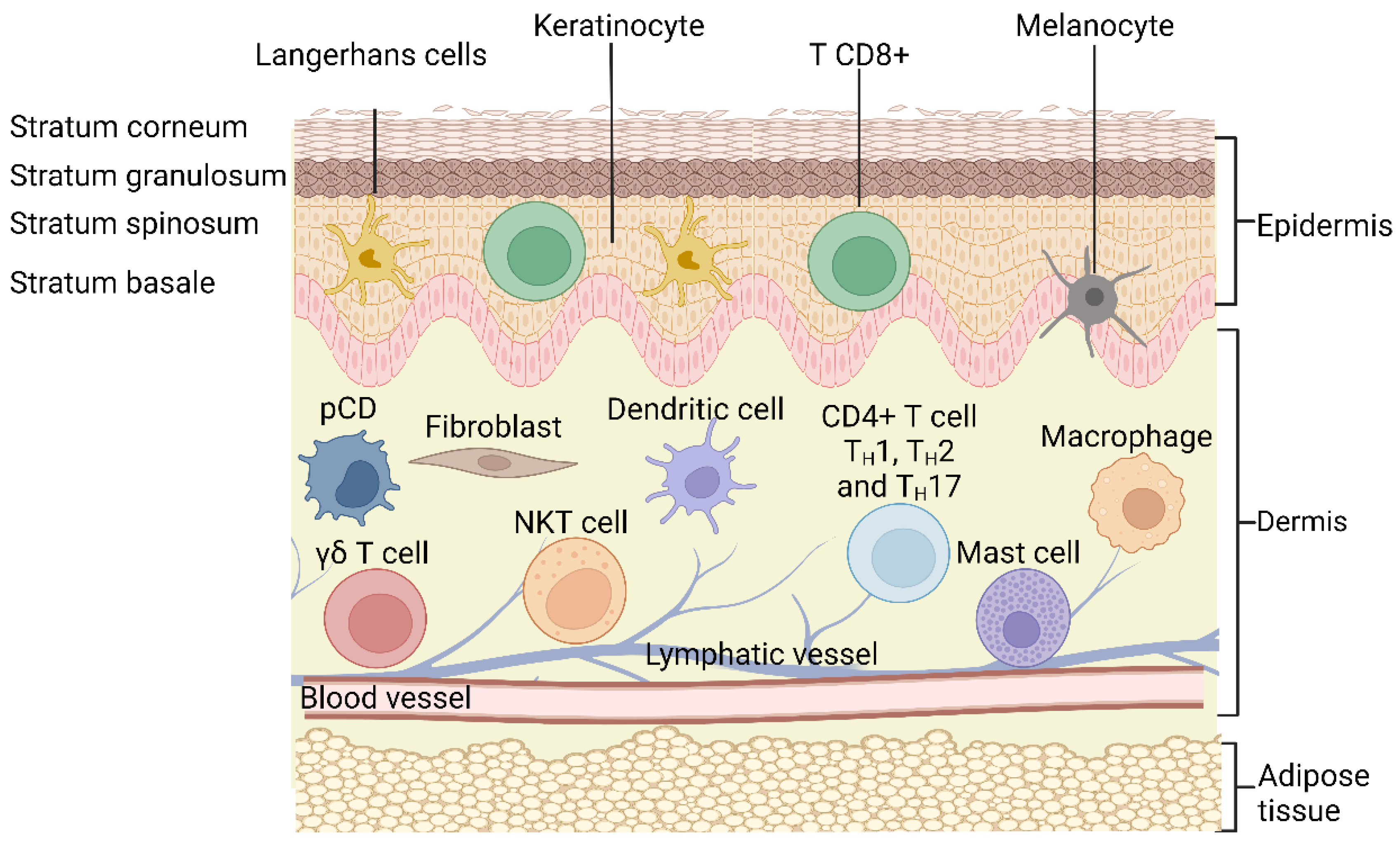
2. Structure of the Skin

The skin is the largest organ of the human body (1.5–2 m2 of the surface), it is a complex barrier between the biological system and the external environment [8]. It consists of three regions (Figure 1): a 50–150 μm thick outer epidermis (biological barrier), a 250 μm thick dermis (thermal barrier), and the innermost subcutaneous adipose tissue (mechanical cushion) [9].
The epidermis is a multilayer barrier composed mainly of keratinocytes with different degrees of differentiation, while the dermis is a connective tissue rich in collagen fibers [10,11].
The epidermis consists of four layers. The basal layer is the lowest layer of the epidermis and is responsible for the constant turnover of epidermal cells. This layer contains a single row of undifferentiated epidermal cells called basal keratinocytes that are constantly dividing. Basal keratinocytes differentiate and migrate to the next layer, called the spinous layer, to initiate maturation, but also divide to complete the basal layer. The cells that enter the spinous layer change from columnar to polygonal and begin to synthesize keratin, which is different from basal keratin. The next layer, called granulosa, is characterized by having keratinocytes with cytoplasmic material with dark clumps, these cells actively produce keratin and lipids [11]. Finally, the stratum corneum is the outermost of the four layers of the epidermis and is primarily responsible for the barrier function of the skin. The cells of this layer, called corneocytes, are dead cells derived from keratinocytes without organelles. They act as a barrier against many harmful substances and prevent dehydration [12].
Damage to the natural barrier components of the skin may contribute to inflammatory skin diseases. For example, the skin of patients with atopic dermatitis showed decreased expression of zonule binding molecules-1 and claudin-1 [13].
Under the epidermis, in the dermis, are fibroblasts, myofibroblasts, and immune cells such as macrophages, lymphocytes and mast cells. Fibroblasts form an extracellular matrix of collagen, proteoglycans and elastic fibers that maintain the structural integrity of the dermis [14].

3. Cutaneous Inflammation

Inflammation is a physiological defense response to various stimuli, such as infection and tissue damage. Inflammation can be acute, for example, in response to tissue damage, or chronic, with pathological consequences [2].
Ultraviolet (UV) rays, trauma, irritants, infections, or any type of barrier disruption trigger a coordinated immune response to maintain skin homeostasis. The immune cells present in the skin are the main ones responsible for restoring homeostasis, but they can also be effector cells in histopathological processes such as dermatitis and psoriasis [11,15].
The epidermis contains immune cells, mainly CD8+ effector T cells and LCs [16]. The dermis contains a more diverse population of immune cells, including CD4+ helper T cells, γδ-T cells, dermal dendritic cells, innate lymphocytes, pDC cell, NK cells, macrophages, mast cells, and fibroblasts [10,11,17]. Likewise, the dermis is drained of lymphatic and blood vessels, which are pathways for cell migration [16].
Although the skin’s immune system is strong, human skin harbors a rich and diverse collection of bacteria, fungi, and viruses. This group of microorganisms constitutes the skin microbiota. In recent years, the skin microbiota has been shown to play an important role in inflammatory skin conditions [18].

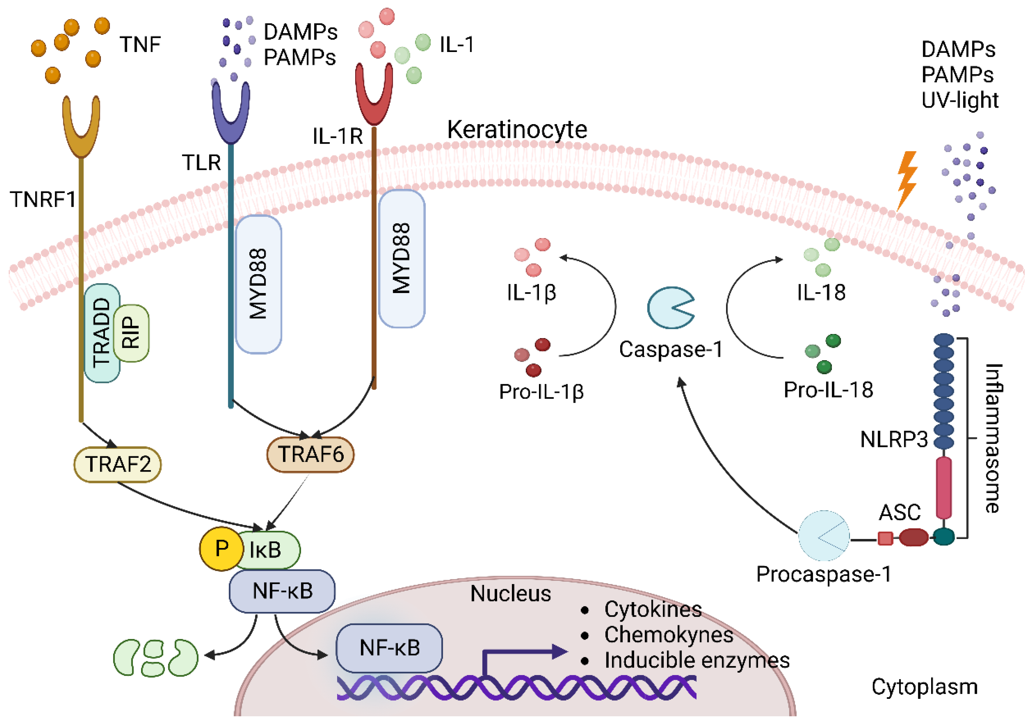
3.1. Keratinocytes as Triggers of Inflammation in the Skin

Damaged keratinocytes send out the first signals called alarmins, which consist of high mobility group 1 box protein (HMGB1), heat shock protein (HSP), antimicrobial peptides (defensins, cathelicidin, calgranulin A/B), interleukin (IL)-1a, IL-33, and IL-8 [15]. These endogenous molecules are considered to be damage-associated molecular patterns (DAMPs) recognized by pattern recognition receptors (PRRs) present in both immune and nonimmune cells to elicit an immune response, with Toll-like receptors (TLRs) being the major subset of the PRRs receptor family. PRRs can also recognize molecules derived from the presence of pathogens through pathogen-associated molecular patterns (PAMPs), such as lipopolysaccharide [19]. Activation of the pathway leads to translocation of the nuclear factor-kappa B (NF-κB) into the nucleus for transcription of downstream target genes, such as the proinflammatory cytokines IL-1, IL-6, tumor necrosis factor alpha (TNF-α), the chemokine IL-8, or the enzyme cyclooxygenase-2 (COX-2) [20].
In addition to TLRs, keratinocytes also express other receptors in response to injury, such as the IL-1 receptor (IL-1R) and tumor necrosis factor receptor 1 (TNFR1) [15].
The family IL-1 is divided into three subfamilies: IL-1, IL-18, and IL-36. IL-36 induces the production of other proinflammatory cytokines and antimicrobial peptides (AMP) as well as its own expression in an autocrine manner. IL-18 Like IL-1β is synthesized as an inactive precursor that is activated by the enzyme caspase-1. These cytokines are ligands of the IL-1R subfamily of receptors that contain the Toll/IL-1R domain [21].
After stimulation with IL-1 or TLR ligands, myeloid differentiation factor 88 (MyD88) recruits IL-1R-associated kinases (IRAK) and TNF receptor-associated factor 6 (TRAF6) for the assembly of the MyD88 signaling complex. Once activated in the MyD88 complex, it enables phosphorylation of the inhibitory protein IkB. This releases NF-κB from IkB binding in the cytoplasm and translocates it to the nucleus [15].
TNF acts on keratinocytes by binding to TNFR1, which attracts the TNFR1-associated death domain protein (TRADD). TRADD interacts with TNF receptor-associated factor 2 (TRAF2) through its binding domain to the N-terminal of TRAF2 and with receptor-interacting protein (RIP) through the death domain. TRAF2 and RIP mediate the recruitment of IKK (IκB kinase), an essential component of the NFκB activation pathway [22].
Another response mechanism of keratinocytes and immune cells to the presence of DAMPs, and PAMPs, is the activation of a large multiprotein oligomer complex in the cytoplasm called an inflammasome. Inflammasomes belong to the NOD-like receptor family, which is composed of 3 subfamilies: NOD (nucleotide-binding oligomerization domain), NLRC (NOD-like receptor caspase activation and recruitment CARD domain containing), and NLRP (NOD-like receptor pyrin domain containing), which is associated with the formation of inflammasomes. There are 14 different NLRPs known, of which NLRP3 has been the best studied to date [23]. This multimeric complex is formed by NLRP3, an adapter protein apoptosis-associated speck-like protein containing a caspase recruitment domain (ASC), and procaspase 1. Its assembly activates caspase-1, which activates IL-1β precursors, IL-18 to their active forms (Figure 2) [17].
Cytokines and chemokines produced by keratinocytes recruit and attract neutrophils to the site of injury, where they remain for 2 to 5 days before becoming apoptotic in the absence of infection. In addition to killing pathogens, neutrophils secrete TNF-α, IL-1β, and IL-6, which promote keratinocyte proliferation and the immune response [24] and stimulate monocytes to differentiate into M1 macrophages [25].

3.2. Dendritic Cells

The DCs of the skin can be classified according to their location in the different skin layers: LCs, which are constitutively located between keratinocytes in the epidermis, and dermal DCs, which are located in the dermis just below the dermal–epidermal junction and are scattered throughout the skin compartment [26]. In addition to their different location in the skin, the different types of DC may have specific functional properties, such as secretion of proinflammatory mediators (inflammatory DC), production of type I interferon (pDC), or cross-presentation (DC103) [11].
LCs are characterized by high expression of the major histocompatibility complex class II (MHCII), the presence of Langerin + Birbeck granules, and are located in the epidermis and are responsible for the uptake, processing and presentation of antigens to lymphocytes in the local lymph nodes [27].

3.3. Macrophages

Macrophages can be distinguished according to their pro-inflammatory (M1) and anti-inflammatory (M2) functions. The unique ability of macrophages to generate these types of polar opposite responses provides primary protection to the host and maintains tissue homeostasis. M1 macrophages are characterized by phagocytic activity and expression of certain proinflammatory cytokines such as TNFα, IL-1β, and IL-6 and proinflammatory mediators such as inducible nitric oxide synthase (iNOS) [28].

3.4. Mast Cells

Mast cells are located in the dermal layer. They require cytokines such as IL-3, IL-4, IL-9 and IL-10 to induce and promote their proliferation. They contain granules with preformed mediators such as histamine, sulfated proteoglycans, serotonin, and tryptase and/or chymase. They produce and release large amounts of histamine, especially in allergic reactions. They also produce large amounts of prostaglandin D2 and leukotrienes, lipid-derived inflammatory mediators, and cytokines such as TNFα and IL-1β [29].

3.5. T Cells

During inflammation, the immigration of T lymphocytes into the skin is facilitated and promoted by the local production of proinflammatory cytokines and chemokines. In the skin, a proportion of recruited T cells become resident memory T cells (TRMs), which are long-lived and distinct from their circulating counterparts. These cells provide local surveillance and are maintained in the tissue [30,31].
T cells are classified on the basis of their T receptors (TCRs) into αβ-T cells, which make up 99%, and γδ-T cells, which make up about 1%. αβ-T cells are in turn divided into CD8+ and CD4+ depending on the expression of the respective co-receptor [32].
CD8+ T lymphocytes are sentinel cells that can recruit other lymphocytes in the skin. They also exert antiviral effects on the skin via the interferon-γ (IFN-γ) mechanism. Skin CD8+ T cells in atopic dermatitis are a potent source of IL-13, IFN-γ, and IL-22, suggesting a pathogenic contribution to inflammation in the disease [30].
The three main types of CD4+ T cells, TH1, TH2, and TH17, are found in the skin in various inflammatory diseases. For example, when the skin is infected with intracellular organisms, TH1 cells are present to produce IFN-γ and lymphotoxins and activate macrophages to kill intracellular organisms. TH1 cell responses have been associated with autoimmune and immune diseases such as psoriasis, while TH2 cell responses have been associated with allergic diseases such as asthma and atopic dermatitis. However, TH17 cells have been shown to play a role in both psoriasis and atopic dermatitis [11].
γδ-T lymphocytes maintain skin homeostasis by balancing keratinocyte differentiation and proliferation with destruction of infected or malignant cells. Cutaneous γδ-T cells isolated from psoriasis patients produced more IL-17 after IL-23 stimulation. These cytokines lead to the recruitment of numerous lymphocytes, neutrophils, and myeloid cells, creating a positive feedback loop that maintains skin inflammation and stimulates epidermal hyperplasia [33].
Invariant natural killer cells (iKNT) are involved in hypersensitivity reactions because they produce IL-4 in response to haptens in the skin, leading to the accumulation of sensitized iNKT cells. They exhibit antitumor activity and are also characterized by the ability to self-recognize and rapidly release cytokines such as IFN-γ [29].

3.6. The Skin Microbiome in Inflammatory Skin Diseases

The skin microbiome comprises the totality of microorganisms (microbiota), their genomes and environmental factors. Four dominant bacterial phyla living on the skin are: Actinobacteria, Firmicutes, Proteobacteria, and Bacteroidetes, with Corynebacterium, Cutibacterium, and Staphylococcus being the most prevalent among the more than 40 bacterial genera identified [34].
Staphylococcus epidermidis accounts for more than 90% of the total resident aerobic microbiota and has many mutualistic anti-inflammatory effects that promote barrier function and inhibit colonization by potentially pathogenic strains. These include production of antimicrobial peptides, immunomodulatory properties, and increased expression of tight junction proteins. Many of these effects are mediated by activation of innate immune receptors on keratinocytes and other local immune cells via Toll-like TLRs [18].
Naik et al. [35] found that resident commensals in mice are required for optimal IL-1 signaling in the skin to promote local effector responses. These findings support the idea that defects in T cell function in the steady state or during inflammation are the result of impaired dialog with skin commensals.
A change in the composition of the microbiome can lead to changes in the reactivity of the immune system and thus to the development of inflammatory diseases such as atopic dermatitis, seborrheic dermatitis, psoriasis, acne and rosacea [36]. Thus, microbiome studies in patients with atopic dermatitis showed a decrease in bacterial community biodiversity and a significant increase in Staphylococcus aureus colonization compared to healthy individuals, in whom this species rarely invades [37]. On the other hand, the most common bacteria harboring psoriatic lesions are those of the phylum Firmicutes, which are present in a higher proportion in the psoriatic skin than in the skin of healthy individuals [38].

4. Ethical and Legal Considerations
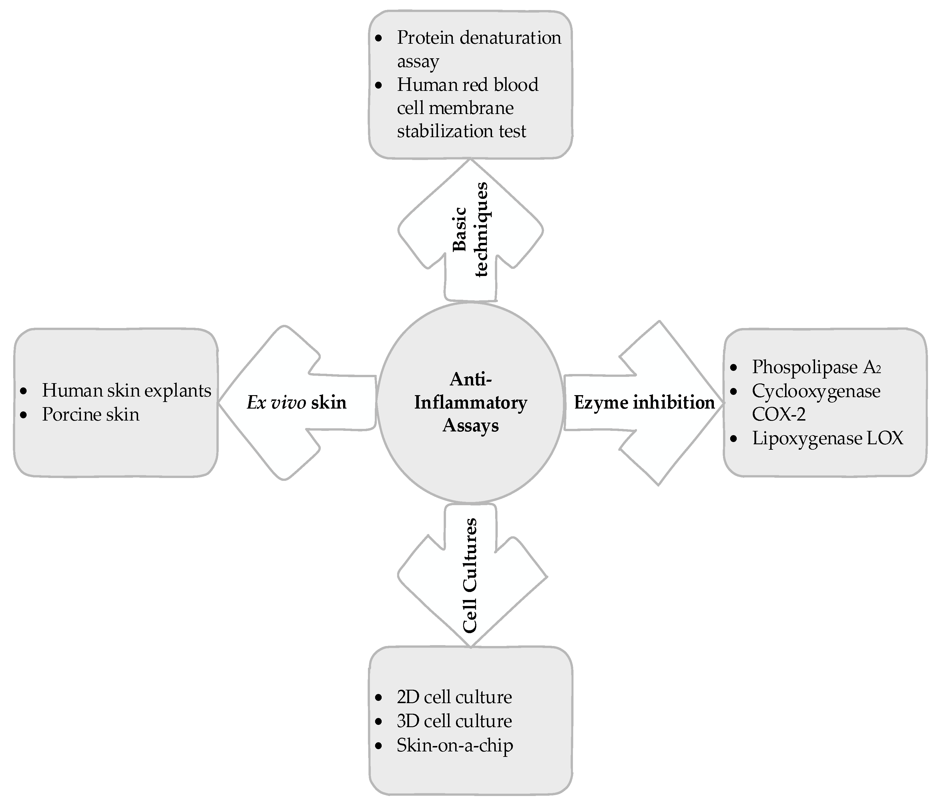
Inflammation itself is a source of discomfort and a major cause of the pathophysiological processes involved in the development and progression of many diseases. There are several models to test the anti-inflammatory effects of drugs. In vivo testing is the most reliable means to evaluate the anti-inflammatory effects of topically applied agents. Historically, animal testing has been considered ethical because of its benefits, but the form or method by which these benefits are obtained has not always been evaluated. It has been scientifically proven that animals are sensitive, meaning they can feel pain and experience both positive and negative emotional states of which they are aware because they have a limbic system that allows them to generate emotions and sensations [39]. This has led to measures being taken to limit the potential harm due to experimental use and to apply rules to ensure their rights.
Russell and Burch [40] published a guide to pain relief in animals. Since then, the concept of “applying humane techniques to animal research” has been defined as the principle of the three Rs: Replacement, Reduction, and Refinement.
Specifically, replacement refers to substituting the use of animals for in silico and in vitro techniques with the use of tissues of ethical origin or, in part, with the use of protozoa, insects, and nematodes. When the use of animals in experiments is essential, reduction refers to collecting enough data to answer research questions with a minimal number of animals, while refinement means finding less invasive experimental procedures to reduce pain, distress, and discomfort and improve animal welfare from birth to death [41].
The three ‘R’ principles have become ethical guidelines for research aimed at reducing harm to animals and have been incorporated into the legislation of several countries.
Currently, many countries, including Mexico, have banned the use of laboratory animals for cosmetic purposes. Legal restrictions and ethical considerations in animal testing require the development and evaluation of in vitro and ex vivo models. Here are some of them that can be considered before deciding to test on animals (Figure 3).

5. Basic Techniques

5.1. Protein Denaturation Assay

Denaturation of tissue proteins is one of the causes of inflammatory and arthritic diseases leading to the formation of autoantigens [42].
Several authors describe the methodology for evaluating the anti-inflammatory effect of topical formulations using this technique [43,44]. Babu and Noor [45] investigated the anti-inflammatory potential of the peptide/polypeptide fraction of Aloe vera. In this study, homogenized aloe gel was found to inhibit heat-induced protein denaturation with an IC50 value of (218.9 ± 15.6) μg/mL. Its anti-inflammatory potential was confirmed under inflammatory conditions in vivo using a rat paw edema model.
The test consists of measuring the degree of inhibition of denaturation of proteins, generally albumin, by a rise in temperature. Albumin stability is monitored by measuring absorbance at 660 nm and comparing with a negative control [46]. Reference molecules (diclofenac, ibuprofen) can also be included to obtain an optimal degree of inhibition.
Therefore, the protein denaturation assay is used as a simple screening test for anti-inflammatory drugs. The protein-centered hypothesis has some inconsistencies. For example, the denaturation-inhibiting effect occurs at relatively higher concentrations than at anti-inflammatory concentrations. In addition, anti-denaturing drugs may not have an anti-inflammatory effect [47].

5.2. Human Red Blood Cell Membrane Stabilization Test

The erythrocyte membrane is analogous to the lysosomal membrane. Stabilization of the lysosomal membrane is important for limiting the inflammatory response by inhibiting the release of lysosomal components from activated neutrophils. Damage to the lysosomal membrane often triggers the release of phospholipase A2, which is involved in the hydrolysis of phospholipids and produces inflammatory mediators [48]. Inhibition of erythrocyte membrane degradation is a measure of a molecule’s anti-inflammatory activity [49].
Hemolysis of erythrocytes can be induced by a hypotonic solution or by heat. Xiao et al. [50] reported the anti-inflammatory potential of Cinnamomum camphora essential oil. The nanoemulsion of the essential oil inhibited heat-induced hemolysis of erythrocytes (the half maximal inhibitory concentration IC 50 = 5.29 mg/mL) and hypotonic solution-induced hemolysis of erythrocytes (IC 50 = 0.26 mg/mL) and showed high skin permeability in vitro, which seemed to enhance its topical anti-inflammatory effect. Finally, its anti-inflammatory potential was demonstrated in a mouse model of acute inflammation (xylene-induced ear edema).
Paul et al. [51] reported that Aloe vera gel homogenate shows anti-inflammatory activity through lysosomal membrane stabilization. Aloe vera gel inhibits red cell membrane lysis induced by hypotension 74.89 ± 1.26% and heat 20.86 ± 0.77%, respectively, at a concentration of 1000 μg/mL compared with the indomethacin standard 80.52 ± 0.65% and 43.98 ± 1.52% at a concentration of 200 µg/mL, respectively. A possible explanation for the membrane stabilizing activity of plant extracts could be an increase in the cell surface area/volume ratio or by a stabilization of skeletal proteins such as tropomyosin.

6. Enzyme Inhibition

6.1. Phospholipase A2 Inhibitory Activity

PLA2 enzymes are divided into distinct classes based on cellular location, molecular weight, disulfide bond pattern, calcium dependence, sequence, and pH of activity. Secreted, cytosolic, lipoprotein-associated and Ca-independent PLA2 (sPLA2, cPLA2, lpPLA2 and iPLA2, respectively) are highlighted.
PLA2 enzymes are lipolytic enzymes that catalyze the hydrolysis of sn-2 ester bonds in various phospholipids, releasing arachidonic acid. Free arachidonic acid serves as a precursor molecule for eicosanoids [52].
Inflammatory/allergic skin diseases, particularly atopic dermatitis, contact dermatitis, and psoriasis, are associated with elevated levels of proinflammatory eicosanoids, suggesting increased PLA2 activity. It has long been known that inhibition of the release of arachidonic acid from membrane glycerophospholipids catalyzed by phospholipase A2 can block the synthesis of all eicosanoids [52].
PLA2 inhibitors have recently emerged as potential therapeutic targets for psoriasis. As mentioned earlier, sPLA2 is a general term for a group of enzymes that includes many different subtypes. Therefore, further studies on the role of sPLA2 in pathogenesis are needed to select the appropriate subtype to target to accelerate the development of innovative drugs [53].
Thibane et al. [54] studied the modulation of the activity of extracts from some medicinal plants used for skin care and beauty. The assay measured free 5-thio-2-nitrobenzoic acid after hydrolysis of diheptanoylthio-phosphatidylcholine by sPLA2. Aqueous extracts of Cassipourea flanaganii showed remarkable inhibitory activity against sPLA2 with an IC value 50 of 12.34 µg/mL compared to other aqueous extracts, however, these results were not lower than those of the positive control.

6.2. COX-2 Inhibitory Activity

The enzyme COX is responsible for the conversion of arachidonic acid into prostaglandins, which are secondary signaling molecules. It has two isoforms: cyclooxygenase-1 (COX-1), which is constitutively expressed in the skin, and COX-2, which is produced after stimulation with cytokines and mitogens and is mainly located in suprabasal keratinocytes. It is known that COX-2 is involved in the inflammatory processes of the skin through the production of prostaglandins and thromboxanes [55,56]. Targeting prostaglandin production by inhibiting the rate-limiting enzyme COX-2 is an option for effective treatment of inflammatory skin diseases [56].
Drugs with a lower inhibitory effect against COX-1 are recommended based on the reported beneficial effects associated with COX-1 activity [57]. Recently, many studies have been conducted to find new selective COX-2 inhibitors with safety profiles.
Thibane et al. [54] found that the activity of COX-2 was selectively inhibited by extracts of Cassipourea flanaganii, Plantago lanceolata, and Rorippa capuchina-aquaticum. The assay measures the peroxidase activity of ovine COX-1 and human COX-2, by monitoring the appearance of oxidized N,N,N′,N′-tetramethyl-p-phenylenediamine.
In other studies, Kola-Mustapha and Khalid-Salako [58] reported the selective COX-2 inhibition of emulgels containing ethanolic extracts of Cola millenii and Chandrakanthan et al. [59] in essential oils of Alpinia calcarata.

6.3. Lipoxygenase (LOX) Inhibitory Activity

Lipoxygenases are dioxygenases that catalyze the formation of hydroperoxides and leukotrienes from polyunsaturated fatty acids such as linoleic acid and arachidonic acid.
LOX Enzymes are expressed in immune cells, epithelial cells, and tumor cells, which have a variety of physiological functions, including inflammation, skin disease, and tumor formation [60]. Lipoxygenases are divided into 5-, 8-, 12-, and 15-lipoxygenases based on their selectivity for oxygenation of fatty acids at a specific position [61].
The 15-lipoxygenase metabolite 15-(S)-HETE was found to increase the degradation of IκBα, allowing nuclear translocation of NF-κB subunits in the progression of arthritis, and may be involved in the inflammatory effect induced by TNF-α and IL-1β. Therefore, 15-LOX is a good target for drug development for the treatment of arthritis [62]. On the other hand, leukotrienes (LT), synthesized by 5-LOX, are thought to play an important role in inflammatory skin diseases such as psoriasis and atopic dermatitis [63].
Appropriate 5-lipoxygenase inhibitors can be used to prevent and ameliorate symptoms of leukotriene-related inflammatory diseases. The study by Todaa et al. [64] shows that proanthocinidide from red rice inhibits the activity of 5-LOX in vitro and decreases the synthesis of leukotrione LTB4 in psoriasis-like mouse skin, with an IC 50 of 15.1 μM.

7. Cell Cultures

7.1. Two-Dimensional Cell Culture

Culturing single cell types, be they immune cells or keratinocytes, can be considered the simplest in vitro model for analyzing the cellular phenomena involved in the epidermal inflammatory disease process. Of course, these models do not take into account the complex structures and interactions within the tissue, but they have the great advantage of being able to accurately assess the response of specific cells to a given stimulus [65]. Cell cultures may be of the primary type if the cells are obtained directly from living tissue and then cultured. These cells have a short survival time and are therefore of limited scientific use. However, there are many commercially available immortalized cell lines that have been modified to survive indefinitely with continued cell division. Cancer cells are an example of the type of cells that can be used to create immortalized cell lines due to their genetic mutations and ability to continuously divide [66].
The work of Carola et al. [67] is a clear example of a study of topical anti-inflammation using single cell culture techniques with a focus on atopic dermatitis. In this study, they tested the anti-inflammatory effect of a cornflower extract containing N-feruloylserotonin. They used the human keratinocyte cell line (HaCaT) stimulated by the chemokines CCL17 and CCL22, which upregulates the transcription of inflammatory biomolecules such as IL-18, the chemokine CCL26, the chemokine CCL5, and matrix metalloproteinase 2 (MMP-2) were stimulated, the extract significantly reduced gene expression of these inflammatory biomarkers. The same authors used a human monocyte cell line (THP-1) differentiated into macrophages and stimulated with lipopolysaccharide (LPS) for the production of COX-2 and LOX, which showed inhibition of the enzymes by the extracts.
Monocytes are circulating bone marrow-derived progenitor cells that can transform into macrophages or dendritic cells as they migrate from blood to tissue. THP-1 and U937 are the most commonly used monocyte cell lines and can differentiate into various types of macrophages or dendritic cells in vitro. The fundamental difference between U937 and THP-1 cells is their origin and maturation stage. U937 cells originate from tissue and are therefore at a more mature stage, whereas THP-1 cells originate from blood leukemia and are at a less mature stage [68].
Varma et al. [69] investigated the anti-inflammatory and skin-protective effects of virgin coconut oil in the same cell cultures. In THP-1 cells stimulated with LPS, TNF-α, IFN-γ, IL-6, IL-8, and IL-5 were inhibited, whereas in HaCaT culture, the expression of aquaporin-3 (AQP3), involucrin (INV) and filaggrin (FLG) were increased, in addition to the photoprotective effects.
Another commonly used cell culture for anti-inflammatory studies is RAW 264.7 from LPS-stimulated murine macrophages, which was used by Mokdad et al. [70] to demonstrate the anti-inflammatory effects of free and liposome encapsulated thermal water on skin. A significant reduction in the production of nitric oxide (NO) and a modulation of the production of TNF-α were observed.
Although single cell cultures are a simple and useful research material, they do not take into account the cell communication that normally exists in the skin, which is why several authors have performed 2D cocultures of cell lines to better understand the etiology of inflammatory diseases in the skin.
Wu et al. [71] established a coculture of HaCaT and U937 cell lines to find bromodomain and extra-terminal domain (BET) antagonists for inhibition of skin inflammatory genes. Effective BET inhibitors were tested in a mouse model of imiquimod-induced psoriasiform dermatitis. This in vitro selection accurately predicted therapeutic efficacy in vivo.

7.2. Three-Dimensional Cell Culture

Today, most cells are grown using two-dimensional (2D) methods, but new and improved methods for performing 3D cell culture provide compelling evidence that more advanced experiments can be performed that provide valuable insights. By performing cell culture experiments in 3D, the cell environment can be manipulated to mimic the cell environment in vivo and provide more accurate data on cell interactions [72].
The properties of the human epidermis can be mimicked by two models: the reconstructed human epidermis (RHE) and the full thickness human skin equivalent (FTSE). Both models reproduce the in vivo properties of human skin in terms of epidermis morphology, differentiation, and barrier function. RHE includes only the epidermis, as only keratinocytes are grown at the air-liquid interface. FTSE has both the epidermis and dermis. In FTSE models, keratinocytes are seeded onto dermis-like structures such as type I collagen gel with fibroblasts, de-epidermized dermis (DED), or self-assembled dermis sheets. To mimic immune responses in skin, the simplest method is to add relevant cytokines to the 3D skin equivalents, or immune cells can be co-cultured with 3D skin for more physiological models [73,74].
These models are used to evaluate chemicals and other constituents, such as skin irritation, corrosion, absorption, and penetration, and some of them have been commercialized: EpiCS ® (CellSystems, Troisdorf, Germany), Epiderm™ (MatTek, Campbell, CA, USA) and SkinEthic™ RHE (L’Oreal, Lyon, France). They are validated and standardized by the European Center for the Evaluation of Alternative Methods (ECVCAM) and the Organization for Economic Cooperation and Development (OECD) [75].
Recently, RHE and FTSE cell cultures have gained great interest in drug discovery, testing, and validation [76]. For example, there are commercially available 3D in vitro models of psoriatic skin, two of which are commonly used: Creative Bioarray and MatTek Corporation’s model. The MatTek Corporation psoriasis model can also be used to test dermatological products, as it is a 3D model composed of psoriatic fibroblasts and normal human epidermal keratinocytes [66].
Clarysse et al. [77] developed 3D skin equivalents for atopic dermatitis and psoriasis. To induce dermatitis, 3D skin equivalents were stimulated with recombinant human IL-4 and IL-13 and for psoriasis by incubation with IL-17A, IL-22, and TNFα. Cultures were treated with tofacitinib, a Jano-kinase (JAK1/3) inhibitor. Epidermal morphology and filaggrin expression were restored in the atopic dermatitis model. Similarly, tofacitinib suppressed IL-20 and IL-1β expression in the psoriasis model.
Van den Bogaard et al. [78] developed an in vitro 3D model of keratinocytes and activated CD4+ T cells as well as polarized Th1 and Th17 T cells to study inflammatory skin diseases. In this model, clinically used drugs targeting crosstalk between keratinocytes and T cells successfully reduced the inflammatory phenotype. The authors therefore suggest that the model may serve as a very suitable platform for drug screening in a preclinical setting.
The change in the composition of the microbiome can lead to a change in the reactivity of the immune system and subsequently to the development of inflammatory diseases. For this reason, the development of a novel model involving the microbiome in the pathogenesis of inflammatory skin diseases has attracted considerable interest in recent years and several companies are developing microbiome-based topical skin therapies [79].
Since conventional monolayer cultures lack the stratum corneum, bacteria come into direct contact with keratinocytes, which is not normally the case. Therefore, 3D models and ex vivo explants are preferred to study host-microbiome interactions [80].
A study of methicillin-resistant S. aureus wound infection after thermal injury in an FTSE model showed significant growth of S. aureus after 24–48 h. Skin exposure to S. aureus increased the expression of inflammatory mediators such as TLR2, IL-6, and IL-8, as well as antimicrobial proteins [81].

7.3. Skin on a Chip

The vasculature plays an important role in mediating inflammatory responses because it is the pathway by which immune cells migrate between lymphoid organs and peripheral tissues. Organs-on-a-chip are microfluidic cultures that mimic the physiological cycle at the organ level [74]. With these technological advances, skin-on-a-chip has been developed, which consists of culturing skin tissue in a microfluidic system that more accurately replicates the physiology of the skin than static and conventional 3D culture, and allows better control of the cellular microenvironment [82,83].
Skin-on-a-chip devices have numerous biomedical applications, including: skin disease modeling, drug screening, cosmetic development, skin health monitoring sensor development, and intra/transdermal drug and cosmetic delivery efficiency studies [84].
Risueño et al. [82] point out that devices can be classified according to how the skin on the chip was generated: In the first method, a piece of skin from a biopsy or an equivalent of human skin is inserted directly into the chip (transferred-skin-on-a-chip); in the second method, the tissue is generated directly on the chip (in situ-skin-on-a-chip).
Wufuer et al. [85] built a skin-on-a-chip device to simulate skin inflammation and edema. The model consists of three layers: HaCaT in the epidermis, fibroblasts in the dermis, and human umbilical vein endothelial cells (HUVECs) for the endothelium. Each skin layer can be supplied with different culture media and different flow rates. Each layer is separated by a porous membrane coated with fibronectin to simulate the extracellular matrix and allow communication between cells. Inflammation and edema were induced by TNF-α. Administration of dexamethasone resulted in a decrease in TNF-α-induced proinflammatory factors (IL-β, IL-6, and IL-8) and a decrease in edema, as shown by the results of permeability and tight junction staining. These experiments simulated the use of ointments to treat inflammation and edema, so the device has the potential to be used for drug screening and as a replacement for animal testing.
In another study, Biglari et al. [86] developed a skin-on-a-chip device to simulate inflammation in a wound. The device consists of three channels. HUVEC cells formed a 3D vascular structure in the center, while fibroblasts and macrophages formed a 2D monolayer in the side channels and migrated slightly into the center channel. Inflammation was induced with TNF-α, and then the interaction with M1 and M2 macrophages was examined. Administration of an anti-inflammatory drug (dexamethasone) improved endothelial cell binding and decreased expression of the proinflammatory cytokines IL-1β, IL-6, and IL-8. The device has the potential to aid in the search for effective therapeutics for inflammation and is a promising tool for future preclinical investigation.

8. Ex Vivo Skin Tests

8.1. Human Skin

The human skin explant from plastic surgery is a suitable model for studying the effects of products applied to the skin. It is important to preserve the structure of the native skin and dermal matrix so that the effects of the applied molecules are similar to the in vivo responses of human skin [87].
Neil et al. [88] describe a structurally viable and metabolically active human skin explant model for 9 days. This culture can be used for preclinical testing of skin treatment delivery and efficacy. In another study, Neil et al. [89] developed an ex vivo skin culture model that maintained skin barrier integrity and metabolic activity for 5 days and was stimulated with cytokines to induce a Th1-mediated inflammatory state. The effects of pimecrolimus and clobetasol propionate in models of inflammatory dermatoses were evaluated and compared with an in vivo clinical study that showed similar reductions in inflammatory gene expression with both drugs.
Anitua et al. [90] studied the anti-inflammatory effects of formulations of growth factor-rich plasma (PRGF) and autologous topical serum (ATS) in a human skin model stimulated ex vivo with IL-4 and IL-13. The treatments increased tissue viability and decreased free radical accumulation and cutaneous production of cytokines such as TNF-α and IL-1β.
In another study, the anti-inflammatory effect of a complex of bakuchiol, Ginkgo biloba extract, and mannitol was demonstrated ex vivo by a decrease in IL-8 and TNF-α by −45% and −46%, p < 0.01, respectively, in human skin explants exposed to the lyophilized strain of Propionibacterium acnes and to which the cream containing the complex was applied twice daily for 3 days [91].

8.2. Pig Skin

Pig skin is very similar to human skin. The validity of the pig skin model is supported by numerous articles comparing it to human skin in terms of permeability to foreign substances, architecture, and immunology [92,93].
Authors such as Heard [7] and Hwang et al. [94], believe that the use of pig skin for ex vivo testing is consistent with the 3Rs (Reduction, Refinement, and Replacement) philosophy because pig skin can be obtained inexpensively as a byproduct from animals already slaughtered for food purposes at local abattoirs, so no additional animals would need to be sacrificed for research.
Freshly excised pig skin contains elevated levels of inducible short-lived COX-2. Under viable conditions, COX-2 and its eicosanoid products continue to be produced until tissue necrosis occurs, so that the anti-inflammatory or pro-inflammatory activity of the compounds can be evaluated. It is important to emphasize that due to the death of the pig and the gross processing of the ear tissue, a state of inflammation already occurs before the experiment, so no prior induction treatment is required [7].
Ex vivo porcine skin models have been used by several researchers. Ouitas and Heard [95] tested the anti-inflammatory (COX-2) potential of six commercial formulations of devil’s claw in an ex vivo porcine skin model. Houston et al. [96] used an ex vivo porcine skin model to evaluate the anti-inflammatory (COX-2) potential of pomegranate peel extracts. Hwang et al. [94] studied the ex vivo viable pig skin model to evaluate the safety and skin barrier-improving effects of hydroxy acids, which are commonly used in exfoliating cosmetics.

9. Perspectives

Advances in in vitro and ex vivo models to study the anti-inflammatory effects of topical drugs will ultimately reduce animal testing, accelerate drug discovery, and can be used to screen new or unproven chemicals that may exhibit skin toxicity.
Skin-on-chip devices show promise in replicating the complex physiological interactions in the human body. However, systematic characterization and validation is still required before they can be used as sensitive in vitro platforms for drug testing [83].
It remains a challenge to mimic the complexity of human skin, including vascularity, immunity, and the presence of appendages. Currently, models of human skin are being developed using specific cell types such as keratinocytes and fibroblasts, as many skin cells are difficult to isolate and culture. Induced pluripotent stem cells can differentiate into almost all skin cell types and could be used to develop skin models with relevant components such as immune cells and appendages [97].
Miyake and Shimada [98] describe the protocol for generating a 3D culture with keratinocytes derived from induced pluripotent stem cells. Models have also been developed with melanocytes [99], endothelial cells [100], skin sensory neurons [101] and adipocytes [102].
Models based on induced pluripotent stem cells are a great innovation and allow for cell versatility as opposed to traditional cell isolation and culturing techniques. However, targeted differentiation of cells remains a challenge, and generation of a variety of immune cells for use in in vitro skin models may not be realistic [76].
In addition to the skin barrier, the microbiome is also of great importance for skin homeostasis. Disruption of this barrier can lead to a persistent inflammatory state and poor response to pathogens. This alteration can lead to inflammatory skin diseases such as psoriasis and atopic dermatitis [75,103].
Currently, there are very few studies on the interactions between microorganisms and 3D skin models. Furthermore, only the effects of individual microorganisms have been studied, which do not truly resemble the full interaction of the human skin microbiome; future studies should eventually allow for modulation and analysis of the entire microbiome in vitro [75]. Identification of the bacterial markers associated with inflammatory skin diseases in particular, would greatly expedite the diagnostic process and reduce the cost of treatment. The study and determination of the microbiota could lead to the identification of possible causes of skin diseases that have remained undetected by simpler methods [36].
Ex vivo models can in some cases more accurately represent human skin in terms of structure, cell signaling mechanisms, metabolism, coherence of skin markings, and absorption effects. However, they are hampered by donor diversity, availability, and biological limitations [88]. Although the anatomy, physiology, and immune systems of human and porcine skin share many similarities, future studies, such as transcriptomics, are needed to confirm the similarities between the cell populations. Advances in the study of porcine skin immunology will allow the development of a wider range of models than are currently used [104]. Table 1 summarizes the advantages and limitations of each technique.

10. Conclusions

The studies discussed here show a broad panorama for searching and evaluating the topical anti-inflammatory effects of both herbal and synthetic molecules at different levels of the inflammatory process. Although models are becoming more robust and specific, the skin inflammatory process cannot be fully reproduced at present. There is a clear need to develop models that cover complex functions of human skin such as immune cell integration and the microbiome. This could provide an alternative to in vivo testing, which is limited due to ethical issues as well as the discrepancy between human and animal responses. Existing studies show potential for developing a definitive model representing the inflammatory process. Despite their imperfections, they have an important role in the study of topical anti-inflammatory drugs.

Author Contributions

Conceptualization, J.A.G.-I. and M.R.M.-J.; investigation, J.L.P.-S., N.E.R.-G., R.F.G.-L. and L.M.-T.; resources, J.A.G.-I. and M.R.M.-J.; writing—original draft preparation, J.L.P.-S.; writing—review and editing, J.A.G.-I. and R.F.G.-L.; visualization, J.L.P.-S. and M.R.M.-J.; supervision, J.A.G.-I.; project administration M.R.M.-J.; funding acquisition, J.A.G.-I. and N.E.R.-G. All authors have read and agreed to the published version of the manuscript.

Funding

This research was funded by TecNM, grant number 13987.22-P.

Data Availability Statement

Not applicable.

Acknowledgments

J.L.P.-S. acknowledge the Conacyt scholarship for doctoral studies.

Conflicts of Interest

The authors declare no conflict of interest.

References

  1. García Barreno, P. Inflamación. Cienc. Exact. Fís. Nat. 2008, 102, 91–159. Available online: http://www.rac.es/ficheros/doc/00681.pdf (accessed on 21 January 2023).
  2. Kindt, T.J.; Goldsby, R.A.; Osborne, B.A.; Kuby, J. Kuby Immunology, 6th ed.; W.H. Freeman: New York, NY, USA, 2007; pp. 350–356. [Google Scholar]
  3. Bordbar-Khiabani, A.; Yarmand, B.; Sharifi-Asl, S.; Mozafari, M. Improved Corrosion Performance of Biodegradable Magnesium in Simulated Inflammatory Condition via Drug-Loaded Plasma Electrolytic Oxidation Coatings. Mater. Chem. Phys. 2020, 239, 122003. [Google Scholar] [CrossRef]
  4. Bordbar-Khiabani, A.; Bahrampour, S.; Mozafari, M.; Gasik, M. Surface Functionalization of Anodized Tantalum with Mn3O4 Nanoparticles for Effective Corrosion Protection in Simulated Inflammatory Condition. Ceram. Int. 2022, 48, 3148–3156. [Google Scholar] [CrossRef]
  5. Lenz, A.; Franklin, G.A.; Cheadle, W.G. Systemic Inflammation after Trauma. Injury 2007, 38, 1336–1345. [Google Scholar] [CrossRef] [PubMed]
  6. Eze, F.I.; Uzor, P.F.; Ikechukwu, P.; Obi, B.C.; Osadebe, P.O. In Vitro and In Vivo Models for Anti-Inflammation: An Evaluative Review. ITPS 2019, 2, 3–15. [Google Scholar] [CrossRef] [Green Version]
  7. Heard, C.M. An Ex Vivo Skin Model to Probe Modulation of Local Cutaneous Arachidonic Acid Inflammation Pathway. J. Biol. Methods 2020, 7, e138. [Google Scholar] [CrossRef]
  8. Richardson, M. Understanding the Structure and Function of the Skin. Nurs. Times 2003, 99, 46–48. [Google Scholar] [PubMed]
  9. Gupta, M.; Agrawal, U.; Vyas, S.P. Nanocarrier-Based Topical Drug Delivery for the Treatment of Skin Diseases. Expert Opin. Drug Deliv. 2012, 9, 783–804. [Google Scholar] [CrossRef] [PubMed]
  10. Cruz, M.S.; Diamond, A.; Russell, A.; Jameson, J.M. Human Aβ and Γδ T Cells in Skin Immunity and Disease. Front. Immunol. 2018, 9, 1304. [Google Scholar] [CrossRef]
  11. Nestle, F.O.; Di Meglio, P.; Qin, J.Z.; Nickoloff, B.J. Skin Immune Sentinels in Health and Disease. Nat. Rev. Immunol. 2009, 9, 679–691. [Google Scholar] [CrossRef] [PubMed] [Green Version]
  12. Proksch, E.; Brandner, J.M.; Jensen, J.M. The Skin: An Indispensable Barrier. Exp. Dermatol. 2008, 17, 1063–1072. [Google Scholar] [CrossRef]
  13. Yuki, T.; Tobiishi, M.; Kusaka-Kikushima, A.; Ota, Y.; Tokura, Y. Impaired Tight Junctions in Atopic Dermatitis Skin and in a Skin-Equivalent Model Treated with Interleukin-17. PLoS ONE 2016, 11, e0161759. [Google Scholar] [CrossRef] [Green Version]
  14. Brown, T.M.; Krishnamurthy, K. Histology, Dermis. In StatPearls; StatPearls Publishing: Treasure Island, FL, USA, 2022. [Google Scholar]
  15. Juráňová, J.; Franková, J.; Ulrichová, J. The Role of Keratinocytes in Inflammation. J. Appl. Biomed. 2017, 15, 169–179. [Google Scholar] [CrossRef]
  16. Pasparakis, M.; Haase, I.; Nestle, F.O. Mechanisms Regulating Skin Immunity and Inflammation. Nat. Rev. Immunol. 2014, 14, 289–301. [Google Scholar] [CrossRef] [PubMed]
  17. Di Meglio, P.; Perera, G.K.; Nestle, F.O. The Multitasking Organ: Recent Insights into Skin Immune Function. Immunity 2011, 35, 857–869. [Google Scholar] [CrossRef] [PubMed] [Green Version]
  18. Balato, A.; Cacciapuoti, S.; Di Caprio, R.; Marasca, C.; Masarà, A.; Raimondo, A.; Fabbrocini, G. Human Microbiome: Composition and Role in Inflammatory Skin Diseases. Arch. Immunol. Ther. Exp. 2019, 67, 1–18. [Google Scholar] [CrossRef] [PubMed]
  19. Frevert, C.W.; Felgenhauer, J.; Wygrecka, M.; Nastase, M.V.; Schaefer, L. Danger-Associated Molecular Patterns Derived From the Extracellular Matrix Provide Temporal Control of Innate Immunity. J. Histochem. Cytochem. 2018, 66, 213–227. [Google Scholar] [CrossRef] [PubMed] [Green Version]
  20. Hayden, M.S.; Ghosh, S. NF-κB in Immunobiology. Cell Res. 2011, 21, 223–244. [Google Scholar] [CrossRef] [PubMed] [Green Version]
  21. Garlanda, C.; Riva, F.; Bonavita, E.; Gentile, S.; Mantovani, A. Decoys and Regulatory “Receptors” of the IL-1/Toll-Like Receptor Superfamily. Front. Immunol. 2013, 4, 180. [Google Scholar] [CrossRef] [Green Version]
  22. Pobezinskaya, Y.L.; Liu, Z. The Role of TRADD in Death Receptor Signaling. Cell Cycle 2012, 11, 871–876. [Google Scholar] [CrossRef] [PubMed] [Green Version]
  23. Montaño Estrada, L.D.; Fortoul Van der Goes, T.I.; Rendón Huerta, E.P. ¿Qué son los inflamosomas? El NLRP3 como ejemplo. Rev. Fac. Med. UNAM 2016, 60, 42–49. [Google Scholar]
  24. Piipponen, M.; Li, D.; Landén, N.X. The Immune Functions of Keratinocytes in Skin Wound Healing. Int. J. Mol. Sci. 2020, 21, 8790. [Google Scholar] [CrossRef] [PubMed]
  25. Ellis, S.; Lin, E.J.; Tartar, D. Immunology of Wound Healing. Curr. Derm. Rep. 2018, 7, 350–358. [Google Scholar] [CrossRef] [PubMed] [Green Version]
  26. Tomura, M.; Hata, A.; Matsuoka, S.; Shand, F.H.W.; Nakanishi, Y.; Ikebuchi, R.; Ueha, S.; Tsutsui, H.; Inaba, K.; Matsushima, K.; et al. Tracking and Quantification of Dendritic Cell Migration and Antigen Trafficking between the Skin and Lymph Nodes. Sci. Rep. 2014, 4, 6030. [Google Scholar] [CrossRef] [Green Version]
  27. Deckers, J.; Hammad, H.; Hoste, E. Langerhans Cells: Sensing the Environment in Health and Disease. Front. Immunol. 2018, 9, 93. [Google Scholar] [CrossRef] [PubMed] [Green Version]
  28. Mills, C.D. Anatomy of a Discovery: M1 and M2 Macrophages. Front. Immunol. 2015, 6, 212. [Google Scholar] [CrossRef] [PubMed]
  29. Nguyen, A.V.; Soulika, A.M. The Dynamics of the Skin’s Immune System. Int. J. Mol. Sci. 2019, 20, 1811. [Google Scholar] [CrossRef] [Green Version]
  30. Kortekaas Krohn, I.; Aerts, J.L.; Breckpot, K.; Goyvaerts, C.; Knol, E.; Van Wijk, F.; Gutermuth, J. T-cell Subsets in the Skin and Their Role in Inflammatory Skin Disorders. Allergy 2022, 77, 827–842. [Google Scholar] [CrossRef]
  31. Seidel, J.A.; Vukmanovic-Stejic, M.; Muller-Durovic, B.; Patel, N.; Fuentes-Duculan, J.; Henson, S.M.; Krueger, J.G.; Rustin, M.H.A.; Nestle, F.O.; Lacy, K.E.; et al. Skin Resident Memory CD8+ T Cells Are Phenotypically and Functionally Distinct from Circulating Populations and Lack Immediate Cytotoxic Function. Clin. Exp. Immunol. 2018, 194, 79–92. [Google Scholar] [CrossRef] [Green Version]
  32. Matos, T.R.; O’Malley, J.T.; Lowry, E.L.; Hamm, D.; Kirsch, I.R.; Robins, H.S.; Kupper, T.S.; Krueger, J.G.; Clark, R.A. Clinically Resolved Psoriatic Lesions Contain Psoriasis-Specific IL-17–Producing Aβ T Cell Clones. J. Clin. Investig. 2017, 127, 4031–4041. [Google Scholar] [CrossRef] [Green Version]
  33. Cai, Y.; Shen, X.; Ding, C.; Qi, C.; Li, K.; Li, X.; Jala, V.R.; Zhang, H.; Wang, T.; Zheng, J.; et al. Pivotal Role of Dermal IL-17-Producing Γδ T Cells in Skin Inflammation. Immunity 2011, 35, 596–610. [Google Scholar] [CrossRef] [Green Version]
  34. Grice, E.A.; Kong, H.H.; Conlan, S.; Deming, C.B.; Davis, J.; Young, A.C.; Bouffard, G.G.; Blakesley, R.W.; Murray, P.R.; Green, E.D.; et al. Topographical and Temporal Diversity of the Human Skin Microbiome. Science 2009, 324, 1190–1192. [Google Scholar] [CrossRef] [PubMed] [Green Version]
  35. Naik, S.; Bouladoux, N.; Wilhelm, C.; Molloy, M.J.; Salcedo, R.; Kastenmuller, W.; Deming, C.; Quinones, M.; Koo, L.; Conlan, S.; et al. Compartmentalized Control of Skin Immunity by Resident Commensals. Science 2012, 337, 1115–1119. [Google Scholar] [CrossRef] [PubMed] [Green Version]
  36. Ferček, I.; Lugović-Mihić, L.; Tambić-Andrašević, A.; Ćesić, D.; Grginić, A.G.; Bešlić, I.; Mravak-Stipetić, M.; Mihatov-Štefanović, I.; Buntić, A.-M.; Čivljak, R. Features of the Skin Microbiota in Common Inflammatory Skin Diseases. Life 2021, 11, 962. [Google Scholar] [CrossRef] [PubMed]
  37. Bourrain, M.; Ribet, V.; Calvez, A.; Lebaron, P.; Schmitt, A.-M. Balance between Beneficial Microflora and Staphylococcus Aureus Colonisation: In Vivo Evaluation in Patients with Atopic Dermatitis during Hydrotherapy. Eur. J. Dermatol. 2013, 23, 786–794. [Google Scholar] [CrossRef] [PubMed] [Green Version]
  38. Fahlén, A.; Engstrand, L.; Baker, B.S.; Powles, A.; Fry, L. Comparison of Bacterial Microbiota in Skin Biopsies from Normal and Psoriatic Skin. Arch. Dermatol. Res. 2012, 304, 15–22. [Google Scholar] [CrossRef] [PubMed]
  39. Heredia Antúnez, A.P.; Cantón, B.V.; Santillán Doherty, P. Retos de Los Comités de Ética En Investigación En Animales. Experiencia de México. Rev. Bioética Derecho 2021, 51, 99–121. [Google Scholar] [CrossRef]
  40. Russell, W.M.S.; Burch, R. The Principles of Humane Experimental Technique; Methuen: London, UK, 1959. [Google Scholar]
  41. Balls, M. It’s Time to Reconsider The Principles of Humane Experimental Technique. Altern. Lab. Anim. 2020, 48, 40–46. [Google Scholar] [CrossRef]
  42. Osman, N.; Sidik, N.; Awal, A.; Adam, N.; Rezali, N. In Vitro Xanthine Oxidase (XO) and Albumin Denaturation Inhibition Assay of Barringtonia racemosa L. and Total Phenolic Content Analysis for Potential Anti-Inflammatory Use in Gouty Arthritis. J. Intercult. Ethnopharmacol. 2016, 5, 343. [Google Scholar] [CrossRef]
  43. Samuel, A.J.; Mulla, N. Formulation and Evaluation of Herbal Topical Gel Containing Leaves Extract of Andrographis Paniculata. J. Drug Deliv. Ther. 2020, 10, 48–51. [Google Scholar] [CrossRef]
  44. Sarkar, B.K.; Arya, J.C.; Pal, S.; Dogra, P.; Atteri, S.; Patial, B.; Babu, G.; Meena, A.K. Preparation, standardization and evaluation of preliminary anti-inflammatory activity of herbal formulation of Citrullus colocynthis. J. Adv. Sci. Res. 2021, 12, 127–131. [Google Scholar]
  45. Babu, S.; Noor, A. Aloe Barbadensis Miller Peptide/Polypeptide Fraction Alleviates Inflammation through Inhibition of Proinflammatory Cytokines and Mediators in Vitro and in Rats with Freund’s Adjuvant-Induced Hind Paw Edema. Asian. Pac. J. Trop. Biomed. 2019, 9, 524. [Google Scholar] [CrossRef]
  46. Moni, J.N.R.; Adnan, M.; Tareq, A.M.; Kabir, M.I.; Reza, A.S.M.A.; Nasrin, M.S.; Chowdhury, K.H.; Sayem, S.A.J.; Rahman, M.A.; Alam, A.K.; et al. Therapeutic Potentials of Syzygium Fruticosum Fruit (Seed) Reflected into an Array of Pharmacological Assays and Prospective Receptors-Mediated Pathways. Life 2021, 11, 155. [Google Scholar] [CrossRef] [PubMed]
  47. Silvestrini, B.; Silvestrini, M. Medical Implications of the Relationships among Protein Denaturation, Necrosis and Inflammation: An Intriguing Story. In Tendons-Trauma, Inflammation, Degeneration, and Treatment; IntechOpen: London, UK, 2022. [Google Scholar] [CrossRef]
  48. García Candela, J.L.E.; Pariona Velarde, C.D.; Londoñe Bailon, R.P. Actividad antiinflamatoria in vitro de los polisacáridos sulfatados de Patallus mollis extraídos mediante digestión enzimática. Rev. Peru. Med. Integr. 2017, 2, 759–764. [Google Scholar] [CrossRef] [Green Version]
  49. Phanse, M.A. In-Vivo and in-Vitro Screening of Medicinal Plants for Their Anti-Inflammatory Activity: An Overview. J. Appl. Pharm. Sci. 2012, 2, 19–33. [Google Scholar] [CrossRef] [Green Version]
  50. Xiao, S.; Yu, H.; Xie, Y.; Guo, Y.; Fan, J.; Yao, W. The Anti-Inflammatory Potential of Cinnamomum Camphora (L.) J.Presl Essential Oil in Vitro and in Vivo. J. Ethnopharmacol. 2021, 267, 113516. [Google Scholar] [CrossRef] [PubMed]
  51. Paul, S.; Modak, D.; Chattaraj, S.; Nandi, D.; Sarkar, A.; Roy, J.; Chaudhuri, T.K.; Bhattacharjee, S. Aloe Vera Gel Homogenate Shows Anti-Inflammatory Activity through Lysosomal Membrane Stabilization and Downregulation of TNF-α and Cox-2 Gene Expressions in Inflammatory Arthritic Animals. Future J. Pharm. Sci. 2021, 7, 12. [Google Scholar] [CrossRef]
  52. Dan, P.; Rosenblat, G.; Yedgar, S. Phospholipase A2 Activities in Skin Physiology and Pathology. Eur. J. Pharmacol. 2012, 691, 1–8. [Google Scholar] [CrossRef]
  53. Shao, S.; Chen, J.; Swindell, W.R.; Tsoi, L.C.; Xing, X.; Ma, F.; Uppala, R.; Sarkar, M.K.; Plazyo, O.; Billi, A.C.; et al. Phospholipase A2 Enzymes Represent a Shared Pathogenic Pathway in Psoriasis and Pityriasis Rubra Pilaris. JCI Insight 2021, 6, e151911. [Google Scholar] [CrossRef] [PubMed]
  54. Thibane, V.S.; Ndhlala, A.R.; Finnie, J.F.; Van Staden, J. Modulation of the Enzyme Activity of Secretory Phospholipase A2, Lipoxygenase and Cyclooxygenase Involved in Inflammation and Disease by Extracts from Some Medicinal Plants Used for Skincare and Beauty. S. Afr. J. Bot. 2019, 120, 198–203. [Google Scholar] [CrossRef]
  55. Acheva, A.; Schettino, G.; Prise, K.M. Pro-Inflammatory Signaling in a 3D Organotypic Skin Model after Low LET Irradiation—NF-κB, COX-2 Activation, and Impact on Cell Differentiation. Front. Immunol. 2017, 8, 82. [Google Scholar] [CrossRef] [PubMed] [Green Version]
  56. Termer, M.; Carola, C.; Salazar, A.; Keck, C.M.; Hemberger, J.; von Hagen, J. Activity-Guided Characterization of COX-2 Inhibitory Compounds in Waltheria indica L. Extracts. Molecules 2021, 26, 7240. [Google Scholar] [CrossRef]
  57. Suleyman, H.; Demircan, B.; Karagoz, Y. Anti-inflammatory and side effects of cyclo-oxygenase inhibitors. Pharmacol. Rep. 2007, 59, 247. [Google Scholar] [PubMed]
  58. Kola-Mustapha, A.T.; Khalid-Salako, F.A. Herbal Emulgels Incorporated with Cola millenii K. Schum Stem Bark Ethanol Extract Potentially for the Management of Rheumatoid Arthritis in-Vitro. Phytomed. Plus 2021, 1, 100033. [Google Scholar] [CrossRef]
  59. Chandrakanthan, M.; Handunnetti, S.M.; Premakumara, G.S.A.; Kathirgamanathar, S. Topical Anti-Inflammatory Activity of Essential Oils of Alpinia calcarata Rosc., Its Main Constituents, and Possible Mechanism of Action. Evid. Based Complement. Altern. Med. 2020, 2020, 2035671. [Google Scholar] [CrossRef] [PubMed]
  60. Mashima, R.; Okuyama, T. The Role of Lipoxygenases in Pathophysiology; New Insights and Future Perspectives. Redox Biol. 2015, 6, 297–310. [Google Scholar] [CrossRef] [PubMed] [Green Version]
  61. Wisastra, R.; Dekker, F. Inflammation, Cancer and Oxidative Lipoxygenase Activity Are Intimately Linked. Cancers 2014, 6, 1500–1521. [Google Scholar] [CrossRef] [Green Version]
  62. Wu, M.Y.; Lin, T.H.; Chiu, Y.C.; Liou, H.C.; Yang, R.S.; Fu, W.M. Involvement of 15-Lipoxygenase in the Inflammatory Arthritis. J. Cell. Biochem. 2012, 113, 2279–2289. [Google Scholar] [CrossRef]
  63. Krieg, P.; Fürstenberger, G. The Role of Lipoxygenases in Epidermis. Biochim. Biophys. Acta Mol. Cell Biol. Lipids 2014, 1841, 390–400. [Google Scholar] [CrossRef]
  64. Toda, K.; Tsukayama, I.; Nagasaki, Y.; Konoike, Y.; Tamenobu, A.; Ganeko, N.; Ito, H.; Kawakami, Y.; Takahashi, Y.; Miki, Y.; et al. Red-Kerneled Rice Proanthocyanidin Inhibits Arachidonate 5-Lipoxygenase and Decreases Psoriasis-like Skin Inflammation. Arch. Biochem. Biophys. 2020, 689, 108307. [Google Scholar] [CrossRef]
  65. De Vuyst, E.; Salmon, M.; Evrard, C.; Lambert de Rouvroit, C.; Poumay, Y. Atopic Dermatitis Studies through In Vitro Models. Front. Med. 2017, 4, 119. [Google Scholar] [CrossRef] [Green Version]
  66. Teimouri, A.; Yeung, P.; Agu, R. 2D vs. 3D Cell Culture Models for In Vitro Topical (Dermatological) Medication Testing. In Cell Culture; Ali Mehanna, R., Ed.; IntechOpen: London, UK, 2019. [Google Scholar] [CrossRef] [Green Version]
  67. Carola, C.; Salazar, A.; Rakers, C.; Himbert, F.; Do, Q.T.; Bernard, P.; von Hagen, J. A Cornflower Extract Containing N-Feruloylserotonin Reduces Inflammation in Human Skin by Neutralizing CCL17 and CCL22 and Inhibiting COX-2 and 5-LOX. Mediat. Inflamm. 2021, 2021, 6652791. [Google Scholar] [CrossRef]
  68. Chanput, W.; Peters, V.; Wichers, H. THP-1 and U937 Cells. In The Impact of Food Bioactives on Health; Verhoeckx, K., Cotter, P., López-Expósito, I., Kleiveland, C., Lea, T., Mackie, A., Requena, T., Swiatecka, D., Wichers, H., Eds.; Springer International Publishing: Cham, Switzerland, 2015; pp. 147–159. [Google Scholar] [CrossRef] [Green Version]
  69. Varma, S.R.; Sivaprakasam, T.O.; Arumugam, I.; Dilip, N.; Raghuraman, M.; Pavan, K.B.; Rafiq, M.; Paramesh, R. In Vitro Anti-Inflammatory and Skin Protective Properties of Virgin Coconut Oil. J. Tradit. Complement. Med. 2019, 9, 5–14. [Google Scholar] [CrossRef]
  70. Mokdad, R.; Seguin, C.; Fournel, S.; Frisch, B.; Heurtault, B.; Hadjsadok, A. Anti-Inflammatory Effects of Free and Liposome-Encapsulated Algerian Thermal Waters in RAW 264.7 Macrophages. Int. J. Pharm. 2022, 614, 121452. [Google Scholar] [CrossRef]
  71. Wu, X.; Shi, Z.; Hsu, D.K.; Chong, J.; Huynh, M.; Mendoza, L.; Yamada, D.; Hwang, S.T. A Monocyte-Keratinocyte-Derived Co-Culture Assay Accurately Identifies Efficacies of BET Inhibitors as Therapeutic Candidates for Psoriasiform Dermatitis. J. Dermatol. Sci. 2020, 100, 31–38. [Google Scholar] [CrossRef]
  72. Jensen, C.; Teng, Y. Is It Time to Start Transitioning from 2D to 3D Cell Culture? Front. Mol. Biosci. 2020, 7, 33. [Google Scholar] [CrossRef] [Green Version]
  73. Griffoni, C.; Neidhart, B.; Yang, K.; Groeber-Becker, F.; Maniura-Weber, K.; Dandekar, T.; Walles, H.; Rottmar, M. In Vitro Skin Culture Media Influence the Viability and Inflammatory Response of Primary Macrophages. Sci. Rep. 2021, 11, 7070. [Google Scholar] [CrossRef] [PubMed]
  74. Moon, S.; Kim, D.H.; Shin, J.U. In Vitro Models Mimicking Immune Response in the Skin. Yonsei Med. J. 2021, 62, 969. [Google Scholar] [CrossRef]
  75. Niehues, H.; Bouwstra, J.A.; El Ghalbzouri, A.; Brandner, J.M.; Zeeuwen, P.L.J.M.; van den Bogaard, E.H. 3D Skin Models for 3R Research: The Potential of 3D Reconstructed Skin Models to Study Skin Barrier Function. Exp. Dermatol. 2018, 27, 501–511. [Google Scholar] [CrossRef] [PubMed] [Green Version]
  76. Hennies, H.C.; Poumay, Y. Skin Disease Models In Vitro and Inflammatory Mechanisms: Predictability for Drug Development. In Organotypic Models in Drug Development; Schäfer-Korting, M., Stuchi Maria-Engler, S., Landsiedel, R., Eds.; Handbook of Experimental Pharmacology; Springer International Publishing: Cham, Switzerland, 2021; Volume 265, pp. 187–218. [Google Scholar] [CrossRef]
  77. Clarysse, K.; Pfaff, C.M.; Marquardt, Y.; Huth, L.; Kortekaas Krohn, I.; Kluwig, D.; Lüscher, B.; Gutermuth, J.; Baron, J. JAK1/3 Inhibition Preserves Epidermal Morphology in Full-Thickness 3D Skin Models of Atopic Dermatitis and Psoriasis. J. Eur. Acad. Dermatol. Venereol. 2019, 33, 367–375. [Google Scholar] [CrossRef] [PubMed]
  78. van den Bogaard, E.H.; Tjabringa, G.S.; Joosten, I.; Vonk-Bergers, M.; van Rijssen, E.; Tijssen, H.J.; Erkens, M.; Schalkwijk, J.; Koenen, H.J.P.M. Crosstalk between Keratinocytes and T Cells in a 3D Microenvironment: A Model to Study Inflammatory Skin Diseases. J. Investig. Dermatol. 2014, 134, 719–727. [Google Scholar] [CrossRef] [Green Version]
  79. Schmidt, C. Out of Your Skin. Nat. Biotechnol. 2020, 38, 392–397. [Google Scholar] [CrossRef] [PubMed]
  80. Fournière, M.; Latire, T.; Souak, D.; Feuilloley, M.G.J.; Bedoux, G. Staphylococcus epidermidis and Cutibacterium acnes: Two Major Sentinels of Skin Microbiota and the Influence of Cosmetics. Microorganisms 2020, 8, 1752. [Google Scholar] [CrossRef]
  81. Haisma, E.M.; Rietveld, M.H.; de Breij, A.; van Dissel, J.T.; El Ghalbzouri, A.; Nibbering, P.H. Inflammatory and Antimicrobial Responses to Methicillin-Resistant Staphylococcus Aureus in an In Vitro Wound Infection Model. PLoS ONE 2013, 8, e82800. [Google Scholar] [CrossRef] [Green Version]
  82. Risueño, I.; Valencia, L.; Jorcano, J.L.; Velasco, D. Skin-on-a-Chip Models: General Overview and Future Perspectives. APL Bioeng. 2021, 5, 030901. [Google Scholar] [CrossRef]
  83. Zhang, Q.; Sito, L.; Mao, M.; He, J.; Zhang, Y.S.; Zhao, X. Current Advances in Skin-on-a-Chip Models for Drug Testing. Microphysiol. Syst. 2018, 2, 4. [Google Scholar] [CrossRef] [PubMed]
  84. Cui, M.; Wiraja, C.; Zheng, M.; Singh, G.; Yong, K.; Xu, C. Recent Progress in Skin-on-a-Chip Platforms. Adv. Ther. 2022, 5, 2100138. [Google Scholar] [CrossRef]
  85. Wufuer, M.; Lee, G.; Hur, W.; Jeon, B.; Kim, B.J.; Choi, T.H.; Lee, S. Skin-on-a-Chip Model Simulating Inflammation, Edema and Drug-Based Treatment. Sci. Rep. 2016, 6, 37471. [Google Scholar] [CrossRef] [Green Version]
  86. Biglari, S.; Le, T.Y.L.; Tan, R.P.; Wise, S.G.; Zambon, A.; Codolo, G.; De Bernard, M.; Warkiani, M.; Schindeler, A.; Naficy, S.; et al. Simulating Inflammation in a Wound Microenvironment Using a Dermal Wound-on-a-Chip Model. Adv. Healthc. Mater. 2019, 8, 1801307. [Google Scholar] [CrossRef] [Green Version]
  87. Eberlin, S.; da Silva, M.S.; Facchini, G.; da Silva, G.H.; Pinheiro, A.L.T.A.; Eberlin, S.; da Pinheiro, A.S. The Ex Vivo Skin Model as an Alternative Tool for the Efficacy and Safety Evaluation of Topical Products. Altern. Lab. Anim. 2020, 48, 10–22. [Google Scholar] [CrossRef]
  88. Neil, J.E.; Brown, M.B.; Williams, A.C. Human Skin Explant Model for the Investigation of Topical Therapeutics. Sci. Rep. 2020, 10, 21192. [Google Scholar] [CrossRef] [PubMed]
  89. Neil, J.E.; Brown, M.B.; Lenn, J.D.; Williams, A.C. Accelerating Topical Formulation Development for Inflammatory Dermatoses; an Ex Vivo Human Skin Culture Model Consistent with Clinical Therapeutics. Int. J. Pharm. 2022, 618, 121648. [Google Scholar] [CrossRef]
  90. Anitua, E.; Pino, A.; Aspe, L.; Martínez, M.; García, A.; Goñi, F.; Troya, M. Anti-Inflammatory Effect of Different PRGF Formulations on Cutaneous Surface. J. Tissue Viability 2021, 30, 183–189. [Google Scholar] [CrossRef]
  91. Trompezinski, S.; Weber, S.; Cadars, B.; Larue, F.; Ardiet, N.; Chavagnac-Bonneville, M.; Sayag, M.; Jourdan, E. Assessment of a New Biological Complex Efficacy on Dysseborrhea, Inflammation, and Propionibacterium Acnes Proliferation. Clin. Cosmet. Investig. Dermatol. 2016, 9, 233–239. [Google Scholar] [CrossRef] [Green Version]
  92. Barbero, A.M.; Frasch, H.F. Pig and Guinea Pig Skin as Surrogates for Human in Vitro Penetration Studies: A Quantitative Review. Toxicol. In Vitro 2009, 23, 1–13. [Google Scholar] [CrossRef]
  93. Meurens, F.; Summerfield, A.; Nauwynck, H.; Saif, L.; Gerdts, V. The Pig: A Model for Human Infectious Diseases. Trends Microbiol. 2012, 20, 50–57. [Google Scholar] [CrossRef] [PubMed]
  94. Hwang, J.; Jeong, H.; Lee, N.; Hur, S.; Lee, N.; Han, J.J.; Jang, H.W.; Choi, W.K.; Nam, K.T.; Lim, K.M. Ex Vivo Live Full-Thickness Porcine Skin Model as a Versatile In Vitro Testing Method for Skin Barrier Research. Int. J. Mol. Sci. 2021, 2, 657. [Google Scholar] [CrossRef] [PubMed]
  95. Ouitas, N.A.; Heard, C.M. A Novel Ex Vivo Skin Model for the Assessment of the Potential Transcutaneous Anti-Inflammatory Effect of Topically Applied Harpagophytum procumbens Extract. Int. J. Pharm. 2009, 376, 63–68. [Google Scholar] [CrossRef]
  96. Houston, D.M.J.; Bugert, J.; Denyer, S.P.; Heard, C.M. Anti-Inflammatory Activity of Punica granatum L. (Pomegranate) Rind Extracts Applied Topically to Ex Vivo Skin. Eur. J. Pharm. Biopharm. 2017, 112, 30–37. [Google Scholar] [CrossRef] [PubMed]
  97. Ponmozhi, J.; Dhinakaran, S.; Varga-Medveczky, Z.; Fónagy, K.; Bors, L.A.; Iván, K.; Erdő, F. Development of Skin-On-A-Chip Platforms for Different Utilizations: Factors to Be Considered. Micromachines 2021, 12, 294. [Google Scholar] [CrossRef]
  98. Miyake, T.; Shimada, M. 3D Organoid Culture Using Skin Keratinocytes Derived from Human Induced Pluripotent Stem Cells. In Induced Pluripotent Stem (iPS) Cells; Nagy, A., Turksen, K., Eds.; Methods in Molecular Biology; Springer US: New York, NY, USA, 2021; Volume 2454, pp. 285–295. [Google Scholar] [CrossRef]
  99. Gledhill, K.; Guo, Z.; Umegaki-Arao, N.; Higgins, C.A.; Itoh, M.; Christiano, A.M. Melanin Transfer in Human 3D Skin Equivalents Generated Exclusively from Induced Pluripotent Stem Cells. PLoS ONE 2015, 10, e0136713. [Google Scholar] [CrossRef]
  100. Abaci, H.E.; Gledhill, K.; Guo, Z.; Christiano, A.M.; Shuler, M.L. Pumpless Microfluidic Platform for Drug Testing on Human Skin Equivalents. Lab Chip 2015, 15, 882–888. [Google Scholar] [CrossRef] [PubMed] [Green Version]
  101. Muller, Q.; Beaudet, M.J.; De Serres-Bérard, T.; Bellenfant, S.; Flacher, V.; Berthod, F. Development of an Innervated Tissue-Engineered Skin with Human Sensory Neurons and Schwann Cells Differentiated from IPS Cells. Acta Biomater. 2018, 82, 93–101. [Google Scholar] [CrossRef] [Green Version]
  102. Mohsen-Kanson, T.; Hafner, A.-L.; Wdziekonski, B.; Takashima, Y.; Villageois, P.; Carrière, A.; Svensson, M.; Bagnis, C.; Chignon-Sicard, B.; Svensson, P.A.; et al. Differentiation of Human Induced Pluripotent Stem Cells into Brown and White Adipocytes: Role of Pax3. Stem Cells 2014, 32, 1459–1467. [Google Scholar] [CrossRef] [PubMed]
  103. Kuo, I.H.; Yoshida, T.; De Benedetto, A.; Beck, L.A. The Cutaneous Innate Immune Response in Patients with Atopic Dermatitis. J. Allergy Clin. Immunol. 2013, 131, 266–278. [Google Scholar] [CrossRef] [PubMed]
  104. Summerfield, A.; Meurens, F.; Ricklin, M.E. The Immunology of the Porcine Skin and Its Value as a Model for Human Skin. Mol. Immunol. 2015, 66, 14–21. [Google Scholar] [CrossRef] [PubMed]
Figure 1. Structure of the skin and cellular effects. The epidermis consists of four layers, the stratum basalis, the stratum spinosum, the stratum granulosum, and the outermost layer, the stratum corneum, which is responsible for the vital barrier function of the skin. Cells in the epidermis include melanocytes, which produce pigment (melanin), Langerhans cells (LCs), and CD8+ cytotoxic T cells. The dermis is composed of collagen, elastic tissue, and reticular fibers. Its specialized cells include subsets of dendritic cells (DC), including dermal DC and plasmacytoid DC (pDC), and subsets of T cells, including CD4+ TH1, TH2, and TH17, T-γδ, and natural killer (NK) cells. In addition, macrophages, mast cells, and fibroblasts are present. Blood and lymphatic vessels are also present throughout the dermis. Adipose tissue is located in the innermost layer.
Scipharm 91 00020 g001
Figure 2. Keratinocytes as triggers of inflammation. During acute inflammation, keratinocytes, particularly with the canonical NF-κβ pathway, respond through several mechanisms that include proinflammatory molecules such as IL-1 cytokines, TNF-α, and recognition of molecular patterns PAMPs and DAMPs by various receptors (TNFR1, TLR, IL-1R, and NLRP3). When TNFR1 interacts with TNF, it recruits the adaptor protein TRADD, which in turn recruits the protein RIP and TRAF2. The receptors TLR and IL-1R, when interacting with their ligands, recruit MyD88 and TRAF6 to form a signaling complex. All this leads to the activation of the IKK complex, which enables the phosphorylation of the inhibitory protein IkB for the release of NF-κB, which translocates to the nucleus and upregulates the expression of cytokines, chemokines and inducible enzymes. On the other hand, the NLRP3 receptor recognizes PAMPs and DAMPs in the cytoplasm to assemble the inflammasome with ASC and procaspase-1 and trigger activation of caspase-1, which contributes to inflammation through proteolytic processing of IL-1β and IL-18 these cytokines may act as a positive feedback loop for NF-κB activation and amplify the inflammatory response.
Scipharm 91 00020 g002
Figure 3. In vitro and ex vivo anti-inflammatory assays. Anti-inflammatory drug evaluation tests have been divided into basic tests and in vitro enzyme inhibition tests. Specifically for topical inflammation, cell culture tests and skin explants are addressed.
Scipharm 91 00020 g003
Table 1. Advantages and limitations of each technique.
TechniqueAdvantageLimitations
Basic techniques and enzyme inhibition in vitro
  • Low cost
  • Rapid tests
  • Relatively simple tests
  • Availability of commercial kits
  • They are not specific for anti-inflammatory skin studies.
  • It is difficult to reproduce the same results in vivo due to pharmacokinetic and pharmacodynamic parameters.
2D Cell culture
  • Highly replicable and easy to interpret
  • Precisely evaluate the mechanism of the anti-inflammatory activity of drugs in specific cells
  • Availability of immortalized cell lines
  • There are no cell-to-cell or cell-to-extracellular matrix interactions.
  • Protein and gene expression levels are often very different from in vivo models
  • Drugs can easily induce apoptosis
3D Cell culture
  • Better imitation of in vivo responses
  • Anti-inflammatory evaluation with stratum corneum as skin barrier
  • Cell–cell communication
  • Commercially available ECVCAM approved model
  • Immunocompetent systems can be manufactured
  • Difficulty replicating experiments
  • Static model
  • High development costs
Skin-on-a-chip
  • Better representation of drug effects
  • Dynamic system
  • Better control in the cellular microenvironment
  • Communication between cells
  • Difficulty in monitoring and analyzing drug effects
  • Limitation of cell types for its manufacture
Ex vivo skin tests
  • Native and fully differentiated barrier function
  • Anti-inflammatory effect depending on drug penetration
  • Low cost
  • Large testing areas
  • Short viability time
  • Donor variability
  • Variation according to the region of the body from which it is obtained
Disclaimer/Publisher’s Note: The statements, opinions and data contained in all publications are solely those of the individual author(s) and contributor(s) and not of MDPI and/or the editor(s). MDPI and/or the editor(s) disclaim responsibility for any injury to people or property resulting from any ideas, methods, instructions or products referred to in the content.

