Article

Antimicrobial Activity of Novel Deep Eutectic Solvents

1
College of Arts and Sciences, American University of Sharjah, University City, Sharjah 26666, United Arab Emirates
2
Department of Clinical Sciences, College of Medicine, University of Sharjah, University City, Sharjah P.O. Box 27272, United Arab Emirates
3
Department of Medical Biology, Faculty of Medicine, Istinye University, Istanbul 34010, Turkey
4
Department of Chemical Engineering, American University of Sharjah, Sharjah P.O. Box 26666, United Arab Emirates
5
Department of Clinical Laboratory Sciences, College of Applied Medical Sciences, Taif University, Taif 21944, Saudi Arabia
6
Department of Medical Microbiology, Faculty of Medicine, Al-Baha University, P.O. Box. 1988, Al-Baha 65799, Saudi Arabia
*
Author to whom correspondence should be addressed.
Sci. Pharm. 2023, 91(1), 9; https://doi.org/10.3390/scipharm91010009
Submission received: 7 November 2022 / Revised: 2 February 2023 / Accepted: 3 February 2023 / Published: 8 February 2023

Abstract

:
Herein, we utilized several deep eutectic solvents (DES) that were based on hydrogen donors and hydrogen acceptors for their antibacterial application. These DES were tested for their bactericidal activities against Gram-positive (Streptococcus pyogenes, Bacillus cereus, Streptococcus pneumoniae, and methicillin-resistant Staphylococcus aureus) and Gram-negative (Escherichia coli K1, Klebsiella pneumoniae, Pseudomonas aeruginosa, and Serratia marcescens) bacteria. Using lactate dehydrogenase assays, DES were evaluated for their cytopathic effects towards human cells. Results from antibacterial tests revealed that DES prepared from the combination of methyl-trioctylammonium chloride and glycerol (DES-4) and DES prepared form methyl-trioctylammonium chloride and fructose (DES-11) at a 2 µL dose showed broad-spectrum antibacterial behavior and had the highest bactericidal activity. Moreover, DES-4 showed 40% and 68% antibacterial activity against P. aeruginosa and E. coli K1, respectively. Similarly, DES-11 eliminated 65% and 61% E. coli K1 and P. aeruginosa, respectively. Among Gram-positive bacteria, DES-4 showed important antibacterial activity, inhibiting 75% of B. cereus and 51% of S. pneumoniae. Likewise, DES-11 depicted 70% B. cereus and 50% S. pneumoniae bactericidal effects. Finally, the DES showed limited cytotoxic properties against human cell lines with the exception of the DES prepared from Methyltrioctylammonium chloride and Citric acid (DES-10), which had 88% cytotoxic effects. These findings suggest that DES depict potent antibacterial efficacies and cause minimal damage to human cells. It can be concluded that the selected DES in this study could be utilized as valuable and novel antibacterial drugs against bacterial infections. In future work, the mechanisms for bactericides and the cytotoxicity effects of these DES will be investigated.

1. Introduction

The morbidity and mortality associated with bacterial infections have remained significant despite advances in antimicrobial chemotherapy and supportive care [1]. In part, this is due to the ability of bacteria to develop resistance and it has become a major global health concern for humans, animals, and the environment. There are several factors that lead to antibacterial resistance including misuse of antibiotics in animals and humans, poor sanitary conditions, and leakage of non-metabolized antimicrobials and their derivatives into the environment [2,3,4,5]. Recent reports from Centers for Disease Control and Prevention (CDC) revealed that antibiotic resistance has resulted in over 23,000 mortalities annually in the United States, 25,000 deaths in the European Union, 58,000 deaths of babies in India, 38,000 deaths in Thailand, and millions of cases of diseases and hospitalizations worldwide [6,7,8,9]. These findings suggest that there is an urgent need to identify novel antimicrobials and/or disinfection strategies to overcome bacterial infections [10]. Worryingly, research in drug development and discovery has fallen short in identifying novel antimicrobials [11].
DES are defined as the combination of two or more substances in specific mole ratio that display a considerable reduction in the melting point of the aggregates compared to those of the pure starting materials [12,13,14,15,16]. DES are primarily used as extraction solvents or as a vehicle for drug delivery. They are also used to extract biologically active molecules from a variety of biomass sources such as phenolic plant metabolites [17,18,19]. DES have been widely employed in the biomedical industry to dissolve and harvest biopolymers, as well as to preserve biomolecules such as cells, DNA, and G-quadruplexes [20,21]. Recent studies revealed that DES display remarkable antibacterial activity against pathogenic bacteria [22]. Hence, it has been suggested that DES have great potential to be used as antimicrobials for treatment and prevention purposes [23,24,25,26,27,28].
The objective of this study was to determine the antibacterial efficacy of DES against several Gram-negative and Gram-positive bacteria. In addition, the cytotoxic effects of these DES were assessed against human cells. This research provides insight into the potential potency of DES, which could be utilized as broad-spectrum antibacterials.

2. Materials and Methods

2.1. Materials and Instrumentation

The following materials were used in this study: methyl-trioctylammonium chloride (≥97%, Sigma-Aldrich, Darmstadt, Germany), 2-hydroxy benzoic acid (≥99.0%, Sigma-Aldrich, Darmstadt, Germany), naphthoic acid (≥96%, Sigma-Aldrich, Darmstadt, Germany), malonic acid (≥99%, Merck, Darmstadt, Germany), glycerol (≥99.5%, LabChem, Zelienople, PA, USA), dodecanoic acid (≥98%, Sigma-Aldrich, Darmstadt, Germany), 4-tert-butylbenzoic acid (≥99%, Sigma Aldrich, Darmstadt, Germany), glucose (≥98%, LabChem, Zelienople, PA, USA), ethylene glycol (100%, LabChem, Zelienople, PA, USA), urea (≥99%, LabChem, Zelienople, PA, USA), fructose (≥99%, Sigma-Aldrich, Darmstadt, Germany), yeast extract (Sigma-Aldrich, Darmstadt, Germany), protease peptone (Sigma-Aldrich, Darmstadt, Germany), bacteriological agar (Sigma-Aldrich, Darmstadt, Germany), and phosphate-buffered saline (Sigma-Aldrich, Darmstadt, Germany). A GENESYS spectrophotometer (Thermo Fisher, Waltham, MA, USA) was used to adjust bacterial cultures’ optical density. A New Brunswick shaker incubator (Eppendorf, The Netherlands) was used for culture incubation. The DES and their components were analyzed using a PerkinElmer Fourier transform infrared spectrophotometer (FTIR) (Thermo Scientific, Waltham, MA, USA). Muller Hinton broth (Himedia, India) was used for MIC50 analysis HeLa cells (CCL-2) were purchased from the American Type Culture Collection (ATCC). The Roswell Park Memorial Institute (RPMI) (Sigma-Aldrich, Darmstadt, Germany) was used to culture human cells. A cytotoxicity detection kit (Roche Diagnostics, Indianapolis, IN, USA) was used for the in vitro cytotoxicity analysis.

2.2. Preparation of Deep Eutectic Solvents

All the DES were prepared as described earlier [29,30,31]. Methyl-trioctylammonium chloride as a hydrogen-bond acceptor (HBA) was stirred at 60 °C for 1 h and heated with several hydrogen-bond donor (HBD) compounds until a uniform liquid mixture was achieved. The composition, structure, chemical names, and mole ratio for each DES are detailed in Table 1.

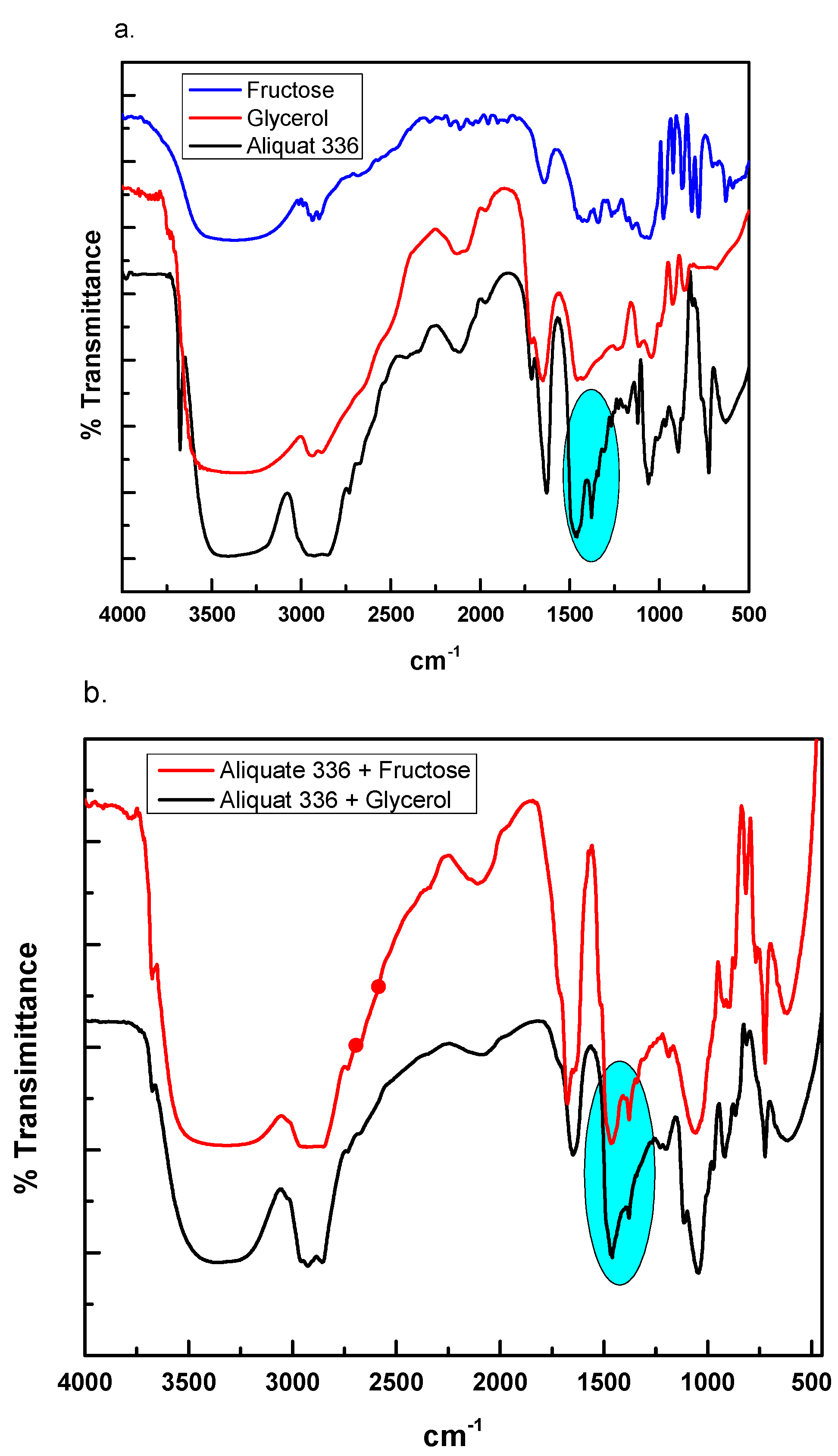
2.3. Fourier Transform Infrared Spectroscopy

To determine the functional groups of separate components and synthesized DES, Fourier transform infrared spectrophotometry (FTIR) was performed using the pressed potassium bromide (KBr) pellet method [32]. The variations in the functional groups before and after DES formation were analyzed by taking the average of 64 scans at 4 cm−1 resolution to obtain the spectra between 500 and 4000 cm−1 using a PerkinElmer Fourier transform infrared spectrophotometer (FTIR) (Thermo Scientific, Waltham, MA, USA).

2.4. Bacterial Cultures

Several Gram-positive (B. cereus, S. pneumoniae, methicillin-resistant S. aureus (MRSA), and S. pyogenes) and Gram-negative (K. pneumoniae, E. coli K1, P. aeruginosa, and S. marcescens) bacteria were used in this study (Table 2). MRSA and E. coli K1 were purchased from the Microbial Type Culture Collection (MTCC). All other bacteria in the study, such as B. cereus, S. pyogenes, S. pneumoniae, K. pneumoniae, P. aeruginosa and S. marcescens, were derived from clinical specimens, purchased from ATCC and MTCC (Table 2). These bacteria were grown aerobically in Luria–Bertani (LB) broth culture at 37 °C overnight [10,33].

2.5. Antibacterial Assays

DES were tested for their bactericidal properties against bacteria using antibacterial assays [34,35]. Briefly, the optical density of overnight-grown bacterial cultures was set to 0.22 at 595 nm using LB broth (approximately 1 × 10⁸ CFU per mL). After this, 1 × 10⁶ bacterial CFU (10 µL) were treated with 2 µL of DES for 2 h at 37 °C, and the final volume was adjusted to 200 µL with phosphate-buffered saline (PBS). All the DES were prepared by combining the methyl-trioctylammonium chloride and all the hydrogen-bond donors (Table 1), which were then used at a 2 µL dose in a final volume of 200 µL in the antibacterial assay. The treated cultures were then ten-fold serially diluted from 10−1 to 10−6, and dilutions 10−3 to 10−6 were plated onto nutrient agar plates. The plates were then incubated at 37 °C for 24 h and the colony-forming units were estimated by counting the viable bacterial colonies. For controls, bacteria grown in phosphate-buffered saline (PBS) and bacteria in water were taken as the negative and solvent control, respectively, while bacteria incubated with gentamicin (100 µg/mL) were considered the positive control (PC). In some experiments, DES were tested at different doses to determine their MIC50 against E. coli K1 and B. cereus. The two-fold dilutions (0.5 µL, 1 µL, 2 µL, and 4 µL) of the tested DES were incubated with 1 × 10⁴ bacteria/well in Muller Hinton broth using broth micro-dilution assays and the final volume was adjusted to 200 µL [22]. Briefly, all the DES were two-fold serially diluted in 100 µL of MHB broth in a 96-well plate and 100 µL of bacterial cultures having 1 × 10⁴ bacterial cells and incubated for overnight at 37 °C. MHB alone was used as control. The next day, the optical density was measured using a spectrophotometer at 595 nm. The results from all treatments were compared with MHB alone and data were recorded.

2.6. In Vitro Host Cell Cytotoxicity

The cytotoxic effects of DES were evaluated by performing lactate dehydrogenase (LDH) assays [36,37]. Briefly, tests were accomplished in a 96-well plate having confluent monolayers of HeLa cells (P15). The cells were incubated with 2 µL of different DES at 37 °C with 5% CO2 and 95% humidity for 24 h. The next day, cell supernatant, having LDH and LDH kit reagents (cytotoxicity detection kit; Roche Diagnostics, Indianapolis, IN, USA), was combined equally to determine the cytotoxic effects by measuring the amount of LDH generated by HeLa cells. HeLa cells cultured in RPMI alone were treated as negative controls, and cells incubated with 0.1% triton X-100 (100 percent LDH release) were treated as positive controls. The following formula was used to calculate the percent cell cytotoxicity:
% cell cytotoxicity = (sample value–negative control value)/(positive control value–negative control value) × 100.

2.7. Statistical Analysis

Student’s t-test was used to evaluate statistical significance in antibacterial investigations. To assure accuracy, all of the tests were conducted in triplicate. The results are expressed as the mean standard error of three biological replicates conducted in duplicates. p-values of less than 0.05 were deemed statistically significant. GraphPad Prism software was used to calculate the cytotoxic dose (MIC50).

3. Results

3.1. DES Characterization

Figure 1a,b shows the FTIR spectra for synthesized DES (DES4 and DES11) and their components. The FTIR spectra show that the HBA (Aliquat 336) have peaks at various positions. The peak at 2927 cm−1 is dedicated to the –CH3 group. The peaks located at 1462 cm−1 and 1375 cm−1 are because of the quaternary ammonium group [38,39]. After DES formation with glycerol and fructose, the peaks become broader and move to 1468 cm−1 and 1378 cm−1 and their intensity decreases considerably (Figure 1a,b) [39]. Similarly, for other DES such as Figures S1–S4, the changes in their functional groups were observed after the formation of the DES from their corresponding HBA and HBD. This shifting of the ammonium group after mixing with glycerol and fructose with Aliquat 336 confirms the successful formation of the synthesized DES.

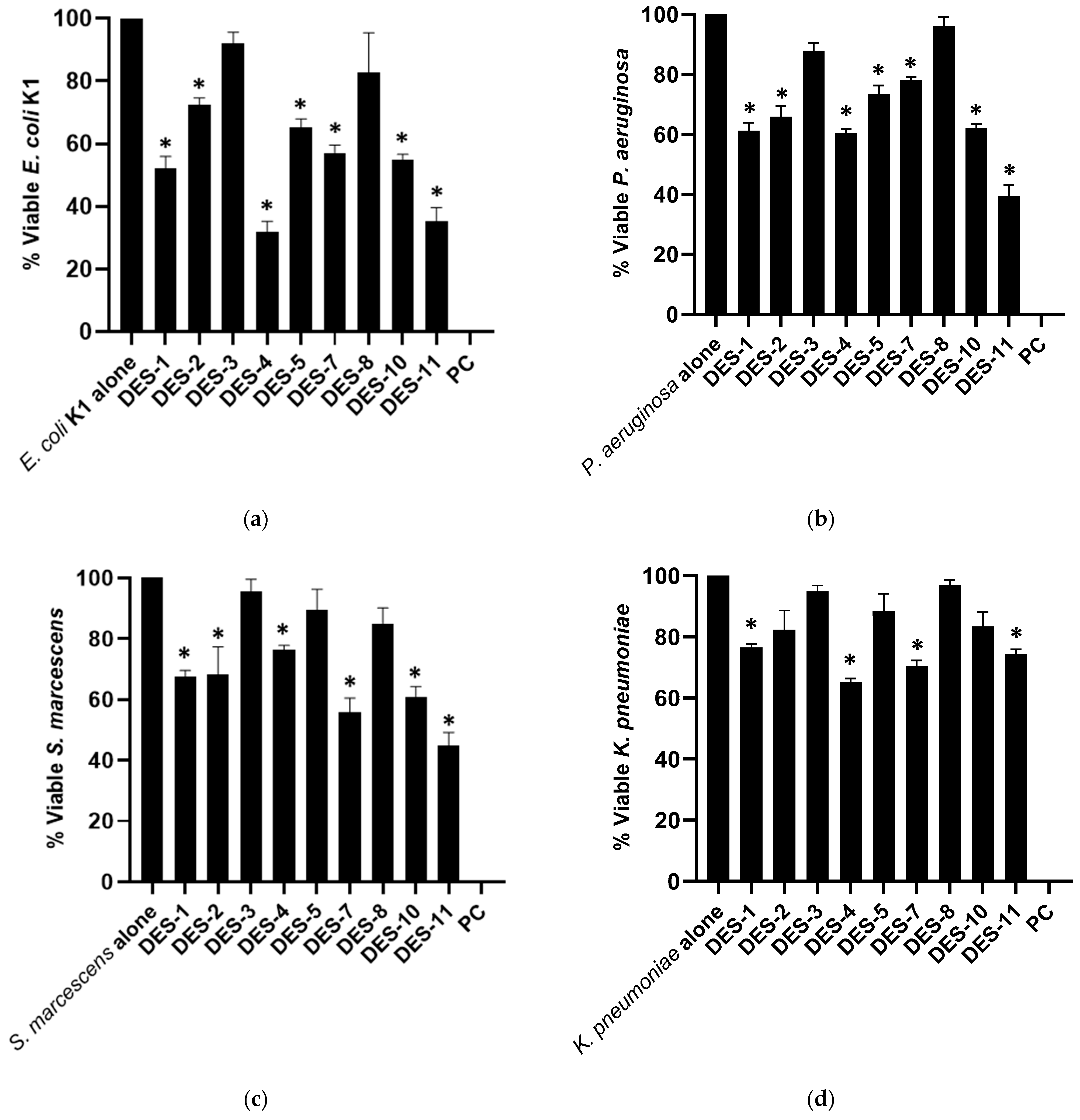
3.2. DES Presented Potent Bactericidal Effects against Multi-Drug-Resistant Pathogens

The results from bactericidal assays have shown that upon successful creation of the DES, all DES with the exception of DES-3 and DES-8 showed significant bacterial inhibition activity against P. aeruginosa and E. coli K1 (p < 0.05) (Figure 2a,b). Of all the DES tested, DES-4 and DES-11 showed the highest bacterial inhibition properties against these two bacteria. DES-4 possessed 40% and 68% antibacterial properties against P. aeruginosa and E. coli K1, whereas DES-11 showed 65% and 61% bacterial inhibition effects against these bacteria (Figure 2a,b). Similarly, DES-1, DES-10, and DES-7 exhibited 48%, 45%, and 43% bactericidal effects against E. coli K1, respectively (Figure 2a). DES-1 and DES-10 presented 39% and 38% bactericidal activities against P. aeruginosa (Figure 2b). When tested against S. marcescens, DES-11 and DES-7 showed 55% and 45% antibacterial activities (Figure 2c), while DES-1, DES-4, DES-7, and DES-11 revealed significant antibacterial properties against K. pneumoniae (Figure 2d) upon their successful formation from their corresponding HBD and HBA.
Correspondingly, when tested against Gram-positive bacteria, all DES after their successful formation, excluding DES-3 and DES-8, demonstrated substantial bactericidal effects against B. cereus (p < 0.05) (Figure 3a). DES-4 and DES-11 had the best bacterial inhibition properties, eradicating 75% and 70% of bacterial population. DES-1 and DES-2 had bactericidal activities of 53% and 46%, while DES-10 and DES-5 exhibited 51% and 43% antibacterial effects, respectively, against B. cereus (Figure 3a). Against MRSA, only DES-1, DES-2, DES-4, and DES-11 indicated significant antibacterial properties, while all other DES failed to show the activity (Figure 3b). All DES except DES-3, DES-7, and DES-8 revealed important bactericidal activity against S. pyogenes (Figure 3c). DES-4 eliminated 47% bacterial viability (Figure 3c). Consistently, DES-4 and DES-11 showed the highest bactericidal activity against S. pneumoniae, abolishing 51% and 50% bacterial growth (Figure 3d). Overall, DES revealed remarkable bactericidal efficacy against both the Gram-positive as well as Gram-negative superbugs. Table 3 summarizes the 50% minimum inhibitory concentration (MIC50) values of the DES using broth micro-dilution assays. All the DES were tested at their doses to assess MIC50 as described in Materials and Methods. Out of all the DES, DES-4 (methyl-trioctylammonium chloride–glycerol) and DES-11 (Methyl-trioctylammonium chloride–fructose) presented MIC50 values of 1.44 µL and 1.53 µL, respectively. Similarly, DES-1 (methyl-trioctylammonium chloride–2-hydroxy benzoic acid), DES-10 (methyl-trioctylammonium chloride–citric acid) and DES-7 (methyl-trioctylammonium chloride–glucose) showed MIC50 values of 2.08 µL, 2.22 µL, and 2.32 µL, respectively, against E. coli K1 (Table 3). Against B. cereus, DES-4 and DES-11 exhibited MIC50 values of 2.66 µL and 2.85 µL, whereas DES-1, DES-10 and DES-7 presented MIC50 values of 3.77 µL, 3.92 µL, and >4 µL, respectively (Table 3).

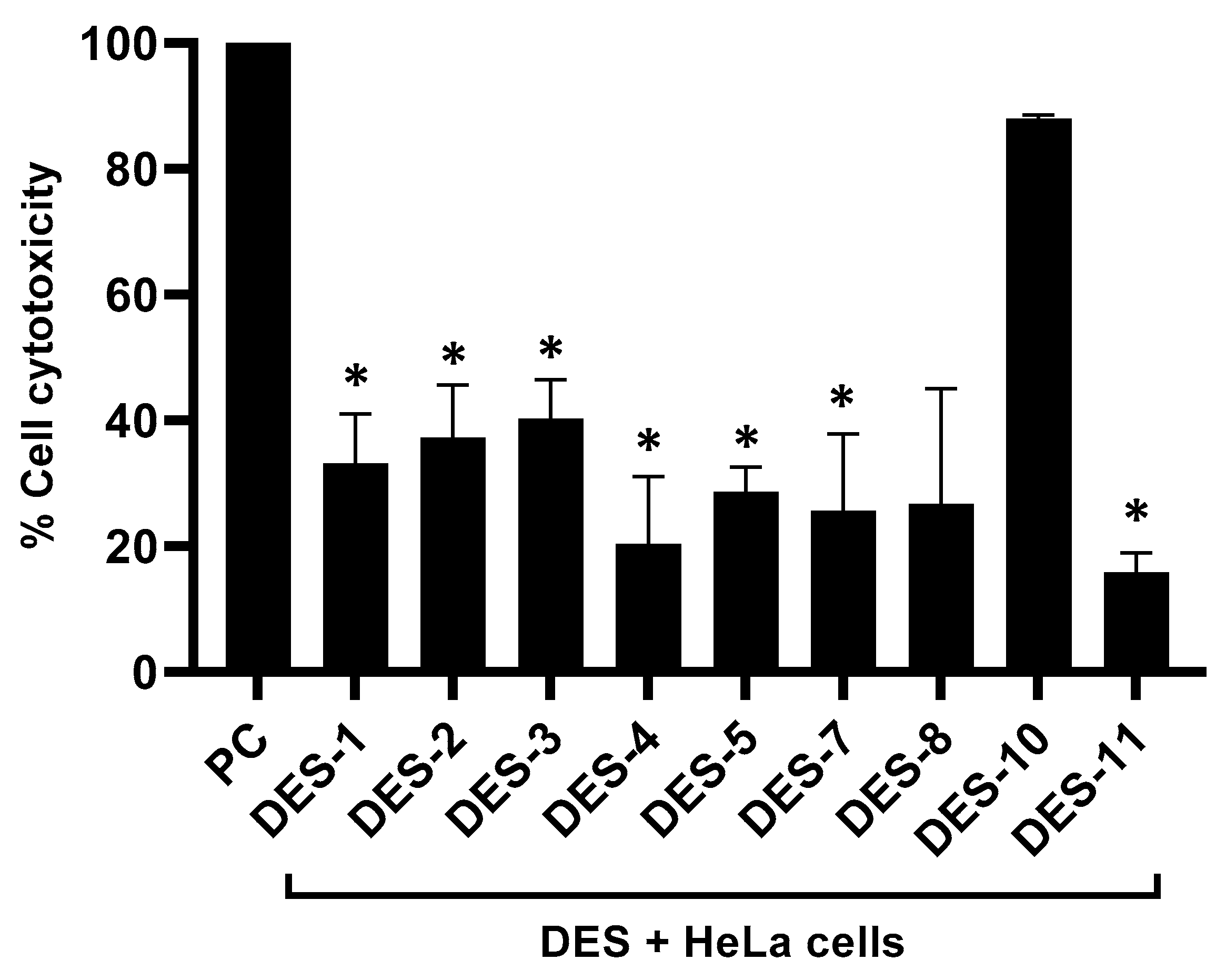
3.3. DES Showed Limited Cytotoxicity against Human Cells

Results from LDH assays revealed that all the DES except DES-10 showed partial cytotoxic properties towards human cells. DES-11 and DES-4 showed the lowest cell cytotoxicity, showing 15% and 20% cytotoxicity against human cells (p ≤ 0.05) (Figure 4). Similarly, DES-7 and DES-8 exhibited 25% and 27% cytotoxic effects, while DES-5, DES-1, DES-2, and DES-3 revealed 29%, 33%, 37%, and 40% cytotoxic properties, respectively, against human cells. DES-10 was potently cytotoxic and presented 88% cytotoxicity (Figure 4).

4. Discussion

Antibiotics have been extensively utilized to treat and prevent serious infections since the 1940s [40]. Over the years, chemical modification of natural compounds seems to be the primary method for discovering novel antibacterials for infections such as bacteria-associated pneumonia, dysentery, gonorrhea, and other bacterial ailments. Unfortunately, increased antibiotic resistance has rendered many of these drugs ineffective due to over-prescription and inappropriate off-purpose use in animal husbandry. As a result, new approaches to the development of antibacterial agents are being developed [41,42]. In this study, different deep eutectic solvents were formed by reacting hydrogen-bond donors and acceptors. Antibacterial tests were used to evaluate the bactericidal properties of DES molecules. Additionally, the cytotoxicity profiles of synthesized DES against human cell lines were also determined using LDH assays. DES revealed broad-spectrum antibacterial properties against several bacteria. Our findings are consistent with previous research. For instance, fatty acid-based DES presented important antibacterial activity against S. epidermis, methicillin-resistant S. aureus, and methicillin-resistant Staphylococcus aureus [22]. In another study, natural DES were investigated for antimicrobial properties against several bacteria and yeast. The DES presented essential antibacterial activity against test bacteria and C. albicans [25]. Aroso et al. discovered that choline chloride- or menthol-based therapeutic DES have broad-spectrum antibacterial action [43].
Among all the DES tested, DES-4 showed potent broad-spectrum antibacterial activity against bacteria. DES-4 is formed by conjugating methyl-trioctylammonium chloride (Aliquat 336) with glycerol at a 1:1 molar ratio. Similarly, DES-11 formed by Aliquat 336 and fructose exerted consistent bactericidal activity against both the Gram-positive bacteria and Gram-negative isolates. Choline chloride-based DES showed noticeable antibacterial properties against Gram-positive and Gram-negative bacteria [28]. Grozdanova et al., in 2020, reported that medicinal plants’ extracts in combination with glycerol-based choline chloride with citric acid-1,2-propanediol (1:4) and 30% H2O presented potent bactericidal effects and limited cytotoxic properties towards human cells [44]. In a recent study, choline chloride-based natural DES extracted from phenolic compounds revealed improved antibacterial activity against Escherichia coli, Staphylococcus aureus, Pseudomonas aeruginosa, and Salmonella enterica [45]. Phosphonium-based deep eutectic solvents produced worthy antibacterial effects against Gram-negative bacteria and Gram-positive pathogens [24]. The DES used in this study are hydrophobic and the hydrophobicity of the DES increases as the alkyl chain length grows. As a result, the hydrophobic phase of these DES may interact better with bacteria and exert antibacterial activity [46].
In the present study, DES were tested at different doses to determine the dose that can inhibit/kill 50% (MIC50) of bacterial populations. Zhao et al. reported the broad-spectrum minimal inhibitory and bactericidal concentration of choline chloride-based DES [28]. The MIC50 values in this study are quite promising, showing the minimum inhibitory strength at microliter doses, whereas the choline chloride-based DES revealed MIC and MBC at millimolar concentration. This indicates the significance of our synthesized DES. Similarly, in another study, Silva et al. reported the MIC and MBC of fatty acid-based DES against Staphylococcus species, which were relatively high against the test bacteria [22]. Further work, using the log-fold method, can also be carried out to understand the potency of these DES.
The biocompatibility of numerous natural DES has been evaluated against several bacterial species (Salmonella enteritidis, S. aureus, Vibrio fischeri, and E. coli), fungi, and cell lines [28,47]. Choline, being a component of vitamin B, plays a significant function in cellular metabolism; the majority of the natural DES investigated were based on choline [28,48]. In the present study, the biocompatibility of the DES was evaluated against HeLa cells and DES showed limited cytotoxicity towards human cells. Nonetheless, future studies to examine any anti-apoptotic effects, or cytotoxicity effects evaluated against other human cell lines, should be carried out in addition to in vivo studies. Interestingly, DES-4 and DES-11, with potent antibacterial activity, presented the least cytotoxic effects against human cells, according to our data. Rodríguez-Juan et al. reported the non-toxic effects of several choline chloride-based DES against several human cell lines [49]. However, recent research has indicated that different DES have the potential to be hazardous and cytotoxic [12,50]. These investigations do, in fact, reveal the toxicity of DES. For example, natural DES with organic acids, such as malonic acid, have been shown to have higher-toxicity profiles than sugar-based natural DES [51]. Moreover, the chain length of the DES determines their cytotoxicity profiles. The larger the chain length, the more toxic the DES [52]. The toxicity of ionic liquids (ILs) is known to be influenced by both their cationic and anionic natures, according to published research [53,54]. The size of the functional groups and the length of the alkyl chain have been revealed to be key determinants of the ILs’ toxicity [55,56]. The toxicity of ILs increases with the lengthening of the alkyl chain on cations. As the length of the alkyl chain grows from C7 to C12, it has been found that the toxicity of guanidinium-based ILs toward Vibrio fischeri increases [57]. Furthermore, Aliquat 336 is known to possess cytotoxicity to humans and aquatic organisms. However, incorporating certain functional groups onto the alkyl chain may reduce toxicity and augment their biodegradability [58,59].
In conclusion, methyl-trioctylammonium chloride-based DES were produced by coupling with different hydrogen-bond donor molecules. The DES showed remarkable bacterial inhibition properties against a panel of Gram-positive and Gram-negative bacteria. The DES revealed MIC50 at micromolar dose against bacteria. Intriguingly, the DES exhibited limited cytotoxic effects against human cells. Our data supports the notion that methyl-trioctylammonium chloride-based DES are anticipated chemotherapeutic agents against bacteria. Future studies are needed to understand the specific mechanism of action and in vivo effects of these DES against pathogenic bacteria in addition to in vivo research and the construction of infection models for bacterial infections.

Supplementary Materials

The following supporting information can be downloaded at: https://www.mdpi.com/article/10.3390/scipharm91010009/s1, Figure S1: DES 2; Figure S2: DES 3; Figure S3: DES 7; Figure S4: DES 8.

Author Contributions

R.S. and N.A.K. conceived the idea. The materials and resources were provided by T.I., M.K., A.S.K., A.M.A. and H.A. N.A. performed all experimentation under the supervision of R.S., N.A.K. and R.S. and N.A. wrote the first draft of the manuscript. R.S., A.M.A., H.A., M.K., T.I. and N.A.K. corrected the manuscript. All authors have read and agreed to the published version of the manuscript.

Funding

This research received no external funding.

Institutional Review Board Statement

Not applicable.

Informed Consent Statement

Not applicable.

Data Availability Statement

The data presented in this study are available on request from the corresponding author.

Acknowledgments

The authors thank the American University of Sharjah and the University of Sharjah for their contributions to this research.

Conflicts of Interest

There is no conflict of interest, and the article was submitted with the permission of all of the authors.

References

  1. Davies, J.; Davies, D. Origins and Evolution of Antibiotic Resistance. Microbiol. Mol. Biol. Rev. 2010, 74, 417–433. [Google Scholar] [CrossRef] [PubMed]
  2. Van Boeckel, T.P.; Brower, C.; Gilbert, M.; Grenfell, B.T.; Levin, S.A.; Robinson, T.P.; Teillant, A.; Laxminarayan, R. Global trends in antimicrobial use in food animals. Proc. Natl. Acad. Sci. USA 2015, 112, 5649–5654. [Google Scholar] [CrossRef] [PubMed]
  3. Du, W.; Chen, H.; Xiao, S.; Tang, W.; Shi, G. New insight on antimicrobial therapy adjustment strategies for gram-negative bacterial infection: A cohort study. Medicine 2017, 96, e6439. [Google Scholar] [CrossRef] [PubMed]
  4. Lozano, C.; Torres, C. Actualización en la resistencia antibiótica en Gram positivos. Enferm. Infecc. Y Microbiol. Clínica 2017, 35, 2–8. [Google Scholar] [CrossRef]
  5. Sun, Y.; Ye, J.; Hou, Y.; Chen, H.; Cao, J.; Zhou, T. Predation Efficacy of Bdellovibrio bacteriovorus on Multidrug-Resistant Clinical Pathogens and Their Corresponding Biofilms. J. Infect. Dis. 2017, 70, 485–489. [Google Scholar] [CrossRef]
  6. CDC. Centers for Disease Control, Antibiotic Resistance the Global Threat. 2019. Available online: https://www.cdc.gov/drugresistance/biggest-threats.html (accessed on 14 March 2022).
  7. Laxminarayan, R.; Duse, A.; Wattal, C.; Zaidi, A.K.M.; Wertheim, H.F.L.; Sumpradit, N.; Vlieghe, E.; Hara, G.L.; Gould, I.M.; Goossens, H.; et al. Antibiotic resistance—The need for global solutions. Lancet Infect. Dis. 2013, 13, 1057–1098. [Google Scholar] [CrossRef]
  8. Sumpradit, N.; Chongtrakul, P.; Anuwong, K.; Pumtong, S.; Kongsomboon, K.; Butdeemee, P.; Khonglormyati, J.; Chomyong, S.; Tongyoung, P.; Losiriwat, S.; et al. Antibiotics Smart Use: A workable model for promoting the rational use of medicines in Thailand. Bull. World Health Organ. 2012, 90, 905–913. [Google Scholar] [CrossRef]
  9. Akova, M. Epidemiology of antimicrobial resistance in bloodstream infections. Virulence 2016, 7, 252–266. [Google Scholar] [CrossRef]
  10. Akbar, N.; Khan, N.A.; Sagathevan, K.; Iqbal, M.; Tawab, A.; Siddiqui, R. Gut bacteria of Cuora amboinensis (turtle) produce broad-spectrum antibacterial molecules. Sci. Rep. 2019, 9, 17012. [Google Scholar] [CrossRef]
  11. Rolain, J.-M.; Abat, C.; Jimeno, M.-T.; Fournier, P.-E.; Raoult, D. Do we need new antibiotics? Clin. Microbiol. Infect. 2016, 22, 408–415. [Google Scholar] [CrossRef] [Green Version]
  12. Smith, E.L.; Abbott, A.P.; Ryder, K.S. Deep Eutectic Solvents (DESs) and Their Applications. Chem. Rev. 2014, 114, 11060–11082. [Google Scholar] [CrossRef]
  13. Liu, P.; Hao, J.-W.; Mo, L.-P.; Zhang, Z.-H. Recent advances in the application of deep eutectic solvents as sustainable media as well as catalysts in organic reactions. RSC Adv. 2015, 5, 48675–48704. [Google Scholar] [CrossRef]
  14. Tang, B.; Zhang, H.; Row, K.H. Application of deep eutectic solvents in the extraction and separation of target compounds from various samples: Other Techniques. J. Sep. Sci. 2015, 38, 1053–1064. [Google Scholar] [CrossRef]
  15. Mouden, S.; Klinkhamer, P.G.L.; Choi, Y.H.; Leiss, K.A. Towards eco-friendly crop protection: Natural deep eutectic solvents and defensive secondary metabolites. Phytochem. Rev. 2017, 16, 935–951. [Google Scholar] [CrossRef]
  16. Mbous, Y.P.; Hayyan, M.; Hayyan, A.; Wong, W.F.; Hashim, M.A.; Looi, C.Y. Applications of deep eutectic solvents in biotechnology and bioengineering—Promises and challenges. Biotechnol. Adv. 2017, 35, 105–134. [Google Scholar] [CrossRef]
  17. Ruesgas-Ramón, M.; Figueroa-Espinoza, M.C.; Durand, E. Application of Deep Eutectic Solvents (DES) for Phenolic Compounds Extraction: Overview, Challenges, and Opportunities. J. Agric. Food Chem. 2017, 65, 3591–3601. [Google Scholar] [CrossRef]
  18. Kumar, A.K.; Sharma, S.; Shah, E.; Patel, A. Technical assessment of natural deep eutectic solvent (NADES) mediated biorefinery process: A case study. J. Mol. Liq. 2018, 260, 313–322. [Google Scholar] [CrossRef]
  19. Sed, G.; Cicci, A.; Jessop, P.G.; Bravi, M. A novel switchable-hydrophilicity, natural deep eutectic solvent (NaDES)-based system for bio-safe biorefinery. RSC Adv. 2018, 8, 37092–37097. [Google Scholar] [CrossRef]
  20. Castro, V.I.B.; Craveiro, R.; Silva, J.M.; Reis, R.L.; Paiva, A.; Duarte, C.A.R. Natural deep eutectic systems as alternative nontoxic cryoprotective agents. Cryobiology 2018, 83, 15–26. [Google Scholar] [CrossRef]
  21. Paiva, A.; Craveiro, R.; Aroso, I.; Martins, M.; Reis, R.L.; Duarte, A.R.C. Natural Deep Eutectic Solvents—Solvents for the 21st Century. ACS Sustain. Chem. Eng. 2014, 2, 1063–1071. [Google Scholar] [CrossRef]
  22. Silva, J.M.; Silva, E.; Reis, R.L.; Duarte, A.R.C. A closer look in the antimicrobial properties of deep eutectic solvents based on fatty acids. Sustain. Chem. Pharm. 2019, 14, 100192. [Google Scholar] [CrossRef]
  23. García-Argüelles, S.; Serrano, M.C.; Gutiérrez, M.C.; Ferrer, M.L.; Yuste, L.; Rojo, F.; del Monte, F. Deep Eutectic Solvent-Assisted Synthesis of Biodegradable Polyesters with Antibacterial Properties. Langmuir 2013, 29, 9525–9534. [Google Scholar] [CrossRef] [PubMed]
  24. Hayyan, M.; Hashim, M.A.; Al-Saadi, M.A.; Hayyan, A.; AlNashef, I.M.; Mirghani, M.E.S. Assessment of cytotoxicity and toxicity for phosphonium-based deep eutectic solvents. Chemosphere 2013, 93, 455–459. [Google Scholar] [CrossRef] [PubMed]
  25. Radošević, K.; Čanak, I.; Panić, M.; Markov, K.; Bubalo, M.C.; Frece, J.; Srček, V.G.; Redovniković, I.R. Antimicrobial, cytotoxic and antioxidative evaluation of natural deep eutectic solvents. Environ. Sci. Pollut. Res. 2018, 25, 14188–14196. [Google Scholar] [CrossRef]
  26. Wikene, K.O.; Bruzell, E.; Tønnesen, H.H. Characterization and antimicrobial phototoxicity of curcumin dissolved in natural deep eutectic solvents. Eur. J. Pharm. Sci. 2015, 80, 26–32. [Google Scholar] [CrossRef]
  27. Zakrewsky, M.; Banerjee, A.; Apte, S.; Kern, T.L.; Jones, M.R.; Sesto, R.E.D.; Koppisch, A.T.; Fox, D.T.; Mitragotri, S. Choline and Geranate Deep Eutectic Solvent as a Broad-Spectrum Antiseptic Agent for Preventive and Therapeutic Applications. Adv. Healthc. Mater. 2016, 5, 1282–1289. [Google Scholar] [CrossRef]
  28. Zhao, B.-Y.; Xu, P.; Yang, F.-X.; Wu, H.; Zong, M.-H.; Lou, W.-Y. Biocompatible Deep Eutectic Solvents Based on Choline Chloride: Characterization and Application to the Extraction of Rutin from Sophora japonica. ACS Sustain. Chem. Eng. 2015, 3, 2746–2755. [Google Scholar] [CrossRef]
  29. Farooq, M.Q.; Abbasi, N.M.; Anderson, J.L. Deep eutectic solvents in separations: Methods of preparation, polarity, and applications in extractions and capillary electrochromatography. J. Chromatogr. A 2020, 1633, 461613. [Google Scholar] [CrossRef]
  30. Kohli, K.; Katuwal, S.; Biswas, A.; Sharma, B.K. Effective delignification of lignocellulosic biomass by microwave assisted deep eutectic solvents. Bioresour. Technol 2020, 303, 122897. [Google Scholar] [CrossRef]
  31. Grillo, G.; Calcio Gaudino, E.; Rosa, R.; Leonelli, C.; Timonina, A.; Grygiškis, S.; Tabasso, S.; Cravotto, G. Green Deep Eutectic Solvents for Microwave-Assisted Biomass Delignification and Valorisation. Molecules 2021, 26, 798. [Google Scholar] [CrossRef]
  32. Kumar, A.K.; Parikh, B.S.; Pravakar, M. Natural deep eutectic solvent mediated pretreatment of rice straw: Bioanalytical characterization of lignin extract and enzymatic hydrolysis of pretreated biomass residue. Environ. Sci. Pollut. Res 2016, 23, 9265–9275. [Google Scholar] [CrossRef]
  33. Akbar, N.; Siddiqui, R.; Iqbal, M.; Khan, N.A. Antibacterial Activities of Selected Pure Compounds Isolated from Gut Bacteria of Animals Living in Polluted Environments. Antibiotics 2020, 9, 190. [Google Scholar] [CrossRef]
  34. Akbar, N.; Siddiqui, R.; Iqbal, M.; Sagathevan, K.; Khan, N.A. Gut bacteria of cockroaches are a potential source of antibacterial compound(s). Lett. Appl. Microbiol. 2018, 66, 416–426. [Google Scholar] [CrossRef]
  35. Ali, S.M.; Siddiqui, R.; Ong, S.-K.; Shah, M.R.; Anwar, A.; Heard, P.J.; Khan, N.A. Identification and characterization of antibacterial compound(s) of cockroaches (Periplaneta americana). Appl. Microbiol. Biotechnol. 2017, 101, 253–286. [Google Scholar] [CrossRef]
  36. Akbar, N.; Siddiqui, R.; Sagathevan, K.; Iqbal, M.; Khan, N.A. Gut Bacteria of Water Monitor Lizard (Varanus salvator) Are a Potential Source of Antibacterial Compound(s). Antibiotics 2019, 8, 164. [Google Scholar] [CrossRef]
  37. Akbar, N.; Siddiqui, R.; Iqbal, M.; Sagathevan, K.; Kim, K.S.; Habib, F.; Khan, N.A. Gut bacteria of Rattus rattus (Rat) produce broad-spectrum antibacterial lipopeptides. ACS Omega 2021, 6, 12261–12273. [Google Scholar] [CrossRef]
  38. Cui, H.; Chen, J.; Yang, H.; Wang, W.; Liu, Y.; Zou, D.; Liu, W.; Men, G. Preparation and application of Aliquat 336 functionalized chitosan adsorbent for the removal of Pb (II). Chem. Eng. J. 2013, 232, 372–379. [Google Scholar] [CrossRef]
  39. Van Osch, D.J.; Zubeir, L.F.; van den Bruinhorst, A.; Rocha, M.A.; Kroon, M.C. Hydrophobic deep eutectic solvents as water-immiscible extractants. Green Chem. 2015, 17, 4518–4521. [Google Scholar] [CrossRef]
  40. Li, L.-L.; Xu, J.-H.; Qi, G.-B.; Zhao, X.; Yu, F.; Wang, H. Core–Shell Supramolecular Gelatin Nanoparticles for Adaptive and “On-Demand” Antibiotic Delivery. ACS Nano 2014, 8, 4975–4983. [Google Scholar] [CrossRef]
  41. Bassetti, M.; De Waele, J.J.; Eggimann, P.; Garnacho-Montero, J.; Kahlmeter, G.; Menichetti, F.; Nicolau, D.P.; Paiva, J.A.; Tumbarello, M.; Welte, T.; et al. Preventive and therapeutic strategies in critically ill patients with highly resistant bacteria. Intensive Care Med. 2015, 41, 776–795. [Google Scholar] [CrossRef]
  42. McKinney, C.W.; Pruden, A. Ultraviolet Disinfection of Antibiotic Resistant Bacteria and Their Antibiotic Resistance Genes in Water and Wastewater. Environ. Sci. Technol. 2012, 46, 13393–13400. [Google Scholar] [CrossRef] [PubMed]
  43. Aroso, I.M.; Silva, J.C.; Mano, F.; Ferreira, A.S.D.; Dionísio, M.; Sá-Nogueira, I.; Barreiros, S.; Reis, R.L.; Paiva, A.; Duarte, A.R.C. Dissolution enhancement of active pharmaceutical ingredients by therapeutic deep eutectic systems. Eur. J. Pharm. Biopharm. 2016, 98, 57–66. [Google Scholar] [CrossRef] [PubMed]
  44. Grozdanova, T.; Trusheva, B.; Alipieva, K.; Popova, M.; Dimitrova, L.; Najdenski, H.; Zaharieva, M.M.; Ilieva, Y.; Vasileva, B.; Miloshev, G.; et al. Extracts of medicinal plants with natural deep eutectic solvents: Enhanced antimicrobial activity and low genotoxicity. BMC Chem. 2020, 14, 73. [Google Scholar] [CrossRef] [PubMed]
  45. Jurić, T.; Mićić, N.; Potkonjak, A.; Milanov, D.; Dodić, J.; Trivunović, Z.; Popović, B.M. The evaluation of phenolic content, in vitro antioxidant and antibacterial activity of Mentha piperita extracts obtained by natural deep eutectic solvents. Food Chem. 2021, 362, 130226. [Google Scholar] [CrossRef]
  46. Syed, U.T.; Leonardo, I.C.; Mendoza, G.; Gaspar, F.B.; Gámez, E.; Huertas, R.M.; Crespo, M.T.; Arruebo, M.; Crespo, J.G.; Sebastian, V.; et al. On the role of components of therapeutic hydrophobic deep eutectic solvent-based nanoemulsions sustainably produced by membrane-assisted nanoemulsification for enhanced antimicrobial activity. Sep. Purif. Technol. 2022, 285, 120319. [Google Scholar] [CrossRef]
  47. Sada Khan, A.; Ibrahim, T.; Akbar, N.; Khamis, M.I.; Siddiqui, R.; Nancarrow, P.; Sabri Mjalli, F.; Ahmed Khan, N.; Abdel Jabbar, N. Application of protic ammonium-based ionic liquids with carboxylate anions for phenol extraction from aqueous solution and their cytotoxicity on human cells. J. Mol. Liq. 2021, 342, 117447. [Google Scholar] [CrossRef]
  48. Florindo, C.; Oliveira, F.S.; Rebelo, L.P.N.; Fernandes, A.M.; Marrucho, I.M. Insights into the Synthesis and Properties of Deep Eutectic Solvents Based on Cholinium Chloride and Carboxylic Acids. ACS Sustain. Chem. Eng. 2014, 2, 2416–2425. [Google Scholar] [CrossRef]
  49. Rodríguez-Juan, E.; López, S.; Abia, R.; Muriana, J.G.F.; Fernández-Bolaños, J.; García-Borrego, A. Antimicrobial activity on phytopathogenic bacteria and yeast, cytotoxicity and solubilizing capacity of deep eutectic solvents. J. Mol. Liq. 2021, 337, 116343. [Google Scholar] [CrossRef]
  50. Kudłak, B.; Owczarek, K.; Namieśnik, J. Selected issues related to the toxicity of ionic liquids and deep eutectic solvents—A review. Environ. Sci. Pollut. Res. 2015, 22, 11975–11992. [Google Scholar] [CrossRef]
  51. Hayyan, M.; Mbous, Y.P.; Looi, C.Y.; Wong, W.F.; Hayyan, A.; Salleh, Z.; Mohd-Ali, O. Natural deep eutectic solvents: Cytotoxic profile. SpringerPlus 2016, 5, 913. [Google Scholar] [CrossRef] [Green Version]
  52. Stolte, S.; Matzke, M.; Arning, J.; Böschen, A.; Pitner, W.R.; Welz-Biermann, U.; Jastorff, B.; Ranke, J. Effects of different head groups and functionalised side chains on the aquatic toxicity of ionic liquids. Green Chem. 2007, 9, 1170–1179. [Google Scholar] [CrossRef]
  53. Pham, T.P.T.; Cho, C.W.; Yun, Y.S. Environmental fate and toxicity of ionic liquids: A review. Water Res. 2010, 44, 352–372. [Google Scholar] [CrossRef]
  54. Bubalo, M.C.; Radošević, K.; Redovniković, I.R.; Halambek, J.; Srček, V.G. A brief overview of the potential environmental hazards of ionic liquids. Ecotoxicol. Environ. Saf. 2014, 99, 1–12. [Google Scholar] [CrossRef]
  55. Mizuuchi, H.; Jaitely, V.; Murdan, S.; Florence, A.T. Room temperature ionic liquids and their mixtures: Potential pharmaceutical solvents. Eur. J. Pharm. Sci. 2008, 33, 326–331. [Google Scholar] [CrossRef]
  56. Ghobadi, A.F.; Taghikhani, V.; Elliott, J.R. Investigation on the solubility of SO2 and CO2 in imidazolium-based ionic liquids using NPT Monte Carlo simulation. J. Phys. Chem. B 2011, 115, 13599–13607. [Google Scholar] [CrossRef]
  57. Ventura, S.P.; Marques, C.S.; Rosatella, A.A.; Afonso, C.A.; Goncalves, F.; Coutinho, J.A. Toxicity assessment of various ionic liquid families towards Vibrio fischeri marine bacteria. Ecotoxicol. Environ. Saf. 2012, 76, 162–168. [Google Scholar] [CrossRef]
  58. Rajadurai, V.; Anguraj, B.L. Ionic liquids to remove toxic metal pollution. Environ. Chem. Lett. 2021, 19, 1173–1203. [Google Scholar] [CrossRef]
  59. Khan, A.S.; Ibrahim, T.H.; Jabbar, N.A.; Khamis, M.I.; Nancarrow, P.; Mjalli, F.S. Ionic liquids and deep eutectic solvents for the recovery of phenolic compounds: Effect of ionic liquids structure and process parameters. RSC Adv. 2021, 11, 12398–12422. [Google Scholar] [CrossRef]
Figure 1. FTIR spectra for Aliquat 336, glycerol and fructose, and their synthesized DES (DES-4 and DES-11) before and after formation. (a) represents the FTIR spectra of individual components whereas (b) represents the FTIR spectra of two separate synthesized DESs.
Scipharm 91 00009 g001
Figure 2. Bactericidal effects of DES against Gram-negative bacteria. (a) represents antibacterial effects towards E. coli K1, (b) against P. aeruginosa, (c) against S. marcescens, and (d) against K. pneumoniae. The results are shown as the means ± standard errors from three separate tests conducted in duplicate. (*) denotes p ≤ 0.05.
Scipharm 91 00009 g002
Figure 3. Antibacterial properties of DES against Gram-positive bacteria. (a) represents bactericidal effects against B. cereus, (b) represents bactericidal activity against MRSA, (c) against S. pyogenes, and (d) against S. pneumoniae. The data are articulated as the means ± standard errors from three independent trials carried out in duplicate. (*) signifies p ≤ 0.05.
Scipharm 91 00009 g003
Figure 4. Deep eutectic solvents revealed limited cytotoxicity against HeLa cells. Human cells grown until confluence were treated with DES with 5% CO2 and 95% humidity overnight at 37 °C. Cells alone in serum-free RPMI and with 0.1% Triton X-100 were considered as negative and positive controls, respectively. Data were investigated with Graph PadPrism software (8.0.2). The data are presented as mean ± SE of several independent trials conducted in duplicates. (*) denotes p ≤ 0.05.
Scipharm 91 00009 g004
Table 1. Deep eutectic solvents, their structures, and molar ratios.
DESHBAHBDComposition of DES in Mole Fraction (HBAs:HBDs)
Chemical NameChemical FormulaChemical NameChemical Formula
DES-1Methyltrioctylammonium chloride or Aliquat 336Scipharm 91 00009 i0012-Hydroxy benzoic acidScipharm 91 00009 i0021:1
DES-21-Naphthoc acidScipharm 91 00009 i0031:1
DES-3Malonic acidScipharm 91 00009 i0041:1
DES-4GlycerolScipharm 91 00009 i0051:1
DES-5Dodecanoic acidScipharm 91 00009 i0061:1
DES-7GlucoseScipharm 91 00009 i0071:2
DES-8Ethylene GlycolScipharm 91 00009 i0081:1
DES-10Citric acidScipharm 91 00009 i0091:1
DES-11FructoseScipharm 91 00009 i0101:1.25
Table 2. Types of bacteria used in this study together with their strain and source.
BacteriaStrain
Escherichia coli K1MTCC 710859
Pseudomonas aeruginosaATCC 10145
Serratia marcescensMTTC 13880
Klebsiella pneumoniaeATCC 13883
Bacillus cereusMTCC 131621
Methicillin-resistant Staphylococcus aureusMTCC 381123
Streptococcus pyogenesATCC 12344
Streptococcus pneumoniaeATCC 33400
Table 3. 50% minimum inhibitory concentration (MIC50) of deep eutectic solvents against bacteria (µL).
Deep Eutectic Solvent (DES)E. coli K1B. cereus
MIC50MIC50
DES-12.083.77
DES-23.56>4
DES-3>4>4
DES-41.442.66
DES-52.85>4
DES-72.32>4
DES-8>4>4
DES-102.223.92
DES-111.532.85
Disclaimer/Publisher’s Note: The statements, opinions and data contained in all publications are solely those of the individual author(s) and contributor(s) and not of MDPI and/or the editor(s). MDPI and/or the editor(s) disclaim responsibility for any injury to people or property resulting from any ideas, methods, instructions or products referred to in the content.

