Article

Carthamus tinctorius Suppresses LPS-Induced Anti-Inflammatory Responses by Inhibiting the MAPKs/NF-κB Signaling Pathway in HaCaT Cells

1
School of Natural Resources and Environmental Science, Kangwon National University, Chuncheon 24341, Republic of Korea
2
Department of Botany, Kongunadu Arts and Science College, Coimbatore 641029, India
3
Chuncheon Center, Korea Basic Science Institute (KBSI), Chuncheon 24341, Republic of Korea
*
Author to whom correspondence should be addressed.
These authors contributed equally to this work.
Sci. Pharm. 2023, 91(1), 14; https://doi.org/10.3390/scipharm91010014
Submission received: 8 February 2023 / Revised: 25 February 2023 / Accepted: 1 March 2023 / Published: 6 March 2023
(This article belongs to the Special Issue Feature Papers in Scientia Pharmaceutica)

Abstract

:
This study aimed to elucidate the anti-inflammatory activity of C. tinctorius leaves by measuring inflammatory parameters such as nitric oxide (NO) production and mRNA expression of iNOS, interleukin-6 (IL-6), and IL-1β in lipopolysaccharide (LPS)-induced HaCaT cells. Further, the effect of C. tinctorius ethanol extract on the MAPKs/NF-κB signaling pathway was examined in HaCaT cells. The phytochemical profile of the ethanol extract of C. tinctorius leaves was determined using UPLC-QTOF-MS/MS. The results indicated that the ethanol extract of C. tinctorius effectively attenuated LPS-induced secretion of NO, IL-6, and IL-1β in HaCaT cells. Further, LPS-stimulated mRNA and protein expressions of iNOS were decreased by pre-treatment with C. tinctorius ethanol extract at the transcriptional level in HaCaT cells. Moreover, the ethanol extract of C. tinctorius suppressed NF-κB signaling in LPS-induced HaCaT cells. This suppression was mediated by MAPKs/NF-κB signaling, inhibiting the phosphorylation of p38 and p65 in HaCaT cells. However, there is no significant effect on the phosphorylation of JNK by the ethanol extract. The QTOF-MS/MS analysis revealed the identification of 27 components in the ethanol extract of C. tinctorius leaves. The data demonstrate that the ethanol extract of C. tinctorius leaves protects the LPS-induced HaCaT cells by inhibiting the expression of iNOS, IL-6, and IL-1β and suppressing the phosphorylation of the p38, p65, p-JNK via inactivation of MAPKs/NF-κB signaling pathway. These results demonstrate that C. tinctorius leaves may serve as a potential candidate to prevent inflammation-related diseases.

1. Introduction

Inflammation is an essential part of the defense mechanism of the host activated by the invasion of pathogens, damaged cells, and toxic compounds. Although inflammation plays an important role in the healing process, it is important pathogenesis of various chronic diseases, such as cardiovascular, diabetes, arthritis, and cancer [1]. In particular, the overproduction of nitric oxide (NO) is involved in inflammatory conditions, so it is regarded as an important pro-inflammatory mediator. NO synthases (NOS) enzymes are primarily responsible for the production of NO in mammalian cells [2,3]. NOS consists of three members which are neuronal NOS, endothelial NOS, and inducible NOS (iNOS). In these, iNOS is highly involved in the production of NO [4,5]. The inflammatory response is distinguished by producing several pro-inflammatory cytokines, including IL-1β, IL-6, TNF-α, etc. [6]. Overproduction of IL-1β upregulates adhesion molecule expression in endothelial cells to activate the translocation of leukocytes, which is related to hyperalgesia and fever [7,8].
Bacterial lipopolysaccharide (LPS) has been exhibited to stimulate macrophages, resulting in the production of pro-inflammatory cytokines via activating NF-κB. NF-κB plays a major role in the regulation of gene expression that is associated with immune and inflammatory responses [9,10]. In this context, NF-κB response genes have been suggested as a promising strategy to treat different inflammatory diseases by inhibiting the production of inflammatory mediators [11,12]. MAPK and NF-κB, signaling molecules play a role in the Toll-like receptor (TLR) pathway [13,14]. Shao et al. [15] reported that the NF-κB signaling molecule regulates inflammatory responses by expressing inflammatory mediators as well as pro-inflammatory cytokines. Under non-induced conditions, the heterodimers of the NF-κB components, especially p50/p65, bind to the inactive form IκB protein in the cytoplasm.
However, NF-κB (p50/p65) is released via the degradation and phosphorylation of IκB by LPS stimulation. Consequently, NF-κB p65 plays an essential role in cellular inflammation response, mainly entering the nucleus and encoding the various chemokines and cytokines [16,17]. MAPKs refer to a special type of serine/thrombin kinase that responds to extracellular signals, including JNK, p38, and ERK1/2. Additionally, the MAPK signaling pathway is a response to LPS-stimulated iNOS expression in activated inflammatory cells [18]. Furthermore, MAPKs are mainly responsible for the activation of NF-κB [19]. Therefore, the potential therapeutic approach for inflammatory injury may be the inactivation of NF-κB and MAPK pathways [20].
Safflower (Carthamus tinctorius L.) belongs to the family of Asteraceae and is commercially cultivated to produce edible oils, dyes, and medicines from seeds and flowers [21]. C. tinctorius has been traditionally used to treat diseases of the cardiovascular system, connective tissues, blood circulation, and musculoskeletal system in Korea [22,23]. According to traditional Chinese medicine, C. tinctorius is used to regulate menstruation, alleviate pain, promote blood circulation, and remove blood stasis [24]. C. tinctorius also exhibited biological activities such as antimicrobial, antithrombotic, anticoagulant, antinociceptive, antitumor, and anti-inflammatory activity [24,25,26]. Previously, many studies reported on the phytochemical analysis and biological properties of florets of C. tinctorius [24]. However, scientific studies on the biological properties of leaves of C. tinctorius are meager.
Hence, this study aimed to evaluate the anti-inflammatory effect of the ethanol extract from the leaves of C. tinctorius via the inhibition of NO production and mRNA expression of iNOS, IL-6, and IL-1β in LPS-induced HaCaT cells. Further, the anti-inflammatory mechanisms of the ethanol extract of C. tinctorius leaves on MAPKs/NF-κB signaling pathway were examined in HaCaT cells. In addition, the phytochemical profile of ethanol extract of C. tinctorius leaves was evaluated by UPLC/QTOF-MS/MS.

2. Materials and Methods

2.1. Collection of Plant Sample

The leaves of C. tinctorius were collected during the flowering stage in Jeollabuk-do ARES Herb and Wild Plant Experimental Farm, Namwon (N 35°25′02.47 E 127°31′34.12), South Korea. The plant sample was authenticated by a plant taxonomist Dr. Jang Geun Jung at Kangwon National University and deposited in Kangwon National University Herbarium, Chuncheon, Republic of Korea, with voucher number KPNS-0952.

2.2. Preparation of Ethanol Extract

The shade-dried leaves of C. tinctorius were powdered using a grinder about the size of 0.6 mm. The powdered leaves (1 kg) were extracted twice with 95% ethanol (4 L) at temperature of 25 ± 2 °C for 2 days. Then, the obtained filtrates were combined and concentrated using a rotary vacuum evaporator at 40 °C (EYELA NE-1101, Tokyo Rikakikai Co., Ltd., Tokyo, Japan). Then, the obtained extract was dried using a lyophilizer (FD5505, ILSHIN BIOBASE, Dongducheon, Republic of Korea). The ethanol extract of C. tinctorius was dissolved with ethanol (at 10 mg/mL) for further analysis.

2.3. Determination of Total Phenolic Content

The total phenolic content of ethanol extract of C. tinctorius leaves was determined by the modified Folin–Ciocalteu method [27].

2.4. Cell Culture

HaCaT cells (gifted by Prof. Lee OH, Food Chemistry laboratory, Kangwon National University) (CVCL-0038) were maintained in DMEM containing 10% fetal bovine serum (FBS) and 100 units/mL penicillin/streptomycin at 37 °C with 5% CO2 [28].

2.5. Cell Viability Assay

The MTT assay was performed to determine the cell viability of HaCaT cells. The cells were treated with C. tinctorius ethanol extract at 30–1000 µg/mL for 24 h. The cytotoxicity of ethanol extract of C. tinctorius in these cells was tested based on the method described by Kim et al. [29].

2.6. Nitric Oxide Production Assay

HaCaT cells were pretreated with the ethanol extract of C. tinctorius (12.5–100 µg/mL) for 1 h and then stimulated with LPS (1 µg/mL) for 24 h. The nitrite concentration in the medium was calculated from a sodium nitrite (NaNO2) standard curve [28,30].

2.7. Isolation of Total RNA and Real Time-Polymerase Chain Reaction (RT-PCR)

The mRNA expression of iNOS, IL-6, and IL-1β was determined by RT-PCR analysis using QuantStudio 3 (Applied Biosystems, Foster City, CA, USA) system with FG POWER SYBR Green PCR master mix and gene-specific primers (Table 1) [28]. Total RNA was isolated from HaCaT cells by RNAiso PLUS. Total RNA (1 µg) was used to generate cDNA by reverse transcription using All-in-One First-Strand cDNA Synthesis SuperMix [31].

2.8. Enzyme-Linked Immunosorbent Assay (ELISA)

HaCaT cells were pretreated with ethanol extract of C. tinctorius at 12.5–100 µg/mL for 1 h and then treated with LPS at 1 µg/mL concentration for 24 h. The amount of IL-6 and IL-1β was measured from the culture supernatants of HaCaT cells using ELISA kits (Invitrogen, Carlsbad, CA, USA) [29].

2.9. Western Blot Analysis

For Western blot analysis, total proteins were extracted from HaCaT cells using a lysis buffer with cocktails of protein inhibitors [32]. Total cellular protein concentration was determined using the Bradford assay. A 20 µg protein per well was loaded to 10% SDS-PAGE and then transferred to PVDF membranes [33]. The membranes were blocked with 5% skimmed milk for 2 h and then incubated with primary antibodies against p-JNK, 1:1000; JNK, 1:1000; p-ERK, 1:1000; ERK, 1:1000; p-p38, 1:1000; p38, 1:1000; p-p65, 1:500; p65, 1:500; iNOS, 1:500; β-actin, 1:1000 at 4 °C overnight (Cell Signaling Technology, Danvers, MA, USA). Then, the washed membranes were incubated with secondary antibodies (Cell Signaling, 1:1000) for 2 h at room temperature.

2.10. Phytochemical Analysis of C. tinctorius by UPLC-QTOF-MS/MS

The LC/MS systems consisted of a Waters Acquity UPLC I-Class system (Waters Co., Milford, MA, USA) with Waters Xeo G2 QTOF MS (Waters MS Technologies, Manchester, UK). An amount of 1 mg of different fractions (hexane, dichloromethane, and ethyl acetate) obtained from the ethanol extract of C. tinctorius was dissolved in 10 mL of 70% ethanol for LC-MS analysis. Then, 2 μL of diluted solution of C. tinctorius was injected into a Waters ACQUITY UPLC BEH C18 column (50 × 2.1 mm, 1.7 μm. The column conditions and the phytochemical characterization were followed based on the method described by Shin et al. [34]. The compounds in the ethanol extract of C. tinctorius were identified by UNIFI 1.8 (Waters, Milford, CT, USA). The traditional Chinese medicine library and the in-house library were used for the tentative identification of chemical components.

2.11. Statistical Analysis

GraphPad Prism Version 8.0 (GraphPad, La Jolla, CA, USA) software was used for data analyses. The values are expressed as the mean of three independent determinations ± SEM. Statistical differences were assessed using the Student–Newman–Keuls test for multiple comparisons after being analyzed with a one-way ANOVA. The p values < 0.05 were considered statistically significant.

3. Results

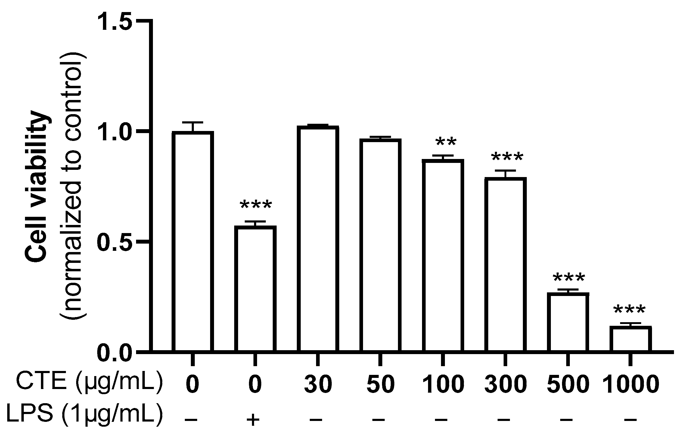
3.1. The Effect of Ethanol Extract of C. tinctorius on the Viability of HaCaT Cells

Previously, several studies reported atopic dermatitis-related inflammation in macrophages and keratinocytes [35,36,37]. Therefore, we investigated whether ethanol extract of C. tinctorius exhibits LPS-induced anti-inflammatory effects on HaCaT cells. For this purpose, the cytotoxicity of the ethanol extract of C. tinctorius was determined by MTT assays after incubating HaCaT cells with different concentrations of the ethanol extract of C. tinctorius (0–1000 µg/mL) for 24 h. After the treatment, there was no significant effect observed in HaCaT with up to 100 µg/mL concentration of the ethanol extract of C. ticntorius (cell viability > 90%) (Figure 1). Hence, we selected the concentration of C. tinctorius ethanol extract up to 100 µg/mL for further experiment.

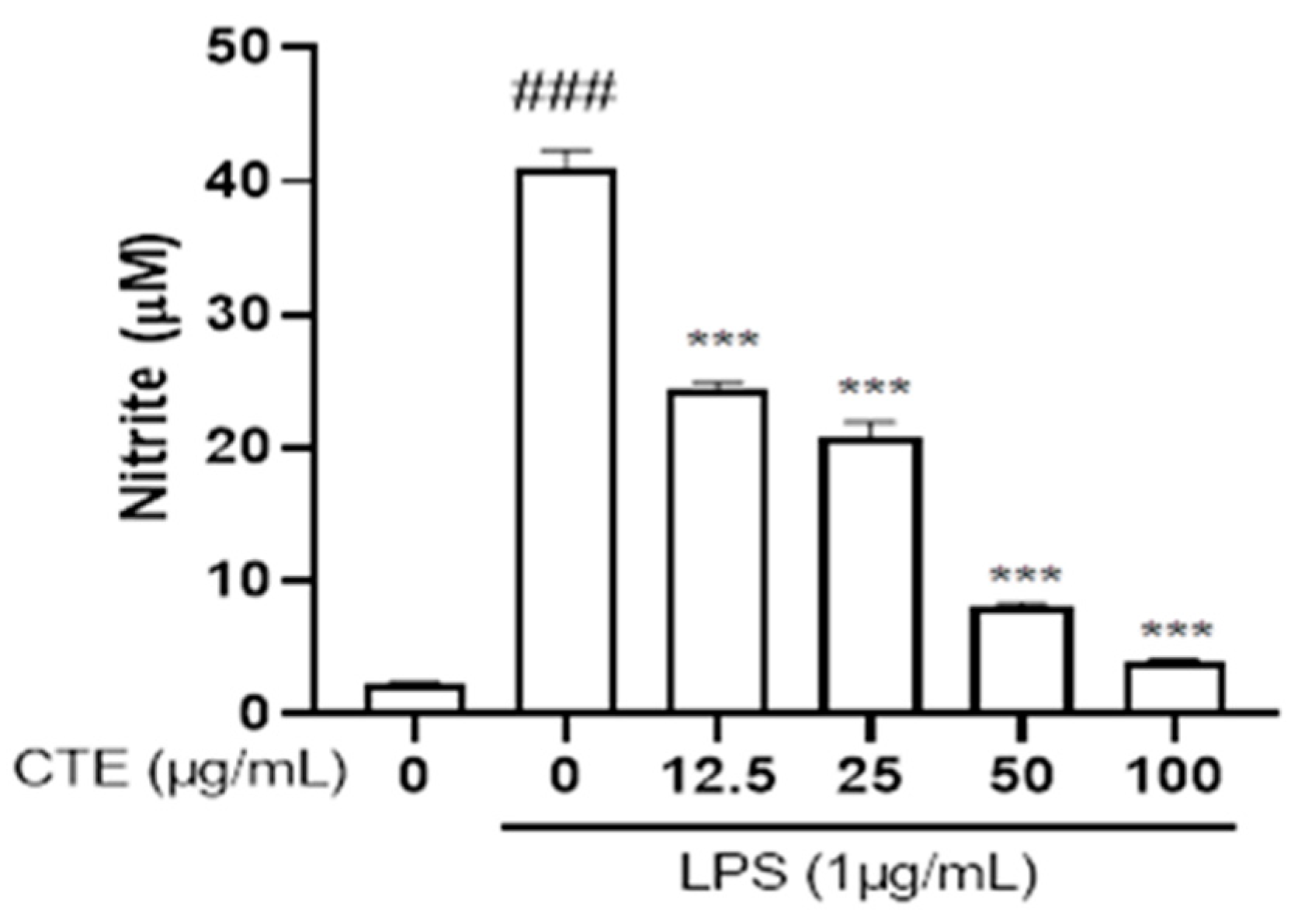
3.2. Effect of C. tinctorius Ethanol Extract on Nitric Oxide Production in LPS-Stimulated HaCaT Cells

It is well known that LPS stimulation is responsible for the release of different inflammatory mediators, especially NO. NO is an important mediator that regulates the molecules with different biological functions including pathological processes in normal physiological conditions. However, excessive production of NO induces inflammation in abnormal physiological conditions [38,39]. Therefore, the inhibitory effect of C. tinctorius ethanol extract on NO production in LPS-induced HaCaT cells was evaluated (Figure 2). The NO production was significantly increased by accumulating a higher level of nitrite in HaCaT cells (40.97 µM). To investigate the effect of C. tinctorius extract on NO production, HaCaT cells were simultaneously treated with 1 µg/mL LPS and various concentrations of extracts (12.5–100 µg/mL) separately for 24 h. When compared to the untreated control, HaCaT cells pre-treated with ethanol extract of C. tinctorius significantly (p < 0.001) reduced the nitrite concentration in the medium to 4.02 µM at the concentration of 100 µg/mL (Figure 2).

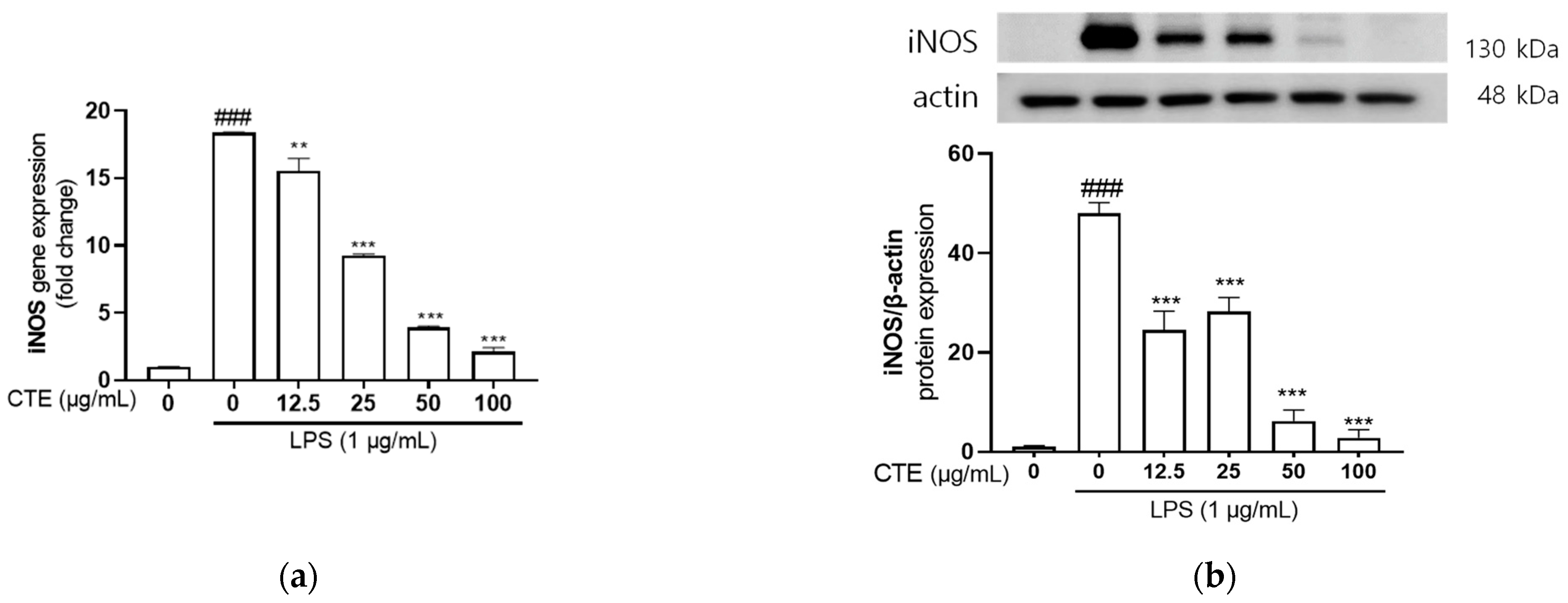
3.3. The Effect of Ethanol Extract of C. tinctorius on mRNA and Protein Expressions of iNOS in LPS-Stimulated HaCaT Cells

To determine the inhibitory action of C. tinctorius ethanol extract on NO production from HaCaT cells, we calculated the mRNA expression level of iNOS by RT-PCR and protein expression level by Western blot analysis. In response to LPS, the iNOS mRNA levels were significantly increased in HaCaT cells. However, the pre-treatment with ethanol extract of C. tinctorius drastically suppressed the mRNA expression of iNOS in HaCaT cells (Figure 3a). Western blot analysis also demonstrated that LPS-treated HaCaT cells markedly increased the protein expression of iNOS. However, pre-treatment with ethanol extract of C. tinctorius showed strong inhibitory activity against iNOS protein expression HaCaT cells (Figure 3b).

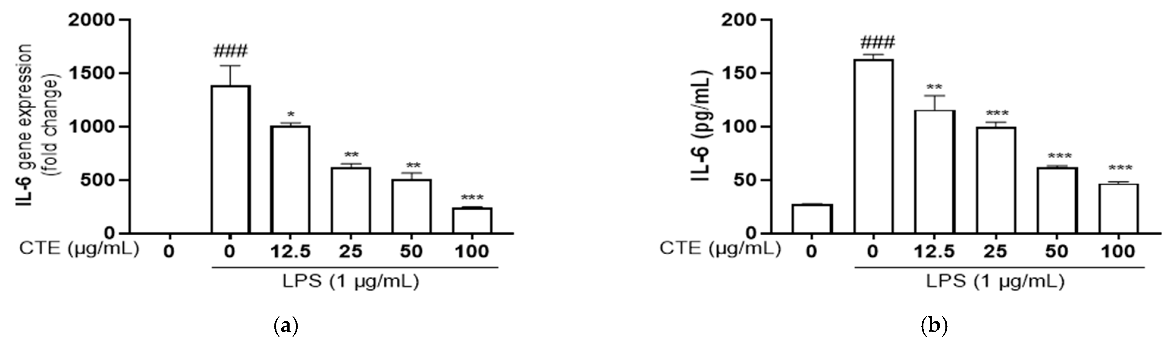
3.4. The Effect of Ethanol Extract of C. tinctorius on Pro-Inflammatory Cytokines in LPS-Stimulated HaCaT Cells

We also examined the inhibitory effects of C. tinctorius on the release of IL-6 and IL-1β in HaCaT cells induced by LPS. HaCaT cells were pretreated with different concentrations of C. tinctorius ethanol extract for 1 h and then stimulated with LPS (1 µg/mL) separately for 24 h. The mRNA expression level of IL-6 and IL-1β was determined in HaCaT cells by RT-PCR analysis. C. tinctorius ethanol extract significantly inhibited the mRNA expression levels of IL-6 and IL-1β in LPS-induced HaCaT cells (Figure 4) compared to that of the control.

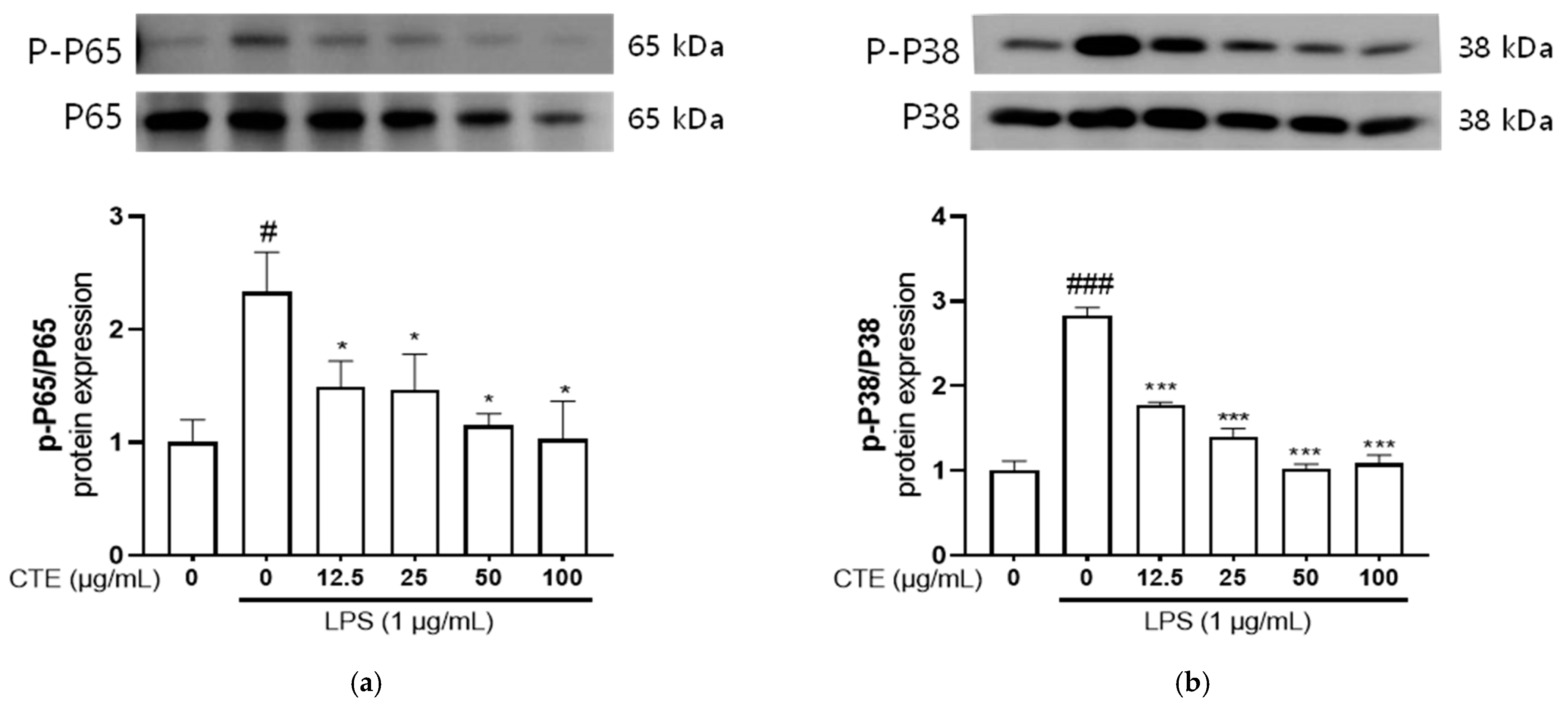
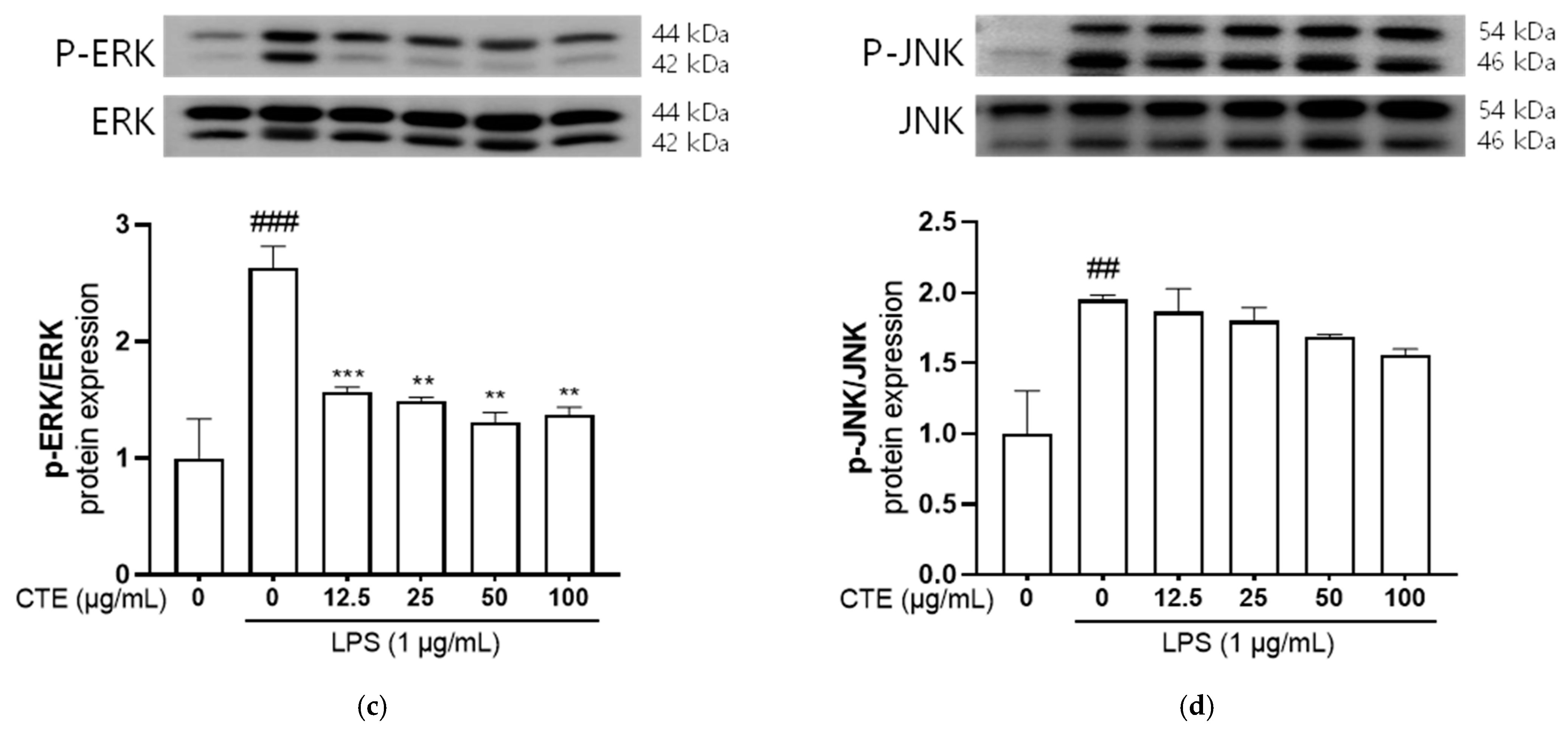
3.5. The Effect of C. tinctorius Ethanol Extract on MAPKs/NF-κB Activation in LPS-Stimulated HaCaT Cells

We investigated whether C. tinctorius ethanol extract inhibited the phosphorylation of p65 (a subunit of NF-κB) in HaCaT cells. The LPS treatment increased the phosphorylation of p65 in HaCaT cells, but C. tinctorius ethanol extract significantly attenuated the phosphorylation of P65 in LPS-stimulated HaCaT cells (Figure 5a). Subsequently, we identified which kinase was involved in the regulation of MAPKs/NF-κB activity in HaCaT cells. The results revealed that the phosphorylation of p38, ERK, and JNK (Figure 5b–d) was increased in HaCaT cells by LPS stimulation and the ethanol extract of C. tinctorius effectively inhibited the phosphorylation of p38 and ERK in HaCaT cells. However, the ethanol extract of C. tinctorius did not exhibit any effect on the phosphorylation of JNK in HaCaT cells. These results demonstrated that C. tinctorius ethanol extract exhibited anti-inflammatory activity on LPS-induced HaCaT cells by inactivating the MAPKs/NF-κB signaling pathway.

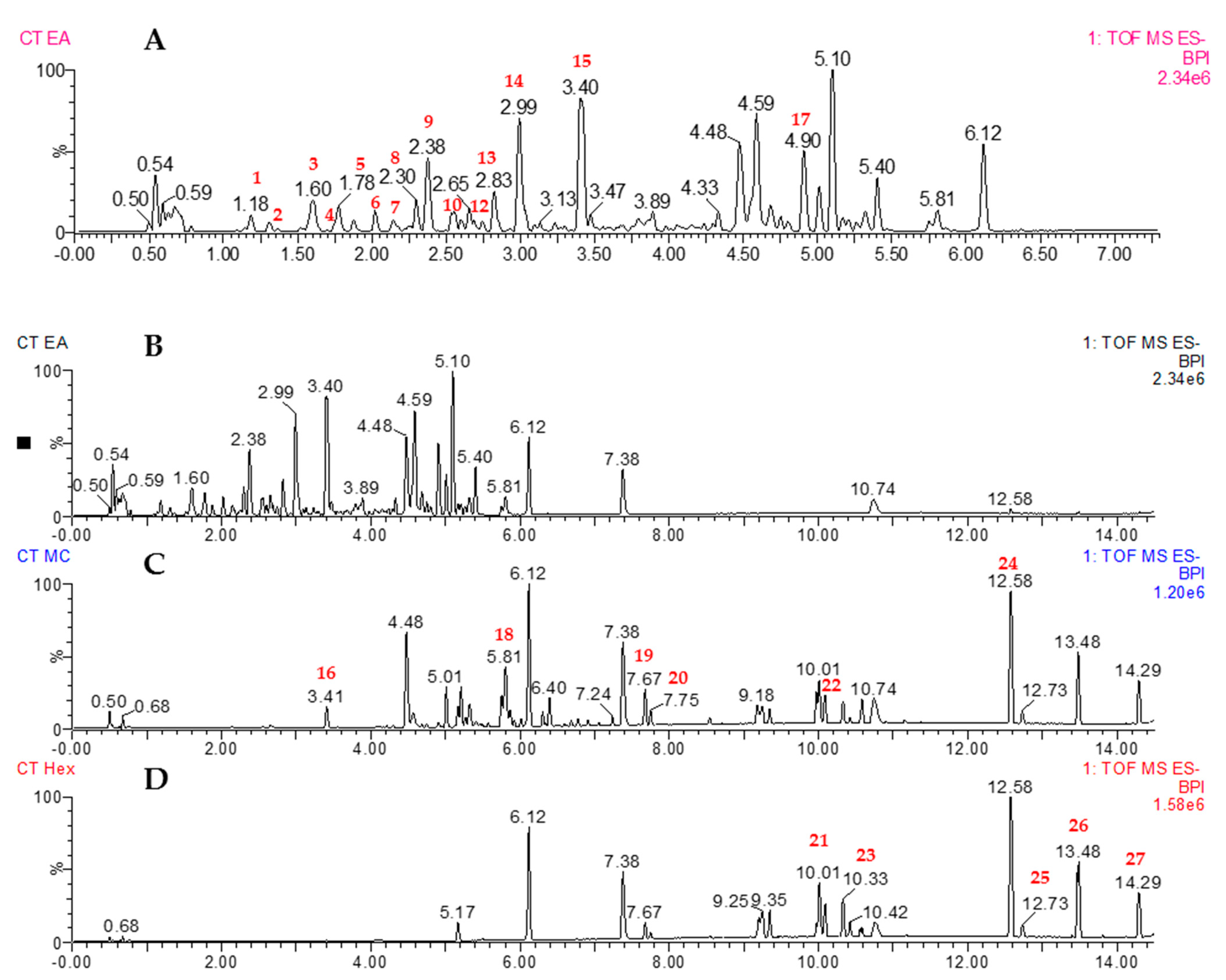
3.6. UPLC-QTOF-MS/MS Analysis of C. tinctorius Leaves

The total phenolic content of the ethanol extract of C. tinctorius leaves was 18.27 ± 0.13 mg GAE/g. QTOF-MS/MS analysis was performed to detect the chemical profile in the ethanol extract of the leaves of C. tinctorius. For better separation of components, the ethanol extract was fractionated first with hexane, dichloromethane, and ethyl acetate successively to produce fractions of different polarities. Figure 6 shows the base peak intensity (BPI) chromatogram of different components from C. tinctorius leaves. The results revealed the presence of 27 different chemical components in the leaves of C. tinctorius (Table 2). The major components in the leaves of C. tinctorius are including protocatechuic acid (C7H6O4) caffeic acid (C9H8O4), coumaroylquinic acid (C16H18O8), coumaric acid (C9H8O3), quercetin hexoside (C21H20O12), kaempferol hexoside (C21H20O11), carthamine (C43H42O22) and kaempferol-3-O-β-rutinoside (C27H30O11).

4. Discussion

Carthamus tinctorius L. is utilized to treat numerous ailments, including inflammation-mediated diseases in Asian countries. Inflammation is an essential part of our body’s defense mechanism that can be activated by various factors. However, the mechanisms of the anti-inflammatory activity of C. tinctorius leaves have not been evaluated. In this study, the LPS-stimulated anti-inflammatory activity of C. tinctorius ethanol extract in HaCaT cells was demonstrated. LPS can generate many pro-inflammatory cytokines, including NO. The excessive production of NO results in many inflammatory diseases and it is an important inflammatory mediator [48,49,50]. In the cytotoxicity study, C. tinctorius ethanol extract did not produce any effect on the viability of HaCaT cells at 100 µg/mL concentration (Figure 1). We evaluated the inhibitory effect of C. tinctorius on the NO production of HaCaT cells stimulated with LPS. The results demonstrated that C. tinctorius ethanol extract effectively inhibited the LPS-stimulated NO production in HaCaT cells (Figure 2) and downregulated mRNA and protein expressions of iNOS. The LPS-induced HaCaT cells produce a rapid inflammatory reaction that can release pro-inflammatory cytokines (IL-6 and IL1β) and inflammatory mediators (iNOS) [51].
Attracting circulatory immune function cells, especially, neutrophils to fight infection is beneficial [52]. Yet, the excessive inflammatory reaction can lead to injury of tissues and organs. Hence, during the inflammatory response, the production of inflammatory mediators and pro-inflammatory cytokines should be strictly controlled [53,54]. We revealed that C. tinctorius ethanol extract inhibited the mRNA and protein expressions of iNOS in HaCaT cells (Figure 3). In addition, we determined the inhibitory effects of C. tinctorius on the release of IL-6 and IL-1β in LPS-induced HaCaT cells. We found that C. tinctorius ethanol extract also inhibited IL-6 and IL-1β mRNA expression in HaCaT cells. A previous study reported that C. tinctorius has a significant effect on the downregulation of iNOS and IL-1β in LPS-activated RAW 264.7 cells [55]. NF-κB is a regulatory transcription factor and is mainly involved in cellular responses to stimuli for the expression of TNFα, IL-1β, IL-6, iNOS, and COX-2 [50].
Dimers of NF-κB (p50/p65) are released and phosphorylated by LPS stimulation, immediately enter the nucleus, and bind exclusively with DNA sequences for promoting the expression of target genes [17,56]. Therefore, the inhibitory effect of C. tinctorius ethanol extracts on LPS-stimulated NF-κB was evaluated. This study found that phosphorylation of p65 was significantly increased by LPS stimulation for 1 h, which means increased NF-κB activation. However, C. tinctorius ethanol extract reduced the phosphorylation of p65 in HaCaT cells, revealing a notable inhibitory effect on NF-κB activity. Consequently, C. tinctorius ethanol extract inhibited the inflammatory mediator and pro-inflammatory cytokines expression in LPS-induced HaCaT cells by downregulating the NF-κB. A previous study reported that C. tinctorius aqueous extract proved to suppress bleomycin-stimulated mRNA levels of IL-1β, TNF-α, and TGF-β1 in lung homogenates. Furthermore, it inhibits the increased activity of NF-κB and p38 MAPK phosphorylation in lung tissues [57]. C. tinctorius methanol extract showed anti-inflammatory activity through increased HO-1 induction via Nrf-2 signals by inhibiting LPS-induced expression of iNOS and COX-2 and TNFα-mediated VCAM-1 upregulation in RAW 264.7 cells [58].
A recent study proved that C. tinctorius honey extract inhibited the NO production, suppressed iNOS, IL-1β, TNF-α, and MCP-1 expressions, decreased the phosphorylation of IκBα, inhibited the NF-κB-p65 protein, in LPS-induced RAW 264.7 cells through the activation of Nrf-2/HO-1 signaling pathway [59]. Moschamine isolated from C. tinctorius significantly suppressed mRNA and protein expression of COX-2, mPGES-1, iNOS, IL-6, and IL-1β by downregulating STAT1/3 activation in RAW 264.7 cells induced with LPS [60]. The florets of C. tinctorius contain a major active constituent called safflower yellow. This constituent inhibited inflammation in TNF-α-induced rat chondrocytes by regulating the NF-κB/SIRT1/AMPK pathways and endoplasmic reticulum stress [61]. Another study found that aqueous extracts from the petals of C. tinctorius and its main constituent safflower yellow prevented NO production and PGE2 in LPS-stimulated RAW264.7 cells by downregulating iNOS and COX-2 expressions [62]. Polyacetylene glucosides isolated from florets of C. tinctorius significantly inhibited NO production in LPS-stimulated RAW264.7 cells [27]. Hydroxysafflor yellow A from C. tinctorius L showed an anti-inflammatory effect in LPS-induced microglia activation by activating a nicotinamide adenine dinucleotide-dependent enzyme, SIRT1 [63]. In addition, dichloromethane extract and its water-ethanolic part of Carthamus lanatus aerial parts exhibited anti-inflammatory activity in activated human neutrophils [64].
The MAPK family, including p38, JNK, and ERK, play an essential role in the transcriptional regulation of the LPS-stimulated expression of iNOS [65]. MAPKs are the upstream activator of NF-κB via blocking the transcriptional activation of NF-κB by MAPK inhibitors [66,67]. In this study, p38, JNK, and ERK phosphorylation dramatically increased LPS-stimulated HaCaT cells, suggesting that LPS upregulated the MAPK pathway in HaCaT cells. C. tinctorius ethanol extract inhibited p38 and ERK phosphorylation in HaCaT cells. A previous study reported that schizandrin A showed a significant protective effect of LPS-induced inflammation in HaCaT cells by suppressing apoptosis, promoting cell viability, inhibiting the expression of IL-1β, IL-6, and TNF-α, downregulating the miR-127 expression, inactivating the p38MAPK/ERK and JNK pathways [68]. Liu et al. [69] demonstrated that sinomenine retards LPS-induced phosphorylation of p65, IκBα, and p38MAPK in HaCaT cells by downregulating the long non-coding RNA colon cancer-related transcript-1. Tanshinol showed a protective effect on LPS-induced HaCaT cells by downregulating the miR-122 and inhibiting the JNK and NF-κB pathways [70]. This study suggested that the anti-inflammatory effect of C. tinctorius ethanol extract was associated with the regulation of MAPKs/NF-κB signaling pathways in LPS-stimulated HaCaT cells.
In this study, 27 different components were identified from the dried leaves of C. tinctorius by UPLC-QTOF. Previous studies reported that derivatives of quinic acid, coumaric acid, quercetin, and kaempferol attenuated immune responses through various mechanisms [71,72,73,74]. In particular, kaempferol and quercetin effectively downregulated the activation of important transcription factors for iNOS (NF-κB and STAT-1) in LPS-stimulated macrophages [71]. In LPS-induced RAW 264.7 cells, ferulic acid and its derivatives inhibited the production of NO through the suppression of iNOS expression and decreasing the production of prostaglandin E2 and TNF-α [75]. Quinic acid derivatives from Uncaria tomentosa also inhibited NF-κB activation in TNF-α-stimulated type II alveolar epithelial-like, A549 cells [72]. In another study, Zhao et al. [76] reported that p-coumaric acid inhibited inflammatory cytokines production in LPS-stimulated RAW 264.7 cells by blocking NF-kB and MAPK signaling pathways. In HaCaT cells, caffeic acid and ferulic acid inhibited UVA-stimulated matrix metalloproteinase-1 by regulating antioxidant defense systems [77]. Flavonoids, kaempferol-3-O-rutinoside, and kaempferol-3-O-glucoside isolated from flowers of C. tinctorius showed remarkable anti-nociceptive and anti-inflammatory activities [78]. Therefore, the presence of various bioactive metabolites in the ethanol extract of C. tinctorius leaves might be ascribed to its anti-inflammatory potential.

5. Conclusions

The results demonstrated that C. tinctorius ethanol extract inhibited LPS-stimulated NO, IL-1β, and IL-6 production as well as protein and mRNA expressions of iNOS in HaCaT cells. Further, the mechanistic study revealed that the anti-inflammatory effect of C. tinctorius ethanol extract is mediated via MAPKs/NF-κB by inhibiting the phosphorylation of p65, p38, and ERK in HaCaT cells. It could be concluded that the ethanol extract of C. tinctorius leaves can be utilized for the development of an anti-inflammatory agent.

Author Contributions

Conceptualization, S.K. and S.J.P.; methodology, S.K. and S.J.P.; formal analysis, M.H. and S.-Y.K.; investigation, M.H., S.-Y.K., and S.P.; resources, M.H. and S.K.; data curation, S.-Y.K., M.H., S.P. and P.D.; writing—original draft preparation, P.D. and K.S.; writing—review and editing, S.K. and K.S.; supervision, S.K. and S.J.P. All authors have read and agreed to the published version of the manuscript.

Funding

This research received no external funding.

Institutional Review Board Statement

Not applicable.

Informed Consent Statement

Not applicable.

Data Availability Statement

Not applicable.

Acknowledgments

This study was carried out with the support of the “Cooperative Research Program for Agriculture Science and Technology Development (Project No. PJ01669705)”, Rural Development Administration, Republic of Korea.

Conflicts of Interest

The authors declare no conflict of interest.

References

  1. Chen, L.; Deng, H.; Cui, H.; Fang, J.; Zuo, Z.; Deng, J.; Li, Y.; Wang, X.; Zhao, L. Inflammatory responses and inflammation-associated diseases in organs. Oncotarget 2017, 9, 7204–7218. [Google Scholar] [CrossRef] [PubMed] [Green Version]
  2. Stuehr, D.J.; Vasquez-Vivar, J. Nitric oxide synthases-from genes to function. Nitric Oxide 2017, 63, 29. [Google Scholar] [CrossRef] [Green Version]
  3. Kanwar, J.R.; Kanwar, R.K.; Burrow, H.; Baratchi, S. Recent advances on the roles of NO in cancer and chronic inflammatory disorders. Curr. Med. Chem. 2009, 16, 2373–2394. [Google Scholar] [CrossRef]
  4. Minghetti, L.; Pocchiari, M. Cyclooxygenase-2, prostaglandin E2, and microglial activation in prion diseases. Int. Rev. Neurobiol. 2007, 82, 265–275. [Google Scholar] [PubMed]
  5. Zheng, L.; Cao, H.; Qiu, J.; Chi, C. Inhibitory Effect of FMRFamide on NO Production During Immune Defense in Sepiella japonica. Front. Immunol. 2022, 13, 825634. [Google Scholar] [CrossRef]
  6. Jeong, J.W.; Lee, H.H.; Han, M.H.; Kim, G.Y.; Hong, S.H.; Park, C.; Choi, Y.H. Ethanol extract of Poria cocos reduces the production of inflammatory mediators by suppressing the NF-kappaB signaling pathway in lipopolysaccharide-stimulated RAW 264.7 macrophages. BMC Complement. Altern. Med. 2014, 14, 101. [Google Scholar] [CrossRef] [Green Version]
  7. Dinarello, C.A. A clinical perspective of IL-1β as the gatekeeper of inflammation. Eur. J. Immunol. 2011, 41, 1203–1217. [Google Scholar] [CrossRef]
  8. Mert, T.; Sahin, M.; Sahin, E.; Yaman, S. Anti-inflammatory properties of Liposome-encapsulated clodronate or Anti-Ly6G can be modulated by peripheral or central inflammatory markers in carrageenan-induced inflammation model. Inflammopharmacology 2019, 27, 603–612. [Google Scholar] [CrossRef] [PubMed]
  9. Mitchell, J.P.; Carmody, R.J. NF-κB and the Transcriptional Control of Inflammation. Int. Rev. Cell Mol. Biol. 2018, 335, 41–84. [Google Scholar]
  10. Barnabei, L.; Laplantine, E.; Mbongo, W.; Rieux-Laucat, F.; Weil, R. NF-κB: At the Borders of Autoimmunity and Inflammation. Front. Immunol. 2021, 12, 716469. [Google Scholar] [CrossRef]
  11. Salminen, A.; Kauppinen, A.; Kaarniranta, K. Phytochemicals suppress nuclear factor-κB signaling: Impact on health span and the aging process. Curr. Opin. Clin. Nutr. Metab. Care. 2012, 15, 23–28. [Google Scholar] [CrossRef]
  12. Yu, H.; Lin, L.; Zhang, Z.; Zhang, H.; Hu, H. Targeting NF-κB pathway for the therapy of diseases: Mechanism and clinical study. Signal Transduct. Target. Ther. 2020, 5, 209. [Google Scholar] [CrossRef] [PubMed]
  13. Kim, K.S.; Cui, X.; Lee, D.S.; Sohn, J.H.; Yim, J.H.; Kim, Y.C.; Oh, H. Anti-inflammatory effect of neoechinulin a from the marine fungus Eurotium sp. SF-5989 through the suppression of NF-κB and p38 MAPK pathways in lipopolysaccharide-stimulated RAW264.7 macrophages. Molecules 2013, 18, 13245–13259. [Google Scholar] [CrossRef] [Green Version]
  14. Lu, Y.; Yeh, W.; Ohashi, P. LPS/TLR4 signal transduction pathway. Cytokine 2008, 42, 145–151. [Google Scholar] [CrossRef] [PubMed]
  15. Shao, J.; Li, Y.; Wang, Z.; Xiao, M.; Yin, P.; Lu, Y.; Qian, X.; Xu, Y.; Liu, J. A novel naphthalimide derivative, exhibited anti-inflammatory effects via targeted inhibiting TAK1 following down-regulation of ERK1/2- and p38 MAPKmediated activation of NF-κB in LPS-stimulated RAW264.7 macrophages. Int. Immunopharmacol. 2013, 17, 216–228. [Google Scholar] [CrossRef]
  16. Ren, Q.; Guo, F.; Tao, S.; Huang, R.; Ma, L.; Fu, P. Flavonoid fisetin alleviates kidney inflammation and apoptosis via inhibiting Src-mediated NF-κB p65 and MAPK signaling pathways in septic AKI mice. Biomed. Pharmacother. 2020, 122, 109772. [Google Scholar] [CrossRef]
  17. Zeng, Y.; Zhu, G.; Zhu, M.; Song, J.; Cai, H.; Song, Y.; Wang, J.; Jin, M. Edaravone Attenuated Particulate Matter-Induced Lung Inflammation by Inhibiting ROS-NF-κB Signaling Pathway. Oxidative Med. Cell. Longev. 2022, 2022, 6908884. [Google Scholar] [CrossRef] [PubMed]
  18. Li, F.; Fu, Y.; Bo, L.; Liu, Z.; Li, D.; Liang, D.; Wen, Z.; Cao, Y.; Zhang, N.; Zhang, X. Stevioside suppressed inflammatory cytokine secretion by Downregulation of NF-κB and MAPK signaling pathways in LPS-stimulated RAW264.7 cells. Inflammation 2012, 35, 1669–1675. [Google Scholar]
  19. Carter, A.B.; Knudtson, K.L.; Monick, M.M.; Hunninghake, G.W. The p38 mitogenactivated protein kinase is required for NF-kappaB-dependent gene expression. The role of TATA-binding protein (TBP). J. Biol. Chem. 1999, 274, 30858–30863. [Google Scholar] [CrossRef] [Green Version]
  20. Dong, J.; Li, J.; Cui, L.; Wang, Y.; Lin, J.; Qu, Y.; Wang, H. Cortisol modulates inflammatory responses in LPS-stimulated RAW264.7 cells via the NF-κB and MAPK pathways. BMC Vet. Res. 2018, 14, 30. [Google Scholar] [CrossRef] [Green Version]
  21. Asgary, S.; Rahimi, P.; Mahzouni, P.; Madani, H. Antidiabetic effect of hydroalcoholic extract of Carthamus tinctorius L. in alloxan-induced diabetic rats. J. Res. Med. Sci. 2012, 17, 386–392. [Google Scholar]
  22. Chang, Y.; Moore, P.S.; Talbot, S.J.; Boshoff, C.H.; Zarkowska, T.; Godden, K.; Paterson, H.; Weiss, R.A.; Mittnacht, S. Cyclin encoded by KS herpesvirus. Nature 1996, 382, 410. [Google Scholar] [CrossRef] [PubMed]
  23. Park, H.; Uhm, C.; Bae, C. Effects of safflower (Carthamus tinctorius L.) seed powder on fracture healing in rats. Korean J. Electron. Microsc. 2001, 31, 307–314. [Google Scholar]
  24. Zhou, X.; Tang, L.; Xu, Y.; Zhou, G.; Wang, Z. Towards a better understanding of medicinal uses of Carthamus tinctorius L. in traditional Chinese medicine: A phytochemical and pharmacological review. J. Ethnopharmacol. 2014, 151, 27–43. [Google Scholar] [CrossRef] [PubMed]
  25. Hiramatsu, M.; Takahashi, T.; Komatsu, M.; Kido, T.; Kasahara, Y. Antioxidant and neuroprotective activities of Mogami-benibana (safflower, Carthamus tinctorius Linne). Neurochem. Res. 2009, 34, 795–805. [Google Scholar] [CrossRef] [PubMed]
  26. Li, X.R.; Liu, J.; Peng, C.; Zhou, Q.M.; Liu, F.; Guo, L.; Xiong, L. Polyacetylene glucosides from the florets of Carthamus tinctorius and their anti-inflammatory activity. Phytochemistry 2021, 187, 112770. [Google Scholar] [CrossRef]
  27. Aarland, R.C.; Bañuelos-Hernández, A.E.; Fragoso-Serrano, M.; Sierra-Palacios, E.D.; Díaz de León-Sánchez, F.; Pérez-Flores, L.J.; Rivera-Cabrera, F.; Mendoza-Espinoza, J.A. Studies on phytochemical, antioxidant, anti-inflammatory, hypoglycaemic and antiproliferative activities of Echinacea purpurea and Echinacea angustifolia extracts. Pharm. Biol. 2017, 55, 649–656. [Google Scholar] [CrossRef] [Green Version]
  28. Yang, Y.I.; Woo, J.H.; Seo, Y.J.; Lee, K.T.; Lim, Y.; Choi, J.H. Protective effect of brown alga phlorotannins against hyperinflammatory responses in lipopolysaccharide-induced sepsis models. J. Agric. Food Chem. 2016, 64, 570–578. [Google Scholar] [CrossRef] [PubMed]
  29. Kim, S.Y.; Hong, M.; Kim, T.H.; Lee, K.Y.; Park, S.J.; Hong, S.H.; Sowndhararajan, K.; Kim, S. Anti-inflammatory effect of liverwort (Marchantia polymorpha L.) and racomitrium moss (Racomitrium canescens (Hedw.) Brid.) growing in Korea. Plants 2021, 10, 2075. [Google Scholar] [CrossRef] [PubMed]
  30. Yang, Y.I.; Shin, H.C.; Kim, S.H.; Park, W.Y.; Lee, K.T.; Choi, J.H. 6,6′-Bieckol, isolated from marine alga Ecklonia cava, suppressed LPS-induced nitric oxide and PGE2 production and inflammatory cytokine expression in macrophages: The inhibition of NF-κB. Int. Immunopharmacol. 2012, 12, 510–517. [Google Scholar] [CrossRef]
  31. Sapkota, A.; Gair, B.P.; Kang, M.G.; Choi, J.W. S1P2 contributes to microglial activation and M1 polarization following cerebral ischemia through ERK1/2 and JNK. Sci. Rep. 2019, 9, 12106. [Google Scholar] [CrossRef] [Green Version]
  32. Baek, H.S.; Min, H.J.; Hong, V.S.; Kwon, T.K.; Park, J.W.; Lee, J.; Kim, S. Anti-Inflammatory Effects of the Novel PIM Kinase Inhibitor KMU-470 in RAW 264.7 Cells through the TLR4-NF-kappaB-NLRP3 Pathway. Int. J. Mol. Sci. 2020, 21, 5138. [Google Scholar] [CrossRef] [PubMed]
  33. Hirai, S.; Horii, S.; Matsuzaki, Y.; Ono, S.; Shimmura, Y.; Sato, K.; Egashira, Y. Anti-inflammatory effect of pyroglutamyl-leucine on lipopolysaccharide-stimulated RAW 264.7 macrophages. Life Sci. 2014, 117, 1–6. [Google Scholar] [CrossRef]
  34. Shin, S.; Saravanakumar, K.; Sathiyaseelan, A.; Mariadoss, A.V.A.; Park, S.; Park, S.; Han, K.; Wang, M.-H. Phytochemical profile and antidiabetic effect of the bioactive fraction of Cirsium setidens in streptozotocin-induced type 2 diabetic mice. Process Biochem. 2022, 116, 60–71. [Google Scholar] [CrossRef]
  35. Wang, H.; Peters, T.; Sindrilaru, A.; Kochanek, K.S. Key role of macrophages in the pathogenesis of CD18 hypomorphic murine model of psoriasis. J. Investg. Dermatol. 2009, 129, 1100–1114. [Google Scholar] [CrossRef] [Green Version]
  36. Kasraie, S.; Werfel, T. Role of macrophages in the pathogenesis of atopic dermatitis. Mediat. Inflamm. 2013, 2013, 942375. [Google Scholar] [CrossRef] [PubMed] [Green Version]
  37. Colombo, I.; Sangiovanni, E.; Maggio, R.; Mattozzi, C.; Zava, S.; Corbett, Y.; Fumagalli, M.; Carlino, C.; Corsetto, P.A.; Scaccabarozzi, D.; et al. HaCaT cells as a reliable in vitro differentiation model to dissect the inflammatory/repair response of human keratinocytes. Mediat. Inflamm. 2017, 2017, 7435621. [Google Scholar] [CrossRef] [PubMed] [Green Version]
  38. Zhong, Y.; Chiou, Y.S.; Pan, M.H.; Shahidi, F. Anti-inflammatory activity of lipophilic epigallocatechin gallate (EGCG) derivatives in LPS-stimulated murine macrophages. Food Chem. 2012, 134, 742–748. [Google Scholar] [CrossRef]
  39. Gasparrini, M.; Forbes-Hernandez, T.Y.; Giampieri, F.; Afrin, S.; Mezzetti, B.; Quiles, J.L.; Bompadre, S.; Battino, M. Protective effect of strawberry extract against inflammatory stress induced in human dermal fibroblasts. Molecules 2017, 22, 164. [Google Scholar] [CrossRef] [PubMed] [Green Version]
  40. Rea Martinez, J.; Montserrat-de la Paz, S.; De la Puerta, R.; Garcia-Gimenez, M.D.; Fernandez-Arche, A. Characterization of bioactive compounds in defatted hempseed (Cannabis sativa L.) by UHPLC-HRMS/MS and anti-inflammatory activity in primary human monocytes. Food Funct. 2020, 11, 4057–4066. [Google Scholar] [CrossRef]
  41. Ciucure, C.T.; Geana, E.-I.; Sandru, C.; Tita, O.; Botu, M. Phytochemical and Nutritional Profile Composition in Fruits of Different Sweet Chestnut (Castanea sativa Mill.) Cultivars Grown in Romania. Separations 2022, 9, 66. [Google Scholar] [CrossRef]
  42. Liu, M.; He, M.; Gao, H.; Guo, S.; Jia, J.; Ouyang, H.; Feng, Y.; Yang, S. Strategy for rapid screening of antioxidant and anti-inflammatory active ingredients in Gynura procumbens (Lour.) Merr. based on UHPLC-Q-TOF-MS/MS and characteristic ion filtration. Biomed. Chromatogr. 2019, 33, e4635. [Google Scholar] [PubMed]
  43. Wang, C.; Zhang, N.; Wang, Z.; Qi, Z.; Zhu, H.; Zheng, B.; Li, P.; Liu, J. Nontargeted Metabolomic Analysis of Four Different Parts of Platycodon grandiflorum Grown in Northeast China. Molecules 2017, 22, 1280. [Google Scholar] [CrossRef] [Green Version]
  44. Liao, Y.; Liang, F.; Liu, H.; Zheng, Y.; Li, P.; Peng, W.; Su, W. Safflower yellow extract inhibits thrombus formation in mouse brain arteriole and exerts protective effects against hemorheology disorders in a rat model of blood stasis syndrome. Biotechnol. Biotechnol. Equip. 2018, 32, 487–497. [Google Scholar] [CrossRef] [Green Version]
  45. Suleimanov, T.A. Phenolic Compounds from Carthamus tinctorius. Chem. Nat. Compd. 2004, 40, 13–15. [Google Scholar] [CrossRef]
  46. Razgonova, M.; Zakharenko, A.; Pikula, K.; Manakov, Y.; Ercisli, S.; Derbush, I.; Kislin, E.; Seryodkin, I.; Sabitov, A.; Kalenik, T.; et al. LC-MS/MS Screening of Phenolic Compounds in Wild and Cultivated Grapes Vitis amurensis Rupr. Molecules 2021, 26, 3650. [Google Scholar] [CrossRef]
  47. Zhou, Y.Z.; Ma, H.Y.; Chen, H.; Qiao, L.; Yao, Y.; Cao, J.Q.; Pei, Y.H. New acetylenic glucosides from Carthamus tinctorius. Chem. Pharm. Bull. 2006, 54, 1455–1456. [Google Scholar] [CrossRef] [Green Version]
  48. Cinelli, M.A.; Do, H.T.; Miley, G.P.; Silverman, R.B. Inducible nitric oxide synthase: Regulation, structure, and inhibition. Med. Res. Rev. 2020, 40, 158–189. [Google Scholar] [CrossRef]
  49. Guzik, T.J.; Korbut, R.; Adamek-Guzik, T. Nitric oxide and superoxide in inflammation and immune regulation. J. Physiol. Pharmacol. 2003, 54, 469–487. [Google Scholar]
  50. Oh, Y.C.; Cho, W.K.; Jeong, Y.H.; Im, G.Y.; Yang, M.C.; Hwang, Y.H.; Ma, J.Y. Anti-inflammatory effect of citrus unshiu peel in LPS-stimulated RAW 264.7 macrophage cells. Am. J. Chin. Med. 2012, 40, 611–629. [Google Scholar] [CrossRef]
  51. Zong, Y.; Sun, L.; Liu, B.; Deng, Y.S.; Zhan, D.; Chen, Y.L.; He, Y.; Liu, J.; Zhang, Z.J.; Sun, J. Resveratrol inhibits LPS-induced MAPKs activation via activation of the phosphatidylinositol 3-kinase pathway in murine RAW 264.7 macrophage cells. PloS ONE 2012, 7, e44107. [Google Scholar] [CrossRef]
  52. Porcherie, A.; Cunha, P.; Trotereau, A.; Roussel, P.; Gilbert, F.B.; Rainard, P.; Germon, P. Repertoire of Escherichia coli agonists sensed by innate immunity receptors of the bovine udder and mammary epithelial cells. Vet. Res. 2012, 43, 14. [Google Scholar] [CrossRef] [PubMed] [Green Version]
  53. Qin, Q.; Li, R.; Li, L.; Zhang, Y.; Deng, S.; Zhu, L. Multi-target regulation of pro-inflammatory cytokine production by transcription factor Blimp-1. Inflamm. Res. 2023, 72, 217–220. [Google Scholar] [CrossRef] [PubMed]
  54. Scheibel, M.; Klein, B.; Merkle, H.; Schulz, M.; Fritsch, R.; Greten, F.R.; Arkan, M.C.; Schneider, G.; Schmid, R.M. IκBβ is an essential co-activator for LPS-induced IL-1β transcription in vivo. J. Exp. Med. 2010, 207, 2621. [Google Scholar] [CrossRef] [Green Version]
  55. Liao, H.; Li, Y.; Zhai, X.; Zheng, B.; Banbury, L.; Zhao, X.; Li, R. Comparison of inhibitory effects of safflower decoction and safflower injection on protein and mRNA expressions of iNOS and IL-1β in LPS-activated RAW264.7 cells. J. Immunol. Res. 2019, 2019, 1018274. [Google Scholar] [CrossRef] [Green Version]
  56. Hoffmann, A.; Baltimore, D. Circuitry of nuclear factor kappaB signaling. Immunol. Rev. 2006, 210, 171–186. [Google Scholar] [CrossRef]
  57. Wu, Y.; Wang, L.; Jin, M.; Zang, B.X. Hydroxysafflor yellow alleviates early inflammatory response of bleomycin-induced mice lung injury. Biol. Pharm. Bull. 2012, 35, 515–522. [Google Scholar] [CrossRef] [PubMed] [Green Version]
  58. Jun, M.S.; Ha, Y.M.; Kim, H.S.; Jang, H.J.; Kim, Y.M.; Lee, Y.S.; Kim, H.J.; Seo, H.G.; Lee, J.H.; Lee, S.H.; et al. Anti-inflammatory action of methanol extract of Carthamus tinctorius involves in heme oxygenase-1 induction. J. Ethanopharm. 2011, 133, 524–530. [Google Scholar] [CrossRef] [PubMed]
  59. Sun, L.P.; Shi, F.F.; Zhang, W.W.; Zhang, Z.H.; Wang, K. Antioxidant and anti-inflammatory activities of Safflower (Carthamus tinctorius L.) honey extract. Foods 2020, 9, 1039. [Google Scholar] [CrossRef]
  60. Jo, A.R.; Han, H.S.; Seo, S.; Shin, J.S.; Lee, J.Y.; Kim, H.J.; Lee, K.T. Inhibitory effect of moschamine isolated from Carthamus tinctorius on LPS-induced inflammatory mediators via AP-1 and STAT1/3 inactivation in RAW 264.7 macrophages. Bioorg. Med. Chem. Lett. 2017, 27, 5245–5251. [Google Scholar] [CrossRef]
  61. Wang, C.; Gao, Y.; Zhang, Z.; Chi, Q.; Liu, Y.; Yang, L.; Xu, K. Safflower yellow alleviates osteoarthritis and prevents inflammation by inhibiting PGE2 release and regulating NF-κB/SIRT1/AMPK signaling pathways. Phytomedicine 2020, 78, 153305. [Google Scholar] [CrossRef] [PubMed]
  62. Wang, C.C.; Choy, C.S.; Liu, Y.H.; Cheah, K.P.; Li, J.S.; Wang, J.T.; Yu, W.Y.; Lin, C.W.; Cheng, H.W.; Hu, C.M. Protective effect of dried safflower petal aqueous extract and its main constituent, carthamus yellow, against lipopolysaccharide-induced inflammation in RAW264.7 macrophages. J. Sci. Food Agric. 2011, 91, 218–225. [Google Scholar] [CrossRef] [PubMed]
  63. Qin, X.; Chen, J.; Zhang, G.; Li, C.; Zhu, J.; Xue, H.; Li, J.; Guan, T.; Zheng, H.; Liu, Y.; et al. Hydroxysafflor Yellow A exerts anti-inflammatory effects mediated by SIRT1 in lipopolysaccharide-induced microglia activation. Front. Pharmacol. 2020, 11, 1315. [Google Scholar] [CrossRef] [PubMed]
  64. Jalil, S.; Mikhova, B.; Taskova, R.; Mitova, M.; Duddeck, H.; Choudhary, M.I.; Atta-ur-Rahman. In vitro anti-inflammatory effect of Carthamus lanatus L. Z. Nat. C J. Biosci. 2003, 58, 830–832. [Google Scholar] [CrossRef] [Green Version]
  65. Pratheeshkumar, P.; Kuttan, G. Modulation of immune response by L. inhibits the proinflammatory cytokine profile, iNOS, and COX-2 expression in LPS-stimulated macrophages. Immunopharmacol. Immunotoxicol. 2011, 33, 73–83. [Google Scholar] [CrossRef] [PubMed]
  66. Berghe, W.V.; Plaisance, S.; Boone, E.; Bosscher, K.D.; Schmitz, M.L.; Fiers, W.; Haegeman, G. p38 and extracellular signal-regulated kinase mitogen activated protein kinase pathways are required for nuclear factor-kappaB p65 transactivation mediated by tumor necrosis factor. J. Biol. Chem. 1998, 273, 3285–3290. [Google Scholar] [CrossRef] [PubMed] [Green Version]
  67. An, Y.; Zhang, H.; Wang, C.; Jiao, F.; Xu, H.; Wang, X.; Luan, W.; Ma, F.; Ni, L.; Tang, X.; et al. Activation of ROS/MAPKs/NF-κB/NLRP3 and inhibition of efferocytosis in osteoclast-mediated diabetic osteoporosis. FASEB J. 2019, 33, 12515–12527. [Google Scholar] [CrossRef] [Green Version]
  68. Li, S.; Xie, R.; Jiang, C.; Liu, M. Schizandrin A alleviates LPS-induced injury in human keratinocyte cell HaCaT through a microRNA-127-dependent regulation. Cell Physiol. Biochem. 2018, 49, 2229–2239. [Google Scholar] [CrossRef]
  69. Liu, Y.; Zhao, C.; Ma, Q.; Li, Y. Sinomenine retards LPS-elicited inflammation via down-regulating CCAT1 in HaCaT cells. Life Sci. 2019, 233, 116703. [Google Scholar] [CrossRef]
  70. Wang, H.; Wei, S. Tanshinol relieves lipopolysaccharide-induced inflammatory injury of HaCaT cells via down-regulation of microRNA-122. Phytother. Res. 2019, 33, 910–918. [Google Scholar] [CrossRef]
  71. Hämäläinen, M.; Nieminen, R.; Vuorela, P.; Heinonen, M.; Moilanen, E. Anti-inflammatory effects of flavonoids: Genistein, kaempferol, quercetin, and daidzein inhibit STAT-1 and NF-kappaB activations, whereas flavone, isorhamnetin, naringenin, and pelargonidin inhibit only NF-kappaB activation along with their inhibitory effect on iNOS expression and NO production in activated macrophages. Mediators Inflamm. 2007, 2007, 45673. [Google Scholar]
  72. Zeng, K.; Thompson, K.E.; Yates, C.R.; Miller, D.D. Synthesis and biological evaluation of quinic acid derivatives as anti-inflammatory agents. Bioorg. Med. Chem. Lett. 2009, 19, 5458–5460. [Google Scholar] [CrossRef]
  73. Pragasam, S.J.; Venkatesan, V.; Rasool, M. Immunomodulatory and anti-inflammatory effect of p-coumaric acid, a common dietary polyphenol on experimental inflammation in rats. Inflammation 2013, 36, 169–176. [Google Scholar] [CrossRef] [PubMed]
  74. Karuppagounder, V.; Arumugam, S.; Thandavarayan, R.A.; Sreedhar, R.; Giridharan, V.V.; Watanabe, K. Molecular targets of quercetin with anti-inflammatory properties in atopic dermatitis. Drug Discov. Today 2016, 21, 632–639. [Google Scholar] [CrossRef] [PubMed]
  75. Alam, M.A. Anti-hypertensive effect of cereal antioxidant ferulic acid and its mechanism of action. Front. Nutr. 2019, 6, 121. [Google Scholar] [CrossRef]
  76. Zhao, Y.; Liu, J.; Liu, C.; Zeng, X.; Li, X.; Zhao, J. Anti-Inflammatory Effects of p-coumaric Acid in LPS-Stimulated RAW264.7 Cells: Involvement of NF-κB and MAPKs Pathways. Med. Chem. 2016, 6, 327–330. [Google Scholar] [CrossRef]
  77. Pluemsamran, T.; Onkoksoong, T.; Panich, U. Caffeic acid and ferulic acid inhibit UVA-induced matrix metalloproteinase-1 through regulation of antioxidant defense system in keratinocyte HaCaT cells. Photochem. Photobiol. 2012, 88, 961–968. [Google Scholar] [CrossRef]
  78. Wang, Y.; Chen, P.; Tang, C.; Wang, Y.; Li, Y.; Zhang, H. Antinociceptive and anti-inflammatory activities of extract and two isolated flavonoids of Carthamus tinctorius L. J. Ethnopharmacol. 2014, 151, 944–950. [Google Scholar] [CrossRef]
Figure 1. The effect of C. tinctorius ethanol extract on the cell viability of HaCaT cells. CTE, C. tinctorius ethanol extract (0–1000 µg/mL). Values are expressed as mean (n = 5) ± SEM. ** p < 0.01, *** p < 0.001 vs. 1% ethanol alone.
Scipharm 91 00014 g001
Figure 2. Inhibitory effect of C. tinctorius ethanol extract on NO production by LPS-stimulated HaCaT cells. CTE, Carthamus tinctorius ethanol extract (0–100 µg/mL). Values are expressed as mean (n = 3) ± SEM. ### p < 0.001 vs. 1% ethanol alone, *** p < 0.001 vs. LPS alone.
Scipharm 91 00014 g002
Figure 3. Effects of ethanol extract of C. tinctorius on mRNA and protein expressions of iNOS in LPS-stimulated HaCaT cells. (a) mRNA expression of iNOS in HaCaT cells; (b) protein expression of iNOS in HaCaT cells. Values are expressed as mean (n = 3) ± SEM. ### p < 0.001 vs. 1% ethanol alone, ** p < 0.01, *** p < 0.001 vs. LPS alone.
Scipharm 91 00014 g003
Figure 4. Effects of C. tinctorius ethanol extract on pro-inflammatory cytokines in LPS-stimulated HaCaT cells. (a) mRNA expression of IL-6; (b) protein production of IL-6; (c) mRNA expression of IL-1β; (d) protein production of IL-1β. Values are expressed as mean (n = 3) ± SEM. ### p < 0.001 vs. 1% ethanol alone, * p < 0.05, ** p < 0.01, *** p < 0.001 vs. LPS alone.
Scipharm 91 00014 g004aScipharm 91 00014 g004b
Figure 5. Effects of C. tinctorius on the phosphorylation of P65, P38, ERK, and JNK in LPS-stimulated HaCaT cells. (a) The immunoreactivity and quantitative analysis of p65 and phosphorylated p65 (p-p65); (b) p38 and p-p38; (c) ERK, and p-ERK; (d) JNK, and p-JNK. Values are expressed as mean (n = 3) ± SEM. # p < 0.05, ## p < 0.01, ### p < 0.001 vs. 1% ethanol alone, * p < 0.05, ** p < 0.01, *** p < 0.001 vs. LPS alone.
Scipharm 91 00014 g005aScipharm 91 00014 g005b
Figure 6. UPLC-QTOF-MS/MS analysis of different fractions obtained from ethanol extract of C. tinctorius leaves. (A) CT EA, ethyl acetate fraction (expanded chromatogram for 0.00 to 7.00 min); (B) CT EA, ethyl acetate fraction; (C) CT MC, dichloromethane fraction; (D) CT Hex, hexane fraction. Numerical in red font denotes the compound number listed in Table 2.
Scipharm 91 00014 g006
Table 1. Primer sequences used for quantitative real-time PCR analysis.
Target GenePrimer SequenceAccession
Number
iNOSForward5′-CATGCTACTGGAGGTGGGTG-3′NM_010927
Reverse5′-CATTGATCTCCGTGACAGCC-3′
IL-6Forward5′-GAGGATACCACTCCCAACAGACC-3′NM_031168
Reverse5′-AAGTGCATCATCGTTGTTCATACA-3′
IL-1βForward5′-ACCTGCTGGTGTGTGACGTT-3′NM_008361
Reverse5′-TCGTTGCTTGGTTCTCCTTG-3′
β-actinForward5′-ATCACTATTGGCAACGAGCG-3′NM_007393
Reverse5′-TCAGCAATGCCTGGGTACAT-3′
Table 2. The list of components identified in the ethanol extract of C. tinctorius leaves by UPLC-QTOF-MS/MS analysis.
S. NO.RT
(min)
Tentative IdentificationFormulaChemical Classm/z
[M-H]-
Mass Error
(ppm)
ResponseFragmentation (m/z)Reference
11.18Protocatechuic acidC7H6O4Phenolic acid153.0192–0.9259240109.0297[40]
21.31Dihydrocaffeic acid C9H10O4Phenylpropanoid181.0503–1.6148736135.0448[41]
31.60Salicylic acidC7H6O3Beta hydroxy acid137.02450.460434393.0354[40]
41.76EsculetinC9H6O4Hydroxycoumarin177.0191–1.1150029133.0295[42]
51.78Caffeic acidC9H8O4Hydroxycinnamic acid179.0347–1.4415615135.045[42]
61.89Coumaroylquinic acidC16H18O8Hydroxycinnamic acid337.09300.386393191.0563[42,43]
72.15Benzoic acid C7H6O2Carboxylic acid121.02982.622638693.0344[42]
82.306-Hydroxykaempferol hexosideC21H20O12Flavonoid463.08830.3299116301.0359[44]
92.38Coumaric acidC9H8O3Hydroxycinnamic acid163.0399–1.11097613119.0504[44]
102.503-(β-D-Glucopyranosyloxy)-5-octenoic acidC14H24O8Fatty acid319.1394–1.327071157.0862Pubmed
112.61Quercetin hexosideC21H20O12Flavonoid463.08840.4127814300.0288[44]
122.69Ferulic acid C10H10O4Hydroxycinnamic acid193.0503–1.6106615134.0371, 149.060[40]
132.83Kaempferol-3-O-β-rutinoside C27H30O15Flavonoid593.15181.1334736285.0413[44]
142.99Kaempferol hexosideC21H20O11Flavonoid447.09360.7985035285.0402[44]
153.40Azelaic acidC9H16O4Dicarboxylic acid187.09770.52328525143.1076[40,41,42]
163.41Carthamine C43H42O22Hydroxycinnamic acid449.10930.8371603287.0562[45,46]
174.904,6-Decadiyne-1-O-β-D-glucopyranosideC16H24O6Polyacetylene glycoside311.15030.9878235-[47]
185.81Trihydroxyoctadecenoic acidC18H34O5Fatty acid329.2333–0.2273521211.1338[41,43]
197.67Hydroperoxyoctadecadienoic acidC18H32O4Fatty acid311.2225–0.7281517223.1670[41]
207.759-Octadecenedioic acidC18H32O4Fatty acid311.2226–0.6144857-[41]
2110.01Hydroxyoctadecadienoic acid C18H32O3Fatty acid295.2274–1.4376589277.2175[41]
2210.09OxylipinC18H32O3Fatty acid295.2275–1.1249495277.2176[41]
2310.33Oxooctadecadienoic acidC18H30O3Fatty acid293.2115–2.4227547277.2175[41]
2412.58Linolenic acidC18H30O2Fatty acid277.21750.71114693-[41,42]
2512.73n-PentadecanalC15H30OFatty aldehyde225.22210149217197.1912[43]
2613.48Linoleic acidC18H32O2Fatty acid279.2328–0.5583977-[42]
2714.29Palmitic acidC16H32O2Fatty acid255.2325–1.8182918-[41,42]
Disclaimer/Publisher’s Note: The statements, opinions and data contained in all publications are solely those of the individual author(s) and contributor(s) and not of MDPI and/or the editor(s). MDPI and/or the editor(s) disclaim responsibility for any injury to people or property resulting from any ideas, methods, instructions or products referred to in the content.

