Article

Preclinical In Vitro Investigation of MDM2 Inhibition in Combination with Antiangiogenic Therapy for Breast Cancer Treatment

1
Department of Pharmacology, College of Medicine, Imam Mohammad Ibn Saud Islamic University (IMSIU), Riyadh 5701, Saudi Arabia
2
Department of Pharmacology and Toxicology, College of Pharmacy, King Saud University, Riyadh 2457, Saudi Arabia
3
Department of Biochemistry, College of Medicine, Al Imam Mohammad Ibn Saud Islamic University (IMSIU), Riyadh 5701, Saudi Arabia
4
Department of Physiology, College of Medicine, Imam Mohammad Ibn Saud Islamic University (IMSIU), Riyadh 5701, Saudi Arabia
5
Department of Pathology, College of Medicine, Imam Mohammad Ibn Saud Islamic University (IMSIU), Riyadh 5701, Saudi Arabia
*
Author to whom correspondence should be addressed.
These authors contributed equally to this work.
Sci. Pharm. 2023, 91(1), 12; https://doi.org/10.3390/scipharm91010012
Submission received: 3 January 2023 / Revised: 8 February 2023 / Accepted: 13 February 2023 / Published: 20 February 2023

Abstract

:
Background: Combining antiangiogenic drugs with other chemotherapeutic drugs has been found to produce superior therapeutic outcomes and prevent drug resistance in a variety of cancers. Methods: Experimental assays such as the MTT assay, flow cytometry, western blotting, and qPCR have been used to evaluate the efficacy of combination therapy. Results: When compared to controls and monotherapies, the combination treatment of axitinib and idasanutlin demonstrated a substantial decrease in cell viability at lower doses, a significant decrease in migration, and a shift toward early and late apoptosis. This study examined major apoptotic, metastatic, and angiogenic factors, including MDM2, p21, BCL-2, BCL-XL, and MMP9, which have showed differential expressions at the protein and mRNA levels after combination. Axitinib and idasanutlin decreased tumorigenesis and migration in vitro in the MCF-7 cell line when compared to other chemotherapeutic medications. The suggested mechanisms of the antitumorigenic effect of the combination therapy may depend on its capacity to promote the production of apoptotic markers and reduce antiapoptotic markers. Conclusions: Treatments with axitinib and idasanutlin demonstrated effective therapeutic targeting of the primary angiogenic growth factor and, consequently, the pro-metastatic arbitrators. This will not only eliminate cancer cells but also stop other malignant processes and ultimately reduce the metastatic cascade.

1. Introduction

Breast cancer is one of the most common cancers, with few treatment options. Early breast cancer, however, is considered curable when the tumor is confined to the breast and nearby tissue. Although challenging, advanced stages of breast cancer can also be treatable with the main objective of improving overall survival and disease-free survival [1]. However, therapeutic resistance and metastasis are major stumbling blocks in the journey of breast cancer treatment. These hurdles might be attributed to many non-mutually exclusive causes, including late diagnosis and limited availability of better treatment options. However, advances in diagnostic technology and the availability of various treatment methods, including surgery, radiation therapy, and gene therapy, have significantly reduced the cancer death rate in recent years. Breast cancer, like other types of cancer, is driven by continuous genetic and epigenetic alterations that allow the development of solid tumors. About 10% of breast cancers are inherited and linked to family history, and loss-of-function mutations remarkably represent a 70% risk of obtaining invasive breast cancer by age 70 [2,3,4]. Furthermore, oncogene and tumor suppressor gene dysregulation are driving forces for many genetic alterations and are considered major contributors to uncontrolled growth, including the development of breast tumors.
P53, known as the guardian of the genome, is a tumor suppressor that preserves DNA integrity during cellular replication. In this line, TP53 is a highly mutated gene in many breast cancer subtypes, including triple-negative breast cancer, the most aggressive subtype with limited treatment options. Similarly, MDM2 (murine double minute 2) is a multifunctional oncogene that is dysregulated in many types of cancer while, in normal conditions, MDM2 primarily inhibits the antiapoptotic effects of p53 and thus promotes cellular hemostasis. In cooperation with MDM2, p53 regulates a variety of cellular processes, such as promoting cell cycle arrest, by inducing p21 and thereby facilitating DNA repair [5]. Currently, several MDM2 antagonists have undergone advanced pre-clinical and clinical studies, including phase II and III clinical trials evaluating the therapeutic activity and tolerability of these inhibitors. For instance, idasanutlin is an oral, second-generation MDM2 inhibitor with potential antineoplastic activities in many other cancer types, including non-hematological tumors. Idasanutlin has been evaluated in phase III clinical trials in patients with relapsed or refractory acute myeloid leukemia [6]. Idasanutlin binds to MDM2 and prevents the interaction between MDM2 and p53. As a result, p53 transcriptional activity is restored by protecting p53 from enzymatic degradation [7].
Angiogenesis is an essential step for tumor growth; therefore, targeting angiogenesis is potentially an effective therapeutic approach to prevent uncontrolled growth. It is widely believed that the vascular endothelial growth factor (VEGF) signaling pathway plays a central role in promoting angiogenesis. Consequently, targeting VEGF and its signaling pathways would restrict angiogenesis and promote a preferential therapeutic outcome [8]. Axitinib is an oral, second-generation pan-VEGFR antagonist that binds with high affinity in the kinase domains of VEGFR-1, 2, and 3 and is approved for advanced renal cell carcinoma (RCC) treatment [9,10]. To date, many angiogenesis inhibitors have been clinically proven to be efficacious against diverse types of cancer. Existing treatment regimens, however, have been restricted by imperfect tolerability and poor clinical outcomes. Therefore, pursuing a new treatment approach that includes combining different molecular targets to attain an optimal therapeutic response is a subject of intensive research. The combination of an MDM2 inhibitor (idasanutlin) and a VEGF inhibitor (axitinib) has not yet been studied and represents unique molecular targets in the breast cancer model. Therefore, we postulated that combining an MDM2 antagonist with a VEGF inhibitor produces a maximum anticancer effect with potentially low toxicity in the MCF-7 breast cancer model.

2. Materials and Methods

2.1. Materials and Chemicals

Dulbecco’s modified Eagle’s medium (DMEM), fetal bovine serum (FBS), penicillin-streptomycin antibiotic, trypsin-EDTA, and all other cell culture reagents were purchased from Thermo Fisher (GibcoTM, Waltham, MA, USA). Axitinib, idasanutlin, and SYBR Green qPCR Master Mix were purchased from MedChemExpress (South Brunswick Township, NJ, USA). Primers of BCL-XL, BCL-2, p21, and GAPDH were purchased from Integrated DNA Technologies (Coralville, IA, USA). MTT, Annexin V, and propidium iodide (PI) were purchased from Thermo Fisher (Waltham, MA, USA). Primary antibodies p21, BCL2, BCL-XL, and GAPDH were purchased from Cell Signaling (Beverly, MA, USA). Primary antibody MDM2 and dimethyl sulfoxide (DMSO) were purchased from Sigma-Aldrich (St. Louis, MO, USA).

2.2. Cell Culture

Human breast cancer cell lines MCF-7 (positive for estrogen receptor (ER), progesterone receptor (PR), and human epidermal growth receptor 2 (HER2)) were deployed in this study as a human breast cancer model and was obtained from ATCC (Manassas, VA, USA). Cells were cultured in a DMEM medium with 10% FBS, 1% penicillin 100 U/mL, and streptomycin. Cultured cells were raised in an incubator (ThermoTM) at 37 °C in a humidified atmosphere containing 5% carbon dioxide (CO2) and were passaged when obtaining 80 to 90% confluence. Briefly, the culture medium was discarded during cell passage. Next, phosphate-buffered saline (PBS) was used to wash cells, and then cells were detached using trypsin-EDTA 0.25% for 5 min at 37 °C. Next, the cell suspension was centrifuged briefly for 5 min. Finally, the supernatant was discarded, and the cell pellet was suspended in DMEM medium accompanied by 10% FBS and 1% antibiotic in a new culture flask.

2.3. Cell Viability Analysis by MTT Assay

The effects of axitinib and idasanutlin monotherapy, or in combination on the viability of breast cancer cells, were investigated by assessing the reducing enzymes of the viable cells to transform MTT compound (3-[4,5-dimethylthiazol-2-yl]-2,5-diphenyl tetrazolium bromide) into formazan crystals. Axitinib and idasanutlin started at a concentration ranging from 10 nM, 100 nM, 1000 nM, 10 μM, and 30 μM for 24 h. Next, cells were cultured in the complete medium and were seeded into 96-Well Microtiter™ Microplates (Thermo Scientific, Waltham, MA, USA) with 1 × 10⁴ cells per well and incubated at 37 °C under a humidified atmosphere of 5% CO2 for 24 h. Then, the medium was removed and replaced by MTT (5 mg/mL) in Phosphate Buffered Saline (PBS) and incubated for 2 to 4 h at 37 °C. Formazan crystals were formed inside the bottom of the well. Next, isopropyl alcohol 100 μM was added to each well, and the plates were put on a shaker for one hour at room temperature to dissolve formazan crystals. Then, the absorbance at 549 nm was checked through microwell plate reader Bio-Tek Instruments (Winooski, VT, USA).

2.4. Apoptosis Analysis by Flow Cytometry

Apoptosis, caused by axitinib and idasanutlin monotherapy, or combination, was investigated using Annexin V-FITC/propidium iodide (PI) assay. The effects of different concentrations of axitinib and idasanutlin on the cell cycle progression were investigated by flow cytometry following staining with PI. The MCF-7 cells were seeded in 6 well plates at a density of 1 × 10⁶ cells per well and were incubated at 37 °C in a humidified 5% CO2 incubator for 24 h. Cells were treated with axitinib (1 μm) or idasanutlin (1 μM) monotherapies. Axitinib and idasanutlin—in combination—were treated at a concentration of 1 μM for 24 h periods at 37 °C in a humidified 5% CO2 atmosphere. Suspended cells in culture medium were collected after treatment. Trypsinization was used to gather detached cells. The cells were washed twice in sterile PBS that had been pre-warmed. Cells were treated with Annexin V-FITC/propidium iodide (PI) at a concentration of 5 μM in HEPES buffer at 25 °C in the darkroom for 15 min. After treatment, the cells were washed once with HEPES buffer and analyzed with a (BD LSRFortessaTM Cell Analyzer) (BD Biosciences, San Jose, CA, USA). The cells were divided into four quadrants: viable cells, early apoptotic cells, necrosis cells, and late apoptotic cells.

2.5. Reverse Transcription Polymerase Chain Reaction (RT-PCR)

Cells cultured in the complete medium were seeded into Thermo Scientific 25-flask Microtiter™ Microplates at a density of 0.7 × 10⁶ cells per well and incubated at 37 °C under a humidified atmosphere of 5% CO2. MCF-7 cells were treated with axitinib (5 μM) or idasanutlin (5 μM) monotherapies and a combination at a concentration of 1 μM for 24 h periods at 37 °C in a humidified 5% CO2 atmosphere. The total RNA was isolated from cells using triazole [11]. The total RNA yield and quality were spectrophotometrically determined using a 260 and 260/280 nm absorbance ratio, respectively. One microgram of total RNA was reverse-transcribed using the MCE® Reverse Transcription Kit (MedChemExpress South Brunswick Township, NJ, USA) into single-stranded complementary DNA (cDNA). Maxima® SYBR Green/Fluorescein qPCR Master Mix, forward and reverse primers, DNA, and RNase-Free H2O (MedChemExpress, South Brunswick Township, NJ, USA) were used to determine the BAX, BCL-2, p21, VEGF, TGF-β, MMP9, and p53. Meanwhile, GAPDH expression was used as a housekeeping gene and an internal reference control. Each qRT-PCR reaction volume of 20 μL consisted of 10 μL SYBR Master mix (2×), 0.4 μL forward primer and 0.4 μL reverse primer, 1 μL DNA, and 8.2 μL RNase free water. The reaction started with denaturation at 95 °C for 5 min. The reaction was amplified for 40 cycles at 95 °C for 15 s, 60 °C for 30 s, and 72 °C for 30 s. The holding curve analysis happened at 4 °C for 10 min. Table 1 shows the forward and reverse primer sequences used in the experiments. Each sample was amplified in triplicate on a Real-time PCR instrument (Applied Biosystems, Waltham, MA, USA). The 2−ΔΔCT method was used to analyze comparative gene expression data.

2.6. Western Blot Analysis

Breast cancer cell lines MCF-7 have been treated with axitinib (5 μM) or idasanutlin (5 μM) monotherapies and combination at a concentration of 1 μM for 24 h. Cells cultured in the complete medium were seeded into Thermo Scientific 25-flask Microtiter™ Microplates with 0.7 × 10⁶ cells per well and incubated at 37 °C under a humidified atmosphere of 5% CO2. Cells were washed with PBS twice, then harvested by Radioimmunoprecipitation assay buffer (RIPA buffer). The total protein contents were determined using Bicinchoninic acid assay (BCA) with RIPA Buffer. Protein was separated via sodium dodecyl sulfate (SDS)—polyacrylamide gel and transferred onto polyvinylidene fluoride (PVDF) membranes. They were then blocked by 5% bovine serum albumin for 1 h. The membranes were incubated with the primary antibody overnight then washed more than twice with Tris Buffered Saline Tween (TBST) buffer. Membranes were incubated with the secondary antibodies for 2 h on the shaker at 37 °C room temperature and then washed. The antibody was detected by using a chemiluminescence detection kit. The proteins BCL-2, BCL-XL, p21, and MDM2 were measured, with GAPDH as a housekeeping protein (Table 2).

2.7. Scratch Assay

Cell migrations were assessed by scratch assay. Monolayers of MCF-7 cells were seeded in 6-well plates close to 80% confluency. Using a sterile 200 µL tip, a single straight-line scratch was made in each well. Then, the wells were washed with phosphate-buffered saline (PBS) and refilled with growth medium containing different treatments (Cis, Ym, Cis + Ym). Cells were treated with axitinib (5 μM) or idasanutlin (5 μM) monotherapies. Axitinib and idasanutlin in combination regimen at a concentrations 100 nM, 500 nM, and 1 μM were kept for 24 h periods at 37 °C in a 5% CO2 atmosphere. Images were taken at 0 h and 24 h by EVOS XL Core Microscope (Thermo Fisher, Waltham, MA, USA). ImageJ software (Bethesda, MD, USA) was used to quantify the area migrated by cultured cells.

2.8. Statistical Analysis

Statistical analysis was conducted by GraphPad Prism software version 8. All data were represented as mean ± standard deviation (SD). One-way ANOVA followed by Tukey’s test, were used to carry out the statistical analysis among various treatment groups. Treatments are considered statistically significant at p < 0.05. The inhibitory concentration was calculated using concentration–inhibition multiple slope equation with GraphPad Prism X software.

3. Results

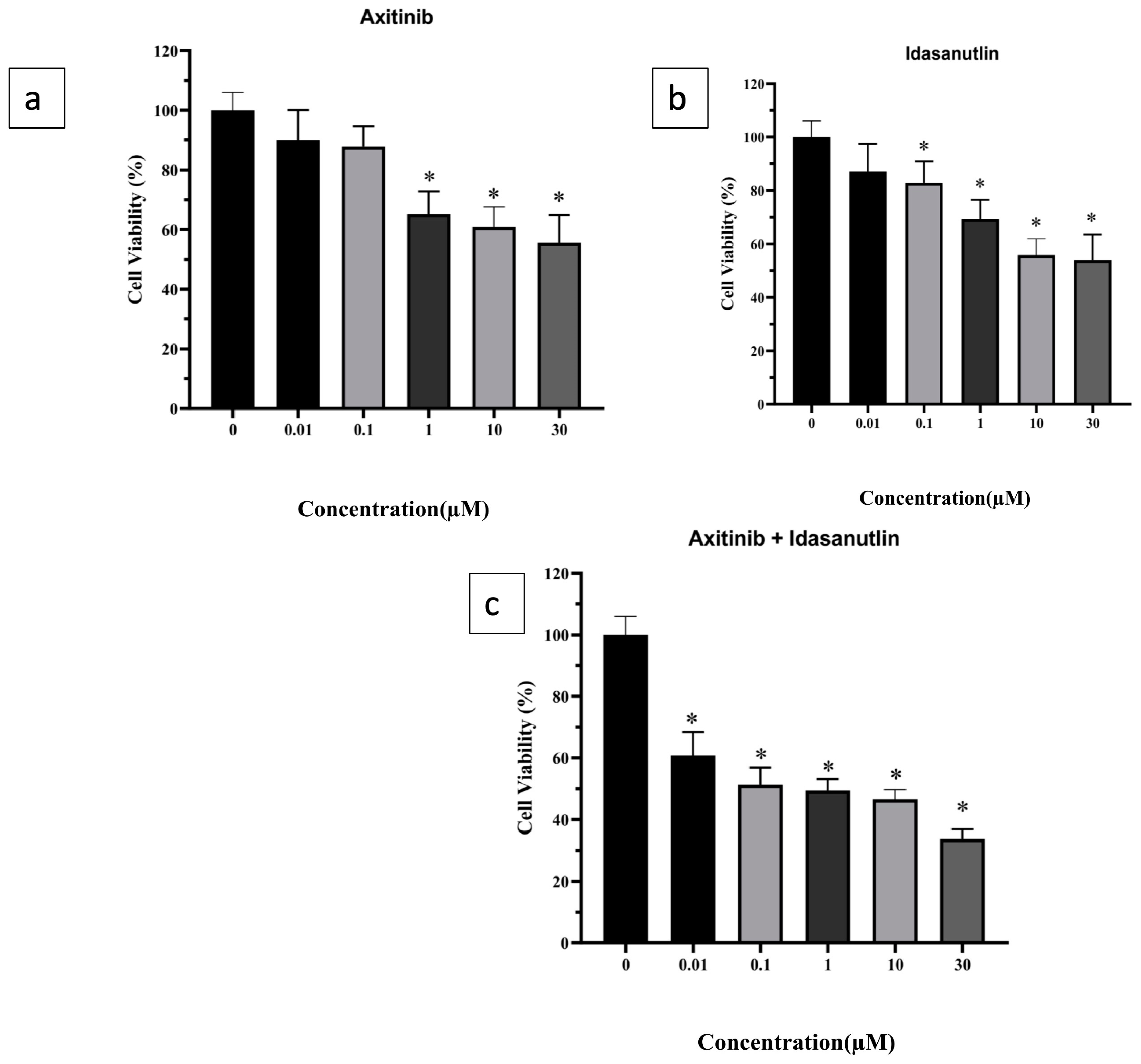
3.1. Evaluation of Cytotoxicity of Axitinib and Idasanutlin on Human Breast Cancer Cell-Line

MTT assay was deployed to evaluate the antiproliferative and cytotoxic effects of axitinib and idasanutlin on the human breast cancer cell-line (MCF-7). Cells were treated with 10-fold serial dilutions of axitinib and idasanutlin (10 nM, 100 nM, 1000 nM, 10 μM, and 30 μM) for 24 h. The inhibitory concentration (IC50) of idasanutlin (10 μM) as a monotherapy on MCF-7 was markedly lower than axitinib (30 μM) as a monotherapy (Figure 1a,b) after 24 h of treatment. The combination therapy of axitinib and idasanutlin showed a further reduction in cell viability at lower concentrations compared to control and monotherapies (Figure 1c).

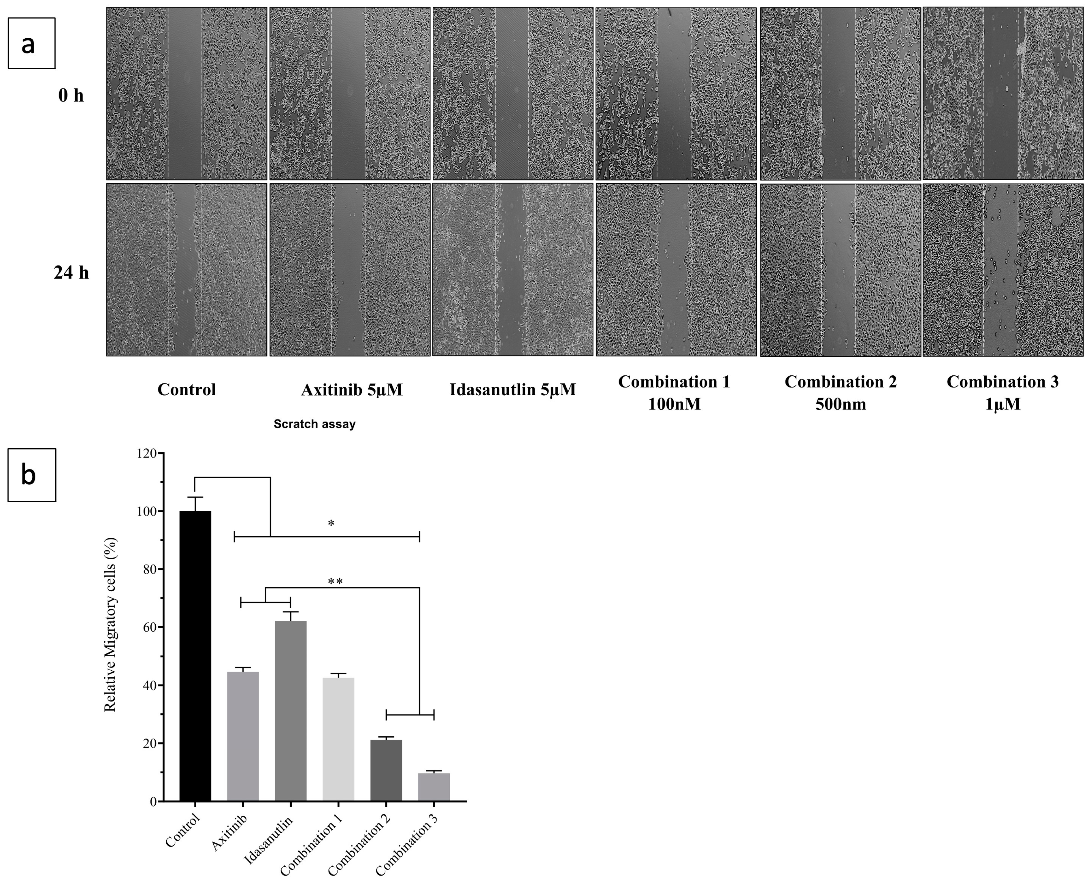
3.2. Inhibitory Effect of Axitinib and Idasanutlin on Cell Migration

A scratch assay was performed to study the inhibitory effects of axitinib and idasanutlin on the migration of human breast cancer cell line Figure 2a. MCF-7 cells were treated with axitinib (5 μM) or idasanutlin (5 μM) monotherapies, and the results showed a moderate reduction in cell migration compared to the control (43% and 62% area migration, respectively). Moreover, the combination therapy of axitinib and idasanutlin, at concentrations 500 nM and 1 μM, demonstrated the most prominent reduction in the ability of MCF-7 cells to migrate compared to monotherapies (22% and 16% area migration, respectively, p < 0.01) in Figure 2b.

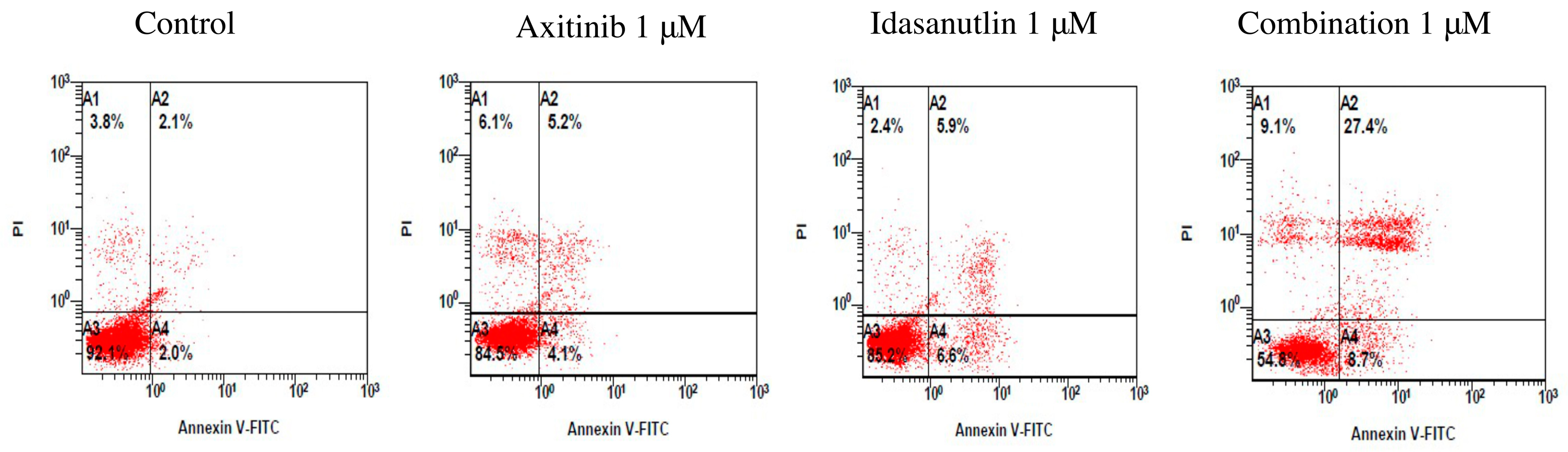
3.3. Evaluation of Cell Apoptosis Induced by Axitinib and Idasanutlin

Annexin V/propidium iodide assay was performed to evaluate the apoptosis effects of axitinib and idasanutlin. MCF-7 cells were treated with axitinib, idasanutlin, or the combination of both axitinib and idasanutlin for 24 h, followed by Annexin V/PI staining to detect apoptotic cells using flow cytometry. The top right quadrant represents late apoptotic cells with high levels of Annexin V and PI staining. The bottom right quadrant represents early apoptotic cells with high levels of Annexin V and low levels of PI staining. Axitinib monotherapy showed a slight increase in MCF-7 apoptotic cells, whereas idasanutlin monotherapy demonstrated a moderate increase in apoptotic cells compared to the control (9.3% and 12.5% apoptotic cells, respectively), as shown in Figure 3. On the other hand, the combination therapy of axitinib and idasanutlin demonstrated a significant shift in the pattern of viable cells to early apoptosis and late apoptosis. It was observed that early apoptosis had increased by 8.7%, whereas late apoptosis had increased by 27.4% when compared with the control group. The findings showed that after 24 h of exposure to the combination therapy of axitinib and idasanutlin, the proportion of apoptotic cells had increased significantly compared to monotherapies and control (Figure 3).

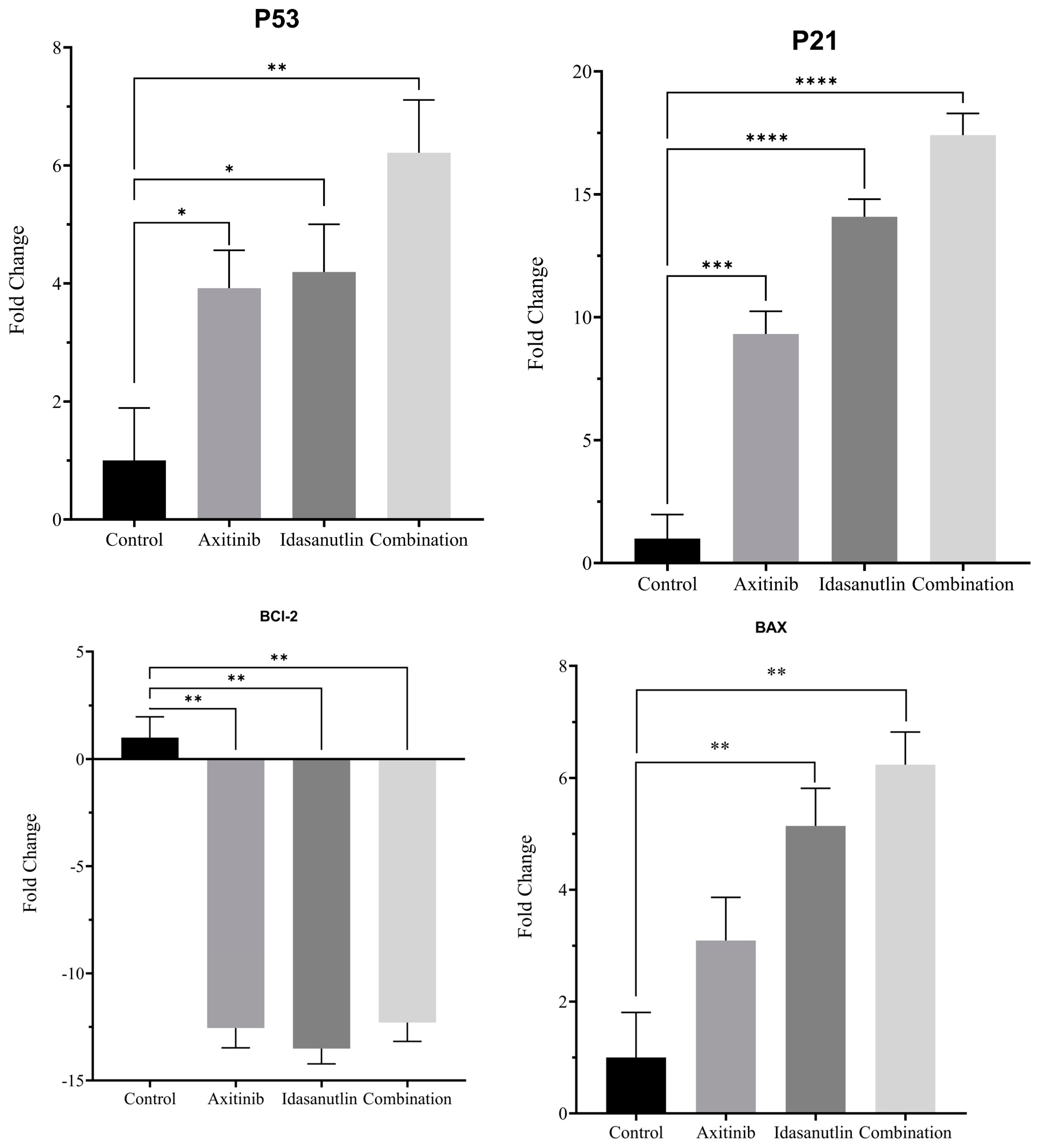
3.4. Effect of Axitinib and Idasanutlin on Gene Expression of Apoptosis Markers

Quantitative real-time PCR was performed to investigate the mRNA levels of apoptotic markers involving p53, p21, BAX, and BCL-2 in MCF-7 cells treated with axitinib (5 μM) or idasanutlin (5 μM) monotherapies or (axitinib 1 μM + idasanutlin 1 μM) treatment for 24 h. Our findings revealed that all treatment groups had significantly higher levels of expression of the apoptotic genes p53 and p21, while the antiapoptotic gene BCL-2 was significantly lower (p < 0.01) compared to the control group (Figure 4). In addition, idasanutlin alone and combined with axitinib significantly increased the expression of BAX (a pro-apoptotic member of the BCL-2 family) compared with the control group (p < 0.01), whereas the increase in axitinib-treated cells was not significant.

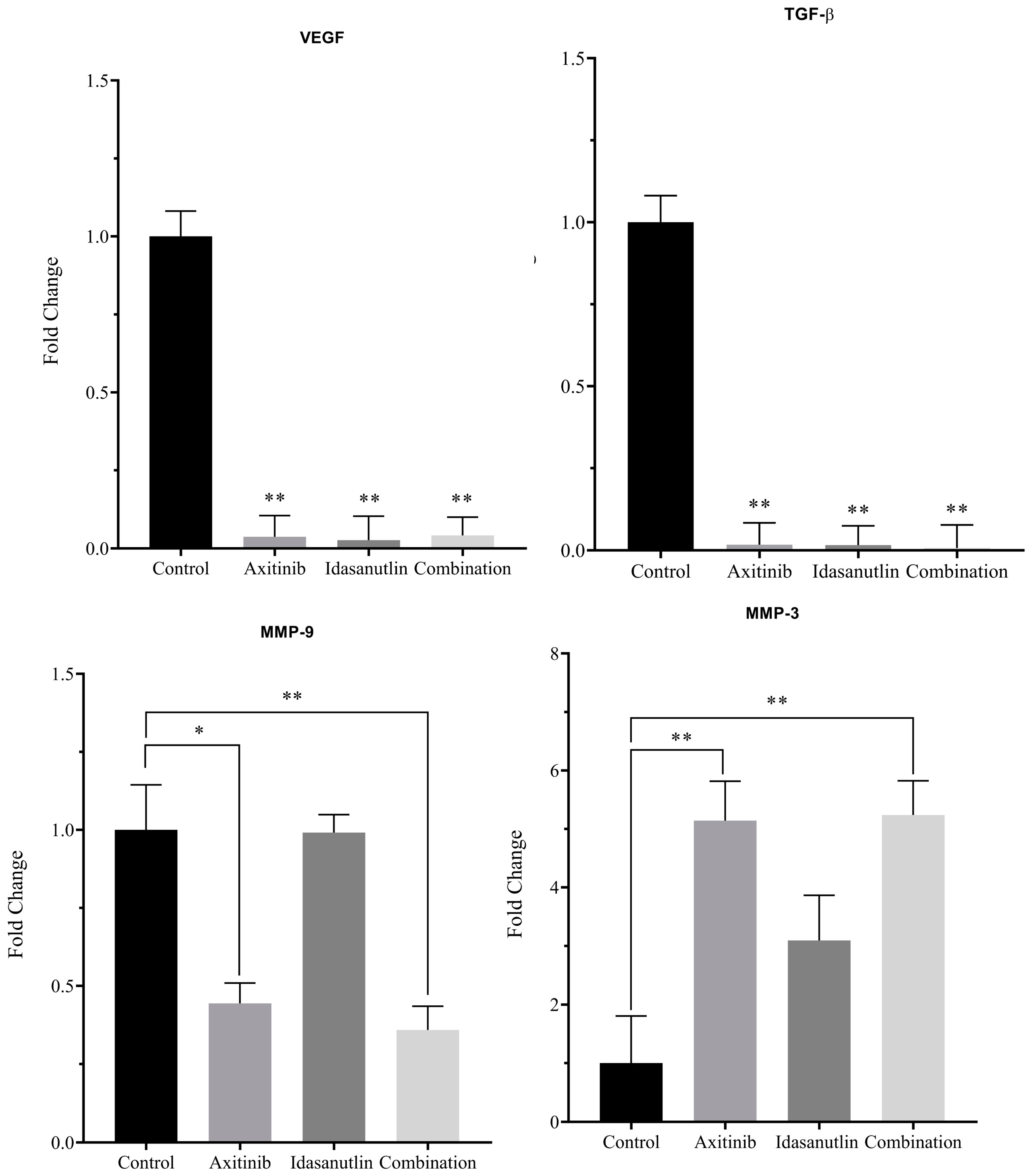
3.5. Effect of Axitinib and Idasanutlin on Gene Expression of VEGF, TGF-β, MMP9, and MMP3

Axitinib and idasanutlin, both alone and in combination, significantly reduced expression of the metastasis genes VEGF and TGF-β (p < 0.01) when compared to the control group. Furthermore, axitinib alone and in combination with idasanutlin significantly reduced MMP9 gene expression compared to the control group (p < 0.05 and p < 0.01, respectively). Additionally, cells treated with idasanutlin showed no change. However, the level of MMP3 expression did not change among the various treatment groups (Figure 5).

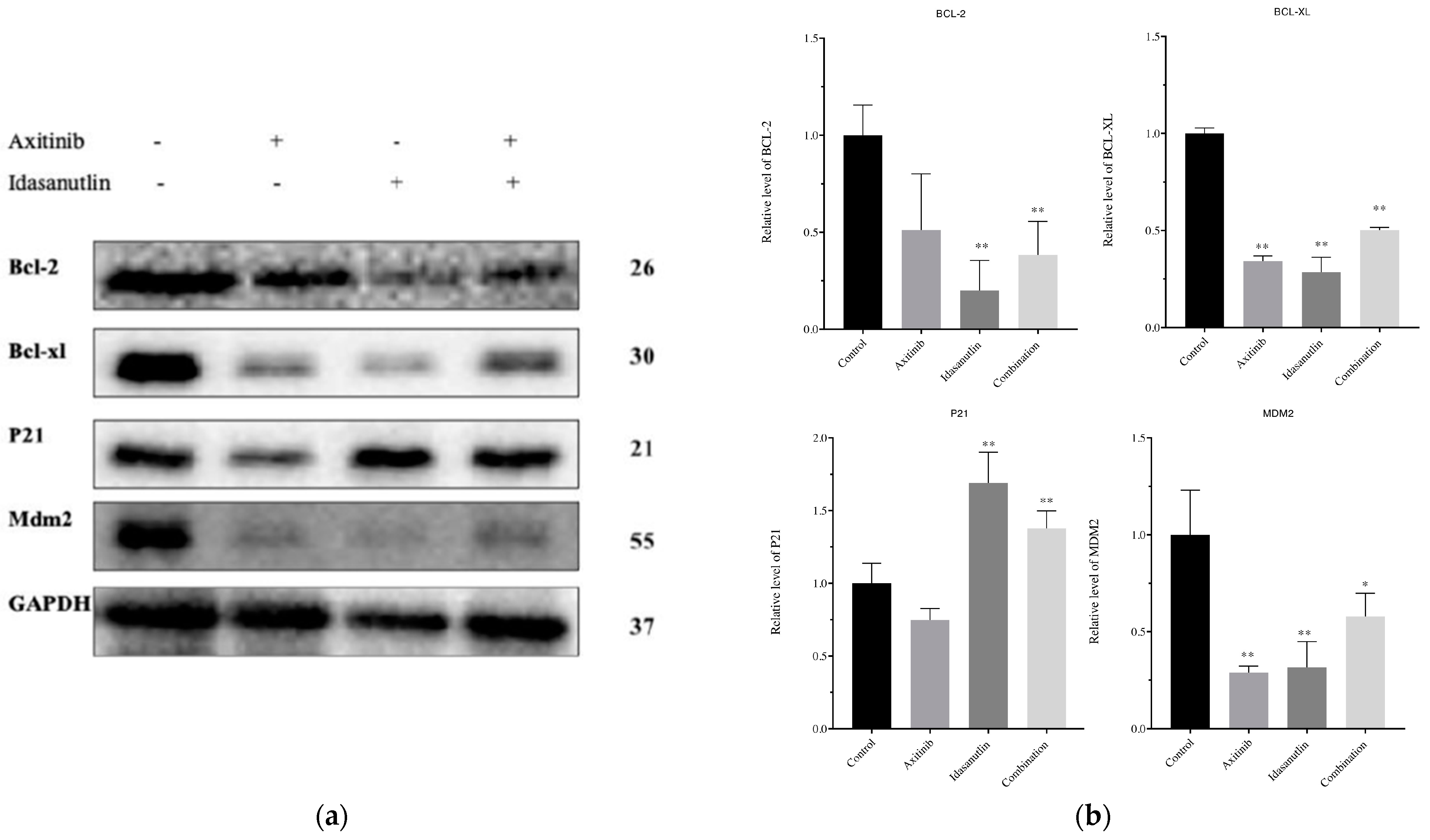
3.6. Effect of Axitinib and Idasanutlin on BCL-2, BCL-XL, p21, and MDM2 Protein Expression Levels in MCF-7 Cells

Western blot experiments were conducted to detect the modifications in protein levels, such as BCL-2, BCL-XL, p21, and MDM2 (Figure 6a). They were analyzed after axitinib (5 μM), idasanutlin (5 μM), or the combination of axitinib and idasanutlin (1 μM) treatment for 24 h. When compared to a control group, BCL-2 and BCL-XL protein levels were significantly lower in all treatment groups (p < 0.01). Furthermore, axitinib and idasanutlin significantly decreased MDM2 protein levels compared to the control group (p < 0.01 and p < 0.05, respectively). In addition, idasanutlin alone and in combination with axitinib elevated the p21 protein (p < 0.01), but there was no significant difference with axitinib alone.

4. Discussion

A treatment strategy that combines antiangiogenic agents with other chemotherapeutic agents has shown optimal therapeutic outcomes with reduced drug resistance in many malignancies [12]. Conversely, treatment failure, commonly seen in monotherapy regimens, subsequent disappointing outcomes owing to lack of efficacy, severe toxicities, and drug resistance are major reasons for therapeutic failure. Inhibiting the MDM2–p53 axis and targeting the VEGF receptor potentially maximize the anticancer effect and minimize the toxicity of both agents. In solid tumors, it has been shown that axitinib alone demonstrated a preferential anticancer effect compared to targeted chemotherapeutic agents, such as sorafenib, in renal cell carcinoma treatment [13,14,15]. Axitinib effect inhibited cell proliferation in various cancer types, including non-small cell lung cancer [16], prostate [17], and neuroblastoma [18]. Similarly, compared to nutlin-3 (a first-generation MDM2 antagonist), idasanutlin demonstrated higher efficacy, selectivity, and oral bioavailability to inhibit the MDM2–p53 interaction and restore p53 protective functions [19,20]. In this present study, we further confirmed that axitinib inhibited cell growth in MCF-7 with an inhibitory concentration (IC50) of around 30 μmol/L. Additionally, we found that idasanutlin exhibited tumor cytostatic activity in MCF-7 with an inhibitory concentration (IC50) of around 10 μmol/L. These findings are consistent with previous reports evaluating the inhibition of proliferation in a variety of cancer types, such as neuroblastoma [21] and breast cancer in vitro models [22]. In our experiments, the estimated difference in the antiproliferative effects of combining axitinib and idasanutlin therapy on MCF-7 showed considerable synergistic effects, with a significant reduction in cell viability. The observed effects can be explained by the antitumorigenic effect of axitinib, which has been shown to cause cell cycle arrest at the G2/M phase, apoptosis, and antiangiogenic activities [23,24]. Similarly, inhibiting the MDM2–p53 interaction and increasing p53 activity resulted in the expression of downstream transcriptional targets, such as cell cycle regulators, including p21. Of note, p21 plays an essential role in cell cycle processes, and its upregulation triggered a cell cycle arrest and a reduction in tumor volume in mice with a prostate tumor [25,26]. Our results showed that idasanutlin, either as monotherapy or in combination with axitinib, increased the expression of p21 protein levels, while axitinib showed no significant difference, suggesting optimal molecular anticancer effects. We found that treating MCF-7 with axitinib or idasanutlin increased the mRNA levels of apoptotic BAX, p21, and p53. It significantly decreased the expression of the antiapoptotic gene BCL-2 (p < 0.01), as previously shown [27,28]. Based on the present findings, axitinib, idasanutlin, or a combination of both, decreased the expression of the antiapoptotic proteins BCL-2 and BCL-XL. Overexpression of antiapoptotic proteins BCL-2 and BCL-XL inhibits cytochrome C release in response to apoptotic stimuli, which is essential for regulating the mitochondrial “intrinsic” apoptotic pathway [29,30,31]. Proapoptotic and cell cycle regulators represent an efficient treatment goal, potentially when treated by targeted therapy with an optimal safety profile.
In this present study, idasanutlin treatment triggered the killing of cancer cells through cell cycle arrest and apoptosis, and this cytotoxic effect is a well-known feature of MDM2–p53 inhibitors as previously reported in a variety of in vitro and tumor-implemented animal cancer models [28,32,33,34]. In addition, our findings showed that a lower dose (1 μM) combining axitinib and idasanutlin significantly induced cellular apoptosis, which appears to be very superior to monotherapy therapy. In turn, employing agents with divergent molecular targets will conceivably improve the safety profile without compromising the therapeutic effect of both agents.
MMP9, TGF-β, and VEGF expression are all linked to tumor progression, invasion, and metastasis. As shown in Figure 5, axitinib treatment and in combination with idasanutlin significantly reduce VEGF, MMP9, and TGF-β mRNA expression levels, indicating that the combination treatment has the best antimetastatic effect (Figure 3). MMP9 is particularly important due to the observed increase in premetastatic lung endothelial cells and macrophages through VEGFR1 activation [35]. Although TGF-β shows a paradoxical effect in cancer, TGF-β-pathway-signaling activation enhances migration and metastatic dissemination into blood arteries and lymph nodes [36,37]. This current study’s findings suggest that the VEGF/MMP9/TGF-β pathway may play a significant role in promoting breast tumorigenesis and the subsequent lethal metastasis stage.

5. Conclusions

In conclusion, our study examines the effect of axitinib, idasanutlin, and the combination of both on a human breast cancer in vitro model (MCF-7). The results of the current study indicate that combining axitinib and idasanutlin inhibited cancer growth and migration in the MCF-7 cell line in a superior way when compared to many therapies. The postulated mechanisms of the antitumorigenic effect might be attributed to the ability of the combination therapy to induce the expression of apoptotic markers such as p53, p21, and BAX and the suppression of antiapoptotic markers BCL-2 and BCL-XL. Additionally, after axitinib and/or idasanutlin treatment, predominant angiogenic growth factor (VEGF) and pro-metastatic mediator (TGF-β) targeting was achieved. This, in turn, will not only kill cancer cells but also limit other malignant processes and impede the metastatic cascade. Limitations include the fact that the cell-specific effect of MCF-7 cannot be excluded after treatment with MDM2 and VEGF inhibitors. Advanced molecular analysis of the treated cells might offer additional mechanistic discovery and open a new avenue in the management of breast cancer. Moreover, other in vitro breast cancer models with distinct molecular features might be exploited to assess the visibility of targeting other breast cancer types. Furthermore, an in vivo model should be examined to further validate these findings.

Author Contributions

Conceptualization, A.M.A. and K.A.; methodology, K.A. and A.M.A.; software, K.A. and A.M.A.; validation, A.M.A., K.A., A.S.A. and Y.A.; formal analysis, K.A. and Y.A.; investigation, K.A., A.M.A., A.Z.A. and Y.A.; resources, A.Z.A.; data curation, K.A.; writing—original draft preparation, Y.A., A.M.A. and K.A.; writing—review and editing, A.M.A., A.S.A., F.T.A., J.A.A., T.J., M.M.A. and G.A.; visualization, J.A.A. and G.A.; supervision, K.A. and A.Z.A.; project administration, A.M.A. and K.A.; funding acquisition, A.M.A. and K.A. All authors have read and agreed to the published version of the manuscript.

Funding

This research was supported by the Deanship of Scientific Research, Imam Mohammad Ibn Saud Islamic University (IMSIU), Saudi Arabia, Grant No. (191213005).

Institutional Review Board Statement

Not applicable.

Informed Consent Statement

Not applicable.

Data Availability Statement

Not applicable.

Acknowledgments

The author extends his appreciation to the Deanship of Scientific Research, Imam Mohammad Ibn Saud Islamic University (IMSIU), Saudi Arabia and the research center, College of Pharmacy, King Saud University for financial support.

Conflicts of Interest

The authors declare no conflict of interest.

References

  1. McDonald, E.S.; Clark, A.S.; Tchou, J.; Zhang, P.; Freedman, G.M. Clinical Diagnosis and Management of Breast Cancer. J. Nucl. Med. 2016, 57 (Suppl 1), 9S–16S. [Google Scholar] [CrossRef] [PubMed] [Green Version]
  2. Sever, R.; Brugge, J.S. Signal transduction in cancer. Cold Spring Harb. Perspect. Med. 2015, 5, a006098. [Google Scholar] [CrossRef] [PubMed] [Green Version]
  3. Shiovitz, S.; Korde, L.A. Genetics of breast cancer: A topic in evolution. Ann. Oncol. 2015, 26, 1291–1299. [Google Scholar] [CrossRef] [PubMed]
  4. Loman, N.; Johannsson, O.; Bendahl, P.; Borg, Å.; Fernö, M.; Olsson, H. Steroid receptors in hereditary breast carcinomas associated with BRCA1 or BRCA2 mutations or unknown susceptibility genes. Cancer Interdiscip. Int. J. Am. Cancer Soc. 1998, 83, 310–319. [Google Scholar] [CrossRef]
  5. Moll, U.M.; Petrenko, O. The MDM2-p53 interaction. Mol. Cancer Res. 2003, 1, 1001–1008. [Google Scholar]
  6. Duffy, M.J.; Synnott, N.C.; O’Grady, S.; Crown, J. Targeting p53 for the treatment of cancer. Semin. Cancer Biol. 2022, 79, 58–67. [Google Scholar] [CrossRef]
  7. Lehmann, C.; Friess, T.; Birzele, F.; Kiialainen, A.; Dangl, M. Superior anti-tumor activity of the MDM2 antagonist idasanutlin and the Bcl-2 inhibitor venetoclax in p53 wild-type acute myeloid leukemia models. J. Hematol. Oncol. 2016, 9, 50. [Google Scholar] [CrossRef] [Green Version]
  8. Folkman, J. Role of angiogenesis in tumor growth and metastasis. In Seminars in Oncology; Elsevier: Amsterdam, The Netherlands, 2002; Volume 29, pp. 15–18. [Google Scholar]
  9. Soleimani, M.; Nappi, L.; Kollmannsberger, C. Avelumab and axitinib combination therapy for the treatment of advanced renal cell carcinoma. Futur. Oncol. 2020, 16, 3021–3034. [Google Scholar] [CrossRef]
  10. Bellesoeur, A.; Carton, E.; Alexandre, J.; Goldwasser, F.; Huillard, O. Axitinib in the treatment of renal cell carcinoma: Design, development, and place in therapy. Drug Des. Devel. Ther. 2017, 11, 2801–2811. [Google Scholar] [CrossRef] [Green Version]
  11. Chomczynski, P. A reagent for the single-step simultaneous isolation of RNA, DNA and proteins from cell and tissue samples. Biotechniques 1993, 15, 532–534. [Google Scholar]
  12. Boere, I.A.; Hamberg, P.; Sleijfer, S. It takes two to tango: Combinations of conventional cytotoxics with compounds targeting the vascular endothelial growth factor-vascular endothelial growth factor receptor pathway in patients with solid malignancies. Cancer Sci. 2010, 101, 7–15. [Google Scholar] [CrossRef]
  13. Motzer, R.J.; Escudier, B.; Tomczak, P.; Hutson, T.E.; Michaelson, M.D.; Negrier, S.; Oudard, S.; Gore, M.E.; Tarazi, J.; Hariharan, S.; et al. Axitinib versus sorafenib as second-line treatment for advanced renal cell carcinoma: Overall survival analysis and updated results from a randomised phase 3 trial. Lancet. Oncol. 2013, 14, 552–562. [Google Scholar] [CrossRef]
  14. Cohen, E.E.W.; Rosen, L.S.; Vokes, E.E.; Kies, M.S.; Forastiere, A.A.; Worden, F.P.; Kane, M.A.; Sherman, E.; Kim, S.; Bycott, P. Axitinib is an active treatment for all histologic subtypes of advanced thyroid cancer: Results from a phase II study. J. Clin. Oncol. 2008, 26, 4708. [Google Scholar] [CrossRef] [PubMed]
  15. Schiller, J.H.; Larson, T.; Ou, S.-H.I.; Limentani, S.; Sandler, A.; Vokes, E.; Kim, S.; Liau, K.; Bycott, P.; Olszanski, A.J. Efficacy and safety of axitinib in patients with advanced non–small-cell lung cancer: Results from a phase II study. J. Clin. Oncol. 2009, 27, 3836–3841. [Google Scholar] [CrossRef] [PubMed]
  16. Hu-Lowe, D.D.; Zou, H.Y.; Grazzini, M.L.; Hallin, M.E.; Wickman, G.R.; Amundson, K.; Chen, J.H.; Rewolinski, D.A.; Yamazaki, S.; Wu, E.Y. Nonclinical antiangiogenesis and antitumor activities of axitinib (AG-013736), an oral, potent, and selective inhibitor of vascular endothelial growth factor receptor tyrosine kinases 1, 2, 3. Clin. Cancer Res. 2008, 14, 7272–7283. [Google Scholar] [CrossRef] [PubMed] [Green Version]
  17. Canu, B.; Fioravanti, A.; Orlandi, P.; Di Desidero, T.; Alì, G.; Fontanini, G.; Di Paolo, A.; Del Tacca, M.; Danesi, R.; Bocci, G. Irinotecan synergistically enhances the antiproliferative and proapoptotic effects of axitinib in vitro and improves its anticancer activity in vivo. Neoplasia 2011, 13, 217-IN3. [Google Scholar] [CrossRef] [PubMed]
  18. Rössler, J.; Monnet, Y.; Farace, F.; Opolon, P.; Daudigeos-Dubus, E.; Bourredjem, A.; Vassal, G.; Geoerger, B. The selective VEGFR1-3 inhibitor axitinib (AG-013736) shows antitumor activity in human neuroblastoma xenografts. Int. J. cancer 2011, 128, 2748–2758. [Google Scholar] [CrossRef] [PubMed]
  19. Ding, Q.; Zhang, Z.; Liu, J.-J.; Jiang, N.; Zhang, J.; Ross, T.M.; Chu, X.-J.; Bartkovitz, D.; Podlaski, F.; Janson, C. Discovery of RG7388, a potent and selective p53–MDM2 inhibitor in clinical development. J. Med. Chem. 2013, 56, 5979–5983. [Google Scholar] [CrossRef] [PubMed]
  20. Tovar, C.; Graves, B.; Packman, K.; Filipovic, Z.; Xia, B.H.M.; Tardell, C.; Garrido, R.; Lee, E.; Kolinsky, K.; To, K.-H. MDM2 small-molecule antagonist RG7112 activates p53 signaling and regresses human tumors in preclinical cancer models. Cancer Res. 2013, 73, 2587–2597. [Google Scholar] [CrossRef] [PubMed] [Green Version]
  21. Vernooij, L.; Bate-Eya, L.T.; Alles, L.K.; Lee, J.Y.; Koopmans, B.; Jonus, H.C.; Schubert, N.A.; Schild, L.; Lelieveld, D.; Egan, D.A. High-throughput screening identifies idasanutlin as a resensitizing drug for venetoclax-resistant neuroblastoma cells. Mol. Cancer Ther. 2021, 20, 1161–1172. [Google Scholar] [CrossRef] [PubMed]
  22. Bianco, G.; Coto-Llerena, M.; Gallon, J.; Taha-Mehlitz, S.; Kancherla, V.; Konantz, M.; Srivatsa, S.; Montazeri, H.; Panebianco, F.; De Menna, M. GATA3 and MDM2 are synthetic lethal in estrogen receptor-positive breast cancers. bioRxiv 2020. [Google Scholar] [CrossRef]
  23. Stehle, F.; Schulz, K.; Fahldieck, C.; Kalich, J.; Lichtenfels, R.; Riemann, D.; Seliger, B. Reduced immunosuppressive properties of axitinib in comparison with other tyrosine kinase inhibitors. J. Biol. Chem. 2013, 288, 16334–16347. [Google Scholar] [CrossRef] [PubMed] [Green Version]
  24. Zhang, R.-R.; Wang, H.; Hui, N.; Zhang, P. Enhanced antitumor effect of axitinib synergistic interaction with AG490 via VEGFR2/JAK2/STAT3 signaling mediated epithelial-mesenchymal transition in cervical cancer in vitro. Asian Biomed. 2013, 7, 39–49. [Google Scholar]
  25. Shamloo, B.; Usluer, S. p21 in cancer research. Cancers 2019, 11, 1178. [Google Scholar] [CrossRef] [PubMed] [Green Version]
  26. Eastham, J.A.; Hall, S.J.; Sehgal, I.; Wang, J.; Timme, T.L.; Yang, G.; Connell-Crowley, L.; Elledge, S.J.; Zhang, W.-W.; Harper, J.W. In vivo gene therapy with p53 or p21 adenovirus for prostate cancer. Cancer Res. 1995, 55, 5151–5155. [Google Scholar] [PubMed]
  27. Peng, N.X.; Liu, C.X.; Wang, X.S.; Zhang, Z.J.; Liao, S.C. Combination of axitinib and dasatinib for anti-cancer activities in two prostate cancer cell lines. Bangladesh J. Pharmacol. 2016, 11, 130–137. [Google Scholar] [CrossRef] [Green Version]
  28. Zanjirband, M.; Edmondson, R.J.; Lunec, J. Pre-clinical efficacy and synergistic potential of the MDM2-p53 antagonists, Nutlin-3 and RG7388, as single agents and in combined treatment with cisplatin in ovarian cancer. Oncotarget 2016, 7, 40115. [Google Scholar] [CrossRef] [Green Version]
  29. Li, F.; Ambrosini, G.; Chu, E.Y.; Plescia, J.; Tognin, S.; Marchisio, P.C.; Altieri, D.C. Control of apoptosis and mitotic spindle checkpoint by survivin. Nature 1998, 396, 580–584. [Google Scholar] [CrossRef]
  30. Tawfik, K.; Kimler, B.F.; Davis, M.K.; Fan, F.; Tawfik, O. Prognostic significance of Bcl-2 in invasive mammary carcinomas: A comparative clinicopathologic study between “triple-negative” and non–“triple-negative” tumors. Hum. Pathol. 2012, 43, 23–30. [Google Scholar] [CrossRef]
  31. Danial, N.N.; Korsmeyer, S.J. Cell death: Critical control points. Cell 2004, 116, 205–219. [Google Scholar] [CrossRef] [Green Version]
  32. Moreno-Smith, M.; Lakoma, A.; Chen, Z.; Tao, L.; Scorsone, K.A.; Schild, L.; Aviles-Padilla, K.; Nikzad, R.; Zhang, Y.; Chakraborty, R. p53 nongenotoxic activation and mTORC1 inhibition lead to effective combination for neuroblastoma therapy. Clin. Cancer Res. 2017, 23, 6629–6639. [Google Scholar] [CrossRef] [Green Version]
  33. Cui, Y.; Zhou, J.; Rong, F. Combination of metformin and RG7388 enhances inhibition of growth and induction of apoptosis of ovarian cancer cells through the PI3K/AKT/mTOR pathway. Biochem. Biophys. Res. Commun. 2020, 533, 665–671. [Google Scholar] [CrossRef]
  34. Fan, X.; Wang, Y.; Song, J.; Wu, H.; Yang, M.; Lu, L.; Weng, X.; Liu, L.; Nie, G. MDM2 inhibitor RG7388 potently inhibits tumors by activating p53 pathway in nasopharyngeal carcinoma. Cancer Biol. Ther. 2019, 20, 1328–1336. [Google Scholar] [CrossRef] [PubMed]
  35. Hiratsuka, S.; Nakamura, K.; Iwai, S.; Murakami, M.; Itoh, T.; Kijima, H.; Shipley, J.M.; Senior, R.M.; Shibuya, M. MMP9 induction by vascular endothelial growth factor receptor-1 is involved in lung-specific metastasis. Cancer Cell 2002, 2, 289–300. [Google Scholar] [CrossRef] [PubMed] [Green Version]
  36. Dai, M.; Al-Odaini, A.A.; Fils-Aimé, N.; Villatoro, M.A.; Guo, J.; Arakelian, A.; Rabbani, S.A.; Ali, S.; Lebrun, J.J. Cyclin D1 cooperates with p21 to regulate TGFβ-mediated breast cancer cell migration and tumor local invasion. Breast Cancer Res. 2013, 15, 3246. [Google Scholar] [CrossRef] [PubMed] [Green Version]
  37. Zhang, M.; Zhang, Y.Y.; Chen, Y.; Wang, J.; Wang, Q.; Lu, H. TGF-β Signaling and Resistance to Cancer Therapy. Front. Cell Dev. Biol. 2021, 9, 786728. [Google Scholar] [CrossRef]
Figure 1. Cytotoxicity of axitinib (a), idasanutlin (b), and combination (c) of both in MCF-7 cells. MCF-7 cells were treated with 10-fold serial dilution increase in axitinib or idasanutlin concentrations (10 nM–30 μM) and with an increased concentration of the combination therapy of axitinib and idasanutlin. The MTT assay was used to investigate the effects of axitinib, idasanutlin, and the combination on the cell viability. Cell viability was expressed as a percentage of live cells relative to 0 nm of the treatment. Medications showed a concentration-dependent reduction in cell viability. Results were expressed as mean ± standard deviation (SD). * p < 0.05 vs. control group.
Scipharm 91 00012 g001
Figure 2. Effects of axitinib and idasanutlin on the migration of MCF-7 cells. (a) Representative microscopic image showing MCF-7 cells after scratches were made (0 h). Cells were treated for 24 h with axitinib ± idasanutlin at a concentration (axitinib 5 μM, idasanutlin 5 μM), combination 1 (axitinib + idasanutlin 100 nM), combination 2 (axitinib + idasanutlin 500 nM), and combination 3 (axitinib + idasanutlin 1 μM). The scratch edges were outlined with dashed white lines, and microscopic photos were taken at 0 h and later at 24 h (×40 magnification). (b) Bar chart representing the quantification of the area of scratch width that was refilled with the migrated MCF-7 cells. Results were expressed as mean ± standard deviation (SD). * p < 0.05 vs. control. ** p < 0.01 vs. monotherapies.
Scipharm 91 00012 g002
Figure 3. Effect of axitinib and idasanutlin on apoptosis induction. MCF-7 cells were treated with axitinib (1 μM) or idasanutlin (1 μM) monotherapies or the combination therapy (axitinib + idasanutlin, 1 μM) for 24 h. Treated cells were stained with Annexin V and PI and analyzed by flow cytometry. The percentage of apoptotic cells was slightly higher in idasanutlin compared to axitinib monotherapy (12.5% vs. 9.3%). The combination therapy showed a significant increase in the apoptotic population (36.1%).
Scipharm 91 00012 g003
Figure 4. Effect of axitinib and idasanutlin on mRNA level of BAX, p53, p21, and BCL-2 in MCF-7 cells. The MCF-7 cells were treated with axitinib (5 μM) or idasanutlin (5 μM) monotherapies and combination (axitinib 1 μM + idasanutlin 1 μM) for 24 h. The mRNA levels were determined by qRT-PCR. The data is presented as a mean ± standard deviation (SD). * p < 0.05; ** p < 0.01 vs. control group, *** p < 0.001 and **** p < 0.0001 vs. control group.
Scipharm 91 00012 g004
Figure 5. Effect of axitinib and idasanutlin on VEGF, TGF-β, MMP9, and MMP3 mRNA levels in MCF-7 cells. The MCF-7 cells were treated with axitinib (5 μM), idasanutlin (5 μM), and combination (axitinib 1 μM + idasanutlin 1 μM) for 24 h. The mRNA levels were determined by qRT-PCR. The data is presented as a mean ± standard deviation (SD). * p < 0.05 vs. control group, ** p < 0.01 vs. control group.
Scipharm 91 00012 g005
Figure 6. Effect of axitinib ± idasanutlin on protein levels in MCF-7 cells. MCF-7 cells were treated with axitinib (5 μM), idasanutlin (5 μM), or the combination of axitinib and idasanutlin (1 μM) for 24 h. (a) Representative immunoblots for protein levels of BCL-2, BCL-XL, MDM2, and p21 were determined by Western blot analysis. (b) The levels of BCL-2, BCL-XL, MDM2, and p21were depicted. * p < 0.05 vs. control group, and ** p < 0.01 vs. control group.
Scipharm 91 00012 g006
Table 1. The Sequence of Primers Used in qPCR Analysis.
GenePrimer
P53Forward Sequence CCCCTCCTGGCCCCTGTAATCTTC
Reverse Sequence GCAGCGCCTCACAACCTCGGTCAT
P21Forward Sequence GTTCCTTGTGGAGCCGGAGC
Reverse Sequence GGTACAAGACAGTGACAGGTC
BAXForward Sequence GTTTCATCCAGGATCGAGCAG
Reverse Sequence CATCTTCTTCCAGATGGTGA
BCL2Forward Sequence CCTGTGGATGACTGAGTACC
Reverse Sequence GAGACAGCCAGGAGAAATCA
GAPDHForward Sequence GTCTCCTCTGACTTCAACAGCG
Reverse Sequence ACCACCCTGTTGCTGTAGCCAA
MMP3Forward Sequence TATGAAGGAGAGGCTGATATAATG
Reverse Sequence TGTGAGTGAGTGATAGAGTGG
MMP9Forward Sequence GCCACTACTGTGCCTTTGAGTC
Reverse Sequence CCCTCAGAGAATCGCCAGTACT
VEGFForward Sequence TTGCCTTGCTGCTCTACCTCCA
Reverse Sequence GATGGCAGTAGCTGCGCTGATA
Table 2. List of Antibodies Used in The Western Blot Experiments.
ProteinMolecular WeightCompanyDilution Ratio
P2121 kDaCell Signaling Technology, USA1:1000
BCL226 kDaCell Signaling Technology, USA1:1000
BCL-XL30 kDaCell Signaling Technology, USA1:1000
MDM255 kDaSigma-Aldrich. (USA)1:1000
GAPDH37 kDaCell Signaling Technology, USA1:5000
Disclaimer/Publisher’s Note: The statements, opinions and data contained in all publications are solely those of the individual author(s) and contributor(s) and not of MDPI and/or the editor(s). MDPI and/or the editor(s) disclaim responsibility for any injury to people or property resulting from any ideas, methods, instructions or products referred to in the content.

