Article

Long-Term Paracetamol Treatment Impairs Cognitive Function and Brain-Derived Neurotrophic Factor in Adult Rat Brain

by
Laddawan Lalert
1,2,*,
Nutnicha Tantarungsee
3,
Tipthanan Chotipinit
4,
Wilawan Ji-au
4,
Anan Srikiatkhachorn
5 and
Supang Maneesri-le Grand
4,*
1
School of Medicine, Walailak University, Nakhon Si Thammarat 80160, Thailand
2
Research Center in Tropical Pathobiology, Walailak University, Nakhon Si Thammarat 80160, Thailand
3
Department of Biochemistry, Faculty of Medicine Siriraj Hospital, Mahidol University, Bangkok 10700, Thailand
4
Department of Pathology, Faculty of Medicine, Chulalongkorn University, Bangkok 10330, Thailand
5
Faculty of Medicine, King Mongkut’s Institute of Technology Ladkrabang, Bangkok 10520, Thailand
*
Authors to whom correspondence should be addressed.
Sci. Pharm. 2023, 91(1), 11; https://doi.org/10.3390/scipharm91010011
Submission received: 6 January 2023 / Revised: 4 February 2023 / Accepted: 9 February 2023 / Published: 13 February 2023

Abstract

:
Paracetamol (acetaminophen, APAP) is known as a safe pain reliever; however, its negative effects on the central nervous system have gradually been reported. We examined alterations in learning and memory, and brain-derived neurotrophic factor (BDNF) expression in the frontal cortex and hippocampus at different durations of APAP treatment in rats. Novel object recognition (NOR) and Morris water maze (MWM) paradigms were used to assess learning and memory in rats fed with 200 mg/kg APAP at single-dose, 15-day or 30-day treatments. BDNF expression was evaluated through immunohistochemistry and Western blotting. The single-dose APAP treatment did not alter the NOR performance. However, deficits in the NOR and MWM capacities were detected in the rats with longer durations of APAP treatment. An analysis of BDNF expression revealed no significant change in BDNF expression in the single-dose APAP treatment, while rats given APAP for extended periods as treatment showed a significant decrement in this protein in the frontal cortex and hippocampus. Short-term APAP treatment has no effect on learning and memory, or BDNF expression; however, long-term APAP exposure causes cognitive impairment. The diminishment of the BDNF level in the frontal cortex and hippocampus due to the long period of treatment with APAP may at least in part be involved in altered learning and memory in rats.

1. Introduction

Paracetamol or acetaminophen (APAP) is usually selected as the first drug of choice for both acute and chronic pain treatments due to its properties such as its high availability, inexpensiveness, and minimal side effects. However, several studies have linked the adverse effects of APAP treatment to homeostasis disturbances in the central nervous system (CNS) [1,2,3,4,5]. It has previously been demonstrated that a single-dose treatment with APAP at a dose under the dose required to induce hepatotoxicity could induce neuronal apoptosis in a rat cortex [3]. The adverse effects of a high-dose APAP treatment for a short period on neurobehaviors, including learning and memory, and anxiety-like behavior, have been established [2,6]. Additionally, in our earlier report, the hippocampus and frontal cortex, two parts of the brain known for learning and memory processes, experienced an elevation in oxidative stress and impaired synaptic integrity after having received a 30-day treatment of 200 mg/kg APAP [7].
It is known that the brain-derived neurotrophic factor (BDNF) is a growth factor that plays a key role in several neuronal activities, including synaptic plasticity, and learning and memory processes [8,9,10,11,12]. Among all brain regions, the distribution of BDNF and its major receptor, tyrosine receptor kinase B (TrkB), are highest in the hippocampus and cerebral cortex [13,14]. Several recent studies suggest a link between developing brain APAP exposure and a fluctuation in BDNF levels in several brain regions, as well as behavioral consequences later in life including recognition memory, anxiolytic-like behavior, sociability, and emotionality [15,16,17]. Nevertheless, knowledge related to the impact of APAP treatment on the BDNF expression in adults with a fully developed brain has never been explored.
This study, therefore, aimed to investigate alterations in learning and memory abilities, and BDNF expression in adult Wistar rats that received APAP at the dose of 200 mg/kg in three different regimens of treatment: single-dose (0-day), 15-day, and 30-day treatments.

2. Materials and Methods

2.1. Animals

Sixty adult male Wistar rats (weighing between 250 and 300 g) were purchased from the National Laboratory Animal Center, Mahidol University, Thailand. The rats were housed as five per cage in a controlled atmosphere (20 ± 22 °C) with a 12 h cycle of darkness and light. Standard food and drinking water were freely accessed by the animals. The protocols carried out in this study have already been accepted by the Animal Ethical Committee of the Faculty of Medicine, Chulalongkorn University, Thailand (CU–ACUC No. 23/57).

2.2. Drug Treatment

In this study, APAP (TYLENOL®, OLIC (Thailand) Ltd., Ayutthaya, Thailand), at a dose of 200 mg/kg was employed. This dose, after being converted from an animal dose in mg/kg to a human equivalent dose (HED) in mg/kg by the formula described by the US Food and Drug Administration (FDA), is equal to 32.25 mg/kg or 1935 mg per day for humans with a weight of 60 kg [18]. Since the FDA has recommended the dose of 4000 mg (in any 24 h period) as a maximum dose of APAP for treatment [19], it can be indicated that the dose of APAP used in the present study was within the therapeutic dose range [5,7].

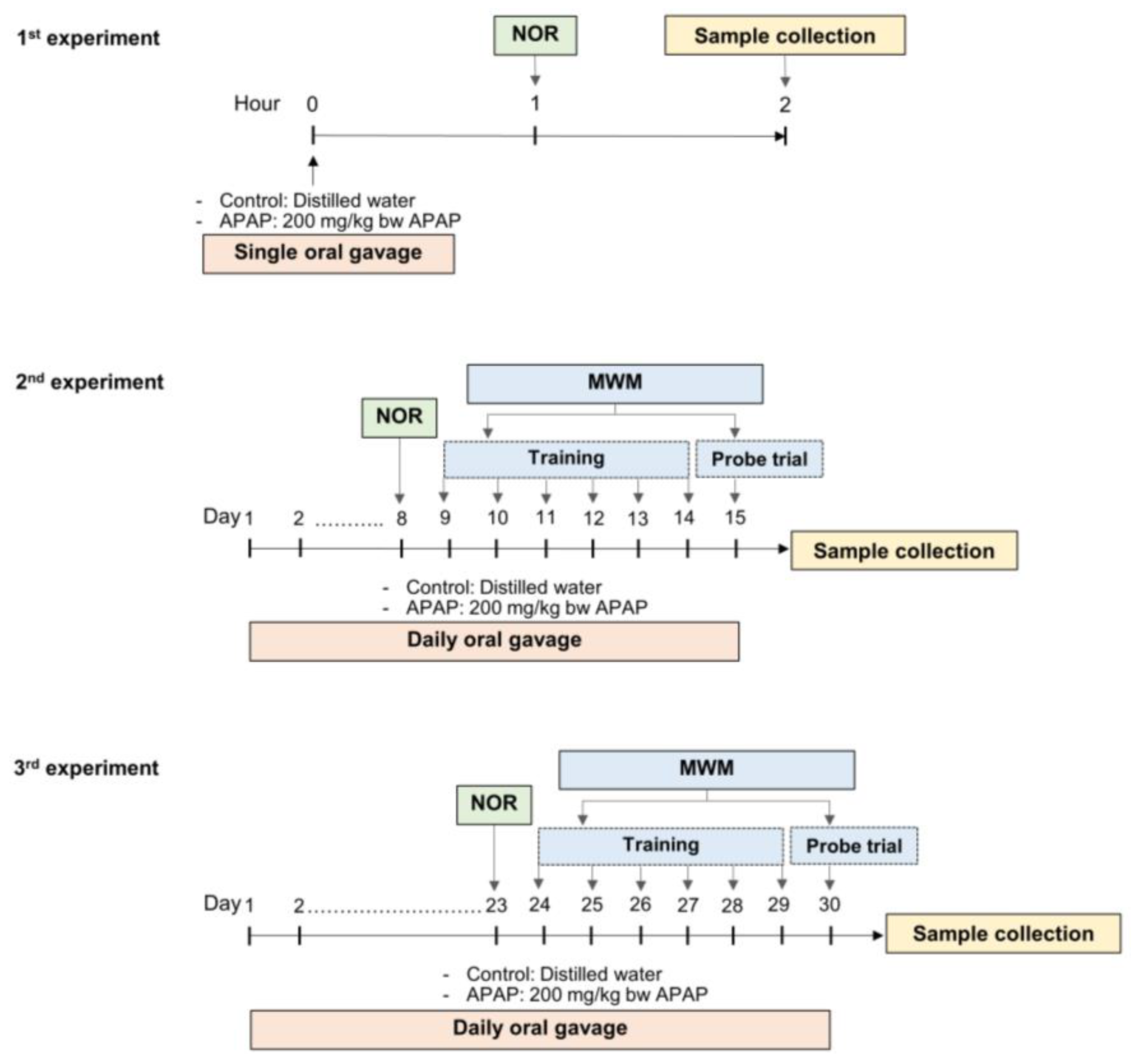
2.3. Experimental Design

The experiment in this study was separated into three experiments. In the first experiment, twenty rats were randomly selected and separated into the control and APAP-treated groups (n = 10). One hour prior to being assessed with the NOR test, 200 mg/kg APAP was fed to the rats in the APAP-treated group, whereas the control group received the same volume of distilled water. In the second experiment, APAP at the dose of 200 mg/kg or distilled water was given to the rats in the APAP-treated or control groups (n = 10) for 15 days, respectively. On day 8 of the treatment, the NOR test was performed 1 h following the treatment, and on day 9 of the treatment, the MWM test was initiated. For the MWM test, the rats were trained to find the hidden platform for 6 consecutive days, and their spatial memory was evaluated with the probe trial on the last day of the treatment. For the third experiment, APAP or distilled water was given to the rats for 30 days (n = 10). The NOR test was conducted on day 23, and the MWM test was performed between days 24 and 30 of the treatment. All behaviors were analyzed from videotape by experimenters who were blinded to the experimental conditions. The experimental design of this present study is demonstrated in Figure 1.

2.4. Novel Object Recognition (NOR) Test

The NOR test was conducted according to the procedure described by Morley et al. [20] with a minor adjustment. The test was performed in an acrylic box (100 × 100 cm wide floor and 50 cm high wall), and all the rats’ behaviors throughout the test were captured by a digital video camera positioned above the box. For the test procedure, the rats were habituated in the empty box for 5 min. After a 1 h delayed stay in their home cage, the rats were trained by experience with two identified objects (objects A and B) for 10 min. Ten min later, the rats were again exposed to two objects, where one old object was replaced by a new one (object C) for 10 min. The exploration behaviors of the rats were defined as sniffing or having their head directed toward the object within a 2 cm perimeter around the object. At the end of each trial, the arena and all objects were cleaned with 70% ethanol to eliminate olfactory cues. The calculation of preference and recognition indexes were based on Equations (1) and (2).
The   preference   index   ( % ) = Total   time   spent   for   exploring   object   A Total   time   spent   for   exploring   both   objects   during   training   trial × 100
The   recognition   index   ( % ) = Total   time   spent   for   exploring   object   C Total   time   spent   for   exploring   both   objects   during   testing   trial × 100

2.5. Morris Water Maze (MWM) Test

The MWM test was carried out in accordance with the procedure introduced by Morris in 1981 [21] with minor modifications. The test apparatus consisted of a circular pool with a 200 cm diameter and 60 cm depth, filled with water (23–24 °C) to a height of 30 cm above the base. Different visual cues were labeled on the wall of the pool, which were obviously seen by the animals. A hidden platform with diameter of 20 cm was submerged 2 cm below the water surface and constantly located in one of the quadrants of the pool. In the training session, the rats were trained on 6 consecutive days with 3 trials per day to find the hidden platform. If the rats failed to reach the hidden platform within 90 s, they were guided by placing them on the platform for 15 s. The average time spent to reach the platform during the training trial was reported as the escape latency, which indicated rats’ spatial learning capacity. In the probe trial, the hidden platform was removed, and the rats were allowed to freely swim for 90 s in the pool. The time spent within the zone where the platform was previously located was recorded to determine a spatial memory.

2.6. Sample Collection

In the first experiment, rats were euthanized by intraperitoneal injection with 60 mg/kg of sodium pentobarbital after 1 h of the treatment. The whole blood (5 mL) was collected from all rats by the cardiac puncture method. To perform the histochemistry, the rats were transcardially perfused with precooled 250 mL of 0.1 M phosphate-buffered saline (PBS), pH 7.4, and then with a 4% paraformaldehyde solution until the rat was stiff. The brain and liver were collected, immerged in the fixative solution, and maintained at 4 °C for 48 h. For the Western blotting, after perfusion with PBS, the rats’ brains were removed, and the frontal cortex and hippocampus were quickly dissected on ice, immediately frozen in liquid nitrogen, and then kept at –80 °C for further investigation. For the second and third experiments, all rats were euthanized 24 h after the last treatment for sample collection, which was performed according to the first experiment’s procedures.

2.7. Immunohistochemistry (IHC)

The paraformaldehyde-fixed brains were dehydrated, processed, and embedded in paraffin wax. The sections with a 5 μm thickness were cut and placed on Superfrost plus slides (Thermoscientific, Portsmouth, NH, USA). After deparaffinization, the sections were incubated with citrate buffer pH 6.0 (Dako, Glostrup, Denmark), 3% hydrogen peroxide, and 3% normal horse serum (PAN Biotech GmbH, Aidenbach, Germany) in PBS. The sections were exposed to primary rabbit anti-BDNF antibody (1:2000; Abcam, Cambridge, UK) for 37 min at 37 °C. The signal of BDNF immunostaining was visualized using an ultraView Universal DAB Detection Kit (Ventana Medical Systems, Inc., Oro Valley, AZ, USA), which was processed in an automatic slide staining machine (Benchmark XT, Ventana Medical Systems, Inc., Oro Valley, AZ, USA). All slides were counterstained with hematoxylin, dehydrated, mounted, and cover-slipped with a mounting media before scanning using a slide scanner (Aperio ScanScope, Aperio, Vista, CA, USA).

2.8. Western Blotting (WB)

The frontal cortex and hippocampal proteins were extracted and measured in accordance with the prior study’s protocol [7]. The proteins were electrically separated in a 12.5% SDS–polyacrylamide gel and blotted onto nitrocellulose membranes (GE Healthcare Life Sciences, Buckinghamshire, UK). After blocking with 5% w/v of bovine serum albumin (Merck Millipore, Burlington, MA, USA) in tris-buffered saline containing 0.1% tween-20 pH 7.4 (TBS–T), the blots were incubated with rabbit anti-BDNF (1:500; Abcam, Cambridge, UK) or mouse anti-β-actin (1:3000; Sigma, St. Louis, MO, USA) antibodies overnight at 4 °C. After washing, the blots were exposed to appropriated horseradish peroxidase-coupled secondary antibodies (1:10,000; Sigma, St. Louis, MO, USA) and enhanced chemiluminescence system (ECL, GE Healthcare Life Sciences, Buckinghamshire, UK). The quantitative analysis of the protein band densities was conducted by using ImageJ software (National Institute of Health, Bethesda, MD, USA).

2.9. Statistical Analysis

A two-way analysis of variance (ANOVA) with a post hoc Bonferroni test was employed to analyze the escape latency in the MWM test, while a student’s t-test was applied for other parameters. Statistical significance was defined as p < 0.05.

3. Results

It is known that hepatotoxicity can interfere with neuronal homeostasis of the brain [22]. Therefore, in this study, the liver morphology as well as the liver enzymes indicating liver function (alanine aminotransferase, aspartate aminotransferase, and alkaline phosphatase) were monitored in all rats. The findings revealed that neither the hepatic morphology nor the levels of liver enzymes obtained from the rats receiving APAP treatment and the control group differed significantly (data not shown), suggesting that any alteration shown in the rats treated with APAP in the present study did not result from hepatotoxicity.

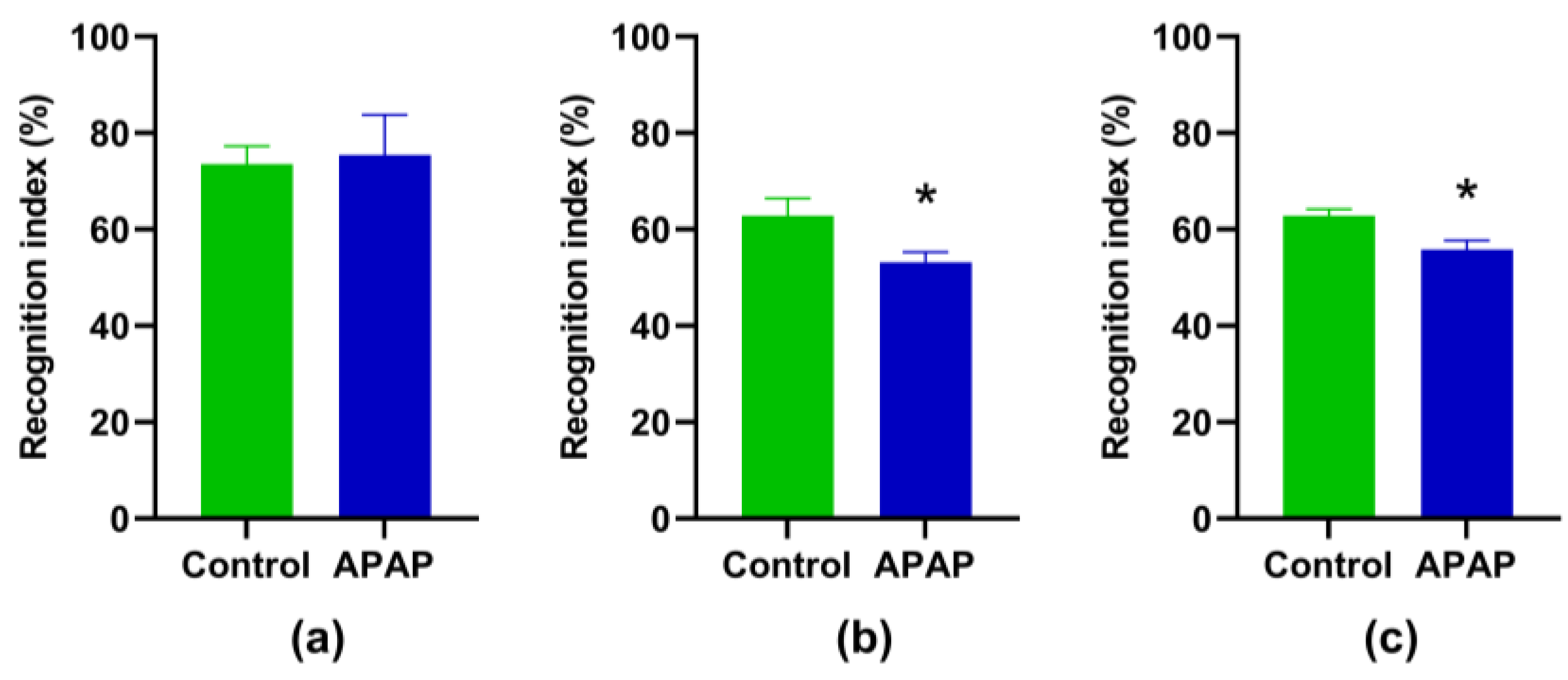
3.1. Effect of APAP Treatment on NOR Memory

The results demonstrated that there was no difference in the preference index among all groups (data not shown). When compared to the control, a single-dose APAP administration did not affect the recognition index (p > 0.05, Figure 2a). However, the rats given APAP treatment for the longer durations of treatment (8 and 23 days) showed a significantly lower recognition index (p < 0.05, Figure 2b,c).

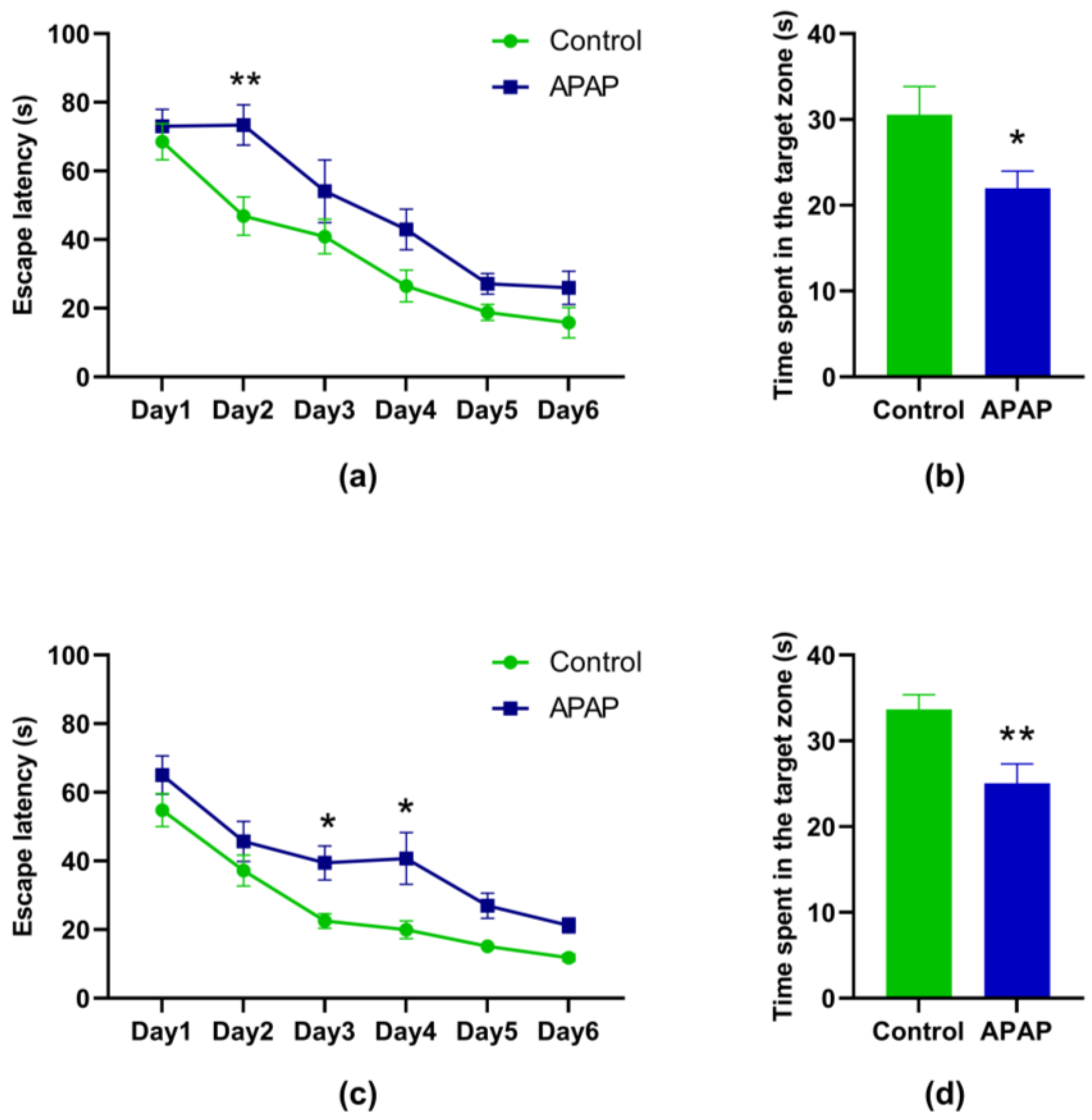
3.2. Effect of the APAP Treatment on the MWM Performance

To evaluate the MWM performance, the animals needed to be trained consecutively for 6 days; therefore, the MWM performance was not monitored in the rats with acute APAP treatment.
In the experiment with the 15-day treatment, the rats treated with APAP showed a significant increase in escape latency on the second day of the training trial (p < 0.01, Figure 3a), and a significant decrease in the time spent in the target zone was also detected in these rats as compared with those observed in the control group (p < 0.05, Figure 3b). The results obtained from the 30-day treatment were in line with what was observed in the experiment with the 15-day treatment. A significant increase in escape latency (on the third and fourth days of the training trial) and a significant decrease in the time spent in the target zone were observed in the 30-day APAP-treated rats as compared to the control group (p < 0.01, Figure 3c,d).

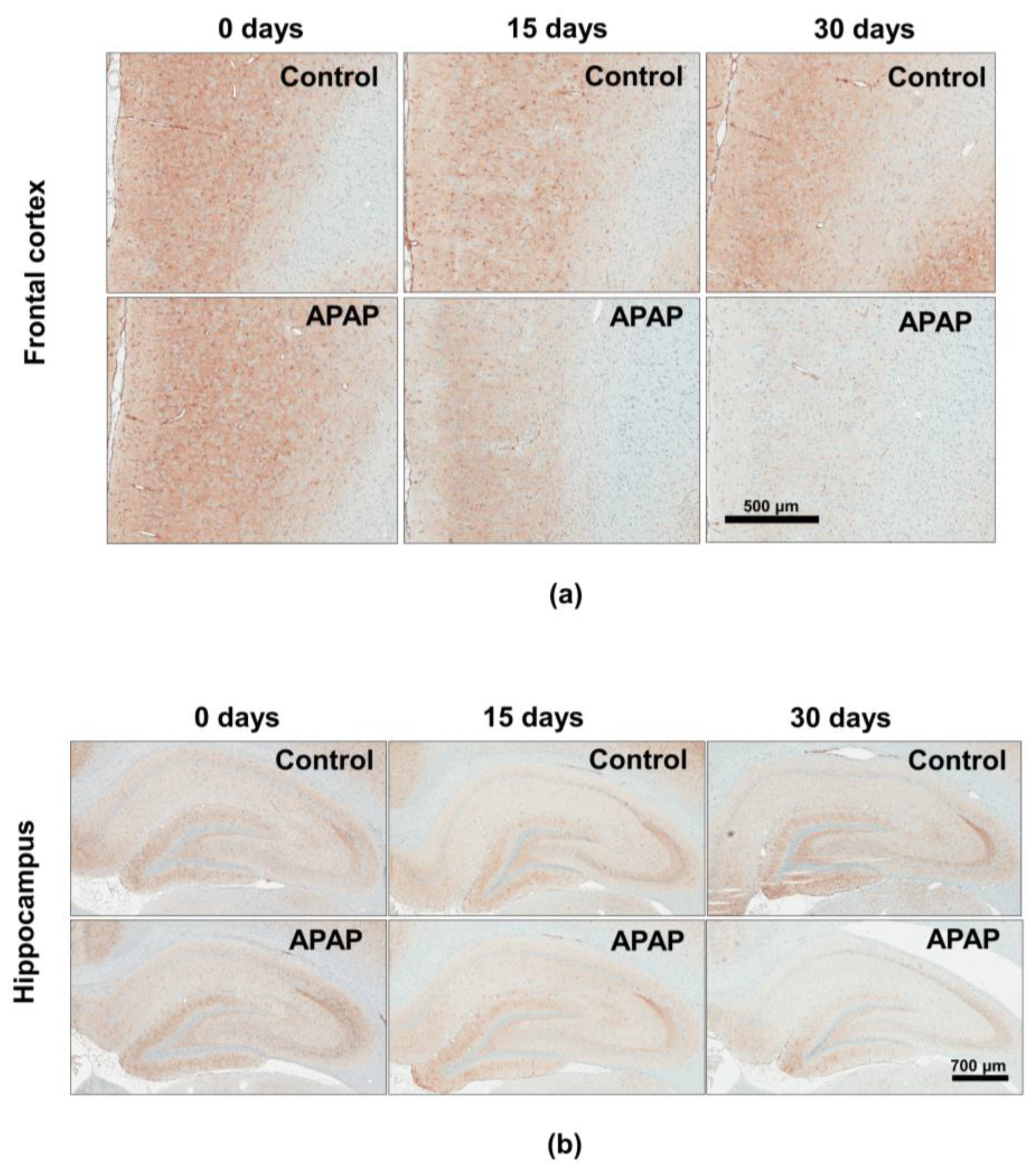
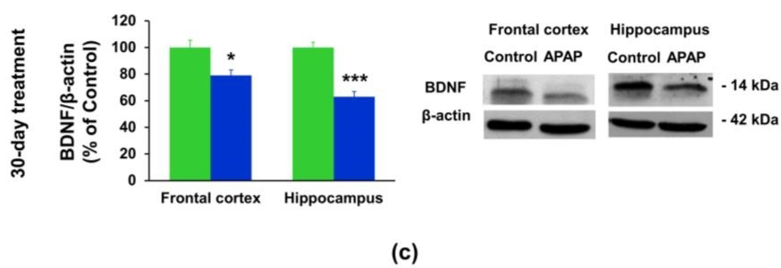
3.3. Effect of APAP Treatment on the BDNF Protein Expression

By using IHC, a strong staining (brown color of DAB staining) was observed in both the frontal cortex and hippocampus for the control and 0-day APAP-treated groups, while a weak staining was obviously observed in the 30-day APAP-treated group. This indicates that chronic APAP treatment decreased the expression of BDNF protein in both the frontal cortex and hippocampus of rats (Figure 4). The results obtained in the WB were in agreement with those observed in the IHC. It was shown that a single-dose APAP treatment had no effect on the expression of the BDNF protein in either the frontal cortex or the hippocampus (p > 0.05, Figure 5a). However, the results obtained from the study in the 15-day treatment demonstrated a significant reduction in BDNF protein in the hippocampus in the APAP-treated rats (p < 0.05, Figure 5b). The alteration in BDNF protein expression was clearly demonstrated in the experiment with the 30-day treatment. It was shown that the expression of BDNF protein in both the frontal cortex and hippocampus of APAP-treated rats was significantly reduced when compared with the control (p < 0.01 and p < 0.001, respectively, Figure 5c).

4. Discussion

The effects of APAP treatment on learning and memory have been shown in several studies. However, the precise mechanism underlying how APAP exposure affects cognitive outcomes in adults has not been concluded to date. The results obtained from this study have shown that long-term APAP exposure affects learning and memory, and BDNF expression differently from acute treatment.
It is well accepted that APAP can easily penetrate the blood–brain barrier (BBB). After entering the cerebral blood flow, APAP can be quickly distributed into the brain and converted by cytochrome P450 2E1 (CYP2E1) enzymes to form the toxic metabolite known as N-acetyl-p-benzoquinone imine (NAPQI) [3]. Normally, small fractions of NAPQI can be rapidly captured by glutathione (GSH) to form a non-toxic byproduct and excreted through the urine [3,23]. Our findings demonstrated that treatment with a single dose of 200 mg/kg of APAP had no impact on learning and memory abilities, and BDNF expression in both the frontal cortex and hippocampus. It is possible that, with acute treatment, NAPQI can be completely detoxified by existing GSH. In this case, the homeostasis in the brain is not disturbed, which then results in no changes in the learning and memory performance. Our findings in this part support a previous document claiming that APAP is a safe drug when administered at a therapeutic dose for a short period of time [19].
However, the results obtained from the rats that received APAP treatment for longer periods show different outcomes. The performance in both NOR and MWM tasks was significantly decreased in the APAP-treated rats. In a previous study, rats receiving low doses of APAP (10 and 50 mg/kg) for 8 weeks exhibited an enhancement in working spatial memory for the short period [1]. Moreover, the neuroprotective effect of a chronic low dose of APAP treatment (15.1 mg/kg) has been reported in mice with colchicine-induced cognitive dysfunction [24]. Zhao et al. [25] also demonstrated that APAP treatment at a dose of 100 mg/kg could attenuate lipopolysaccharide-induced spatial memory deficit. In addition, a recent study by Garrone et al. provided evidence supporting the neuroprotective effects of APAP. They found that treatment with APAP (75 and 150 mg/kg) in short periods could prevent cognitive disturbance and allodynia in the animal model of post-operative cognitive decline by modulating hippocampal cytokines and markers of microtubule dynamics [26]. As compared to those studies, our present results demonstrated the opposite effect of APAP treatment on learning and memory. It has to be noted here that the dose of APAP used in the present study was different from those employed in the previous studies [1,24]. While the animals were treated with APAP at low doses (10–50 mg/kg) in those studies, a higher dose of APAP (200 mg/kg) was given to the rats in our present study. Furthermore, the duration of APAP treatment in our study was longer than those reported by Garrone et al. and Zhao et al. [25,26]. We suggest that the difference in concentration and duration of the treatment might result in a different amount of reactive metabolite accumulation in the brain.
In parallel with the diminishing of learning and memory, a downregulation of the BDNF protein in both the frontal cortex and hippocampus was also demonstrated in rats that received the APAP treatment for a long period (15 and 30 days). The impact of APAP treatment on the BDNF expression was first reported by Viberg et al. in 2014. This study demonstrated that neonatal APAP exposure could induce a fluctuation in BDNF levels in the parietal and prefrontal cortex, and neurobehavioral dysfunctions were detected in adulthood [17]. Recently, Blecharz-Klin et al. have proved that impaired recognition and sociability in prenatal APAP treatment were accompanied by a decrement in BDNF protein in the striatum [15]. In our present study, the effect of APAP treatment in adult rats with a fully matured brain is clearly demonstrated. There is insufficient evidence to explain why BDNF fluctuates in different brain regions and different stages of age following APAP exposure. The possible reasons for this may be the different amounts of the compound reaching specific brain regions or retention of the compound due to blood flow to those specific parts. Moreover, different stages of the brain might have differential metabolic pathways that affect the mode of action of the compound.
The brain changes caused by APAP intoxication are well known to be secondary to hepatotoxicity. The key factor for producing the toxic metabolite of APAP, NAPQI, is P450 enzymes (especially CYP2E1), which is mainly found in the liver. However, the contribution of CYP2E1 has also been reported in several brain areas, and a high expression of this enzyme has been demonstrated in the cortex and hippocampus [27]. This indicates that the toxic metabolite NAPQI can be produced in the critical brain areas for learning and memory processes. Even though the direct toxic effects of APAP on the brain are the subject of only a few published studies, Posadas et al. have demonstrated that APAP at a dose lower than that required to produce hepatotoxicity (250 and 500 mg/kg) could increase neuronal CYP2E1 enzymatic activity and protein levels, which are the mechanisms proposed to be involved in neuronal cell death in the rat cerebral cortex [3]. In addition to increasing CYP2E1 activity, APAP can induce the generation of reactive oxygen species (ROS) and decrease GSH levels in neuroblastoma cells [28]. The high vulnerability of the brain to APAP intoxication might be due to the fact that the brain utilizes oxygen more than other organs, is rich in lipids with unsaturated fatty acids, and is not particularly enriched with protective antioxidant enzymes or antioxidant compounds [29].
In an animal with long-term exposure to APAP, the toxic NAPQI molecule can be generated continuously and cannot be completely detoxified by GSH. This phenomenon can then result in the accumulation of NAPQI and an increase in oxidative stress in the brain. Simultaneously, NAPQI itself can interact with cellular proteins, especially mitochondrial proteins, resulting in protein adducts and malfunctions that can finally lead to cell damage and death [30]. Regarding our findings in the present study, the reduction in BDNF and cognitive functions was exhibited without the presence of hepatic dysfunction or injury since we could not detect a significant difference in the liver function enzymes and the morphology of the liver among experimental groups. Despite the fact that we could not measure an increment in NAPQI due to the limitations of our research technique, the depletion of GSH and the increment in protein oxidation in the brains of the rats with 30-day APAP treatment were demonstrated in our previous study. These alterations were accompanied by impairment of the synaptic plasticity in the frontal cortex and hippocampus [7]. Altogether, our findings support the idea that excessive use or a long period of APAP treatment (even within a therapeutic dose range) has a direct toxic effect on the brain, and the generation of toxic metabolites and ROS may, at least in part, produce deleterious effects. Although the action of oxidative stress in interfering with the expression of BDNF is very complicated, several studies have suggested a link between an elevated oxidative stress and downregulated BDNF levels. Increased oxidative stress could decrease the DNA-binding activities of the activator protein-1 [31] and cause a dysfunction of the N-methyl-D-aspartate (NMDA) channel due to energy depletion [32,33]. These phenomena have been proposed as hidden mechanisms to lower the BDNF expression.
Additionally, the mechanism underlying the analgesic and anti-anxiolytic effects of APAP has newly been proposed to be involved in the modulation of the endogenous cannabinoid system. After being delivered to the brain, APAP is deacetylated to p-aminophenol, which undergoes conjugation with arachidonic acid by fatty acid amide hydrolase (FAAH) enzymes to form analgesic and anti-anxiolytic compounds known as N-arachidinoyl-phenolamine (AM404) [34]. AM404 can indirectly activate the cannabinoid receptors due to the inhibition of the anandamide membrane transporter, which can lead to an increase in endocannabinoid levels in both human and rodent brains [34,35]. Previous studies have linked a modulation of the cannabinoid system with changes in BDNF levels. One preliminary clinical study found that light users of cannabinoids had lower basal BDNF levels in their serum. This effect is believed to be a possible mechanism underlying the consequences of exposure to cannabis and altered neurodevelopment, such as schizophrenia [36]. The formation of AM404 in the brain is also supposed to be involved in the fluctuation of BDNF levels in the brains of mice with neonatal APAP exposure [17]. According to the findings of our study, we suggest that a decrease in BDNF levels after long-term APAP treatment could also be attributed to a modulation of the endocannabinoids system in the brain via metabolite AM404 production.
Regarding the association between BDNF protein and synaptogenesis, BDNF can enhance synaptic connectivity and play an important role in learning and memory [37,38,39]. Lacking the BDNF receptor (TrkB receptor) or dysfunction of the BDNF/TrkB signaling could result in decrements in both synaptic vesicles and total synapse number [40,41,42]. Learning and memory formation are well known to be predominately facilitated by synaptic plasticity in the frontal cortex and hippocampus [43,44]. Therefore, an impairment of the synaptic plasticity in those brain areas can be one explanation for the decline in learning and memory performance demonstrated in the 30-day APAP-treated rats in the present study. With these cumulative data, we suggest that a long-term APAP-treatment-induced downregulation of BDNF protein expression is related to impaired synaptic integrity in key brain areas, which can further decrease the capacity for learning and memory in adult rat brains.

5. Conclusions

Based on the results of this study, we suggest that chronic exposure to APAP, even at doses that are therapeutic, can impair the expression of BDNF protein in key brain regions and may consequently result in learning and memory deficits. Our present results strengthen the evidence that, contrary to short-term usage of APAP, long-term usage of this drug can possibly be detrimental to the brain. Therefore, using APAP as a drug for chronic pain management should strictly follow the guidelines.

Author Contributions

Conceptualization, L.L. and S.M.-l.G.; methodology, L.L. and S.M.-l.G.; validation, L.L., S.M.-l.G., N.T., T.C. and W.J.-a.; investigation, L.L., S.M.-l.G., N.T., T.C. and W.J.-a.; writing—original draft preparation, L.L. and S.M.-l.G.; writing—review and editing, L.L. and S.M.-l.G.; supervision, S.M.-l.G. and A.S.; funding acquisition, S.M.-l.G. and A.S. All authors have read and agreed to the published version of the manuscript.

Funding

This research was funded by the National Research Council of Thailand (grant number: NRCT, GB-B_60_074_30_18) and the Thailand Research Fund (grant number: PHD/0109/2557).

Institutional Review Board Statement

Not applicable.

Informed Consent Statement

Not applicable.

Data Availability Statement

The data will be made available upon request.

Conflicts of Interest

The authors declare no conflict of interest.

References

  1. Blecharz-Klin, K.; Piechal, A.; Pyrzanowska, J.; Joniec-Maciejak, I.; Kiliszek, P.; Widy-Tyszkiewicz, E. Paracetamol—The outcome on neurotransmission and spatial learning in rats. Behav. Brain Res. 2013, 253, 157–164. [Google Scholar] [CrossRef] [PubMed]
  2. Chen, Z.; Wei, H.; Pertovaara, A.; Wang, J.; Carlson, S. Anxiety- and activity-related effects of paracetamol on healthy and neuropathic rats. Pharmacol. Res. Perspect. 2018, 6, e00367. [Google Scholar] [CrossRef] [PubMed]
  3. Posadas, I.; Santos, P.; Blanco, A.; Munoz-Fernandez, M.; Cena, V. Acetaminophen induces apoptosis in rat cortical neurons. PLoS ONE 2010, 5, e15360. [Google Scholar] [CrossRef]
  4. Supornsilpchai, W.; le Grand, S.M.; Srikiatkhachorn, A. Cortical hyperexcitability and mechanism of medication-overuse headache. Cephalalgia 2010, 30, 1101–1109. [Google Scholar] [CrossRef]
  5. Yisarakun, W.; Supornsilpchai, W.; Chantong, C.; Srikiatkhachorn, A.; Maneesri-le Grand, S. Chronic paracetamol treatment increases alterations in cerebral vessels in cortical spreading depression model. Microvasc. Res. 2014, 94, 36–46. [Google Scholar] [CrossRef] [PubMed]
  6. Ishida, T.; Sato, T.; Irifune, M.; Tanaka, K.; Nakamura, N.; Nishikawa, T. Effect of acetaminophen, a cyclooxygenase inhibitor, on Morris water maze task performance in mice. J. Psychopharmacol. 2007, 21, 757–767. [Google Scholar] [CrossRef] [PubMed]
  7. Lalert, L.; Ji-Au, W.; Srikam, S.; Chotipinit, T.; Sanguanrungsirikul, S.; Srikiatkhachorn, A.; Maneesri-le Grand, S. Alterations in Synaptic Plasticity and Oxidative Stress Following Long-Term Paracetamol Treatment in Rat Brain. Neurotox. Res. 2020, 37, 455–468. [Google Scholar] [CrossRef] [PubMed]
  8. Chao, M.V. Neurotrophins and their receptors: A convergence point for many signalling pathways. Nat. Rev. Neurosci. 2003, 4, 299. [Google Scholar] [CrossRef]
  9. Cirulli, F.; Berry, A.; Chiarotti, F.; Alleva, E. Intrahippocampal administration of BDNF in adult rats affects short-term behavioral plasticity in the Morris water maze and performance in the elevated plus-maze. Hippocampus 2004, 14, 802–807. [Google Scholar] [CrossRef]
  10. Heldt, S.A.; Stanek, L.; Chhatwal, J.P.; Ressler, K.J. Hippocampus-specific deletion of BDNF in adult mice impairs spatial memory and extinction of aversive memories. Mol. Psychiatry 2007, 12, 656–670. [Google Scholar] [CrossRef] [Green Version]
  11. Minichiello, L. TrkB signalling pathways in LTP and learning. Nat. Rev. Neurosci. 2009, 10, 850. [Google Scholar] [CrossRef] [PubMed]
  12. Montkowski, A.; Holsboer, F. Intact spatial learning and memory in transgenic mice with reduced BDNF. Neuroreport 1997, 8, 779–782. [Google Scholar] [CrossRef]
  13. Hofer, M.; Pagliusi, S.R.; Hohn, A.; Leibrock, J.; Barde, Y.A. Regional distribution of brain-derived neurotrophic factor mRNA in the adult mouse brain. EMBO J. 1990, 9, 2459–2464. [Google Scholar] [CrossRef] [PubMed]
  14. Lu, B.; Nagappan, G.; Lu, Y. BDNF and Synaptic Plasticity, Cognitive Function, and Dysfunction. In Neurotrophic Factors; Lewin, G.R., Carter, B.D., Eds.; Springer: Berlin/Heidelberg, Germany, 2014; pp. 223–250. [Google Scholar]
  15. Blecharz-Klin, K.; Wawer, A.; Jawna-Zboinska, K.; Pyrzanowska, J.; Piechal, A.; Mirowska-Guzel, D.; Widy-Tyszkiewicz, E. Early paracetamol exposure decreases brain-derived neurotrophic factor (BDNF) in striatum and affects social behaviour and exploration in rats. Pharmacol. Biochem. Behav. 2018, 168, 25–32. [Google Scholar] [CrossRef]
  16. Klein, R.M.; Rigobello, C.; Vidigal, C.B.; Moura, K.F.; Barbosa, D.S.; Gerardin, D.C.C.; Ceravolo, G.S.; Moreira, E.G. Gestational exposure to paracetamol in rats induces neurofunctional alterations in the progeny. Neurotoxicol. Teratol. 2019, 77, 106838. [Google Scholar] [CrossRef] [PubMed]
  17. Viberg, H.; Eriksson, P.; Gordh, T.; Fredriksson, A. Paracetamol (acetaminophen) administration during neonatal brain development affects cognitive function and alters its analgesic and anxiolytic response in adult male mice. Toxicol. Sci. 2014, 138, 139–147. [Google Scholar] [CrossRef]
  18. FDA. Guidance for Industry: Estimating the Maximum Safe Starting Dose in Initial Clinical Trials for Therapeutics in Adult Healthy Volunteers: U.S. Food and Drug Administration. 2005. Available online: https://www.fda.gov/ucm/groups/fdagov-public/@fdagov-drugs-gen/documents/document/ucm078932.pdf (accessed on 15 December 2022).
  19. FDA. FDA Drug Safety Communication: Prescription Acetaminophen Products to Be Limited to 325 mg Per Dosage Unit; Boxed Warning Will Highlight Potential for Severe Liver Failure: U.S. Food and Drug Administration; 2018 [updated 02/07/2018]. Available online: https://www.fda.gov/drugs/drug-safety-and-availability/fda-drug-safety-communication-prescription-acetaminophen-products-be-limited-325-mg-dosage-unit (accessed on 15 December 2022).
  20. Morley, K.C.; Gallate, J.E.; Hunt, G.E.; Mallet, P.E.; McGregor, I.S. Increased anxiety and impaired memory in rats 3 months after administration of 3,4-methylenedioxymethamphetamine (“ecstasy”). Eur. J. Pharmacol. 2001, 433, 91–99. [Google Scholar] [CrossRef] [PubMed]
  21. Morris, R.G.M. Spatial localization does not require the presence of local cues. Learn. Motiv. 1981, 12, 239–260. [Google Scholar] [CrossRef]
  22. Ghanem, C.I.; Pérez, M.J.; Manautou, J.E.; Mottino, A.D. Acetaminophen from liver to brain: New insights into drug pharmacological action and toxicity. Pharmacol. Res. 2016, 109, 119–131. [Google Scholar] [CrossRef]
  23. Manyike, P.T.; Kharasch, E.D.; Kalhorn, T.F.; Slattery, J.T. Contribution of CYP2E1 and CYP3A to acetaminophen reactive metabolite formation. Clin. Pharmacol. Ther. 2000, 67, 275–282. [Google Scholar] [CrossRef]
  24. Pitchaimani, V.; Arumugam, S.; Thandavarayan, R.A.; Thiyagarajan, M.K.; Aiyalu, R.; Sreedhar, R.; Nakamura, T.; Watanabe, K. Nootropic activity of acetaminophen against colchicine induced cognitive impairment in rats. J. Clin. Biochem. Nutr. 2012, 50, 241–244. [Google Scholar] [CrossRef] [PubMed]
  25. Zhao, W.X.; Zhang, J.H.; Cao, J.B.; Wang, W.; Wang, D.X.; Zhang, X.Y.; Yu, J.; Zhang, Y.Y.; Zhang, Y.Z.; Mi, W.D. Acetaminophen attenuates lipopolysaccharide-induced cognitive impairment through antioxidant activity. J. Neuroinflamm. 2017, 14, 17. [Google Scholar] [CrossRef] [PubMed]
  26. Garrone, B.; Durando, L.; Prenderville, J.; Sokolowska, E.; Milanese, C.; Di Giorgio, F.P.; Callaghan, C.; Bianchi, M. Paracetamol (acetaminophen) rescues cognitive decline, neuroinflammation and cytoskeletal alterations in a model of post-operative cognitive decline (POCD) in middle-aged rats. Sci. Rep. 2021, 11, 10139. [Google Scholar] [CrossRef]
  27. Upadhya, S.C.; Tirumalai, P.S.; Boyd, M.R.; Mori, T.; Ravindranath, V. Cytochrome P4502E (CYP2E) in brain: Constitutive expression, induction by ethanol and localization by fluorescence in situ hybridization. Arch. Biochem. Biophys. 2000, 373, 23–34. [Google Scholar] [CrossRef] [PubMed]
  28. Posadas, I.; Santos, P.; Cena, V. Acetaminophen induces human neuroblastoma cell death through NFKB activation. PLoS ONE 2012, 7, e50160. [Google Scholar] [CrossRef]
  29. Halliwell, B.; Gutteridge, J.M. The importance of free radicals and catalytic metal ions in human diseases. Mol. Asp. Med. 1985, 8, 89–193. [Google Scholar] [CrossRef]
  30. Ramachandran, A.; Jaeschke, H. Oxidative Stress and Acute Hepatic Injury. Curr. Opin. Toxicol. 2018, 7, 17–21. [Google Scholar] [CrossRef] [PubMed]
  31. Iwata, E.; Asanuma, M.; Nishibayashi, S.; Kondo, Y.; Ogawa, N. Different effects of oxidative stress on activation of transcription factors in primary cultured rat neuronal and glial cells. Brain Res. Mol. Brain Res. 1997, 50, 213–220. [Google Scholar] [CrossRef]
  32. Light, K.E.; Ge, Y.; Belcher, S.M. Early postnatal ethanol exposure selectively decreases BDNF and truncated TrkB-T2 receptor mRNA expression in the rat cerebellum. Brain Res. Mol. Brain Res. 2001, 93, 46–55. [Google Scholar] [CrossRef]
  33. Lu, W.; Man, H.; Ju, W.; Trimble, W.S.; MacDonald, J.F.; Wang, Y.T. Activation of synaptic NMDA receptors induces membrane insertion of new AMPA receptors and LTP in cultured hippocampal neurons. Neuron 2001, 29, 243–254. [Google Scholar] [CrossRef] [Green Version]
  34. Högestätt, E.D.; Jönsson, B.A.; Ermund, A.; Andersson, D.A.; Björk, H.; Alexander, J.P.; Cravatt, B.F.; Basbaum, A.I.; Zygmunt, P.M. Conversion of acetaminophen to the bioactive N-acylphenolamine AM404 via fatty acid amide hydrolase-dependent arachidonic acid conjugation in the nervous system. J. Biol. Chem. 2005, 280, 31405–31412. [Google Scholar] [CrossRef]
  35. Muramatsu, S.; Shiraishi, S.; Miyano, K.; Sudo, Y.; Toda, A.; Mogi, M.; Hara, M.; Yokoyama, A.; Kawasaki, Y.; Taniguchi, M.; et al. Metabolism of AM404 From Acetaminophen at Human Therapeutic Dosages in the Rat Brain. Anesth. Pain Med. 2016, 6, e32873. [Google Scholar] [CrossRef] [PubMed]
  36. D’Souza, D.C.; Pittman, B.; Perry, E.; Simen, A. Preliminary evidence of cannabinoid effects on brain-derived neurotrophic factor (BDNF) levels in humans. Psychopharmacology 2009, 202, 569–578. [Google Scholar] [CrossRef] [PubMed]
  37. Bamji, S.X.; Rico, B.; Kimes, N.; Reichardt, L.F. BDNF mobilizes synaptic vesicles and enhances synapse formation by disrupting cadherin-beta-catenin interactions. J. Cell Biol. 2006, 174, 289–299. [Google Scholar] [CrossRef]
  38. Bramham, C.R.; Messaoudi, E. BDNF function in adult synaptic plasticity: The synaptic consolidation hypothesis. Prog. Neurobiol. 2005, 76, 99–125. [Google Scholar] [CrossRef] [PubMed]
  39. Liu, R.; Liu, I.Y.; Bi, X.; Thompson, R.F.; Doctrow, S.R.; Malfroy, B.; Baudry, M. Reversal of age-related learning deficits and brain oxidative stress in mice with superoxide dismutase/catalase mimetics. Proc. Natl. Acad. Sci. USA 2003, 100, 8526–8531. [Google Scholar] [CrossRef] [PubMed]
  40. Luikart, B.W.; Nef, S.; Virmani, T.; Lush, M.E.; Liu, Y.; Kavalali, E.T.; Parada, L.F. TrkB has a cell-autonomous role in the establishment of hippocampal Schaffer collateral synapses. J. Neurosci. 2005, 25, 3774–3786. [Google Scholar] [CrossRef] [PubMed]
  41. Martinez, A.; Alcantara, S.; Borrell, V.; Del Rio, J.A.; Blasi, J.; Otal, R.; Campos, N.; Boronat, A.; Barbacid, M.; Silos-Santiago, I.; et al. TrkB and TrkC signaling are required for maturation and synaptogenesis of hippocampal connections. J. Neurosci. 1998, 18, 7336–7350. [Google Scholar] [CrossRef] [PubMed]
  42. Otal, R.; Martinez, A.; Soriano, E. Lack of TrkB and TrkC signaling alters the synaptogenesis and maturation of mossy fiber terminals in the hippocampus. Cell Tissue Res. 2005, 319, 349–358. [Google Scholar] [CrossRef]
  43. Hou, Y.; Zhou, L.; Yang, Q.D.; Du, X.P.; Li, M.; Yuan, M.; Zhou, Z.W. Changes in hippocampal synapses and learning-memory abilities in a streptozotocin-treated rat model and intervention by using fasudil hydrochloride. Neuroscience 2012, 200, 120–129. [Google Scholar] [CrossRef]
  44. Xiao, Y.; Fu, H.; Han, X.; Hu, X.; Gu, H.; Chen, Y.; Wei, Q.; Hu, Q. Role of Synaptic Structural Plasticity in Impairments of Spatial Learning and Memory Induced by Developmental Lead Exposure in Wistar Rats. PLoS ONE 2014, 9, e115556. [Google Scholar] [CrossRef] [PubMed] [Green Version]
Figure 1. Illustration of the experimental design. Abbreviation: APAP, paracetamol; NOR, novel object recognition; MWM, Morris water maze.
Scipharm 91 00011 g001
Figure 2. Effect of APAP treatment on the NOR performance. The recognition index in control and APAP-treated groups are shown; (a) 0-day, (b) 8-day and (c) 23-day treatments. The data are represented as mean ± SEM (n = 10). * Significant decrease in recognition index when compared to the control group, p < 0.05.
Scipharm 91 00011 g002
Figure 3. Effect of APAP treatment on the MWM performance. The escape latency (a) and the time spent in the target zone (b) for the rats in the experiment with the 15-day treatment. The escape latency (c) and the time spent in the target zone (d) for the rats in the experiment with the 30-day treatment. The data are represented as mean ± SEM (n = 10). * Significant increase in escape latency when compared to the control group, p < 0.05. ** Significant decrease in time spent in the target zone when compared to the control group, p < 0.01.
Scipharm 91 00011 g003
Figure 4. Effect of APAP treatment on the BNDF protein expression using the IHC method. Photomicrographs showing BDNF-immunoreactivity (brown color of DAB staining) in the frontal cortex (a) and the hippocampus (b) obtained from the control and APAP-treated groups for the experiments with the 0-, 15- and 30-day treatments (n = 2); scale bar: 500 and 700 μm for the frontal cortex and hippocampus, respectively.
Scipharm 91 00011 g004
Figure 5. Effect of APAP treatment on the BNDF protein expression using WB technique. The expression of BDNF protein in the control and APAP−treated groups for the experiments with the (a) 0–day, (b) 15–day, and (c) 30–day treatments. The data are represented as mean ± SEM (n = 4–6). * and *** Significant reduction in BDNF protein when compared to the control group, p < 0.05, and p < 0.001, respectively.
Scipharm 91 00011 g005aScipharm 91 00011 g005b
Disclaimer/Publisher’s Note: The statements, opinions and data contained in all publications are solely those of the individual author(s) and contributor(s) and not of MDPI and/or the editor(s). MDPI and/or the editor(s) disclaim responsibility for any injury to people or property resulting from any ideas, methods, instructions or products referred to in the content.

