
Promising Antimicrobial Action of Sustained Released Curcumin-Loaded Silica Nanoparticles against Clinically Isolated Porphyromonas gingivalis

 , ,
, ,
Abstract
:1. Introduction
2. Material and Methods
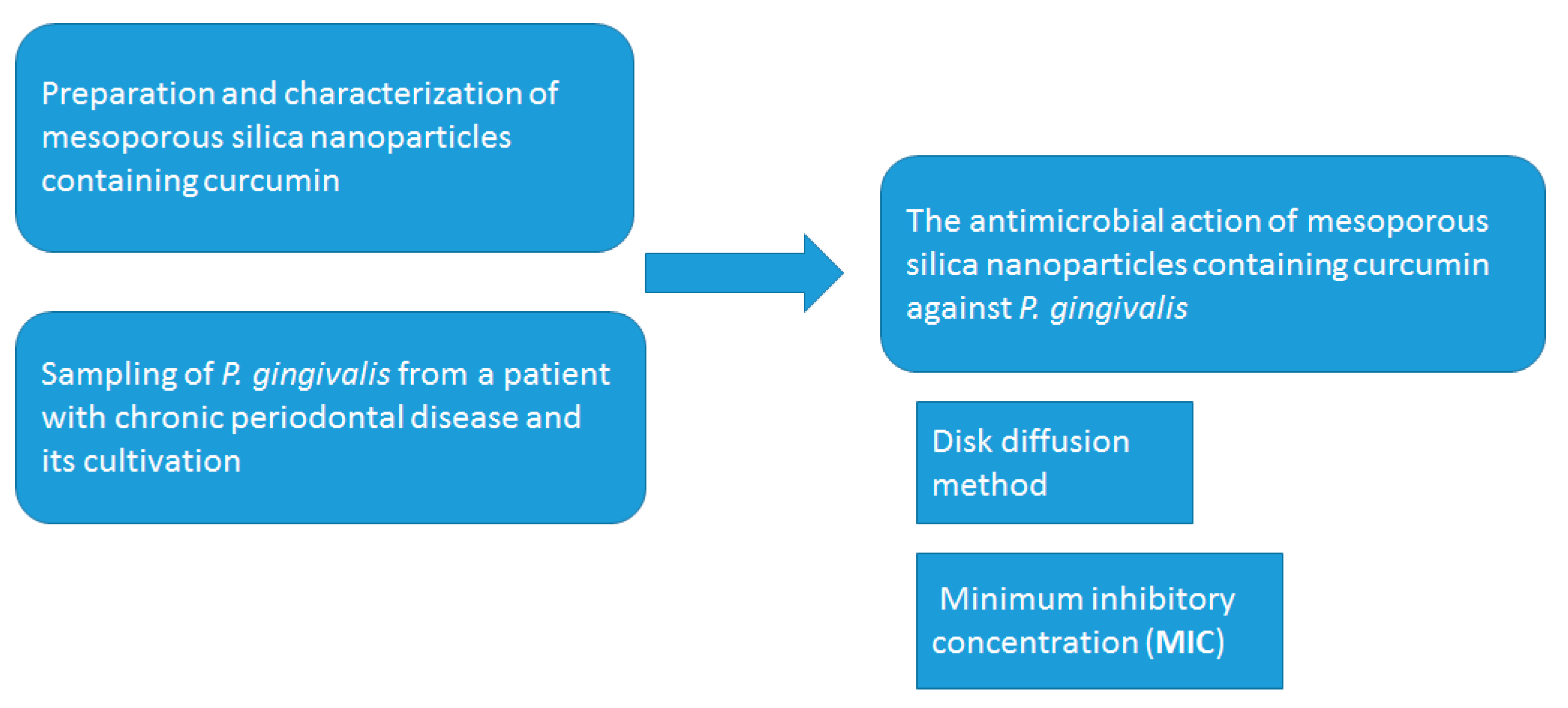
2.1. Preparation of Mesoporous Silica Nanoparticles Containing Curcumin
2.2. Sampling of P. gingivalis
2.3. Cultivation of P. gingivalis
2.4. Characterization of the Nanoparticles
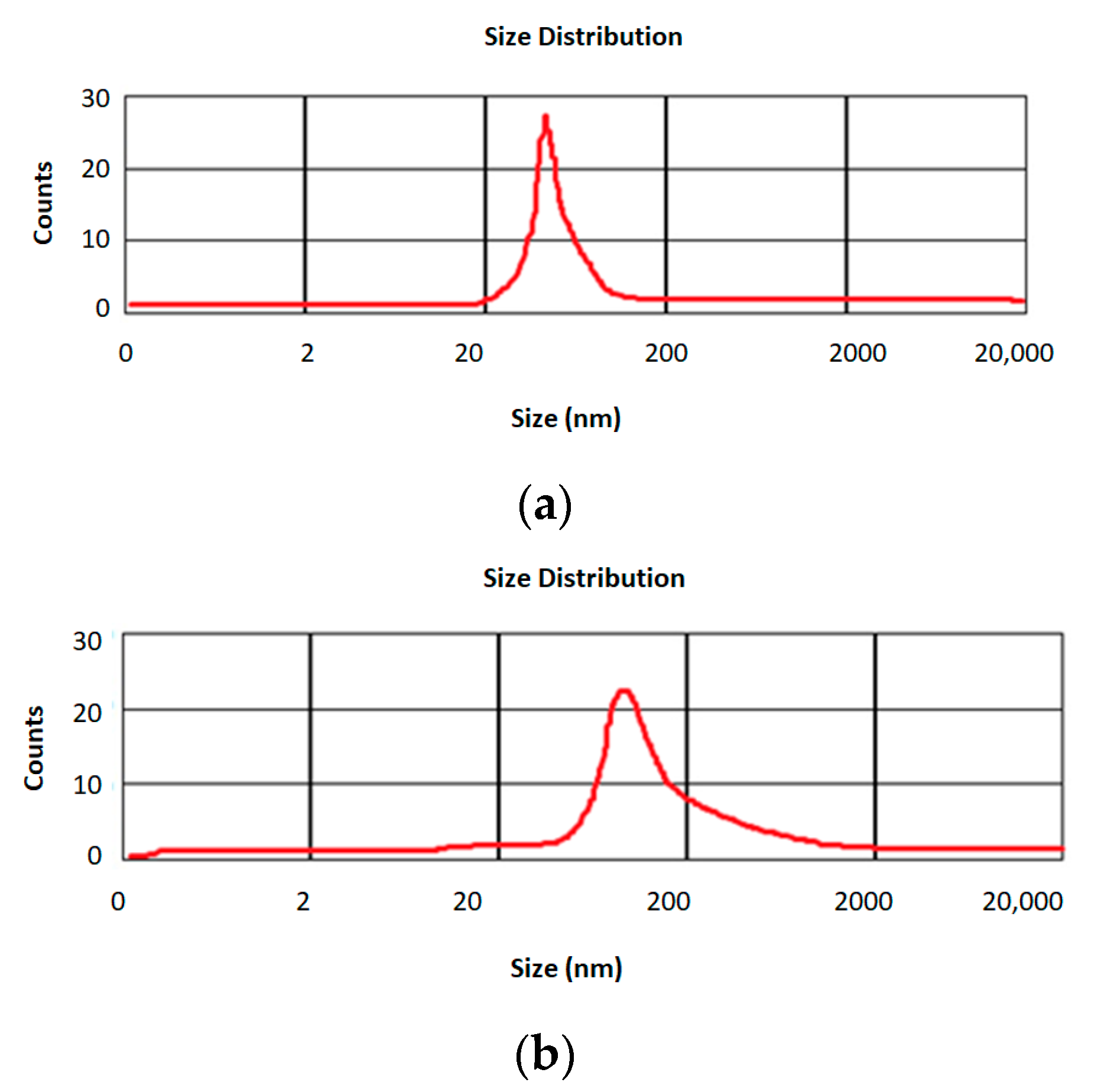
2.4.1. The Particle Size of Nanoparticles
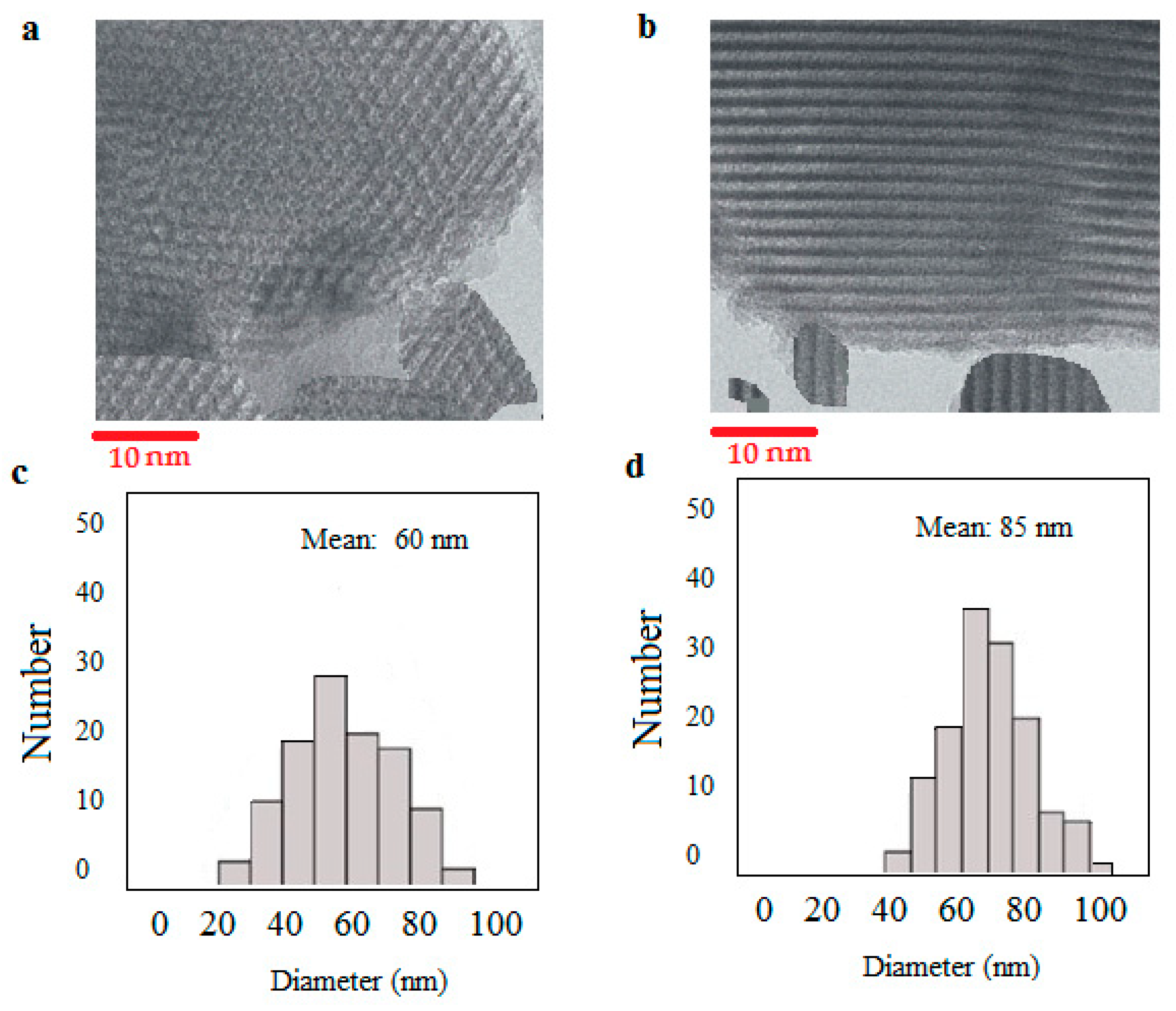
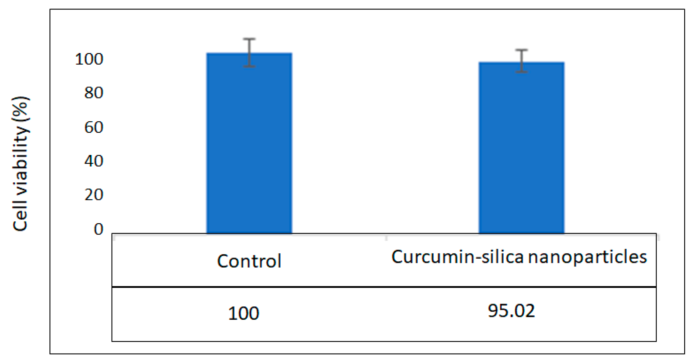
2.4.2. Morphology and the Cytotoxicity Investigation
2.4.3. Determination of Curcumin Loading Inside the Nanoparticles
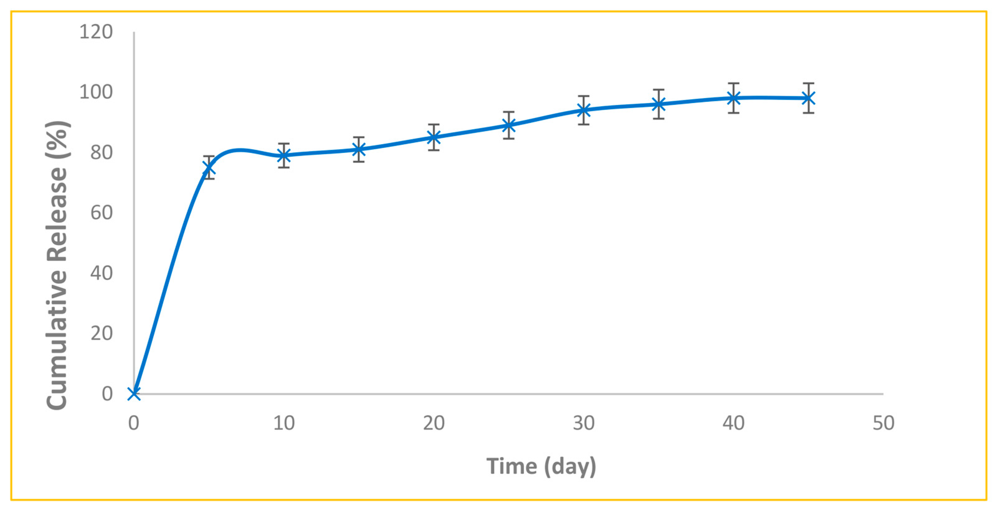
2.4.4. Evaluation of Release Pattern
2.4.5. The Antimicrobial Action of Nanoparticles
3. Statistical Analysis
4. Results and Discussion
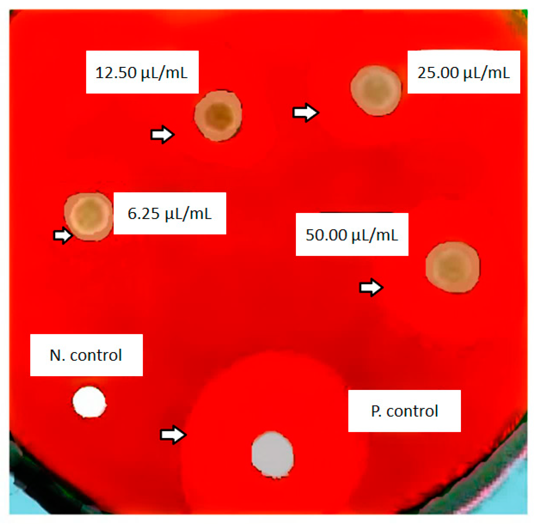
Antimicrobial Action
5. The Strengths and Limitations
6. Suggestions and Future Perspective
7. Conclusions
Author Contributions
Funding
Institutional Review Board Statement
Informed Consent Statement
Data Availability Statement
Acknowledgments
Conflicts of Interest
References
- Shirmohammadi, A.; Babaloo, A.; Dizaj, S.M.; Lotfipour, F.; Sharifi, S.; Ghavimi, M.A.; Khezri, K. A View on Polymerase Chain Reaction as an Outstanding Molecular Diagnostic Technique in Periodontology. BioMed Res. Int. 2021, 2021, 1–8. [Google Scholar] [CrossRef]
- Kumar, M.; Prakash, S.; Radha; Lorenzo, J.M.; Chandran, D.; Dhumal, S.; Dey, A.; Senapathy, M.; Rais, N.; Singh, S.; et al. Apitherapy and Periodontal Disease: Insights into in vitro, in vivo, and Clinical Studies. Antioxidants 2022, 11, 823. [Google Scholar] [CrossRef]
- Fragkioudakis, I.; Riggio, M.P.; Apatzidou, D.A. Understanding the microbial components of periodontal diseases and periodontal treatment-induced microbiological shifts. J. Med. Microbiol. 2021, 70, jmm001247. [Google Scholar] [CrossRef]
- Armitage GCJAop. Development of a classification system for periodontal diseases and conditions. Ann. Periodontol. 1999, 4, 1–6. [Google Scholar] [CrossRef]
- Norowski, P.A., Jr.; Bumgardner, J.D. Biomaterial and antibiotic strategies for peri-implantitis: A review. J. Biomed. Mater. Res. Part B Appl. Biomater. Off. J. Soc. Biomater. Jpn. Soc. Biomater. Aust. Soc. Biomater. Korean Soc. Biomater. 2009, 88, 530–543. [Google Scholar] [CrossRef]
- Lafzi, A.; Shirmohammadi, A.; Faramarzi, M.; Jabali, S.; Shayan, A. Clinical Comparison of Autogenous Bone Graft with and without Plasma Rich in Growth Factors in the Treatment of Grade II Furcation Involvement of Mandibular Molars. J. Dent. Res. Dent. Clin. Dent. Prospect. 2013, 7, 22–29. [Google Scholar] [CrossRef]
- Yue, C.; Zhao, B.; Ren, Y.; Kuijer, R.; Van Der Mei, H.; Busscher, H.; Rochford, E. The implant infection paradox: Why do some succeed when others fail? Opinion and discussion paper. Eur. Cells Mater. 2015, 29, 303–313. [Google Scholar] [CrossRef] [Green Version]
- Chapple, C.C.; Kumar, R.; Hunter, N. Vascular remodelling in chronic inflammatory periodontal disease. J. Oral Pathol. Med. 2000, 29, 500–506. [Google Scholar] [CrossRef]
- Mitwalli, H.; Alsahafi, R.; Balhaddad, A.A.; Weir, M.D.; Xu, H.H.K.; Melo, M.A.S. Emerging Contact-Killing Antibacterial Strategies for Developing Anti-Biofilm Dental Polymeric Restorative Materials. Bioengineering 2020, 7, 83. [Google Scholar] [CrossRef]
- Chi, M.; Qi, M.; Wang, P.; Weir, M.D.; Melo, M.A.; Sun, X.; Dong, B.; Li, C.; Wu, J. Novel Bioactive and Therapeutic Dental Polymeric Materials to Inhibit Periodontal Pathogens and Biofilms. Int. J. Mol. Sci. 2019, 20, 278. [Google Scholar] [CrossRef] [Green Version]
- Zhu, J.; Huang, Y.; Chen, M.; Hu, C.; Chen, Y. Functional Synergy of Antimicrobial Peptides and Chlorhexidine Acetate against Gram-Negative/Gram-Positive Bacteria and a Fungus in vitro and in vivo. Infect. Drug Resist. 2019, 12, 3227–3239. [Google Scholar] [CrossRef] [Green Version]
- Saidin, S.; Jumat, M.A.; Amin, N.A.A.M.; Al-Hammadi, A.S.S. Organic and inorganic antibacterial approaches in combating bacterial infection for biomedical application. Mater. Sci. Eng. C 2020, 118, 111382. [Google Scholar] [CrossRef]
- Cieplik, F.; Jakubovics, N.S.; Buchalla, W.; Maisch, T.; Hellwig, E.; Al-Ahmad, A. Resistance toward chlorhexidine in oral bacteria–is there cause for concern? Front. Microbiol. 2019, 10, 587. [Google Scholar] [CrossRef] [Green Version]
- Ahmed, A.; Khan, A.K.; Anwar, A.; Ali, S.A.; Shah, M.R. Biofilm inhibitory effect of chlorhexidine conjugated gold nanoparticles against Klebsiella pneumoniae. Microb. Pathog. 2016, 98, 50–56. [Google Scholar] [CrossRef]
- Addy, M. Oral hygiene products: Potential for harm to oral and systemic health? Periodontology 2008, 48, 54–65. [Google Scholar] [CrossRef]
- Njoroge, T.; Genco, R.J.; Sojar, H.T.; Hamada, N.; Genco, A.C. A role for fimbriae in Porphyromonas gingivalis invasion of oral epithelial cells. Infect. Immun. 1997, 65, 1980–1984. [Google Scholar] [CrossRef] [PubMed] [Green Version]
- Mohseni, M.; Samadi, N.; Ghanbari, P.; Yousefi, B.; Tabasinezhad, M.; Sharifi, S.; Nazemiyeh, H. Co-treatment by docetaxel and vinblastine breaks down P-glycoprotein mediated chemo-resistance. Iran. J. Basic Med. Sci. 2016, 19, 300–309. [Google Scholar] [CrossRef]
- El-Saadony, M.T.; Zabermawi, N.M.; Zabermawi, N.M.; Burollus, M.A.; Shafi, M.E.; Alagawany, M.; Yehia, N.; Askar, A.M.; Alsafy, S.A.; Noreldin, A.E.; et al. Nutritional Aspects and Health Benefits of Bioactive Plant Compounds against Infectious Diseases: A Review. Food Rev. Int. 2021, 37, 1–23. [Google Scholar] [CrossRef]
- Hinault, M.-P.; Farina-Henriquez-Cuendet, A.; Goloubinoff, P. Molecular Chaperones and Associated Cellular Clearance Mechanisms against Toxic Protein Conformers in Parkinson’s Disease. Neurodegener. Dis. 2011, 8, 397–412. [Google Scholar] [CrossRef]
- Soory, M. Inflammatory Mechanisms and Redox Status in Periodontal and Cardiometabolic Diseases: Effects of Adjunctive Nutritional Antioxidants and Statins. Infect. Disord. Drug Targets 2012, 12, 301–315. [Google Scholar] [CrossRef]
- Zambrano, L.M.G.; Brandao, D.A.; Rocha, F.R.G.; Marsiglio, R.P.; Longo, I.B.; Primo, F.L.; Tedesco, A.C.; Guimaraes-Stabili, M.R.; Junior, C.R. Local administration of curcumin-loaded nanoparticles effectively inhibits inflammation and bone resorption associated with experimental periodontal disease. Sci. Rep. 2018, 8, 1–11. [Google Scholar] [CrossRef] [PubMed] [Green Version]
- Wahab, S.; Ahmad, P.; Hussain, A.; Qadir, S.F.A. Nanomateria, ls for the delivery of Herbal Bioactive Compounds. Curr. Nanosci. 2021, 18, 425–441. [Google Scholar] [CrossRef]
- Banerjee, H.N.; Verma, M. Application of nanotechnology in cancer. Technol. Cancer Res. Treatment 2008, 7, 149–154. [Google Scholar] [CrossRef] [Green Version]
- Abhilash, P.P.; Nayak, D.K.; Sangoju, B.; Kumar, R.; Kumar, V. Effect of nano-silica in concrete; a review. Constr. Build. Mater. 2021, 278, 122347. [Google Scholar] [CrossRef]
- Saravanakumar, P.; Arunkumar, S.; Mansingh, B.B.; Kumar, P.M.; Subbiah, R.; Eswarlal, V.K. Investigating the effect of thermal cycling on thermal characteristics of the nano-silica based phase changing material (PCM). Mater. Today Proc. 2021, 50, 1502–1507. [Google Scholar] [CrossRef]
- Said, A.; Zeidan, M.; Bassuoni, M.; Tian, Y. Properties of concrete incorporating nano-silica. Constr. Build. Mater. 2012, 36, 838–844. [Google Scholar] [CrossRef]
- Ambrogio, M.W.; Thomas, C.R.; Zhao, Y.-L.; Zink, J.I.; Stoddart, J.F. Mechanized Silica Nanoparticles: A New Frontier in Theranostic Nanomedicine. Accounts Chem. Res. 2011, 44, 903–913. [Google Scholar] [CrossRef] [Green Version]
- Lam, M.; Migonney, V.; Falentin-Daudre, C. Review of silicone surface modification techniques and coatings for antibacterial/antimicrobial applications to improve breast implant surfaces. Acta Biomater. 2020, 121, 68–88. [Google Scholar] [CrossRef]
- Massa, M.A.; Covarrubias, C.; Bittner, M.; Fuentevilla, I.A.; Capetillo, P.; Von Marttens, A.; Carvajal, J.C. Synthesis of new antibacterial composite coating for titanium based on highly ordered nanoporous silica and silver nanoparticles. Mater. Sci. Eng. C 2014, 45, 146–153. [Google Scholar] [CrossRef]
- Patra, D.; Karaman, D.; Desai, D.; El Khoury, E.; Rosenholm, J.M. Preparation of curcumin loaded mesoporous silica nanoparticles: Determining polarizability inside the mesopores. Mater. Res. Bull. 2016, 84, 267–272. [Google Scholar] [CrossRef]
- Hunt, D.; Jones, J.; Dowell, V., Jr. Selective medium for the isolation of Bacteroides gingivalis. J. Clin. Microbiol. 1986, 23, 441–445. [Google Scholar] [CrossRef] [Green Version]
- Dowding, J.M.; Das, S.; Kumar, A.; Dosani, T.; McCormack, R.; Gupta, A.; Sayle, T.X.T.; Sayle, D.C.; von Kalm, L.; Seal, S.; et al. Cellular Interaction and Toxicity Depend on Physicochemical Properties and Surface Modification of Redox-Active Nanomaterials. ACS Nano 2013, 7, 4855–4868. [Google Scholar] [CrossRef] [Green Version]
- Bakand, S.; Hayes, A.J. Toxicological considerations, toxicity assessment, and risk management of inhaled nanoparticles. Int. J. Mol. Sci. 2016, 17, 929. [Google Scholar] [CrossRef]
- Mitchell, M.J.; Billingsley, M.M.; Haley, R.M.; Wechsler, M.E.; Peppas, N.A.; Langer, R. Engineering precision nanoparticles for drug delivery. Nat. Rev. Drug Discov. 2020, 20, 101–124. [Google Scholar] [CrossRef]
- Fissan, H.; Ristig, S.; Kaminski, H.; Asbach, C.; Epple, M. Comparison of different characterization methods for nanoparticle dispersions before and after aerosolization. Anal. Methods 2014, 6, 7324–7334. [Google Scholar] [CrossRef] [Green Version]
- Marambio-Jones, C.; Hoek, E.M.V. A review of the antibacterial effects of silver nanomaterials and potential implications for human health and the environment. J. Nanopart. Res. 2010, 12, 1531–1551. [Google Scholar] [CrossRef]
- Salatin, S.; Maleki Dizaj, S.; Yari Khosroushahi, A. Effect of the surface modification, size, and shape on cellular uptake of nanoparticles. Cell Biol. Int. 2015, 39, 881–890. [Google Scholar] [CrossRef]
- Yin, I.X.; Zhang, J.; Zhao, I.S.; Mei, M.L.; Li, Q.; Chu, C.H. The antibacterial mechanism of silver nanoparticles and its application in dentistry. Int. J. Nanomed. 2020, 15, 2555–2562. [Google Scholar] [CrossRef] [PubMed] [Green Version]
- Geng, Y.; Dalhaimer, P.; Cai, S.; Tsai, R.; Tewari, M.; Minko, T.; Discher, D.E. Shape effects of filaments versus spherical particles in flow and drug delivery. Nat. Nanotechnol. 2007, 2, 249–255. [Google Scholar] [CrossRef]
- Li, D.; Tang, Z.; Gao, Y.; Sun, H.; Zhou, S. A Bio-Inspired Rod-Shaped Nanoplatform for Strongly Infecting Tumor Cells and Enhancing the Delivery Efficiency of Anticancer Drugs. Adv. Funct. Mater. 2015, 26, 66–79. [Google Scholar] [CrossRef]
- Zhao, Y.; Wang, Y.; Ran, F.; Cui, Y.; Liu, C.; Zhao, Q.; Gao, Y.; Wang, D.; Wang, S. A comparison between sphere and rod nanoparticles regarding their in vivo biological behavior and pharmacokinetics. Sci. Rep. 2017, 7, 4131. [Google Scholar] [CrossRef] [Green Version]
- Parani, M.; Lokhande, G.; Singh, A.; Gaharwar, A.K. Engineered Nanomaterials for Infection Control and Healing Acute and Chronic Wounds. ACS Appl. Mater. Interfaces 2016, 8, 10049–10069. [Google Scholar] [CrossRef]
- Bhattacharyya, S.; Wang, H.; Ducheyne, P. Polymer-coated mesoporous silica nanoparticles for the controlled release of macromolecules. Acta Biomater. 2012, 8, 3429–3435. [Google Scholar] [CrossRef] [PubMed]
- Memar, M.Y.; Yekani, M.; Ghanbari, H.; Nabizadeh, E.; Vahed, S.Z.; Dizaj, S.M.; Sharifi, S. Antimicrobial and antibiofilm activities of meropenem loaded-mesoporous silica nanoparticles against carbapenem-resistant Pseudomonas aeruginosa. J. Biomater. Appl. 2021, 36, 605–612. [Google Scholar] [CrossRef]
- Wang, L.; Hu, C.; Shao, L. The antimicrobial activity of nanoparticles: Present situation and prospects for the future. Int. J. Nanomed. 2017, 12, 1227–1249. [Google Scholar] [CrossRef] [PubMed] [Green Version]
- Memar, M.Y.; Yekani, M.; Ghanbari, H.; Shahi, S.; Sharifi, S.; Dizaj, S.M. Biocompatibility, cytotoxicity and antibacterial effects of meropenem-loaded mesoporous silica nanoparticles against carbapenem-resistant Enterobact. Artif. Cells Nanomed. Biotechnol. 2020, 48, 1354–1361. [Google Scholar] [CrossRef] [PubMed]
- Shahzad, M.; Millhouse, E.; Culshaw, S.; Edwards, C.A.; Ramage, G.; Combet, E. Selected dietary (poly)phenols inhibit periodontal pathogen growth and biofilm formation. Food Funct. 2014, 6, 719–729. [Google Scholar] [CrossRef] [PubMed] [Green Version]
- Mandroli, P.S.; Bhat, K. An in-vitro evaluation of antibacterial activity of curcumin against common endodontic bacteria. J. Appl. Pharm. Sci. 2013, 3, 16. [Google Scholar]
- Izui, S.; Sekine, S.; Maeda, K.; Kuboniwa, M.; Takada, A.; Amano, A.; Nagata, H. Antibacterial Activity of Curcumin Against Periodontopathic Bacteria. J. Periodontol. 2016, 87, 83–90. [Google Scholar] [CrossRef]
- Bomdyal, R.S.; Shah, M.U.; Doshi, Y.S.; Shah, V.A.; Khirade, S.P. Antibacterial activity of curcumin (turmeric) against periopathogens-An in vitro evaluation. J. Adv. Clin. Res. Insights 2017, 4, 175–180. [Google Scholar] [CrossRef]
- Maleki Dizaj, S.; Shokrgozar, H.; Yazdani, J.; Memar, M.Y.; Sharifi, S.; Ghavimi, M.A.J.C. Antibacterial Effects of Curcumin Nanocrystals against Porphyromonas gingivalis Isolated from Patients with Implant Failure. Clin. Praqct. 2022, 12, 809–817. [Google Scholar] [CrossRef] [PubMed]
- Shang, W.; Zhao, L.-J.; Dong, X.-L.; Zhao, Z.-M.; Li, J.; Zhang, B.-B.; Cai, H. Curcumin inhibits osteoclastogenic potential in PBMCs from rheumatoid arthritis patients via the suppression of MAPK/RANK/c-Fos/NFATc1 signaling pathways. Mol. Med. Rep. 2016, 14, 3620–3626. [Google Scholar] [CrossRef] [PubMed] [Green Version]
- Cirano, F.; Pimentel, S.; Casati, M.; Corrêa, M.; Pino, D.; Messora, M.; Silva, P.; Ribeiro, F. Effect of curcumin on bone tissue in the diabetic rat: Repair of peri-implant and critical-sized defects. Int. J. Oral Maxillofac. Surg. 2018, 47, 1495–1503. [Google Scholar] [CrossRef] [PubMed]
- Xu, C.S.; Ip, M.; Leung, A.W.; Wang, X.N.; Yang, Z.R.; Zhang, B.T.; Ip, S.P. Sonodynamic bactericidal activity of curcumin against foodborne bacteria. Hong Kong Med. J. 2018, 24, 43–44. [Google Scholar]
- Kumbar, V.M.; Peram, M.R.; Kugaji, M.S.; Shah, T.; Patil, S.P.; Muddapur, U.M.; Bhat, K.G. Effect of curcumin on growth, biofilm formation and virulence factor gene expression of Porphyromonas gingivalis. Odontology 2021, 109, 18–28. [Google Scholar] [CrossRef]
- Chen, D.; Nie, M.; Fan, M.-W.; Bian, Z. Anti-Inflammatory Activity of Curcumin in Macrophages Stimulated by Lipopolysaccharides from Porphyromonas gingivalis. Pharmacology 2008, 82, 264–269. [Google Scholar] [CrossRef] [PubMed]


| Samples | The Mean Growth Inhibition Zone (mm) |
|---|---|
| Curcumin-loaded silica nanoparticles (50 µg/mL) | 15 ± 1.2 |
| Curcumin-loaded silica nanoparticles (25 µg/mL) | 12.23 ± 0.8 |
| Curcumin-loaded silica nanoparticles (12.5 µg/mL) | 10.24 ± 1.2 |
| Curcumin-loaded silica nanoparticles (6.25 µg/mL) | 7.59 ± 1.4 |
| Metronidazole as the positive control | 19.20 ± 1.2 |
| Blank disk (water) as the negative control | 0 |
Disclaimer/Publisher’s Note: The statements, opinions and data contained in all publications are solely those of the individual author(s) and contributor(s) and not of MDPI and/or the editor(s). MDPI and/or the editor(s) disclaim responsibility for any injury to people or property resulting from any ideas, methods, instructions or products referred to in the content. |
© 2023 by the authors. Licensee MDPI, Basel, Switzerland. This article is an open access article distributed under the terms and conditions of the Creative Commons Attribution (CC BY) license (https://creativecommons.org/licenses/by/4.0/).
Share and Cite
Shirmohammadi, A.; Maleki Dizaj, S.; Sharifi, S.; Fattahi, S.; Negahdari, R.; Ghavimi, M.A.; Memar, M.Y. Promising Antimicrobial Action of Sustained Released Curcumin-Loaded Silica Nanoparticles against Clinically Isolated Porphyromonas gingivalis. Diseases 2023, 11, 48. https://doi.org/10.3390/diseases11010048
Shirmohammadi A, Maleki Dizaj S, Sharifi S, Fattahi S, Negahdari R, Ghavimi MA, Memar MY. Promising Antimicrobial Action of Sustained Released Curcumin-Loaded Silica Nanoparticles against Clinically Isolated Porphyromonas gingivalis. Diseases. 2023; 11(1):48. https://doi.org/10.3390/diseases11010048
Chicago/Turabian StyleShirmohammadi, Adileh, Solmaz Maleki Dizaj, Simin Sharifi, Shirin Fattahi, Ramin Negahdari, Mohammad Ali Ghavimi, and Mohammad Yousef Memar. 2023. "Promising Antimicrobial Action of Sustained Released Curcumin-Loaded Silica Nanoparticles against Clinically Isolated Porphyromonas gingivalis" Diseases 11, no. 1: 48. https://doi.org/10.3390/diseases11010048