Article

Hot Springs Cyanobacteria Endowed with Biological Activities for Cosmetic Applications: Evaluation of On-Site Collected Communities and Isolated Strains

by
Natascia Biondi
1,*,
Maria Raffaella Martina
1,
Marisanna Centini
2,
Cecilia Anselmi
2 and
Mario R. Tredici
1,†
1
Department of Agriculture, Food, Environment and Forestry (DAGRI), University of Florence, Piazzale delle Cascine 18, 50144 Florence, Italy
2
Department of Biotechnology, Chemistry and Pharmacy, University of Siena, Via Aldo Moro 2, 53100 Siena, Italy
*
Author to whom correspondence should be addressed.
This article is dedicated to the memory of Mario R. Tredici, who passed away during the writing of the manuscript.
Cosmetics 2023, 10(3), 81; https://doi.org/10.3390/cosmetics10030081
Submission received: 7 April 2023 / Revised: 2 May 2023 / Accepted: 11 May 2023 / Published: 17 May 2023
(This article belongs to the Collection Feature Papers in Cosmetics in 2023)

Abstract

:
Microbial communities growing around hot springs (bioglea), of which cyanobacteria are the main components, contribute to beneficial properties of thermal muds and often enter cosmetic treatment products. Bioglea is obtainable via alternative approaches to harvesting from nature: direct cultivation of whole communities and isolation and cultivation of the isolated strains. Cultivation represents a step towards industrial production, guaranteeing higher availability, quality and safety. In this work, the biochemical composition of natural and cultivated bioglea collected in different hot springs and of cyanobacterial strains isolated thereof was analysed. Lipophilic and hydrophilic extracts were tested for antimicrobial and radical scavenging activities and toxicity against Artemia salina. Higher antimicrobial activity was found in lipophilic than hydrophilic bioglea extracts and the opposite in isolates’ extracts. Thermal water extracts also showed some activity. No toxicity was observed. Bioglea radical scavenging activity positively correlated with carotenoids (lipophilic extracts) and phycobiliproteins (hydrophilic extracts). No correlation was observed for isolated strains. Cultivation at the 10 L scale of Chroococcidiopsis BIOG3 evidenced bioactivity changes with the growth phase and life cycle stage. From activity comparison, communities showed higher potential over single strains cultivation; however, further investigations should elucidate isolates’ full potential also by combining them into synthetic communities to simplify production and standardisation.

1. Introduction

Microalgae and cyanobacteria have received increasing attention for their application in cosmetic products [1,2,3,4,5]. Several components of these microorganisms have the characteristics to become cosmetic ingredients. Polysaccharides may be used as thickeners [6] and moisturizing agents [1,5] and might be endowed with antioxidant, anti-inflammatory and anti-ageing activity [4,7]. Pigments have a dual nature of colourants and antioxidant compounds [7]. Other molecules of interest are phenolics, fatty acids, sunscreens such as scytonemin and mycosporine-like amino acids, and biologically active peptides [7,8].
Spas are increasingly becoming wellness centres proposing cosmetic treatments and programmes to improve lifestyle (e.g., weight loss, fitness, relaxation programmes), often disjunct from the presence of site-specific thermal waters, unless traditional health treatments (e.g., for respiratory, dermatological and bone diseases) are run in parallel [9]. In traditional spas, the health-promoting action of treatments is connected to the specific characteristics of the waters springing on-site, and an important role is played by mud therapy or pelotherapy [10,11]. Beneficial effects (medical and cosmetic) of thermal muds are partly due to physical properties (heat) and chemical components (e.g., minerals); nevertheless, additional functions (e.g., antioxidant and anti-inflammatory) may originate from the maturation process carried out in the presence of microorganisms, which usually include an important phototrophic component [12].
The complex microbial communities developing around hot springs have long been named in different ways, among which are bioglée or bioglea [12,13]. Bioglea was described by Laporte et al. [13] as a microbial community associated with a mineral component assuming a slimy consistency. In more recent definitions, it is considered as an organic peloid composed of algae and bacteria with a liquid component, generally mineral water bearing reduced sulphur; in other terms, it is a biofilm consisting of a gelatinous pellicle, which may assume different colours (yellowish, greenish, greyish or reddish), occurring at the surface or dispersed in thermal or non-thermal sulphur-bearing spring waters [12]. Bioglea, as mentioned above, is a fundamental component in the process of thermal mud maturation [14,15,16,17]; moreover, it has been used in spas for direct treatments [14,18,19] or to extract bioactives to enrich spa own-branded cosmetic products (e.g., Saturnia Bioplancton® from Terme di Saturnia; brands from Dax, Acquadax™; and the patented Algodax).
In Dax (France), a process to obtain a mature peloid for spa treatments has been patented [20], and, as part of this process, an interesting bioglea cultivation step is performed. In brief, the virgin clay is mixed with thermal water, the biomass of a Clostridium strain is isolated from the mud and cultivated in fermenters, and the bioglea is grown under a greenhouse on inclined planes sprinkled with thermal water to allow the development of a thick mat dominated by cyanobacteria. The mix is left to mature for a pre-determined period and then is packed and distributed to spas for pelotherapy and cosmetic applications [20]. Production processes such as this, which represent an upscale compared to traditional methods, are of great interest for the development of the sector, as they transform bioglea from a “gift of nature” collected as it is, into an actual industrial product that may be obtained following more standardisable protocols. This process permits obtaining a higher amount of product with a higher quality (avoiding contamination with exogenous chemicals) and partially modulating production to favour a stable community with the required characteristics in terms of composition and/or bioactivity.
Another strategy to improve standardisation of the product is to isolate and separately cultivate bioglea components, which would improve the reproducibility of the product and the optimisation of culture conditions to increase the target molecule production in a scaling-up perspective. This approach is applied in Iceland, where Cyanobacterium aponium isolated from the Blue Lagoon, is cultivated indoors in tubular photobioreactors using geothermal water added with nutrients [21]. On the other hand, the cultivation of isolated strains leads to the loss of the interactions among the microbial community components, which could play a role in triggering the production of bioactive molecules.
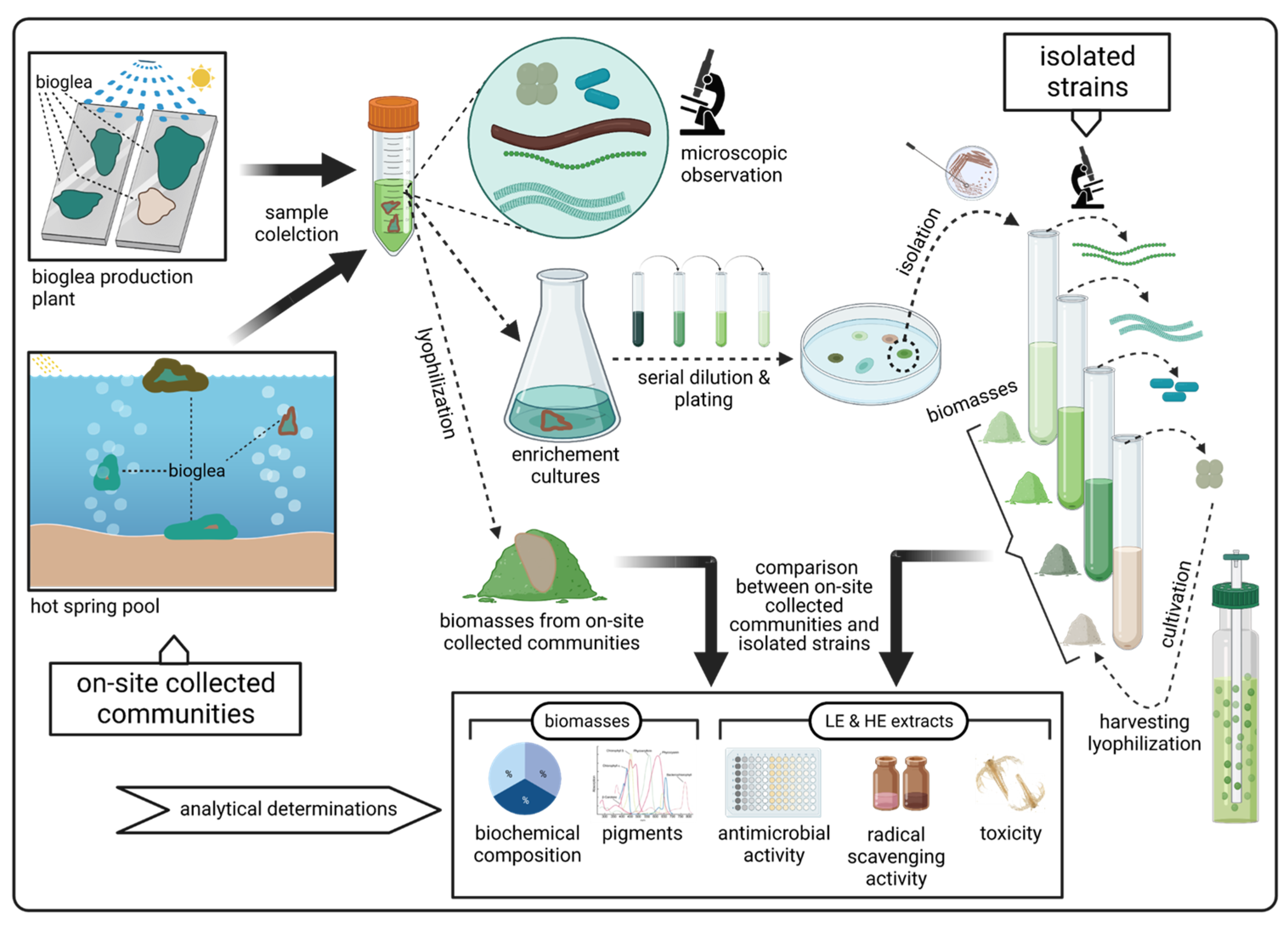
In this work, a comparison between the biochemical composition and the activity of isolated strains and microbial communities collected on site (bioglea) was performed to verify whether strains isolated from bioglea may replicate the effect of the community from which they originated. Bioactivity tests (antimicrobial activity, radical scavenging activity and toxicity) were performed on samples from microbial communities collected in spas with different water physico-chemical characteristics and on strains isolated thereof.

2. Materials and Methods

2.1. Experimental Plan

Bioglea samples collected from natural hot spring pools or from cultivation plants were used for microscopic observation and set-up of enrichment cultures, while an aliquot was lyophilised for analytical determinations (biochemical composition and pigment content) and for extraction (lipophilic and hydrophilic extracts) (Figure 1). The extracts were tested for antimicrobial activity against a panel of fungi and bacteria, for radical scavenging activity (DPPH assay), and for toxicity against the model organism Artemia salina. The strains isolated from the enrichment cultures were also tested as described above, and the results compared with those from on-site collected communities (Figure 1).

2.2. Sampling

Samples were collected from four hot springs with different physical and chemical characteristics. In Saturnia spa (Grosseto, Tuscany, Italy), sulphurous water (13 mg L−1 of H2S) containing sulphate (1508 mg L−1), bicarbonate (667 mg L−1), calcium (578 mg L−1) and magnesium (122 mg L−1) as main constituents, springs at the bottom of a partially natural pool at a temperature of 37.5 °C and pH 6.3 [22]. Petriolo (Siena, Tuscany, Italy) is also a sulphurous spring (11 mg L−1 of H2S) with water at 44 °C, pH 6.5, and a composition similar to that of Saturnia except for a two-fold bicarbonate concentration and high sodium (163 mg L−1) and chlorine (120 mg L−1) concentrations [22]. In Bagno di Romagna (Forlì, Emilia Romagna, Italy), water springs at 44 °C, pH 8.7, with a low sulphide concentration (2 mg L−1 of H2S) and bicarbonate (855 mg L−1) as the main constituent. In Dax (Landes, Nouvelle-Aquitaine, France), water has a temperature of 55–61 °C; pH 7.0–7.4; and sulphate (370 mg L−1), bicarbonate (159 mg L−1), calcium (125 mg L−1), sodium (128 mg L−1) and chlorine (157 mg L−1) as main constituents [23].
Samples collected and analysed in this work (indicated by codes) are listed in Table 1. In Saturnia and Petriolo, bioglea grows in pools with a rocky or silty bottom. Samples were also collected from muds produced by mixing clays with thermal water and microorganisms (Saturnia, Bagno di Romagna and Dax). In Bagno di Romagna, samples included a cultivated bioglea, used to inoculate mud for maturation, grown on inclined planes where thermal water flowed from a tank placed at the upper end of the plane. Similarly, samples from Dax were obtained from a cultivated bioglea, used to inoculate maturing mud (see Introduction Section for detailed process description) [20]. All the samples, except aliquots for immediate analyses, were frozen, lyophilised (HETO Sicc, HETO Lab Equipment, Allerød, Denmark) and stored at −20 °C until further use.

2.3. Microscopic Observation of the Samples

Fresh on-site collected samples (Table 1) were checked under the microscope (Eclipse E200, Nikon, Tokyo, Japan). The precise determination of all the components of the microbial communities was beyond the scope of this work, and observations were limited to oxygenic phototrophs, which were the main components, to provide a basic description of the community. For Saturnia samples collected from April to November, checks also aimed at evaluating the main seasonal fluctuations in the phototrophic community composition. A semi-quantitative determination of the frequency of each component was performed. Equally sized samples were evenly spread on a slide, then the number of trichomes or unicells (cyanobacteria and eukaryotic algae) were counted in microscope fields along the median lines of the slide. For each sample, a number of microscope fields sufficient to count at least 100 cells/trichomes was analysed. At least three replicates per sample were checked to increase count representativeness.
Besides microscopic analysis, when possible, in vivo absorption spectra were determined on fresh material dispersed in water by sonication (VCX130 Vibra Cell®, Sonics, Newtown, CT, USA). The measured optical density spectrum (Cary 50 spectrophotometer, Agilent Technologies, Santa Clara, CA, USA) was converted to total absorption by the formula: a(λ) = 2.303OD(λ)/0.01, where a(λ) is the absorption coefficient (m−1) at wavelength λ, OD is optical density and 0.01 is the path-length (m). The absorption spectrum due to pigmented material was finally calculated by subtracting from total absorption the absorption due to debris and colourless particles obtained by the model equation reported in Roesler et al. [24]. The final absorption spectra were normalised to their spectral mean (mean of the absorption values) to compare shapes beyond differences in material concentrations. For each site sampled within the same spa (e.g., surface bioglea, bottom bioglea), an average ± standard deviation spectrum was calculated from the spectra measured at different times.

2.4. Isolation and Cultivation of Cyanobacterial and Microalgal Strains

Sample aliquots were streaked on agarised BG11 medium [25] after homogenisation by vortexing (Vibromix, PBI, Milan, Italy) with glass beads (2–3 mm diameter). Other aliquots were enriched in both thermal water and BG11 medium, unadded or added with 200 µg mL−1 of actidione (Merck Life Science, Milan, Italy) to inhibit eukaryote growth. The enrichment cultures were periodically checked and streaked. Isolation from agarised plates was performed by transferring colonies to liquid BG11 medium or thermal water using a Pasteur pipette tip.
The isolated strains were cultivated in 0.5–1 L tubes bubbled with an air/CO2 mixture (98/2, v/v) at 27 ± 3 °C and a continuous illumination of 50 µmol photons m−2 s−1 provided by daylight fluorescent tubes (Osram Lumilux L 58W; Osram, Munich, Germany). Strains BIOG5 and BIOG6, unable to grow in BG11 and under bubbling, were cultivated under the same environmental conditions in 1 L flasks (0.4 L culture volume) kept in an orbital shaker flushed with CO2 (5%, v/v) using thermal water:BG11 1:1 as culture medium.
Strain BIOG3 was cultivated in fed-batch regime in a 10 L bubbled tube in BG11 medium. The tube had an external diameter of 10 cm and a height of 140 cm, with an operative volume of 9 L. The light was provided continuously by daylight fluorescent tubes (Osram Lumilux L 58W) at an intensity of 125 µmol photons m−2 s−1. The temperature was not controlled and was, on average, 28.3 ± 0.9 °C, while pH was maintained at 7.7 ± 0.1 by bubbling an air/CO2 mixture (98/2, v/v). Nutrients were integrated according to productivity, considering N as 10%, P as 1%, Mg and Ca as 0.5% and Fe as 0.2% of dry biomass.
Harvesting was performed by filtration on a nylon net (12 µm nominal mesh size) for filamentous strains and by centrifugation (8000× g, 20 min) for the unicellular ones. Biomasses were washed with a 1 g L−1 NaCl solution to eliminate excess salts, frozen, lyophilised and stored at −20 °C until analysis or extraction.

2.5. Biochemical Composition Analyses

On-site community and isolated strain biomasses were analysed in lyophilised form for ash, protein, carbohydrate, lipids, phycobiliproteins (phycocyanin, allophycocyanin, phycoerythrin), chlorophyll and total carotenoids content. The organic matter content was determined by ashing pre-weighted dried samples in a muffle furnace (L(T)5/11, Nabertherm, Lilienthal, Germany) at 450 °C overnight. Protein, carbohydrate and lipids were determined following Lowry et al. [26], the phenol–sulphuric acid method [27], and Marsh and Weinstein [28], respectively. Chlorophylls and total carotenoids were measured on samples extracted with 90% acetone in water after cell wall disruption using glass beads (0.4 mm diameter). Following spectrophotometric measurement, concentrations were calculated according to the equations reported in Jeffrey and Humphrey [29] for chlorophylls and Parsons and Strickland [30] for total carotenoids. Phycobiliproteins were extracted in phosphate buffer saline (0.01 M, pH 7.0, 0.15 M NaCl), and concentrations were calculated according to Bennett and Bogorad [31].

2.6. Preparation of the Extracts

Lyophilised bioglea and mud samples, as well as isolated strain biomasses, were extracted using two consecutive solvents. From 1.5 to 2.0 g (dry weight) of bioglea and isolated strain biomasses and about 20 g (dry weight) of mud samples were extracted overnight in 100 mL of chloroform at room temperature. After filtration on paper and washing with chloroform (about half the volume used for extraction), the pellet was let to dry and then extracted again overnight in 100 mL of a water:ethanol 70:30 solution at room temperature. For both extraction steps, the solvent ratio was 50–60 mL per gram of sample organic matter. The same procedure was used for water lyophilised samples. Solvents were evaporated under a vacuum (Rotavapor® R3, Büchi, Flawil, Switzerland). The dry residue from chloroform extraction was resuspended in methanol to avoid incompatibility with plasticware used for bioactivity tests, finally obtaining the lipophilic extract (LE), while the dry residue of the water:ethanol 70:30 extraction was resuspended in the same solvent, obtaining the hydrophilic extract (HE). Detailed steps of the Extraction Protocol are described in the Supplementary Materials.

2.7. Antimicrobial Activity Determination

Antimicrobial activity was determined for LEs and HEs of all samples. The concentrations tested were: 1.6–100 g of extracted dry material L−1 for bioglea and isolated strain biomasses, 15.6–1000 g of extracted dry mud sample L−1, and 0.3–20 L of extracted water L−1 for the water sample. Results for bioglea and isolates strains are reported in g L−1 of extracted organic matter.
The antimicrobial activity tests were carried out in 96-well microplates according to the procedure described in Biondi et al. [32]. Aliquots of 100 µL for bioglea and isolates’ extracts and 200 µL for mud extracts were evaporated in the first and second well of the microplate row, then resuspended in 10 and 20 µL, respectively, of 10% dimethyl sulfoxide (DMSO) in water. From the second well, 1:2 serial dilutions were performed in 10% DMSO. All the wells were then filled with the inoculated culture medium up to the final test volume of 100 µL so as to obtain a non-toxic concentration of 1% DMSO. The negative control was also prepared with a 1% DMSO concentration in culture medium without extract. Each extract was tested in three replicates.
Antibacterial activity was determined on the opportunistic skin bacterium Staphylococcus epidermidis (S. epidermidis) LMG 10,273 (BCCM, Ghent, Belgium). The test was prepared as described above using Nutrient Broth (Merck Life Science) as a culture medium. The bacterium was inoculated in the exponential growth phase at a final OD of 0.125. The colour of the extracts could interfere with the optical density measurement at 590 nm; thus, a blank microplate with the extracts at the same concentrations without bacterial inoculum was prepared. Test and blank plates were read using a microplate reader (DV990BV4, GDV, Milan, Italy) after 24 h of incubation in the dark at 30 °C. The optical density of the extract without bacterial inoculum was subtracted from that of the inoculated extract. Extracts were considered active when they inhibited bacterial growth by 75%. For Saturnia samples collected in October, due to a technical problem, antibacterial activity was determined by the disk diffusion assay [33], using paper disks (N.1, Whatman, Maidstone, UK) of 6 mm diameter, soaked with the extract (40 and 10 µL, corresponding to 20 and 5 mg of extracted dry material, respectively) and placed on top of Nutrient Agar on which the bacterial inoculum (obtained as already described) had been homogeneously spread. The plates were incubated in the dark at 30 °C for up to 48 h and checked daily for bacterial development and the presence of inhibition zones. The negative control was prepared by soaking the disk with 40 µL of the same solvent of the extracts. The activity was measured by the diameter of the inhibition zone and considered active when growth was reduced by at least 75%.
Antifungal activity was determined against Aspergillus niger (A. niger), Penicillium expansum (P. expansum) and Arthroderma uncinatum (A. uncinatum). The first two strains were available internally and were tested in Difco Potato Dextrose Broth (Thermo Fisher Scientific, Milan, Italy), while A. uncinatum IHEM 17776 was purchased (BCCM, Brussels, Belgium) and tested in Sabouraud Dextrose Broth (Merck Italia, Rome, Italy). A. niger and Penicillium sp. pl. are common contaminants of cosmetic products, while A. uncinatum is a soil fungus that may occur on human skin as a saprophyte. A. niger and P. expansum were inoculated as suspensions of conidia (4 × 10⁴ conidia mL−1) and A. uncinatum of macroconidia (scrapings of cultures in the solidified medium were diluted from 30 to 50 times according to density). Microplates were incubated at 25 °C for 72 h. Extracts were considered active if they inhibited fungal growth of at least 75% compared to the negative control. Growth was visually rated.
Two positive controls for antibacterial and antifungal activity were tested in three replicates: a 2.5% solution of methyl-paraben in ethanol and a 1% solution of Kathon CG in water (10,000 ppm of active components, methylchloroisothiazolinone (MCI) and methylisothiazolinone (MI) 3:1). An aliquot (10 µL) of the methyl-paraben solution was evaporated, resuspended in DMSO 10% and serially diluted 1:2. The Kathon CG solution (10 µL) was diluted in water. The concentrations tested were 0.0013–2.5 mL L−1 for methyl-paraben and 0.0005–1.0 mL L−1 for Kathon CG. For the disk diffusion assay, 10 µL of Kathon CG 1% was used as a positive control.

2.8. Radical Scavenging Activity

The radical scavenging activity was determined using the DPPH assay, following Blois [34] and Yan et al. [35]. Both lipophilic and hydrophilic extracts were tested at a concentration of 40 g of organic matter per litre. The test was performed by adding 0.5 mL of a 3 × 10⁻⁴ M solution of 2,2-diphenyl-1-picrylhydrazil (DPPH) radical (Merck Life Science) in DMSO to 0.5 mL of each extract at the test concentration. OD was read at 517 nm after 1 h of incubation in the dark. As most of the hydrophilic extracts strongly absorbed at 517 nm because of phycobiliproteins, a blank was prepared by adding 0.5 mL of pure DMSO to 0.5 mL of extract. Controls were prepared by adding 0.5 mL of DPPH solution to 0.5 mL of methanol (for LE) or water:ethanol 70:30 (for HE). The radical scavenging activity (RSA), in percent, was calculated as RSA = [1 − (ODE+DPPH − ODE+DMSO)/ODC] × 100, where ODE+DPPH is the optical density of the extract added with the DPPH solution, ODE+DMSO is the optical density of the extract added only with DMSO, and ODC is the optical density of the control. Samples reaching RSA values higher than 50% were tested at decreasing concentrations (from 40 to 5 g of organic matter per litre) to obtain a dose–response curve.
Vitamin C (Merck Life Science) and butylated hydroxytoluene (BHT, Merck Life Science) were used as positive controls. For both, stock solutions of 500 µg mL−1 in methanol were used to set up dose–response curves. Vitamin C Equivalent Antioxidant Capacity (VCEAC) was also calculated for those extracts, for which calculation of 50% Inhibiting Concentration (IC50) was possible. IC50 was expressed as g of extracted organic matter per litre, while VCEAC was expressed as μg of vitamin C per gram of extracted organic matter.

2.9. Toxicity

Cytotoxicity was determined by using the lethality test on Artemia salina (A. salina), a model crustacean [36,37]. The A. salina nauplii were obtained from lyophilised eggs (Premium, SHG, Alessandria, Italy), hatched at 25 °C in naturally filtered seawater and bubbled with sterile air. All the extracts were tested at the concentration of 50 g of extracted material per litre, except those from water samples, tested at 10 L of extracted water per litre. Tests were performed in 96-well microplates. Each extract was evaporated in the well and resuspended in 10 µL of DMSO 10% for a final non-toxic DMSO concentration of 1%. Two controls were prepared, one with 10 µL of DMSO 10% and one with 10 µL of water. Eight replicate wells were prepared for each extract, and each of them was inoculated with five A. salina nauplii using a micropipette. Seawater was added up to the final 100 µL volume. After 24 h of incubation at 25 °C, the number of living nauplii in each well was counted under the microscope (Nikon Eclipse E200). Nauplii were considered dead when no movement was detected in a reasonable time of observation. Mortality was calculated as follows: M (% vs. control) = ((Lc − Le)/(Lc)) 100, where M is mortality, Lc is the number of living nauplii in the control, and Le is the number of living nauplii in the extract [38].

3. Results

3.1. Microbial Communities and Isolated Strains

Taxonomic characterisation of bioglea and isolated strains was beyond the scope of this work; however, to frame the differences in bioactivity, a preliminary characterisation was performed. Saturnia spa was the most investigated environment, and several sites therein were sampled in different seasons. The main one was a semi-natural pool at the bottom of which thermal water springs. The silty bottom was covered by a multilayered biofilm, which, during the day, tended to detach and float to the surface of the water, assuming the form of a soft mat. The surface layer of this mat (BS, Figure 2) was mainly composed of a thick (8.5 ± 1.2 µm) dark brown Oscillatoria (sp. 1). The interior of the mat was a blue-green layer dominated by Spirulina cf. labyrinthiformis (S. cf. labyrinthiformis) and/or Leptolyngbya. In August, BS was dominated by the green mat organisms, as Oscillatoria in the external layer were heavily degraded. The biofilm covering the bottom of the pool (BB, Figure 2) was dominated by S. cf. labyrinthiformis, except in August and in October, when the dark brown Oscillatoria and Synechococcus, respectively, prevailed. The bioglea sampled in the canal conveying excess pool water (BC) in August was dominated by S. cf. labyrinthiformis (>95%) with rare dark brown Oscillatoria and Leptolyngbya, whereas in October, the same three cyanobacteria were almost equally present. The mats formed on the walls of the canal where water sprays had a round-shaped multilayer structure with a leathery texture. In May, the external layer composition was similar to BS, except for naviculoid diatoms. The sub-superficial layer was dominated by Leptolyngbya with S. cf. labyrinthiformis and naviculoid diatoms, and the innermost layer by Leptolyngbya, while the part adhering to the wall was rich in diatoms and purple bacteria. In October, Leptolyngbya and Chroococcus were dominant in all surface layers. Besides phototrophs, all samples contained many colourless bacteria, such as Beggiatoa.
The average in vivo absorption spectra of Saturnia bioglea are reported in Figure 3. BS and BB spectra showed low variability and a shape typical of cyanobacterial pigments’ absorption, with peaks due to chlorophyll a (440, 676 nm) and phycocyanin (620 nm). BS spectrum also exhibited a phycoerythrin peak (560 nm), while bacteriochlorophyll c peak (750 nm) in BB spectrum indicated the presence of green sulphur bacteria. BC and Mat average spectra showed a higher variability. BC spectrum was similar to BB one (peaks at 440, 676, 620 and 750 nm). Mat spectrum also had a shape similar to BB, besides low absorption peaks at 800 and 850 nm, typical of bacteriochlorophyll a, present in green sulphur and purple bacteria (these latter also detectable in microscopic observations).
In the microbial community constituting the cultivated bioglea from Bagno di Romagna (BdR CB), purple bacteria dominated with colourless bacteria and few cyanobacteria (Leptolyngbya and sporadic Spirulina), naviculoid diatoms and coccoid green algae. The latter two represented the major components of the mud population (BdR Mud). The in vivo absorption spectrum of BdR CB (Figure 3) showed sharp bacteriochlorophyll a peaks (378, 592, 800 and 850 nm), underlining the high proportion of purple bacteria with respect to oxygenic phototrophs, only detectable by a small chlorophyll a peak (675 nm). Absorption at the typical carotenoids peak wavelengths (490 and 510 nm) was also detectable.
The Petriolo sample was dominated by a blue-green Oscillatoria and two different Leptolyngbya, while Synechococcus and S. cf. labyrinthiformis, as well as Beggiatoa and other colourless bacteria, were also present. Finally, the cultivated bioglea from Dax mainly included Phormidium and Mastigocladus, with a minor presence of Chroococcus cf. minutus.
Eighteen strains were isolated (Table 2), nine from Saturnia (BIOG, TSAT), two from Petriolo (TPET), three from Bagno di Romagna (BdR) and four from Dax (Dx). The putative correspondence of each isolate with the oxygenic phototrophs observed in the on-site communities is illustrated in Table S1.

3.2. Biochemical Composition

A summary of the biochemical composition of natural communities and isolated strains of biomasses is reported in Table 3, while detailed data are illustrated in Figure S1A. The mean protein content, about 40%, was similar in Saturnia bioglea collected from the surface and the bottom of the pool and in the isolated strains’ biomasses (Table 3). Cultivated bioglea from Dax showed a lower protein content (27%). Carbohydrate content was more variable, with the highest value in Dax cultivated bioglea (about 54%) and in the isolated strains (about 43%) and the lowest in bottom bioglea (about 18%). Lipids were generally low in all samples, reaching maximum values in surface bioglea (about 17%).
Chlorophyll a content was similar in surface bioglea samples and isolated strains (11 mg g−1), while the lowest values were found in the canal bioglea (<3 mg g−1) (Table 3). Carotenoids were rather constant among different bioglea and were higher the in isolated strains’ biomasses. Phycobiliprotein content (Table 3) was rather variable, especially among isolated strains. Phycoerythrin was mainly present in surface bioglea samples. Detailed data for pigments are reported in Figure S1B,C.

3.3. Antimicrobial Activity

The antimicrobial activity of extracts obtained from water samples collected in Saturnia and Bagno di Romagna is reported in Table 4. HE from BdR Water was active against all the microorganisms tested, whereas that from SAT Water inhibited only A. uncinatum and S. epidermidis. LEs from both samples were active only against A. uncinatum. The activity of the positive controls (methyl-paraben and Kathon CG) is also reported.
The extracts obtained from on-site microbial communities and isolated strain biomasses were mainly active against A. uncinatum (Table 5). Interestingly, communities’ samples showed activity in LE (20 active extracts out of 21 tested), with a minor part active also in HE (five extracts, plus two producing partial inhibition), whereas among the isolated strains, none of the LEs was active and seven out of the eighteen HEs were active. The active extracts were from Leptolyngbya (BIOG1 and BdR1), Spirulina (BIOG5), Chroococcidiopsis (BIOG3), Chroococcus (Dx1) and Tolypothrix (Dx4). Two HEs, from Oscillatoria (BIOG6) and Chroococcus (TSAT1), showed partial inhibition.
Overall, among on-site collected samples (including water), 96% of LEs were active against A. uncinatum compared to 43% of HEs, about one-third of which only showed partial inhibition. Among the isolated strains, none of the LEs showed inhibition, not even partial, whereas 39% of HEs showed some inhibition, of which about 25% were only partial.
Few extracts were active against the other organisms of the panel (Table 6). Only HEs from the Bagno di Romagna communities (CB and Mud) and the isolated strain Tolypothrix Dx4 were active against A. niger and P. expansum. HE from BdR CB also showed antibacterial activity against S. epidermidis. Three LEs (BB, BC and Mat) from the October Saturnia samples inhibited, at least partially, S. epidermidis growth.

3.4. Radical Scavenging Activity

Radical scavenging activity was evaluated by the DPPH assay. Most of the extracts showed some ability to scavenge the DPPH radical, although only a few showed an inhibition higher than 50%, mainly LEs (Table 7 and Figure S2). On average, higher activity was found in LEs from on-site collected communities (RSA 47%) with respect to isolated strains (RSA 23%), whereas a similar average scavenging activity was observed for HEs (23 and 20%, respectively) (Figure 4). Water extracts showed no or very little radical scavenging activity. LEs from Saturnia bioglea exhibited high activity from June to November with similar values (>50%) in BS and BB extracts. HE activity was usually lower in BB samples. Mat extracts from August and October samples showed the opposite behaviour, as in the former HE and in the latter LE was the more active. Bagno di Romagna and Dax samples had a scavenging activity never higher than 26%.
Among the isolated strains (Figure 4), the most active were Synechococcus TSAT2 (LE), Tolypothrix Dx4 (LE), and Leptolyngbya BdR1 (HE), above 50%, and Oscillatoria TPET1 (LE), above 40%. HEs from Leptolyngbya BdR2, Chlorogleopsis Dx3, Leptolyngbya TPET2 and Oscillatoria BIOG6 showed activities about 30%.
Extracts with activity higher than 50% were diluted to obtain dose–response curves (Figure S2), used to calculate linear regressions to be used in IC50 calculations (Table S2). Positive controls (vitamin C and BHT) curves were also determined and linear regression for vitamin C was also determined to be used in VCEAC calculations. Extracts’ IC50 (Table 7) varied between 10 and 37 g of extracted organic matter L−1, while VCEAC varied from 3.3 to 0.9 μg g−1. The average IC50 (22.6 ± 8.4 g L−1) for Saturnia surface bioglea LEs was similar to that found for bottom bioglea (23.3 ± 5.9 g L−1). Tolypothrix Dx4 showed the lowest IC50 value among isolated strains’ LEs. Usually, HEs showed higher IC50 values than LEs.
In order to verify whether biomass composition might account for the radical scavenging activity observed, the latter was correlated with the content of the main biochemical components (protein, carbohydrate, lipid) and pigments (chlorophyll, carotenoids, phycobiliproteins) (Figure 5). Data for bioglea and isolated strains are reported separately. Regarding LEs, both isolated strains (p < 0.05) and bioglea (p < 0.01) RSA positively correlated with lipid content, while only bioglea RSA positively correlated with carotenoids (p < 0.01) (Figure 5). No correlation was found with chlorophyll a (Figure S3).
HE RSA did not show a correlation with carbohydrate and protein content (Figure S3). A positive correlation was found for both bioglea and isolates’ RSA with chlorophyll a (p < 0.01 and p < 0.5, respectively) and between bioglea RSA and phycobiliproteins (p < 0.001) (Figure 5), or phycocyanin alone (Figure S3).

3.5. Toxicity

No toxicity in the A. salina lethality test was detected in LEs neither from on-site communities nor from isolated strains. HEs from most isolated strains (except Oscillatoria BIOG6, Leptolyngbya Dx2 and Tolypothrix Dx4) and a few BS (April, May and August) tested at the same concentration led to the death of more than 25% of the nauplii.

3.6. Cultivation of Chroococcidiopsis BIOG3 at 10 L Scale

To verify the influence of the culture phase and the life cycle stage prevalent in the culture on bioactivity, a 10 L culture of Chroococcidiopsis BIOG3 was performed, and samples were collected at different times during the cultivation period (Figure 6). A stepwise growth (Figure 6A) was observed, with a halt from day 6 to day 10 and in the last two days of the trial. Maximum concentration reached 4 g L−1, while average productivity attained 0.27 g L−1 day−1. Chroococcidiopsis has several phases in its life cycle [39], and the distribution of the culture among these different stages, established through microscopic observations, is schematised in Figure 6B. At the start, the population was composed exclusively of unicells evenly distributed in the culture medium, which immediately started to undergo multiple divisions with the successive formation of the typical cubic form and release of beocytes, the spore-like stage. This process was at its maximum on day 2 when few mature unicells remained in the culture. This latter started to increase again from day 3 when beocytes transformed into mature cells. From day 7 to day 9, cells undergoing division were less visible, and in the last days, cells tended to aggregate to form unstructured clusters instead of the cubic form, and very few beocytes were visible.
When the gross biochemical composition was analysed (Figure 6C), little variation along the curve was visible, except for a slight decrease in lipids and an increase in ash content. Total pigment content showed a stepwise increase. In particular, from day 7 to day 10, there was an increase in phycobiliprotein content, followed by a further increase in the last day of the trial, accompanied by a chlorophyll a decrease (Figure 6C). Bioactivity (Figure 6D) was evaluated as scavenging of DPPH radical and as antifungal activity against A. uncinatum. RSA increased both for HE and LE from day 0 to day 3, in correspondence to pigment increase, and to a much greater extent on day 14, although only for LE. This is not in accordance with pigment variations, as on this latter day of the trial, phycobiliproteins increased, while HE activity remained constant. At the start of the trial, only a weak antifungal activity (94.4 g L−1 of extracted organic matter) in HE was detectable, as already observed when the same strain was grown in 1 L bubbled tubes (Table 5). Then, activity in HE disappeared until day 14, whereas activity in LE was detected starting from day 10 (45.6 g L−1 of extracted organic matter) and increased on day 14 (5.8 g L−1).

4. Discussion

This work aimed at comparing the composition and biological activities of communities collected in different spas with strains isolated thereof. This type of comparison is seldom dealt with in the literature. At present, the increasing request for natural products is accompanied by the need to meet stringent quality and safety standards. In this respect, the analysis carried out in this work can provide some indications useful to improve the exploitation of microbial resources from hot springs. In particular, we considered microbial communities naturally developing in hot spring environments and communities cultivated using hot spring waters and compared them with isolated strains grown under laboratory conditions. These latter represented the “proxy” for a production process performed independently of the hot spring environment. However, the results showed higher activities for communities than for isolated strains, particularly antimicrobial and radical scavenging activity of lipophilic extracts, while general biochemical composition did not significantly differ in bioglea and isolated strains, except for the bioglea sampled at the bottom of the pool in Saturnia, which showed a lower carbohydrate content. In the next paragraphs, these findings are discussed in detail.
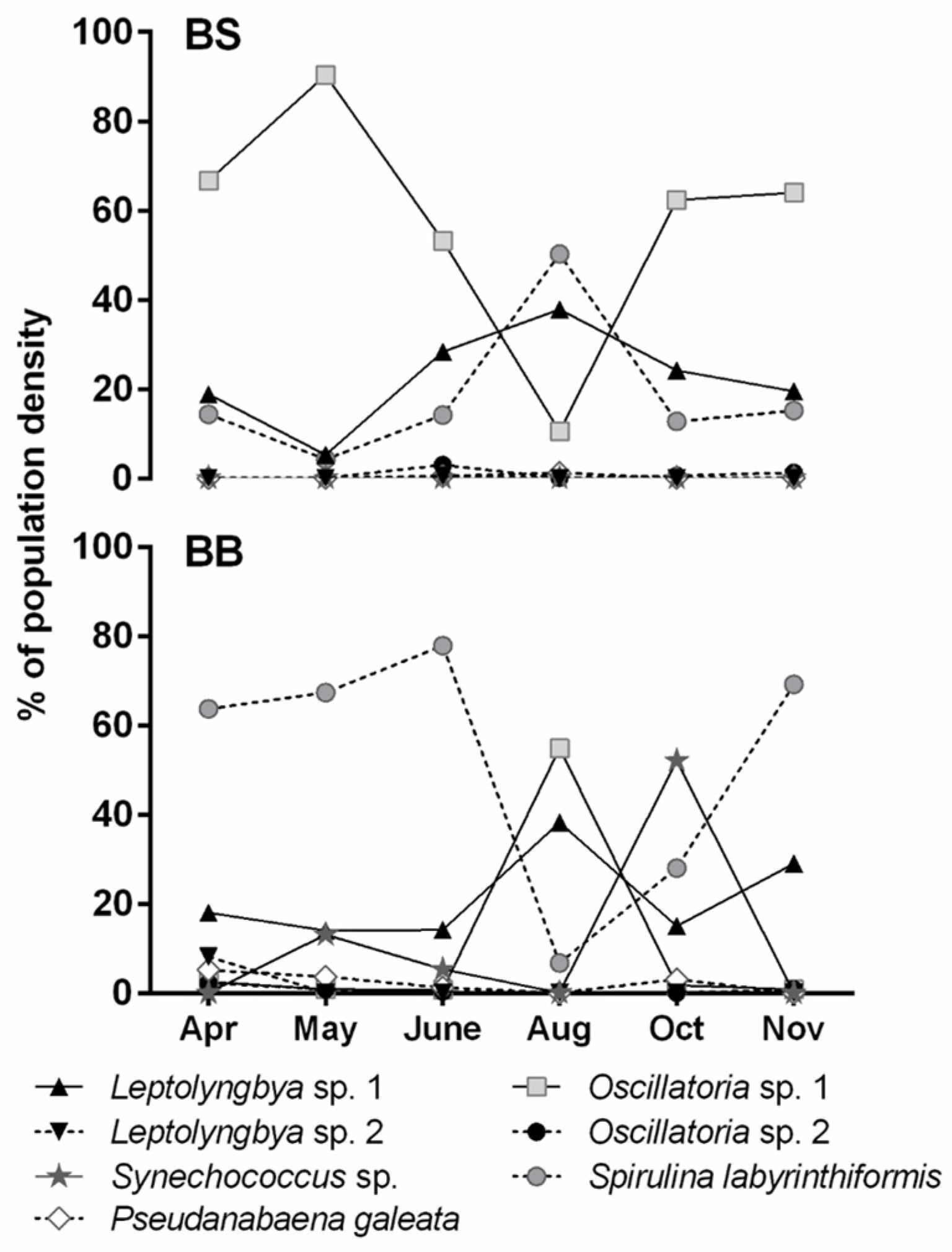
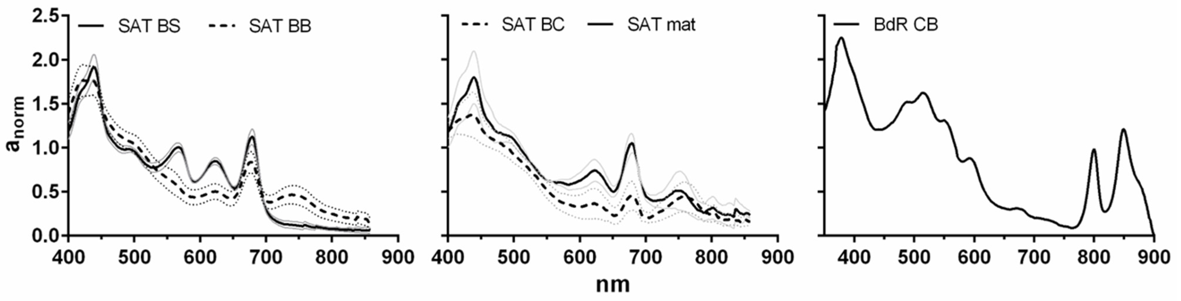
The phototrophic communities developing in the hot springs analysed had different phototrophic community compositions according to the chemico-physical characteristics of the spring water. When seasonal samples were collected (Saturnia), the cyanobacterial community appeared rather stable in its major components, thanks to constant temperature and pH. The variations in the abundance of different taxa were, most probably, due to seasonal variations in solar radiation or to the enrichment of thermal water with exogenous nutrients (e.g., following heavy precipitations). Rather little variations in the correspondent pigment signatures were found because spectra also recorded pigments from degraded trichomes, which were excluded from microscopic counts. Few studies analyse seasonal variation in cyanobacterial composition in hot springs, evidencing the role of different factors, such as sulphide concentration or heavy rainfalls [40,41]. In-depth characterisation of the microbial community composition was beyond the scope of the present work, nevertheless, it would be of great interest as future work to thoroughly characterise both phototrophic and chemotrophic members of these communities, by applying multifaceted approaches, including genomic techniques, both isolation-based and isolation-independent [42,43,44].
When the biological activity of on-site communities is considered, besides natural variation in the ratios among the oxygenic phototrophs, several sources of variability may explain activity differences during seasons and between bioglea samples and the isolated strains, considering that more than one factor can concur. It is also worth mentioning that it was not always possible to isolate the dominant component of the community (e.g., Saturnia dark brown Oscillatoria) because of a too strict dependency on on-site conditions not reproducible in the laboratory.
As stated previously, extracts from on-site communities usually showed higher antimicrobial activity than isolated strains, particularly considering lipophilic extracts. Antimicrobial activity was searched for on a panel including common contaminants of cosmetic products (e.g., moulds such as Penicillium and Aspergillus) and potential pathogens, such as dermatophytes (here represented by A. uncinatum as a model organism) or opportunistic bacteria (S. epidermidis). Positive results on the two types of targets would have led to different potential benefits, for users’ health or for technological applications. However, moulds were not affected, except for hydrophilic extracts from water, cultivated bioglea and mud from Bagno di Romagna. Dermatophytes were previously reported to be more sensitive to cyanobacterial extracts compared to other groups of fungi [45], and the results obtained in this work on A. uncinatum compared to moulds seem to confirm this higher sensitivity. Dermatophyte resistance to antifungal agents commonly used to treat their infections is a growing concern; thus, new antifungal agents would be of great importance [46,47]. To this end, extracts from both on-site communities and isolated strains have to confirm their effects on pathogenic dermatophytes.
Considering the total lack of antimicrobial activity in lipophilic extracts from isolated strains, a first hypothesis could be that thermal water accounts for the activity observed against A. uncinatum in lipophilic extracts of bioglea and mud samples from Saturnia and Bagno di Romagna, as well as for antibacterial activity against S. epidermidis of the hydrophilic extract from cultivated bioglea. Activity intensity varied among different community samples, notwithstanding that in the same hot spring, they were exposed to the same water environment. Different hydration levels of the collected samples could explain the variability, which, on the other hand, might also suggest that other antimicrobial molecules besides those present in water are involved. Moreover, if different hydration levels were responsible for variations in lipophilic extract activity, a similar activity pattern should have also been found for hydrophilic extracts. However, a lower frequency of activity (43%) was found in the latter. These considerations suggest that the observed activity in on-site communities was mainly not originated from water.
Cyanobacteria and microalgae were subjected to different environmental conditions in their habitat of origin with respect to laboratory cultures for, e.g., temperature, “medium” composition, light intensity, and bubbling. Pigments (chlorophyll a and its degradation products, and carotenoids) and fatty acids were the major components of the extracts. It is possible that in the mats grown in hot spring environments, a higher amount of chlorophyll degradation products was present compared to biomasses from laboratory-grown isolates. Phaeophytin a, as well as phaeophorbide a, has been shown to have antimicrobial activity [48,49]. Accumulation of cell degradation products in mats and biofilms might also have led to some fatty acids or their derivatives accumulation compared to laboratory-obtained biomasses. Fatty acids can also be endowed with antifungal activity [50]. Finally, in Saturnia, but also in Petriolo, microbial communities develop in pools open to the surrounding environment in which people bathe, so exogenous active substances may be absorbed/adsorbed in the microbial biofilms and mats, contributing to their antimicrobial activity. In the case of Saturnia bioglea, Centini et al. [51] actually found some contaminant cosmetics preservatives (e.g., sorbic acid, 4-hydroxy benzoic acid, phenoxyethanol) in the chloroform extracts (including those from the same samples analysed in this work), besides plant-derived molecules with known antimicrobial activity (abietic and dehydroabietic acids) [52] and several phenolics (e.g., methoxy cinnamic acid), which origin is not known but might also have antimicrobial activity [53]. The hydrophilic extracts contained mainly hydrophilic pigments (phycobiliproteins) and other protein-derived compounds (e.g., some free amino acids). They also contain few fatty acids. Lower activity was found in these extracts, either from bioglea and or isolated strains, compared to lipophilic extracts, most probably because a lower antimicrobial activity is associated with the main components of these extracts. The antifungal activity of phycobiliprotein-containing crude extracts has been tested for several cyanobacterial and microalgal strains, usually resulting low or absent unless purified compounds were prepared [54].
Besides abiotic conditions, biotic factors could also have impacted the activity. In their habitat, cyanobacteria and microalgae are growing amidst other oxygenic phototrophs, anoxygenic phototrophs, and chemoautotrophic and heterotrophic bacteria, thus needing to establish a network of interactions, among which competition through the production of antimicrobial or allelopathic metabolites [55,56]. The simplified biological community present in the laboratory cultures (including only some non-photosynthetic bacteria besides the oxygenic phototroph) may have reduced the necessity to invest in the production of antimicrobial molecules. Moreover, in laboratory cultures, bioactivity can vary with the growth phase, as observed for several cyanobacteria [57,58,59,60,61]; thus, the time of harvesting of the isolated strains may have affected both the presence and the intensity of antimicrobial activity. The antifungal activity during 
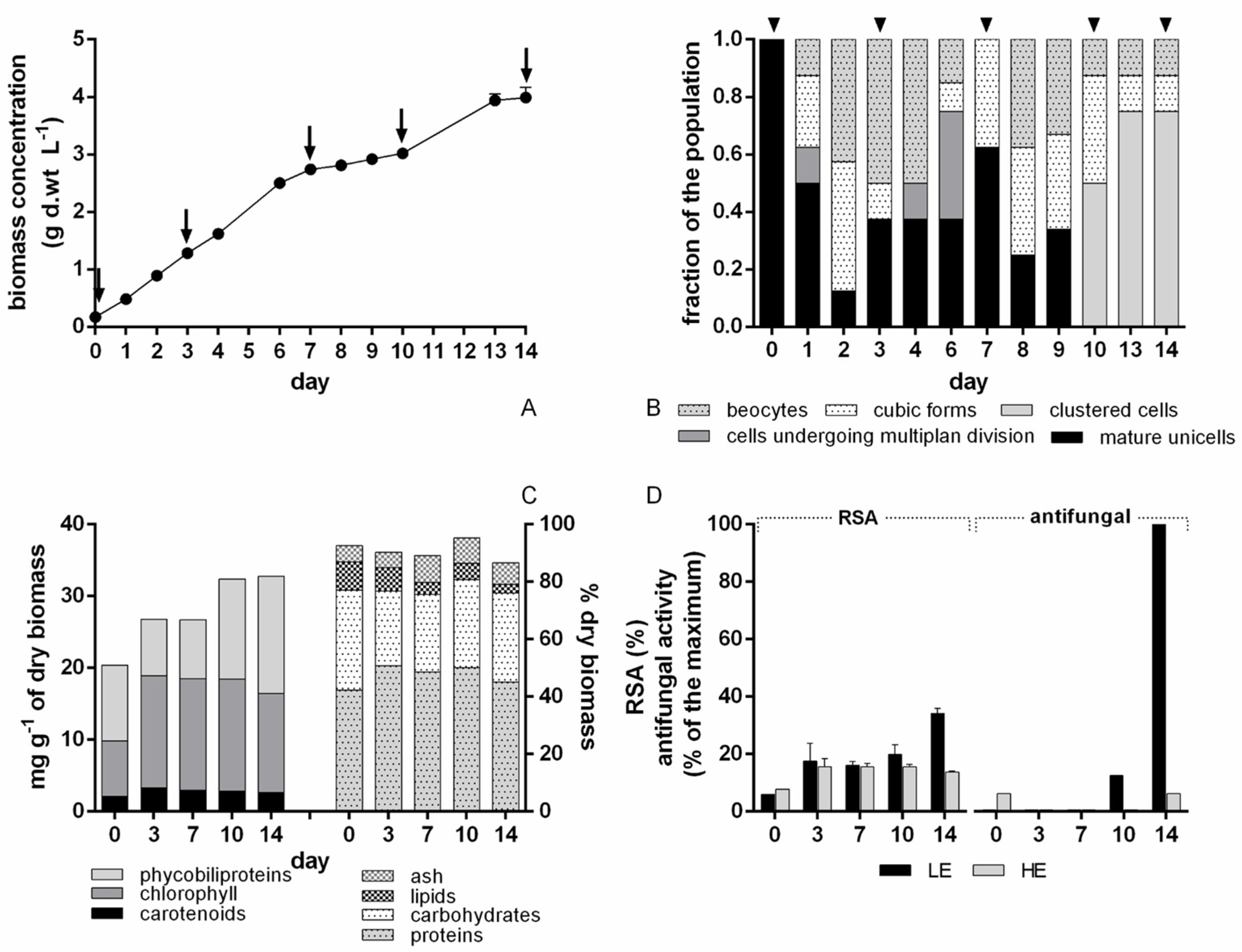
Chroococcidiopsis BIOG3 cultivation at the 10 L scale varied throughout the growth curve and life cycle stages, also appearing in the lipophilic extract (contrary to what was found at the 1 L scale). This highlights the necessity to establish for each isolated strain the best growth phase (and, if appropriate, life cycle stage) in which to harvest the biomass to maximise the active molecule production.
Little literature on the antimicrobial properties of on-site microbial communities is available. Dobretsov et al. [62] evidence complex interactions in hot spring communities from Oman, as they found antibacterial activity in cyanobacterial mats’ extracts from two of the four analysed springs, for one of which only non-polar extract and for the other both polar and non-polar extracts were active, but against different targets. Additionally, inhibition of quorum sensing was observed in extracts from three of the springs, whereas anti-diatom activity from all four suggested the production of metabolites to regulate interactions among different microbial groups. In contrast to what was observed in our work, cyanobacterial strains isolated from the four hot springs showed a wider and more potent antibacterial activity compared to microbial mats, besides showing antifungal activity [63]. Antimicrobial properties of strains isolated from hot springs, also for non-photosynthetic microorganisms, have been reported. Methanolic extracts from seven cyanobacterial strains (belonging to Oscillatoria, Synechococcus and Synechocystis) from Geno hot spring (Iran) showed activity mainly against Gram-positive bacteria, but few also against Escherichia coli and Candida [64]. Two cyanobacteria (Gleocapsa and Synechocystis) and two green algae (Scenedesmus and Chlorella) isolated from Rupite hot spring (Bulgaria) showed activity against Gram-positive bacteria in either exopolysaccharide or fatty acid fractions and, only exopolysaccharides from Gleocapsa, against the yeast Candida albicans [65]. Capsular polysaccharide from a Mastigocladus laminosus isolate from a hot spring in Néris-les-Bains (France) showed weak antibacterial activity [66]. A peptide from a thermal spring Bacillus licheniformis showed strong activity against Gram-positive bacteria [67]. A Beggiatoa-like strain from baregine showed bacteriostatic activity against three pathogens [68]. Non-photosynthetic bacteria might also contribute to the activity of microbial communities (and isolated strains) analysed in the present work.
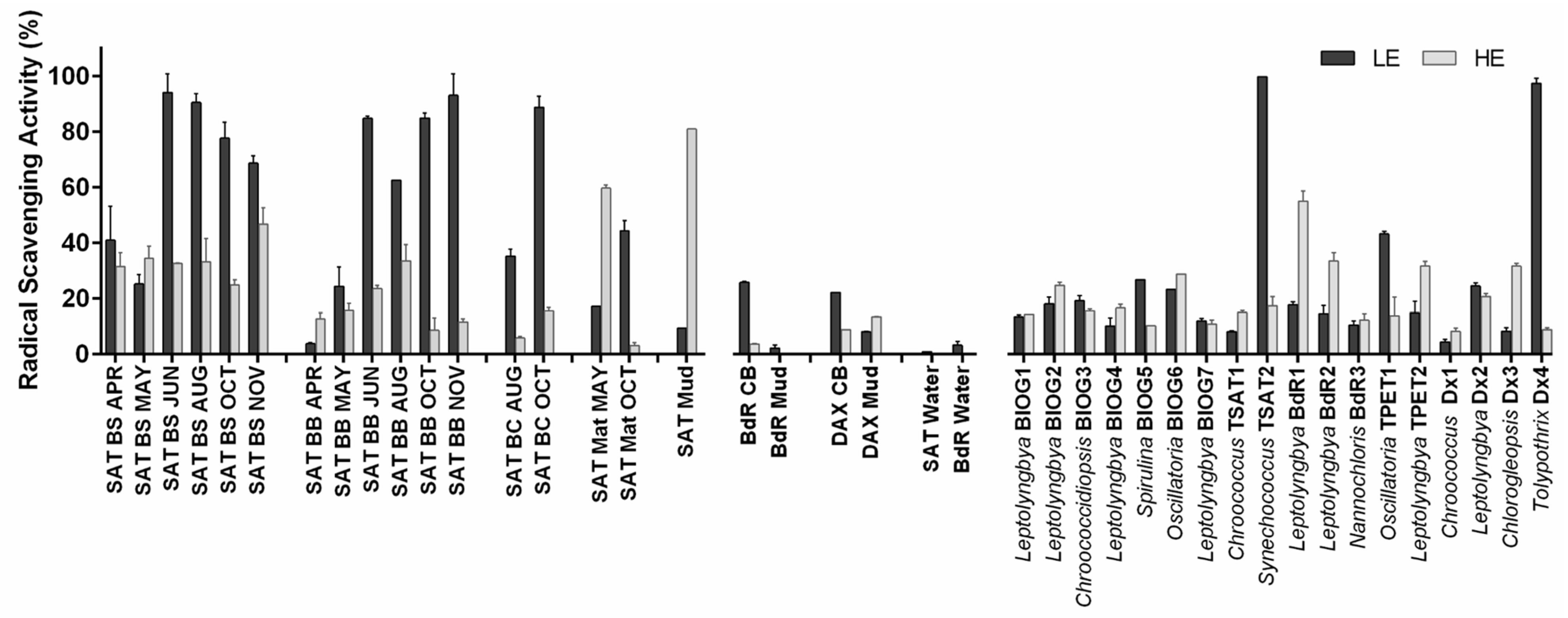
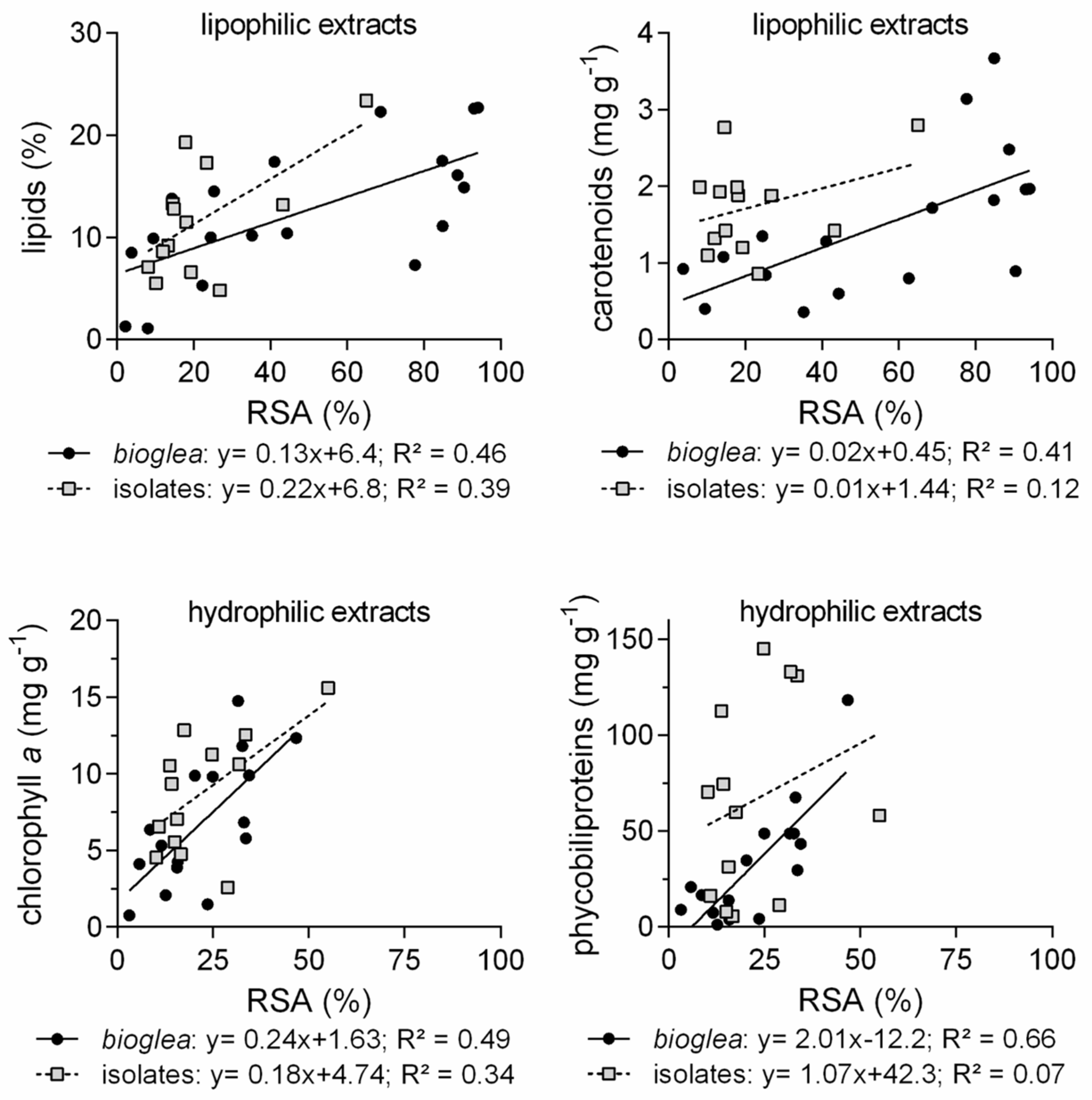
In our study, radical scavenging activity was usually higher in lipophilic extracts from on-site communities than in those from isolated strains, whereas more similar activities were observed in hydrophilic extracts. Regarding bioglea, biotic components were the main determinant of radical scavenging activity, as thermal waters were almost inactive. Radical scavenging activity of lipophilic extracts positively correlated with total lipids and, only for communities, with carotenoid content, which was higher in summer and autumn samples. Chlorophyll and its derivatives might also have contributed to radical scavenging activity, as they are known as radical scavengers [69]. In Saturnia bioglea extracts, several lipophilic compounds with antioxidant activity were detected in chloroform extracts, such as the chlorophyll-derived alcohol phytol, the carotenoid lycopersene and some cinnamic acid derivatives [51], which might have contributed to the radical scavenging activity observed. Determination of phenolics content and profile in the isolated strains will help to understand whether this component may be responsible for the observed radical scavenging activity. Phospholipid derivatives, such as diacylglycerols might also contribute. Furthermore, exogenous contaminants due to people bathing in the pool can also be responsible for part of the observed radical scavenging activity in Saturnia bioglea, as antioxidant chemicals deriving from cosmetic products, such as BHT and squalene, were found in the extracts [51].
Hydrophilic extracts contained phycobiliproteins, which are well-known antioxidant and anti-inflammatory molecules [70]. Radical scavenging activity of hydrophilic extracts from on-site communities positively correlated with phycobiliprotein content, while no correlation was found for isolated strains. The high variability in phycobiliprotein content, very low (<1% of dry biomass) in some strains (e.g., Chroococcus TSAT1 and Leptolyngbya BIOG4) and rather high (13–14%) in others (Leptolyngbya strains BIOG2, TPET2 and BdR2), was paired with a fairly low and little variable radical scavenging activity. Positive correlation with chlorophyll a in both bioglea and isolates’ extracts could be due to the presence of more hydrophilic chlorophyll derivatives. Although in this work total carbohydrate content did not correlate with the radical scavenging activity of the hydrophilic extracts, the polysaccharidic fraction, which plays an important role in the architecture of mats [71,72] and represents a sink of excess energy and carbon under high radiations [73,74], may also have contributed to the radical scavenging activity, a well-documented property of these polymers, often coupled with anti-inflammatory activity [66,75,76]. However, high total carbohydrate content (>50%) in isolated strains did not correspond to high radical scavenging activity. Other components, such as enzymes (e.g., peroxidases), might have been responsible for the observed activity in a higher proportion in isolated strains exposed to a potentially stressful regime of continuous light than in communities. Further analysis will be needed to confirm this hypothesis. Moreover, further testing of the extracts aimed at fully establishing antioxidant activity will be needed.
Both extracts from on-site communities and isolated strains were non-toxic toward the model crustacean Artemia salina. However, some of the hydrophilic extracts (mainly from isolates) were too dense and led to increased nauplii death because of poor gas exchange. These extracts derived from biomasses richer in carbohydrates and/or, in some cases, proteins. It seems that a carbohydrate + protein content above 2–3 g L−1 might have led to lethal physical effects on more than 50% of the nauplii. For comparison, a similar outcome was observed with hydrophilic extracts from the food-grade cyanobacterium Arthrospira platensis at the same extracted material concentration, while a 1:2 dilution of the extract totally removed the lethal effects (Biondi et al., unpublished data).
Two of the most interesting isolates emerging from this work are Tolypothrix Dx4 and Leptolyngbya BdR1. Both strains were isolated from cultivated bioglea. In spite of lower radical scavenging and, only in the case of Dax, antimicrobial activity, these communities represent an interesting biotechnological approach for spa development. The lower performance of Dax cultivated bioglea is easily explained by the time of sampling, i.e., during a maintenance period in autumn, in which the community was at its minimal development. For Bagno di Romagna, the good performances in terms of antimicrobial activity were most probably largely due to the action of thermal water, whereas the low radical scavenging activity may have originated from the reduced development of the oxygenic phototrophic community, usually a main driver of this activity. Optimisation of production and composition of the cultivated bioglea may lead to improvement and increased stability of their biological activity, an important feature for the application of these communities either as a component in peloid production [20] or for direct application in cosmetic and wellness treatments [14,18,19], not to mention extract preparation for cosmetic products. For production, e.g., in Saturnia or Petriolo, purposely designed plants would be necessary to optimise exposure to light and moisture, thus allowing the development of a bioglea similar to that growing in the pools and with comparable bioactivity profiles. The use of cultivated bioglea instead of isolated strains would allow for preserving the interactions within the microbial community, which may be responsible for part of the activity observed. On the other side, cultivation of isolated strains, which potential will need to be fully elucidated by further studies under culture conditions closer to those of hot springs, together with chemical characterisation of the extracts, would guarantee a higher standardisation of the product and reliability of activity. To this end, future studies, including a comparison between whole bioglea and synthetic communities built by the cultivation of multiple isolated strains under the same conditions of bioglea, would provide decisive information to lead these interesting microbial substrates towards actual industrial-level exploitation in terms of availability of the product, quality and safety.

5. Conclusions

Interesting antimicrobial and radical scavenging activities were found in microbial communities from different hot springs, thermal water-driven cultivations and mud production facilities. These properties were only in part reflected in cyanobacterial/microalgal strains isolated from these communities. Thermal water components, together with pigments and fatty acids, may explain the antimicrobial activity found in bioglea. Radical scavenging activity seems completely related to molecules produced by microorganisms, in particular pigments (carotenoids and phycobiliproteins), although other components, such as phenolics and polysaccharides, may also contribute. Non-pigment components should account for most of the activity found in the isolated strains. The findings suggest that to improve production, on-site cultivation of whole microbial communities would be the best solution for the present, while future research should have to investigate the possibility of combining isolated strains to obtain synthetic communities mimicking better than single strains the important bioactivities of natural communities, providing at the same time better standardisation prospects than the cultivation of whole communities. In both cases, for mass production, purposely designed plants, based on the exploitation of thermal waters, will need to be built so as to guarantee high yields and reliability of biological activities.

Supplementary Materials

The following supporting information can be downloaded at: https://www.mdpi.com/article/10.3390/cosmetics10030081/s1, Extraction Protocol; Table S1: Relation between isolated strains and on-site communities; Table S2: Equations of the regressions used to calculate IC50 values for radical scavenging activity determined with DPPH assay; Figure S1: Biochemical composition (A), phycobiliprotein (B), and chlorophyll and carotenoid (C) content in hot spring microbial communities samples and of the isolated strains; Figure S2: Dose–response curves for radical scavenging activity with DPPH assay; Figure S3: Linear regressions between components of bioglea/isolate biomasses and radical scavenging activity of lipophilic and hydrophilic extracts.

Author Contributions

Conceptualisation, N.B. and M.R.T.; methodology, N.B.; investigation, N.B., M.R.M. and M.C.; formal analysis, N.B.; validation, N.B. and M.C.; writing—original draft preparation, N.B.; writing—review and editing, all authors; funding acquisition, C.A. and M.R.T. All authors have read and agreed to the published version of the manuscript.

Funding

This research received no external funding.

Institutional Review Board Statement

Not applicable.

Informed Consent Statement

Not applicable.

Data Availability Statement

The data presented in this study are all available within the article and Supplementary Materials.

Acknowledgments

This article is dedicated to the memory of Mario R. Tredici, who passed away during the writing of the manuscript. Figure 1 was created with BioRender.com.

Conflicts of Interest

The authors declare no conflict of interest.

References

  1. Bueno Ariede, M.; Candido Marcílio, T.; Jacome Morocho, A.L.; Velasco Robles, M.V.; de Carvalho, J.C.M.; Rolim Baby, A. Cosmetic attributes of algae–A review. Algal Res. 2017, 25, 483–487. [Google Scholar] [CrossRef]
  2. Morone, J.; Alfeus, A.; Vasconcelos, V.; Martins, R. Revealing the potential of cyanobacteria in cosmetics and cosmeceuticals—A new bioactive approach. Algal Res. 2019, 41, 101541. [Google Scholar] [CrossRef]
  3. Morone, J.; Lopes, G.; Preto, M.; Vasconcelos, V.; Martins, R. Exploitation of filamentous and picoplanktonic cyanobacteria for cosmetic applications: Potential to improve skin structure and preserve dermal matrix components. Mar. Drugs 2020, 18, 486. [Google Scholar] [CrossRef] [PubMed]
  4. Thiyagarasaiyar, K.; Goh, B.-H.; Jeon, Y.-J.; Yow, Y.-Y. Algae metabolites in cosmeceutical: An overview of current applications and challenges. Mar. Drugs 2020, 18, 323. [Google Scholar] [CrossRef]
  5. Yarkent, Ç.; Gürlek, C.; Oncel, S.S. Potential of microalgal compounds in trending natural cosmetics: A review. Sustain. Chem. Pharm. 2020, 17, 100304. [Google Scholar] [CrossRef]
  6. Bernaerts, T.M.M.; Gheysen, L.; Foubert, I.; Hendrickx, M.E.; Van Loey, A.M. The potential of microalgae and their biopolymers as structuring ingredients in food: A review. Biotechnol. Adv. 2019, 37, 107419. [Google Scholar] [CrossRef]
  7. Couteau, C.; Coiffard, L. Microalgal application in cosmetics. In Microalgae in Health and Disease Prevention; Levine, I., Fleurence, J., Eds.; Academic Press: London, UK, 2018; pp. 317–323. [Google Scholar] [CrossRef]
  8. Stoyneva-Gärtner, M.; Uzunov, B.; Gärtner, G. Enigmatic microalgae from aeroterrestrial and extreme habitats in cosmetics: The potential of the untapped natural sources. Cosmetics 2020, 7, 27. [Google Scholar] [CrossRef]
  9. Dimitrovski, D.; Todorović, A. Clustering wellness tourists in spa environment. Tour. Manag. Perspect. 2015, 16, 259–265. [Google Scholar] [CrossRef]
  10. Antonelli, M.; Donelli, D.; Veronesi, L.; Vitale, M.; Pasquarella, C. Clinical efficacy of medical hydrology: An umbrella review. Int. J. Biometeorol. 2021, 65, 1597–1614. [Google Scholar] [CrossRef]
  11. Tian, X.; Zhang, Y.; Li, H.; Jiao, Y.; Wang, Q.; Zhang, Y.; Ma, N.; Wang, W. Property of mud and its application in cosmetic and medical fields: A review. Environ. Geochem. Health 2022, 44, 4235–4251. [Google Scholar] [CrossRef]
  12. Gomes, C.; Carretero, M.I.; Pozo, M.; Maraver, F.; Cantista, P.; Armijo, F.; Legido, J.L.; Teixeira, F.; Rautureau, M.; Delgado, R. Peloids and pelotherapy: Historical evolution, classification and glossary. Appl. Clay Sci. 2013, 75–76, 28–38. [Google Scholar] [CrossRef]
  13. Laporte, G.; Laurent, M.; Kaiser, P.; Deluzarches, M. Biologie du péloïde de Dax et de l’eau associée. Ann. Inst. Pasteur 1965, 109, 563–593. [Google Scholar]
  14. Francois, G.; Micollier, A.; Rouvie, I. Atelier Santé Environnement. In Les Boues Thermales; ESPN: Paris, France, 2005; pp. 1–41. [Google Scholar]
  15. Centini, M.; Tredici, M.R.; Biondi, N.; Buonocore, A.; Maffei Facino, R.; Anselmi, C. Thermal mud maturation: Organic matter and biological activity. Int. J. Cosmet. Sci. 2015, 37, 339–347. [Google Scholar] [CrossRef]
  16. Carretero, M.I. Clays in pelotherapy. A review. Part II: Organic compounds, microbiology and medical applications. Appl. Clay Sci. 2020, 189, 105531. [Google Scholar] [CrossRef]
  17. Gris, B.; Treu, L.; Zampieri, R.M.; Caldara, F.; Romualdi, C.; Campanaro, S.; La Rocca, N. Microbiota of the therapeutic Euganean thermal muds with a focus on the main cyanobacteria species. Microorganisms 2020, 8, 1590. [Google Scholar] [CrossRef] [PubMed]
  18. Mazzulla, S.; Chimenti, R.; Sesti, S.; De Stefano, S.; Morrone, M.; Martino, G. Effetto delle Bioglee solfuree su lesioni psoriasiche. Clin. Ter. 2004, 155, 499–504. [Google Scholar]
  19. Dupire, J. Le plancton thermal de Moltig-les-Bains: Actions anti-inflammatoire, anti-oxydante et cicatrisante dans la dermatite atopique. Presse Therm. Clim. 2008, 145, 143–153. [Google Scholar]
  20. Counilh, P.; Gibert, J.L. Procédé de Préparation de Boues Thermales, Boues Obtenues et Leurs Applications. EP0646647 B1, 15 January 2003. [Google Scholar]
  21. Svavarsson, H.G.; Valberg, J.E.; Arnardottir, H.; Brynjolfsdottir, A. Carbon dioxide from geothermal gas converted to biomass by cultivating coccoid cyanobacteria. Environ. Technol. 2018, 39, 2097–2104. [Google Scholar] [CrossRef]
  22. Mantelli, F.; Menichetti, S.; Calà, P. Principali Emergenze Termali in Toscana. Idrogeologia e Chimica Delle Acque; Regione Toscana & ARPAT: Firenze, Italy, 2014; pp. 1–378. [Google Scholar]
  23. Berard, P.; Chéry, L.; Lopoukhine, M.; Platel, J.P.; Sourisseau, B. Synthèse des Connaissances Hydrogéologiques sur les Ressources Thermo-Minérales du Bas-Adours; BRGM: Pessac, France, 1997; pp. 1–329. [Google Scholar]
  24. Roesler, C.S.; Perry, M.J.; Carder, K.L. Modeling in situ phytoplankton absorption from total absorption spectra in productive inland marine waters. Limnol. Oceanogr. 1989, 34, 1510–1523. [Google Scholar] [CrossRef]
  25. Rippka, R.; Deruelles, J.; Waterbury, J.B.; Herdman, M.; Stanier, R.Y. Generic assignments, strain histories and properties of pure cultures of cyanobacteria. J. Gen. Microbiol. 1979, 111, 1–61. [Google Scholar] [CrossRef]
  26. Lowry, O.H.; Rosebrough, N.J.; Farr, A.L.; Randall, R.J. Protein measurement with the Folin phenol reagent. J. Biol. Chem. 1951, 193, 265–275. [Google Scholar] [CrossRef] [PubMed]
  27. Dubois, M.; Gilles, K.A.; Hamilton, J.K.; Rebers, P.; Smith, F. Colorimetric method for determination of sugars and related substances. Anal. Chem. 1956, 28, 350–356. [Google Scholar] [CrossRef]
  28. Marsh, J.B.; Weinstein, D.B. Simple charring methods for determination of lipids. J. Lipid Res. 1966, 7, 574–576. [Google Scholar] [CrossRef] [PubMed]
  29. Jeffrey, S.W.; Humphrey, G.F. New spectrophotometric equations for determining chlorophylls a, b, c1 and c2 in higher plants, algae and natural phytoplankton. Biochem. Physiol. Pflanz. 1975, 167, 191–194. [Google Scholar] [CrossRef]
  30. Parsons, T.R.; Strickland, J.D.H. Discussion of spectrophotometric determination of marine plant pigments, with revised equations for ascertaining chlorophylls and carotenoids. J. Mar. Res. 1963, 21, 155–161. [Google Scholar] [CrossRef]
  31. Bennett, A.; Bogorad, L. Complementary chromatic adaptation in a filamentous blue-green alga. J. Cell Biol. 1973, 58, 419–435. [Google Scholar] [CrossRef]
  32. Biondi, N.; Piccardi, R.; Margheri, M.C.; Rodolfi, L.; Smith, G.D.; Tredici, M.R. Evaluation of Nostoc strain ATCC 53789 as a potential source of natural pesticides. Appl. Environ. Microb. 2004, 70, 3313–3320. [Google Scholar] [CrossRef]
  33. Das, K.; Tiwari, R.K.S.; Shrivastava, D.K. Techniques for evaluation of medicinal plant products as antimicrobial agent: Current methods and future trends. J. Med. Plants Res. 2010, 4, 104–111. [Google Scholar]
  34. Blois, M.S. Antioxidant determinations by the use of a stable free radical. Nature 1958, 4617, 1199–1200. [Google Scholar] [CrossRef]
  35. Yan, X.; Nagata, T.; Fan, X. Antioxidative activities in some common seaweeds. Plant Food Hum. Nutr. 1998, 52, 253–262. [Google Scholar] [CrossRef] [PubMed]
  36. Carballo, J.L.; Hernández-Inda, Z.L.; Pérez, P.; García-Grávalos, M.D. A comparison between two brine shrimp assays to detect in vitro cytotoxicity in marine natural products. BMC Biotechnol. 2002, 2, 17. [Google Scholar] [CrossRef]
  37. Lopes, V.R.; Fernández, N.; Martins, R.F.; Vasconcelos, V. Primary screening of the bioactivity of brackish water cyanobacteria: Toxicity of crude extracts to Artemia salina larvae and Paracentrotus lividus embryos. Mar. Drugs 2010, 8, 471–482. [Google Scholar] [CrossRef] [PubMed]
  38. Abbott, W.S. A method of computing the effectiveness of an insecticide. J. Am. Mosq. Control Assoc. 1987, 3, 302–303. [Google Scholar] [CrossRef]
  39. Waterbury, J.B. Subsection II. Order Pleurocapsales Geitler 1925, emend. Waterbury and Stanier 1978. In Bergey’s Manual of Systematic Bacteriology; Staley, J.T., Bryant, M.P., Pfennig, N., Holt, J.G., Eds.; Williams & Wilkins: Baltimore, MD, USA, 1989; Volume 3, pp. 1710–1727. [Google Scholar]
  40. Lacap, D.C.; Barraquio, W.; Pointing, S.B. Thermophilic microbial mats in a tropical geothermal location display pronounced seasonal changes but appear resilient to stochastic disturbance. Environ. Microbiol. 2007, 9, 3065–3076. [Google Scholar] [CrossRef] [PubMed]
  41. Reul, A.; Martín-Clemente, E.; Melero-Jiménez, I.J.; Bañares-España, E.; Flores-Moya, A.; García-Sánchez, M.J. What triggers the annual cycle of cyanobacterium Oscillatoria sp. in an extreme environmental sulfide-rich spa? Water 2020, 12, 883. [Google Scholar] [CrossRef]
  42. Prieto-Barajas, C.M.; Valencia-Cantero, E.; Santoyo, G. Microbial mat ecosystems: Structure types, functional diversity, and biotechnological application. Electron. J. Biotechnol. 2018, 31, 48–56. [Google Scholar] [CrossRef]
  43. Yasir, M.; Qureshi, A.K.; Khan, I.; Bibi, F.; Rehan, M.; Khan, S.B.; Azhar, E.I. Culturomics-based taxonomic diversity of bacterial communities in the hot springs of Saudi Arabia. OMICS J. Integr. Biol. 2019, 23, 17–27. [Google Scholar] [CrossRef]
  44. Strunecký, O.; Kopejtka, K.; Goecke, F.; Tomasch, J.; Lukavský, J.; Neori, A.; Kahl, S.; Pieper, D.H.; Pilarski, P.; Kaftan, D.; et al. High diversity of thermophilic cyanobacteria in Rupite hot spring identified by microscopy, cultivation, single-cell PCR and amplicon sequencing. Extremophiles 2019, 23, 35–48. [Google Scholar] [CrossRef]
  45. Issa, A.A. Antibiotic production by the cyanobacteria Oscillatoria angustissima and Calothrix parietina. Environ. Toxicol. Pharmacol. 1999, 8, 33–37. [Google Scholar] [CrossRef]
  46. Gnat, S.; Łagowski, D.; Nowakiewicz, A. Major challenges and perspectives in the diagnostics and treatment of dermatophyte infections. J. Appl. Microbiol. 2020, 129, 212–232. [Google Scholar] [CrossRef] [PubMed]
  47. Muhaij, F.F.; George, S.J.; Nguyen, C.D.; Tyring, S.K. Antimicrobials and resistance part II: Antifungals, antivirals, and antiparasitics. J. Am. Acad. Dermatol. 2022, 86, 1207–1226. [Google Scholar] [CrossRef]
  48. Albuquerque Gomes, R.; Ferreira Teles, Y.C.; de Oliveira Pereira, F.; de Sousa Rodrigues, L.A.; de Oliveira Lima, E.; de Fátima Agra, M.; de Fátima Vanderlei de Souza, M. Phytoconstituents from Sidastrum micranthum (A. St.-Hil.) Fryxell (Malvaceae) and antimicrobial activity of pheophytin a. Braz. J. Pharm. Sci. 2015, 51, 861–867. [Google Scholar] [CrossRef]
  49. Ji, J.-Y.; Yang, J.; Zhang, B.-W.; Wang, S.-R.; Zhang, G.-C.; Lin, L.-N. Sodium pheophorbide a controls cherry tomato gray mold (Botrytis cinerea) by destroying fungal cell structure and enhancing disease resistance-related enzyme activities in fruit. Pest. Biochem. Phys. 2020, 166, 104581. [Google Scholar] [CrossRef] [PubMed]
  50. Guimarães, A.; Venâncio, A. The potential of fatty acids and their derivatives as antifungal agents: A review. Toxins 2022, 14, 188. [Google Scholar] [CrossRef] [PubMed]
  51. Centini, M.; Tredici, M.R.; Biondi, N.; Buonocore, A.; Maffei Facino, R.; Anselmi, C. Bioglea as a source of bioactive ingredients: Chemical and biological evaluation. Cosmetics 2020, 7, 81. [Google Scholar] [CrossRef]
  52. Helfenstein, A.; Vahermo, M.; Nawrot, D.A.; Demirci, F.; İşcan, G.; Krogerus, S.; Yli-Kauhaluoma, J.; Moreira, V.M.; Tammela, P. Antibacterial profiling of abietane-type diterpenoids. Bioorg. Med. Chem. 2017, 25, 132–137. [Google Scholar] [CrossRef] [PubMed]
  53. Guzman, J.D. Natural cinnamic acids, synthetic derivatives and hybrids with antimicrobial activity. Molecules 2014, 19, 19292–19349. [Google Scholar] [CrossRef]
  54. Dranseikienė, D.; Balčiūnaitė-Murzienė, G.; Karosienė, J.; Morudov, D.; Juodžiukynienė, N.; Hudz, N.; Gerbutavičienė, R.J.; Savickienė, N. Cyano-phycocyanin: Mechanisms of action on human skin and future perspectives in medicine. Plants 2022, 11, 1249. [Google Scholar] [CrossRef]
  55. Leão, P.; Vasconcelos, M.T.S.D.; Vasconcelos, V.M. Allelopathy in freshwater cyanobacteria. Crit. Rev. Microbiol. 2009, 35, 271–282. [Google Scholar] [CrossRef]
  56. Hom, E.F.Y.; Aiyar, P.; Schaeme, D.; Mittag, M.; Sasso, S. A chemical perspective on microalgal–microbial interactions. Trends Plant Sci. 2015, 20, 689–693. [Google Scholar] [CrossRef]
  57. Armstrong, J.E.; Janda, K.E.; Alvarado, B.; Wright, A.E. Cytotoxin production by a marine Lyngbya strain (cyanobacterium) in a large-scale laboratory bioreactor. J. Appl. Phycol. 1991, 3, 277–282. [Google Scholar] [CrossRef]
  58. Pushparaj, B.; Pelosi, E.; Jüttner, F. Toxicological analysis of the marine cyanobacterium Nodularia harveyana. J. Appl. Phycol. 1999, 10, 527–530. [Google Scholar] [CrossRef]
  59. Suikkanen, S.; Fistarol, G.O.; Granéli, E. Allelopathic effects of the Baltic cyanobacteria Nodularia spumigena, Aphanizomenon flos-aquae and Anabaena lemmermannii on algal monocultures. J. Exp. Mar. Biol. Ecol. 2004, 308, 85–101. [Google Scholar] [CrossRef]
  60. El-Sheek, M.M.; Khairy, H.M.; El-Shenody, R.A. Allelopathic effects of cyanobacterium Microcystis aeruginosa Kützing on the growth and photosynthetic pigments of some algal species. Allelopath. J. 2010, 26, 275–290. [Google Scholar]
  61. Wang, L.; Zi, J.; Xu, R.; Hilt, S.; Hou, X.; Chang, X. Allelopathic effects of Microcystis aeruginosa on green algae and a diatom: Evidence from exudates addition and co-culturing. Harmful Algae 2017, 61, 56–62. [Google Scholar] [CrossRef]
  62. Dobretsov, S.; Abed, R.M.M.; Al Maskari, S.M.S.; Al Sabahi, J.N.; Victor, R. Cyanobacterial mats from hot springs produce antimicrobial compounds and quorum-sensing inhibitors under natural conditions. J. Appl. Phycol. 2011, 23, 983–993. [Google Scholar] [CrossRef]
  63. Sherwani, N.; Abed, R.M.M.; Dobretsov, S.; Mary, S. Antibacterial and antifungal activities of cyanobacterial strains isolated from hot springs in Oman. Sultan Qaboos Univ. J. Sci. 2015, 20, 11–19. [Google Scholar] [CrossRef]
  64. Heidari, F.; Rihai, H.; Yousefzadi, M.; Asadi, M. Antimicrobial activity of cyanobacteria isolated from hot spring of Geno. Middle East J. Sci. Res. 2012, 12, 336–339. [Google Scholar]
  65. Najdenski, H.M.; Gigova, L.G.; Iliev, I.I.; Pilarski, P.S.; Lukavský, J.; Tsvetkova, I.V.; Ninova, M.S.; Kussovski, V.K. Antibacterial and antifungal activities of selected microalgae and cyanobacteria. Int. J. Food Sci. Technol. 2013, 48, 1533–1540. [Google Scholar] [CrossRef]
  66. Gloaguen, V.; Garbacki, N.; Petit, D.; Morvan, H.; Hoffmann, L. Bioactive capsular polysaccharide from the thermophilic Mastigocladus laminosus (Cyanophyceae/Cyanobacteria): Demonstration of anti-inflammatory properties. Arch. Hydrobiol. Suppl. Algol. Stud. 2003, 108, 63–73. [Google Scholar] [CrossRef]
  67. Mendo, S.; Faustino, N.A.; Sarmento, A.C.; Amado, F.; Moir, A.J.G. Purification and characterization of a new peptide antibiotic produced by a thermotolerant Bacillus licheniformis strain. Biotechnol. Lett. 2004, 26, 115–119. [Google Scholar] [CrossRef] [PubMed]
  68. Maillard, F.; Shepherd, D. Process for the Production of a Substance Having Bacteriostatic Activity. US Patent 4,419,449, 6 December 1983. [Google Scholar]
  69. Hsu, C.-Y.; Chao, P.-Y.; Hu, S.-P.; Yang, C.-M. The antioxidant and free radical scavenging activities of chlorophylls and pheophytins. Food Nutr. Sci. 2013, 4, 1–8. [Google Scholar] [CrossRef]
  70. Pagels, F.; Guedes, A.C.; Amaro, H.M.; Kijjoa, A.; Vasconcelos, V. Phycobiliproteins from cyanobacteria: Chemistry and biotechnological applications. Biotechnol. Adv. 2019, 37, 422–443. [Google Scholar] [CrossRef] [PubMed]
  71. Veerabadhran, M.; Chakraborty, S.; Mitra, S.; Karmakar, S.; Mukherjee, J. Effects of flask configuration on biofilm growth and metabolites of intertidal cyanobacteria isolated from a mangrove forest. J. Appl. Microbiol. 2018, 125, 190–202. [Google Scholar] [CrossRef] [PubMed]
  72. Rossi, F.; De Philippis, R. Role of cyanobacterial exopolysaccharides in phototrophic biofilms and in complex microbial mats. Life 2015, 5, 1218–1238. [Google Scholar] [CrossRef]
  73. Cogne, G.; Gros, J.-B.; Dussap, C.-G. Identification of a metabolic network structure representative of Arthrospira (Spirulina) platensis metabolism. Biotechnol. Bioeng. 2003, 84, 667–676. [Google Scholar] [CrossRef]
  74. Otero, A.; Vincenzini, M. Nostoc (Cyanophyceae) goes nude: Extracellular polysaccharides serve as a sink for reducing power under unbalanced C/N metabolism. J. Phycol. 2004, 40, 74–81. [Google Scholar] [CrossRef]
  75. Gongi, W.; Cordeiro, N.; Gomez Pinchetti, J.L.; Ben Ouada, H. Functional, rheological, and antioxidant properties of extracellular polymeric substances produced by a thermophilic cyanobacterium Leptolyngbya sp. J. Appl. Phycol. 2022, 34, 1423–1434. [Google Scholar] [CrossRef]
  76. Zampieri, R.M.; Adessi, A.; Caldara, F.; De Philippis, R.; Dalla Valle, L.; La Rocca, N. In vivo anti-inflammatory and antioxidant effects of microbial polysaccharides extracted from Euganean therapeutic muds. Int. J. Biol. Macromol. 2022, 209, 1710–1719. [Google Scholar] [CrossRef]
Figure 1. Experimental plan. LE—lipophilic extract; HE—hydrophilic extract.
Cosmetics 10 00081 g001
Figure 2. Seasonal variation in relative frequency of Saturnia hot spring cyanobacteria. Cyanobacterial composition of surface (BS) and bottom (BB) bioglea in different seasons.
Cosmetics 10 00081 g002
Figure 3. In vivo absorption spectra of bioglea and mats normalised to their spectral mean. Samples were collected in different sites at Saturnia and Bagno di Romagna. Thick lines—average spectrum of all the samples collected in the same site: thin lines—standard deviation. Sample names indicate: sampling spa: SAT—Saturnia spa; BdR—Bagno di Romagna spa, and site of sampling within the spa: BS—surface bioglea; BB—bottom bioglea; BC—canal bioglea; Mat—coriaceous microbial mat; CB—cultivated bioglea.
Cosmetics 10 00081 g003
Figure 4. Extracts’ radical scavenging activity determined by DPPH assay. Activity of lipophilic (LE) and hydrophilic (HE) extracts from on-site collected communities, mud and water samples, and isolated strains are reported. Sample names indicate: sampling spa (SAT—Saturnia spa; BdR—Bagno di Romagna spa; DAX—Dax thermal area), location of sampling within the spa (BS—surface bioglea; BB—bottom bioglea; BC—canal bioglea; Mat—coriaceous microbial mat; Mud—thermal mud; CB—cultivated bioglea), and, where needed, month of sampling (APR—April; JUN—June; AUG—August, OCT—October; NOV—November).
Cosmetics 10 00081 g004
Figure 5. Linear regressions between components of bioglea/isolates’ biomasses and radical scavenging activity of lipophilic and hydrophilic extracts. Equations and R2 values of the interpolated lines are also reported. Correlation analysis: lipophilic extract RSA vs. lipids—bioglea, p < 0.01, n = 19–isolates, p < 0.05, n = 13; lipophilic extract RSA vs. carotenoids—bioglea, p < 0.01, n = 17–isolates, p > 0.05, n = 13; hydrophilic extract RSA vs. chlorophyll abioglea, p < 0.01, n = 16–isolates, p < 0.05, n = 13; hydrophilic extract RSA vs. phycobiliproteins—bioglea, p < 0.001, n = 16–isolates, p > 0.05, n = 13.
Cosmetics 10 00081 g005
Figure 6. Cultivation of Chroococcidiopsis BIOG3 in a 10 L bubbled tube: (A) growth curve, arrows indicate sample collection for analyses; (B) fraction of the population in the culture in the different growth stages, arrow meaning as in (A); (C) biomass gross composition and main pigments’ content during the trial; (D) bioactivity, evaluated as radical scavenging activity (RSA) and antifungal activity against A. uncinatum of lipophilic (LE) and hydrophilic (HE) extract during the trial.
Cosmetics 10 00081 g006
Table 1. List of the hot spring samples and description of the collection site. Codes include indication of hot spring of origin, type of material sampled and month of sampling (where needed).
Sample IDOrigin
SAT BS APR; SAT BS MAY; SAT BS JUN; SAT BS AUG; SAT BS OCT; SAT BS NOVSaturnia spa, bioglea harvested at midday while floating at the surface (BS) of the natural pool
SAT BB APR; SAT BB MAY; SAT BB JUN; SAT BB AUG; SAT BB OCT; SAT BB NOVSaturnia spa, bioglea harvested at midday from the bottom of the natural pool (BB)
SAT BC AUG; SAT BC OCTSaturnia spa, bioglea harvested at midday from the bottom of the canal connecting the natural pool to artificial pools
SAT Mat MAY; SAT Mat OCTSaturnia spa, microbial mat harvested from the wall where the natural pool water cascades into the canal
SAT MudSaturnia spa, 5-month matured thermal mud prepared by mixing virgin mud with thermal water and periodically stirring manually
SAT WaterSaturnia spa, water directly springing from the thermal source
BdR MudBagno di Romagna spa, thermal mud prepared by mixing virgin mud with thermal water and cultivated bioglea
BdR CBBagno di Romagna spa, bioglea cultivated on an inclined plane where water flows from a tank placed in the upper end of the plane
BdR WaterBagno di Romagna spa, water from the pool
TPET BPetriolo spa, bioglea from the pool (used only to perform strain isolation)
DAX MudDax, mud prepared and packed by the Régie Municipale des Eaux et des Boues with the brand name Terdax®
DAX CBDax, bioglea cultivated on inclined planes using thermal water at the Régie Municipale des Eaux et des Boues as a component in Terdax® preparation
Table 2. Cyanobacterial and microalgal isolated strains. Isolations were carried out from samples collected from different hot spring sites (see Table 1 and Table S1).
OrderGenus/SpeciesStrain
SynechococcalesSynechococcus sp. TSAT2
Leptolyngbya sp.BIOG1, BIOG2, BIOG4, BIOG7, TPET2, BdR1, BdR2
ChroococcalesChroococcus sp.TSAT1
Chroococcus cf. minutusDx1
ChroococcidiopsidalesChroococcidiopsis sp.BIOG3
OscillatorialesOscillatoria sp.BIOG6, TPET1
Phormidium sp.Dx2
SpirulinalesSpirulina cf. labyrinthiformisBIOG5
NostocalesChlorogleopsis sp.Dx3
Tolypothrix sp.Dx4
ChlorellalesNannochloris sp.BdR3
Table 3. Biochemical composition of bioglea and isolated strains. Mean ± standard deviation values for each site within the spas and for isolated strains are reported.
SamplesProteinsCarbohydratesLipidsChlorophyll aCarotenoidsPhycocyaninAllophycocyaninPhycoerythrin
% Organic Mattermg g−1 Organic Matter% Organic Matter
SAT BS42.2 ± 6.831.6 ± 6.316.6 ± 5.510.9 ± 2.71.7 ± 0.929.7 ± 11.812.9 ± 7.420.1 ± 10.2
SAT BB41.5 ± 6.318.3 ± 3.315.6 ± 7.04.7 ± 1.51.8 ± 1.16.4 ± 6.21.5 ± 1.02.6 ± 4.4
SAT BC61.9 ± 15.424.4 ± 12.012.4 ± 5.02.7 ± 1.71.4 ± 1.513.8 ± 1.42.9 ± 3.30.6 ± 0.2
DAX CB27.2 ± 0.853.8 ± 3.66.7 ± 0.3ntntntntnt
Isolated strains38.4 ± 8.843.4 ± 13.812.6 ± 7.011.1 ± 9.22.4 ± 2.443.4 ± 35.018.7 ± 19.03.9 ± 5.2
SAT—Saturnia spa; DAX—Dax thermal area; BS—surface bioglea; BB—bottom bioglea; BC—canal bioglea; CB—cultivated bioglea; nt—not tested.
Table 4. Antimicrobial activity of spring waters’ extracts and positive control standards. Results of tests against the whole target panel are reported.
Aspergillus nigerArthroderma uncinatumPenicillium expansumStaphylococcus epidermidis
LEHELEHELEHELEHE
SampleL L−1L L−1L L−1L L−1L L−1L L−1L L−1L L−1
SAT Waternana510nanana20
BdR Waterna2.555na2.5na10
mL L−1mL L−1mL L−1mL L−1
Methyl-Paraben1.251.250.310
Kathon CG0.0310.0140.0260.250
LE—lipophilic extract; HE—hydrophilic extract; na—no activity; SAT—Saturnia spa; BdR—Bagno di Romagna spa.
Table 5. Antifungal activity against Arthroderma uncinatum. Data for extracts from on-site collected communities and isolated strains are reported.
SampleArthroderma uncinatumStrainArthroderma uncinatum
LEHE LEHE
g L−1g L−1 g L−1g L−1
SAT BS APR6.3naLeptolyngbya BIOG1na44.2
SAT BS MAY16.0naLeptolyngbya BIOG2nana
SAT BS JUN4.04.0Chroococcidiopsis BIOG3na89.6
SAT BS AUG8.132.5Leptolyngbya BIOG4nana
SAT BS OCT0.812.4 pi (50–75%)Spirulina BIOG5na23.5
SAT BS NOV4.9naOscillatoria BIOG6na43.6 pi (50–75%)
SAT BB APR1.0naLeptolyngbya BIOG7nana
SAT BB MAY5.6naChroococcus TSAT1na87.3 pi (25%)
SAT BB JUN1.6naSynechococcus TSAT2nana
SAT BB AUG17.9naOscillatoria TPET1nana
SAT BB OCT1.0naLeptolyngbya TPET2nana
SAT BB NOV5.1naLeptolyngbya BdR1na42.3
SAT BC AUG8.717.4Leptolyngbya BdR2nana
SAT BC OCT0.721.2 pi (50–75%)Nannochloris BdR3na91.1
SAT Mat MAY3.1naChroococcus Dx1na45.1
SAT Mat OCT1.9naLeptolyngbya Dx2nana
SAT Mud3.8naChlorogleopsis Dx3nana
BdR CB35.435.4Tolypothrix Dx4na45.1
BdR Mud1.22.4
DAX CBna10.9 pi (<25%)
DAX Mud59.1na
LE—lipophilic extract; HE—hydrophilic extract; na—no activity; pi—partial inhibition. Sample names indicate: sampling spa: SAT—Saturnia spa; BdR—Bagno di Romagna spa; DAX—Dax thermal area, location of sampling within the spa: BS—surface bioglea; BB—bottom bioglea; BC—canal bioglea; Mat—coriaceous microbial mat; Mud—thermal mud; CB—cultivated bioglea, and month of sampling (where necessary): APR—April; JUN—June; AUG—August; OCT—October; NOV—November.
Table 6. Antimicrobial activity against the other components of the target panel. Data for the extracts from on-site collected communities and isolated strains showing activity against targets other than A. uncinatum are reported.
Sample/StrainAspergillus nigerPenicillium expansumStaphylococcus epidermidis
SAT BB OCTnanaLE, 20 mg
SAT BC OCTnanaLE, 5 mg
SAT Mat OCTnanaLE, 20 mg, pi (50–75%)
BdR MudHE, 4.8 g L−1HE, 4.8 g L−1na
BdR CBHE, 17.7 g L−1HE, 17.7 g L−1HE, 35.4 g L−1
Tolypothrix Dx4HE, 45.1 g L−1HE, 45.1 g L−1na
LE—lipophilic extract; HE—hydrophilic extract; na—no activity; pi—partial inhibition. Sample names indicate: sampling spa: SAT—Saturnia spa; BdR—Bagno di Romagna spa, lLocation of sampling within the spa: BB—bottom bioglea; BC—canal bioglea; Mat—coriaceous microbial mat; Mud—thermal mud; CB—cultivated bioglea, and month of sampling (where necessary): OCT—October.
Table 7. IC50 values for radical scavenging activity determined with DPPH assay. The IC50 values were calculated for the extracts reported in Figure S2 using equations reported in Table S2.
ExtractIC50 (g L−1)VCEAC (μg g−1)
Lipophilic extracts
SAT BS JUN10.33.18
SAT BS AUG26.41.24
SAT BS OCT24.61.34
SAT BS NOV29.11.13
SAT BB JUN22.71.45
SAT BB AUG31.81.04
SAT BB OCT20.51.61
SAT BB NOV18.31.80
SAT BC OCT16.91.95
Synechococcus TSAT218.41.79
Tolypothrix Dx416.42.01
Hydrophilic extracts
SAT Mat MAY36.50.90
SAT Mud29.91.10
Leptolyngbya BdR137.50.88
IC50—50% Inhibiting Concentration; VCEAC—vitamin C equivalent antioxidant capacity. Sample names indicate: sampling spa (SAT—Saturnia spa), location of sampling within the spa (BS—surface bioglea; BB—bottom bioglea; BC—canal bioglea; Mat—coriaceous microbial mat; Mud—thermal mud) and, where needed, month of sampling (JUN—June; AUG—August, OCT—October; NOV—November).
Disclaimer/Publisher’s Note: The statements, opinions and data contained in all publications are solely those of the individual author(s) and contributor(s) and not of MDPI and/or the editor(s). MDPI and/or the editor(s) disclaim responsibility for any injury to people or property resulting from any ideas, methods, instructions or products referred to in the content.

