Review

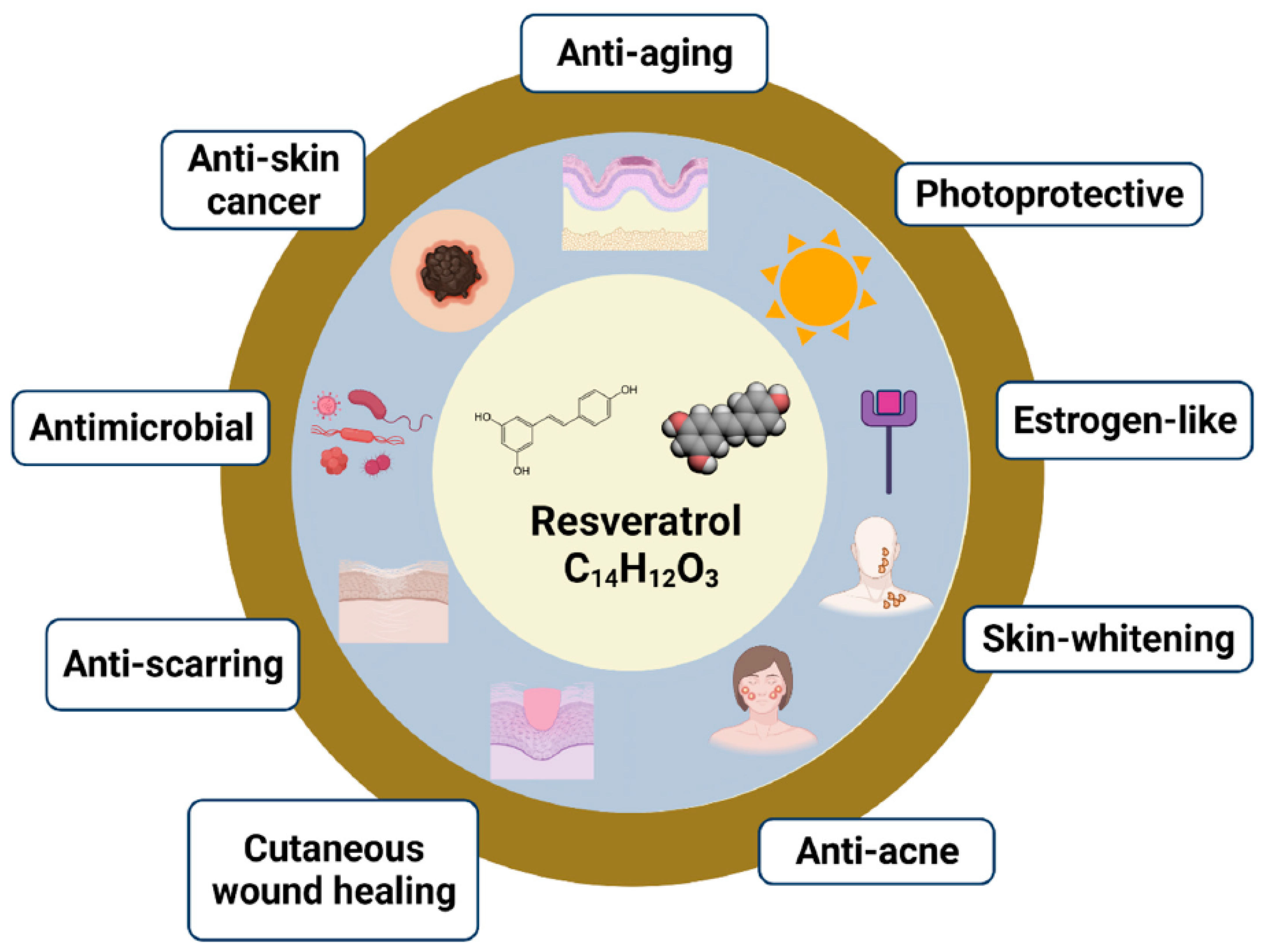
Dermatological Bioactivities of Resveratrol and Nanotechnology Strategies to Boost Its Efficacy—An Updated Review

by
Filipa Mascarenhas-Melo
1,2,*,
André R. T. S. Araújo
3,4,
Márcio Rodrigues
3,5,
Ankita Mathur
6,
Maria Beatriz S. Gonçalves
1,
Kalpita Tanwar
7,
Fariba Heidarizadeh
8,
Fereshteh Nejaddehbashi
9,
Abbas Rahdar
10,
Priscila Gava Mazzola
11,
Francisco Veiga
1,2 and
Ana Cláudia Paiva-Santos
1,2,*
1
Laboratory of Development and Drug Technologies, Faculty of Pharmacy, University of Coimbra, Azinhaga Sta. Comba, 3000-548 Coimbra, Portugal
2
LAQV, REQUIMTE, Department of Pharmaceutical Technology, Faculty of Pharmacy, University of Coimbra, Azinhaga Sta. Comba, 3000-548 Coimbra, Portugal
3
CPIRN-UDI-IPG—Center for Potential and Innovation of Natural Resources, Research Unit for Inland Development, Polytechnic Institute of Guarda, Av. Dr. Francisco de Sá Carneiro, 6300-559 Guarda, Portugal
4
LAQV, REQUIMTE, Laboratory of Applied Chemistry, Department of Chemical Sciences, Faculty of Pharmacy, Porto University, Rua Jorge Viterbo Ferreira, No. 228, 4050-313 Porto, Portugal
5
CICS-UBI—Health Sciences Research Center, University of Beira Interior, Av. Infante D. Henrique, 6200-506 Covilhã, Portugal
6
Research and Development Department, Abode Biotech India Pvt. Limited, MLA Colony, Banjara Hills Hyderabad-Telangana, Hyderabad 500033, Telangana, India
7
Department of Bioinformatics, Martin Luther Christian University, Dongktieh, Block-1, Nongrah, Shillong 793006, Meghalaya, India
8
Department of Chemistry, Shahid Chamran University of Ahvaz, Ahvaz 6135743169, Iran
9
Cellular and Molecular Research Center, Medical Basic Sciences Institute, Ahvaz Jundishapur University of Medical Sciences, Ahvaz 61357-1579, Iran
10
Department of Physics, University of Zabol, Zabol 98613-35856, Iran
11
Faculty of Pharmaceutical Sciences, University of Campinas, Campinas 13083-871, SP, Brazil
*
Authors to whom correspondence should be addressed.
Cosmetics 2023, 10(3), 68; https://doi.org/10.3390/cosmetics10030068
Submission received: 18 March 2023 / Revised: 3 April 2023 / Accepted: 17 April 2023 / Published: 24 April 2023

Abstract

:
Resveratrol is a polyphenolic phytoalexin, whose main natural sources are red grapes, red wine, berries, and peanuts. When applied to the skin, resveratrol evidences a good penetrating capacity and low degradation compared to the oral route, allowing for a longer and improved topical effect. This bioactive molecule has been gaining interest in dermo-pharmacy and cosmetics for the prevention and treatment of skin alterations. Its inherent meritorious biomedical potentials, including anti-aging, photoprotective, estrogen-like, skin-whitening, anti-acne, wound healing, anti-scarring, antimicrobial, and anti-skin cancer properties are the most evident. However, resveratrol evidences stability hurdles, becoming an unstable molecule when exposed to ultraviolet radiation. Several technological strategies have been developed to improve its stability, efficacy, and safety. We have described the main topical properties of resveratrol, and the involved mechanisms of action, and a great focus was placed on the technological strategies for the efficient application of resveratrol in dermatological and cosmetic formulations, including nanotechnology.

1. Introduction

The skin is an essential mechanical barrier in protecting the human body from external undesirable environmental aggressions and is considered the largest human organ, comprising three main layers: epidermis, dermis, and subcutaneous tissue [1,2]. The skin plays a key role in metabolism, thermoregulation, and electrolyte homeostasis [3]. Thus, anything that alters the integrity of the skin will have repercussions on the individual’s health and well-being [4]. A change in skin permeability can lead to increased production and release of proinflammatory cytokines, which may underlie inflammatory skin diseases, as well as increased penetration of pathogens into the skin [5]. The growing increase in skin cancer, primarily due to prolonged exposure to ultraviolet light and skin aging, is also a concern [6]. Considering the relevance of the skin barrier in preventing various cutaneous disorders, it is increasingly important to understand which active ingredients can contribute more effectively to this functionality. The recent trend toward using natural ingredients is increasingly present, to the detriment of synthetic substances, as they are cheaper, safer, and easier to obtain [5]. Resveratrol (RES) is one of the best-known bioflavonoids, which is mostly found in red grapes, particularly in their skin and seeds [7]. RES can also be found in other foods, such as peanuts, tomatoes, and beverages such as champagne and wine. However, RES content may vary according to the part of the plant, soil type (chemical composition and environmental conditions), and the age of the beverage [5]. In addition, RES is a polyphenol phytoalexin that has two isomeric forms, 3,4′,5-trihydroxy-cis-stilbene and 3,4′, 5-trihydroxy-trans-stilbene, the latter of which is the biologically active form [8]. When given orally, RES is rapidly absorbed and has a very short half-life. It essentially undergoes hepatic metabolization through phase II reactions, forming metabolites that are then eliminated through the urine. Thus, the pharmacokinetics of RES compromise its efficacy due to its low bioavailability [9]. Despite the recognition of the beneficial use of RES, namely in the cardiovascular system, this polyphenol has also been highlighted in other areas of interest due mainly to its antioxidant effect, but also due to anti-inflammatory, antiproliferative, anti-angiogenic and even antimicrobial properties [8]. In the field of dermo-pharmacy and cosmetics, RES has been gaining a lot of interest due to its easy penetration into the skin and lower degradation, when compared to oral administration, which allows its effect to be prolonged [9,10]. The greater penetration of RES into the skin results from its physico-chemical properties, such as low molecular weight (228.25 Da); low degree of ionization (log P n-octanol/water = 3.32), which gives it high lipophilicity; and low polar surface area (60.69 Å2); additionally, the lowest degradation of the molecule is due to the least metabolization in the skin [8]. RES has been shown to have several benefits for skin function. Some studies have found that RES inhibits keratinocyte proliferation and contributes to the differentiation of keratinocytes, which is key in determining skin permeability [11,12,13,14]. Moreover, data from various sources have reported effects of antioxidant defense, contributing to a decrease in aging and to the prevention and treatment of various skin diseases [15,16,17,18] and mitigating the damage caused by exposure to ultraviolet (UV) radiation, such as skin cancer, photoaging, and sunburns [19,20,21]. Studies have also reported anticancer properties, namely by inhibiting melanocyte proliferation [13,18,22,23], anti-inflammation effects, contributing to a decrease in inflammatory dermatoses [24,25,26,27,28,29], and accelerated wound healing, through collagen deposition, neovascularization, and fibroblast maturation [30,31,32,33,34].
Despite strong evidence of the effectiveness of RES when applied topically, debate continues about the best strategies for the management of this active ingredient. Thus, in order to improve its bioavailability and decrease its instability, some lipophilic RES derivatives can also be used in cosmetic formulations [35]. Moreover, technological systems, based on nanocarriers, have been developed in order to protect the trans-RES from isomerization into the cis-form, which results from the action of UV radiation. These nanocarriers also aim to increase bioavailability, facilitate skin penetration, and allow controlled release of the active ingredient at the intended site [9]. Although some research has been carried out on new nanotechnology for skin RES delivery, their biocompatibility and toxicity in humans have not yet been established.
The main purpose of this manuscript is to develop an understanding of the skin activities of RES and what mechanisms of action are involved, namely in its anti-aging, photoprotective, estrogen-like, skin-whitening, anti-acne, wound healing, anti-scarring, antimicrobial, and anti-skin cancer properties. Furthermore, this review aims to explore the development of topical formulations containing resveratrol, using nanotechnology strategies, to increase their efficacy while ensuring the safety of this active ingredient.

2. Mechanisms of Action and Skin Effects of Resveratrol

2.1. Mechanisms of Action

Resveratrol has been extensively researched because of its renowned biological activities, with important dermatological and cosmetic effects.RES plays a valuable role in the reduction in reactive oxygen species (ROS) production and the prevention of anti-aging events [20,30]. This molecule exhibits a photoprotection effect by upregulating HSP27 expression, enhancing the Bcl-2/Bax ratio, inhibiting caspase-3 activity and p65 expression [36], and partially lowering the activation of caspase-3, caspase-8, and caspase-9 in RES-pretreated (human epidermal keratinocyte) HaCaT cells [37], as well as reducing ultraviolet B (UVB) induced activation of the nuclear factor-kappa B (NF-κB) pathway in human-derived cell lines (hTERT-RPE retinal pigment epithelial cells and CRL-11147 melanoma cancer cells) [38]. The ability of RES to bind to estrogen receptors (both α and β with comparable affinity) is also important in anti-aging cosmetology, as it can inhibit the collagenase and elastin activity and inhibit matrix metalloproteinases (MMPs) [39,40,41,42]. It has been verified that tissues in which β-estrogen receptor expression is higher than that of α-estrogen receptors could be more prone to RES as an agonist of estrogen receptors [39]. In human keratinocytes, it prevented oxidative stress-induced injury [43], and in HaCaT cells, reduced UVB-induced [19] and inflammatory injury [25]. Regarding skin-whitening activity, RES decreases the post-transcription of the tyrosinase enzyme in melanocytes [44], inhibits melanogenesis through the downregulation of microphthalmia-associated transcription factor and tyrosinase through the extracellular signal-regulated kinase (ERK) pathway, and forkhead box O 3a activation is involved in depigmenting effects [45]. On the other hand, due to its anti-keratogenesis, anti-inflammatory, and antimicrobial activities, it could be used in keratinocyte hyperproliferation (basis of the follicular obstruction) and inflammatory processes involved in acne vulgaris [46]. Indeed, RES impacts inflammation as it regulates intracellular signaling cascades, converging with the activation of nuclear factor-kB and activator protein-1, which act independently or in combination to regulate the expression of target genes, including tumor necrosis factor-α (TNFα), interleukin (IL) -1β (IL-1β), and MMPs. Additionally, it inhibits Propionibacterium acnes (P. acnes) growth [47], and, on the other hand, inhibits the proliferation of sebocytes, and, thus, sebum production [48,49]. Cutaneous wound healing is another RES application, due to the stimulation of keratinocyte differentiation and antimicrobial peptide expression, cutaneous inflammation, and inhibition of melanogenesis [5]. RES activates sirtuin 1 and nuclear factor erythroid 2-related factor 2 and inhibits mitogen-activated protein kinase signaling [5]. The anti-scarring effect has also been highlighted. RES has been noted as an effective anti-scarring agent due to its polyphenolic content. It retards the cell cycle by ultimately inhibiting the apoptosis process [50]. In hypertrophic scars, RES contributes to the initiation of the process of autophagy and suppression of Rheb. In addition, RES plays an important role as an antimicrobial agent. At a sublethal concentration dosage, RES can act as a bacteriostatic or bactericidal agent. The action of RES leads to morphological changes in the cell by activating ROS, thereby inhibiting the infection and ultimately protecting the cell [51]. With regard to viral infection, RES inhibits the reverse transcription process and blocks the expression of the protein; thus, it can also be regarded as an antiviral agent [52]. At a particular dose, RES can serve as a protective shield against dermophytes, as the keratinocytes divide into cornified cells that are taken up by the dermophytes [53]. In the case of parasitic infection, RES inhibits the epimastigote’s growth, further causing histone deacetylation and controlling the expression of the gene [54].
The topical application of RES on the skin inhibits photocarcinogenesis [55]. RES can reduce tyrosinase activity [56]. It has the potential to modulate diverse mechanisms that participate in cell cycle regulation (cyclin kinase inhibitors, cyclins, and cyclin-dependent kinases); apoptosis (survivin, second mitochondria-derived activator of caspases/Direct IAP-binding protein with low pI (Smac/DIABLO), and p53); autophagy (light chain and Rictor); ROS production (lipid peroxidation (LPO)); cell proliferation (proliferating cell nuclear antigen (PCNA), NF-κB, and Ki-67); and tumor promotion (cyclooxygenase-2 (COX-2) and ornithine decarboxylase (ODC)). Thus, RES has an effective protective role against UV radiation-induced skin cancer and may be used preventively and therapeutically in the management of UV radiation (UVR) induced skin carcinogenesis [57,58].
Figures illustrating the main mechanisms of action presented can be observed in detail in [5,20,43,45,52,55,57,58]. However, Figure 1 summarizes the main dermatological and cosmetic biological activities of resveratrol on the skin, which will be discussed in detail in the following sections.

2.2. Skin Effects

2.2.1. Anti-Aging

Resveratrol has been investigated with regard to a group of distinct anti-aging effects, in particular due to its antioxidant effects [59,60].
Miura et al. (2000) showed that RES had strong antioxidant activity, acting by inhibiting iron-catalyzed and UV light-induced lipid peroxidation, and as a pro-oxidant of deoxyribonucleic acid (DNA) by decreasing ADP-Fe3+ in the presence of H2O2 [61]. The anti-aging potential use of the RES alone, and also of the binary systems of inclusion complexes RES/β-cyclodextrin and RES/polyethylene glycol (PEG), was studied. The cytotoxicity and inhibitory effects of ROS following hydrogen peroxide challenge in the presence of RES, with or without excipients, were investigated on HaCaT cells. The RES alone significantly reduced hydrogen peroxide-induced ROS production, more so than the binary system RES/PEG. Finally, the effect of a cream containing RES on different skin parameters was evaluated on human volunteers to assess their protective effect against photoaging injuries. A visible improvement in clinical conditions of all patients was observed, with a notable decrease in aging signs. These results indicated that different parameters of the skin, including hydration, luminosity, and skin elasticity improved with statistical significance, demonstrating its clinical efficacy. In addition, this therapy was considered safe, and there were no reported adverse effects [62].
Lephart et al. (2014) showed that RES can improve skin aging by influencing the gene expression positively by stimulating sirtuin 1 (silent mating type information regulation 2 homolog 1 (SIRT1)), extracellular matrix proteins (collagen and elastin), and antioxidants, and also by reducing inflammatory and dermal-aging biomarkers. RES anti-aging protection mainly involved the enhancement of specific dermal biomarkers through several mechanisms, and, therefore, demonstrates its capacity to improve human skin [63].
The effect of RES on the recovery and rejuvenation of rat skin submitted to chemical peeling was analyzed. It was observed that it led to increased epidermal thickness, suggesting that RES interfered with the epidermal renewal process, and increased dermal thickness, which could be related with superior collagen formation, which could improve skin firmness and elasticity, and contribute to skin rejuvenation and wrinkle decrease [64].
Another study demonstrated that RES controlled skin aging caused by UVB exposure through downregulation of the oxidative stress-mediated aging, inflammation-induced skin aging, and apoptosis-mediated skin aging, as well as wrinkle appearance. However, RES showed normal toxicity to human dermal fibroblasts. The authors also used a genetically engineered vehicle for RES (rice), called RES-enriched rice, that did not have cellular toxicity and elicited an additive effect to attenuate UVB-ROS-induced skin aging, and therefore considered it a useful ingredient for the formulation of cosmectics for anti-aging purposes [20].
Abbas et al. (2018) prepared lipid Compritol® from Gattefossè (Saint-Priest Cedex, France) ATO-based RES colloidal carriers. Following exposure to UV radiation, in vivo investigations were conducted to evaluate the antioxidant markers catalase (CAT), lowered glutathione (GSH), and superoxide dismutase (SOD) (Figure 2(A1)); anti-inflammatory markers IL-6, IL-8, and rat NF-κB) (Figure 2(A2)); and anti-wrinkling and anti-photoaging markers MMP-1 and granulocyte-macrophage colony-stimulating factor (GM-CSF) (Figure 2(A3)). Rats who were pre-treated with the RES aqueous suspension displayed a meaningful improvement, as only a few wrinkles and scar traces were observed, and the levels of biochemical markers were significantly different compared to the positive control group’s levels (Figure 2(A1–A3)), despite the higher effect observed with colloidal carrier formulations. In addition, the considerable protective action on the skin was proved by visual (Figure 2(B1,B2)) and histopathological analysis (Figure 2(C1,C2)) of the rats’ skin: in the group subjected to UVB radiation, serious wrinkling was verified in the external surface of the epidermis related to acanthosis and prickle cell layer degeneration. The RES suspension treatment resulted in moderate wrinkling in the outermost cell layer of the epidermis, and no signs of inflammation were observed [65].

2.2.2. Photoprotective

Several studies have demonstrated the effects of UV radiation from the sun, specifically UVB radiation (280–320 nm), in various skin diseases, namely erythema, edema, hyperplasia, hyperpigmentation, sunburn cells, immunosuppression, skin cancers, and premature skin aging and photoaging [66]. These skin diseases occurred after chronic UV radiation and are caused by excessive inflammation induction, oxidative stress and DNA damage, premature skin aging, and various effects on the immune system [37,67,68].
In a study in SKH-1 hairless mice, it was described that topical application of RES before UVB radiation decreased skin thickness and punch weight of the ear in comparison with controls [57]. Moreover, another study assessed the involvement of the inhibitor of apoptosis protein family survivin during RES-mediated protection from various exposures of UVB radiation in the hairless mouse skin. The results showed that previous topical application of RES resulted in significant inhibition of enhanced cellular proliferation provoked by UVB radiation, protein levels of epidermal COX-2 and ODC, recognized markers of tumor promotion, proteins, and messenger ribonucleic acid (mRNA) levels of survivin, as well as phosphorylation of survivin in hairless mice’s skin. Previous treatment with RES in mouse skin also caused the reversal of the UVB-mediated decline in Smac/DIABLO, and improvement in UVB-induced apoptosis [69].
Chen et al. (2006) evaluated the addition of RES immediately after UVA radiation of HaCaT cells, which resulted in complete protection against damages caused by UVA exposure in keratinocyte proliferation, and an increase in malondialdehyde concentration. Additionally, both SOD and glutathione peroxidase contents in keratinocytes exposed to UVA were equilibrated by dose-dependent therapy [70]. In another study, RES again demonstrated an increase in HaCaT cell viability after UVA radiation and protected them from the subsequent oxidative stress. However, in this study, the effects on the proteins and mRNA of nuclear factor erythroid 2–related factor 2 (Nrf2) and Kelch-like-ECH-associated protein 1 (Keap1) were shown: after UVA exposure to the cells, RES increased the level of Nrf2 protein. In this study, it was found that a known repressor of Nrf2 in the cytoplasm (Keap1 protein) was diminished by treatment with RES, and the level of Keap1 mRNA was still enhanced. Thus, RES promotes the degradation of Keap1 protein and then increases Nrf2 stabilization and promotes its nucleus translocation, protecting HaCaT cells from UVA-induced oxidative stress [71].
Additionally, it was observed that HaCaT cells markedly decreased cell proliferation rates in a dose-dependent manner after UVB radiation. The pretreatment of RES on UVB radiation increased cell survival, which concomitantly reduced ROS formation, leading to the mitigation of UV-induced DNA injury. Furthermore, the activations of caspase-3, -8, and -9 were partially decreased in RES-pretreated HaCaT cells, suggesting that the attenuation of these caspases is involved in cell survival after UVB radiation [37].
Pre-treatment with RES protected against UVB- and H2O2-induced cell death and apoptosis in HaCaT cells through the activation of SIRT1, which is functionally expressed in cultured skin keratinocytes. These results suggest that SIRT1 might be a new target that protects against UV/ROS-induced cell damage on the skin conducive to photoaging and cancer of the skin [72]. Vitale et al. (2013) also found that HaCaT cells pre-treated with RES and subsequently exposed to UVB radiation showed a reduction in the induced UVB effects such as ROS formation and detrimental counteraction of HaCaT cell viability, as well as increased caspase-8, Poly(ADP-ribose) polymerase cleavage, and induced autophagy [19].
Another study also showed that the viability of UVB-irradiated HaCaT cells pretreated with RES was significantly increased. RES inhibited UVB-induced apoptosis by upregulation of HSP27 expression and through intervention on the mitochondrial caspase-dependent apoptotic pathway, by decreasing the formation of proapoptotic proteins (p65, Bax, and cleaved caspase-3), and by promoting the expression of anti-apoptotic protein Bcl-2. Although UVB radiation on HaCaT cells pretreated with RES resulted in the upregulation of Bax, downregulation of Bcl-2, and activation of p65 and caspase-3 after silencing of the HSP27 gene, it was pointed out that the inhibition of HSP27 expression may, in part, reverse the anti-apoptotic action of RES, and confirmed that RES can regulate HSP27, thereby regulating the activation of p65 and caspase-3 and, thus, preventing skin photoaging [36].
In normal human epidermal keratinocytes, the contribution of nuclear factor kappa B (NF-kB) was studied as the mechanism of chemoprevention of UV damage by RES. It was found that RES blocked the activation of NF-kB caused by UVB in a dose-dependent and time-dependent manner [73]. Furthermore, treatment of normal human epidermal keratinocytes with RES before UVA and UVB radiation reduced the expression of proinflammatory cytokines (IL-6, IL-8, and TNF-α) [24]. Likewise, the addition of RES after UVA and UVB radiation also decreased the expression of IL-1β and IL-6 [74]. Lee et al. (2012) tested RES in two human-derived cell lines (hTERT-RPE and CRL-11147), which are in vitro models of photooxidative stress caused by exposure to the UV-visible (VIS) broadband. Regarding untreated cells, UV-VIS broadband exposure caused a reduction in proliferative capacity, an activation of NF-κB, and an enhancement of protein carbonyl adducts after 24 h of exposure, while previous treatment of the cells with RES significantly lowered the phosphorylated NF-κB and reduced the burden of light-induced protein carbonyl adducts in exposed cells after 24 h of exposure. These results point out that RES modulates the cellular response to photochemical stress by interplaying with particular cell-signaling pathways [38]. More recently, in vivo rat studies were performed in order to investigate the photoprotective action of RES suspension, or encapsulation in surfactant-based elastic vesicles (spanlastics). The application of RES in the suspension showed a protective effect from UVB radiation, namely from edema, erythema, and scars (inflammation signs) and irritation. It was observed in markers, namely an increase in antioxidants (CAT, GSH, and SOD), and a reduction in the anti-inflammatory (IL-6, IL-8, and NF-κB) and anti-wrinkling (MMP-1 and GM-CSF) markers in the group in the RES suspension, compared to the positive control, which was exposed to UVB radiation. These results were also confirmed by histopathological examination: in the rats to which the RES suspension was applied, the effects caused by the UVB radiation were reduced, in that the hair follicles exhibited slight epithelial hyperplasia with little cystic dilatation; meanwhile, in the rats of the positive control group, epithelial hyperplasia, cystic dilatation, and central keratosis were observed [75].

2.2.3. Estrogen-like

The decline of 17β-estradiol levels after menopause is associated with both general and skin-related health concerns, such as collagen loss, elastin, fibroblast function, vascularity, and enhanced MMP enzymatic effects, resulting in cellular and extracellular degradation that contributes to dryness, wrinkles, atrophy, impaired wound healing/barrier function, reduced antioxidant effects, attractiveness and psychological health, and improved awareness of aging [76]. However, hormone replacement has been shown to increase the content of collagen, hydration, skin elasticity, and skin thickness, and also decrease skin wrinkles [77,78]. RES is one polyphenolic/phytoestrogen compound that has an identical chemical structure and some overlapping biological activities to 17β-estradiol (Lephart, 2017). RES presents mixed agonist/antagonist activity over the α- and β-estrogen receptors and differentially influences the transcriptional effect of α- and β-estrogen receptors in an estrogen response element sequence-dependent way. The RES in tissues in which β-estrogen receptor expression is superior to that of α-estrogen receptors can be more prone to RES, as an agonist of estrogen receptors [39]. In previous work, it was also shown that RES inhibited estradiol’s binding to the estrogen receptor and activated the transcription of estrogen-responsive reporter genes at levels similar to those necessary for its additional biological activities. This transcriptional activation was estrogen receptor-dependent, needed an estrogen-response component in the reporter gene, and was inhibited by specific antagonists of estrogen type [40]. Anti-estrogenic effects of RES have also been reported in the presence of estradiol, which was associated with cancer chemoprevention effects [79].
Furthermore, RES, on cultured skin fibroblast cells can inhibit the activity of collagenase, increasing the production of collagen [41]. In addition, RES inhibited the β-estrogen receptor by 210%, but caused no change in gene expression in the α-estrogen receptor. It was also noted that RES stimulated the collagen and elastin, and inhibited MMPs by stimulating tissue inhibitors of MMP-1, thus inhibiting the degradation of collagen and causing anti-aging activity [63]. Despite the known involvement of the estrogen receptors on the skin, and that it is known that RES binds to the estrogen receptors, further studies evaluating the action of RES specifically are lacking.

2.2.4. Skin-Whitening

The control of the formation of melanin is a relevant strategy to treat pigmentation disorders. In particular, in the process of melanogenesis, it is essential to act directly/indirectly on the enzyme tyrosinase and its related signaling pathways [80].
Bernard and Berthon [81] evaluated the effect of RES on tyrosinase activity and reported that RES behaves as a substrate of tyrosinase and is biotransformed into an oxidated form, becoming a powerful inhibitor of tyrosinase. In addition, Satooka et al. (2012) reported that oxidation of RES and inhibition of L-tyrosine oxidation suggested the inhibitory activities of metabolites of RES on tyrosinase through k(cat) inhibition. In in vitro studies using B16-F10 murine melanoma cells, melanin production was inhibited without cytotoxic effects up to 200 µM. These results suggested the use of RES as a promising tyrosinase inhibitor and a melanogenesis inhibitor [82]. In a previous study, it was reported that it was not due to alterations in microphthalmia-associated transcription factor, but was justified by both direct tyrosinase inhibition and post-transcriptional activity that decreased the amount of completely processed tyrosinase [44].
Another study reported that RES inhibits tyrosinase activity and melanin synthesis in murine melanoma B16 cells [83]. Similarly, Park et al. (2013) reported that RES inhibits the tyrosinase activity in transformed non-melanocytic human embryonic kidney 293 cells [84]. Thereafter, the potent inhibition effects of RES against cellular melanin synthesis in B16/F10 cells and human epidermal melanocytes was reported: it was found that RES also suppressed the protein expression of tyrosinase in human epidermal melanocytes stimulated with L-tyrosine. RES also suppressed L-3,4-dihydroxyphenylalanine chrome tautomerase and microphthalmia-associated transcription factor expression at 10 µM. Furthermore, the anti-melanogenic effects were evaluated in tissues using a 3D reconstituted skin model, MelanoDermTM from MatTek Corporation (Ashland, MA, USA), and a decrease in the melanin content using RES, in comparison with the vehicle control, was observed [85].
In other studies, the effect of RES in the presence of exposure to UVB was evaluated. In a study using dark-skinned Yucatan swine, topical therapy with 1% RES over 8 weeks provoked visible skin lightening without signs of irritation or additional undesired effects [86]. In an additional study using light-skinned Yucatan swine, skin tanning was promoted by exposing the animals to one minimal erythema dose of UVB, on three alternate days. Treatment with 1% RES over 2 weeks, after each UVB exposure, and on non-UVB exposure days, diminished UVB-induced pigment deposition. Lee et al. (2014) evaluated the influence of RES on pigmentation in brownish guinea pig skin with UVB-induced hyperpigmentation. In addition, the effects of RES in stimulated α-melanocyte-stimulating hormone (α-MSH) B16F10 mouse melanoma cells were also evaluated. The results showed that RES reduced melanin production and the underlying mechanisms are the reduced expressions of melanogenesis-related proteins tyrosinase, tyrosinase-related proteins (TRP) 1 and -2 (TRP-2), and microphthalmia-associated transcription factors (MITF) in melanoma cells (Figure 3(A1)). The expressions of tyrosinase, TRP-1 and TRP-2, and microphthalmia-associated transcription factor in the skin tissue of guinea pigs were also evaluated, showing a reduction (Figure 3(A2)). Furthermore, the dorsal skin of brown guinea pigs of the UVB-negative group (Figure 3(B1)), UVB-positive group (Figure 3(B2)), and UVB+ and 1% RES (Figure 3(B3)) group treated for 2 weeks showed that the topical application of RES significantly decreases hyperpigmentation on UVB-stimulated guinea pig skin in vivo. Based on histological results using Fontana-Masson stain for melanin in the skin of brown guinea pigs from the UVB-negative group (Figure 3(C1)), UVB-positive group (Figure 3(C2)), and UVB+ and 1% RES (Figure 3(C3)) group treated for 2 weeks, RES inhibited melanin synthesis, supporting RES as a depigmentation agent. Thus, these results showed that RES inhibits melanin synthesis in both models, and also supports the fact that RES could be a potential skin-whitening active ingredient [87].

2.2.5. Anti-Acne

The main pathogenic factors concerned to acne vulgaris are ductal hyperkeratinization, obstruction of sebaceous follicles, stimulation of sebaceous gland secretion by androgens, and microbial colonization of pilosebaceous units by P. acnes, which induces perifollicular inflammation [47]. Resveratrol also can limit acne vulgaris, due to its antibacterial effects against P. acnes, and due to the ability to reduce sebum production [88].
In a study, RES was incorporated into a carboxymethylcellulose-based hydrogel that was administered to 20 patients affected by acne vulgaris. The RES-containing formulation was applied daily on one side of the face for 60 days, while the hydrogel vehicle was administered to the other side of the face (control). In the patients, the number and type of lesions were recorded and compared using the Global Acne Grading System (GAGS) score initially and at the end of the investigation. Furthermore, with the recent method of follicular biopsy, areas of acneic skin were prepared for histopathologic analysis. The average area occupied by microcomedones at baseline was compared with that at the completion of therapy. The therapy was well tolerated, and no side effects were verified. An average 53.75% reduction in the GAGS score on the RES-treated sides of the face was observed, in comparison with the 6.10% on the vehicle-treated side. These results were supported by histology, which depicted a 66.7% mean lowering in the average area of microcomedones on the RES-treated sides of the face compared to a 9.7% reduction on the vehicle-treated side of the face. This study pointed to potential for the use of RES in acne and should be regarded as a starting point for further investigation of the efficiency of this compound [47].
It was also observed that RES had strong and delayed antibacterial activity against P. acnes, whereas benzoyl peroxide, a commonly applied antibacterial drug for acne, exhibited a short-term bactericidal response (effective initially, but not sustained beyond the first 24 h period). Low levels of bacteria were found when RES and benzoyl peroxide were combined and, therefore, they may complement each other when used in combination treatment in vivo, improving therapeutic results. Electron microscopy of P. acnes treated with RES showed alterations in bacterial morphology, with a reduction in membrane definition and in well-defined extracellular fimbrial structures. In addition, RES was significantly less cytotoxic compared to benzoyl peroxide, which may translate to decreased irritation in vivo [89].
The possible cellular pathways associated with RES on human SZ95 sebocytes were also investigated. The results revealed that RES inhibited the proliferation of sebocytes, which was shown to be involved in the inactivation of ERK, Akt (protein kinase B), and peroxisome proliferator-activated receptor (PPAR)-γ. In order to investigate the antiproliferative effects of RES, the levels of cell cycle control proteins were evaluated. RES inhibited cyclin D1 synthesis, whereas it stimulated p21 and p27 protein synthesis. Additionally, it was proved that the RES-mediated cell cycle arrest was caused by the inactivation of the Akt pathway. It was also found that RES blocked the linoleic acid-induced transcription of PPAR-γ and sebocyte lipogenesis [48].
Dos Santos et al. (2019) evaluated the in vitro antimicrobial activities against P. acnes and the topical anti-edematogenic and anti-inflammatory activities of RES in vivo in mice with ear edema using several biochemical markers. Mice were challenged, by intradermal route, with a suspension comprising P. acnes, after which groups of mice were diversely treated with RES. The results were most pronounced in the chronic phase. Mice treated with RES produced a 40% reduction in edema, a 50% reduction in IL-1β, and a 35% reduction in myeloperoxidase. In addition, it provoked an enhancement of GSH levels in comparison to the treatment with benzoyl peroxide and the control, and also evidenced increased SOD activity compared to the control. In terms of CAT, none of the treatment groups evidenced any significant results. Compared to mice in the control group and ones treated with benzoyl peroxide, mice treated with RES showed lower levels of thiobarbituric acid-reactive substances [90].
Wei et al. (2021) elucidated the involved mechanisms by which RES presents sebo-suppressive and anti-inflammatory activities on linoleic acid-induced lipogenesis and peptidoglycan-induced inflammation in human SZ95 sebocytes, respectively. RES inhibited the lipogenesis-related pathway and NF-κB pathway in SZ95 sebocytes. It additionally downregulated linoleic acid-induced lipogenesis, the expression of lipid-related proteins, and the concentrations of unsaturated fatty acids. Likewise, RES increased the expression of SIRT1 and the deacetylation of the NF-κB p65 subunit, which decreased IL-1β and IL-6 secretion under peptidoglycan induction. Adenosine monophosphate-activated protein kinase (AMPK) inhibitor pretreatment eliminated RES-mediated sebo-suppressive and anti-inflammatory effects. Meanwhile, SIRT1 silencing abolished the anti-inflammatory ability of RES. Thus, in SZ95 sebocytes, RES exhibited sebo-suppressive and anti-inflammatory activities partially via the activation of the AMPK signaling pathway, which may explain the effect of RES in the therapy of acne vulgaris [49].

2.2.6. Cutaneous Wound Healing

Wound healing is a dynamic and complex biological process: an organism’s natural response, which involves interplay among cellular components, growth factors, cytokines, and the extracellular matrix [91]. It involves the repair of the tensile strength of injured/wounded skin and tissue repair following surgery [92].
Wound healing is mostly divided into three phases: inflammation, proliferation, and remodeling [91]. During this process, several growth factors (GFs) and their receptors are altered, namely platelet-derived GF, fibroblast GF, epidermal GF, tumor GF, vascular endothelial GF, hepatocyte GF, and keratinocyte GF [91]. Thus, cutaneous wound healing comprises the proliferation of fibroblasts, keratinocytes, and collagen deposition [5].
Open cutaneous wounds exhibit lesions that show the damaged parts in either the epidermis or the dermis (Bilgic et al., 2021). The disruption of the permeability of the epidermis leads to the release of cytokines (proinflammatory) in the skin and circulation (Ye et al., 2019). This further induces the penetration and activation of the inflammatory cells in the skin, which further leads to the development of inflammatory dermatoses. Disruption of the barrier affects homeostasis and involves epidermal proliferation, creating a risk of the penetration of microbial pathogens into the skin (Wen et al., 2020). Resveratrol has several benefits for cutaneous functions, such as the stimulation of keratinocyte differentiation, antimicrobial peptide expression, cutaneous inflammation, and inhibition of melanogenesis. Resveratrol activates sirtuin 1 and nuclear factor erythroid 2-related factor 2, and blocks the signaling of mitogen-activated protein kinase [5].
RES accelerates the wound healing process and vascularization in the aged group of animal models via the upregulation of SIRT1 and AMPK pathways and activation of antioxidant enzymes (thioredoxin-1 and heme oxygenase-1) (Zhao et al., 2017) (Lakshmanan et al., 2019). Topical application of RES to the wound surface of diabetic animal models inhibited the apoptosis of the endothelial cells, which accelerated wound healing phenomenon and also protected the endothelial cells from oxidative stress (Wen et al., 2020).
The purpose of cutaneous wound healing involves the development and formation of an unbroken permeability barrier that involves both the production of lipid production and the differentiation of keratinocytes. RES accelerates cutaneous wound healing by stimulating keratinocyte differentiation and lipid production (Wen et al., 2020).
A novel wound dressing that consists of chitosan-sodium hyaluronate-RES exhibited benefits in cutaneous wound healing (Berce et al., 2018). According to a study, HaCaT cells exposed to different doses of RES found that lower concentrations of RES exhibited less impact on the viability of the cell. Along with this, LPS (Lipopolysaccharide) treatment with RES depicted an LPS-induced improvement in keratinocyte injury (Liu et al., 2021). Therefore, RES plays a crucial role in the healing of the wounds by enhancing the proliferation of keratinocytes and their migration. A skin wound healing animal model is represented in Figure 4. The animals were categorized into four groups and treated with RES, consisting of negative control (NC) inhibitor or miR-212 inhibitor. Subsequently, the wounds were measured for a period of 10 days (days 0, 4, 7, and 10) (Figure 4(C1–C4)). Figure 3A depicts the measurement of miR-212 and CASP8 with relative mRNA expressions and relative protein levels (Figure 4(B)) via qrt-PCR and western blot in both wound and normal tissues. In wound tissues, miR-212 expressions were reduced as compared to the control group, whereas CASP8 activity enhanced abundantly. It was thus observed that treatment with RES significantly improved wound healing from days 7 to 10 (Figure 4(C2)) and this effect was reversed with the miR-212 knockdown. On the final day, i.e., day 10, the wounded tissues were exposed to paraffin embedding and were sliced into 5µm sections. Subsequently, the tissues were hematoxylin and eosin stained and examined under a microscope (Figure 4(E1–E4)). A clear depiction of wound healing with RES treatment was observed on day 10 (Figure 4(E4)).
RES assists in the regulation of wounds produced by diabetes through the SIRT1-FOXO1-c Myc signaling angiogenesis pathway (Huang et al., 2019). RES also promotes cell proliferation and migration in HaCaT cell lines by regulating miR-212/CASP8. This study demonstrates a new mechanism of RES in wound healing. In human health, RES has a protective and pro-apoptotic role at a low to moderate dose and high dose, respectively (Liu et al., 2021).

2.2.7. Anti-Scarring

Scarring is the process in which many different types of tissues, especially collagenous ones, combine with each other and renew the integrity of the structure such that the tensile strength is maintained, and its function is retained. Resveratrol has been studied as a famous anti-scarring agent due to its polyphenolic content, and it possesses the capacity to reduce the proliferation of cells by arresting the cell cycle and generating apoptosis [50]. According to Zeng et al., (2013) RES has shown that hypertrophic scar-derived fibroblasts inhibited and arrested the cell cycle in a time- and dose-dependent manner [94]. The evidence of the phototrophic effect of RES through the regulation of heat shock proteins and an experimental set-up for the anti-apoptotic effect via inhibition of NF-kB and caspase was defined significantly [36]. In a recent study, RES was utilized in the form of vehicle-enabling hydrogels (peptides) that inhibit scar formation. Resveratrol is released slowly from the hydrogels, and peptide hydrogel-RES is commonly used in rat models to study wound healing, scar prevention, and inflammation processes [95]. RES promotes inhibition of hypertrophic scars by initiating autophagy with the help of miR-4645, and suppresses Rheb by binding to the 3′ domain (untranslated) region. Autophagy in this process was found to be a decline in dose in cell viability studies [50]. Recently, there have been various drug delivery vehicles utilized, such as RES-loaded cellulose acetate butyrate nanoparticles. These act as biodegradable, biocompatible, and non-toxic materials. These are embedded in a wafer and evaluated in animal models [96]. RES may act as an important and potent anti-scarring agent in fibroblasts, such as keloid and burn-related scarring. The mechanism behind this property has also highlighted the signaling pathway of Shh through SIRT1 mediation; thereby, the transcriptional activity of Gli-1 cells is enhanced [97]. RES utilization as an anti-scarring agent has a promising role in cesarian section surgery, where the uterus undergoes the process of scarring. RES has provided a significant contribution in the formation of the endometrium and in the development of epithelial tissue. According to a study [98], RES has been potent in reducing scarring in the majority of the patients, approximately 87.3%, as compared to the control. In the case of pathological scar formation, especially in fibroblasts, the expression levels of the mechanistic target of rapamycin (mTOR), Akt, and phosphoinositide 3-kinase (PI3K) molecules in the mTOR signaling pathway enhanced in the case of scar formation of fibroblasts, as compared to the control. With the treatment by RES, a dose-dependent relationship was followed and expression quantity for the protein decreased as compared to the control. This may suggest the role of RES in the inhibition of molecules in the signaling pathway and downregulating of the expressional levels [99]. The effect of RES in the case of pathological scars has a greater impact on its mTOR signaling pathway. Expression of Akt and PI3K molecules is enhanced with different RES concentrations in the mTOR signaling pathway [99]. RES has also been used in combination with a drug such as praziquantel to treat major parasitic diseases including schistosomiasis. This disease may include abdominal pain, diarrhea, joint pain, and even changes in internal organ tissues. Histological examination of the mouse models suffering from the disease exhibited damage in mitochondria and changes in the expression level of fibrosis related to collagen. The treatment with these drugs has improved scars, further elevated the expression levels of mitochondrial genes as compared to the control, and also upregulated the biogenesis process [100]. Resveratrol decreases TGF-β1, collagen in keloid-derived factors from the proliferation of fibroblasts, and thereby induces the apoptosis process, but on the other hand, RES does not have a similar type of effect on smooth muscle cells and collagen if the fibroblast present is normal [101].

2.2.8. Antimicrobial

Resveratrol has played a significant role as an antibacterial, antiviral, antifungal, and antiparasitic compound. Recently, this compound exhibited its usage both on its own and with antibiotics. At a sublethal concentration, RES can inhibit the virulent effect of pathogenic bacteria and can pave an inhibition in biofilm. The antioxidant effect of RES makes it an ideal candidate against many topical agents. Trans-RES, an isoform of RES, has been identified as a phytoalexin, and its minimum inhibitory concentration (MIC) is active against a broad range of microorganisms.

Antibacterial

Resveratrol has been studied profoundly for its antibacterial activity. For a limited number of bacterial species, RES exhibits a MIC of less than 100 μg/mL. MIC ranged between 100 and 200 μg/mL for gram-positive pathogenic bacteria and less than 200 μg/mL for gram-negative bacteria, [102]. The main mechanism that has been studied in the case of gram-negative bacteria is the lower penetration power of RES and, subsequently, expulsion due to the efflux system. A similar mechanism was also supported by Paulo et al., (2010) showing that the presence of RES exhibits a decline in growth, whereas in the case of gram-positive bacteria, there was a halt in growth. Resveratrol exhibits the binding of adenosine tri-phosphate synthase in facultative aerobic bacteria, and also activates DNA fragmentation, as well as the stress response, in E. coli [103]. Several studies related to the bacteriostatic ability of RES, rather than bactericidal effects [51]. The action of RES also led to changes in the morphology of the bacteria, activating SOD and subsequently inhibiting the cell [104]. In an experimental culture utilizing E. coli and Staphylococcus as a model, with RES in the culture medium along with antimicrobials, ROS levels were reduced to sub-lethal in the mutagenic culture. Meanwhile, in the absence of RES, the ROS levels became elevated to their maximum levels, thereby predicting the role of antioxidants in the antimicrobial species [105].

Antiviral

The specific potential antiviral activity of RES has been studied in several viruses, such as the major family Reoviridae. A study with Caco-2 cells was incubated with different varying concentrations of RES ranging between 20 and 80 μg/mL. This protocol [52] was utilized with different incubation times. The cytotoxicity assays in this study exhibited a significant decline in the concentration of RES, suggesting that RES was highly effective in inhibiting Reoviridae infection by blocking the viral protein expressions and genomic expressions.
The potential active interaction of Zika virus with natural compounds such as RES may play a significant role in intercellular signaling and inhibit the Zika virus cytopathy effect. A dose-dependent inhibition is the usual trend, and the mechanism involves inhibitory activity of pathways such as NF-kB, activator protein-1, protein synthesis inhibition, and a decrease in ROS production. During viral infection, a consequent regulatory response is an inflammation that can act as tumor development and proliferation [106]. Resveratrol has been active against Epstein virus, rotavirus, and vesicular stomatitis virus, and leads to a mechanistic role in blocking protein synthesis, decreasing the level of ROS, thereby inducing activation of transcription factors and, thus, affecting replication [107]. It has been reported that low doses of RES can inhibit or even block HIV infection by causing interference in the reverse transcription process [108].

Antifungal

Resveratrol exhibited a superior antifungal effect as compared to other antifungal agents. Specific antifungal agents available on the market, such as imidazoles and amphotericin, follow the ergosterol pathway of the cell membrane of fungus, while RES inhibits the cell cycle and follows the NF-kB pathway. Concerning the specific concentration, i.e., 25–50 µg/mL of RES on specific fungus strains Saccharomyces, Candida, and Trichoderma could inhibit the dermophytes. In the epidermis, keratinocytes differentiate into cornified cells that synthesize keratin, which is used by the dermatophytes [53]. In fungal infections with Aspergillus, RES inhibits the fungal growth within the skin and protects from the infection of invasive aspergillosis. Using scanning electron microscopy, cells in the absence of RES exhibited the sleek shape of hyphal growth, whereas in the presence of RES, the morphology was changed and they started disaggregating. Subsequently, at an ultrastructural level, nuclear and mitochondrial membranes are affected foremost, leading to the disorganization of cell organelles and the cell membrane. There are some alterations in the cell of the fungus: (a) production of the secondary and tertiary germ tubes, (b) curved germ tubes, and (c) granulation of conidia in hyphal cells. The two compounds that are best utilized in anticandidal activities are cis- and trans-RES.
The use of RES allows for decreasing the dosage of ketoconazole and itraconazole against Candida albicans infections (Wang et al., 2021). A concentration range from 50 to 4000 μg/ ml was utilized for both (cis and trans), and was compared to the control. Trans-RES exhibited a better role in treating fungal strains. The utilization of a combination of RES and azoles can decrease the dosage of specific azoles, and can be used in clinical treatments alone [109].

Antiparasitic

Toxoplasma gondi is a single-celled obligate parasite that causes severe toxoplasmosis. This is treated by several courses of drugs, including a combination of sulfadiazine and pyrimethamine, which exhibit elevated side effects with resistance mechanisms. Therefore, the options for natural therapy, including the utilization of RES, were much needed. The properties of RES as an anti-toxoplasma inhibit the extracellular and intracellular parasites by interrupting redox activities, further assisting macrophages by removing tachyzoites (intracellularly) via apoptotic and autophagic pathways [110]. Resveratrol reduces the infection rate and maintains homeostasis induced by parasitic infection. Leishmaniasis is one of the global problems caused by the parasite Leishmania, and there have been studies for treating this parasitic infection with RES. Trypanosomiasis is a parasitic infectious disease, Chagas disease. This parasite develops in the host cell, mainly in two forms: proliferative (epimastigote) and infective (promastigote) forms. A range of 0.4–40 μM of RES was utilized to treat infections caused by Trypanosoma cruzi (Figure 5). Figure 5(A1) represents epimastigotes incubated for 48 h, a linear graph depicting a growth with varying concentrations of TSA (trichostatin), as compared to the control, i.e., without any infection; similarly, Figure 5(A2) depicts the growth curve of parasites with different sirtinol concentrations; Figure 5(A3) shows a prediction of the growth curve with varying concentration of RES; Figure 5(B) shows that IC50 (inhibitory medium concentration) values were calculated by counting live parasites; in Figure 5(C1), the incubation of parasites with TSA (Trichostatin) exhibits varying concentrations of the compound, which displayed a significant rise (around 10–20%); a similar condition was observed in Figure 5(C2), where sirtinol also showed significant elevation; with RES, conversely (Figure 5(C3), parasitic infection declined as compared to the control. Therefore, this compound inhibits epimastigotes’ growth and in vitro infection. Regarding the mechanism of action, trichostatin and sirtinol inhibitors cause histone deacetylation, whereas the RES compound regulated the control of gene expression and transcript level [54].

2.2.9. Anti-Skin Cancer

Environmental elements can accumulate in the skin and cause stress that leads to skin carcinogenesis through a multi-step process: initiation, promotion, and progression leading to skin cancer [111]. There are two major groups of skin cancer: (1) keratinocyte cancers, or non-melanoma skin cancers (NMSCs), the most usual groups of which comprise basal cell carcinoma (BCC) and cutaneous squamous cell carcinoma/squamous cell carcinoma (SSC); and (2) melanoma, which can be classified into cutaneous and non-cutaneous forms [112]. There are also other rarer skin tumors: Merkel cell carcinoma, Kaposi sarcoma, and cutaneous lymphoma [113].
Due to skin exposure to UV radiation, carcinogenic photons are released, which results in reactive oxidative stresses [114] and, in the formation of thymine dimers in the nucleus, causes DNA damage, directly/indirectly leading to skin cell inflammation, causing skin cancer [111].
Resveratrol inhibits UVB-induced lipid peroxidase or blocks UV-mediated activation of NF-kB. It decreases ROS production and inhibits DNA polymerase and deoxyribonucleotide synthesis, stopping the cell cycle [55,115]. The effectiveness of RES in combination therapy with other compounds shows a significant improvement on skin/skin cells [13,56]. Imran, et al. (2021) used nanostructured lipid carrier (NLC) gel and demonstrated that it might be suitable as a potential delivery carrier for quercetin & RES into deeper layers of the skin [116]. This demonstrated enhanced antitumor activity, based on cytotoxicity, stopping the cell cycle, and apoptosis tests, and served as a promising combination for skin cancer treatment [114,116].
Additionally, topical treatment of RES on the skin of hairless animal models before UVB exposure resulted in a substantial suppression of UVB-mediated upregulation of both cell proliferation-Ki-67 and tumor promotion of the markers COX-2 and ODC [58].

Non-Melanoma Skin Cancer (NMSC)

The origin of NMSC is in the keratinocytes, hence termed keratinocyte carcinomas/cancers [112]. There has been a 5.5% increase in NMSC per year in the last four decades. NMSCs have a low mortality rate [112,117,118]. BCC prevails in 70% of the population, whereas SCC comprises 25% [112].
  • Basal cell carcinoma (BCC)
BCC/basal cell epithelioma is the most common and least lethal skin cancer. BCC has different subtypes, and the most typical are nodular, micronodular, superficial, morphea form, infiltrative, and fibroepithelial (fibroepithelioma of Pinkus). However, combinations of these subtypes can also occur [119].
On UVB-mediated photocarcinogenesis and UVB-induced skin hyperplasia in animal models, there was a topical application of RES for a few weeks. Resveratrol prevented the onset of skin tumors: there was an increase in apoptosis, p53, Bax, cytochrome C, and apoptotic protease activation factor, and there was a decline in COX-2 inhibitors, lipid peroxidation, ODC, hyperplasia, mRNA, and protein. The topical application of RES on the skin inhibits photocarcinogenesis [55].
The topical treatment with RES before and after exposure to UVB inhibited the development of skin tumors in an animal model study (Ko et al., 2017). A key regulator, called survivin, is involved in the survival and death of cells, and is over-expressed in skin cancer [120]. The mechanism depicted in the animal model explains that the topical application of RES protects against UVB exposure via inhibition of survivin-phosphorylation at Thr34. Thereby, a decrease in survivin leads to the upregulation of proapoptotic Smac/DIABLO (mitochondrial protein) in skin tumors and the cascade of events that leads to apoptotic death of premalignant/malignant cells (Aziz, Reagan-Shaw, et al., 2005).
Another importance of RES exhibiting chemopreventive effects on UVB-mediated skin tumorigenesis in animal models was determined. Topical application of RES either pre- or post-UVB inhibits the onset of tumorigenesis. In a similar kind of chemical carcinogenesis report, utilization of a two-stage (DMBA-initiated and TPA-promoted) model of skin cancer depicted a 98% decrease in skin tumors and a 60% reduction in papillomas (Athar et al., 2007). This confirms the RES cytotoxic and free radical scavenging activities. Hence, RES imparts a strong chemopreventive effect against exposure to UVB-mediated skin cancers [69,121].
2.
Squamous cell carcinoma (SCC)
In SSC, cancer originates from the malignant spread of the keratinocytes from the epidermis [13,122]. In a study, the HaCaT and human A431 epidermoid carcinoma cells were treated with the combination therapy of aminolevulinic acid photodynamic therapy (ALA-PDT) and RES [13]. RES enhanced the ALA-PDT effect on apoptosis and A431 cell proliferation. The p-ERK, p-p38, p53, and caspase-3 expressions were raised [13]. RES increases p53 expression, which induces skin cancer cell apoptosis via stimulation of the mitogen-activated protein kinase (MAPK)/ERK pathway [123]. The inhibitory and apoptotic activities of RES on the development of two different human melanoma cells were investigated [123]. There was a rise in cell viability, arrest in the G1 phase, and apoptotic rates (in a concentration-dependent manner). The expression of Bcl-2 was reduced, but the expressions of Bax, p53, caspase-3, and caspase-9 were noticeably increased when compared to the control [123].
RES plays an important role in photochemoprevention by inducing autophagy [57,58]. Autophagy is the catabolic process that ensures the timely removal of damaged and potentially carcinogenic cells. It plays a protective role that causes the recycling and degradation of organelles and macromolecules in response to stress [58]. Light chain (LC) 3 protein is an important marker of autophagy. According to a study by Vitale et al., in 2013, the pre-treatment of RES in the HaCaT cell lines (in vitro) enhanced the conversion of LC3- I to LC3- II, and autophagosome–lysosome fusion led, consequently, to the degradation of LC3. The results of the study suggested that RES influences autophagic flux due to increased LC3 degradation which established its photochemopreventive properties [19,58,124].
RES boosts premature senescence in human A431 SCC cells [58]. Furthermore, RES treatment lessened both the levels of S473 Akt1 phosphorylation and the mTOR signaling components, as well as rictor in the A431 cells. RES showed downregulation of rictor (a constituent of mTORC2) that led to a decrease in RhoA-GTPase and a change in the cytoskeleton of actin; it showed an increase in senescence-associated β-gal expression and its activity. Thus, RES-mediated downregulation of rictor decreases the autophagic process and prevents UV-induced skin carcinogenesis Thus, the rictor downregulation mediated by RES decreases the autophagic process and abolishes UV-induced skin carcinogenesis [124].
According to a study, A431 cells were subjected to treatment with varying amounts of RES (0.1–0.3 mg/mL). This study depicted the morphological changes in the cells and RES inhibited cell proliferation. The hTERT protein and the activity of telomerase were inhibited with RES. Therefore, it could be one of the significant impacts of RES to inhibit telomerase activity [125].

Melanoma Skin Cancer

The deadliest type of skin cancer [111,117], melanoma originates from unusual melanin-producing cells (melanocytes) with extreme proliferation that leads to metastatic effects. Melanocytes produce melanin within melanosomes, which regulate skin pigmentation and safeguard from UV radiation [112]. Tyrosinase is an important enzyme for melanin production. Tyrosinase enzyme-inhibiting activity is very effective in controlling melanoma cell growth. Application of RES in vivo reduces tyrosinase activity by 30 to 45%, thus reducing skin damage and skin cancer rates [56].
It was seen that, in B16-F10 cells, RES treatment upregulated beclin 1 and LC3 expression and downregulated p62 level expression. RES stopped the in vitro growth of melanoma via an autophagy-mediated manner through PI3K/Akt/mTOR axis inhibition [126]. Wu et al. (2015) incubated A375 and SK-MEL-31 cells for 24 h with varying amounts of RES (Figure 6). The cell viability was determined through a 3-(4,5-dimethylthiazol-2-thiazolyl)-2,5-diphenyltetrazolium bromide test and these cells were analyzed by flow cytometric analysis (Figure 6(B)). RES inhibited the proliferation of these cells (for 48 h), which was observed under the microscope with x100 magnification (Figure 6(A1,A2)). Resveratrol induced the cell cycle expression and apoptotic-related proteins in A375 cells and SK-MEL-31; in the G1 phase, it was observed that the cell population in A375 cells increased to 78–93%, and in Sk-MEL-31 cells, the cell population increased to 78–94%, along with a reduction in cells in the S phase. Figure 6(C1,C2), thus, show inhibited proliferation of these cells through inducing G1/S cell cycle arrest, leading to apoptosis [115].
Studies in vivo and in vitro showed that RES inhibited survivin, a pro-apoptosis protein (which is part of the inhibitor of the apoptosis-IAP gene family) which resulted in upregulation of Smac/DIABLO. This study proposed that the downregulation of survivin reduced carcinogenesis. Hence, the modulations in survivin and its related events can be considered as one of the mechanisms for the chemopreventive effects of RES in UVB-mediated melanoma [58]. In an in vitro study, RES was found to sensitize cell lines to UVA-induced apoptosis through extreme oxidative stress that leads to decreased mitochondrial membrane potential, resulting in an opening of mitochondrial pores, which ultimately leads to apoptosis [127]. Thus, in UVR-induced skin cancer, RES acts as a pro-apoptotic agent that promotes programmed cell death, safeguarding normal cells [58,127].
The inhibitory and apoptotic promoting activities of RES on the proliferation of two human melanoma cells have been determined in vitro [123]. There was an increment in cell viability, arrest in the G1 phase, and apoptotic rates (in a concentration-dependent manner). The expression of Bcl-2 was lowered, but the expressions of Bax, p53, caspase-3, and caspase-9 were noticeably improved when compared to the control [123]. p53 expression was increased, and induced the apoptosis of skin cancer cells via the activation of the MAPK/ERK pathway [123]. Thus, RES inhibits tumor progression by suppressing various growth factors, cell signaling pathways, and cell cycle arrest [56].
In an in vitro study, when SK-Mel-28 and Colo-38 human melanoma cell lines received RES treatment, there was an arrest in the G1/S phase and downregulation of Bcl-xL proteins, and there was also inhibition of the NF-kB signaling pathway [128]. On providing RES treatment to A375 human melanoma cell lines (in vitro), there was an arrest of the S-phase and a decrease in the G2/M phase, and inhibition of cell division was also observed [129]. There was an escalation in necrotic area, as well as an inflammatory infiltration of melanoma tumors when B16-F10 (animal cell lines) in vitro were treated with RES [130].
RES plays an important role in skin cancers. These also have an important role in cell cycle regulation and modulate various mechanisms, including the activity of cyclin kinase inhibitors, cyclins, and cyclin-dependent kinases. RES also contributes to regulating mechanisms of apoptosis that consist of key regulators, such as survivin, Smac/DIABLO, and p53 (Aziz &Aziz, 2018). These also have an important property of analyzing ROS and checking cell proliferation (Pavel et al., 2020). The key regulators, such as E-Cadherin and other cascade molecules, together with RES regulate in the gene expression for cancer (Liu et al., 2017) (Aziz & Aziz, 2018).

3. Boosting the Efficacy of Resveratrol-Based Topical Formulations Using Nanotechnology

Considering the different pharmacological activities of RES in the skin already duly established, research studies are currently being conducted to improve its efficacy when incorporated into dermo-formulations, in order to overcome its main limitations, among them its low solubility, bioavailability, and chemical stability. (Moyano-Mendez et al., 2014). As noted previously, when administered orally, RES is rapidly absorbed and metabolized, which decreases its bioavailability. Thus, the topical application becomes an appealing way to apply RES, particularly in the field of skin diseases, for its efficacy, but also for its comfort and because it is a non-invasive method. In addition to its low aqueous solubility, this molecule faces some challenges, such as its instability in the face of UV, pH, and temperature changes. Thus, concerning its topical use, several technological strategies have been designed to overcome these difficulties and improve its effectiveness. The use of micro- and nanotechnology has proven to be a great weapon for the development and success of RES-based topical formulations.
In vitro studies indicate that the use of RES-loaded nanoemulsions allows a significant increase in both the transdermal amount and deposition in the skin when compared to the use of the RES-saturated solution, and that this increase is inversely proportional to the size of the nanoemulsion. It also appears that the smaller the amount of surfactant used in the preparation of the nanoemulsion the greater its permeation capacity. In vivo, there is an increase in the relative bioavailability of RES when administered topically compared to oral administration, as well as a lower skin irritation effect with the RES-loaded nanoemulsion. It is also noteworthy that these nanoemulsions, when subjected to thermodynamic stability testing, showed good physical stability, therefore highlighting good strategies for topical RES-based formulations [131]. The use of microemulsions has also been studied. These technological systems are characterized by being thermodynamically stable and have a longer shelf-life than emulsions or even nanoemulsions. Microemulsions increase the solubility of substances that are poorly soluble in water, and are being studied with interest for the delivery of RES to the skin. Microemulsions using two oils—tea tree oil and medium-chain triglycerides—in different ratios, along with surfactant/co-surfactant combinations, were developed. The composition of the microemulsions was found to influence not only their stability, but also the release and permeation of RES, in vitro, into the skin. Microemulsions are being viewed as a promising alternative for the delivery of active ingredients into the skin [132]. A RES-loaded NLC hydrogel (RES-NLC gel) for enhancing the anti-UV radiation and antioxidant efficacy was developed. These effects were confirmed due to the ability of RES-NLC gel, in vivo, to inhibit ROS formation, thus protecting keratinocytes from UV radiation, and also increasing the antioxidant activity of enzymes such as peroxidase, catalase, superoxide dismutase, and glutathione. Furthermore, it has been shown that NLC gel increases the stability of RES when subjected to UV radiation and that, in vivo, the efficacy of sunscreen against UV-induced erythema increases in the presence of a higher RES content in RES-NLC gel. This anti-UV radiation effect results from the high affinity between NLC and the constituent lipids of the stratum corneum, which decreases the percutaneous absorption of NLC, and also from the possibility of having a reorganization of the NLC structure that will prevent its penetration through the stratum corneum. In addition, the size of the particles also conditions their penetration into the keratinocytes (the RES-NLC gel prepared was larger than the intercellular size of the keratinocytes). In this study, the strategy also considered the charges of both the keratinocyte membranes (negative) and the RES-NLC gel (also negatively charged), which prevent the penetration of the particle into the cell [133]. In order to overcome the low solubility and stability of RES while avoiding the use of aggressive organic solvents and oils harmful to humans and the environment, recent studies have proposed the use of multifunctional dendrimers. The proposal is to formulate a dendrimer with RES, considered a water-based “green” formulation that is safe for both the environment and the skin. This dendrimer not only confers greater solubility and stability to RES, but allows for increased RES content, as well as its penetration through the skin. This product has also been shown to be feasible at scale-up for the preparation of anti-aging creams with an innovative, effective, and safe system [134]. The use of transfersomes to encapsulate RES has also been tested. The presence of non-ionic edge activators in transfersomes gives them properties of increased flexibility and ultradeformability, which allows them to better penetrate the drugs into the skin. In terms of antioxidant activity, no advantages were found, but an increase in the transdermal delivery of RES, and a decrease in cytotoxicity, was found in vitro. Transfersomes may also be a good alternative, as they increase the solubility and stability of RES, as well as its safety. In general, the literature highlights that nanotechnology strategies are effective and safe in overcoming the limitations associated with the use of RES, in various applications on the skin. However, we also verified the lack of in vivo studies, mainly in clinical trials in this area of interest. Given the evidence of the efficacy of RES in combating various skin disorders and, on the other hand, the demonstrated importance of nanosystems for the encapsulation of this molecule, it becomes imperative at this moment for the transposition of this nanotechnology to the human scale, with solid studies, for further development and commercialization of RES-based topical formulations in the dermatological and cosmetic area.

4. Conclusions

Resveratrol is a natural component, found mainly in red grapes and some berries. Its growing research interest has demonstrated its biological activity in several fields. The antioxidant, anti-inflammatory, anti-aging, photoprotective, estrogen-like, skin-whitening, anti-acne, cutaneous wound healing, anti-scarring, antimicrobial, and anti-skin cancer effects have been explored for the use of RES in cosmetic and dermatological applications, with clear results of efficacy in these fields. However, its instability requires the development of therapeutic strategies that maintain its integrity, and consequent bioactivity, for its effective and safe use. In this sense, nanotechnology-based formulations have been developed to increase its bioavailability, facilitate its penetration into the skin, and allow controlled release. Although the results are quite consistent and promising, the lack of in vivo studies is a limitation to the implementation of these nanosystems in formulations for human use. As there is no doubt about the added value of RES, not only for its intrinsic properties, but also as a sustainable alternative for the management of several skin disorders, it is imperative to develop ex vivo tests and clinical trials that confirm the safety and efficacy of these nanosystems, allowing the evaluation of their feasibility when transposed to the industrial scale. If the promising results of the trials are confirmed, both in terms of the biological activity of RES and in terms of the benefit of the use of nanosystems, this may be, along with other natural molecules, an alternative with great impact on the therapeutic outcomes of patients, along with an undeniable contribution to the sustainability of the cosmetic industry.

Author Contributions

F.M.-M.: Supervision, Writing—Original Draft, Review & Editing, Conceptualization; A.R.T.S.A.: Writing—Original Draft; M.R.: Writing—Original Draft; A.M.: Writing—Original Draft; M.B.S.G.: Writing—Original Draft; K.T.: Writing—Original Draft; F.H.: Writing—Review & Editing, Software; F.N.: Writing—Review & Editing, Software; A.R.: Writing—Review & Editing, Software; P.G.M.: Supervision, Writing—Review & Editing, Conceptualization; F.V.: Supervision, Writing—Review & Editing, Conceptualization; A.C.P.-S.: Supervision, Writing—Review & Editing, Conceptualization. All authors have read and agreed to the published version of the manuscript.

Funding

This research received no external funding.

Institutional Review Board Statement

Not applicable.

Informed Consent Statement

Not applicable.

Data Availability Statement

Not applicable.

Conflicts of Interest

The authors declare no conflict of interest.

Correction Statement

Change the affiliation from 'Jondishapur' to “Jundishapur” 'Cellular and Molecular Research Center, Medical Basic Sciences Institute, Ahvaz Jundishapur University of Medical Sciences'

Abbreviations

Aktprotein kinase B
ALA-PDTaminolevulinic acid-photodynamic therapy
AMPKadenosine monophosphate-activated protein kinase pathway
BCCbasal cell carcinoma
CATcatalase
COX-2cyclooxygenase-2
DNAdeoxyribonucleic acid
ERKextracellular signal-regulated kinase
GAGSGlobal Acne Grading System
GFGrowth factor
GM-CSFgranulocyte-macrophage colony-stimulating factor
GSHreduced glutathione
HaCaThuman epidermal keratinocyte
HEhematoxylin and eosin
ILinterleukin
LClight chain
LPOlipid peroxidation
LPSlipopolysaccharide
MAPKmitogen-activated protein kinase
MICminimum inhibitory concentration
MMPmatrix metalloproteinase
mRNAmessenger ribonucleic acid
α-MSHα-melanocyte-stimulating hormone
mTORmechanistic target of rapamycin
NCnegative control
NF-kBnuclear factor kappa B
NLCnanostructured lipid carrier
NMSCnon-melanoma skin cancer
ODCornithine decarboxylase
PCNAproliferating cell nuclear antigen
PEGpolyethylene glycol
PI3Kphosphoinositide 3-kinase
PPAR-γperoxisome proliferator-activated receptor
RESresveratrol
ROSreactive oxygen species
SIRT1sirtuin 1 (silent mating type information regulation 2 homolog 1)
Smac/DIABLOsecond mitochondria-derived activator of caspases/Direct IAP-binding protein with low pI
SODsuperoxide dismutase
SSCsquamous cell carcinoma
TBARSthiobarbituric acid reactive substances
TGFtumor growth factor
TNFαtumor necrosis factor-α
TRPtyrosinase-related protein
UVultraviolet
UV-VISultraviolet-visible

References

  1. Bouwstra, J.A.; Honeywell-Nguyen, P.L.; Gooris, G.S.; Ponec, M. Structure of the skin barrier and its modulation by vesicular formulations. Prog. Lipid Res. 2003, 42, 1–36. [Google Scholar] [CrossRef] [PubMed]
  2. Szulc-Musioł, B.; Sarecka-Hujar, B. The Use of Micro- and Nanocarriers for Resveratrol Delivery into and across the Skin in Different Skin Diseases-A Literature Review. Pharmaceutics 2021, 13, 451. [Google Scholar] [CrossRef] [PubMed]
  3. Lai-Cheong, J.E.; McGrath, J.A. Structure and function of skin, hair and nails. Medicine 2017, 45, 347–351. [Google Scholar] [CrossRef]
  4. Ndiaye, M.; Philippe, C.; Mukhtar, H.; Ahmad, N. The grape antioxidant resveratrol for skin disorders: Promise, prospects, and challenges. Arch. Biochem. Biophys. 2011, 508, 164–170. [Google Scholar] [CrossRef][Green Version]
  5. Wen, S.; Zhang, J.; Yang, B.; Elias, P.M.; Man, M.-Q. Role of Resveratrol in Regulating Cutaneous Functions. Evid.-Based Complement. Altern. Med. 2020, 2020, 2416837. [Google Scholar] [CrossRef][Green Version]
  6. Craythorne, E.; Nicholson, P. Diagnosis and management of skin cancer. Medicine 2021, 49, 435–440. [Google Scholar] [CrossRef]
  7. Baur, J.A.; Sinclair, D.A. Therapeutic potential of resveratrol: The in vivo evidence. Nat. Rev. Drug Discov. 2006, 5, 493–506. [Google Scholar] [CrossRef]
  8. Ratz-Łyko, A.; Arct, J. Resveratrol as an active ingredient for cosmetic and dermatological applications: A review. J. Cosmet. Laser Ther. 2019, 21, 84–90. [Google Scholar] [CrossRef]
  9. Gugleva, V.; Zasheva, S.; Hristova, M.; Andonova, V. Topical use of resveratrol: Technological aspects. Pharmacia 2020, 67, 89–94. [Google Scholar] [CrossRef]
  10. Edwards, J.A.; Beck, M.; Riegger, C.; Bausch, J. Safety of resveratrol with examples for high purity, trans-resveratrol, resVida(®). Ann. N. Y. Acad. Sci. 2011, 1215, 131–137. [Google Scholar] [CrossRef]
  11. Wu, Z.; Uchi, H.; Morino-Koga, S.; Shi, W.; Furue, M. Resveratrol inhibition of human keratinocyte proliferation via SIRT1/ARNT/ERK dependent downregulation of aquaporin 3. J. Dermatol. Sci. 2014, 75, 16–23. [Google Scholar] [CrossRef] [PubMed]
  12. Pastore, S.; Lulli, D.; Maurelli, R.; Dellambra, E.; De Luca, C.; Korkina, L.G. Resveratrol induces long-lasting IL-8 expression and peculiar EGFR activation/distribution in human keratinocytes: Mechanisms and implications for skin administration. PLoS ONE 2013, 8, e59632. [Google Scholar] [CrossRef][Green Version]
  13. Zhang, X.; Liu, X.; Kang, S.; Liu, C.; Hao, Y. Resveratrol enhances the effects of ALA-PDT on skin squamous cells A431 through p38/ MAPK signaling pathway. Cancer Biomark. 2018, 21, 797–803. [Google Scholar] [CrossRef] [PubMed]
  14. Arun, S.N.; Xie, D.; Dodd, M.E.; Zhong, X.; Bollag, W.B. The potential use of protein kinase D inhibitors for prevention/treatment of epidermal tumors. J. Dermatol. Sci. 2010, 60, 29–39. [Google Scholar] [CrossRef] [PubMed][Green Version]
  15. Soeur, J.; Eilstein, J.; Léreaux, G.; Jones, C.; Marrot, L. Skin resistance to oxidative stress induced by resveratrol: From Nrf2 activation to GSH biosynthesis. Free Radic. Biol. Med. 2015, 78, 213–223. [Google Scholar] [CrossRef]
  16. Bastianetto, S.; Dumont, Y.; Duranton, A.; Vercauteren, F.; Breton, L.; Quirion, R. Protective action of resveratrol in human skin: Possible involvement of specific receptor binding sites. PLoS ONE 2010, 5, e12935. [Google Scholar] [CrossRef][Green Version]
  17. Sticozzi, C.; Belmonte, G.; Cervellati, F.; Muresan, X.M.; Pessina, F.; Lim, Y.; Forman, H.J.; Valacchi, G. Resveratrol protects SR-B1 levels in keratinocytes exposed to cigarette smoke. Free Radic. Biol. Med. 2014, 69, 50–57. [Google Scholar] [CrossRef][Green Version]
  18. Alonso, C.; Martí, M.; Barba, C.; Carrer, V.; Rubio, L.; Coderch, L. Skin permeation and antioxidant efficacy of topically applied resveratrol. Arch. Dermatol. Res. 2017, 309, 423–431. [Google Scholar] [CrossRef]
  19. Vitale, N.; Kisslinger, A.; Paladino, S.; Procaccini, C.; Matarese, G.; Pierantoni, G.M.; Mancini, F.P.; Tramontano, D. Resveratrol couples apoptosis with autophagy in UVB-irradiated HaCaT cells. PLoS ONE 2013, 8, e80728. [Google Scholar] [CrossRef][Green Version]
  20. Subedi, L.; Lee, T.H.; Wahedi, H.M.; Baek, S.H.; Kim, S.Y. Resveratrol-Enriched Rice Attenuates UVB-ROS-Induced Skin Aging via Downregulation of Inflammatory Cascades. Oxid. Med. Cell. Longev. 2017, 2017, 8379539. [Google Scholar] [CrossRef][Green Version]
  21. Sirerol, J.A.; Feddi, F.; Mena, S.; Rodriguez, M.L.; Sirera, P.; Aupí, M.; Pérez, S.; Asensi, M.; Ortega, A.; Estrela, J.M. Topical treatment with pterostilbene, a natural phytoalexin, effectively protects hairless mice against UVB radiation-induced skin damage and carcinogenesis. Free Radic. Biol. Med. 2015, 85, 1–11. [Google Scholar] [CrossRef] [PubMed]
  22. Kim, A.L.; Zhu, Y.; Zhu, H.; Han, L.; Kopelovich, L.; Bickers, D.R.; Athar, M. Resveratrol inhibits proliferation of human epidermoid carcinoma A431 cells by modulating MEK1 and AP-1 signalling pathways. Exp. Dermatol. 2006, 15, 538–546. [Google Scholar] [CrossRef] [PubMed]
  23. Tyagi, A.; Gu, M.; Takahata, T.; Frederick, B.; Agarwal, C.; Siriwardana, S.; Agarwal, R.; Sclafani, R.A. Resveratrol selectively induces DNA Damage, independent of Smad4 expression, in its efficacy against human head and neck squamous cell carcinoma. Clin. Cancer Res. 2011, 17, 5402–5411. [Google Scholar] [CrossRef] [PubMed][Green Version]
  24. Potapovich, A.I.; Lulli, D.; Fidanza, P.; Kostyuk, V.A.; De Luca, C.; Pastore, S.; Korkina, L.G. Plant polyphenols differentially modulate inflammatory responses of human keratinocytes by interfering with activation of transcription factors NFκB and AhR and EGFR-ERK pathway. Toxicol. Appl. Pharmacol. 2011, 255, 138–149. [Google Scholar] [CrossRef]
  25. Wang, X.; Zhang, Y. Resveratrol alleviates LPS-induced injury in human keratinocyte cell line HaCaT by up-regulation of miR-17. Biochem. Biophys. Res. Commun. 2018, 501, 106–112. [Google Scholar] [CrossRef]
  26. Carbone, M.L.; Lulli, D.; Passarelli, F.; Pastore, S. Topical Plant Polyphenols Prevent Type I Interferon Signaling in the Skin and Suppress Contact Hypersensitivity. Int. J. Mol. Sci. 2018, 19, 2652. [Google Scholar] [CrossRef][Green Version]
  27. Kang, M.C.; Cho, K.; Lee, J.H.; Subedi, L.; Yumnam, S.; Kim, S.Y. Effect of Resveratrol-Enriched Rice on Skin Inflammation and Pruritus in the NC/Nga Mouse Model of Atopic Dermatitis. Int. J. Mol. Sci. 2019, 20, 1428. [Google Scholar] [CrossRef][Green Version]
  28. Karuppagounder, V.; Arumugam, S.; Thandavarayan, R.A.; Pitchaimani, V.; Sreedhar, R.; Afrin, R.; Harima, M.; Suzuki, H.; Nomoto, M.; Miyashita, S.; et al. Resveratrol attenuates HMGB1 signaling and inflammation in house dust mite-induced atopic dermatitis in mice. Int. Immunopharmacol. 2014, 23, 617–623. [Google Scholar] [CrossRef]
  29. Kjær, T.N.; Thorsen, K.; Jessen, N.; Stenderup, K.; Pedersen, S.B. Resveratrol ameliorates imiquimod-induced psoriasis-like skin inflammation in mice. PLoS ONE 2015, 10, e0126599. [Google Scholar] [CrossRef][Green Version]
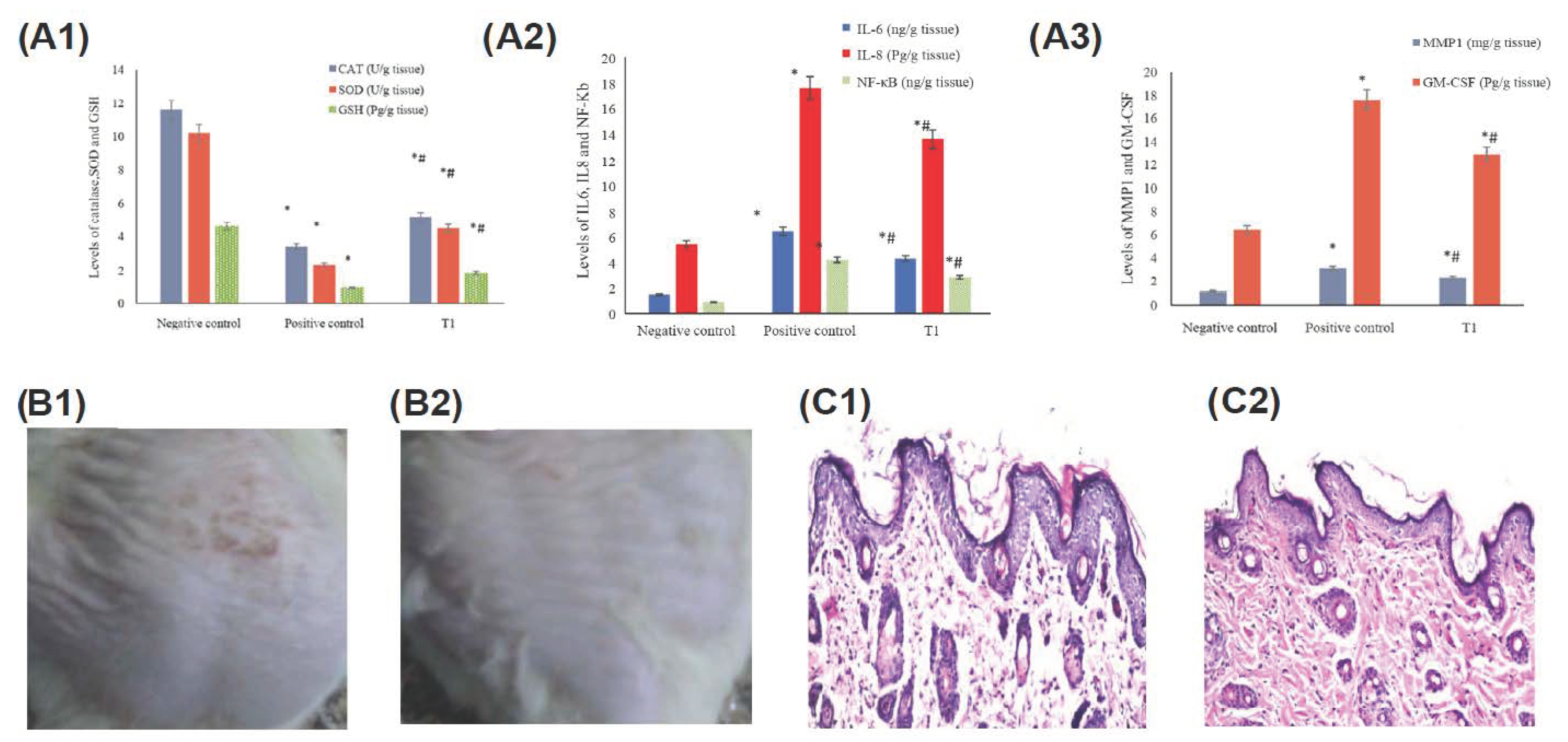
  30. Zhao, P.; Sui, B.D.; Liu, N.; Lv, Y.J.; Zheng, C.X.; Lu, Y.B.; Huang, W.T.; Zhou, C.H.; Chen, J.; Pang, D.L.; et al. Anti-aging pharmacology in cutaneous wound healing: Effects of metformin, resveratrol, and rapamycin by local application. Aging Cell 2017, 16, 1083–1093. [Google Scholar] [CrossRef]
  31. Berce, C.; Muresan, M.S.; Soritau, O.; Petrushev, B.; Tefas, L.; Rigo, I.; Ungureanu, G.; Catoi, C.; Irimie, A.; Tomuleasa, C. Cutaneous wound healing using polymeric surgical dressings based on chitosan, sodium hyaluronate and resveratrol. A preclinical experimental study. Colloids Surf. B Biointerfaces 2018, 163, 155–166. [Google Scholar] [CrossRef] [PubMed]
  32. Lakshmanan, R.; Campbell, J.; Ukani, G.; O’Reilly Beringhs, A.; Selvaraju, V.; Thirunavukkarasu, M.; Lu, X.; Palesty, J.A.; Maulik, N. Evaluation of dermal tissue regeneration using resveratrol loaded fibrous matrix in a preclinical mouse model of full-thickness ischemic wound. Int. J. Pharm. 2019, 558, 177–186. [Google Scholar] [CrossRef] [PubMed]
  33. Huang, X.; Sun, J.; Chen, G.; Niu, C.; Wang, Y.; Zhao, C.; Sun, J.; Huang, H.; Huang, S.; Liang, Y.; et al. Resveratrol Promotes Diabetic Wound Healing via SIRT1-FOXO1-c-Myc Signaling Pathway-Mediated Angiogenesis. Front. Pharmacol. 2019, 10, 421. [Google Scholar] [CrossRef] [PubMed][Green Version]
  34. Gokce, E.H.; Tuncay Tanrıverdi, S.; Eroglu, I.; Tsapis, N.; Gokce, G.; Tekmen, I.; Fattal, E.; Ozer, O. Wound healing effects of collagen-laminin dermal matrix impregnated with resveratrol loaded hyaluronic acid-DPPC microparticles in diabetic rats. Eur. J. Pharm. Biopharm. 2017, 119, 17–27. [Google Scholar] [CrossRef] [PubMed]
  35. Lephart, E.D.; Andrus, M.B. Human skin gene expression: Natural (trans) resveratrol versus five resveratrol analogs for dermal applications. Exp. Biol. Med. 2017, 242, 1482–1489. [Google Scholar] [CrossRef][Green Version]
  36. Zhou, F.; Huang, X.; Pan, Y.; Cao, D.; Liu, C.; Liu, Y.; Chen, A. Resveratrol protects HaCaT cells from ultraviolet B-induced photoaging via upregulation of HSP27 and modulation of mitochondrial caspase-dependent apoptotic pathway. Biochem. Biophys. Res. Commun. 2018, 499, 662–668. [Google Scholar] [CrossRef]
  37. Park, K.; Lee, J.H. Protective effects of resveratrol on UVB-irradiated HaCaT cells through attenuation of the caspase pathway. Oncol. Rep. 2008, 19, 413–417. [Google Scholar] [CrossRef][Green Version]
  38. Lee, Y.H.; Kumar, N.C.; Glickman, R.D. Modulation of photochemical damage in normal and malignant cells by naturally occurring compounds. Photochem. Photobiol. 2012, 88, 1385–1395. [Google Scholar] [CrossRef]
  39. Bowers, J.L.; Tyulmenkov, V.V.; Jernigan, S.C.; Klinge, C.M. Resveratrol acts as a mixed agonist/antagonist for estrogen receptors alpha and beta. Endocrinology 2000, 141, 3657–3667. [Google Scholar] [CrossRef]
  40. Gehm, B.D.; McAndrews, J.M.; Chien, P.Y.; Jameson, J.L. Resveratrol, a polyphenolic compound found in grapes and wine, is an agonist for the estrogen receptor. Proc. Natl. Acad. Sci. USA 1997, 94, 14138–14143. [Google Scholar] [CrossRef][Green Version]
  41. Giardina, S.; Michelotti, A.; Zavattini, G.; Finzi, S.; Ghisalberti, C.; Marzatico, F. Efficacy study in vitro: Assessment of the properties of resveratrol and resveratrol + N-acetyl-cysteine on proliferation and inhibition of collagen activity. Minerva Ginecol. 2010, 62, 195–201. [Google Scholar] [PubMed]
  42. Lephart, E.D. Resveratrol, 4′ Acetoxy Resveratrol, R-equol, Racemic Equol or S-equol as Cosmeceuticals to Improve Dermal Health. Int. J. Mol. Sci. 2017, 18, 1193. [Google Scholar] [CrossRef] [PubMed][Green Version]
  43. Sticozzi, C.; Cervellati, F.; Muresan, X.M.; Cervellati, C.; Valacchi, G. Resveratrol prevents cigarette smoke-induced keratinocytes damage. Food Funct. 2014, 5, 2348–2356. [Google Scholar] [CrossRef] [PubMed]
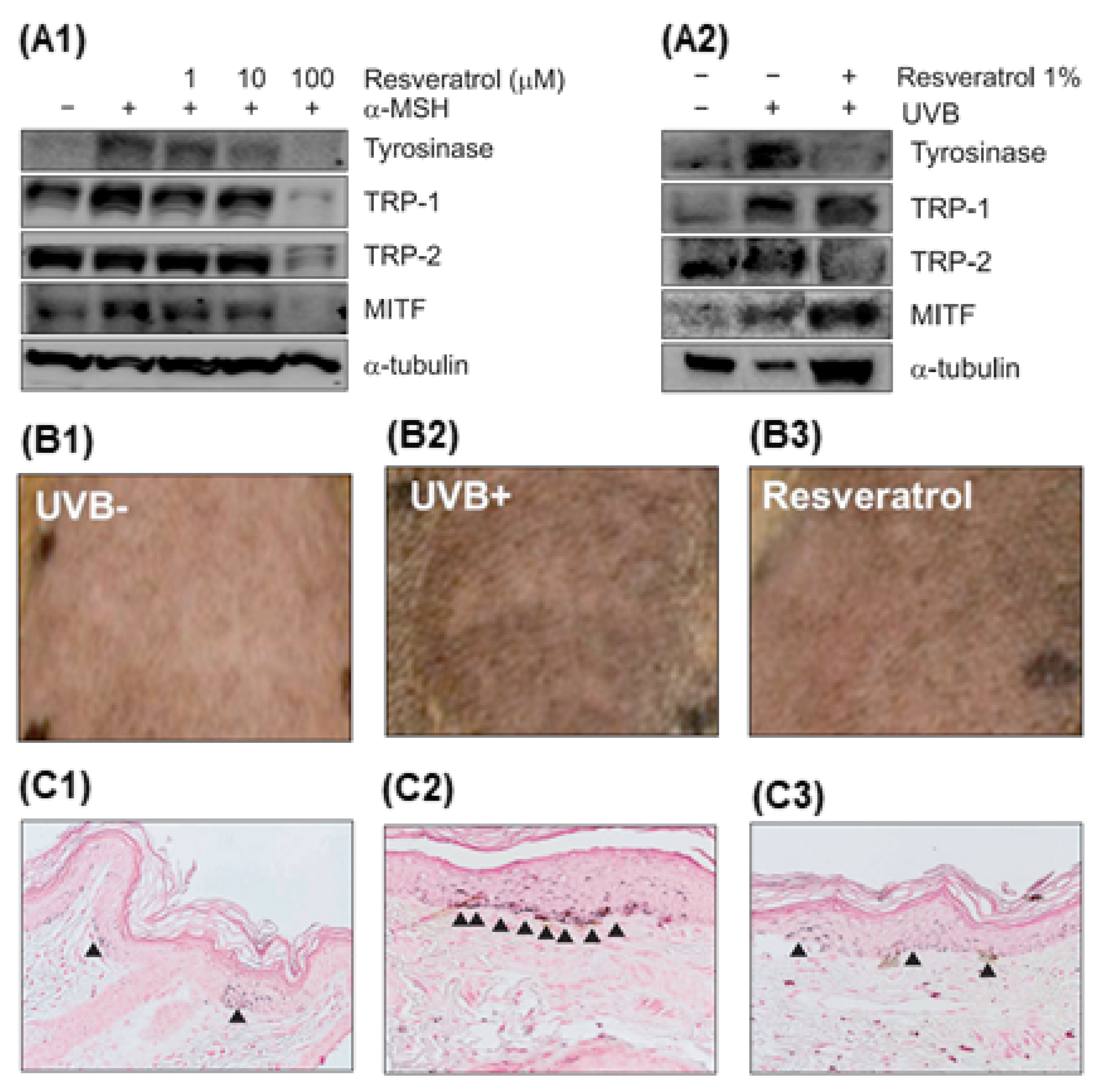
  44. Newton, R.A.; Cook, A.L.; Roberts, D.W.; Leonard, J.H.; Sturm, R.A. Post-transcriptional regulation of melanin biosynthetic enzymes by cAMP and resveratrol in human melanocytes. J. Investig. Dermatol. 2007, 127, 2216–2227. [Google Scholar] [CrossRef] [PubMed]
  45. Kwon, S.H.; Choi, H.R.; Kang, Y.A.; Park, K.C. Depigmenting Effect of Resveratrol Is Dependent on FOXO3a Activation without SIRT1 Activation. Int. J. Mol. Sci. 2017, 18, 1213. [Google Scholar] [CrossRef] [PubMed][Green Version]
  46. Angellotti, G.; Murgia, D.; Presentato, A.; D’Oca, M.C.; Scarpaci, A.G.; Alduina, R.; Raimondi, M.V.; De Caro, V. Antibacterial PEGylated Solid Lipid Microparticles for Cosmeceutical Purpose: Formulation, Characterization, and Efficacy Evaluation. Materials 2020, 13, 2073. [Google Scholar] [CrossRef]
  47. Fabbrocini, G.; Staibano, S.; De Rosa, G.; Battimiello, V.; Fardella, N.; Ilardi, G.; La Rotonda, M.I.; Longobardi, A.; Mazzella, M.; Siano, M.; et al. Resveratrol-containing gel for the treatment of acne vulgaris: A single-blind, vehicle-controlled, pilot study. Am. J. Clin. Dermatol. 2011, 12, 133–141. [Google Scholar] [CrossRef]
  48. Kim, S.Y.; Hyun, M.Y.; Go, K.C.; Zouboulis, C.C.; Kim, B.J. Resveratrol exerts growth inhibitory effects on human SZ95 sebocytes through the inactivation of the PI3-K/Akt pathway. Int. J. Mol. Med. 2015, 35, 1042–1050. [Google Scholar] [CrossRef][Green Version]
  49. Wei, Z.; Chen, G.; Hu, T.; Mo, X.; Hou, X.; Cao, K.; Wang, L.; Pan, Z.; Wu, Q.; Li, X.; et al. Resveratrol ameliorates lipid accumulation and inflammation in human SZ95 sebocytes via the AMPK signaling pathways in vitro. J. Dermatol. Sci. 2021, 103, 156–166. [Google Scholar] [CrossRef]
  50. Pang, K.; Li, B.; Tang, Z.; Yang, W.; Hao, L.; Shi, Z.; Zhang, J.; Cai, L.; Li, R.; Liu, Y.; et al. Resveratrol inhibits hypertrophic scars formation by activating autophagy via the miR-4654/Rheb axis. Mol. Med. Rep. 2020, 22, 3440–3452. [Google Scholar] [CrossRef]
  51. Nawrocki, E.M.; Bedell, H.W.; Humphreys, T.L. Resveratrol is cidal to both classes of Haemophilus ducreyi. Int. J. Antimicrob. Agents 2013, 41, 477–479. [Google Scholar] [CrossRef] [PubMed][Green Version]
  52. Huang, H.; Liao, D.; Zhou, G.; Zhu, Z.; Cui, Y.; Pu, R. Antiviral activities of resveratrol against rotavirus in vitro and in vivo. Phytomedicine 2020, 77, 153230. [Google Scholar] [CrossRef] [PubMed]
  53. Chan, M.M. Antimicrobial effect of resveratrol on dermatophytes and bacterial pathogens of the skin. Biochem. Pharmacol. 2002, 63, 99–104. [Google Scholar] [CrossRef]
  54. Campo, V.A. Comparative effects of histone deacetylases inhibitors and resveratrol on Trypanosoma cruzi replication, differentiation, infectivity and gene expression. Int. J. Parasitol. Drugs Drug Resist. 2017, 7, 23–33. [Google Scholar] [CrossRef]
  55. Ko, J.H.; Sethi, G.; Um, J.Y.; Shanmugam, M.K.; Arfuso, F.; Kumar, A.P.; Bishayee, A.; Ahn, K.S. The Role of Resveratrol in Cancer Therapy. Int. J. Mol. Sci. 2017, 18, 2589. [Google Scholar] [CrossRef][Green Version]
  56. Annaji, M.; Poudel, I.; Boddu, S.H.S.; Arnold, R.D.; Tiwari, A.K.; Babu, R.J. Resveratrol-loaded nanomedicines for cancer applications. Cancer Rep. 2021, 4, e1353. [Google Scholar] [CrossRef]
  57. Afaq, F.; Adhami, V.M.; Ahmad, N. Prevention of short-term ultraviolet B radiation-mediated damages by resveratrol in SKH-1 hairless mice. Toxicol. Appl. Pharmacol. 2003, 186, 28–37. [Google Scholar] [CrossRef]
  58. Aziz, S.W.; Aziz, M.H. Protective molecular mechanisms of resveratrol in UVR-induced Skin carcinogenesis. Photodermatol. Photoimmunol. Photomed. 2018, 34, 35–41. [Google Scholar] [CrossRef][Green Version]
  59. Baxter, R.A. Anti-aging properties of resveratrol: Review and report of a potent new antioxidant skin care formulation. J. Cosmet. Dermatol. 2008, 7, 2–7. [Google Scholar] [CrossRef]
  60. Farris, P.; Krutmann, J.; Li, Y.H.; McDaniel, D.; Krol, Y. Resveratrol: A unique antioxidant offering a multi-mechanistic approach for treating aging skin. J. Drugs Dermatol. 2013, 12, 1389–1394. [Google Scholar]
  61. Miura, T.; Muraoka, S.; Ikeda, N.; Watanabe, M.; Fujimoto, Y. Antioxidative and prooxidative action of stilbene derivatives. Pharmacol. Toxicol. 2000, 86, 203–208. [Google Scholar] [CrossRef]
  62. Moyano-Mendez, J.R.; Fabbrocini, G.; De Stefano, D.; Mazzella, C.; Mayol, L.; Scognamiglio, I.; Carnuccio, R.; Ayala, F.; La Rotonda, M.I.; De Rosa, G. Enhanced antioxidant effect of trans-resveratrol: Potential of binary systems with polyethylene glycol and cyclodextrin. Drug Dev. Ind. Pharm. 2014, 40, 1300–1307. [Google Scholar] [CrossRef][Green Version]
  63. Lephart, E.D.; Sommerfeldt, J.M.; Andrus, M.B. Resveratrol: Influences on gene expression in human skin. J. Funct. Foods 2014, 10, 377–384. [Google Scholar] [CrossRef]
  64. Gonçalves, G.; Barros, P.; da Silva, G.; dos Santos, E.; Minutti, A. Formulations Containing Curcumin or Trans-Resveratrol Increase Dermal Thickness in Rats Submitted to Chemical Peeling. J. Cosmet. Dermatol. Sci. Appl. 2017, 7, 14–26. [Google Scholar] [CrossRef][Green Version]
  65. Abbas, H.; Kamel, R.; El-Sayed, N. Dermal anti-oxidant, anti-inflammatory and anti-aging effects of Compritol ATO-based Resveratrol colloidal carriers prepared using mixed surfactants. Int. J. Pharm. 2018, 541, 37–47. [Google Scholar] [CrossRef]
  66. Afaq, F.; Mukhtar, H. Botanical antioxidants in the prevention of photocarcinogenesis and photoaging. Exp. Dermatol. 2006, 15, 678–684. [Google Scholar] [CrossRef]
  67. Afaq, F.; Katiyar, S.K. Polyphenols: Skin photoprotection and inhibition of photocarcinogenesis. Mini Rev. Med. Chem. 2011, 11, 1200–1215. [Google Scholar] [CrossRef][Green Version]
  68. Nichols, J.A.; Katiyar, S.K. Skin photoprotection by natural polyphenols: Anti-inflammatory, antioxidant and DNA repair mechanisms. Arch. Dermatol. Res. 2010, 302, 71–83. [Google Scholar] [CrossRef][Green Version]
  69. Aziz, M.H.; Afaq, F.; Ahmad, N. Prevention of ultraviolet-B radiation damage by resveratrol in mouse skin is mediated via modulation in survivin. Photochem. Photobiol. 2005, 81, 25–31. [Google Scholar] [CrossRef]
  70. Chen, M.L.; Li, J.; Xiao, W.R.; Sun, L.; Tang, H.; Wang, L.; Wu, L.Y.; Chen, X.; Xie, H.F. Protective effect of resveratrol against oxidative damage of UVA irradiated HaCaT cells. Zhong Nan Da Xue Xue Bao Yi Xue Ban 2006, 31, 635–639. [Google Scholar]
  71. Liu, Y.; Chan, F.; Sun, H.; Yan, J.; Fan, D.; Zhao, D.; An, J.; Zhou, D. Resveratrol protects human keratinocytes HaCaT cells from UVA-induced oxidative stress damage by downregulating Keap1 expression. Eur. J. Pharmacol. 2011, 650, 130–137. [Google Scholar] [CrossRef]
  72. Cao, C.; Lu, S.; Kivlin, R.; Wallin, B.; Card, E.; Bagdasarian, A.; Tamakloe, T.; Wang, W.J.; Song, X.; Chu, W.M.; et al. SIRT1 confers protection against UVB- and H2O2-induced cell death via modulation of p53 and JNK in cultured skin keratinocytes. J. Cell. Mol. Med. 2009, 13, 3632–3643. [Google Scholar] [CrossRef][Green Version]
  73. Adhami, V.M.; Afaq, F.; Ahmad, N. Suppression of ultraviolet B exposure-mediated activation of NF-kappaB in normal human keratinocytes by resveratrol. Neoplasia 2003, 5, 74–82. [Google Scholar] [CrossRef][Green Version]
  74. Potapovich, A.I.; Kostyuk, V.A.; Kostyuk, T.V.; de Luca, C.; Korkina, L.G. Effects of pre- and post-treatment with plant polyphenols on human keratinocyte responses to solar UV. Inflamm. Res. 2013, 62, 773–780. [Google Scholar] [CrossRef]
  75. Abbas, H.; Kamel, R. Potential role of resveratrol-loaded elastic sorbitan monostearate nanovesicles for the prevention of UV-induced skin damage. J. Liposome Res. 2020, 30, 45–53. [Google Scholar] [CrossRef]
  76. Lephart, E.D.; Naftolin, F. Menopause and the Skin: Old Favorites and New Innovations in Cosmeceuticals for Estrogen-Deficient Skin. Dermatol. Ther. 2021, 11, 53–69. [Google Scholar] [CrossRef]
  77. Stevenson, S.; Thornton, J. Effect of estrogens on skin aging and the potential role of SERMs. Clin. Interv. Aging 2007, 2, 283–297. [Google Scholar] [CrossRef][Green Version]
  78. Verdier-Sévrain, S. Effect of estrogens on skin aging and the potential role of selective estrogen receptor modulators. Climacteric 2007, 10, 289–297. [Google Scholar] [CrossRef]
  79. Ruotolo, R.; Calani, L.; Fietta, E.; Brighenti, F.; Crozier, A.; Meda, C.; Maggi, A.; Ottonello, S.; Del Rio, D. Anti-estrogenic activity of a human resveratrol metabolite. Nutr. Metab. Cardiovasc. Dis. 2013, 23, 1086–1092. [Google Scholar] [CrossRef]
  80. Pillaiyar, T.; Namasivayam, V.; Manickam, M.; Jung, S.H. Inhibitors of Melanogenesis: An Updated Review. J. Med. Chem. 2018, 61, 7395–7418. [Google Scholar] [CrossRef]
  81. Bernard, P.; Berthon, J.Y. Resveratrol: An original mechanism on tyrosinase inhibition. Int. J. Cosmet. Sci. 2000, 22, 219–226. [Google Scholar] [CrossRef] [PubMed]
  82. Satooka, H.; Kubo, I. Resveratrol as a kcat type inhibitor for tyrosinase: Potentiated melanogenesis inhibitor. Bioorg. Med. Chem. 2012, 20, 1090–1099. [Google Scholar] [CrossRef] [PubMed]
  83. Yanagihara, M.; Yoshimatsu, M.; Inoue, A.; Kanno, T.; Tatefuji, T.; Hashimoto, K. Inhibitory effect of gnetin C, a resveratrol dimer from melinjo (Gnetum gnemon), on tyrosinase activity and melanin biosynthesis. Biol. Pharm. Bull. 2012, 35, 993–996. [Google Scholar] [CrossRef] [PubMed][Green Version]
  84. Park, J.; Boo, Y.C. Isolation of resveratrol from vitis viniferae caulis and its potent inhibition of human tyrosinase. Evid. Based Complement. Alternat. Med. 2013, 2013, 645257. [Google Scholar] [CrossRef] [PubMed][Green Version]
  85. Park, J.; Park, J.H.; Suh, H.J.; Lee, I.C.; Koh, J.; Boo, Y.C. Effects of resveratrol, oxyresveratrol, and their acetylated derivatives on cellular melanogenesis. Arch. Dermatol. Res. 2014, 306, 475–487. [Google Scholar] [CrossRef]
  86. Lin, C.B.; Babiarz, L.; Liebel, F.; Roydon Price, E.; Kizoulis, M.; Gendimenico, G.J.; Fisher, D.E.; Seiberg, M. Modulation of microphthalmia-associated transcription factor gene expression alters skin pigmentation. J. Investig. Dermatol. 2002, 119, 1330–1340. [Google Scholar] [CrossRef][Green Version]
  87. Lee, T.H.; Seo, J.O.; Baek, S.H.; Kim, S.Y. Inhibitory effects of resveratrol on melanin synthesis in ultraviolet B-induced pigmentation in Guinea pig skin. Biomol. Ther. 2014, 22, 35–40. [Google Scholar] [CrossRef][Green Version]
  88. Docherty, J.J.; McEwen, H.A.; Sweet, T.J.; Bailey, E.; Booth, T.D. Resveratrol inhibition of Propionibacterium acnes. J. Antimicrob. Chemother. 2007, 59, 1182–1184. [Google Scholar] [CrossRef][Green Version]
  89. Taylor, E.J.; Yu, Y.; Champer, J.; Kim, J. Resveratrol Demonstrates Antimicrobial Effects Against Propionibacterium acnes In Vitro. Dermatol. Ther. 2014, 4, 249–257. [Google Scholar] [CrossRef][Green Version]
  90. Dos Santos, Z.M.Q.; Dos Santos, M.Q.; Zancanaro, V.; Bellaver, E.H.; Nardi, G.M.; Gelinski, J.M.L.; Locatelli, C. Topical application of phenolic compounds suppresses Propionibacterium acnes-induced inflammatory responses in mice with ear edema. Naunyn Schmiedebergs Arch. Pharmacol. 2019, 392, 529–540. [Google Scholar] [CrossRef]
  91. Prakoeswa, C.R.S.; Rindiastuti, Y.; Wirohadidjojo, Y.W.; Komaratih, E.; Nurwasis; Dinaryati, A.; Lestari, N.M.I.; Rantam, F.A. Resveratrol promotes secretion of wound healing related growth factors of mesenchymal stem cells originated from adult and fetal tissues. Artif. Cells Nanomed. Biotechnol. 2020, 48, 1160–1167. [Google Scholar] [CrossRef] [PubMed]
  92. Bilgic, T. Comparison of the Effect of Local and Systemic Injection of Resveratrol on Cutaneous Wound Healing in Rats. Int. J. Low Extrem. Wounds 2021, 20, 55–59. [Google Scholar] [CrossRef] [PubMed]
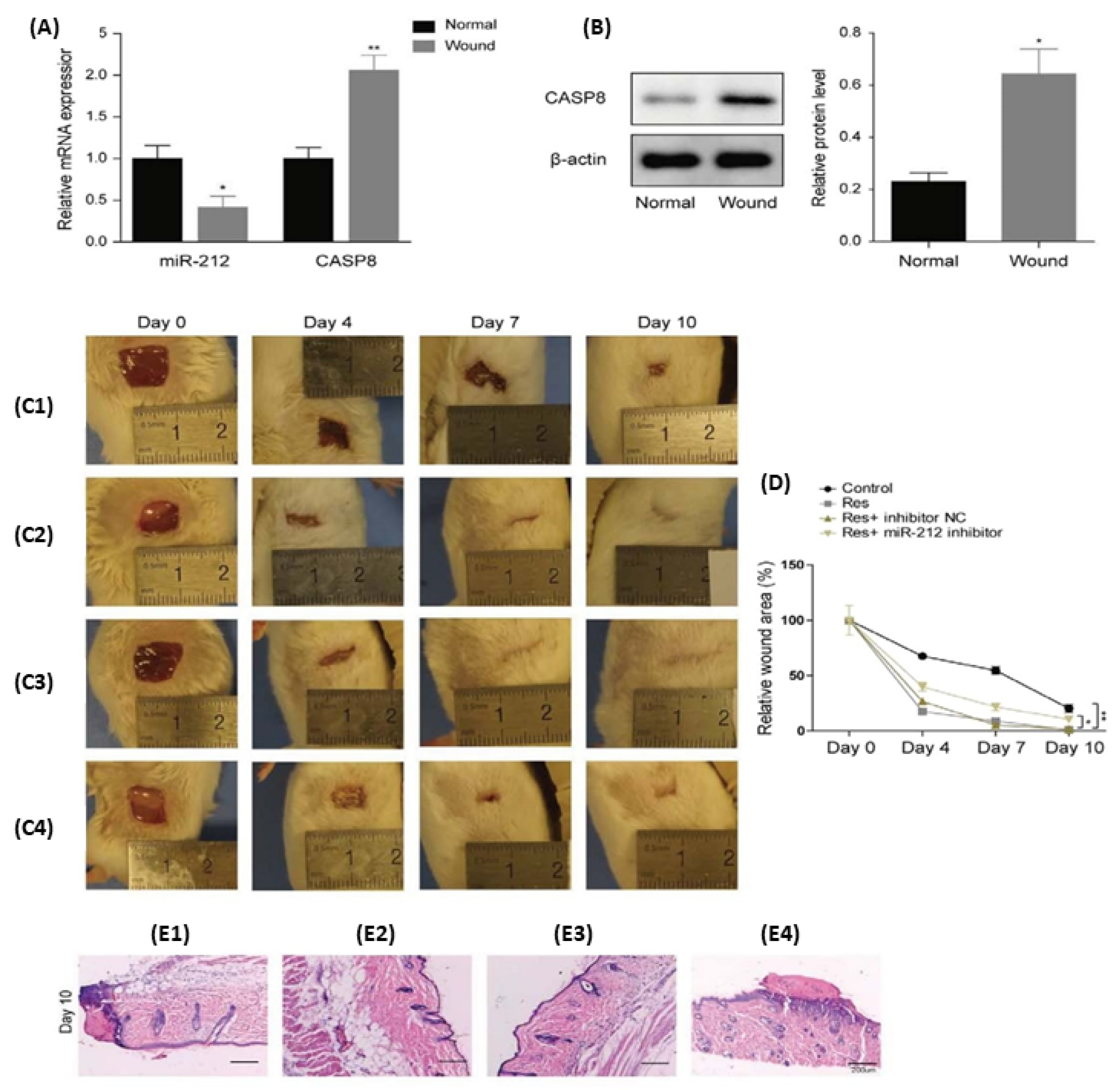
  93. Liu, Y.; Xiong, W.; Wang, C.W.; Shi, J.P.; Shi, Z.Q.; Zhou, J.D. Resveratrol promotes skin wound healing by regulating the miR-212/CASP8 axis. Lab. Investig. 2021, 101, 1363–1370. [Google Scholar] [CrossRef] [PubMed]
  94. Zeng, G.; Zhong, F.; Li, J.; Luo, S.; Zhang, P. Resveratrol-mediated reduction of collagen by inhibiting proliferation and producing apoptosis in human hypertrophic scar fibroblasts. Biosci. Biotechnol. Biochem. 2013, 77, 2389–2396. [Google Scholar] [CrossRef][Green Version]
  95. Zhao, C.C.; Zhu, L.; Wu, Z.; Yang, R.; Xu, N.; Liang, L. Resveratrol-loaded peptide-hydrogels inhibit scar formation in wound healing through suppressing inflammation. Regen. Biomater. 2020, 7, 99–107. [Google Scholar] [CrossRef]
  96. Amanat, S.; Taymouri, S.; Varshosaz, J.; Minaiyan, M.; Talebi, A. Carboxymethyl cellulose-based wafer enriched with resveratrol-loaded nanoparticles for enhanced wound healing. Drug Deliv. Transl. Res. 2020, 10, 1241–1254. [Google Scholar] [CrossRef]
  97. Guo, S.; Liao, H.; Liu, J.; Liu, J.; Tang, F.; He, Z.; Li, Y.; Yang, Q. Resveratrol Activated Sonic Hedgehog Signaling to Enhance Viability of NIH3T3 Cells In Vitro via Regulation of Sirt1. Cell. Physiol. Biochem. 2018, 50, 1346–1360. [Google Scholar] [CrossRef]
  98. Ma, H.; Qiao, Z. Analysis of the efficacy of resveratrol treatment in patients with scarred uterus. Exp. Ther. Med. 2018, 15, 5410–5414. [Google Scholar] [CrossRef]
  99. Tang, Z.; Ding, J.C.; Zhai, X.X. Effects of resveratrol on the expression of molecules related to the mTOR signaling pathway in pathological scar fibroblasts. G. Ital. Dermatol. Venereol. 2020, 155, 161–167. [Google Scholar] [CrossRef]
  100. Chen, T.T.; Peng, S.; Wang, Y.; Hu, Y.; Shen, Y.; Xu, Y.; Yin, J.; Liu, C.; Cao, J. Improvement of Mitochondrial Activity and Fibrosis by Resveratrol Treatment in Mice with Schistosoma japonicum Infection. Biomolecules 2019, 9, 658. [Google Scholar] [CrossRef][Green Version]
  101. Ikeda, K.; Torigoe, T.; Matsumoto, Y.; Fujita, T.; Sato, N.; Yotsuyanagi, T. Resveratrol inhibits fibrogenesis and induces apoptosis in keloid fibroblasts. Wound Repair Regen. 2013, 21, 616–623. [Google Scholar] [CrossRef] [PubMed]
  102. Sun, D.; Hurdle, J.G.; Lee, R.; Lee, R.; Cushman, M.; Pezzuto, J.M. Evaluation of flavonoid and resveratrol chemical libraries reveals abyssinone II as a promising antibacterial lead. ChemMedChem 2012, 7, 1541–1545. [Google Scholar] [CrossRef][Green Version]
  103. Paulo, L.; Ferreira, S.; Gallardo, E.; Queiroz, J.A.; Domingues, F. Antimicrobial Activity and Effects of Resveratrol on Human Pathogenic Bacteria. World J. Microbiol. Biotechnol. 2010, 26, 1533–1538. [Google Scholar] [CrossRef]
  104. Hwang, D.; Lim, Y.H. Resveratrol antibacterial activity against Escherichia coli is mediated by Z-ring formation inhibition via suppression of FtsZ expression. Sci. Rep. 2015, 5, 10029. [Google Scholar] [CrossRef] [PubMed][Green Version]
  105. Liu, Y.; Zhou, J.; Qu, Y.; Yang, X.; Shi, G.; Wang, X.; Hong, Y.; Drlica, K.; Zhao, X. Resveratrol Antagonizes Antimicrobial Lethality and Stimulates Recovery of Bacterial Mutants. PLoS ONE 2016, 11, e0153023. [Google Scholar] [CrossRef]
  106. Li, Q.; Verma, I.M. NF-kappaB regulation in the immune system. Nat. Rev. Immunol. 2002, 2, 725–734. [Google Scholar] [CrossRef]
  107. De Leo, A.; Arena, G.; Lacanna, E.; Oliviero, G.; Colavita, F.; Mattia, E. Resveratrol inhibits Epstein Barr Virus lytic cycle in Burkitt’s lymphoma cells by affecting multiple molecular targets. Antiviral Res. 2012, 96, 196–202. [Google Scholar] [CrossRef]
  108. Chan, C.N.; Trinité, B.; Levy, D.N. Potent Inhibition of HIV-1 Replication in Resting CD4 T Cells by Resveratrol and Pterostilbene. Antimicrob. Agents Chemother. 2017, 61, e00408–e00417. [Google Scholar] [CrossRef][Green Version]
  109. Wang, J.; Zhang, X.; Gao, L.; Wang, L.; Song, F.; Zhang, L.; Wan, Y. The synergistic antifungal activity of resveratrol with azoles against Candida albicans. Lett. Appl. Microbiol. 2021, 72, 688–697. [Google Scholar] [CrossRef]
  110. Chen, Q.-W.; Dong, K.; Qin, H.-X.; Yang, Y.-K.; He, J.-L.; Li, J.; Zheng, Z.-W.; Chen, D.-L.; Chen, J.-P. Direct and Indirect Inhibition Effects of Resveratrol against Toxoplasma gondii Tachyzoites In Vitro. Antimicrob. Agents Chemother. 2019, 63, e01233-18. [Google Scholar] [CrossRef][Green Version]
  111. Zi, C.; Gan, C.; Xu, H.; Sheng, J.; Wang, X. Recent Advances on Anti-skin Cancer Activity of Phytochemicals and Underlying Molecular Mechanisms. Med. Res. 2021, 5, 210006. [Google Scholar]
  112. Kaur, H.; Kesharwani, P. Advanced nanomedicine approaches applied for treatment of skin carcinoma. J. Control. Release 2021, 337, 589–611. [Google Scholar] [CrossRef] [PubMed]
  113. Cullen, J.K.; Simmons, J.L.; Parsons, P.G.; Boyle, G.M. Topical treatments for skin cancer. Adv. Drug Deliv. Rev. 2020, 153, 54–64. [Google Scholar] [CrossRef] [PubMed]
  114. Imran, M.; Iqubal, M.K.; Imtiyaz, K.; Saleem, S.; Mittal, S.; Rizvi, M.M.A.; Ali, J.; Baboota, S. Topical nanostructured lipid carrier gel of quercetin and resveratrol: Formulation, optimization, in vitro and ex vivo study for the treatment of skin cancer. Int. J. Pharm. 2020, 587, 119705. [Google Scholar] [CrossRef] [PubMed]
  115. Wu, Z.; Liu, B.; Liu, J.; Zhang, Q.; Liu, J.; Chen, N.; Chen, R.; Zhu, R. Resveratrol inhibits the proliferation of human melanoma cells by inducing G1/S cell cycle arrest and apoptosis. Mol. Med. Rep. 2015, 11, 400–404. [Google Scholar] [CrossRef][Green Version]
  116. Ravikumar, P.; Katariya, M.; Patil, S.; Tatke, P.; Pillai, R. Skin delivery of resveratrol encapsulated lipidic formulation for melanoma chemoprevention. J. Microencapsul. 2019, 36, 535–551. [Google Scholar] [CrossRef]
  117. Mahamat-Saleh, Y.; Aune, D.; Schlesinger, S. 25-Hydroxyvitamin D status, vitamin D intake, and skin cancer risk: A systematic review and dose-response meta-analysis of prospective studies. Sci. Rep. 2020, 10, 13151. [Google Scholar] [CrossRef]
  118. Pavel, T.I.; Chircov, C.; Rădulescu, M.; Grumezescu, A.M. Regenerative Wound Dressings for Skin Cancer. Cancers 2020, 12, 2954. [Google Scholar] [CrossRef]
  119. McDaniel, B.; Badri, T.; Steele, R.B. Basal Cell Carcinoma. In StatPearls; StatPearls Publishing LLC.: Treasure Island, FL, USA, 2022. [Google Scholar]
  120. Aziz, M.H.; Reagan-Shaw, S.; Wu, J.; Longley, B.J.; Ahmad, N. Chemoprevention of skin cancer by grape constituent resveratrol: Relevance to human disease? FASEB J. 2005, 19, 1193–1195. [Google Scholar] [CrossRef][Green Version]
  121. Athar, M.; Back, J.H.; Tang, X.; Kim, K.H.; Kopelovich, L.; Bickers, D.R.; Kim, A.L. Resveratrol: A review of preclinical studies for human cancer prevention. Toxicol. Appl. Pharmacol. 2007, 224, 274–283. [Google Scholar] [CrossRef][Green Version]
  122. Liu, Z.L.; Li, H.; Liu, J.; Wu, M.L.; Chen, X.Y.; Liu, L.H.; Wang, Q. Inactivated Wnt signaling in resveratrol-treated epidermal squamous cancer cells and its biological implication. Oncol. Lett. 2017, 14, 2239–2243. [Google Scholar] [CrossRef] [PubMed][Green Version]
  123. Hao, Y.; Huang, W.; Liao, M.; Zhu, Y.; Liu, H.; Hao, C.; Liu, G.; Zhang, G.; Feng, H.; Ning, X.; et al. The inhibition of resveratrol to human skin squamous cell carcinoma A431 xenografts in nude mice. Fitoterapia 2013, 86, 84–91. [Google Scholar] [CrossRef] [PubMed]
  124. Back, J.H.; Zhu, Y.; Calabro, A.; Queenan, C.; Kim, A.S.; Arbesman, J.; Kim, A.L. Resveratrol-mediated downregulation of Rictor attenuates autophagic process and suppresses UV-induced skin carcinogenesis. Photochem. Photobiol. 2012, 88, 1165–1172. [Google Scholar] [CrossRef] [PubMed]
  125. Zhai, X.X.; Ding, J.C.; Tang, Z.M.; Li, J.G.; Li, Y.C.; Yan, Y.H.; Sun, J.C.; Zhang, C.X. Effects of resveratrol on the proliferation, apoptosis and telomerase ability of human A431 epidermoid carcinoma cells. Oncol. Lett. 2016, 11, 3015–3018. [Google Scholar] [CrossRef][Green Version]
  126. Gong, C.; Xia, H. Resveratrol suppresses melanoma growth by promoting autophagy through inhibiting the PI3K/AKT/mTOR signaling pathway. Exp. Ther. Med. 2020, 19, 1878–1886. [Google Scholar] [CrossRef][Green Version]
  127. Boyer, J.Z.; Jandova, J.; Janda, J.; Vleugels, F.R.; Elliott, D.A.; Sligh, J.E. Resveratrol-sensitized UVA induced apoptosis in human keratinocytes through mitochondrial oxidative stress and pore opening. J. Photochem. Photobiol. B 2012, 113, 42–50. [Google Scholar] [CrossRef][Green Version]
  128. Cosco, D.; Paolino, D.; Maiuolo, J.; Marzio, L.D.; Carafa, M.; Ventura, C.A.; Fresta, M. Ultradeformable liposomes as multidrug carrier of resveratrol and 5-fluorouracil for their topical delivery. Int. J. Pharm. 2015, 489, 1–10. [Google Scholar] [CrossRef]
  129. Wang, W.; Tang, Q.; Yu, T.; Li, X.; Gao, Y.; Li, J.; Liu, Y.; Rong, L.; Wang, Z.; Sun, H.; et al. Surfactant-Free Preparation of Au@Resveratrol Hollow Nanoparticles with Photothermal Performance and Antioxidant Activity. ACS Appl. Mater. Interfaces 2017, 9, 3376–3387. [Google Scholar] [CrossRef]
  130. Carletto, B.; Berton, J.; Ferreira, T.N.; Dalmolin, L.F.; Paludo, K.S.; Mainardes, R.M.; Farago, P.V.; Favero, G.M. Resveratrol-loaded nanocapsules inhibit murine melanoma tumor growth. Colloids Surf. B Biointerfaces 2016, 144, 65–72. [Google Scholar] [CrossRef]
  131. Tsai, M.J.; Lu, I.J.; Fu, Y.S.; Fang, Y.P.; Huang, Y.B.; Wu, P.C. Nanocarriers enhance the transdermal bioavailability of resveratrol: In-vitro and in-vivo study. Colloids Surf. B Biointerfaces 2016, 148, 650–656. [Google Scholar] [CrossRef]
  132. Das, S.; Lee, S.H.; Chow, P.S.; Macbeath, C. Microemulsion composed of combination of skin beneficial oils as vehicle: Development of resveratrol-loaded microemulsion based formulations for skin care applications. Colloids Surf. B Biointerfaces 2020, 194, 111161. [Google Scholar] [CrossRef] [PubMed]
  133. Miao, L.; Daozhou, L.; Ying, C.; Qibing, M.; Siyuan, Z. A resveratrol-loaded nanostructured lipid carrier hydrogel to enhance the anti-UV irradiation and anti-oxidant efficacy. Colloids Surf. B Biointerfaces 2021, 204, 111786. [Google Scholar] [CrossRef] [PubMed]
  134. Pentek, T.; Newenhouse, E.; O’Brien, B.; Chauhan, A.S. Development of a Topical Resveratrol Formulation for Commercial Applications Using Dendrimer Nanotechnology. Molecules 2017, 22, 137. [Google Scholar] [CrossRef] [PubMed][Green Version]
Figure 1. Main dermatological and cosmetic effects of resveratrol on the skin.
Cosmetics 10 00068 g001
Figure 2. (A1) Antioxidant, (A2) anti-inflammatory, and (A3) anti-aging activities of RES suspension (T1), compared with the control groups (positive and negative). * Statistically significantly different from the normal control group (p < 0.05) # Statistically significantly different from the T1 group (p < 0.05). Photographs of the rats’ dorsal skin: (B1) positive control group and (B2) irradiated followed by topical pre-treatment with RES suspension. Histopathological observation of the rats’ dorsal skin: (C1) positive control group and (C2) irradiated followed by topical pre-treatment with RES suspension. Adapted from [65].
Cosmetics 10 00068 g002
Figure 3. Effects of RES on the expression of proteins involved in melanogenesis: (A1) in B16F10 cells exposed to 1, 10, and 100 µM RES for 48 h; (A2) in the skin of guinea pigs treated with 1% RES for 2 weeks. Effects of RES on melanin synthesis after UVB exposure to induce skin pigmentation of brown guinea pigs: representative images of the dorsal skin of brown guinea pigs of (B1) UVB-negative group (UVB); (B2) UVB-positive group (UVB+); and (B3) UVB+ and 1% RES group treated for 2 weeks. Results of Fontana-Masson stain for melanin in the skin of brown guinea pigs (C1) UVB-, (C2) UVB+, and (C3) UVB+ and 1% RES treated group for 2 weeks (arrowheads indicate melanin). Adapted from [87].
Cosmetics 10 00068 g003
Figure 4. The effect of RES, RES + miR-212, and RES + inhibitor NC on mice wound healing: (A) measure of miR-212 and CASP8 ranges with relative mRNA expressions; (B) measure of miR-212 and CASP8 abundances with relative protein level. Wound area was measured for 10 days in (C1) control group; (C2) RES-treated group; (C3) Res + inhibitor NC-treated animal group; (C4) Res + miR-212 inhibitor-treated animal group. (D) Comparative analysis of all the treated groups determining a relative wound area (%) over several days. (E1) Wound tissues of the control group were stained with HE on day 10; (E2) wound tissues of RES-treated group were stained with HE on day 10; (E3) wound tissues of RES + inhibitor NC-treated group were stained with HE on day 10; (E4) wound tissues of RES + miR-212-treated group were stained with HE on day 10. (* p < 0.05, ** p < 0.005). Adapted from [93].
Cosmetics 10 00068 g004
Figure 5. Values expressed as a percentage of trypomastigotes and epimastigotes, with varying concentrations of each compound to treat the parasitic infection. (A1) represents epimastigotes incubated for 48 h, a linear graph depicting a growth with varying concentrations of TSA (trichostatin); (A2) depicts the growth curve of parasites with different sirtinol concentrations; (A3) shows a prediction of the growth curve with varying concentration of RES; (B) shows that IC50 (inhibitory medium concentration) values were calculated by counting live parasites; (C1) the incubation of parasites with TSA (Trichostatin) exhibits varying concentrations of the compound, which displayed a significant rise (around 10–20%); (C2) sirtinol showed significant elevation; (C3) with RES, parasitic infection declined as compared to the control. The values exhibited statistical significance when compared to the control (** p < 0.005). Adapted from [54].
Cosmetics 10 00068 g005
Figure 6. RES with different concentrations inducing decreased cell viability in A375 and SK-MEL-31 cells. (A1) A375 cells are treated with varying concentrations of RES, which shows RES-induced inhibitory cell proliferation; (A2) SK-MEL-31 cells are treated with varying concentrations of RES, which shows RES-induced inhibitory cell proliferation; (B) inhibition rate (%) vs. RES of A375 and SK-MEL-31 cells; (C1) the A375 cells incubated for 48 h with growing RES amounts; (C2) the SK-MEL-31 cells incubated for 48 h with growing concentration of RES. Adapted from [115].
Cosmetics 10 00068 g006
Disclaimer/Publisher’s Note: The statements, opinions and data contained in all publications are solely those of the individual author(s) and contributor(s) and not of MDPI and/or the editor(s). MDPI and/or the editor(s) disclaim responsibility for any injury to people or property resulting from any ideas, methods, instructions or products referred to in the content.

