Review

In Vitro Models for Anti-Aging Efficacy Assessment: A Critical Update in Dermocosmetic Research

by
Ana M. Cruz
1,†,
Margarida C. Gonçalves
1,†,
Matilde S. Marques
1,†,
Francisco Veiga
1,2,
Ana Cláudia Paiva-Santos
1,2,* and
Patrícia C. Pires
1,2,3,*
1
Department of Pharmaceutical Technology, Faculty of Pharmacy of the University of Coimbra, University of Coimbra, 3000-548 Coimbra, Portugal
2
REQUIMTE/LAQV, Group of Pharmaceutical Technology, Faculty of Pharmacy of the University of Coimbra, University of Coimbra, 3000-548 Coimbra, Portugal
3
Health Sciences Research Centre (CICS-UBI), University of Beira Interior, Av. Infante D. Henrique, 6200-506 Covilhã, Portugal
*
Authors to whom correspondence should be addressed.
These authors contributed equally to this work.
Cosmetics 2023, 10(2), 66; https://doi.org/10.3390/cosmetics10020066
Submission received: 4 April 2023 / Revised: 18 April 2023 / Accepted: 19 April 2023 / Published: 20 April 2023
(This article belongs to the Collection Feature Papers in Cosmetics in 2023)

Abstract

:
The skin is the human body’s largest organ and serves as a crucial boundary between the body and the external environment. As a natural process, skin aging cannot be avoided, and it causes changes in the skin’s strength, structure, elasticity, and integrity. Many approaches have been developed over the years to study the skin, including in vivo and in vitro methods. Nevertheless, animal assays have ethical issues and a lack of reproducibility. Hence, in vitro skin models have been increasingly developed and used. For the assessment of the potential of the anti-aging activity of compounds of different origins, the most commonly used in vitro assays are the ones evaluating antioxidant, anti-collagenase, anti-elastase, anti-hyaluronidase, anti-tyrosinase, anti-inflammatory, antiglycation, or moisturizing activity, and the induction of skin cell proliferation/anti-senescence effects or the inhibition of matrix metalloproteinase production. The purpose of this review is to summarize the most commonly used in vitro models for the evaluation of skin aging and cometic products’ anti-aging efficacy, providing a useful guide for researchers in the field. Overall, these assays provide important data on the safety and efficacy of anti-aging compounds, and a foundation for research on and the eventual introduction of formulations into the cosmetics market.

Graphical Abstract

1. Introduction

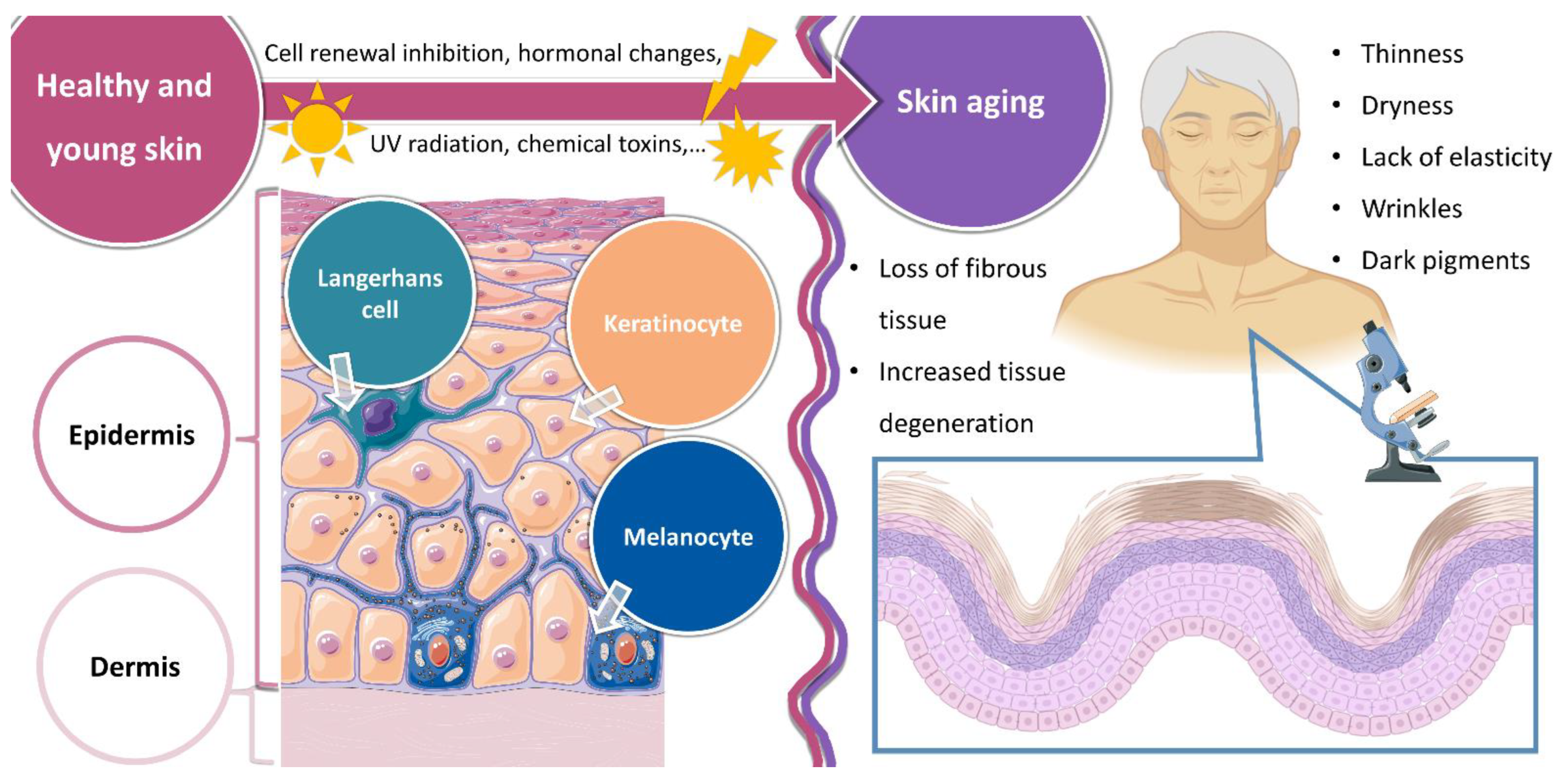
1.1. Healthy Skin and Skin Aging

The skin is the largest organ of the human body. As the first line of defense, it plays a crucial role against external physical, chemical, and biological aggressions. It is rendered under a heterogeneous and complex structure of different tissue origins, organized in three layers: the epidermis, the dermis, and the hypodermis. Histologically, the skin layers have different compositions. The epidermis, a non-vascular tissue, contains keratinocytes (the majority of cells), Langerhans cells, melanocytes, and Merck cells. The dermis is a supportive tissue that protects the epidermis, and it is composed of cells, a ground substance, and fibrous molecules. This skin layer controls the synthesis and degradation of proteins from its composition, maintaining a turnover mechanism. The hypodermis, for some considered to be the subcutaneous tissue, is the innermost layer of the skin, and is mainly composed of adipose tissue (adipocytes), serving as energy storage and connecting the skin to the muscle tissues below it. Collagen fibers are the main components of the skin, specifically collagen-I and collagen-III, and maintain skin firmness and elasticity. Elastic fibers are responsible for the retractile properties of the skin. As the main skin cells of the dermis, fibroblasts synthesize all types of fibers and the ground substance. Involved in thermoregulation, wound healing, immune reactions, and blood pressure maintenance, the skin possesses a rich vascular network [1,2,3,4,5]. A schematic representation of the main skin structure and composition is shown in Figure 1.
Skin aging occurs due to both intrinsic and extrinsic factors. Intrinsic aging occurs naturally with age, due to the inhibition of cell renewal processes and changes in hormone production. Extrinsic aging occurs due to environmental factors such as UV radiation and chemical toxins. The skin aging process includes various physiological changes, such as the degradation of vascular and glandular structures, loss of fibrous tissue, and decreasing cell regeneration. The effects of skin aging include thinness, dryness, a lack of elasticity, a rough texture, wrinkles, and dark pigments, which are all common characteristics of older skin, caused by an increase in tissue degeneration and a decrease in the extracellular matrix. Elastin, collagen, and hyaluronic acid are important components of the extracellular matrix that play a pivotal role in the youthful appearance of the skin. During aging, extracellular matrix proteins are vulnerable to excessive proteolytic enzymes’ activity, such as collagenases and elastases, resulting in considerable changes in the skin. A schematic representation of the main changes in the skin due to aging is shown in Figure 1. To cope with skin aging, research mainly focuses on the reduction of oxidative stress and extracellular matrix degradation, through the use of antioxidants and anti-collagenase and anti-elastase agents, but other methods have also been developed [6,7,8,9,10].

1.2. In Vitro Skin Models

In order to effectively study the skin, one needs a suitable model. In vivo, in vitro, and in silico approaches have been developed over the years for pharmaceutical and cosmetic testing. Nevertheless, there has been increasing concern with regard to reducing animal use in drug and cosmetic testing, in accordance with the ethics of the ‘3 R’ principles: replacement, reduction, and refinement. Furthermore, animal experiments for the evaluation of cosmetic substances have been severely restricted around the world, especially in Europe, where they are only legally permitted for safety (toxicological) tests. Additionally, animal assays have been reported to have reproducibility issues and have been questioned regarding the relevance of the obtained results in predicting skin responses in humans. Hence, non-animal methods, such as in vitro skin models, have been increasingly developed and used, and have been gradually replacing traditional animal tests [11,12,13,14,15,16,17,18].
The availability of in vitro skin models that can faithfully replicate the essential elements of healthy and sick human skin will substantially aid in the creation of novel pharmaceutical and cosmetic products. To achieve this, researchers have created in vitro models that include immunological and structural components, produced utilizing 2D and 3D cell culture systems. Firstly, 2D cell culture systems, which are the most commonly used models, usually utilize monolayers of dermal fibroblasts or/and epidermal keratinocytes, and provide a fast, repeatable way to evaluate skin responses. They have the great advantages of being affordable and simple to maintain. The accuracy of existing 2D models has increased thanks to the advent of 3D cell culture techniques, which have also brought to light the beneficial interactions between various cell types. These models can represent a number of features of human skin in three dimensions, including epidermal and dermal structures, cell-to-cell contact, cell migration, and barrier functions [19,20,21,22,23].
Nevertheless, the contribution and relevance of these models for the evaluation of skin aging is lacking in the scientific literature. Hence, the purpose of this review is to summarize the most commonly used in vitro models for the evaluation of skin aging and cometic products’ anti-aging efficacy, providing a useful guide for researchers in the field.

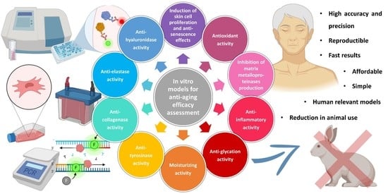
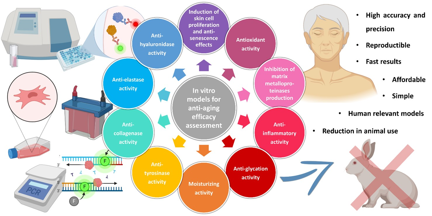
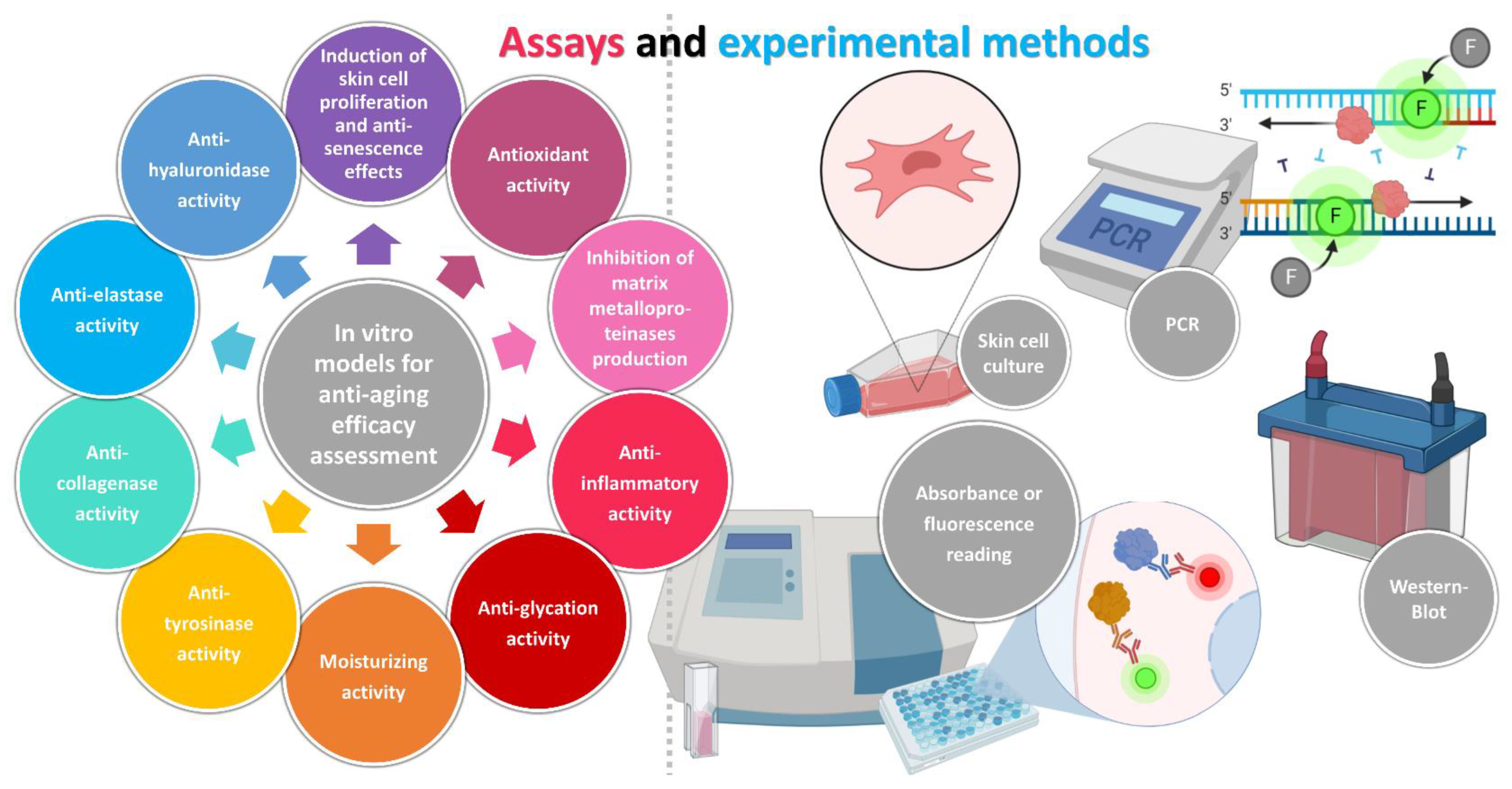
2. In Vitro Models for Anti-Aging Efficacy Assessment

The most common in vitro models for anti-aging activity assessment are the ones evaluating antioxidant, anti-collagenase, anti-elastase, anti-hyaluronidase, anti-tyrosinase, anti-inflammatory, antiglycation, or moisturizing activity, and the induction of skin cell proliferation/anti-senescence effects or the inhibition of matrix metalloproteinase production. Each with their specific characteristics, these assays require the use of skin cell cultures, absorbance or fluorescence reading, polymerase chain reaction (PCR), and/or Western blotting. Each of these methods will be described in the following sections, with illustrative examples, and a schematic representation is presented in Figure 2.

2.1. Induction of Skin Cell Proliferation and Anti-Senescence Effects

Cell proliferation and migration impact the skin aging process and play crucial roles in tissue formation and repair, in order to maintain healthy skin. Through paracrine signaling and gap junctions, biochemical communication induces cellular proliferation and migration. Thereby, aging causes the loss of proliferative capacity and senescent fibroblasts tend to prevail, with skin cells being characterized by their proliferative inability. Hence, the promotion of skin cell proliferation and/or anti-senescence effects are directly related to anti-aging properties [24,25,26,27,28,29].
This type of study has been extensively performed in plant-derived compounds. An example is the study performed by Gu et al. [30], in which the potential anti-senescence effect of bamboo leaf flavonoids on HaCaT cell proliferation was determined. HaCaT cells are immortalized human keratinocytes, and since they have been widely used for epidermal pathophysiology study, they offer a good model for the evaluation of anti-aging effects. HaCaT cells, in which senescence was chemically induced by previous contact with 2,2-azobis(2-amidinopropane) dihydrochloride (AAPH), were treated with varying concentrations of the compounds for 48 h. Then, the common 3-[4,5-dimethylthiazole-2-yl]-2,5-diphenyltetrazolium bromide (MTT) assay was performed, in order to assess cell proliferation. This assay is performed to assess the proliferation capacity, if cell viability increases, and cytotoxic potential, if cell viability is compromised, of different compounds in cell lines. Results showed that the test compounds at concentrations ranging from 5 to 80 μg/mL effectively mitigated the inhibitory effect of AAPH on HaCaT cell proliferation (Figure 3A). However, at a concentration of 160 μg/mL, they exhibited an increased inhibitory effect on cell proliferation, indicating potential dose-dependent effects (Figure 3A). Further investigation of the effects of the test flavonoids on cell senescence was conducted by mRNA sequencing and Western blotting. The results showed that the compounds inhibited senescence by reducing p53, p21, and p16 gene expression, and increasing Lamin B1 gene expression (Figure 3B), all common markers used to detect cell senescence (related to cell cycle arrest). Fluorescent staining of the cells also led to the same conclusions (Figure 3C,D). These findings suggest that bamboo leaf flavonoids could be a potential therapeutic strategy for age-related diseases.
A different study, conducted by Quiles et al. [28], studied the anti-aging efficacy of a combination of four different plant polyphenolic extracts: pomegranate fruit (Punica granatum), containing punicalagins; sweet orange (Citrus aurantium), enriched in hesperidin; Herba Cistanche stem, containing phenylpropanoids; and Centella asiatica, standardized in triterpenoids. Normal human dermal fibroblast (NHDF) cells were used, and cell proliferation was established by the MTT assay. Cells were pretreated with H2O2 for aging induction. Epidermal growth factor was used as a positive control, and NHDF without H2O2 pretreatment as a non-aging control. NHDF cells pretreated with pro-oxidant-conditioned medium, which reproduced the reduced proliferation rate in aged skin fibroblasts, induced a significant decrease in the cell proliferation rate when compared to untreated NHDF cells. Absorbance values lower than those of control cells indicated a reduction in the rate of cell proliferation, and a higher absorbance rate indicated an increase in cell proliferation. Results showed that cells treated with the extract combination recovered their proliferation capabilities significantly.
Another study regarding plant-derived compounds, performed by Panichakul et al. [29], showed that Ipomoea pes-caprae extracts have potential for use in skin anti-aging cosmeceutical preparations. At noncytotoxic concentrations, the extracts significantly increased CCD-986sk (human fibroblasts) cell proliferation. Cell proliferation was examined using the PrestoBlue® cell viability reagent, a purified resazurin for high-sensitivity tests. The results were interpreted as the percentage of cell proliferation and showed that ethanolic extracts in concentrations between 0.031 and 0.062 mg/mL were able to induce cell proliferation, compared with the untreated control cells. Additionally, the expression of FGF2 genes was monitored in the treated cells, using real-time PCR, and the extracts significantly upregulated the mRNA levels of this gene. The expression of FGF2 is involved in encoding growth factors that stimulate cellular proliferation, and hence it was determined that the cell proliferation induction of the tested extracts was linked to this gene.
One other study, performed by Lee et al. [31], determined the anti-aging and protective effects of galangin (3,5,7-trihydroxyflavone), a flavanol compound found in Alpinia officinarum and other types of ginger. The study was done in HS68 (human dermal fibroblast) cells, which were exposed to H2O2/UVB radiation for chemical and radiation cell proliferation reduction. The MTT assay results showed that treatment with galangin led to the protection of these cells against H2O2- and UVB-induced cell death, with an optimal dose of 30 µM.
In another study, by Li et al. [32], the antagonistic effects of Dendrobium nobile Lindl., an orchid species, on UVA-induced photoaging was investigated. The experiments were performed on HFF-1 (human foreskin fibroblast) cells, and cell viability under UVA irradiation and the level of proliferation activity after treatment were assessed by the CCK-8 method (Cell-Counting-Kit-8 assay kit, using 2-(2-methoxy-4-nitrophenyl)-3-(4-nitrophenyl)-5-(2,4-disulfophenyl)-2H-tetrazolium). Results showed that the tested plant was successful in restoring the proliferative viability of UVA-injured cells. With the increase in compound concentration, the proliferation activity of cells in the treatment group was significantly improved.
Ouyang et al. [33] also studied the anti-aging effects of a plant-derived compound, the water-soluble heteropolysaccharide extracted from Gracilaria lemaneiformis. This study explored the efficacy of the test compound against skin photoaging in HaCaT cells under UVB irradiation. Exposure to UVB irradiation significantly increased the apoptosis rate of HaCaT cells, but heteropolysaccharide pretreatment effectively inhibited the pro-apoptotic effect of UVB on these cells. Hence, the results obtained in this study indicated that the tested compound could protect HaCaT cells from UVB-induced damage. Furthermore, the results suggested that the tested molecule modulates cell apoptosis via downregulating the expression of apoptosis-related protein caspase-3, and accelerates cell proliferation, migration, and repair.

2.2. Anti-Collagenase Activity

Collagen is an essential component for human skin health, since it provides it with structural support, elasticity, firmness, and flexibility. Collagenases are enzymes that degrade collagen in the skin, causing wrinkles as we age. Therefore, compounds with the potential to inhibit this enzyme can delay wrinkle formation and, consequently, aging. Hence, assays that quantify anti-collagenase effects are used in the analysis of the anti-aging activity of different compounds [34,35,36,37].
In a study performed by Barak et al. [38], the authors investigated the anti-aging potential of Maclura pomifera methanolic extract (MPM). Clostridium histolyticum was used as the source of the collagenase enzyme (bacterial origin), and N-[3-(2-furyl)acryloyl]-Leu-Gly-Pro-Ala (FALGPA) was used as a substrate. In order to evaluate the anti-collagenase activity of MPM, the extracts were incubated with the collagenase enzyme for 15 min, and then the substrate was added. Absorbance was immediately measured, and results showed that MPM had high collagenase inhibition activity, with 84.55 ± 1.99% inhibition being observed after 20 min of incubation, and reaching 94.68 ± 2.42% inhibition after 40 min of incubation, at a concentration of 1 mg/mL, demonstrating that the inhibitory bioactivity was amplified through time. Epigallocatechin gallate (EGCG) was used as a positive control and showed similar levels of inhibition to MPM. Hence, the findings indicated that MPM may have potential as an anti-collagenase agent.
Eaknai et al. [39] used similar experiments to investigate the anti-aging activity of fenugreek extract encapsulated within a liponiosome (Figure 4A), using an in vitro collagenase inhibitory assay and collagen production assay, in human dermal fibroblast (HDF) cells. The collagenase was sourced from Clostridium histolyticum type IA, and FALGPA was used as a substrate. After incubation with the extract, changes in sample absorbance were measured, at 340 nm, and half minimal inhibitory concentration (IC50) values were calculated by plotting a linear regression curve showing sample concentrations on the x-axis and percentage inhibition on the y-axis. Results showed that the fenugreek extract had significant anti-collagenase activity, with an IC50 value of 0.56 ± 0.02 mg/mL (Figure 4B). The inhibitory concentration of the fenugreek extract was lower than that of ascorbic acid, indicating its potential as an anti-aging agent. The collagen production assay revealed that treatment with the extract at a concentration of 125 μg/mL significantly enhanced the amount of stained collagen in human dermal fibroblast cells, compared to the control, at both day 7 (163%) and day 14 (131%) (Figure 4C). The induction was comparable to the effect of vitamin C-induced collagen production, and significantly higher than rutin-induced collagen production. The researchers also formulated the extract into liponiosomes, in order to improve its potency as an active ingredient in anti-aging products, which proved effective since the results showed even higher anti-collagenase activity.
A different study, by Castejon et al. [40], aimed to investigate the collagenase inhibition activity of different seaweed extracts using a colorimetric assay kit. This time, FALGPA was also determined, but a colorimetric assay kit was used (MAK293). EGCG was also used as a positive control. Results showed that several extracts exhibited positive collagenase inhibition ranging from 68% to 91%, being especially relevant for Alaria esculenta, which is in line with previous research that highlights this extract’s anti-aging properties due to its inhibition of collagenase activity, making it a promising ingredient in anti-aging products.
In another study, by An et al. [41], the in vitro anti-wrinkle efficacy of propolis extract-loaded nanoparticles was investigated, by evaluating intracellular collagen production and inhibitory effects on intracellular collagenase activity. These assays were performed in HDF cells, and procollagen type i c peptide was determined, using an in vitro enzyme immunoassay kit (PIP EIA Kit), with subsequent absorbance measurement. The results revealed that the developed nanoparticles significantly increased intracellular collagen production in HDF, at concentrations of 0.016% to 0.063%, compared to blank nanoparticles (no drug). Therefore, the findings suggest that the developed propolis-containing nanoparticles have the potential to improve skin elasticity and reduce wrinkles by increasing intracellular collagen production.
A study conducted by Moreira et al. [42] investigated the effect of Eucalyptus globulus extract obtained from hydrodistillation residual water on the expression of collagen I in HaCaT cells. The cells were treated with the extract, and the expression of collagen I was measured using real-time PCR. The results showed that extract treatment significantly increased the expression of collagen I compared to the control. This suggests that the extract may have anti-senescent effects in keratinocytes, as collagen I is known to play an important role in maintaining the skin structure and preventing signs of aging. The study suggests that the tested extract may have potential as an anti-aging treatment for the skin.
El-Nashar et al. [43] conducted an in vitro collagenase inhibitory assay to determine the anti-aging activity of a new xanthone glycoside isolated from the methanol extract of Mangifera indica leaves (Anacardiaceae). The assay was performed using a fluorometric collagenase inhibitor screening kit (K833-100, fluorescence-labeled gelatin degradation assessment), at a 490 nm excitation wavelength and a 520 nm emission wavelength, using a microplate reader. The compound exhibited moderate anti-collagenase activity, with an IC50 value of 419.10 μg/mL, as compared to a standard. The findings suggest that the evaluated compound has potential anti-aging properties.

2.3. Anti-Elastase Activity

Elastin is one of the most abundant proteins in healthy human skin, accounting for more than 90% of the elastic fibers that give skin its flexibility. The breakdown of elastin, which is predominantly induced by the enzyme elastase, is the main cause of drooping skin and the production of fine wrinkles. As a result, the assay’s purpose is to inquire about the capacity of different compounds to suppress elastase activity and their potential to delay aging [37,44,45,46,47].
Chaikhong et al. [48] evaluated the anti-elastase activity of several Thai plant extracts (Areca catechu, Anacardium occidentale, Glochidion zeylanicum, and Senna alata) using an elastase inhibition assay, with N-succinyl-triallyl-p nitroanilide (SANA) as a substrate. EGCG was once again used as a positive control, and dimethyl sulfoxide (DMSO) as a negative control. The assay involved adding extracts, pancreatic porcine elastase, and Tris–HCl buffer to a 96-well plate, pre-incubating them at room temperature, adding SANA, and then measuring the absorbance at 734 nm with a microplate reader. Results showed that EGCG had an elastase inhibition level of 45.27%, whereas the evaluated plant extracts had inhibition levels between 73.95% and 88.31%.
In a different study, Ibrahim et al. [49] assessed the anti-elastase activity of Rosmarinus officinalis L. hexane extract, using the EnzCheck® Elastase Assay Kit. The assay also involved incubating the tested compound with a porcine pancreatic elastase stock solution and elastase substrate, but, this time, the substrate was soluble bovine neck ligament elastin. Additionally, incubation was followed by measurement of the fluorescence intensity (instead of absorbance), at specific wavelengths (λex 505 nm, λem 515 nm), using a microplate reader. Moreover, 10-phenathroline was used as a positive control (standard elastase inhibitor). The IC50 value was calculated as the concentration of the extract required to inhibit 50% of elastase activity. The extract showed good dose-dependent inhibition of elastase activity, with an IC50 value of 57.6 μg/mL, which was considered to be similar to the reference standard.
Barak et al. [38] also evaluated the anti-elastase activity of an MPM extract, also using pancreatic porcine elastase, but using a different substrate, N-succinyl-Ala-Ala-p-nitroanilide (AAAPVN). The experiment was carried out using 0.2 mM Tris–HCl buffer solution at pH 8.0. After pre-incubation of 1 μg/mL of the elastase with the MPM extract, for 15 min, at 37 °C, the AAAPVN substrate was added, and incubation was performed again for 15 more minutes. Then, fluorescence was measured, at 365 nm for excitation and 410 nm for emission. The results showed that MPM exhibited significant inhibition activity against elastase, and its inhibition activity increased from 34.7% to 97.4% from 5 min to 30 min, which was higher than the inhibition activity obtained with the positive control (EGCG), at every time point. These findings suggest that the MPM extract has potential as an anti-elastase agent.
Shakour et al. [37] also used AAAPVN as the substrate for the evaluation of the capacity of Moringa oleifera extracts to suppress elastase activity. Each extract, at different concentrations, was individually incubated for 15 min with the elastase enzyme, before adding the substrate. The reaction was then carried out using a mixture of the tested extract, the substrate AAAPVN, the buffer, and the elastase enzyme. After incubation, the absorbance microplate’s absorbance was evaluated at 400 nm, using a microplate reader and EGCG as a positive control. The obtained results highlighted that all the extracts had anti-elastase activity, with the IC50 varying from 0.45 to 1.92 mg/mL.

2.4. Anti-Hyaluronidase Activity

Hyaluronic acid is a major component of the dermal extracellular matrix, preserving the skin’s moisture content, elasticity, and hydration retention. Since hyaluronic acid is depolymerized by hyaluronidase enzymatic degradation, the inhibition of hyaluronidase activity is significant in preventing the breakdown of hyaluronic acid, as it may cause wrinkles, skin dryness, and loss of firmness. Therefore, anti-hyaluronidase activity is essential in delaying the skin aging process [50,51,52,53,54].
Shakour et al. [37] assessed the anti-hyaluronidase activity of Moringa oleifera extracts using a fluorometric assay. Test tubes were prepared by adding hyaluronidase enzyme to the tested extract and calcium chloride. A substrate solution was also prepared, containing hyaluronic acid in an acetate buffer, at different concentrations. Then, potassium borate was added to the test tubes, which were submerged in a water bath and heated to 100 °C for a few minutes, before being cooled down at room temperature. Next, a dimethylaminobenzaldehyde solution was produced and added to the samples, followed by incubation. Finally, sample fluorescence was measured, in 96-well plates, using a microplate reader, at 545 nm excitation and 612 nm emission. Hyaluronidase enzyme inhibition was calculated, and the obtained results highlighted that the evaluated extracts had substantial anti-hyaluronidase activity, with the IC50 ranging from 0.46 to 0.51 mg/mL.
On the other hand, Sklirou et al. [55] investigated the anti-hyaluronidase activity of different Greek flora extracts using a spectrophotometric method. The method consisted of a series of incubations of mixtures mainly containing the tested sample, acetate buffer, a hyaluronidase solution, and a hyaluronic acid solution. Samples were heated and cooled down for several cycles, and then transferred into 96-well microplates, on which their absorbance was quantified at 586 nm. Tannic acid was used as a positive control and DMSO as a negative control. The obtained results highlighted both Sedum sediforme and Umbilicus horizontalis’ significant hyaluronidase inhibitory activity.
El-Nashar et al. [43] evaluated the anti-hyaluronidase activity of a new xanthone glycoside, isolated from the methanol extract of Mangifera indica leaves, using a similar assay. Compound solutions were prepared in different concentrations, and were incubated with bovine hyaluronidase dissolved in acetate buffer. The optical density of the reaction mixture was measured at 585 nm after incubation. The results showed that the tested compound had a strong inhibitory effect on hyaluronidase activity, with an IC50 value of 1.65 μg/mL, which was better than that of 6-O-palmitoyl L-ascorbic acid, used as a positive control (2.55 μg/mL). The findings suggest that the tested compound has potential as an anti-aging agent in skin health-related products.
A different method was used by Ibrahim et al. [49], who performed a turbidimetric assay to evaluate the hyaluronidase inhibitory activity of rosemary extract. The assay utilized the QuantiChromTM Hyaluronidase Inhibitor Screening Assay Kit, and bovine hyaluronidase (type-1-S) as the enzyme. The extract was added to the wells containing the enzyme, and the reaction was stopped using the stop reagent, after incubation for 20 min. The turbidity formed by residual hyaluronic acid was determined by measuring the optical density at 600 nm, after an additional 10 min incubation. The hyaluronidase inhibitory activity of the tested extract was found to be mild, with an IC50 of 448.1 μg/mL.
Another study, performed by Lee et al. [56], assessed the potential of Potentilla paradoxa Nutt using HaCaT cells that produce hyaluronic acid. After HaCaT cells were treated with the plant extract, they assessed the mRNA expression, by PCR, of hyaluronan synthase-1, -2, and -3. Results showed that the mRNA expression of hyaluronan synthase-2, and -3 was increased by the extract. Additionally, the levels of the protein phospho-AKT, which regulates the expression of hyaluronan synthase-2, increased with extract exposure, as well as the protein expression of phosphorylated phosphoinositide 3-kinase, which is responsible for the regulation of the phosphorylation of AKT. Hence, keratinocytes exposed to the tested extract increased the expression of an enzyme that synthesizes hyaluronic acid (hyaluronan synthase) and decreased the expression of an enzyme that degrades hyaluronic acid (hyaluronidase), showing the potential anti-aging effects of Potentilla paradoxa Nutt.

2.5. Anti-Tyrosinase Activity

Tyrosinase is a critical enzyme in melanogenesis. Melanin protects the skin against UV radiation, exposure to which may lead to pathological skin conditions. However, the overproduction and abnormal accumulation of melanin can lead to pigment disorders, including freckles, age spots, and hyperpigmentation. Therefore, the suppression of tyrosinase activity may provoke the downregulation of melanogenesis and a decline in skin hyperpigmentation, contributing to the prevention of premature aging and treatment of abnormal skin pigmentation [57,58,59,60,61].
Jan et al. [62] conducted an assay for the determination of the anti-tyrosinase activity of zinc nanoparticles. Levodopa (L-DOPA) was used as a substrate and the tyrosinase was of mushroom origin. The substrate, the enzyme, and the tested sample were incubated in sodium phosphate buffer. Then, the reaction was tracked by absorbance reading, at 475 nm, using a microplate reader. The extraction solvent was used as a control. The results showed that the nanoparticles had a medium-intensity inhibitory effect on tyrosinase, with a percentage inhibition of 14.56 ± 0.89%.
Shakour et al. [37] evaluated Moringa oleifera extracts’ effects by assessing their capacity to inhibit the enzyme tyrosinase in a similar manner. A mixture of the tested extracts (at various concentrations), a solution containing mushroom tyrosinase, L-DOPA, and phosphate buffer, was prepared. Then, dopachrome formation (L-DOPA-derived compound, with a characteristic color) was observed, and samples’ absorbance was measured at 475 nm, using a microplate reader. Nevertheless, in this assay, the positive control was different, being EGCG. The tyrosinase enzyme inhibition percentage was calculated, and the obtained results highlighted that the butanolic extract was the most effective, having an IC50 value of 0.59 mg/mL.
Sklirou et al. [55] performed a similar assay, investigating the capacity of different plants from Greek flora extracts to inhibit tyrosinase. The extracts were dissolved in DMSO, diluted in phosphate buffer, mixed with mushroom tyrosinase and L-DOPA, and incubated. The difference between this assay and the ones performed by Jan et al. and Sklirou et al. is that a different positive control was used, namely kojic acid, a depigmenting agent commonly used in skin products. Absorbance reading was performed at 475 nm, using a microplate reader, and tyrosinase inhibition activity was found to be more substantial for extracts from the Lamiaceae and Leguminosae families, being highly effective, with inhibitory activity of over 40%.
El-Nashar et al. [43] also evaluated the anti-tyrosinase activity of a specific compound, namely a new xanthone glycoside isolated from the methanol extract of Mangifera indica leaves. Nevertheless, in this study, an assay kit was used, namely the Abcam Tyrosinase Inhibitor Screening Colorimetric Assay Kit. Tyrosinase will catalyze tyrosine oxidation, producing a chromophore that can be detected at a specific wavelength (510 nm). Hence, in the presence of a compound with anti-tyrosinase activity, the measured absorbance will decrease. Results showed that the tested compound demonstrated potential tyrosinase inhibition, with an IC50 value of 0.48 μg/mL, compared to kojic acid as a positive control (0.82 μg/mL).
On the other hand, Moreira et al. [42] investigated the anti-tyrosinase activity of Eucalyptus globulus leaf extracts using B-16V melanocytes. The B-16V cells were treated with 3-isobutyl-1-methylxanthine (IBMX) to induce skin pigmentation, and then treated with the extracts or kojic acid (positive control). L-DOPA was used as the substrate, and tyrosinase activity assessed by measuring the sample absorbance, at 400 nm. Results showed that the Eucalyptus globulus leaf essential oil inhibited tyrosinase in IBMX-treated cells by 90.69%, while the positive control only reduced its activity by 77.92%.

2.6. Antioxidant Activity

The aging process can be induced by endogenous or exogenous factors and is largely associated with oxidative stress, through the formation of reactive oxygen species (ROS), resulting in structural changes in skin cell composition. UV radiation is directly related to the majority of skin problems and is a major cause of photoaging, since it generates ROS formation and, consequently, oxidative stress. ROS directly damage skin cells, mediate inflammatory responses, and contribute to the breakdown of the extracellular matrix. Accordingly, the synthesis and degradation of essential extracellular matrix components, such as collagen and elastin, are perturbed. Furthermore, ROS induces melanin production via the activation of the tyrosinase enzyme and causes hyaluronic acid degradation, which consequently may lead to pigment disorders and improper hydration, respectively. Hence, topical antioxidant application can be quite beneficial against the harmful effects of UV radiation, since it prevents molecular damage and maintains skin homeostasis. In this regard, antioxidant activity may have a protective effect on the skin [63,64,65,66,67].
Dina et al. [68] determined the antioxidant potential of a Rosa damascena-enriched phenolic extract on BJ cells (human diploid skin fibroblasts). Short-term exposure (24 h) of the cells to the extract resulted in the upregulation of nuclear factor erythroid 2-related factor (NRF2), a transcription factor related to the mobilization of genomic responses to oxidative damage (detection by real-time PCR). Additionally, a 2,2-diphenyl-1-picrylhydrazyl (DPPH) antioxidant assay was performed, carried out by mixing a stock solution of DPPH with the sample extract, incubating it for 30 min, and measuring the absorbance at 517 nm. Gallic acid, a naturally occurring polyphenol with antioxidant effects, was used as a positive control. A third antioxidant assay was also performed, the 2,2′-azino-bis(3-ethylbenzothiazoline-6-sulfonic acid) (ABTS) assay, which was carried out by mixing a stock solution of ABTS with the sample extract, incubating it for 10 min, and measuring the absorbance at 734 nm. Trolox (6-hydroxy-2,5,7,8-tetramethylchroman-2-carboxylic acid), a water-soluble analog of vitamin E, was used as a positive control. For both the DPPH and ABTS assays, the percentage of scavenging was calculated, and the evaluated extract showed high antioxidant capability.
Sklirou et al. [55] investigated the antioxidant potential of extracts of different plants from Greek flora using two different assays. One of them was the DPPH free radical scavenging assay. Extracts were dissolved in DMSO, and either the extract solutions or a gallic acid solution (positive control) were mixed with a DPPH solution. The mixtures were placed in a microwell plate and incubated, and after incubation, the absorbance was read at 517 nm. A negative control (DPPH plus DMSO) and a blank sample (sample plus ethanol) were also used. The obtained results highlighted that 167 plant extracts demonstrated substantial scavenging activity, with 99 of them exhibiting inhibition >80%, and the vast majority (85%) being methanolic extracts derived from the Lamiaceae family. An intracellular ROS assay was also performed, in which the capacity of the extracts to scavenge intracellular ROS was analyzed, in BJ cells (with H2O2 pretreatment or not). The cells were seeded in 96-well microplates, and the test extracts were added, at the highest non-cytotoxic concentration. After incubation, 2′,7′-dichlorodihydrofluorescein diacetate was added, and fluorescence was measured (emission at 520 nm, excitation at 485 nm). Results showed that the vast majority of extracts were able to decrease the levels of intracellular ROS.
On the other hand, Lee et al. [31] used HS68 cells (human foreskin fibroblasts) and treated them with H2O2 and UVB radiation to induce oxidative damage. Results showed that H2O2 and UVB treatment induced ROS accumulation, but treatment with galangin (3,5,7-trihydroxyflavone, a flavanol found in Alpinia officinarum; Figure 5A), reduced ROS generation (schematic representation in Figure 5B). The determination consisted of measuring the NRF2-dependent luciferase activity, which was increased when cells were treated with the test compound (Figure 5C,D). Hence, galangin increased NRF2 nuclear translocation and transcriptional activation, which is related to the regulation of antioxidant protein production, which protect dermal fibroblasts against oxidative damage. This suggests that galangin has antioxidant properties and can protect cells from oxidative damage induced by H2O2 and UVB radiation.
A different study, by Barak et al. [38], included several in vitro assays to determine the antioxidant potential of Maclura pomifera methanolic extract. One of these assays was the DPPH radical scavenging activity test, performed as previously described: freshly diluted sample solutions were mixed with methanolic DPPH solution, and, after incubation, the absorbance was measured at 517 nm. Butylated hydroxy toluene was used as a reference. The tested extract exhibited significant DPPH radical scavenging activity, with an IC50 value of 1998.86 ± 0.02 μg/mL. A second assay was performed, namely the ferric reducing antioxidant power (FRAP) test. This assay measures the ability of an antioxidant compound to reduce Fe(III) to Fe(II). In this test, the sample was added to the FRAP reagent (2,4,6-tris (2-pyridyl)-s-triazine and FeCl3.6H2O in acetate buffer) and incubated for 30 min at 37 °C. The absorbance was then measured, at 593 nm. Butylated hydroxy toluene was used as a reference compound, and a ferrous chloride solution was used to obtain a standard curve. The results were reported as mM FeSO4 per gram of dry extract, and the tested extract exhibited notable metal-reducing activity, with a value of 0.191 ± 0.01 mM FeSO4/DE. The third assay that was performed was the cupric-reducing antioxidant capacity (CUPRAC) test. Similarly to the FRAP test, this assay measures the ability of an antioxidant compound to reduce Cu(II) to Cu(I). In this test, CuSO4, neocuproine, and ammonium acetate buffer were mixed in a 96-well plate. A sample solution was then added to the mixture, and the absorbance was measured at 450 nm after incubation. The results were reported as mg ascorbic acid equivalent per gram of dry extract. Again, the evaluated extract showed noteworthy copper-reducing activity, with a value of 73.928 ± 0.01 AAE/g DE. Finally, a fourth test was also performed, namely the total antioxidant capacity (TOAC) test. This assay measures the overall antioxidant capacity of a sample, and a sample solution is mixed with TOAC solution (sodium phosphate monobasic, ammonium molybdate, and H2SO4), and then incubated for 90 min at 95 °C. The absorbance is then measured at 695 nm. Ascorbic acid was used to obtain a standard curve, and the results were reported as mg of Trolox equivalents in 1 g of dry extract. The tested extract exhibited a significant total antioxidant capacity, with a value of 114.43 ± 0.02 AAE/g DE. Overall, all these results suggest that Maclura pomifera methanolic extract has promising antioxidant potential.
On the other hand, Vaz et al. [69] investigated the antioxidant capacity of sulfurous/bicarbonate/sodic natural mineral waters using two different assays: the ROS production assay, and the superoxide dismutase (SOD) activity evaluation assay. In the ROS production assay, sample antioxidant activity was evaluated by ROS quantification through lipopolysaccharide (LPS) exposure. In 96-well microplates containing already cultured NIH/3T3 cells (murine skin fibroblasts), either samples or a control medium were added, as well as LPS. Fluorescence was then measured using a microplate reader (at excitation of 485/530 nm and emission of 350/461 nm). Regarding the second assay that was performed, SOD are antioxidant enzymes essential for regulating cellular homeostasis, due to their ability to eliminate harmful superoxide radicals by converting them into hydrogen peroxide and oxygen. Hence, the SOD activity evaluation assay assesses the capacity of samples to activate SOD. This assay was firstly performed in a similar way as the ROS production assay: in microplates containing NIH/3T3 cells, either samples or a control medium were added, as well as LPS. However, afterwards, the cells were collected, and the protein content was isolated using a specific buffer (RIPA buffer, containing NaCl, Tris–HCl, Nonidet P-40, sodium deoxycholate, sodium dodecyl sulfate, ethylenediaminetetraacetic acid). Then, the level of SOD activity was determined using a WST SOD Assay Kit, which utilizes water-soluble tetrazolium as a substrate because of its capability to produce water-soluble formazan dye when reduced by a superoxide anion. The absorbance was then measured at 450 nm, using a microplate reader, and SOD activity was calculated as the rate of reaction inhibition percentage. Overall, the obtained results highlighted that the studied natural mineral waters had substantial antioxidant effects.
Yang et al. [70] also investigated the ability of plant-derived compounds, in this case Cortex Moutan extracts, to inhibit oxidation, using several different assays. Aside from the already described DPPH and FRAP assays, two other assays were used: the hydroxyl radical scavenging activity assay and the inhibition of β-carotene bleaching assay. Regarding the hydroxyl radical scavenging activity assay, a mixture of sample, ferrous sulfate, salicylic acid, and hydrogen peroxide was incubated for 1 h at 37 °C. Vitamin C was used as a standard antioxidant compound, and 50% ethanol as the control. Then, the absorbance was analyzed by spectrophotometer at 510 nm, in triplicate, and the hydroxyl radical scavenging capability was calculated. Results showed that the tested extracts had a moderate capacity for hydroxyl radical scavenging. As for the inhibition of β-carotene bleaching assay, firstly, a β-carotene solution was prepared, by dissolving β-carotene in chloroform, removing it at 40 °C under a vacuum, and then adding linoleic acid, Tween® 80, and distilled water. Then, this mixture was added to the sample solution and incubated at 50 °C. Ethanol at 50% and the prepared mixture (without the sample) were used as controls. Absorbance was measured at 470 nm. The β-carotene bleaching inhibition was then calculated, and the results showed that the extracts had reasonable activity.

2.7. Inhibition of Matrix Metalloproteinase Production

The skin’s structural framework is provided by the extracellular matrix, which is mainly composed of elastic fibers and collagen. The expression of matrix metalloproteinases (MMP) in epidermal keratinocytes is induced by prolonged UV exposure, which increases skin sagging and wrinkle formation due to the breakdown of extracellular matrix proteins and collagen. Therefore, the inhibition of MMP expression is a potential strategy to inhibit wrinkle formation [71,72,73,74,75].
Kim et al. [75] studied the MMP inhibition activity of 1-monoeicosapentaenoin, a component isolated from a microalgae, Micractinium sp. The expression of MMP-1 and MMP-9 was determined by an enzyme-linked immunosorbent assay (ELISA), in human epidermal keratinocytes irradiated with UVB, after treatment with the compound. Results showed that 1-monoeicosapentaenoin suppressed MMP-1 secretion and MMP-9 protein expression, and hence could be able to inhibit UVB-induced skin damage.
Jia et al. [76] also examined the MMP inhibitory effects of mercaptopurine, an immunologic agent commonly used for the treatment of inflammatory bowel disease and acute lymphoblastic leukemia, but this time on UVB-irradiated HaCaT cells. As expected, the results showed that UVB radiation exposure induced the production of MMP in the cells; however, treatment with mercaptopurine significantly inhibited the upregulation of MMP, thus effectively leading to cell recovery after photoaging. MMP detection was performed by Western blot. Therefore, the findings suggest that mercaptopurine could be a potential therapeutic agent for the prevention and treatment of skin photoaging and acute photodamage caused by UVB radiation.
An et al. [41] also investigated MMP inhibitory effects, using polymeric nanoparticles containing propolis extract. HDF cells were used and MMP-1 levels were measured. HDF cells were incubated with different concentrations of the developed nanoparticles for 48 h, the supernatant was collected, and MMP-1 levels were measured using an ELISA kit, with absorbance reading at 450 nm. Results showed that the developed nanoparticles significantly reduced MMP-1 activity by 7% to 32%, thus suggesting their potential to prevent skin aging.
On the other hand, Sklirou et al. [55] determined the effect of several Greek flora extracts on MMP activity in human skin fibroblasts. Cells were incubated with the extracts, and then with the fluorogenic substrate Dabcyl-Gaba-Pro-Gln-Gly-Leu-Glu-(EDANS)-AIa-Lys-NH2. MMP activity was determined due to the cleavage of the Gly-Leu bond of the substrate by MMP, being proportional to the measured fluorescence (monitored at 480 nm after excitation at 340 nm, using a microplate reader). The obtained results highlighted the extracts’ ability to suppress the MMP activity.
Alternatively, Moreira et al. [42] investigated the inhibitory effects of extracts obtained from Eucalyptus globulus leaves on MMP activity using a cell-free assay. The activity of MMP-1, MMP-9, and MMP-13 was measured in the presence of the test compounds, being performed in a buffer of a specific composition (containing N-2-hydroxyethyl piperazine-N’-2-ethanesulfonic acid, CaCl2, and Brij-35), in 96-well plates. In each well, MMP-1, MMP-9, or MMP-13 enzymes were pre-incubated with the test samples, for 30 min at 37 °C, followed by the addition of an MMP fluorogenic substrate. Then, fluorescence was measured, at an excitation wavelength of 340 nm and an emission wavelength of 440 nm. EDTA, a chelating agent, was used as a positive control. Results showed that the studied samples significantly inhibited MMP-1 activity by 30% to 70%, demonstrating potential applications in preventing skin aging and related diseases using the tested compounds.

2.8. Other Assays: Anti-Inflammatory, Anti-Glycation, and Moisturizing Activity Asessment

An inflammatory response is manifested by elevated levels of pro-inflammatory mediators. Cytokines are small proteins released by cells, including interleukins (IL), which are cytokines produced by one leukocyte and acting on other leukocytes. They may act on the cells that secrete them, on nearby cells, or on distant cells, and there are both pro-inflammatory cytokines and anti-inflammatory cytokines. Certain inflammatory cytokines may be involved in nerve-injury/inflammation-induced central sensitization. As the inflammatory process is closely related to skin aging, the evaluation of anti-inflammatory effects could be an indirect contributor to anti-aging capacity evaluation [77,78,79,80,81].
Jia et al. [76] tested the anti-inflammatory effects of mercaptopurine on HaCaT cells exposed to UVB radiation. UVB radiation can cause the activation of p38 mitogen-activated protein kinases (MAPK), and the upregulation of nuclear factor kappa B, which leads to the expression of inflammatory factors such as IL-1, IL-6, and tumor necrosis factor (TNF) α. However, the results of the study showed that cell treatment with the test compound mitigated the effect of UVB exposure on the expression of these parameters. Hence, after treatment with mercaptopurine, the expression of inflammatory factors in the cells was significantly reduced, suggesting substantial anti-inflammatory effects. Protein and genetic material identification and quantification were performed by Western blot and quantitative real-time PCR, respectively.
Alternatively, Shakour et al. [37] analyzed Moringa oleifera extracts’ anti-inflammatory activity via the assessment of lipoxygenase (LOX) and cyclooxygenase (COX) inhibition. LOX are enzymes that catalyze the oxidation of arachidonic acid, resulting in the generation of pro-inflammatory mediators such as leukotrienes. The increase in their activity leads to the overproduction of pro-inflammatory mediators, which accelerates aging by causing wrinkle development, a decrease in tissue healing ability, and elasticity loss. For this assay, a mixture was prepared by combining glycine max (soybean), LOX solution, and borate buffer. Then, different concentrations of each extract (in ethanol and DMSO) or the standard drug (ibuprofen, in water) were added to the mixture in 96-well microplates. After the preincubation of samples with the substrate (linoleic acid), at 25 °C for 15 min, the absorbance was measured in a microplate reader, at 234 nm. The LOX inhibition percentage was then determined, and the obtained results highlighted that the extracts caused overall effective LOX enzyme inhibition, with the IC50 ranging from 2.51 to 25.00 mg/mL. In turn, COX enzymes are responsible for the conversion of the arachidonic acid into prostaglandins, which are pro-inflammatory mediators. When there is COX overactivation, the excessive prostaglandins can cause persistent tissue inflammation and consequently lead to the destruction of collagen and elastin. Therefore, in this case, it can also accelerate the aging process, causing wrinkles and a loss of skin elasticity. For this assay, the ability of various extracts or the standard drug (ibuprofen) to inhibit COX-1 and COX-2 enzymes was monitored through N,N,N’,N’-tetramethyl-p-phenylenediamine (TMPD) oxidation by arachidonic acid, at 611 nm, in a microplate reader. The inhibition percentage of COX-1 and COX-2 enzymes was calculated, and again the obtained results highlighted that some of the extracts had a significant COX inhibition capacity, with some being more selective to COX-1 and others to COX-2.
On the other hand, Jan et al. [62] investigated the in vitro anti-inflammatory potential of aqueous Aquilegia pubiflorazinc leaf extract zinc nanoparticles, by testing their inhibition of various inflammatory pathways, including COX-1, COX-2, secretory phospholipase A2 (sPLA2), and 15-LOX. For the evaluation of COX-1 and COX-2 inhibition, assays were conducted using specific kits: the Ovine Kit 701050 and the Human Kit 701050, respectively. Both are colorimetric inhibition assays and measure peroxidase activity by determining oxidized TMPD, via absorbance reading at 590 nm. Ibuprofen, a known COX inhibitor, was used as a positive control. The 15-LOX assay was done in a similar manner, using a kit (760700 kit). In a 96-well plate, the developed nanoparticles were mixed and incubated with the enzyme (15-LOX), and then the substrate (arachidonic acid) and chromogen were added. Nordihydroguaiaretic acid was used as a positive control. Absorbance was again measured at 590 nm. Finally, an assay kit was also used to measure the nanoparticles’ activity against sPLA2 (10004883 kit), in which diheptanoyl thio-PC was used as a positive control, and thiotheramide-PC as a substrate. Absorbance was read at 420 nm, detecting the cleavage of diheptanoyl thio-PC ester into free thiols. Results showed that all pathways produced productive outcomes for the inhibitory activity, with the developed nanoparticles exhibiting the highest inhibitory activity for sPLA2, followed by 15-LOX, COX-1, and COX-2. Overall, the results suggest that the developed nanoparticles enable the efficient inhibition of inflammatory processes.
Glycation is a slow chemical reaction between the carbonyl groups of reducing sugars and the free amino acid groups of proteins, which can cause the natural process of skin aging. Advanced glycation end products (AGE), such as Nε-(carboxymethyl) lysine (CML) and pentosidine, which are products of glycation, can also reduce the flexibility of the dermis. Therefore, the ability of different compounds to suppress AGE formation is directly related to their anti-glycation and, consequently, anti-aging activity [82,83,84,85,86]. Shin et al. [87] evaluated methyl gallate’s capacity to suppress AGE formation and inhibit CML production. First, solutions containing bovine serum albumin, glucose, phosphate buffer, and either the test compound or a nucleophilic hydrazine (positive control) were incubated at 37 °C, for 3 weeks. Then, sample fluorescence was measured, at 355/460 nm. The obtained results highlighted that methyl gallate considerably reduced the fluorescence intensity, in a dose-dependent manner, with high AGE formation inhibition. Furthermore, the effects of methyl gallate on CML production were measured using an ELISA kit, and the results showed that the test compound was again effective.
Transepithelial water loss (TEWL) control is a crucial function of the stratum corneum. This can be compromised by hygroscopic molecules and (although not always) can result in a loss of epidermis moisture and elasticity, which can lead to skin aging. Hence, although being indirectly related to skin aging, TEWL measurement can be a relevant parameter for assessment [88,89,90,91,92]. Wang et al. [93] evaluated Benincasa hispida Cogn. polysaccharides’ capacity to prevent the loss of skin moisture, using two different assays: the in vitro moisture absorption assay and the in vitro moisture retention assay. In the in vitro moisture absorption assay, the samples, previously obtained by adding glycerol and the test compounds, were subjected to a saturated ammonium sulfate solution in a closed desiccator (25 °C, 81% relative humidity). Then, the moisture absorption rate was determined, by measuring and recording the mass change of the drying dish and the sample per hour. Results demonstrated that the test compound’s moisture absorption rate gradually increased after drying, first rapidly and then slowly (from 0 to 12 h), and reached the maximum moisture absorption rate after 12 h, demonstrating strong hygroscopic capability. Additionally, in the in vitro moisture retention assay, samples were divided into four groups: test compounds + deionized water, glycerol + deionized water, test compounds + glycerol + deionized water, and water only. Afterwards, each of them was subjected to a closed desiccator (25 °C, below 40% relative humidity), equipped with discolored silica gel. Then, the moisture retention rate was determined, with the sample containing the test compounds + glycerol + deionized water exhibiting the best moisturizing effect.

3. Conclusions

In vitro assays are an essential tool in investigating the potential anti-aging activity of compounds of different origins, with plant-derived molecules or whole extracts being the most commonly studied. These models are regularly used in the cosmetic industry to test the safety and efficacy of new ingredients and formulations before they are introduced to the market, and they allow the reduction or overall replacement of animal use. The most frequently used anti-aging in vitro assays are the ones evaluating antioxidant, anti-collagenase, anti-elastase, anti-hyaluronidase, and anti-tyrosinase activity, being related to the direct effects that aging has on specific skin parameters. Other assays, such as the evaluation of the induction of skin cell proliferation and anti-senescence effects, the inhibition of matrix metalloproteinase production, and anti-inflammatory, anti-glycation, or moisturizing activity assessment, could also be performed to determine the indirect effects of aging on the skin’s characteristics. Hence, overall, these assays provide important data on the safety and efficacy of anti-aging compounds and provide a foundation for research on and the eventual introduction of formulations into the cosmetics market.

Author Contributions

Conceptualization, F.V., A.C.P.-S. and P.C.P.; methodology, F.V., A.C.P.-S. and P.C.P.; investigation, A.M.C., M.C.G. and M.S.M.; data curation, F.V., A.C.P.-S. and P.C.P.; writing—original draft preparation, A.M.C., M.C.G. and M.S.M.; writing—review and editing, F.V., A.C.P.-S. and P.C.P.; supervision, F.V., A.C.P.-S. and P.C.P. All authors have read and agreed to the published version of the manuscript.

Funding

This research received no external funding.

Institutional Review Board Statement

Not applicable.

Informed Consent Statement

Not applicable.

Data Availability Statement

Not applicable.

Conflicts of Interest

The authors declare no conflict of interest.

Abbreviations

AAAPVN—N-succinyl-Ala-Ala-p-nitroanilide; AAPH—2,2-azobis(2-amidinopropane) dihydrochloride; ABTS—2,2′-azino-bis(3-ethylbenzothiazoline-6-sulfonic acid); AGE—advanced glycation end products; CML—Nε-(carboxymethyl) lysine; COX—cyclooxygenase; CUPRAC—cupric-reducing antioxidant capacity; DMSO—dimethyl sulfoxide; DPPH—2,2-diphenyl-1-picrylhydrazyl; EGCG—epigallocatechin gallate; ELISA—enzyme-linked immunosorbent assay; FALGPA—N-[3-(2-furyl)acryloyl]-Leu-Gly-Pro-Ala; FRAP—ferric reducing antioxidant power; HaCaT—immortalized human keratinocytes; HDF—human dermal fibroblasts; HFF-1—human foreskin fibroblasts; IBMX—3-isobutyl-1-methylxanthine; IC50—half minimal inhibitory concentration; IL—interleukins; LOX—lipoxygenase; LPS—lipopolysaccharide; MAPK—mitogen-activated protein kinases; MMP—matrix metalloproteinases; MPM—Maclura pomifera methanolic extract; MTT—3-[4,5-dimethylthiazole-2-yl]-2,5-diphenyltetrazolium bromide; NHDF—normal human dermal fibroblasts; NRF2—nuclear factor erythroid 2-related factor; NIH/3T3—murine skin fibroblasts; PCR—polymerase chain reaction; ROS—reactive oxygen species; SANA—N-succinyl-triallyl-p nitroanilide; SOD—superoxide dismutase; sPLA2—secretory phospholipase A2; TEWL—transepithelial water loss; TMPD—N,N,N’,N’-tetramethyl-p-phenylenediamine; TNF—tumor necrosis factor; TOAC—total antioxidant capacity.

References

  1. Nakajima, S.; Nomura, T.; Common, J.; Kabashima, K. Insights into Atopic Dermatitis Gained from Genetically Defined Mouse Models. J. Allergy Clin. Immunol. 2019, 143, 13–25. [Google Scholar] [CrossRef] [PubMed]
  2. Gilaberte, Y.; Prieto-Torres, L.; Pastushenko, I.; Juarranz, Á. Anatomy and Function of the Skin. In Nanoscience in Dermatology; Elsevie: Amsterdam, The Netherlands, 2016; pp. 1–14. [Google Scholar]
  3. Abdo, J.M.; Sopko, N.A.; Milner, S.M. The Applied Anatomy of Human Skin: A Model for Regeneration. Wound Med. 2020, 28, 100179. [Google Scholar] [CrossRef]
  4. Wong, R.; Geyer, S.; Weninger, W.; Guimberteau, J.-C.; Wong, J.K. The Dynamic Anatomy and Patterning of Skin. Exp. Dermatol. 2016, 25, 92–98. [Google Scholar] [CrossRef] [PubMed]
  5. Kanitakis, J. Anatomy, Histology and Immunohistochemistry of Normal Human Skin. Eur. J. Dermatol. 2002, 12, 390–401. [Google Scholar]
  6. Tobin, D.J. Introduction to Skin Aging. J. Tissue Viability 2017, 26, 37–46. [Google Scholar] [CrossRef]
  7. Russell-Goldman, E.; Murphy, G.F. The Pathobiology of Skin Aging. Am. J. Pathol. 2020, 190, 1356–1369. [Google Scholar] [CrossRef]
  8. Wong, Q.Y.A.; Chew, F.T. Defining Skin Aging and Its Risk Factors: A Systematic Review and Meta-Analysis. Sci. Rep. 2021, 11, 22075. [Google Scholar] [CrossRef]
  9. Ganceviciene, R.; Liakou, A.I.; Theodoridis, A.; Makrantonaki, E.; Zouboulis, C.C. Skin Anti-Aging Strategies. Dermatoendocrinol 2012, 4, 308–319. [Google Scholar] [CrossRef]
  10. Farage, M.A.; Miller, K.W.; Elsner, P.; Maibach, H.I. Characteristics of the Aging Skin. Adv. Wound Care 2013, 2, 5–10. [Google Scholar] [CrossRef]
  11. Neves, L.M.G.; Wilgus, T.A.; Bayat, A. In Vitro, Ex Vivo, and In Vivo Approaches for Investigation of Skin Scarring: Human and Animal Models. Adv. Wound Care 2023, 12, 97–116. [Google Scholar] [CrossRef]
  12. Stokes, W. Animals and the 3Rs in Toxicology Research and Testing. Hum. Exp. Toxicol. 2015, 34, 1297–1303. [Google Scholar] [CrossRef] [PubMed]
  13. John, L.M.; Dalsgaard, C.M.; Jeppesen, C.B.; Conde-Frieboes, K.W.; Baumann, K.; Knudsen, N.P.H.; Skov, P.S.; Wulff, B.S. In Vitro Prediction of in Vivo Pseudo-Allergenic Response via MRGPRX2. J. Immunotoxicol. 2021, 18, 30–36. [Google Scholar] [CrossRef] [PubMed]
  14. Rooney, J.P.; Choksi, N.Y.; Ceger, P.; Daniel, A.B.; Truax, J.; Allen, D.; Kleinstreuer, N. Analysis of Variability in the Rabbit Skin Irritation Assay. Regul. Toxicol. Pharmacol. 2021, 122, 104920. [Google Scholar] [CrossRef] [PubMed]
  15. Niehues, H.; Bouwstra, J.A.; El Ghalbzouri, A.; Brandner, J.M.; Zeeuwen, P.L.J.M.; van den Bogaard, E.H. 3D Skin Models for 3R Research: The Potential of 3D Reconstructed Skin Models to Study Skin Barrier Function. Exp. Dermatol. 2018, 27, 501–511. [Google Scholar] [CrossRef]
  16. Barthe, M.; Bavoux, C.; Finot, F.; Mouche, I.; Cuceu-Petrenci, C.; Forreryd, A.; Chérouvrier Hansson, A.; Johansson, H.; Lemkine, G.F.; Thénot, J.-P.; et al. Safety Testing of Cosmetic Products: Overview of Established Methods and New Approach Methodologies (NAMs). Cosmetics 2021, 8, 50. [Google Scholar] [CrossRef]
  17. Singh, P.; Bhat, S.S.; Singh, N.; Venkanna, B.U.; Mohamed, R.; Rao, R.P. Cell-Based Model Systems for Validation of Various Efficacy-Based Claims for Cosmetic Ingredients. Cosmetics 2022, 9, 107. [Google Scholar] [CrossRef]
  18. Scientific Committee on Consumer Safety (SCCS). The SCCS Notes of Guidance for the Testing of Cosmetic Ingredients and Their Safety Evaluation, 11th ed; European Union: Brussels, Belgium, 2021. [Google Scholar]
  19. Szilágyi, Z.; Németh, Z.; Bakos, J.; Kubinyi, G.; Necz, P.P.; Szabó, E.; Thuróczy, G.; Pinto, R.; Selmaoui, B. Assessment of Inflammation in 3D Reconstructed Human Skin Exposed to Combined Exposure to Ultraviolet and Wi-Fi Radiation. Int. J. Mol. Sci. 2023, 24, 2853. [Google Scholar] [CrossRef]
  20. Goncalves, K.; De Los Santos Gomez, P.; Costello, L.; Smith, L.; Mead, H.; Simpson, A.; Przyborski, S. Investigation into the Effect of Skin Tone Modulators and Exogenous Stress on Skin Pigmentation Utilizing a Novel Bioengineered Skin Equivalent. Bioeng. Transl. Med. 2023, 8, e10415. [Google Scholar] [CrossRef]
  21. Leman, G.; Moosbrugger-Martinz, V.; Blunder, S.; Pavel, P.; Dubrac, S. 3D-Organotypic Cultures to Unravel Molecular and Cellular Abnormalities in Atopic Dermatitis and Ichthyosis Vulgaris. Cells 2019, 8, 489. [Google Scholar] [CrossRef]
  22. Jang, H.-J.; Lee, J.B.; Yoon, J.-K. Advanced In Vitro Three-Dimensional Skin Models of Atopic Dermatitis. Tissue Eng. Regen. Med. 2023. [Google Scholar] [CrossRef]
  23. Moon, S.; Kim, D.H.; Shin, J.U. In Vitro Models Mimicking Immune Response in the Skin. Yonsei Med. J. 2021, 62, 969. [Google Scholar] [CrossRef] [PubMed]
  24. Tigges, J.; Krutmann, J.; Fritsche, E.; Haendeler, J.; Schaal, H.; Fischer, J.W.; Kalfalah, F.; Reinke, H.; Reifenberger, G.; Stühler, K.; et al. The Hallmarks of Fibroblast Ageing. Mech. Ageing Dev. 2014, 138, 26–44. [Google Scholar] [CrossRef] [PubMed]
  25. Wlaschek, M.; Maity, P.; Makrantonaki, E.; Scharffetter-Kochanek, K. Connective Tissue and Fibroblast Senescence in Skin Aging. J. Investig. Dermatol. 2021, 141, 985–992. [Google Scholar] [CrossRef] [PubMed]
  26. Kondo, H.; Yonezawa, Y. Changes in the Migratory Ability of Human Lung and Skin Fibroblasts during in Vitro Aging and in Vivo Cellular Senescence. Mech. Ageing Dev. 1992, 63, 223–233. [Google Scholar] [CrossRef]
  27. Sanchez, M.M.; Bagdasarian, I.A.; Darch, W.; Morgan, J.T. Organotypic Cultures as Aging Associated Disease Models. Aging 2022, 14, 9338–9383. [Google Scholar] [CrossRef]
  28. Quiles, J.; Cabrera, M.; Jones, J.; Tsapekos, M.; Caturla, N. In Vitro Determination of the Skin Anti-Aging Potential of Four-Component Plant-Based Ingredient. Molecules 2022, 27, 8101. [Google Scholar] [CrossRef]
  29. Panichakul, T.; Ponnikorn, S.; Tupchiangmai, W.; Haritakun, W.; Srisanga, K. Skin Anti-Aging Potential of Ipomoea Pes-Caprae Ethanolic Extracts on Promoting Cell Proliferation and Collagen Production in Human Fibroblasts (CCD-986sk Cells). Pharmaceuticals 2022, 15, 969. [Google Scholar] [CrossRef]
  30. Gu, Y.; Xue, F.; Xiao, H.; Chen, L.; Zhang, Y. Bamboo Leaf Flavonoids Suppress Oxidative Stress-Induced Senescence of HaCaT Cells and UVB-Induced Photoaging of Mice through P38 MAPK and Autophagy Signaling. Nutrients 2022, 14, 793. [Google Scholar] [CrossRef]
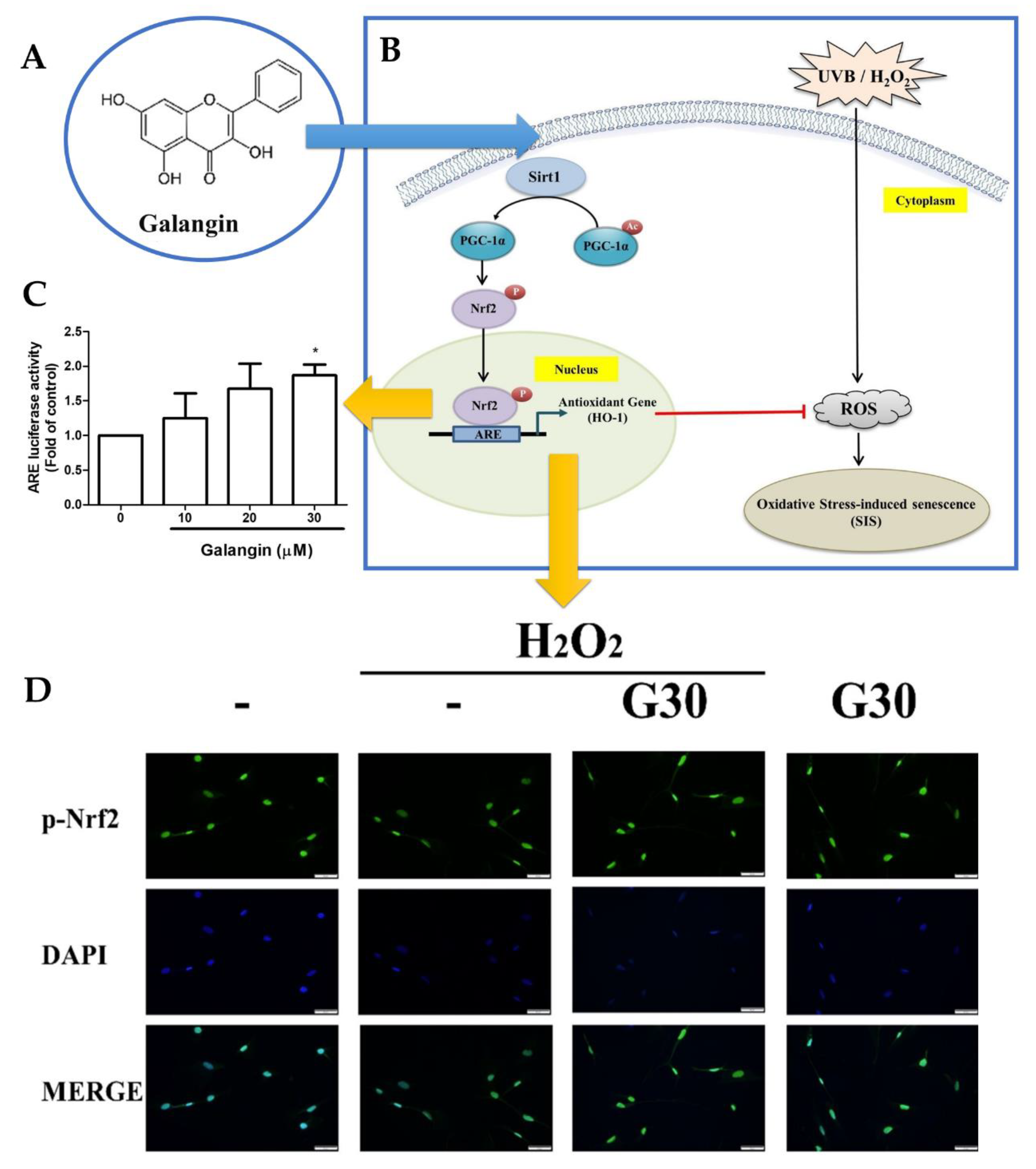
  31. Lee, J.-J.; Ng, S.-C.; Hsu, J.-Y.; Liu, H.; Chen, C.-J.; Huang, C.-Y.; Kuo, W.-W. Galangin Reverses H2O2-Induced Dermal Fibroblast Senescence via SIRT1-PGC-1α/Nrf2 Signaling. Int. J. Mol. Sci. 2022, 23, 1387. [Google Scholar] [CrossRef]
  32. Li, W.; Mu, X.; Wu, X.; He, W.; Liu, Y.; Liu, Y.; Deng, J.; Nie, X. Dendrobium Nobile Lindl. Polysaccharides Protect Fibroblasts against UVA-Induced Photoaging via JNK/c-Jun/MMPs Pathway. J. Ethnopharmacol. 2022, 298, 115590. [Google Scholar] [CrossRef]
  33. Ouyang, Q.; Li, Y.; Mei, S.; Zhang, Q.; Li, X.; Luo, H.; Zhu, Y.; Wu, K. Protective Effects of GLHP from Gracilaria Lemaneiformis against UVB-Induced Photodamage in Human Immortalized Keratinocytes Cells and BALB/c Mice. Exp. Gerontol. 2021, 155, 111550. [Google Scholar] [CrossRef] [PubMed]
  34. Madan, K.; Nanda, S. In-Vitro Evaluation of Antioxidant, Anti-Elastase, Anti-Collagenase, Anti-Hyaluronidase Activities of Safranal and Determination of Its Sun Protection Factor in Skin Photoaging. Bioorg. Chem. 2018, 77, 159–167. [Google Scholar] [CrossRef] [PubMed]
  35. Altyar, A.E.; Ashour, M.L.; Youssef, F.S. Premna Odorata: Seasonal Metabolic Variation in the Essential Oil Composition of Its Leaf and Verification of Its Anti-Ageing Potential via In Vitro Assays and Molecular Modelling. Biomolecules 2020, 10, 879. [Google Scholar] [CrossRef] [PubMed]
  36. Widowati, W.; Ginting, C.N.; Lister, I.N.E.; Girsang, E.; Amalia, A.; Wibowo, S.H.B.; Kusuma, H.; Rizal, R. Anti-Aging Effects of Mangosteen Peel Extract and Its Phytochemical Compounds: Antioxidant Activity, Enzyme Inhibition and Molecular Docking Simulation. Trop. Life Sci. Res. 2020, 31, 127–144. [Google Scholar] [CrossRef]
  37. Abdel Shakour, Z.T.; El-Akad, R.H.; Elshamy, A.I.; El Gendy, A.E.-N.G.; Wessjohann, L.A.; Farag, M.A. Dissection of Moringa Oleifera Leaf Metabolome in Context of Its Different Extracts, Origin and in Relationship to Its Biological Effects as Analysed Using Molecular Networking and Chemometrics. Food Chem. 2023, 399, 133948. [Google Scholar] [CrossRef]
  38. Barak, T.H.; KURT CELEP, İ.; ŞENTÜRK, T.B.; BARDAKCI, H.; CELEP, E. Anti-Aging Potential Evaluation of Maclura Pomifera (Rafin.) Schneider 80% Methanol Extract with Quantitative HPTLC Analysis. Turk J. Pharm. Sci. 2022, 19, 400–407. [Google Scholar] [CrossRef]
  39. Eaknai, W.; Bunwatcharaphansakun, P.; Phungbun, C.; Jantimaporn, A.; Chaisri, S.; Boonrungsiman, S.; Nimmannit, U.; Khongkow, M. Ethanolic Fenugreek Extract: Its Molecular Mechanisms against Skin Aging and the Enhanced Functions by Nanoencapsulation. Pharmaceuticals 2022, 15, 254. [Google Scholar] [CrossRef]
  40. Castejón, N.; Thorarinsdottir, K.A.; Einarsdóttir, R.; Kristbergsson, K.; Marteinsdóttir, G. Exploring the Potential of Icelandic Seaweeds Extracts Produced by Aqueous Pulsed Electric Fields-Assisted Extraction for Cosmetic Applications. Mar. Drugs 2021, 19, 662. [Google Scholar] [CrossRef]
  41. An, J.Y.; Kim, C.; Park, N.R.; Jung, H.S.; Koo, T.; Yuk, S.H.; Lee, E.H.; Cho, S.H. Clinical Anti-aging Efficacy of Propolis Polymeric Nanoparticles Prepared by a Temperature-induced Phase Transition Method. J. Cosmet. Dermatol. 2022, 21, 4060–4071. [Google Scholar] [CrossRef]
  42. Moreira, P.; Sousa, F.J.; Matos, P.; Brites, G.S.; Gonçalves, M.J.; Cavaleiro, C.; Figueirinha, A.; Salgueiro, L.; Batista, M.T.; Branco, P.C.; et al. Chemical Composition and Effect against Skin Alterations of Bioactive Extracts Obtained by the Hydrodistillation of Eucalyptus Globulus Leaves. Pharmaceutics 2022, 14, 561. [Google Scholar] [CrossRef]
  43. El-Nashar, H.A.S.; El-labbad, E.M.; Al-Azzawi, M.A.; Ashmawy, N.S. A New Xanthone Glycoside from Mangifera indica L.: Physicochemical Properties and In Vitro Anti-Skin Aging Activities. Molecules 2022, 27, 2609. [Google Scholar] [CrossRef] [PubMed]
  44. Chaiyana, W.; Charoensup, W.; Sriyab, S.; Punyoyai, C.; Neimkhum, W. Herbal Extracts as Potential Antioxidant, Anti-Aging, Anti-Inflammatory, and Whitening Cosmeceutical Ingredients. Chem. Biodivers 2021, 18, e2100245. [Google Scholar] [CrossRef] [PubMed]
  45. Ebrahim, H.Y.; Mady, M.S.; Atya, H.B.; Ali, S.A.; Elsayed, H.E.; Moharram, F.A. Melaleuca Rugulosa (Link) Craven Tannins: Appraisal of Anti-Inflammatory, Radical Scavenging Activities, and Molecular Modeling Studies. J. Ethnopharmacol. 2022, 298, 115596. [Google Scholar] [CrossRef] [PubMed]
  46. Michalak, M.; Zagórska-Dziok, M.; Klimek-Szczykutowicz, M.; Szopa, A. Phenolic Profile and Comparison of the Antioxidant, Anti-Ageing, Anti-Inflammatory, and Protective Activities of Borago Officinalis Extracts on Skin Cells. Molecules 2023, 28, 868. [Google Scholar] [CrossRef] [PubMed]
  47. Bogdanowicz, P.; Haure, M.-J.; Ceruti, I.; Bessou-Touya, S.; Castex-Rizzi, N. Results from in Vitro and Ex Vivo Skin Aging Models Assessing the Antiglycation and Anti-Elastase MMP-12 Potential of Glycylglycine Oleamide. Clin. Cosmet. Investig. Dermatol. 2016, 9, 143–150. [Google Scholar] [CrossRef]
  48. Chaikhong, K.; Chumpolphant, S.; Rangsinth, P.; Sillapachaiyaporn, C.; Chuchawankul, S.; Tencomnao, T.; Prasansuklab, A. Antioxidant and Anti-Skin Aging Potential of Selected Thai Plants: In Vitro Evaluation and In Silico Target Prediction. Plants 2022, 12, 65. [Google Scholar] [CrossRef]
  49. Ibrahim, N.; Abbas, H.; El-Sayed, N.S.; Gad, H.A. Rosmarinus officinalis L. Hexane Extract: Phytochemical Analysis, Nanoencapsulation, and in Silico, in Vitro, and in Vivo Anti-Photoaging Potential Evaluation. Sci. Rep. 2022, 12, 13102. [Google Scholar] [CrossRef]
  50. Vaithanomsat, P.; Boonlum, N.; Chaiyana, W.; Tima, S.; Anuchapreeda, S.; Trakunjae, C.; Apiwatanapiwat, W.; Janchai, P.; Boondaeng, A.; Nimitkeatkai, H.; et al. Mushroom β-Glucan Recovered from Antler-Type Fruiting Body of Ganoderma Lucidum by Enzymatic Process and Its Potential Biological Activities for Cosmeceutical Applications. Polymers 2022, 14, 4202. [Google Scholar] [CrossRef]
  51. Ndlovu, G.; Fouche, G.; Tselanyane, M.; Cordier, W.; Steenkamp, V. In Vitro Determination of the Anti-Aging Potential of Four Southern African Medicinal Plants. BMC Complement. Altern. Med. 2013, 13, 304. [Google Scholar] [CrossRef]
  52. McCook, J.; Dorogi, P.; Vasily, D.; Cefalo, D. In Vitro Inhibition of Hyaluronidase by Sodium Copper Chlorophyllin Complex and Chlorophyllin Analogs. Clin. Cosmet. Investig. Dermatol. 2015, 8, 443–448. [Google Scholar] [CrossRef]
  53. Murthy, R.; Roos, J.C.P.; Goldberg, R.A. Periocular Hyaluronic Acid Fillers. Curr. Opin. Ophthalmol. 2019, 30, 395–400. [Google Scholar] [CrossRef] [PubMed]
  54. Karamanos, N.K.; Theocharis, A.D.; Piperigkou, Z.; Manou, D.; Passi, A.; Skandalis, S.S.; Vynios, D.H.; Orian-Rousseau, V.; Ricard-Blum, S.; Schmelzer, C.E.H.; et al. A Guide to the Composition and Functions of the Extracellular Matrix. FEBS J. 2021, 288, 6850–6912. [Google Scholar] [CrossRef] [PubMed]
  55. Sklirou, A.D.; Angelopoulou, M.T.; Argyropoulou, A.; Chaita, E.; Boka, V.I.; Cheimonidi, C.; Niforou, K.; Mavrogonatou, E.; Pratsinis, H.; Kalpoutzakis, E.; et al. Phytochemical Study and In Vitro Screening Focusing on the Anti-Aging Features of Various Plants of the Greek Flora. Antioxidants 2021, 10, 1206. [Google Scholar] [CrossRef] [PubMed]
  56. Lee, H.P.; Kim, D.S.; Park, S.H.; Shin, C.Y.; Woo, J.J.; Kim, J.W.; An, R.-B.; Lee, C.; Cho, J.Y. Antioxidant Capacity of Potentilla Paradoxa Nutt. and Its Beneficial Effects Related to Anti-Aging in HaCaT and B16F10 Cells. Plants 2022, 11, 873. [Google Scholar] [CrossRef] [PubMed]
  57. Jdey, A.; Falleh, H.; Ben Jannet, S.; Mkadmini Hammi, K.; Dauvergne, X.; Magné, C.; Ksouri, R. Anti-Aging Activities of Extracts from Tunisian Medicinal Halophytes and Their Aromatic Constituents. EXCLI J. 2017, 16, 755–769. [Google Scholar] [CrossRef]
  58. Majeed, M.; Majeed, S.; Jain, R.; Mundkur, L.; Rajalakshmi, H.; Lad, P.S.; Neupane, P. An Open-Label Single-Arm, Monocentric Study Assessing the Efficacy and Safety of Natural Pterostilbene (Pterocarpus Marsupium) for Skin Brightening and Antiaging Effects. Clin. Cosmet. Investig. Dermatol. 2020, 13, 105–116. [Google Scholar] [CrossRef]
  59. Mostafa, E.S.; Maher, A.; Mostafa, D.A.; Gad, S.S.; Nawwar, M.A.M.; Swilam, N. A Unique Acylated Flavonol Glycoside from Prunus Persica (L.) Var. Florida Prince: A New Solid Lipid Nanoparticle Cosmeceutical Formulation for Skincare. Antioxidants 2021, 10, 436. [Google Scholar] [CrossRef]
  60. Chaikul, P.; Kanlayavattanakul, M.; Somkumnerd, J.; Lourith, N. Phyllanthus emblica L. (Amla) Branch: A Safe and Effective Ingredient against Skin Aging. J. Tradit. Complement. Med. 2021, 11, 390–399. [Google Scholar] [CrossRef]
  61. Saeedi, M.; Eslamifar, M.; Khezri, K. Kojic Acid Applications in Cosmetic and Pharmaceutical Preparations. Biomed. Pharmacother. 2019, 110, 582–593. [Google Scholar] [CrossRef]
  62. Jan, H.; Shah, M.; Andleeb, A.; Faisal, S.; Khattak, A.; Rizwan, M.; Drouet, S.; Hano, C.; Abbasi, B.H. Plant-Based Synthesis of Zinc Oxide Nanoparticles (ZnO-NPs) Using Aqueous Leaf Extract of Aquilegia Pubiflora: Their Antiproliferative Activity against HepG2 Cells Inducing Reactive Oxygen Species and Other In Vitro Properties. Oxid. Med. Cell Longev. 2021, 2021, 1–14. [Google Scholar] [CrossRef]
  63. Hecker, A.; Schellnegger, M.; Hofmann, E.; Luze, H.; Nischwitz, S.P.; Kamolz, L.; Kotzbeck, P. The Impact of Resveratrol on Skin Wound Healing, Scarring, and Aging. Int. Wound J. 2022, 19, 9–28. [Google Scholar] [CrossRef] [PubMed]
  64. Mohd Zaid, N.A.; Sekar, M.; Bonam, S.R.; Gan, S.H.; Lum, P.T.; Begum, M.Y.; Mat Rani, N.N.I.; Vaijanathappa, J.; Wu, Y.S.; Subramaniyan, V.; et al. Promising Natural Products in New Drug Design, Development, and Therapy for Skin Disorders: An Overview of Scientific Evidence and Understanding Their Mechanism of Action. Drug Des. Devel. Ther. 2022, 16, 23–66. [Google Scholar] [CrossRef] [PubMed]
  65. Kammeyer, A.; Luiten, R.M. Oxidation Events and Skin Aging. Ageing Res. Rev. 2015, 21, 16–29. [Google Scholar] [CrossRef] [PubMed]
  66. Gu, Y.; Han, J.; Jiang, C.; Zhang, Y. Biomarkers, Oxidative Stress and Autophagy in Skin Aging. Ageing Res. Rev. 2020, 59, 101036. [Google Scholar] [CrossRef]
  67. Bouzroud, S.; El Maaiden, E.; Sobeh, M.; Merghoub, N.; Boukcim, H.; Kouisni, L.; El Kharrassi, Y. Biotechnological Approaches to Producing Natural Antioxidants: Anti-Ageing and Skin Longevity Prospects. Int. J. Mol. Sci. 2023, 24, 1397. [Google Scholar] [CrossRef]
  68. Dina, E.; Sklirou, A.D.; Chatzigeorgiou, S.; Manola, M.S.; Cheilari, A.; Louka, X.P.; Argyropoulou, A.; Xynos, N.; Skaltsounis, A.-L.; Aligiannis, N.; et al. An Enriched Polyphenolic Extract Obtained from the By-Product of Rosa Damascena Hydrodistillation Activates Antioxidant and Proteostatic Modules. Phytomedicine 2021, 93, 153757. [Google Scholar] [CrossRef]
  69. Vaz, C.; Oliveira, A.; Silva, A.; Cortes, L.; Correia, S.; Ferreira, R.; Breitenfeld, L.; Martinez-de-Oliveira, J.; Palmeira-de-Oliveira, R.; Pereira, C.; et al. Protective Role of Portuguese Natural Mineral Waters on Skin Aging: In Vitro Evaluation of Anti-Senescence and Anti-Oxidant Properties. Int. J. Biometeorol. 2022, 66, 2117–2131. [Google Scholar] [CrossRef]
  70. Yang, S.; Liu, X.; He, J.; Liu, M. Insight into Seasonal Change of Phytochemicals, Antioxidant, and Anti-Aging Activities of Root Bark of Paeonia Suffruticosa (Cortex Moutan) Combined with Multivariate Statistical Analysis. Molecules 2021, 26, 6102. [Google Scholar] [CrossRef]
  71. Wu, Q.; Bai, P.; Guo, H.; Guo, M.S.S.; Xia, Y.; Xia, Y.; Gao, X.; Wang, X.; Wu, J.; Dong, T.T.X.; et al. Capsaicin, a Phytochemical From Chili Pepper, Alleviates the Ultraviolet Irradiation-Induced Decline of Collagen in Dermal Fibroblast via Blocking the Generation of Reactive Oxygen Species. Front. Pharmacol. 2022, 13, 872912. [Google Scholar] [CrossRef]
  72. Kim, H.; Jang, J.; Song, M.J.; Park, C.-H.; Lee, D.H.; Lee, S.-H.; Chung, J.H. Inhibition of Matrix Metalloproteinase Expression by Selective Clearing of Senescent Dermal Fibroblasts Attenuates Ultraviolet-Induced Photoaging. Biomed. Pharmacother. 2022, 150, 113034. [Google Scholar] [CrossRef]
  73. Ellistasari, E.Y.; Kariosentono, H.; Purwanto, B.; Wasita, B.; Riswiyant, R.C.A.; Pamungkasari, E.P.; Soetrisno, S. Exosomes Derived from Secretome Human Umbilical Vein Endothelial Cells (Exo-HUVEC) Ameliorate the Photo-Aging of Skin Fibroblast. Clin. Cosmet. Investig. Dermatol. 2022, 15, 1583–1591. [Google Scholar] [CrossRef] [PubMed]
  74. Donato, A.; Belluzzi, E.; Mattiuzzo, E.; Venerando, R.; Cadamuro, M.; Ruggieri, P.; Vindigni, V.; Brun, P. Anti-Inflammatory and Pro-Regenerative Effects of Hyaluronan-Chitlac Mixture in Human Dermal Fibroblasts: A Skin Ageing Perspective. Polymers 2022, 14, 1817. [Google Scholar] [CrossRef] [PubMed]
  75. Kim, K.M.; Ji, K.-Y.; Choi, Y.J.; Heo, J.B.; Youn, U.J.; Kim, S.; Shim, K.-S.; Lee, J.Y.; Kim, T.S.; Seo, Y.K.; et al. The Safety and Efficacy of 1-Monoeicosapentaenoin Isolated from the Trebouxiophyceae Micractinium on Anti-Wrinkle: A Split-Face Randomized, Double-Blind Placebo-Controlled Clinical Study. J. Clin. Med. 2023, 12, 587. [Google Scholar] [CrossRef] [PubMed]
  76. Jia, H.; Vashisth, M.K.; Ge, Y.; Dai, Q.; He, F.; Wang, X. Anti-Inflammation and Anti-Aging Mechanisms of Mercaptopurine in Vivo and in Vitro. Biochem. Biophys. Res. Commun. 2023, 638, 103–111. [Google Scholar] [CrossRef]
  77. Guimarães, G.R.; Almeida, P.P.; de Oliveira Santos, L.; Rodrigues, L.P.; de Carvalho, J.L.; Boroni, M. Hallmarks of Aging in Macrophages: Consequences to Skin Inflammaging. Cells 2021, 10, 1323. [Google Scholar] [CrossRef] [PubMed]
  78. Zhuang, Y.; Lyga, J. Inflammaging in Skin and Other Tissues—The Roles of Complement System and Macrophage. Inflamm. Allergy-Drug Targets 2014, 13, 153–161. [Google Scholar] [CrossRef]
  79. Fuller, B. Role of PGE-2 and Other Inflammatory Mediators in Skin Aging and Their Inhibition by Topical Natural Anti-Inflammatories. Cosmetics 2019, 6, 6. [Google Scholar] [CrossRef]
  80. Pilkington, S.M.; Bulfone-Paus, S.; Griffiths, C.E.M.; Watson, R.E.B. Inflammaging and the Skin. J. Investig. Dermatol. 2021, 141, 1087–1095. [Google Scholar] [CrossRef]
  81. Zhang, J.-M.; An, J. Cytokines, Inflammation, and Pain. Int. Anesthesiol. Clin. 2007, 45, 27–37. [Google Scholar] [CrossRef]
  82. Pageon, H. Reaction of Glycation and Human Skin: The Effects on the Skin and Its Components, Reconstructed Skin as a Model. Pathol. Biol. 2010, 58, 226–231. [Google Scholar] [CrossRef]
  83. Pageon, H.; Zucchi, H.; Rousset, F.; Monnier, V.M.; Asselineau, D. Skin Aging by Glycation: Lessons from the Reconstructed Skin Model. Clin. Chem. Lab. Med. 2014, 52, 169–174. [Google Scholar] [CrossRef]
  84. Pageon, H.; Bakala, H.; Monnier, V.M.; Asselineau, D. Collagen Glycation Triggers the Formation of Aged Skin in Vitro. Eur. J. Dermatol. 2007, 17, 12–20. [Google Scholar] [CrossRef] [PubMed]
  85. Pageon, H.; Técher, M.-P.; Asselineau, D. Reconstructed Skin Modified by Glycation of the Dermal Equivalent as a Model for Skin Aging and Its Potential Use to Evaluate Anti-Glycation Molecules. Exp. Gerontol. 2008, 43, 584–588. [Google Scholar] [CrossRef] [PubMed]
  86. Pageon, H.; Asselineau, D. An in Vitro Approach to the Chronological Aging of Skin by Glycation of the Collagen: The Biological Effect of Glycation on the Reconstructed Skin Model. Ann. N. Y. Acad. Sci. 2005, 1043, 529–532. [Google Scholar] [CrossRef] [PubMed]
  87. Shin, S.; Lee, J.; Yoon, S.H.; Park, D.; Hwang, J.S.; Jung, E. Anti-glycation Activities of Methyl Gallate in Vitro and in Human Explants. J. Cosmet. Dermatol. 2022, 21, 2602–2609. [Google Scholar] [CrossRef] [PubMed]
  88. Dini, I.; Falanga, D.; Di Lorenzo, R.; Tito, A.; Carotenuto, G.; Zappelli, C.; Grumetto, L.; Sacchi, A.; Laneri, S.; Apone, F. An Extract from Ficus Carica Cell Cultures Works as an Anti-Stress Ingredient for the Skin. Antioxidants 2021, 10, 515. [Google Scholar] [CrossRef]
  89. Leo, T.K.; Tan, E.S.S.; Amini, F.; Rehman, N.; Ng, E.S.C.; Tan, C.K. Effect of Rice (Oryza sativa L.) Ceramides Supplementation on Improving Skin Barrier Functions and Depigmentation: An Open-Label Prospective Study. Nutrients 2022, 14, 2737. [Google Scholar] [CrossRef] [PubMed]
  90. Khan, Z.U.; Khan, T.; Mannan, A.; Ali, A.; Ni, J. In Vitro and Ex Vivo Evaluation of Mangifera indica L. Extract-Loaded Green Nanoparticles in Topical Emulsion against Oxidative Stress and Aging. Biomedicines 2022, 10, 2266. [Google Scholar] [CrossRef]
  91. Kappler, K.; Grothe, T.; Srivastava, S.; Jagtap, M. Evaluation of the Efficacy and Safety of Blue Fenugreek Kale Extract on Skin Health and Aging: In-Vitro and Clinical Evidences. Clin. Cosmet. Investig. Dermatol. 2022, 15, 2051–2064. [Google Scholar] [CrossRef] [PubMed]
  92. Wang, Q.; Hu, F.; Hu, X.; Xie, Y.; Du, L.; Ye, R. The Synergistic Effect of Retinyl Propionate and Hydroxypinacolone Retinoate on Skin Aging. J. Cosmet. Dermatol. 2023. online ahead of print. [Google Scholar] [CrossRef] [PubMed]
  93. Wang, Q.; Yang, X.; Zhu, C.; Liu, G.; Han, W.; Sun, Y.; Qian, L. Valorization of Polysaccharides From Benincasa Hispida: Physicochemical, Moisturizing, and Antioxidant Skincare Properties. Front. Pharmacol. 2022, 13, 912382. [Google Scholar] [CrossRef] [PubMed]
Figure 1. Main skin structure and composition, intrinsic and extrinsic factors that lead to skin aging, and main changes in skin characteristics due to aging (produced with BioRender).
Cosmetics 10 00066 g001
Figure 2. Schematic representation of the assays and corresponding experimental methods that can be used for the assessment of skin anti-aging activity (produced with BioRender).
Cosmetics 10 00066 g002
Figure 3. (A) Effects on HaCaT cell proliferation of different doses of bamboo leaf flavonoids, determined by MTT assay; (B) p53, p21, p16, and Lamin B1 gene expression after treatment with bamboo leaf flavonoids, by quantitative real-time PCR; (C) confocal microscope photographs showing senescence-associated heterochromatin foci formation after treatment with bamboo leaf flavonoids; (D) percentage of senescence-associated heterochromatin foci-positive cells; * p < 0.05, ** p < 0.01; adapted from Gu et al. [30].
Cosmetics 10 00066 g003
Figure 4. (A) Schematic representation of fenugreek extract encapsulation within the developed liponiosomes, and intended effect on the skin; (B) in vitro collagenase inhibition assay results of the fenugreek extract without being encapsulated (“Extract”) and encapsulated within the developed liponiosomes (“LNF”); (C) collagen production after treatment with the fenugreek extract (and controls); * p < 0.05, *** p < 0.001; adapted from Eaknai et al. [39].
Cosmetics 10 00066 g004
Figure 5. (A,B) Schematic representation of the effects of galangin on H2O2 and UVB radiation-induced oxidative damage; (C) NRF2-dependent luciferase activity measurement results after treatment with galangin; (D) NRF2 nuclear translocation determination, acquired by fluorescence microscopy; * p < 0.05; adapted from Lee et al. [31].
Cosmetics 10 00066 g005
Disclaimer/Publisher’s Note: The statements, opinions and data contained in all publications are solely those of the individual author(s) and contributor(s) and not of MDPI and/or the editor(s). MDPI and/or the editor(s) disclaim responsibility for any injury to people or property resulting from any ideas, methods, instructions or products referred to in the content.

