Article

Chemical Characterization and Bioactive Properties of Wine Lees and Diatomaceous Earth towards the Valorization of Underexploited Residues as Potential Cosmeceuticals

by
Cristina N. Duarte
1,2,3,
Oludemi Taofiq
1,2,4,
Maria Inês Dias
1,2,
Sandrina A. Heleno
1,2,
Celestino Santos-Buelga
3,
Lillian Barros
1,2 and
Joana S. Amaral
1,2,*
1
Centro de Investigação de Montanha (CIMO), Instituto Politécnico de Bragança, Campus de Santa Apolónia, 5300-253 Bragança, Portugal
2
Laboratório Associado Para a Sustentabilidade e Tecnologia em Regiões de Montanha (SusTEC), Instituto Politécnico de Bragança, Campus de Santa Apolónia, 5300-253 Bragança, Portugal
3
Grupo de Investigación en Polifenoles (GIP-USAL), Facultad de Farmacia, Universidad de Salamanca, E-37007 Salamanca, Spain
4
Nutrition and Bromatology Group, Department of Analytical Chemistry and Food Science, Faculty of Science, Universidade de Vigo, E-32004 Ourense, Spain
*
Author to whom correspondence should be addressed.
Cosmetics 2023, 10(2), 58; https://doi.org/10.3390/cosmetics10020058
Submission received: 4 March 2023 / Revised: 24 March 2023 / Accepted: 27 March 2023 / Published: 29 March 2023

Abstract

:
Annually, wine production is responsible for generating large quantities of residues, which are frequently disposed of and not valorized. So far, different studies have been conducted on grape pomace, yet less attention has been paid to other residues, such as wine lees and diatomaceous earth used in wine filtration. In this context, this study aimed to evaluate and compare the phenolic profile of these underexploited winemaking residues and assess their biological potential based on their antioxidant, antimicrobial, cytotoxic, and anti-aging activities (inhibition of tyrosinase and collagenase). Twenty-nine phenolic compounds, including twelve anthocyanins, were tentatively identified in the residues, with red grape pomace showing the highest diversity of compounds. The diatomaceous earth presented the highest content of non-anthocyanin phenolic compounds, being particularly rich in flavan-3-ols and myricetin-O-hexoside, and also presenting two anthocyanins. This sample also showed a high antioxidant activity, evidencing the best result in the reducing power assay. The red wine lees extract, despite showing a low content of phenolic compounds and less antioxidant activity, presented the highest inhibition capacity of bacteria growth. The extracts did not exhibit cytotoxicity against keratinocyte (up to 400 μg/mL) and fibroblast (up to 100 μg/mL) skin cell lines. However, the capacity of inhibiting tyrosinase and collagenase was low for the lees and diatomaceous earth, contrary to the grape pomace, seeds, and skins extracts that showed promising results, evidencing its potential as a cosmeceutical. Overall, this study highlights for the first time the potential of diatomaceous earth, an underexploited winemaking waste, in the obtention of added-value extracts and/or ingredients for cosmetic industry.

1. Introduction

Polyphenols are secondary metabolites produced by plants that play important roles in plant systems, such as providing them with protection against different types of attacks, including from parasites, insect pests, ultraviolet (UV) radiation, and other environmental threats [1]. Polyphenols may also have beneficial effects on health as they show different biological activities, including antioxidant, anti-inflammatory, antimutagenic, anticarcinogenic, and antiproliferative properties [2]. One of the most interesting characteristics of polyphenols is their antioxidant activity, owing to their reducing properties and their capacity to neutralize free radicals, such as singlet oxygen, which have been related to the ability to avoid or delay the oxidation of biomolecules (mainly lipids) in biological systems [3,4]. Moreover, the phenolic composition is an important quality parameter in certain foods and drinks, such as wine, contributing to various organoleptic attributes, such as bitterness, astringency, color, flavor, and oxidative stability [5].
Over recent years, different by-products from the food industry have been suggested as potential sources of polyphenols, since a high proportion of interesting bioactive compounds remains in their composition. The winemaking industry is amongst the ones that generate a large number of by-products, mainly grape pomace (consisting of grape skins, seeds, and stems), but also wine lees and other undervalued residues, such as diatomaceous earth used in filtrations. At the end of the winemaking process, residues containing yeasts, bacteria, and organic matter are deposited at the bottom of the wine tanks, corresponding to the lees. To avoid undesirable flavors and aromas, the wine should not remain in contact with these deposits, thus being separated from them [6]. Moreover, to guarantee the desired cleanness and stabilization of the wine, besides transfers, the winemaking industry carries out further operations such as filtration and tartaric stabilization. When properly treated, diatomaceous earth (silica deposits consisting of the accumulation of fossilized shells from microscopic marine algae) yields a material of high porosity non-deformable structure and of a large specific surface. This material is frequently used in wine filtration to eliminate suspended particles, rendering a clear and bright aspect to the wine [7], being an additional residue generated by this industry. While the pomace seeds can be used to produce grapeseed oil and the pomace can further undergo a distillation process or be used as a fertilizer or as animal feed, the stems, lees, and diatomaceous earth are generally discarded and perceived as an environmental problem [8]. However, recent studies suggest that the wine lees can be exploited for their richness in polyphenols, since these compounds can be adsorbed in their colloidal state to the yeast’s cell walls as well as being dissolved in the liquid fraction of the wine lees [9,10]. Nevertheless, so far, only a few studies have advanced beyond the evaluation of the total phenolic compounds and carried out the identification of individual phenolics in wine lees. Most of these works concerned red wine lees [11,12,13,14,15], and only limited information is available on white wine lees [16,17]. Likewise, data on the diatomaceous earth from winemaking residues are very scarce. Recently, the feasibility of applying molecularly imprinted polymers to extracts prepared from winemaking diatomaceous earth residues to obtain quercetin-enriched fractions was demonstrated [18]. However, the study mainly focused on some selected compounds without a detailed phytochemical characterization of this by-product being carried out.
In the framework of the United Nations Sustainable Development Goals (SDGs), particularly SDG12 “Ensure sustainable consumption and production patterns”, and within a circular economy perspective, there is now a considerable interest in changing the paradigm of wine industry wastes by turning them into by-products that other industries can further use. In this sense, grape pomaces from the wine and juice industries have been widely studied as a source of polyphenols with potential nutraceutical properties to be exploited by the food industry [19,20,21]. Furthermore, the use of bioactive compounds extracted from these sources is increasingly common in the cosmetic field, mostly based on their antioxidant capacity. In the human body, oxidative stress occurs when the balance between the generation of reactive oxygen species (ROS) and the antioxidant defense systems is compromised. In the skin, environmental stress, such as ultraviolet light, exacerbates ROS production, resulting in cell damage and degradation of the extracellular matrix proteins such as collagen and elastin, ultimately leading to skin aging. The main changes that take place at the dermal connective tissue level, which are essentially translated into the loss of mature collagen and alterations in the elastic network [22], allow uneven pigmentation of the epidermis to occur with aging due to changes in tyrosinase activity in the melanocytes [23]. Distinct phenolic compounds present in grapes, such as anthocyanins, not only possess renowned antioxidant properties, but can also directly inhibit enzymes involved in the skin aging process—namely, tyrosinase and collagenase [24,25]. For this reason, winemaking by-products can be potential sources of innovative functional ingredients for cosmetics, and more studies are required concerning the so far less studied residues.
This work aimed at evaluating several winemaking by-products, with particular focus on underexploited residues for which there is a scarcity of data and knowledge (wine lees and diatomaceous earth), and aimed to compare those with the more widely studied grape pomace, skins, and seeds. The extracts obtained from the residues have been evaluated for their phenolic compounds profile, their antioxidant properties, and their antimicrobial and anti-fungicidal activity. Moreover, to evaluate their potential as cosmeceutical ingredients, the inhibition of tyrosinase and collagenase, as enzymes involved in the skin aging process, as well as the cytotoxicity in fibroblasts (HFF-1) and keratinocytes (HaCaT) cell lines, have also been assessed.

2. Materials and Methods

2.1. Winemaking Industry by-Products

Winemaking by-products were supplied by Caves Campelo S.A. (Portugal) during the harvest season in September 2021. The seeds and skins were manually separated from the red grape pomace. All the other samples were sent individually. The samples of whole pomace, seeds, skins, and stems from red grapes were arranged on separated trays and dried in an oven (Scientific, Series 9000, Scientific Engineering, Johannesburg, South Africa) at 40 °C for 3 days, and then ground into powder in a mill (IKA, A11 basic) to pass a sieve of 20 mesh. The red lees were sent on a plastic bottle and the separation of phases was visible. Thus, they were placed on falcon tubes and centrifuged at 7871× g for 5 min. The liquid phase was then pipetted into a flask and separated from the solid phase. After eliminating the ethanol by rotoevaporation (Julabo, TW12), the liquid phase was lyophilized (Telstar, LyoQuest—55 Plus). The separated solid phase from red lees as well as the white lees were also submitted to lyophilization. All the samples were stored at −20 °C inside a container with silica, and were protected from light.

2.2. Extraction of Compounds

Phenolic compounds were extracted using ethanol/water 80:20 (v/v). A total of 5 grams of powder of each sample was extracted with 100 mL of solvent by sonication (SONO SWISS, sw1) for 5 min followed by stirring (Lbx instruments, S03 series) for 1 h at ambient temperature and protected from light. The samples were centrifuged (Eppendorf, Centrifuge 5810 R) at 7871× g for 5 min, and the pellet was re-extracted. The supernatants were collected and joined and filtered, and the ethanol (Fisher Scientific, Loughborough, UK) evaporated under vacuum on a rotary evaporator (Julabo, TW12, Julabo Labortechnik, Seelbach, Germany) at 40 °C. Finally, the extracts were lyophilized, and the obtained dry residues were stored at −20 °C in flasks kept inside a container with silica for further use or weighted and redissolved in ethanol/water 80:20 (v/v) to a final concentration of 10 mg/mL.

2.3. Total Phenolic Content

The total phenolic compounds content of the hydroethanolic extracts prepared was determined by spectrophotometry in a microplate reader (Epoch 2, Biotek, BioTek Instruments, Inc., Winooski, VT, USA) using the Folin–Ciocalteu reagent. To obtain the calibration curve, an ethanolic solution of gallic acid was used as a standard, within the range of 0.005–0.5 mg/mL. For analysis, different concentrations of sample extracts were prepared from a stock solution (5 mg/mL) prepared from the lyophilizate. In a test tube, 250 μL of extract from each sample or standard were added with 1.25 mL of Folin-Ciocalteu reagent (1:10 v/v in water) and 1 mL of sodium carbonate (75 g/L). The tubes were vortexed for 15 s and placed to rest in the dark for 30 min at 40 °C for colour development. After this, the tubes were centrifuged at 12,000× g rpm for 2 min and the solution transferred to a 96-well microplate, and the absorbance read at 765 nm [26]. Ethanol was used as blank control. The results were expressed as mg gallic acid equivalents per gram of dry extract (mg GAE/g extract).

2.4. Characterization of Phenolic Compounds by HPLC-DAD-ESI/MSn

Individual phenolic compounds were separated, identified, and quantified by high-performance liquid chromatography coupled to double diode array and tandem mass spectrophotometry detection (HPLC-DAD-ESI-MS/MS). Dry extracts (40 mg) were redissolved in 2 mL of ethanol/water 80:20 (v/v) and filtered through 0.22 μm disposable filter disks. The analysis was carried out in a Dionex Ultimate 3000 UPLC (Thermo Scientific, San Jose, CA, USA) system equipped with a degasser, a quaternary pump, an auto sampler (kept at 5 °C), an automated thermostatically controlled column compartment, and a diode array detector (DAD) coupled to a mass spectrometer with an electrospray ionization (ESI) source.
Chromatographic separation was achieved with a Waters Spherisorb S3 ODS-2C18 (3 μm, 4.6 mm×150 mm, Waters, Mil-ford, MA, USA) column maintained at 35 °C. The solvents used were: (A) 0.1% formic acid in water, and (B) acetonitrile. The elution gradient established was 15% B (5 min), 15% B to 20% B (5 min), 20–25% B (10 min), 25–35% B (10 min), and 35–50% B (10 min), followed by the re-equilibration of the column in a flow rate of 0.5 mL/min. Double online detection was carried out in the DAD using 280, 320, 370, and 520 nm as preferred wavelengths and in a mass spectrometer (MS) connected to HPLC system via the DAD cell outlet. MS detection was performed using a Orbitrap Exploris 120 mass spectrometer (ThermoFinnigan, San Jose, CA, USA) equipped with an ESI source operating in negative or positive mode for non-anthocyanin and anthocyanins, respectively. Nitrogen served as the sheath gas (50 psi), and the system was operated with a spray voltage of 5 kV, a source temperature of 325 °C, and a capillary voltage of −20 V. The tube lens offset was kept at a voltage of −70 V. The full MS and MS2 scans covered the mass range from m/z 100–1200. The collision energy used was 35 (arbitrary units). Data acquisition was carried out with Xcalibur® data system (ThermoFinnigan, San Jose, CA, USA).
The phenolic compounds were identified by comparing their retention times, UV-Vis, and mass spectra with those obtained from standard compounds, when available. Otherwise, compounds were tentatively identified comparing the obtained information with available data reported in the literature. For quantitative analysis, a calibration curve for each available phenolic standard was constructed based on the areas of the peaks recorded at 280 nm, except for flavonols (370 nm) and for anthocyanin compounds (520 nm). For the identified phenolic compounds for which a commercial standard was not available, the quantification was performed through the calibration curve of the most similar available standard. The results were expressed as mg/g of extract (dry weight).

2.5. Antioxidant Activity

Antioxidant activity assays were performed as previously described by Iyda et al. (2019) [26]. The extracts described in 2.1 were re-dissolved in ethanol/water (80:20, v/v) to obtain stock solutions (with various concentrations), which were further diluted to obtain a range of working concentrations to evaluate the antioxidant activity. DPPH radical-scavenging activity was evaluated by using an Epoch 2 microplate reader (BioTek Instruments, Inc., Winooski, VT, USA), and was calculated as a percentage of DPPH discoloration using the formula: [(ADPPH−AS)/ADPPH] × 100, where AS is the absorbance of the sample solution at 515 nm, and ADPPH is the absorbance of the DPPH solution. Reducing power was evaluated by the capacity to convert Fe3+ into Fe2+ by measuring the absorbance at 690 nm, as described in the literature [27], using the same microplate reader. The lipid peroxidation inhibition was evaluated using porcine brain tissue due to its richness in lipids, and the ability of the assay in generating free radicals produced by oxidative stress. Lipid oxidation originates different products, the most common being malonaldehyde (MDA), which is determined based on the reaction with thiobarbituric acid (TBA), generating a complex of pink coloration, the absorbance of which was measured at 532 nm. The results were expressed as EC50 values, corresponding to the sample concentration providing 50% of antioxidant activity or 0.5 of absorbance in the reducing power assay. Trolox was used as positive control.

2.6. Antibacterial Activity

The bacterial strains were clinical isolates obtained from patients hospitalized in various departments at the North-Eastern Local Health Unit (Bragança, Portugal) and Hospital Center of Trás-os-Montes and Alto Douro (Vila Real, Portugal), isolated in a previous work [28]. Five Gram-negative bacteria, namely, Escherichia coli (isolated from urine, VRU12881), Klebsiella pneumoniae (isolated from urine, VRI17214), Proteus mirabilis (isolated from wound exudate, VRI78844), Pseudomonas aeruginosa (isolated from expectoration, VRU14123), and Morganella morganii (isolated from urine, VRU14272), and three Gram-positive bacteria, namely, Enterococcus faecalis (isolated from urine, VRU14041), Listeria monocytogenes (isolated from cerebrospinal fluid, VRU17684), and methicillin-resistant Staphylococcus aureus (MRSA) (isolated from expectoration, VRI17654), were all tested. These microorganisms were selected based on their antibiotic resistance profile previously described [29]. All microorganisms were incubated at 37 °C in appropriate fresh medium during the 24 h before analysis in order to maintain the exponential growth phase. The minimum inhibitory concentration (MIC) determination was conducted based on the colorimetric broth microdilution assay [30]. The samples were first dissolved in 5% (v/v) dimethyl sulfoxide (DMSO) and 95% autoclaved distilled water to give a final concentration of 20 mg/mL for the stock solution. Afterwards, the samples were successively diluted with tryptic soy broth (TSB) culture medium to obtain the concentration ranges of 10 to 0.03125 mg/mL. 10 μL of inoculum (standardized at 1.5 × 10⁶ Colony Forming Unit (CFU)/mL) was then added to each well containing 90 μL of sample dilutions, reaching a final concentration of 1.5 × 10⁵ CFU/mL. Two negative controls were prepared, one with TSB and another with the extract only. Two positive controls were also prepared, one with TSB and each inoculum and another with TSB, antibiotics, and bacteria. Ampicillin was used for all the tested bacteria, and imipenem was used against all the strains, except for E. faecalis and MRSA, in which vancomycin was used. The microplates were incubated at 37 °C for 24 h and the MIC was determined following the addition (40 μL) of 0.2 mg/mL p-iodonitrotetrazolium chloride (INT) and incubation at 37 °C for 30 min. The MIC was defined as the lowest concentration that inhibits the visible bacterial growth determined by color change from yellow to pink if the microorganisms are viable. For the minimum bactericidal concentration (MBC) determination, 10 μL from each well that showed no change in color was plated on Blood agar (7% sheep blood) solid medium and incubated at 37 °C for 24 h. The lowest concentration that yielded no growth was established as the MBC. The MIC and MBC results are expressed in mg/mL.

2.7. Antifungal Activity

The antifungal activity was determined against Aspergillus fumigatus (ATCC 204305) and Aspergillus brasiliensis (ATCC 16404) according to the methodology described in the literature [31]. The micromycetes were maintained on malt agar and the cultures stored at 4 °C. Before the assay, they were further placed in a new medium and incubated at 25 °C for 72 h. To investigate the antifungal activity, the fungal spores were washed from the surface of agar plates with sterile 0.85% saline containing 0.1% Tween 80 (v/v). The spore suspension was adjusted with sterile saline to a concentration of approximately 1.0 × 10⁵ in a final volume of 100 μL per well. The extracts were first dissolved in 5% (v/v) dimethyl sulfoxide (DMSO) and 95% of autoclaved distilled water to give a final concentration of 20 mg/mL for the stock solution. Afterwards, 90 μL of this concentration was added in the first well (96-well microplate) in duplicate with 100 μL of Malt Extract Broth (MEB). The remaining wells were added with 90 μL of MEB, and then the samples were successively diluted to obtain a concentration range from 10 to 0.03125 mg/mL. The MIC was established as the lowest concentration without visible growth (at the binocular microscope). The minimum fungicidal concentration (MFC) was determined by the sub-cultivation of 2 μL of each well that presented no visible growth into microplates containing 100 μL of MEB per well, and then further incubation 72 h at 26 °C. The lowest concentration with no visible growth was defined as MFC, indicating a 99.5% killing of the original inoculum. Commercial fungicide ketoconazole (Frilabo, Porto, Portugal) was used as positive control. The MIC and MFC were expressed in mg/mL.

2.8. Cytotoxicity Assay in Skin Cell Lines

In vitro spontaneously transformed keratinocytes from histologically normal skin (HaCaT) and fibroblast from human foreskin (HFF-1) cells were cultured in Dulbecco’s modified Eagle’s medium containing 10% fetal bovine serum (FBS), 100 U/mL penicillin, and 100 μg/mL streptomycin. The cryopreserved culture of HaCaT was purchased from Cell Line Service (Germany), while HFF-1 was purchased from ATCC. Cells were routinely sub-cultured in 75 cm2 flasks in humidified 5% CO2 atmosphere at 37 °C [32]. After cultured cells reached the appropriate confluence, the culture medium was removed from the culture plate, washed twice with Hank’s Balanced Salt Solution (HBSS), and then trypsinised using 0.25% trypsin/0.53 mM EDTA, and cells were suspended in a fresh medium and seeded into 96-well plates at 2 × 10⁴ cells/well for 24 h. The cytotoxic effect of the extract was investigated by using Sulforhodamine B (SRB). Briefly, different concentrations of extracts were added to each well, and the control wells were exposed to Triton X-100 at a final concentration of 1% (w/v). After the 48 h incubation period, an ice-cold solution of trichloroacetic acid (TCA) (10%, w/v) was added to each well, followed by incubation for 1 h at 4 °C. Afterwards, the microplates were washed with water and dried at room temperature. A solution of SRB (0.057%, w/v) was then added to each well. Wells were washed three times with an acetic acid solution (1%, v/v) and left to dry at room temperature. Finally, the SRB was solubilised with Tris (10 mM, 200 μL), the absorbance was measured at 540 nm, and the results were expressed as the percentage of cell viability.

2.9. Tyrosinase Inhibitory Activity

The tyrosinase enzyme inhibition activity was evaluated using L-3,4-dihydroxyphenylalanine (L-DOPA) as a substrate with a concentration of 5 mM and 3-methyl-2-benzothiazolinone-hydrazonehydrochloride (MBTH) with a concentration of 20.7 mM as a chromogenic stabilizing agent in 96-well microplates, following the procedure previously described [33,34]. Briefly, test samples (10 μL at 10 mg/mL), phosphate buffer (pH 7.1, 0.1 M), L-DOPA (60 μL), and MBTH (87 μL) were mixed and pre-incubated at 25 °C for 10 min. Subsequently, 6 μL of mushroom tyrosinase enzyme (142 Units of enzyme/mL) were added to each well and the plates were incubated for 30 min at 25 °C. The blank was made with a phosphate buffer (pH 7.1, 0.1 M) and butylresorcinol was used as positive control. The formation of the dopaquinone-MBTH complex was evaluated at 505 nm, using a microplate spectrophotometer (SPECTROstar Nano Multi-Detection Microplate Reader; BMG Labtech, Ortenberg, Germany). The percentage of tyrosinase enzyme inhibition was calculated using the following equation:
% tyrosinase inhibition = ((Absorbance Blank − Absorbance Sample)/(Absorbance Blank)) × 100

2.10. Collagenase Activity Colorimetric Assay

The collagenase inhibition assay was performed using the colorimetric assay kit from Sigma-Aldrich (MAK293) and according to manufacturer’s instructions. The eight samples of the extracts from the winemaking residues were prepared with ultrapure water to reach a concentration of 5 mg/mL. A volume of 10 μL of each sample (50 μg of extract) was placed in a 96 well plate in addition with 80 μL of collagenase assay buffer and 10 μL of the provided enzyme (collagenase 0.35 U/mL). For positive control, 10 μL of enzyme only (0.35 U/mL) was used. For inhibitor control, a volume of 10 μL of provided collagenase (0.35 U/mL) and 2 μL of inhibitor (1,10-phenanthroline) was placed into desired wells. The volume of positive control and inhibitor control wells were adjusted to 100 μL with collagenase assay buffer. The plate was incubated at 37 °C for 10 min. A mixture containing 60 μL of collagenase assay buffer and 40 μL of collagenase substrate FALGPA (N-(3-[2-furyl]acryloyl)-L-leucylglycyl-L-prolyl-L-alanine), was prepared and 100 μL of it was added into the desired wells. Subsequently, the absorbance was read at 345 nm in a microplate reader (Epoch 2, BioTek Instruments, Inc., USA) for 1 h. The percentage of collagenase enzyme inhibition was calculated using the following equation:
% collagenase inhibition = ((Activity enzyme − Activity inhibitor)/(Activity enzyme)) × 100
with the enzyme activity being calculated as indicated by the manufacturer––namely, by taking the absorbance (A1 and A2) at two time points (T1 and T2) in the linear range with at least two readings in between and at least 1 min apart. Therefore, T1 was considered the first absorbance reading and T2 was established at a 10 min reading.

2.11. Statistical Analysis

For the eight samples evaluated, all the assays were carried out in triplicate and the results were expressed as mean values and standard deviation (SD). Statistical analysis was performed using one way analysis of variance (ANOVA) followed by Tukey’s HSD Test with p = 0.05. When necessary, a Student’s t-test was used to determine the significant difference between less than three different samples, with p = 0.05. These analyses were carried out using IBM SPSS Statistics for Windows, Version 23.0. (IBM Corp., Armonk, NY, USA).

3. Results and Discussion

3.1. Chemical Characterization

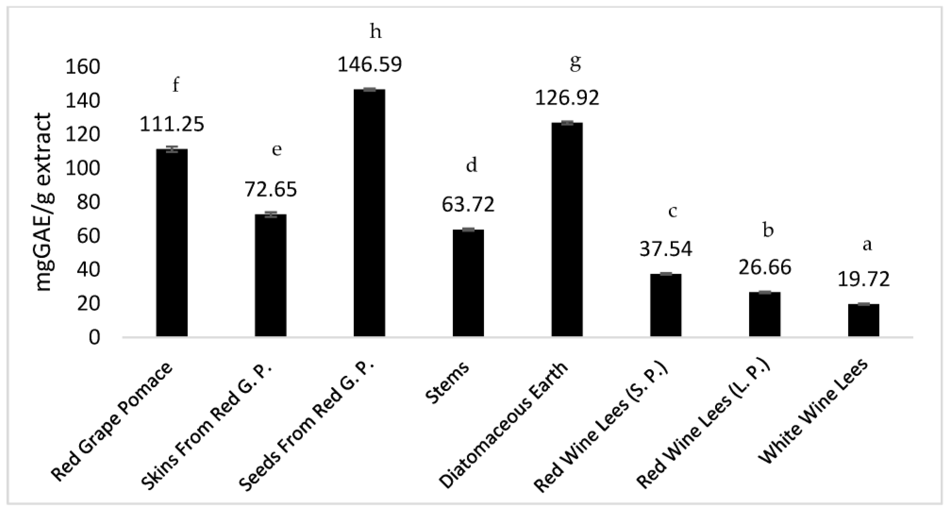
3.1.1. Total Phenolic Compounds

Several works reported on the phenolic composition of different winemaking residues, either regarding their total content in phenolic compounds or by identifying and quantifying the respective individual compounds. However, most focused on grape pomace, particularly from red grape cultivars [35,36,37]. In this work, besides red wine pomace, other less studied residues were also evaluated––namely, wine lees and diatomaceous earth from wine filtration. The wine lees from red wine production were supplied by the industry separated into two phases (solid and liquid phases), which were individually assessed. As far as we know, this is the first report on the chemical characterization and bioactive properties of extracts obtained from winemaking diatomaceous earth residue.
Considering the potential interest as a screening assay for the industry, allowing us to estimate which batches of residues can be more promising to be exploited, all extracts were evaluated for its total phenolic content (TPC) based on the reaction with Folin–Ciocalteu. The results for TPC are presented in Figure 1, showing that the highest content was observed for the extracts of grape seeds from red wine pomace, diatomaceous earth, and red wine pomace with values of 146.6 ± 0.6, 111 ± 2, and 126.9 ± 0.9 mg GAE/g extract, respectively. Interestingly, the results suggest that diatomaceous earth is a very promising residue, since it presented a TPC content even higher than the red wine pomace. On the contrary, the extracts from wine lees showed the lowest contents, particularly the one from white wine production (19.7 ± 0.4 mg GAE/g extract).
The obtained results of TPC are consistent with values previously reported for red wine grape pomace by Maicas et al. (between 55.5 to 153.8 mg GAE/g) [38] and by Matos et al. (83.9 ± 2.0 mg GAE/g extract) [39]. Higher values (254 ± 24 mg GAE/g dry extract) were obtained in other studies [40] when evaluating wine lees from red wine production, extracted with a similar solvent as in the current study here (ethanol:water 75:25). However, this can be related to the fact that the lees sample was obtained after the wine had been aged for 12 months in American oak barrels, while the red wine lees herein analyzed were obtained after the fermentation phase (7 to 10 days) of a red wine produced in stainless steel tanks.

3.1.2. Phenolic Compounds Profile

Twenty-nine phenolic compounds (non-anthocyanins and anthocyanins) were identified or tentatively identified in the ethanol/water (80:20, v/v) extracts prepared from the winemaking residues studied in this work––namely, whole pomace, seeds, skins, stems from red wine production, red and white wine lees, and diatomaceous earth used in red wine production (Table 1). Seventeen non-anthocyanin phenolic compounds were tentatively identified: one phenolic acid (p-coumaric acid hexoside), six flavan-3-ols ((epi)catechin derivatives), four flavonols (O-glycosylated myricetin, and quercetin derivatives), three dihydroflavonols (taxifolin hexosides), two resveratrol derivatives (resveratrol tetramer and trans-resveratrol), and ethyl-gallate. In addition, twelve anthocyanins were also identified, including O-glycosylated and acylated derivatives of delphinidin, cyanidin, malvidin, petunidin, and peonidin.
Most of the detected compounds have been previously described in grape pomace residues from winemaking and other winery byproducts [35,41,42,43,44]. Peaks 4 and 9 ([M-H] at m/z 289) were identified as (+)-catechin and (-)-epicatechin, respectively, by comparing their retention time and UV spectra with the available standard compounds. Taxifolin and myricetin aglycones were previously reported in red wine samples from China [45], but, in the present work, these two compounds were found to be linked to sugar moieties. Taxifolin derivatives, peaks 6, 7, and 8, all with a deprotonated ion [M-H] at m/z 465, presented a unique MS2 fragment at m/z 303 (taxifolin aglycone), corresponding to the loss of 162 u (hexosyl moiety) and thus being tentatively assigned as taxifolin-O-hexosides. Peak 11 ([M-H] at m/z 479), with an MS2 fragment at m/z 317 (-162 u; myricetin aglycone), was tentatively identified as myricetin-O-hexoside. Peak 15 ([M-H]- at m/z 521) also released an MS2 fragment at m/z 317 from the loss of an acetyl-hexoside unit (42 u + 162 u) that allowed its identification as myricetin-O-acetyl-hexoside. Peak 12 presented a deprotonated [M-H] ion at m/z 197 and MS2 fragment ions at m/z 169 ([M-H-28]; gallic acid) and at m/z 125 ([M-H-28-44]) that corresponded to the successive loss of ethyl (28 u) and CO2 residues, thus tentatively identified as ethyl gallate. This compound has been previously reported in wine and grape pomace samples [45,46,47]. In this case, its quantification was not carried out due to a lack of similar standard compounds in our research lab that could be used to construct the calibration curve. Peaks 16 ([M-H] at m/z 905) and 17 ([M-H] at m/z 227) were tentatively identified as stilbene type-compounds––namely, a resveratrol tetramer (cis) and trans-resveratrol, respectively. This tentative identification was performed comparing the obtained results with mass fragmentation data reported in the literature for those compounds [48,49].
Anthocyanins were identified based on their chromatographic and mass information, in agreement with the data reported in the literature [35,50,51,52,53,54]. The presence of acylated anthocyanins in the winemaking residues is particularly interesting, as they can have greater color stability when exposed to light compared with non-acylated anthocyanins [48,49], thereby making them more suitable to be used as food colorants.
Table 2 presents the quantification (mg/g dry extract) of the non-anthocyanin and anthocyanin phenolic compounds found in all the studied samples. The diatomaceous earth and the red wine pomace presented the highest amounts of non-anthocyanin phenolic compounds, which is in good agreement with the data presented in Figure 1. Moreover, the one of diatomaceous earth was also the richer extract in (-)-epicatechin, showing an amount significantly higher of this flavan-3-ol than the remaining extracts. Only the stems and the seeds from red wine pomace revealed the presence of stilbenes, namely, resveratrol tetramer and trans-resveratrol, respectively, and yet only in trace amounts below the level of quantification. For the white wine lees, only two phenolic compounds were identified, although it was the sample that presented the highest amount of (+)-catechin.
In general, the non-anthocyanin and anthocyanin phenolic composition was in accordance with what previously reported in red wine pomaces [35,54]. Interestingly, peonidin 3-O-p-coumarylglucoside and malvidin 3-O-p-coumarylglucoside were also detected in the diatomaceous earth extract, which was also the sample that presented the highest and third highest level of these two compounds, respectively. It should be noticed that the samples supplied by the Caves Campelo S.A. company were obtained as part of their normal routines and seasonal production. Therefore, the sample of diatomaceous earth supplied corresponded to the final discarded residue, which at industry level corresponds to around four tons that were used to filtrate several batches of wine (obtained from different cultivars, geographical origins, etc., with the requisite of all filtrated wines being red or all being white). For this reason, a direct comparison of the composition of the pomace and that of the diatomaceous earth is not possible with the obtained results. Nevertheless, for the first time, these results highlight this residue as being a potential source of phenolic compounds that otherwise would just be discarded as a waste. The high content of polyphenols verified for the diatomaceous earth sample suggest that the compounds become adsorbed in the silicon atoms during wine filtration, which can afterwards be desorbed with an adequate solvent, allowing extracts to be obtained with a high phenolic concentration.

3.2. Bioactive Properties

3.2.1. Antioxidant Activity

Table 3 shows the results of the antioxidant activity of the extracts obtained from the winemaking residues re-dissolved in ethanol/water (80:20) at different concentrations. Overall, the extracts of grape seeds, red grape pomace, and diatomaceous earth gave the lowest EC50 values (meaning the highest antioxidant activity) in all of the assays performed––namely, DPPH scavenging activity, reducing power, and TBARS inhibition. For the TBARS assay, the sample of skins from red grape pomace also performed very well, showing the best activity with an EC50 value of 0.018 mg/mL. The wine lees from red and white wine revealed the highest values (i.e., lowest antioxidant activity), yet still presented an interesting activity since all showed EC50 lower than 1 mg/mL. In general, the results were in good agreement with those obtained for the phenolic content, since the wine lees were also the samples evidencing the lowest contents of phenolic compounds (Figure 1, Table 2). Compared to the herein obtained results, Maluf et al. [54] reported a lower EC50 (6.9 μg/mL) in the DPPH assay for a grape pomace extract. However, the extract was obtained with a different solvent (75% acetone/water) and from a different grape species (Vitis lambrusca), thus resulting in different concentrations of phenolic compounds (69.830 mg EAG/g). Recently, the antioxidant activity of red wine lees extracted with 50% methanol/water was investigated by Lopez-Fernandez et al. [55] that reported an EC50 of 0.0129 mg/mL in DPPH assay, but their sample also presented a higher content of total phenolic compounds (148.03 ± 0.48 mg GAE/g). Greater EC50 than ours was reported in the literature [30] for the skins obtained from red grape pomace (0.563 mg/mL), while a lower value was observed for the seeds (0.023 mg/mL).

3.2.2. Antimicrobial Assays

The results of the antibacterial and antifungal activities are presented in Table 4 and Table 5, respectively. None of the extracts showed bactericidal activity at the tested concentrations. However, for all the bacteria, bacteriostatic activity was observed for at least one of the assayed samples. In general, the extracts were more effective against Gram-positive than Gram-negative bacteria, with the lowest MIC values being obtained for the red wine lees extracts against MRSA (Methicillin-resistant Staphylococcus aureus). Stronger activity against Gram-positive bacteria was also reported [56] when testing extracts obtained from peels, seeds, and stems of two red grape cultivars. Interestingly, the lowest MIC values were generally observed for the extracts of the red wine lees, which together with the extract from white wine lees, were able to inhibit the growth of all tested bacteria. On the other hand, the grape stems presented the weakest activity, since they showed no inhibition of Gram-negative bacteria, despite its activity against Gram-positive strains, particularly L. monocytogenes (MIC = 5 mg/mL).
The results obtained for the antifungal activity were similar to the antibacterial ones, since none of the extracts demonstrated fungicidal potential and most extracts showed only a fungistatic activity at the highest tested concentration (10 mg/mL).

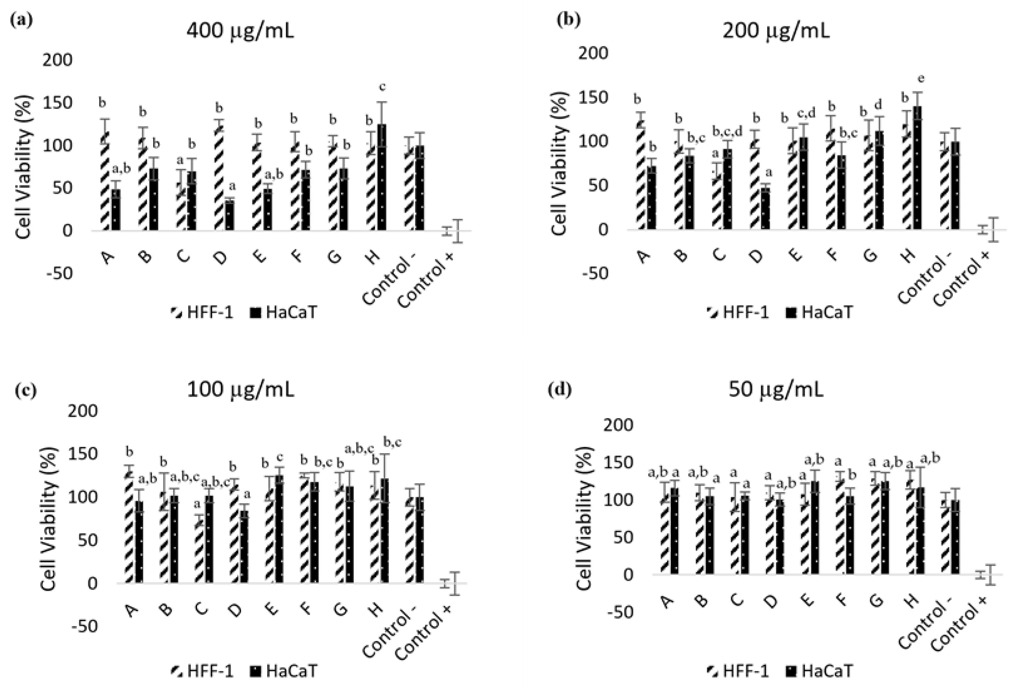
3.2.3. Cytotoxicity Assay in Skin Cell Lines

Due to the possible interest of the studied residues as a source of multifunctional bioactive molecules for cosmetic formulation development, the cell viability effect of the prepared extracts in HaCaT (keratinocytes) and HFF-1 (fibroblast) skin cell lines was also evaluated. As shown in Figure 2, Triton-X was utilised as a positive control to induce a decrease in cell viability. In contrast, cell culture media (DMEM) was used as the negative control, maintaining 100% cell viability. Keratinocytes are a biologically relevant target for skin irritants because they are the first layer of skin cells that come in contact with topically applied compounds. HaCaT cells also present several morphological and functional features typical of normal epidermal keratinocytes, making them a good model for skin toxicity tests [35]. By comprising a mixture of different compounds, the hydroethanolic extracts from winemaking residues may potentiate the development of reactions (allergic or irritant) when applied for dermatologic purposes, thus imposing the need to perform a cytotoxicity evaluation in skin cells and access the minimum concentration, reducing the viability of the tested cell lines. Regarding HaCaT cells, after exposure to the eight extracts (Figure 2), up to 100% cell viability was maintained at 50 μg/mL, while the highest tested concentration (400 μg/mL) was found to significantly inhibit cell viability, particularly for the red wine lees and diatomaceous earth extracts. Concerning HFF-1 cells, the cell viability was maintained up to 100% at 50 μg/mL and more than 100% viability was also observed for all the tested extracts at the highest concentration (400 μg/mL), with one exception (sample C, white wine lees). Therefore, it can be concluded that, in general, all the extracts did not show evidence of affecting the viability of HFF-1 cells. Similar results were observed in previous studies [39] that reported a percentage of cell viability around 100% when HaCaT cells were incubated with 2 mg/mL of Port wine lees extract, and 0.5 mg/mL of red wine lees extract, and when HFF cells were treated with 1 mg/mL and 0.5 mg/mL of the same extracts, respectively.

3.3. Anti-Tyrosinase and Anti-Ageing Activities

3.3.1. Tyrosinase Inhibition

With aging, pigmentation disorders tend to appear on the skin, which has attracted the attention of cosmetic industries and led to the search for compounds with anti-hyperpigmentation potential. These pigmented lesions are caused by alterations resulting in melanin accumulation. Therefore, inhibiting melanin production is the most explored approach in this field. Because tyrosinase is the enzyme limiting the synthesis of melanin, it is a promising target for the development of skin-whitening cosmetic products. Due to their aromatic structural characteristics, phenolic compounds may have some similarities with tyrosine, the tyrosinase substrate that initiates melanin synthesis. Therefore, phenolic compounds have been described as potential tyrosine analogues that can act as competitive inhibitors of the enzyme [57].
The results obtained for the tyrosinase inhibition assay are presented in Table 6 as the percentage of enzyme inhibition. The extract that showed the most promising result was that prepared from the seeds (45.3%). On the other hand, the red lees showed negligible activity, while the white wine lees and the diatomaceous earth did not show any inhibitory activity against tyrosinase enzyme. The inhibitory action of grape pomace extract on tyrosinase enzyme was also reported [39], which demonstrated that the microwave-treated lees extract presented the best inhibitory effect (50% inhibition at 0.14 mg/mL). Red and white grape stems (1 mg/mL) were also reported to inhibit tyrosinase enzyme, with % inhibition ranging from 41.47% and 53.83% for all grape stem extracts [58]. Phenolic compounds such as those belonging to chalcones, simple phenols, and hydroxystilbenes families have been referred in the literature as effective tyrosinase inhibitors [59]. However, in this work, it was not always the extracts with the highest content in non-anthocyanin phenolic and anthocyanin phenolic compounds that translated into better tyrosinase inhibition, such as in the case of diatomaceous earth. This indicates that other biomolecules present in the extract might be the most important contributors to this effect. Butylresorcinol is a well-known inhibitor of tyrosinase enzyme that has presented better effectiveness when compared to kojic acid, arbutin, and hydroquinone [60]. In the present work, butylresorcinol presented 100% inhibition of tyrosinase enzyme at 36.52 ± 2.85 μg/mL. Overall, these findings suggest that these winemaking by-products can be suitable raw materials for recovering biomolecules with potent anti-tyrosinase activity.

3.3.2. Collagenase Inhibition

Collagenase is an enzyme in the matrix metalloproteinase family that breaks down collagen, assisting in the degradation of the extracellular matrix, a key step in the skin aging process. Collagenase has been used to treat Dupuytren’s contracture, a disease characterized by the thickening of connective tissue. In this study, a collagenase activity assay kit was used, providing a quick and easy way to measure the inhibition of collagenase activity by the winemaking residues based on the use of a synthetic peptide (FALGPA) that mimics collagen’s structure. The obtained results are shown in Table 6. The red grape pomace, the skins, the seeds, and the stems were the extracts that evidenced a higher percentage of collagenase inhibition. Hydroethanolic extract (600 μg/mL) from grape seed, abundant in phenolic compounds, also presented 100% inhibition of collagenase enzyme in a previous study [15,61]. The grape pomace extract with abundant anthocyanin phenolic compounds, such as delphidin-3-O-glucoside, malvidin-3-O-glucoside, and malvidin-3-O-acetylglucoside, presented the best collagenase inhibitory effect. In this assay, the diatomaceous earth also presented some activity, but it showed a much weaker performance compared to the pomace extracts. Both the white and red wine lees did not exhibit any activity. However, a previous study proved that both conventional and microwave-pretreated red wine lees extract presented 50% inhibition of MMP-1 activity [39]. MMP-1 is a collagenase enzyme and its inhibition is vital in preventing extracellular matrix degradation and restoring aging skin. These results showed that these wine by-products could be explored as raw materials to scale up the recovery of structurally diverse biomolecules with a broad spectrum of biological activities relevant to skin care, in contrast to the current disposal approach.

4. Conclusions

Grape pomace has been widely studied concerning its chemical composition and bioactivity, since there is considerable interest from the food, cosmetic, and pharmaceutical industries to turn this by-product rich in bioactive compounds into innovative ingredients. However, other residues from the winemaking process, namely, the wine lees and the diatomaceous earth used for wine filtration, are less studied and are underexploited products. In this study, both of these residues demonstrated that they are potential sources of phenolic compounds, with diatomaceous earth being rich in flavan-3-ols and presenting the highest amount of epicatechin among all of the extracts. This extract also showed the highest amount of non-anthocyanin phenolic compounds, mainly due to its relatively high content in myricetin-O-hexoside. As expected, the red grape pomace exhibited the highest amounts of anthocyanins, mainly due to the presence of malvidin derivatives. Interestingly, a few anthocyanins were also found in the diatomaceous earth.
Together with the extracts from the seeds and red pomace, the diatomaceous earth extract also revealed a promising antioxidant activity, since it presented the best results in the reducing power assay. The lees extract, particularly the red ones, showed the highest capacity to inhibit bacterial growth, presenting a bacteriostatic effect against all tested bacteria. In general, the absence of toxicity in HFF-1 cells was observed at the highest tested concentration of extracts. However, some toxicity effects were observed in the HaCaT lines for the highest concentrations of the lees and diatomaceous earth extracts. These extracts also presented the least inhibitory effect against tyrosinase and collagenase enzymes. Therefore, while the extracts from grape pomace, seeds, skins, and stems presented results that suggest their interest as potential ingredients in cosmetic formulations (due to their antioxidant properties and their inhibition of enzymes associated with skin pigmentation and loss of firmness), the extracts of diatomaceous earth and wine lees did not perform as well, since their results suggest a possible cytotoxicity in keratinocytes at high concentrations. Even so, these undervalued residues showed a high potential for producing extracts with high antioxidant properties that can be further exploited by other industries, or for the isolation of added-value compounds, such as anthocyanins, that can be used as natural colorants. For their potential use as cosmetic ingredients, further studies are needed that consider the purification of the extracts in view of decreasing/eliminating the potential cytotoxic effect on keratinocytes.
This work reveals that the wine lees and diatomaceous earth, which are generally discarded and considered by the wine industry as waste, can be valorized as potential sources of bioactive molecules.

Author Contributions

Conceptualization, J.S.A., L.B. and C.S.-B.; methodology, C.N.D., O.T. and M.I.D.; formal analysis, S.A.H., L.B., C.S.-B. and J.S.A.; writing—original draft preparation, C.N.D., O.T. and M.I.D.; writing—review and editing, J.S.A., C.S.-B. and L.B.; supervision, J.S.A., L.B. and C.S.-B.; funding acquisition, J.S.A. and L.B. All authors have read and agreed to the published version of the manuscript.

Funding

This work was funded by “BacchusTech–Integrated Approach for the Valorization of Winemaking Residues” (POCI-01-0247-FEDER-069583), supported by the Competitiveness and Internationalization Operational Program (COMPETE 2020), under the PORTUGAL 2020 Partnership Agreement, through the European Regional Development Fund (ERDF). The authors are grateful to the Foundation for Science and Technology (FCT, Portugal) for financial support through national funds FCT/MCTES (PIDDAC) to CIMO (UIDB/00690/2020 and UIDP/00690/2020) and SusTEC (LA/P/0007/2021), and national funding by FCT, P.I., through the institutional scientific employment program-contract for L. Barros and M.I. Dias, and to COST Action 20133 FULLRECO4US, supported by COST (European Cooperation in Science and Technology). This work is also supported by MICINN for the Juan de la Cierva Formation contract of T. Oludemi (FJC2019-042549-I). The GIP-USAL is indebted to Junta de Castilla y León (Spain) for financial support through the Project SA093P20.

Institutional Review Board Statement

Not applicable.

Informed Consent Statement

Not applicable.

Data Availability Statement

Data available on request.

Acknowledgments

To Óscar Lima (Caves Campelo S.A.) for supplying the winemaking residues used in this work.

Conflicts of Interest

The authors declare no conflict of interest.

References

  1. Troncozo, M.I.; Liesevic, M.; Beskoski, V.; Andelkovic, B.; Ballati, P.; Saparrat, M.C.N. Fungal Transformation and Reduction of Phytotoxicity of Grape Pomace Waste. Chemosphere 2019, 237, 124458. [Google Scholar] [CrossRef]
  2. Gomes, C.; Sadoyan, G.; Dias, R.C.S.; Costa, M.R.P.F.N. Development of Moleculary Imprinted Polymers to Target Polyphenols Present in Plant Extracts. Processes 2017, 5, 72. [Google Scholar] [CrossRef] [Green Version]
  3. Anastasiadi, M.; Pratsinis, H.; Kletsas, D.; Skaltsounis, A.L.; Haroutounian, S.A. Bioactive non-coloured Polyphenols Content of Grapes, Wine and Vinification by-products: Evaluation of the Antioxidant Activities of their Extracts. Food Res. Int. 2010, 43, 805–813. [Google Scholar] [CrossRef]
  4. Gerogiannaki-Christopoulou, M.; Kyriakidis, N.V.; Athanasopoulos, P.E. Effect of Grape Variety (Vitis vinifera L.) and Grape Pomace Fermentation Conditions on some Volatile Compounds of the Produced Grape Pomace Distillate. J. Int. Des Scien. De La Vigne Et Du Vin 2004, 38, 225–230. [Google Scholar] [CrossRef] [Green Version]
  5. Fontana, A.R.; Bottini, R. High-throughput Method Based on Quick, Easy, Cheap, Effective, Rugged and Safe Followed by Liquid Chromatography-multi-wavelength Detection for the Quantification of Multiclass Polyphenols in Wines. J. Chrom. A 2014, 1342, 44–53. [Google Scholar] [CrossRef]
  6. Tao, Y.; Wu, D.; Qing-An, Z.; Da-Wen, S. Ultrasound-assisted Extraction of Phenolics from Wine Lees: Modeling, Optimization and Stability of Extracts During Storage. Ultra. Sonochem. 2014, 21, 706–715. [Google Scholar] [CrossRef]
  7. Abe, L.T.; Vieira da Mota, R.; Lajolo, F.M.; Genovese, M.I. Phenolic Compounds and Antioxidant Activity of Vitis labrusca and Vitis vinifera cultivars. Food Scien. Tech. 2007, 27, 394–400. [Google Scholar] [CrossRef] [Green Version]
  8. Da Silva, F.R.T. Caracterização e Valorização Energética de Resíduos da Poda da Vinha. Master’s Thesis, Instituto Superior de Engenharia do Porto, Porto, Portugal, 2017. Available online: https://recipp.ipp.pt/bitstream/10400.22/11684/1/DM_FilipaSilva_2017_MES.pdf (accessed on 14 December 2022). (In Portuguese).
  9. Guchu, E.; Ebeler, S.E.; Lee, J.; Mitchell, A.E. Monitoring Selected Monomeric Polyphenol Composition in pre- and post- fermentation products of Vitis vinifera L. cv. Airén and cv. Grenache noir. LWT—Food Scien. Tech. 2015, 60, 552–562. [Google Scholar] [CrossRef] [Green Version]
  10. Ricci, A.; Mejia, J.A.A.; Versari, A.; Chiarello, E.; Bordoni, A.; Parpinello, G.P. Microencapsulation of Polyphenolic Compounds Recovered from Red Wine Lees: Process Optimization and Nutraceutical Study. Food Bioprod. Process. 2022, 132, 1–12. [Google Scholar] [CrossRef]
  11. Delgado de la Torre, M.P.; Priego-Capote, F.; Luque de Castro, M.D. Characterization and Comparison of Wine Lees by Liquid Chromatography—Mass Spectrometry in High-Resolution Mode. J. Agric. Food Chem. 2015, 63, 1116–1125. [Google Scholar] [CrossRef]
  12. Dujmic, F.; Ganic, K.K.; Curic, D.; Karlovic, S.; Bosiljkov, T.; Jezek, D.; Vidrih, R.; Hribar, J.; Zlatic, E.; Prusina, T.; et al. Non-Thermal Ultrasonic Extraction of Polyphenolic Compounds from Red Wine Lees. Foods 2020, 9, 472. [Google Scholar] [CrossRef] [Green Version]
  13. Giacobbo, A.; Dias, B.B.; Onorevoli, B.; Bernardes, A.M.; Norberta de Pinho, M.; Camarão, E.B.; Rodrigues, E.; Jacques, R.A. Wine Lees from the 1st and 2nd Rackings: Valuable By-Products. J. Food Sci. Tech. 2019, 56, 1559–1566. [Google Scholar] [CrossRef] [PubMed]
  14. Pérez-Serradilla, J.A.; Luque de Castro, M.D. Microwave-assisted Extraction of Phenolic Compounds from Wine Lees and Spray-Drying of the Extract. Food Chem. 2011, 124, 1652–1659. [Google Scholar] [CrossRef]
  15. Wu, J.J.; Lin, J.C.; Wang, C.H.; Jong, T.T.; Yang, H.L.; Hsu, S.L.; Chang, C.J. Extraction of antioxidative compounds from wine lees using supercritical fluids and associated anti-tyrosinase activity. J. Supercrit. Fluids 2009, 50, 33–41. [Google Scholar] [CrossRef]
  16. Zhijing, Y.; Shavandi, A.; Harrison, R.; Bekhit, A.E. A Characterization of phenolic compounds in wine lees. Antioxidants 2018, 7, 48. [Google Scholar] [CrossRef] [PubMed] [Green Version]
  17. Barcia, M.T.; Pertuzatti, P.B.; Gómez-Alonso, S.; Godoy, H.T.; Hermosín-Gutiérrez, I. Phenolic Composition of Grape and Winemaking By-Products of Brazilian Hybrid Cultivars BRS Violeta and BRS Lorena. Food Chem. 2014, 159, 95–105. [Google Scholar] [CrossRef] [PubMed]
  18. Bzainia, A.; Dias, R.C.S.; Costa, M.R.P.F.N. Enrichment of Quercetin from Winemaking Residual Diatomaceous Earth via a Tailor-Made Imprinted Adsorbent. Molecules 2022, 27, 6406. [Google Scholar] [CrossRef]
  19. Antonic, B.; Jancikova, S.; Dordevic, D.; Tremlova, B. Grape Pomace Valorization: A Systematic Review and Meta-Analysis. Foods 2020, 9, 1627. [Google Scholar] [CrossRef]
  20. Bianchi, F.; Lomuscio, E.; Rizzi, C.; Simonato, B. Predicted Shelf-Life, Thermodynamic Study and Antioxidant Capacity of Breadsticks Fortified with Grape Pomace Powders. Foods 2021, 10, 2815. [Google Scholar] [CrossRef]
  21. Tolve, R.; Simonato, B.; Rainero, G.; Bianchi, F.; Rizzi, C.; Cervini, M.; Giuberti, G. Wheat Bread Fortification by Grape Pomace: Nutritional, Technological, Antioxidant, and Sensory Properties. Foods 2021, 10, 75. [Google Scholar] [CrossRef]
  22. Thakur, R.; Batheja, P.; Kaushik, D.; Michniak, B. Structural and Biochemical Changes in Aging Skin and Their Impact on Skin Permeability Barrier. In Skin Aging Handbook; Andrew, W., Ed.; Elsevier: Amsterdam, The Netherlands, 2009; pp. 55–90. ISBN 9780815519799. [Google Scholar]
  23. Gilchrest, B.A.; Blog, F.B.; Szabo, G. Efects of Aging and Chronic Sun Exposure on Melanocytes in Human Skin. J. Investig. Dermat. 1979, 73, 141–143. [Google Scholar] [CrossRef] [Green Version]
  24. Wittenauer, J.; Mäckle, S.; Sußmann, D.; Schweiggert-Weisz, U.; Carle, R. Inhibitory Effects of Polyphenols from Grape Pomace Extract on Collagenase and Elastase Activity. Fitoterapia 2015, 101, 179–187. [Google Scholar] [CrossRef] [PubMed]
  25. Lin, Y.S.; Chen, H.J.; Huang, J.P.; Lee, P.C.; Tsai, C.R.; Hsu, T.F.; Huang, W.Y. Kinetics of Tyrosinase Inhibitory Activity Using Vitis vinifera Leaf Extracts. BioMed Res. Int. 2017, 2017, 5232680. [Google Scholar] [CrossRef] [PubMed] [Green Version]
  26. Iyda, J.H.; Fernandes, A.; Ferreira, F.D.; Alves, M.J.; Pires, T.C.S.P.; Barros, L.; Amaral, J.S.; Ferreira, I.C.F.R. Chemical composition and bioative properties of the wild edible plant Rapahnus raphanistrum L. Food Res. Int. 2019, 121, 714–722. [Google Scholar] [CrossRef] [PubMed] [Green Version]
  27. Barros, L.; Pereira, E.; Calhelha, R.C.; Dueñas, M.; Carvalho, A.M.; Santos-Buelga, C.; Ferreira, I.C.F.R. Bioactivity and Chemical Characterization in Hydrophilic and Lipophilic Compounds of Chenopodium ambrosioides L. J. Funct. Foods 2013, 5, 1732–1740. [Google Scholar] [CrossRef]
  28. Alves, M.J.; Ferreira, I.C.F.R.; Lourenço, I.; Costa, E.; Martins, A.; Pintado, M. Wild Mushroom Extracts as Inhibitors of Bacterial Biofilm Formation. Pathogens 2014, 3, 667–679. [Google Scholar] [CrossRef] [Green Version]
  29. Dias, M.I.; Barros, L.; Morales, P.; Cámara, M.; Alves, M.J.; Oliveira, M.B.P.P.; Santos-Buelga, C.; Ferreira, I.C.F.R. Wild Fragaria vesca L. fruits: A Rich Source of Bioactive Phytochemicals. Food Funct. 2016, 11, 4471–4722. [Google Scholar] [CrossRef]
  30. Pires, T.C.S.P.; Dias, M.I.; Barros, L.; Calhelha, R.C.; Alves, M.J.; Santos-Buelga, C.; Ferreira, I.C.F.R. Phenolic Compounds Profile, Nutritional Compounds and Bioactive Properties of Lycium barbarum L.: A comparative study with Stems and Fruits. Indust. Crops Prod. 2018, 122, 574–581. [Google Scholar] [CrossRef] [Green Version]
  31. Heleno, S.A.; Stojkovic, D.; Barros, L.; Glamoclija, J.; Sokovic, M.; Martins, A.; Queiroz, M.J.R.P.; Ferreira, I.C.F.R. A Comparative Study of Chemical Composition, Antioxidant, and Antimicrobial Properties of Morchella esculenta (L.) Pers. From Portugal and Serbia. Food Res. Int. 2013, 51, 236–243. [Google Scholar] [CrossRef] [Green Version]
  32. Taofiq, O.; Rodrigues, F.; Barros, L.; Barreiro, M.F.; Ferreira, I.C.F.R.; Oliveira, M.B.P.P. Mushroom Ethanolic Extracts as Cosmeceuticals Ingredients: Safety and ex vivo Skin Permeation Studies. Food Chem. Tox. 2019, 127, 228–236. [Google Scholar] [CrossRef] [Green Version]
  33. Winder, A.J. A Stopped Spectrophotometric Assay for the Dopa Oxidase Activity of Tyrosinase. J. Bio. Bioph. Methods 1994, 28, 173–183. [Google Scholar] [CrossRef]
  34. Taofiq, O.; Heleno, S.H.; Calhelha, R.C.; Alves, M.J.; Barros, L.; González-Paramás, A.M.; Barreiro, M.F.; Ferreira, I.C.F.R. The potential of Ganoderma lucidum extracts as bioactive ingredientes in topical formulations, beyond its nutritional benefits. Food Chem. Toxicol. 2017, 108, 139–147. [Google Scholar] [CrossRef] [PubMed] [Green Version]
  35. Peixoto, C.M.; Dias, M.I.; Alves, M.J.; Calhelha, R.C.; Barros, L.; Pinho, S.; Ferreira, I.C.F.R. Grape Pomace as a Source of Phenolic Compounds and Diverse Bioactive Properties. Food Chem. 2018, 253, 132–138. [Google Scholar] [CrossRef] [PubMed] [Green Version]
  36. Santos-Buelga, C.; Francia-Aricha, E.M.; Escribano-Bailón, M.T. Comparative flavan-3-ol Compostition of Seeds from Different Grape Varieties. Food Chem. 1995, 51, 197–201. [Google Scholar] [CrossRef] [Green Version]
  37. Montealegre, R.R.; Peces, R.R.; Vozmediano, J.L.C.; Gascueña, J.M.; Romero, E.G. Phenolic Compounds in Skins and Seeds of Ten Grape Vitis vinífera Varieties Grown in a Warm Climate. J. Food Comp. Anal. 2006, 19, 687–693. [Google Scholar] [CrossRef]
  38. Maicas, S.; Mateo, J. Sustainability of Wine Production. Sustainability 2020, 12, 559. [Google Scholar] [CrossRef] [Green Version]
  39. Matos, M.S.; Romero-Diaz, R.; Álvarez, A.; Bronze, M.R.; Rodriguez-Rojo, S.; Mato, R.B.; Cocero, M.J.; Matias, A.A. Polyphenol-Rich Extract Obtained from Winemaking Waste Streams as Natural Ingredients with Cosmeceutical Potential. Antioxidants 2019, 8, 355. [Google Scholar] [CrossRef] [Green Version]
  40. Romero-Díez, R.; Rodríguez-Rojo, S.; Cocero, M.J.; Duarte, C.M.M.; Matias, A.A.; Bronze, M.R. Phenolic Characterization of Aging Wine Lees: Correlation with Antioxidant Activities. Food Chem. 2018, 259, 188–195. [Google Scholar] [CrossRef] [Green Version]
  41. Gil-Sánchez, I.; Ayuda-Durán, B.; González-Manzano, S.; Santos-Buelga, C.; Cueva, C.; Martín-Cabrejas, M.A.; Sanz-Buenhombre, M.; Guadamarra, A.; Moreno-Arribas, M.V.; Bartolomé, B. Chemical characterization and in vitro colonic fermentation of grape pomace extracts. J. Sci. Food Agric. 2017, 97, 3433–3444. [Google Scholar] [CrossRef]
  42. Jara-Palacios, M.J.; Hernanz, D.; González-Manzano, S.; Santos-Buelga, C.; Escudero-Gilete, M.L.; Heredia, F.J. Detailed phenolic composition of white grape by-products by RRLC/MS and measurement of the antioxidant activity. Talanta 2014, 125, 51–57. [Google Scholar] [CrossRef]
  43. Jara-Palacios, M.J.; González-Manzano, S.; Escudero-Gilete, M.L.; Hernanz, D.; Dueñas, M.; González-Paramás, A.M.; Heredia, F.J.; Santos-Buelga, C. Study of Zalema grape pomace: Phenolic composition and biological effects in Caenorhabditis elegans. J. Agric. Food Chem. 2013, 61, 5114–5121. [Google Scholar] [CrossRef]
  44. González-Paramás, A.M.; Esteban-Ruano, S.; Santos-Buelga, C.; Pascual-Teresa, S.; Rivas-Gonzalo, J.C. Flavanol content and antioxidant activity in winery byproducts. J. Agric. Food Chem. 2004, 52, 234–238. [Google Scholar] [CrossRef] [PubMed]
  45. Sun, J.; Liang, F.; Bin, Y.; Li, P.; Duan, C. Screening Non-colored Phenolics in Red Wines using Liquid Chromatography/Ultraviolet and Mass Spectrometry/Mass Spectrometry Libraries. Molecules 2007, 12, 679–693. [Google Scholar] [CrossRef] [PubMed] [Green Version]
  46. Milincic, D.D.; Stanisavljevic, N.S.; Kostic, A.Z.; Bajic, S.S.; Kojic, M.O.; Gasic, U.M.; Barac, M.B.; Stanojevic, S.P.; Tesic, Z.L.; Pesic, M.B. Phenolic compounds and biopotential of grape pomace extracts from Prokupac red grape variety. LWT 2021, 138, e110739. [Google Scholar] [CrossRef]
  47. Lingua, M.S.; Fabani, M.P.; Wunderlin, D.A.; Baroni, M.V. In vivo antioxidant activity of grape, pomace and wine from three red varieties grown in Argentina: Its relationship to phenolic profile. J. Funct. Foods 2016, 20, 332–345. [Google Scholar] [CrossRef]
  48. Montero, L.; Sáez, V.; Von Baer, D.; Cifuentes, A.; Herrero, M. Profiling of Vitis vinifera L. canes (poly) phenolic compounds using comprehensive two-dimensional liquid chromatography. J. Chromatogr. A 2018, 1536, 205–215. [Google Scholar] [CrossRef] [PubMed] [Green Version]
  49. Careri, M.; Corradini, C.; Elviri, L.; Nicoletti, I.; Zagnoni, I. Direct HPLC analysis of quercetin and trans-resveratrol in red wine, grape, and winemaking byproducts. J. Agric. Food Chem. 2003, 51, 5226–5231. [Google Scholar] [CrossRef]
  50. Alcalde-Eon, C.; Escribano-Bailón, M.T.; Santos-Buelga, C.; Rivas-Gonzalo, J.C. Changes in the detailed pigment composition of red wine during maturity and ageing. A comprehensive study. An. Chim. Acta 2006, 563, 238–2549. [Google Scholar] [CrossRef]
  51. Hebrero, E.; Santos-Buelga, C.; Rivas-Gonzalo, J.C. High Performance Liquid Chromatography Diode Array Spectroscopy Identification of Anthocyanins of Vitis vinifera variety Tempranillo. Am. J. Enol. Vitic. 1988, 39, 3. [Google Scholar] [CrossRef]
  52. García-Beneytez, E.; Cabello, F.; Revilla, E. Analysis of grape and wine anthocyanins by HPLC-MS. J. Agric. Food Chem. 2003, 51, 5622–5629. [Google Scholar] [CrossRef]
  53. García-Marino, M.; Hernández-Hierro, J.M.; Santos-Buelga, C.; Rivas-Gonzalo, J.C.; Escribano-Bailón, M.T. Multivariate analysis of the polyphenol composition of Tempranillo and Graciano red wines. Talanta 2011, 85, 2060–2066. [Google Scholar] [CrossRef] [PubMed] [Green Version]
  54. Maluf, D.F.; Gonçalves, M.M.; D’Angelo, R.W.O.; Girassol, A.B.; Tulio, A.P.; Pupo, Y.M.; Farago, P.V. Cytoprotection of Antioxidant Biocompounds from Grape Pomace: Further Exfoliant Phytoactive Ingredients for Cosmetic Products. Cosmetics 2018, 5, 46. [Google Scholar] [CrossRef] [Green Version]
  55. López-Férnandez-Sobrino, R.; Margalef, M.; Torres-Fuentes, C.; Ávila-Román, J.; Aragonès, G.; Muguerza, B.; Bravo, F.I. Enzyme-Assisted Extraction to Obtain Phenolic-Enriched Wine Lees with Enhanced Bioactivity in Hypertensive Rats. Antioxidants 2021, 10, 517. [Google Scholar] [CrossRef]
  56. Silva, V.; Igrejas, G.; Falco, V.; Santos, T.P.; Torres, C.; Oliveira, A.M.P.; Pereira, J.E.; Amaral, J.S.; Poeta, P. Chemical composition, antioxidante and antimicrobial activity of phenolic compounds extracted from wine industry by-products. Food Cont. 2018, 92, 516–552. [Google Scholar] [CrossRef] [Green Version]
  57. Castejón, N.; Thorarinsdottir, K.A.; Einarsdottir, R.; Kristbergsson, K.; Mateinsdóttir, G. Exploring the Potential of Icelandic Seaweeds Extracts Produced by Aqueous Electric Fields-Assisted Extraction for Cosmetic Applications. Mar. Drugs 2021, 19, 662. [Google Scholar] [CrossRef] [PubMed]
  58. Leal, C.; Gouvinhas, I.; Santos, R.A.; Rosa, E.; Silva, A.M.; Saavedra, M.J.; Barros, A.I.R.N.A. Potential application of grape (Vitis vinifera L.) stem extracts in the cosmetic and pharmaceutical industries: Valorization of a by-product. Indust. Crops Prod. 2020, 154, 112675. [Google Scholar] [CrossRef]
  59. Panzella, L.; Napolitano, A. Natural and Bioinspired Phenolic Compounds as Tyrosinase Inhibitors for the Treatment of Skin Hyperpigmentation: Recent Advances. Cosmetics 2019, 6, 57. [Google Scholar] [CrossRef] [Green Version]
  60. Kolbe, L.; Mann, T.; Gerwat, W.; Batzer, J.; Ahlheit, S.; Scherner, C.; Wenck, H.; Stab, F. 4-n-butylresorcinol, a highly effective tyrosinase inhibitor for the topical treatment of hyperpigmentation. JEADV 2013, 27 (Suppl. S1), 19–23. [Google Scholar] [CrossRef]
  61. Michailidis, D.; Angelis, A.; Nikolaou, P.E.; Mitakou, S.; Skaltsounis, A.L. Explitation of Vitis vinifera, Foeniculum vulgare, Cannabis sativa and Punica granatum By-Product Seeds as Dermo-Cosmetic Agents. Molecules 2021, 26, 731. [Google Scholar] [CrossRef]
Figure 1. Total phenolic content (TPC) (mg GAE/g dry extract) of hydroethanolic winemaking extracts. (Red Wine Lees (S. P.)—Red Wine Lees Solid Phase; Red Wine Lees L. P.—Red Wine Lees Liquid Phase. Values indicate the means, and error bars represent the standard deviation. In each extract, different letters mean significant differences (p < 0.05)).
Cosmetics 10 00058 g001
Figure 2. Cell viability effects of hydroethanolic extracts from winemaking residues on HFF-1 and HaCAt cells at different concentrations: (a) 400 μg/mL; (b) 200 μg/mL; (c) 100 μg/mL; and (d) 50 μg/mL. Values are expressed as means ± SD. In each extract, different letters mean significant differences between the concentrations (p < 0.05). A—Red Wine Lees (Solid Phase); B—Red Grape Pomace; C—White Wine Lees; D—Diatomaceous Earth; E—Red Wine Lees (Liquid Phase); F—Seeds; G—Skins; H—Stems.
Cosmetics 10 00058 g002
Table 1. Retention time (Rt), wavelength at the maximum absorption (λmáx), mass spectra (m/z), and tentative identification of the non-anthocyanin and anthocyanin phenolic compounds found in winemaking residues from the year 2021.
PeakRt
(min)
λmáx
(nm)
[M-H]
(m/z)
MS2 (m/z)Tentative Identification
Non-anthocyanin phenolic compounds
14.58270865739(8), 577(45), 575(8), 425(20), 407(30), 289(11), 287(25)β-type procyanidin trimer
24.83279577451(100), 575(39), 425(5), 407(5), 289(5), 287(12)β-type procyanidin dimer
35.052781153865(3), 863(14), 577(7),575(5), 289(14), 287(5)β-type procyanidin tetramer
45.45279289245(100), 205(45), 179(13)(+)-Catechin
55.63279577451(100), 575(39), 425(5), 407(5), 289(5), 287(12)β-type procyanidin dimer
66.17298465303(100)Taxifolin-O-hexoside
76.37298465303(100)Taxifolin-O-hexoside
86.7298465303(100)Taxifolin-O-hexoside
96.91282289245(100), 205(25), 179(12), 203(13), 231(5), 271(3), 161(3)(-)-Epicatechin
107.16277325163, 145, 119p-Coumaric acid hexoside
119.8274479317(100)Myricetin-O-hexoside
1212.33277197169(100), 124(25)Ethyl gallate
1313.78353477301(100)Quercetin-O-hexuronoside
1414.37343463301(100)Quercetin-O-hexoside
1518.54358521317(100)Myricetin-O-acetyl-hexoside
1629.64284905811, 717, 357, 451, 611, 887Resveratrol tetramer (cis)
1735.24284227186, 159, 143trans-Resveratrol
PeakRt
(min)
λmax (nm)[H]+
(m/z)
MS2 (m/z)Tentative Identification
Anthocyanin phenolic compounds
1827.23523465303(100)Delphidin-3-O-glucoside
1928.64523449287(100)Cyanidin-3-O-glucoside
2029.61525479317(100)Petunidin-3-O-glucoside
2131.76520463301(100)Peonidin-3-O-glucoside
2232.92526493331(100)Malvidin-3-O-glucoside
2336.78517507303(100)Dephinidin-3-O-acetylglucoside
2440.13529521317(100)Petunidin-3-O-acetylglucoside
2543.06529535331(100)Malvidin-3-O-acetylglucoside
2644.81531655331(100)Malvidin-3-O-caffeoylglucoside
2745.57531625317(100)Petunidin-3-O-p-coumaroylglucoside
2847.42526609301(100)Peonidin-3-O-p-coumaroylglucoside
2947.78531639331(100)Malvidin-3-O-p-coumaroylglucoside
Table 2. Quantification of the non-anthocyanin and anthocyanin phenolic compounds found in the hydroethanolic extracts of winemaking residues from the year 2021 (mean ± standard deviation).
PeakRed Grape
Pomace
SkinsSeedsStemsDiatomaceous
Earth
Red Wine LeesWhite Wine Lees
(Solid Phase)(Liquid Phase)
Non-anthocyanin phenolic compounds (mg/g extract)
10.0686 ± 0.0065 and0.0483 ± 0.0028 b0.0653 ± 0.0014 andndndnd
20.0413 ± 0.0053 end0.0637 ± 0.0101 d0.167 ± 0.002 a0.1554 ± 0.0147 b0.0832 ± 0.001 c0.1713 ± 0.0163 and
30.0234 ± 0.0047ndndndndndndnd
40.0551 ± 0.0066 end0.101 ± 0.0191 d0.139 ± 0.0047 bnd0.098 ± 0.0293 d0.122 ± 0.00 c0.1510 ± 0.0236 a
50.0266 ± 0.0015 cnd0.087 ± 0.0048 bnd0.1208 ± 0.0292 andndnd
60.0424 ± 0.0004 d0.070 ± 0.0034 andndnd0.0536 ± 0.0052 c0.057 ± 0.00 bnd
70.0286 ± 0.0019 dnd0.0335 ± 0.0021 cnd0.0807 ± 0.0171 andnd0.0597 ± 0.0042 b
80.0302 ± 0.0007 cnd0.0525 ± 0.0102 b0.0664 ± 0.0091 andndndnd
90.0591 ± 0.0024 e0.099 ± 0.0141 b0.0337 ± 0.0020 f0.0753 ± 0.0046 d0.254 ± 0.009 a0.0729 ± 0.0007 d0.0851 ± 0.0138 cnd
100.0033 ± 0.0002ndtrndndndndnd
11ndndndnd0.532 ± 0.004ndndnd
12ndndndnd0.201 ± 0.009ndndnd
130.114 ± 0.0004 e0.1140 ± 0.0036 b0.0939 ± 0.0001 e0.1226 ± 0.0036 a0.101 ± 0.0005 d0.1051 ± 0.0010 c0.0944 ± 0.0001 end
140.0973 ± 0.0014 g0.0992 ± 0.0012 d0.1065 ± 0.0011 b0.097 ± 0.0004 c0.108 ± 0.0023 a0.0964 ± 0.0011 e0.0938 ± 0.0002 fnd
150.5016 ± 0.0003 andndndndndndnd
16ndndndtrndndndnd
17ndndtrndndndndnd
TF3O0.27 ± 0.0034 a0.0998 ± 0.0141 h0.334 ± 0.0252 b0.443 ± 0.0254 d0.730 ± 0.0146 c0.254 ± 0.0283 f0.3782 ± 0.0025 e0.1510 ± 0.0236 g
TOF0.81 ± 0.0006 e0.2830 ± 0.001 c0.286 ± 0.0092 d0.2859 ± 0.0058 b0.822 ± 0.0159 a0.2551 ± 0.0032 d0.2460 ± 0.0001 f0.0597 ± 0.0042 g
TPC1.092 ± 0.004 e0.3829 ± 0.015 c0.6202 ± 0.0345 d0.7322 ± 0.0195 b1.553 ± 0.0305 a0.509 ± 0.0315 d0.6242 ± 0.0025 f0.2107 ± 0.0194 g
Anthocyanin phenolic compounds (mg/g extract)
187.568 ± 0.186 a4.134 ± 0.071 b2.25 ± 0.003 d1.161 ± 0.001 end1.075 ± 0.004 f2.82 ± 0.043 cnd
192.886 ± 0.101 a1.562 ± 0.021 d2.24 ± 0.001 c1.166 ± 0.014 end1.067 ± 0.005 f2.673 ± 0.038 bnd
207.481 ± 0.174 a4.35 ± 0.064 b2.009 ± 0.003 d1.074 ± 0.004 end0.985 ± 0.004 f2.446 ± 0.024 cnd
214.273 ± 0.130 a2.182 ± 0.016 c1.997 ± 0.001 d1.144 ± 0.021 end0.964 ± 0.007 f2.485 ± 0.051 bnd
2227.142 ± 0.389 a16.78 ± 0.469 b2.090 ± 0.004 d1.614 ± 0.012 end1.065 ± 0.004 f3.362 ± 0.0359 cnd
232.743 ± 0.208 a1.843 ± 0.04 d2.239±0.001 c1.071 ± 0.013 fnd1.111 ± 0.003 e2.65 ± 0.05 bnd
243.254 ± 0.043 a1.931 ± 0.01 d1.993 ± 0.003 c0.982 ± 0.006 end0.975 ± 0.01 e2.379 ± 0.038 bnd
255.426 ± 0.246 a2.779 ± 0.046 b2.006 ± 0.0004 d1.086 ± 0.009 end1.048 ± 0.008 e2.619 ± 0.004 cnd
264.146 ± 0.00 a2.262 ± 0.076 c2.002 ± 0.003 d0.997 ± 0.007 end0.983 ± 0.013 e2.337 ± 0.036 bnd
272.861 ± 0.169 a2.152 ± 0.038 c1.999 ± 0.0057 d1.014 ± 0.003 end1.031 ± 0.008 e2.345 ± 0.047 bnd
283.157 ± 0.054 b1.545 ± 0.104 e1.995 ± 0.0005 d1.479 ± 0.005 f3.401 ± 0.023 a0.997 ± 0.009 g2.272 ± 0.048 cnd
299.901 ± 0.06 a6.505 ± 0.04 b2.033 ± 0.0002 e1.274 ± 0.009 f3.441 ± 0.012 c1.024 ± 0.004 g2.371 ± 0.007 dnd
TA80.837 ± 0.861 a48.023 ± 0.583 b24.852 ± 0.009 d14.063 ± 0.013 f6.842 ± 0.035 g12.324 ± 0.026 e30.76 ± 0.368 cnd
TF3O—Total flavan-3-ols; TOF—Total Other flavonoids; TPC—Total phenolic compounds; TA—Total anthocyanin. nq—non quantifiable, nd—non detectable, tr—trace amount. Standard calibration curves used for quantification: cyanidin-3-O-glucoside (y = 134,578x − 3 × 10⁶, R2 = 0.9986, LOD = 0.94 μg/mL; LOQ = 3.13 μg/mL, peaks 19, 20, and 24); (-)—epicatechin (y = 13,304x − 7786.3; R2 = 0.9998; LOD = 0.07 μg/mL; LOQ = 0.23 μg/mL, peaks 1, 2, 3, 4, 5, and 9); quercetin-3-O-glucoside (y = 28,555x + 3032.3, R2 = 0.9996, LOD = 0.02 μg/mL; LOQ = 0.07 μg/mL, peaks 11 and 16); p-coumaric acid (y = 76,029x + 102,258, LOD = 0.71 μg/mL; LOQ = 2.38 μg/mL peak 10); peonidin-3-O-glucoside (y = 151,438x − 3 × 10⁶, R2 = 0.9965, LOD = 0.13 μg/mL; LOQ = 0.40 μg/mL, peaks 21, 22, 23, 25, 26, 27, 28, 29, and 30); quercetin-3-O-glucoside (y = 28,555x + 3032.3, R2 = 0.9996, LOD = 0.02 μg/mL; LOQ = 0.07 μg/mL, peaks 13 and 14); taxifolin (y = 39,133x − 13,647, R2 = 0.02; LOD = 0.07 μg/mL; LOQ = 2.02 μg/mL, peaks 6, 7, and 8) and resveratrol (y = 54,835x − 29,986, R2 = 0.9949; LOD = 0.03 μg/mL; LOQ = 0.11 μg/mL, peaks 17 and 18). ANOVA analysis—In each row different letters mean significant differences (p < 0.05).
Table 3. Antioxidant activity of extracts obtained from winemaking residues.
Hydroethanolic ExtractsAntioxidant Activity EC50 Values
(mg/mL)
DPPH Scavenging ActivityReducing PowerTBARS Inhibition
Red Grape Pomace0.123 ± 0.0070.17 ± 0.0040.025 ± 0.0173
Skins From Red Grape Pomace0.217 ± 0.0050.24 ± 0.0040.018 ± 0.0045
Seeds From Red Grape Pomace0.081 ± 0.0050.11 ± 0.0070.021 ± 0.00289
Stems 0.21 ± 0.0050.41 ± 0.0040.11 ± 0.00682
Diatomaceous Earth0.17 ± 0.010.11 ± 0.0040.195 ± 0.0321
Red Wine Lees (solid phase)0.506 ± 0.0070.68 ± 0.0020.609 ± 0.00458
Red Wine Lees (liquid phase)0.578 ± 0.0050.78 ± 0.0010.346 ± 0.0185
White Wine Lees0.93 ± 0.080.83 ± 0.090.376 ± 0.0224
EC50 values correspond to the sample concentration achieving 50% of antioxidant activity or 0.5 of absorbance in reducing power assay. Trolox EC50 values: 0.0536 mg/mL (DDPH), 0.0451 mg/mL (reducing power), and 0.00251 mg/mL (TBARS inhibition).
Table 4. Antimicrobial activity of hydroethanolic extracts (mg/mL) obtained from winemaking residues from the harvest of 2021.
Red Grape
Pomace
SkinsSeedsStemsDiatomaceous EarthRed Wine Lees
(Solid Phase)
MICMBCMICMBCMICMBCMICMBCMICMBCMICMBC
Gram-negative bacteria
Escherichia coli>10>10>10>1010>10>10>1010>1010>10
Klebsiella pneumoniae>10>10>10>10>10>10>10>1010>105>10
Morganella morganii5>105>10>10>10>10>1010>105>10
Proteus mirabilis10>1010>1010>10>10>1010>1010>10
Pseudomonas aeruginosa>10>10>10>10>10>10>10>10>10>1010>10
Gram-positive bacteria
Enterococcus faecalis10>1010>1010>1010>1010>1010>10
Listeria monocytogenes10>1010>105>105>1010>105>10
MRSA10>1010>1010>1010>105>102.5>10
Red Wine Lees
(Liquid Phase)
White Wine LeesAmpicillin
(20 mg/mL)
Imipenem
(1 mg/mL)
Vancomycin
(1 mg/mL)
MICMBCMICMBCMICMBCMICMBCMICMBC
Gram-negative bacteria
Escherichia coli5>1010>10<0.15<0.15<0.0078<0.0078n.t.n.t.
Klebsiella pneumoniae10>1010>1010>10<0.0078<0.0078n.t.n.t.
Morganella morganii5>1010>10>10>10<0.0078<0.0078n.t.n.t.
Proteus mirabilis10>1010>10<015<0.15<0.0078<0.0078n.t.n.t.
Pseudomonas aeruginosa10>1010>10>10>100.51n.t.n.t.
Gram-positive bacteria
Enterococcus faecalis5>1010>10<0.15<0.15n.t.n.t.<0.0078<0.0078
Listeria monocytogenes5>1010>10<0.15<0.15<0.0078<0.0078n.t.n.t.
MRSA2.5.>105>10<0.15<0.15n.t.n.t.0.250.5
MIC values are defined as the lowest concentration of an antimicrobial agent that prevents the visible growth of bacteria. MBC values corresponds to the lowest concentration of an anti-bactericidal agent required to kill the bacteria. MRSA—Methicillin resistant Staphylococcus aureus. n.t.—not tested.
Table 5. Antifungal activity of hydroethanolic extracts (mg/mL) obtained from winemaking residues from the harvest of 2021.
Aspergillus brasiliensisAspergillus fumigatus
MICMICMFCMFC
Red Grape Pomace1010>10>10
Skins from Red Grape Pomace510>10>10
Seeds from Red Grape Pomace>1010>10>10
Stems1010>10>10
Diatomaceous earth>10>10>10>10
Red Wine Lees Solid Phase1010>10>10
Red Wine Lees Liquid Phase>1010>10>10
White Wine Lees1010>10>10
Ketoconazole0.060.50.1251
MIC values are defined as the lowest concentration of an antifungal agent that prevents the visible growth of fungus. MFC values correspond to the lowest concentration of an antifungal agent required to kill the fungus.
Table 6. Percentage of tyrosinase and collagenase inhibition towards extracts of winemaking residues.
Extracts% Collagenase Inhibition% Tyrosinase Inhibition
Red Wine Lees (Solid Phase)NA1.22 ± 0.03 a,b
Red Wine Pomace89 ± 228.2 ± 0.1 c,d
White Wine LeesNANA
Diatomaceous Earth18 ± 3NA
Red Wine Lees (Liquid Phase)NA0.61 ± 0.02 a,b
Seeds87 ± 845.3 ± 0.1 e
Skins75 ± 734.55 ± 0.05 b,c
Stems82 ± 329.93 ± 0.02 d,e
NA—No Activity. The results identified with different letters in the same column are statistically different (p-value ≤ 0.05). Positive controls: tyrosinase assay (butylresorcinol) presented 100% inhibition at 36.52 ± 2.85 μg/mL; collagenase assay (1,10-phenanthroline) presented 100% inhibition at the conditions suggested by the kit’s manufacturer.
Disclaimer/Publisher’s Note: The statements, opinions and data contained in all publications are solely those of the individual author(s) and contributor(s) and not of MDPI and/or the editor(s). MDPI and/or the editor(s) disclaim responsibility for any injury to people or property resulting from any ideas, methods, instructions or products referred to in the content.

