Article

Adaptation of Commensal Escherichia coli in Tomato Fruits: Motility, Stress, Virulence

1
Department of Biology, University of Florence, 50019 Sesto Fiorentino, Italy
2
Department of Biomedical, Experimental and Clinical Sciences, University of Florence, 50134 Florence, Italy
3
Department of Experimental and Clinical Biomedical Sciences, University of Florence, 50134 Florence, Italy
*
Author to whom correspondence should be addressed.
Current address: School of Biosciences and Veterinary Medicine, University of Camerino, 62032 Camerino, Italy.
Biology 2023, 12(4), 633; https://doi.org/10.3390/biology12040633
Submission received: 1 March 2023 / Revised: 7 April 2023 / Accepted: 18 April 2023 / Published: 21 April 2023

Abstract

:

Simple Summary

When pathogenic bacteria contaminate food, they can harm humans, causing, for example, gastrointestinal disorders. This is a main concern, especially in the case of foods that are consumed fresh and/or are not cooked following common hygiene standards. Thus, it is important to understand how bacteria can survive and prosper once they have contaminated fruits and vegetables, regardless of the presence of defense mechanisms in these plant tissues. In this work, we investigated the ability of a harmless E. coli strain’s ability to adapt to a tomato’s external portion, where bacteria encounter harsh growth conditions. E. coli grown in tomatoes was isolated and compared with E. coli grown in standard laboratory conditions: this comparison allowed us to identify potential molecular determinants helping E. coli to survive in tomatoes. This investigation was conducted through sequencing of DNA and through assays proving that growth in tomatoes altered, at least in part, some features of this bacterium, such as its ability to withstand chemical compounds.

Abstract

Food contamination can be a serious concern for public health because it can be related to the severe spreading of pathogens. This is a main issue, especially in the case of fresh fruits and vegetables; indeed, they have often been associated with gastrointestinal outbreak events, due to contamination with pathogenic bacteria. However, little is known about the physiological adaptation and bacterial response to stresses encountered in the host plant. Thus, this work aimed to investigate the adaptation of a commensal E. coli strain while growing in tomato pericarp. Pre-adapted and non-adapted cells were compared and used to contaminate tomatoes, demonstrating that pre-adaptation boosted cell proliferation. DNA extracted from pre-adapted and non-adapted cells was sequenced, and their methylation profiles were compared. Hence, genes involved in cell adhesion and resistance against toxic compounds were identified as genes involved in adaptation, and their expression was compared in these two experimental conditions. Finally, pre-adapted and non-adapted E. coli were tested for their ability to resist the presence of toxic compounds, demonstrating that adaptation exerted a protective effect. In conclusion, this work provides new information about the physiological adaptation of bacteria colonizing the tomato fruit pericarp.

1. Introduction

Members of the Enterobacteriaceae can adapt to a wide variety of environments [1], including plants. Escherichia coli, for example, is a widespread gut commensal and often a versatile pathogen of public health concern [2]. Plant contamination with human pathogens has important implications for human health, as vegetables are increasingly recognized as vehicles of Salmonella enterica, pathogenic E. coli, and other pathogens [3,4,5]. Outbreaks of gastroenteritis caused by pathogenic E. coli strains [4] and related to the consumption of fresh fruit and vegetables show that this human pathogen can contaminate products at any stage of the production cycle [6].
Bacteria can penetrate plant tissue through wounds, and Enterobacteria such as E. coli can duplicate in plants. In the case of tomato fruits, bacteria can penetrate the fruit’s pericarp area and specifically replicate within the intercellular spaces [7]. Nevertheless, only minor information is available about the ecology and adaptation to the host of E. coli in tomato pericarp and, in general, in the plant. In the pericarp tissue, bacteria find a complex environment. Indeed, for example, the production of H2O2 is plentiful in plant tissues, as reported in the case of the intercellular spaces of tomatoes and apples [8,9] and the fruits of Capsicum annuum (bell pepper), which exhibit microbursts of H2O2 when facing particular stressors such as invading pathogens [10].
Recent research has shown that E. coli isolated from colonized leaves could progressively colonize lettuce seedlings to higher titers, suggesting the presence of a fast adaptation process [11]. Moreover, these colonizing E. coli cells isolated from leaves also showed a dramatic rise in tolerance to oxidative stress [11].
Limited information is also available on how this adaptation to the plant environment relates to the stimulation of E. coli during infection in human hosts. For instance, E. coli O157:H7 (EHEC) present on the surface of living radish sprouts showed limited activation of the pathogenicity islands, while the highest transcriptional activity of these genes was observed when E. coli was cultivated in Lysogeny Broth (LB) medium, minimal medium, and after treatment with antibiotics [12]. Lower gene activation in E. coli while persisting in Arabidopsis was also detected in further experiments [13]. It is possible that the progressive adaptation in the plant environment would strengthen the bacterial stress responses.
In this work, we aimed to furtherly study the interaction of a commensal strain of E. coli and tomato fruit, as tomato represents a major commodity for the food industry worldwide [3]. This interaction was investigated by analyzing epigenetic modifications that occurred in the bacterial DNA after growth in tomatoes. In particular, DNA methylation was studied because it is the main epigenetic regulation controlling gene expression in bacteria [14]. Indeed, DNA methylation can control protein binding to target DNA, DNA curvature, and gene transcription, besides being involved in well-studied molecular mechanisms such as chromosome replication, DNA damage repair, defense against restriction enzyme-mediated cleavages, and phase variation [15]. Thus, this work took advantage of third-generation DNA sequencing methods, as they can directly identify chemical modifications of nucleobases; in this case, DNA was sequenced by nanopore technology that can identify modified bases by measuring changes in the electric current while DNA passes through protein pores [16,17].
These results showed that adaptation induced differential nucleotide methylations, leading to reduced cell motility and increased resistance against stress. Moreover, pre-adaptation in tomato fruits fostered elicitation of inflammatory stimuli in human dendritic cells. Other studies have shown that some genotypes of commensal E. coli, isolated from healthy conventional microbiota mice and representing distinct populations of E. coli, elicited strain-specific disease phenotypes and immunopathological changes following treatment with the inflammatory stimulus [18]. This is interesting since, in general, commensal anaerobic gut bacteria can attenuate inflammation [19].

2. Materials and Methods

2.1. Bacterial Strain and Plant Material

The strain Escherichia coli ATCC 35218 was used in this study. Although E. coli ATCC 35218 is classified as a Biosafety Level 1 strain, it holds specific virulence genes shared with different E. coli pathotypes causing human and animal infections, such as fimH, papA, papC, papG, papE, sfaS, hlyA (gene for α-hemolysin), kpsM, fyuA, and ompT [20]. The strain also hosts a bla gene conferring resistance to ampicillin. Tomato fruit cultivar ‘SF’ (Lycopersicum esculentum) was used for the experiments and purchased at the local grocery store. Only tomatoes at the “ripe” stage 6 were used as indicated by the USDA Color Classification Requirements. Fruits were first rinsed with tap water, then with deionized water, and finally wiped with paper. Fruits were not subjected to any further decontamination procedure of the external surface to mimic a realistic day life situation. The quality and integrity of tomatoes were visually checked before and during the incubation with E. coli. All fruits that showed, at any stage of the experiments, any damage or sign of spoilage were discarded.

2.2. Tomato Fruits Contamination

Tomato pericarp contamination was performed as previously described [5]. Briefly, E. coli was grown overnight at 37 °C in Lysogeny Broth (LB) (Fisher Scientific) under shaking (200 rpm). One mL of culture was pelleted, washed three times in PBS (pH 7.0), and diluted in sterile water. Three μL of the suspension (containing a final concentration of about 102 colony-forming units (CFU)) were spotted onto three shallow wounds (~1 mm) under the fruit epidermis. Contaminated fruits were incubated at room temperature for three days. Some of the tomatoes were used for the growth curve: upon completion of the incubation, tomatoes were homogenized in an equal volume of PBS (pH 7.0) (Fisher Scientific) using a stomacher (Sevard) (200 rpm for 1 min), and the suspensions were plated onto MacConkey Agar (Oxoid) plates and incubated at 37 °C overnight. Proliferation was calculated by dividing the total CFU recovered from each tomato by the total CFU inoculated into each fruit. This accounted for differences in tomato sizes. The ratios were further subjected to the log10 transformation. A total of 14 tomatoes were used for the growth curve, while 11 or 12 tomato fruits were used for the adaptation experiments. All fruits were purchased on at least two different days.
Alongside the contamination experiments, control tomato fruits were used to exclude the presence of prior E. coli contamination. In each experiment, these control fruits were tested onto MacConkey Agar plates using swabs from their surface and homogenate (obtained with the stomacher) to detect colonies morphologically similar to those of E. coli ATCC 35218. No E. coli colonies were detected in these controls. Other bacteria present on the external surface of fruits were not monitored. Moreover, presence of other putative ampicillin-resistant bacteria than E. coli ATCC 35218 was monitored and excluded by checking the presence of beta-lactamase resistance genes in the DNA used for the differential methylation analysis (see Section 2.3 for further details).

2.3. Differential Methylation Analysis

2.3.1. Tomato Contamination and E. coli TOM Isolation

E. coli were grown overnight at 37 °C in Lysogeny Broth (LB) (Fisher Scientific) under shaking (180 rpm). One mL of the overnight grown culture was washed twice in PBS (pH 7.0) and finally resuspended in 1 mL of sterile PBS (pH 7.0). The titer of this suspension was determined by counting CFU on MacConkey Agar plates after overnight incubation at 37 °C.
Five hundred μL of washed cells were inoculated in 50 mL of pre-heated LB medium in a 250 mL Erlenmeyer flask. This culture was incubated at 37 °C under shaking (180 rpm) and stopped when the exponential phase was reached. The culture was centrifuged, and the pellet was collected and stored at −20 °C until use: this bacterial pellet (indicated hereinafter as E. coli LB) was used as reference to determine the differential nucleotide methylation profile.
Suspension of the E. coli washed cells was also diluted in PBS, and 3 μL of the 10−3 dilution were spotted into shallow (~1 mm) wounds in tomato epidermis made with a sterile micropipette tip. Each tomato had eight wounds, and twelve tomatoes were used for each experiment: this number of wounds allowed to maximize the yield of E. coli cells used for the differential DNA methylation analysis without compromising fruit integrity. The number of E. coli cells inoculated in each wound was determined by counting CFU on MacConkey Agar plates after overnight incubation at 37 °C: ~103 cells were inoculated in each wound. Contaminated tomatoes were incubated at room temperature for two days. Bacterial cells were harvested from tomatoes using the following protocol: plugs (~1 × 1 × 0.1 cm) centered on the wound used for contamination were collected in 200 mL of sterile PBS in a sterile stomacher plastic bag. Plugs were macerated using a stomacher (Sevard) with four rounds of 230 rpm for 30 s. Stomached tomatoes were filtered with a tea colander to remove large particulate, then with Whatmann n.8 filter paper under vacuum. The filtered suspension was centrifuged at 7000× g for 5 min, and the supernatant was removed, leaving about 5 mL of it. The obtained pellet was resuspended gently without breaking its red portion formed by the debris of tomato, and the supernatant, containing bacterial cells, was transferred in a new centrifuge tube. This final suspension was centrifuged at 10,000× g for 3 min, and the white pellet obtained was resuspended in 200 μL of sterile PBS. The number of harvested cells (E. coli TOM) was estimated by counting CFU on MacConkey Agar plates.

2.3.2. DNA Extraction and Sequencing

Differential methylation sites were determined by comparing the methylation profiles of E. coli TOM and E. coli LB samples, with the latter used as a reference. DNA was extracted using the DNeasy PowerSoil Pro Kit (Qiagen, Hilden, Germany) and following the protocol provided by the manufacturer. DNA extracted from E. coli TOM and E. coli LB was sequenced using MinION (Oxford Nanopore Technology, Oxford, UK) and a PCR-free approach following the native barcoding genomic DNA protocol (version NBE_9065_v109_revY_14Aug2019), as previously described [21]. In brief, 1 μg of each sample DNA was repaired and end-prepped using the NEBNext Companion Module for Oxford Nanopore Technologies Ligation Sequencing (New England Biolabs, Ipswich, MA, USA). End-prepped DNA samples were barcoded using Native Barcoding Expansion 1–12 and NEB Blunt/TA Ligase Master Mix (New England Biolabs). After purification, equimolar amounts of barcoded DNA samples were pooled and ligated to adapters. DNA library was enriched with >3 kb-long fragments using the Long Fragment Buffer of the Ligation Sequencing Kit (Oxford Nanopore Technology). DNA library was immediately sequenced with an R9.4.1 Flow Cell (Oxford Nanopore Technology) and a MinION MK1B (Oxford Nanopore Technology). Methylation analysis was performed with Oxford Nanopore Technologies Tombo v.1.5.1 (nanoporetech.github.io/tombo/). Briefly, multi-read fast5 data were basecalled with guppy v.6.0.1 and demultiplexed with ont_fast5_api tool. Sample-specific fast5 files were resquiggled using the Tombo command tombo-resquiggle, using the fasta file of Escherichia coli ATCC 35218 from ATCC as reference sequence. Differential signals between LB growth and tomato growth were obtained with the Tombo detect_modifications command using the level_sample_compare mode using t-test as the main statistic and extracting -log10 p-values. For circular barplots, the Circos software [22] was used, plotting methylations of forward or reverse strand if p-value of differential methylation was lower than 0.001. The criteria to select differentially methylated C for further investigations were the following: (i) differences in methylation of Cs were in all three biological replicas of E. coli TOM versus E. coli LB and vice versa; (ii) at least 2 differentially methylated Cs were near each other within a 500 nucleotide-long interval. Once these two criteria were met, the expression of the genes having differentially methylated Cs was further investigated.

2.3.3. qPCR Analysis

Total RNA was extracted from E. coli TOM and E. coli LB by using the RNeasy Mini Kit (Qiagen) according to the manufacturer’s instructions. The presence and integrity of RNA were checked by visualizing on 1.3% w/v agarose gel electrophoresis. Samples were quantified with Tecan Spectrophotometer according to manufacturer’s instructions. DNA was degraded with DNAse (Thermo Scientific), and RNeasy MinElute Cleanup Kit (Qiagen) was used to purify the DNA-free RNA. DNA-free RNA was tested via standard PCR amplification to ensure the complete removal of genomic DNA by using 16S rDNA primers (Table S1). cDNA synthesis was performed by using PrimeScript RT reagent Kit (Takara) according to the user manual by using random hexamer primers. qPCR was performed on a QuantStudio 7 Flex (Applied Biosystem, Waltham, MA, USA) instrument and using Powerup SYBR Green Master Mix (Applied Biosystem, Waltham, MA, USA) according to the user guide specifications. Negative control was carried out by using PCR-grade water instead of cDNA template. E. coli genes narG, papA_1, fimH, atoB-C-D, acrF, murE-ftsl-ftsL, fliC, and flHD were tested, whereas rpoD gene, coding for the RNA polymerase σ70 factor, was used as internal reference gene. qPCR was performed by using the following cycles: initial denaturation at 50 °C for 2 min, then 95 °C for 2 min, 40 cycles of denaturation at 95 °C for 15 sec, annealing at 59 °C for 15 sec, and extension at 72 °C for 30 sec. Primers used in PCR reactions are listed in Table S1. Minimum requirement tests to ensure specific amplifications were performed as recommended by the MIQE Guideline [23]. PCR amplification efficiency was established by means of calibration curves on all genes and melting curves to check single amplification. Three biological replicas and three technical replicas were used for each gene. Livak (2−ΔΔCt) method was used to analyze gene expression.

2.4. Growth Curves in the Presence of Antibiotics and H2O2

An overnight-grown culture of E. coli ATCC 35218 was diluted up to OD600 = 0.1 and aliquoted in 250 µL of LB medium in 96-well plate (Sarstedt, Nümbrecht, Germany) with different concentrations of antimicrobials: ampicillin at 0–100 µg/mL, nalidixic acid at 0–1–10 µg/mL, and H2O2 at 0–0.34–3.4 mM. Plates were incubated in a Tecan plate reader (Infinite 200 PRO, Tecan, Männedorf, Switzerland) at 37 °C in slow shaking for 12 h with OD600 measured every 30 min.

2.5. Motility Test

Motility was assessed with the method of MacConkey Agar to compare E. coli TOM and E. coli LB. Briefly, MacConkey Agar (Oxoid) was diluted 1:4 and poured into a sterile tube. When solid, the medium was inoculated with overnight-grown E. coli tester cultures by inserting a sterile loop into the center of the medium. Color variation of the pH indicator in the medium was used to highlight bacterial spread because the medium turned yellow in the case of lactose fermentation.

2.6. Experiments with Human Monocyte-Derived Dendritic Cells

2.6.1. Monocyte-Derived Dendritic Cells Differentiation

Human monocyte-derived dendritic cells (mDCs) were differentiated in vitro. Peripheral blood mononuclear cells (PBMCs) from 2 healthy donors were collected by density gradient centrifugation using Pancoll (density: 1.077 g/mL; Bioclass Srl, Pistoia, Italy) at 1500 rpm for 30 min, RT. From collected PBMCs, CD14+ monocytes were purified by immunomagnetic selection using the human CD14 MicroBeads kit (Miltenyi Biotec, Bergisch Gladbach, Germany) by elution of cell suspension through an LS column placed on a suitable Miltenyi MACS separator, following manufacturer’s protocol. Isolated CD14+ cells were washed once in MACS buffer (PBS 1X with 0.5% bovine serum albumin and 2 mM ethylenediaminetetraacetic acid) at 1500 rpm, RT, for 7 min, and then resuspended in complete RPMI 1640 medium (10% fetal bovine serum; 1% penicillin/streptomycin; 1% sodium pyruvate; 1% L-glutammine; 1% Hepes buffer) and counted. Monocytes were then plated at a density of 2 × 10⁶/mL in complete RPMI 1640 medium, at 37 °C and 5% CO2, for 7 days, in the presence of 100 ng/mL of granulocyte-macrophage colony-stimulating factor (GM-CSF; R&D Systems, Minneapolis, MI, USA) and 50 ng/mL of interleukin-4 (IL-4; R&D Systems, USA).

2.6.2. Viability and Activation of E. coli-Infected Monocyte-Derived Dendritic Cells

After 7 days of differentiation, mDCs were washed once at 1300 rpm, 10 min, RT. Therefore, supernatant was completely discarded, and cells were resuspended in antibiotic-free incomplete RPMI 1640 medium, supplemented with only 10% fetal bovine serum. After being counted, mDCs were plated at a density of 5 × 10⁵ cells/500 μL in incomplete RPMI 1640 medium in the following conditions: (1) unstimulated; (2) stimulated with lipopolysaccharide (LPS); (3) stimulated with E. coli LB (106 CFU/500 μL); (4) stimulated with E. coli TOM (106 CFU/500 μL); and (5) in the presence of non-contaminated tomato, as control. After 2 h of infection, half of the medium (250 μL) from each well was discarded and replaced with 250 μL of complete RPMI 1640 medium, then 1 μg/mL of LPS was added to the appropriated wells, and cells were incubated overnight at 37 °C, 5% CO2. Afterward, mDCs were analyzed by CyFlow Space flow cytometer (Sysmex Partec, Görlitz, Germany) for viability, detecting necrotic cells by propidium iodide (PI; 2.5 μg/mL; Molecular Probes, Eugene, OR, USA), and for activation phenotype by labeling cells for 20 min, RT, in the dark, with the following fluorescent anti-human antibodies: HLA-DR PerCP (clone L243); CD80 FITC (clone 2D10.4); and CD86 PE (clone IT2.2), all from eBioscence (USA). Flow cytometry data were acquired by the FloMax software (Sysmex Partec, Germany).
Buffy coats were collected from 2 anonymous healthy donors at the Transfusion Unit at Careggi University Hospital in Florence, Italy. The utilization of donor material, not destined to diagnostic standard procedures and registered with a traceable numeric code, was authorized by the Careggi Transfusion Unit.

3. Results

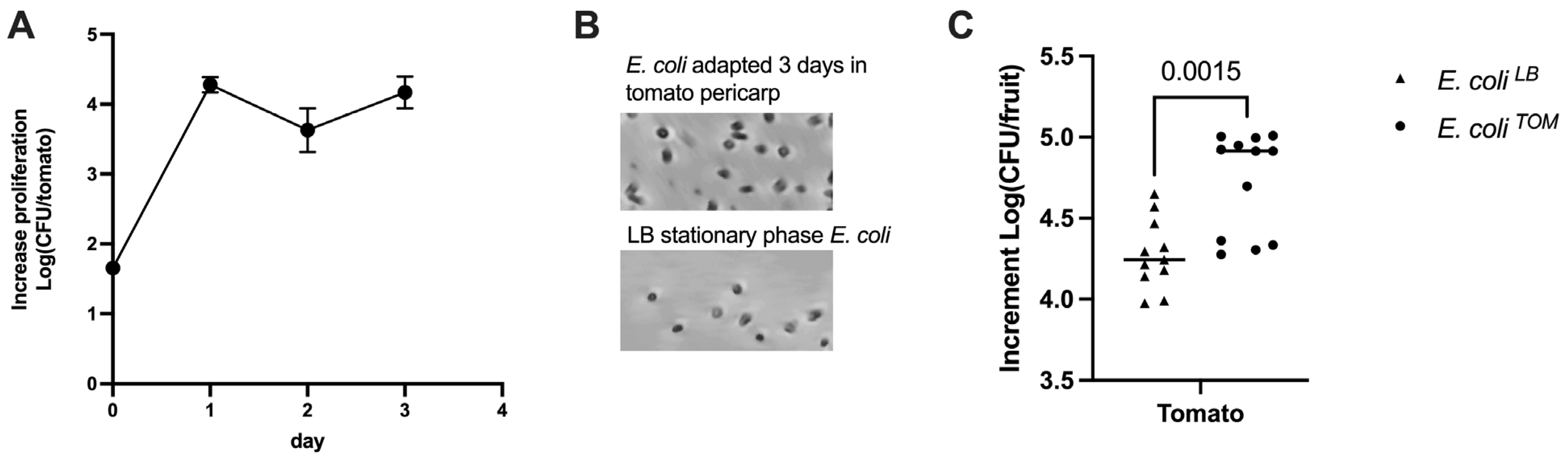
3.1. Pre-Adaptation of E. coli to Tomato Pericarp

About 150 cells of E. coli ATCC 35218 were inoculated in three shallow wounds in the tomato pericarp, showing a growth of about 2 Log(CFU/tomato) within only 24 h (Figure 1A) and reaching the stationary phase within two days.
We further determined whether pre-adaptation of E. coli in tomato pericarp (E. coli TOM) would support an increase in proliferation if transferred to other vegetables. Therefore, other tomato fruits were contaminated with the E. coli TOM strain and the E. coli originating from a stationary LB culture (E. coli LB) (no evident differences in cell morphology were observed in these two bacterial populations used for fruit contamination, Figure 1B). It is reasonable to assume that the pre-adaptation of E. coli TOM would be advantageous, growing at a higher rate when transferred into a new vegetable when compared to E. coli LB. This hypothesis was confirmed, showing that E. coli TOM had an increased—although limited—proliferation in tomatoes of log 4.7 CFU/fruit versus the log 4.2 CFU/fruit of E. coli LB (Figure 1C). Thus, pre-adaption positively affected E. coli fitness even when new vegetables were contaminated.

3.2. Differential Methylation Analysis and Expression of Genes Involved in Bacterial Adaptation

These results suggested that pre-adaptation in tomatoes probably influenced the expression of genes involved in interaction with the host environment. To further investigate this hypothesis, high-throughput Nanopore sequencing was performed to identify genomic loci whose expression could be affected by epigenetic modifications. Therefore, to identify genes putatively involved in the adaptation of E. coli ATCC 35218 to tomato pericarp, differential cytosine methylation was analyzed by sequencing the DNA of E. coli TOM and E. coli LB. We selected a few significantly differentially methylated regions associated with a number of ORFs (open reading frames) (Figure 2, Table 1).
The criteria for choosing genes characterized by differentially methylated cytosines are described in the Materials and Methods section. Thus, narG, papA_1, fimH, atoBCD, acrF, and murE-ftsI-ftsL were selected for further investigations (Table 1).
All selected genes are involved in cell adhesion and resistance against toxic compounds: their expression was therefore studied via qPCR to quantify differences between the two conditions and to find a preliminary relationship between expression levels and methylation profiles. Almost all selected genes were more expressed in E. coli LB, except for acrF, which was slightly more expressed in E. coli TOM (Table 1).

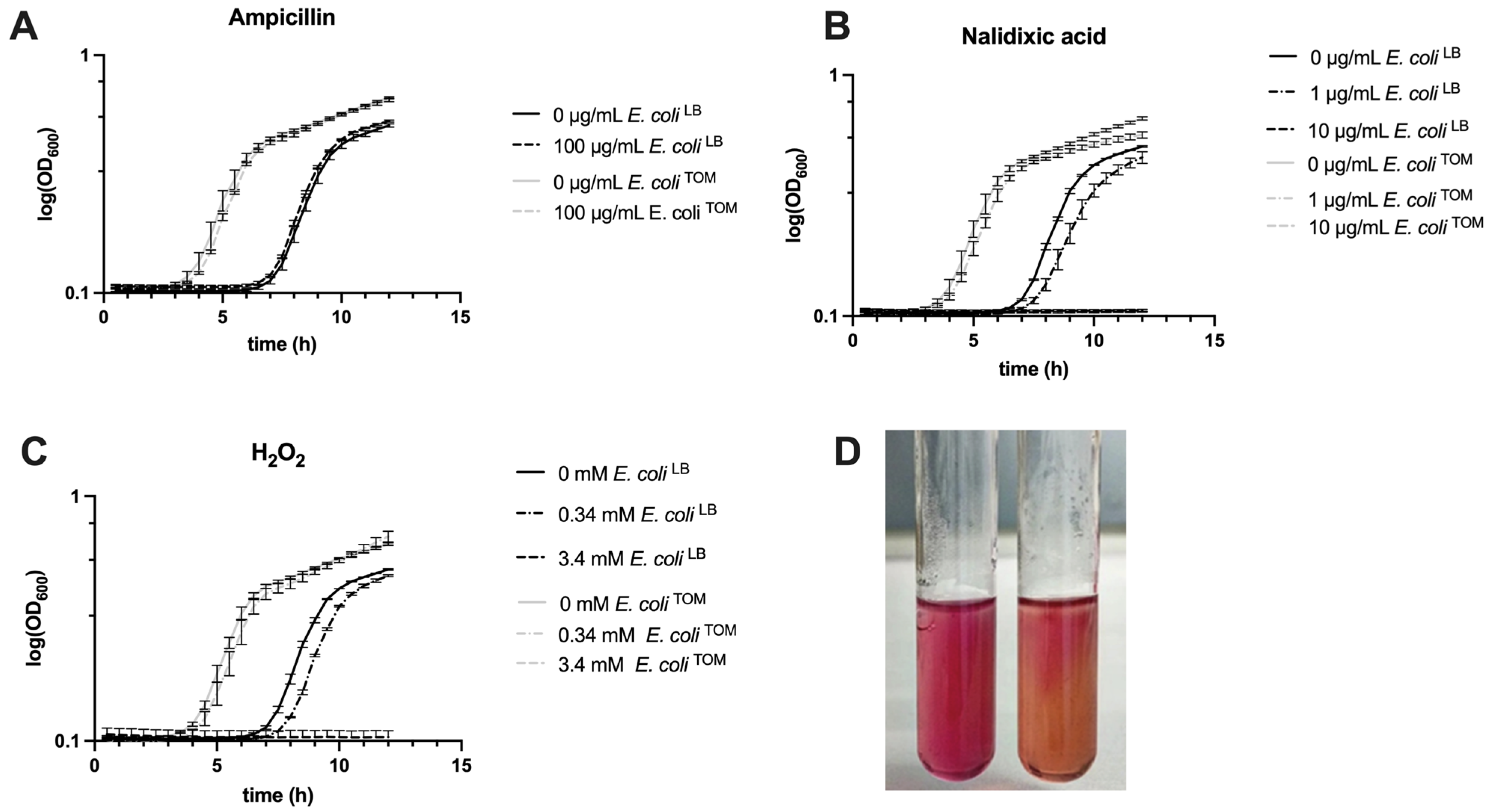
3.3. Effects of Pre-Adaptation on Environmental Stress Resistance and Cell Motility

We further focused on acrF because it encodes for a multidrug export protein and is involved in drug extrusion [24]. Thus, E. coli TOM and E. coli LB were cultivated in the presence of two antibiotics (i.e., ampicillin and nalidixic acid) and H2O2. Ampicillin was used as a control because E. coli ATCC 35218 carries a resistance gene, while H2O2 was tested because it is present in tomato wounds. Growth curves of E. coli TOM and E. coli LB growing in the presence of these compounds were compared and are shown in Figure 3.
In all experiments, the pre-adapted E. coli TOM showed a reduced lag phase, entering the exponential phase about 4 h earlier than E. coli LB. In addition, E. coli LB, which had a lower expression of acrF, was more sensitive to 1 µg/mL nalidixic acid and 0.34 mM H2O2 when compared to E. coli TOM, as represented by the further delay of the curves of E. coli LB.
We, therefore, focused our attention on two other particular genes: papA_1 and fimH, which are remarkably less expressed in E. coli TOM and encode for components of fimbriae. In the literature, it is reported that the decrement of fimbriae could be associated with reduced cell motility [25]. Hence, a motility test was performed, supporting the reduced motility of E. coli TOM compared to E. coli LB (Figure 3D).

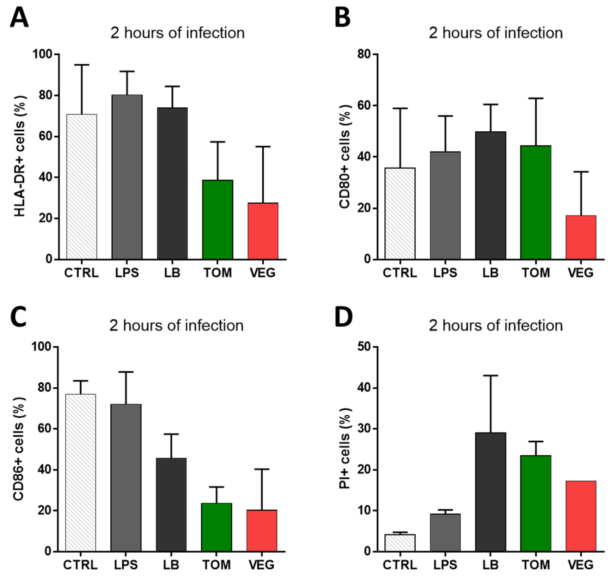
3.4. Effects of Pre-Adaptation on Pathogenicity

Finally, we measured to what extent the pre-adaptation of E. coli TOM induced any inflammation response in human mDCs, as a result of adaptation to the host environment. Indeed, previous studies showed that commensal E. coli could elicit strain-specific inflammatory stimuli [18]. E. coli TOM, E. coli LB, and non-contaminated tomatoes (used as control) were tested on mDCs. The activation status and viability of mDCs after 2 h of in vitro infection with different treatments were evaluated by flow cytometry, analyzing the human leukocyte antigen molecule HLA-DR (HLA) (Figure 4A), analyzing the expression of surface markers CD80 and CD86 (Figure 4B and Figure 4C, respectively), and by assessing the percentage of PI+ cells (necrotic cells) (Figure 4D). CD80 and CD86 are costimulatory molecules necessary for activating naïve T lymphocytes and are typically upregulated on activated mDCs. HLA-DR, also expressed on activated mDCs, is a class II HLA molecule essential for antigen presentation to CD4+ T helper lymphocytes (Th).
The results showed a trend towards the reduced expression of HLA-DR and CD86 in mDCs infected with either E. coli TOM or non-contaminated tomato (Figure 4).
A similar trend was observed with a reduction in the percentage of CD80+ cells in the presence of non-contaminated tomatoes. Regarding viability, the percentage of necrotic PI+ cells tended to increase compared to the controls (i.e., non-infected mDCs and mDCs grown in the presence of LPS), reaching about 20–30% in case of infection with E. coli TOM and E. coli LB. However, these differences were not found to be statistically significant.

4. Discussion

This study aimed to investigate unexplored aspects of the interaction between bacteria and plant hosts, analyzing pre-adaptation effects in an Escherichia coli grown in a fruit endocarp. The underpinning hypothesis is that pre-adaptation may increase bacterial virulence, providing an “epigenetic memory” that would foster replication in vegetables and other hosts such as humans [26].
In this regard, this proof-of-concept study conducted with a non-pathogenic E. coli strain provided indications for future directions. E. coli pre-adapted in tomatoes showed a higher fitness during a second round of fruit contamination, reaching higher titers compared to non-adapted bacteria. Additional studies should be performed in order to further confirm this observation, since although the difference is significant, it is also limited to 0.5 log in tomato. Similar behavior was observed previously in the case of lettuce, where E. coli K12 isolated from colonized leaves progressively colonized lettuce seedlings to a greater extent [11].
Bacteria face a challenging environment when they enter plant tissues, which are rich in both nutrients and stressors. Indeed, depending on the pathogen-associated molecular patterns (PAMP) recognized by the plant, the host may activate the production of reactive oxygen species (ROS) in the intercellular space. For example, tomato exhibits microbursts of H2O2 when facing particular stressors, including invading pathogens [27,28]. Thus, in the context of this pathogen–host competition, it should not surprise that other studies demonstrated that plant-adapted Escherichia coli showed increased resistance to oxidative stress. It is reasonable to assume that several factors are regulated in bacteria facing such a stressful environment, such as those involved in osmoprotection, as reported in the case of E. coli colonizing lettuce [29], and critical de novo biosynthesis of amino acids, as in the case of Salmonella [30]. Conversely, fruits are also rich in nutrients, vitamins, and other essential elements that sustain invasive microbes, leading to their adaption and proliferation. During ripening, the host’s nutrients may modify signaling mechanisms in the pathogen leading to metabolic responses and modification in the synthesis of pH-modulating molecules and in carbon-regulation signaling [31]. These changes occur even though E. coli and its closest taxonomical enterobacteria are commensal rather than plant pathogens; thus, this aspect requires further investigation.
Identification of genes potentially activated/repressed in E. coli upon invasion of tomato pericarp was investigated through a differential methylome analysis. This approach allowed us to identify six differentially expressed genes (i.e., narG, papA_1, fimH, atoBCD, acrF, and murE-ftsI-ftsL). All of them were more expressed in E. coli LB except for acrF, which was more expressed in E. coli TOM. The higher expression of acrF could be related to the increased resistance of E. coli TOM to harmful compounds, as demonstrated here with ampicillin, nalidixic acid, and H2O2, compared to not pre-adapted cells. This finding is in agreement with previous experiments showing a dramatic increase in tolerance to H2O2 in plant-adapted E. coli K12 cells [11]. However, it cannot a priori be excluded that the increased resistance of E. coli TOM could be due to the expression of other genes (e.g., not necessarily those encoding for efflux pumps) and activation of additional detoxification pathways (e.g., genes involved in oxidative stress tolerance), as it was demonstrated by microarray-based whole-genome transcriptional profiling in E. coli harvested from lettuce [11,32].
Finally, we tested to what extent the adaptation of E. coli TOM was able to modify the inflammation response in human mDCs. We did not find significant differences in the activation and viability profile of mDCs after in vitro infection with adapted E. coli when compared with the non-adapted one. However, new experiments should be further conducted: for example, an improved method to isolate E. coli from the tomato pericarp should be established since cell viability was compromised even in the case of non-contaminated tomatoes used as control.

5. Conclusions

In conclusion, the findings reported in this study help to understand the physiology of E. coli during the adaptation process following the colonization of tomato pericarps. It would be relevant to repeat these experiments with human pathogens and those more related to the food industry (e.g., Salmonella and pathogenic E. coli). Furthermore, it would be interesting to couple the characterization of the methylome with a transcriptomic analysis: this approach would allow a more comprehensive genome-wide overview of the bacterial adaptation to the fruit tissues. Previous research has shown that Salmonella replicates differently in relation to the ripening stage (i.e., green, partially ripe, or ripe) [33,34]. This different stage-dependent behavior should also be analyzed in the case of E. coli to better understand to what extent each ripening stage affects E. coli methylome and gene expression.
The biology of the interaction between E. coli and tomatoes should be further studied to obtain more insights into more practical aspects (as has already been performed in the case of Salmonella), such as the effect of humidity on contamination in the field [6], how agronomical practices can prevent E. coli contamination of fruits [35], the possible use of signaling molecules to reduce enteric contamination [36,37], and the selection of cultivars less susceptible to enteric pathogens [38].
Understanding the physiology of human pathogens in vegetables would allow us to better understand their life cycle outside the animal hosts and prevent food contamination.

Supplementary Materials

The following supporting information can be downloaded at https://www.mdpi.com/article/10.3390/biology12040633/s1. Table S1: Primers used in this study.

Author Contributions

Conceptualization, M.M., A.V., D.C. and C.B.; methodology, R.A, A.V. and P.G.; software, M.R., L.C. and M.Z.; data curation, A.V.; writing—original draft preparation, M.M. and A.V.; writing—review and editing, A.V., R.A., M.Z. and M.M.; visualization, L.C., A.V. and R.A.; funding acquisition, M.M. and C.B. All authors have read and agreed to the published version of the manuscript.

Funding

This research was funded by Fondazione Cassa di Risparmio di Firenze, grant number 2020-1423 #47174.

Institutional Review Board Statement

Not applicable.

Informed Consent Statement

Not applicable.

Data Availability Statement

The data presented in this study are available on request from the corresponding author.

Conflicts of Interest

The authors declare no conflict of interest.

References

  1. Holden, N.; Pritchard, L.; Toth, I. Colonization outwith the colon: Plants as an alternative environmental reservoir for human pathogenic enterobacteria: Review article. FEMS Microbiol. Rev. 2009, 33, 689–703. [Google Scholar] [CrossRef] [PubMed]
  2. Conway, T.; Krogfelt, K.A.; Cohen, P.S. The Life of Commensal Escherichia coli in the Mammalian Intestine. EcoSal Plus 2004, 1. [Google Scholar] [CrossRef] [PubMed]
  3. Bartz, J.A.; Marvasi, M.; Teplitski, M. Salmonella and Tomatoes; Academic Press: Cambridge, MA, USA, 2014; ISBN 9780124046115. [Google Scholar]
  4. Luna-Guevara, J.J.; Arenas-Hernandez, M.M.P.; Martínez De La Peña, C.; Silva, J.L.; Luna-Guevara, M.L. The Role of Pathogenic E. coli in Fresh Vegetables: Behavior, Contamination Factors, and Preventive Measures. Int. J. Microbiol. 2019, 2019, 2894328. [Google Scholar] [CrossRef] [PubMed]
  5. Marvasi, M.; Noel, J.T.; George, A.S.; Farias, M.A.; Jenkins, K.T.; Hochmuth, G.; Xu, Y.; Giovanonni, J.J.; Teplitski, M. Ethylene signalling affects susceptibility of tomatoes to Salmonella. Microb. Biotechnol. 2014, 7, 545–555. [Google Scholar] [CrossRef] [PubMed]
  6. Devleesschauwer, B.; Marvasi, M.; Giurcanu, M.C.; Hochmuth, G.J.; Speybroeck, N.; Havelaar, A.H.; Teplitski, M. High relative humidity pre-harvest reduces post-harvest proliferation of Salmonella in tomatoes. Food Microbiol. 2017, 66, 55–63. [Google Scholar] [CrossRef]
  7. Kroupitski, Y.; Golberg, D.; Belausov, E.; Pinto, R.; Swartzberg, D.; Granot, D.; Sela, S. Internalization of Salmonella enterica in Leaves Is Induced by Light and Involves Chemotaxis and Penetration through Open Stomata. Appl. Environ. Microbiol. 2009, 75, 6076–6086. [Google Scholar] [CrossRef]
  8. Lu, S.; Song, J.; Campbell-Palmer, L. A modified chemiluminescence method for hydrogen peroxide determination in apple fruit tissues. Sci. Hortic. 2009, 120, 336–341. [Google Scholar] [CrossRef]
  9. Nasirizadeh, N.; Shekari, Z.; Nazari, A.; Tabatabaee, M. Fabrication of a novel electrochemical sensor for determination of hydrogen peroxide in different fruit juice samples. J. Food Drug Anal. 2016, 24, 72–82. [Google Scholar] [CrossRef]
  10. Hyong, W.C.; Young, J.K.; Sung, C.L.; Jeum, K.H.; Byung, K.H. Hydrogen peroxide generation by the pepper extracellular peroxidase CaPO2 activates local and systemic cell death and defense response to bacterial pathogens. Plant Physiol. 2007, 145, 890–904. [Google Scholar] [CrossRef]
  11. Dublan, M.D.L.A.; Ortiz-Marquez, J.C.F.; Lett, L.; Curatti, L. Plant-adapted Escherichia coli show increased lettuce colonizing ability, resistance to oxidative stress and chemotactic response. PLoS ONE 2014, 9, e110416. [Google Scholar] [CrossRef]
  12. Landstorfer, R.; Simon, S.; Schober, S.; Keim, D.; Scherer, S.; Neuhaus, K. Comparison of strand-specific transcriptomes of enterohemorrhagic Escherichia coli O157: H7 EDL933 (EHEC) under eleven different environmental conditions including radish sprouts and cattle feces. BMC Genom. 2014, 15, 1–25. [Google Scholar] [CrossRef] [PubMed]
  13. Jacob, C.; Velásquez, A.C.; Josh, N.A.; Settles, M.; He, S.Y.; Melotto, M. Dual transcriptomic analysis reveals metabolic changes associated with differential persistence of human pathogenic bacteria in leaves of Arabidopsis and lettuce. G3 Genes Genomes Genet. 2021, 11, jkab331. [Google Scholar] [CrossRef] [PubMed]
  14. Beaulaurier, J.; Schadt, E.E.; Fang, G. Deciphering bacterial epigenomes using modern sequencing technologies. Nat. Rev. Genet. 2018, 20, 157–172. [Google Scholar] [CrossRef] [PubMed]
  15. Sánchez-Romero, M.A.; Casadesús, J. The bacterial epigenome. Nat. Rev. Microbiol. 2019, 18, 7–20. [Google Scholar] [CrossRef]
  16. Rand, A.C.; Jain, M.; Eizenga, J.M.; Musselman-Brown, A.; Olsen, H.E.; Akeson, M.; Paten, B. Mapping DNA methylation with high-throughput nanopore sequencing. Nat. Methods 2017, 14, 411–413. [Google Scholar] [CrossRef]
  17. Simpson, J.T.; Workman, R.E.; Zuzarte, P.C.; David, M.; Dursi, L.J.; Timp, W. Detecting DNA cytosine methylation using nanopore sequencing. Nat. Methods 2017, 14, 407–410. [Google Scholar] [CrossRef]
  18. Kittana, H.; Gomes-Neto, J.C.; Heck, K.; Geis, A.L.; Segura Muñoz, R.R.; Cody, L.A.; Schmaltz, R.J.; Bindels, L.B.; Sinha, R.; Hostetter, J.M.; et al. Commensal Escherichia coli strains can promote intestinal inflammation via differential interleukin-6 production. Front. Immunol. 2018, 9, 2318. [Google Scholar] [CrossRef]
  19. Kelly, D.; Campbell, J.I.; King, T.P.; Grant, G.; Jansson, E.A.; Coutts, A.G.P.; Pettersson, S.; Conway, S. Commensal anaerobic gut bacteria attenuate inflammation by regulating nuclear-cytoplasmic shutting of PPAR-$γ$ and ReIA. Nat. Immunol. 2004, 5, 104–112. [Google Scholar] [CrossRef]
  20. Chapman, T.A.; Wu, X.-Y.; Barchia, I.; Bettelheim, K.A.; Driesen, S.; Trott, D.; Wilson, M.; Chin, J.J.C. Comparison of virulence gene profiles of Escherichia coli strains isolated from healthy and diarrheic swine. Appl. Environ. Microbiol. 2006, 72, 4782–4795. [Google Scholar] [CrossRef]
  21. Semenzato, G.; Alonso-Vásquez, T.; Duca, S.D.; Vassallo, A.; Riccardi, C.; Zaccaroni, M.; Mucci, N.; Padula, A.; Emiliani, G.; Piccionello, A.P.; et al. Genomic Analysis of Endophytic Bacillus-Related Strains Isolated from the Medicinal Plant Origanum vulgare L. Revealed the Presence of Metabolic Pathways Involved in the Biosynthesis of Bioactive Compounds. Microorganisms 2022, 10, 919. [Google Scholar] [CrossRef]
  22. Krzywinski, M.; Schein, J.; Birol, I.; Connors, J.; Gascoyne, R.; Horsman, D.; Jones, S.J.; Marra, M.A. Circos: An information aesthetic for comparative genomics. Genome Res. 2009, 19, 1639–1645. [Google Scholar] [CrossRef] [PubMed]
  23. Bustin, S.A.; Benes, V.; Garson, J.A.; Hellemans, J.; Huggett, J.; Kubista, M.; Mueller, R.; Nolan, T.; Pfaffl, M.W.; Shipley, G.L.; et al. The MIQE Guidelines: Minimum Information for Publication of Quantitative Real-Time PCR Experiments. Clin. Chem. 2009, 55, 611–622. [Google Scholar] [CrossRef] [PubMed]
  24. Zhang, C.-Z.; Chang, M.-X.; Yang, L.; Liu, Y.-Y.; Chen, P.-X.; Jiang, H.-X. Upregulation of AcrEF in quinolone resistance development in Escherichia coli when AcrAB-TolC function is impaired. Microb. Drug Resist. 2018, 24, 18–23. [Google Scholar] [CrossRef] [PubMed]
  25. Lehti, T.A.; Bauchart, P.; Dobrindt, U.; Korhonen, T.K.; Westerlund-Wikström, B. The fimbriae activator MatA switches off motility in Escherichia coli by repression of the flagellar master operon flhDC. Microbiology 2012, 158, 1444–1455. [Google Scholar] [CrossRef]
  26. Riber, L.; Hansen, L.H. Epigenetic Memories: The Hidden Drivers of Bacterial Persistence? Trends Microbiol. 2021, 29, 190–194. [Google Scholar] [CrossRef]
  27. Orozco-Cárdenas, M.L.; Narváez-Vásquez, J.; Ryan, C.A. Hydrogen Peroxide Acts as a Second Messenger for the Induction of Defense Genes in Tomato Plants in Response to Wounding, Systemin, and Methyl Jasmonate. Plant Cell 2001, 13, 179–191. [Google Scholar] [CrossRef]
  28. Waszczak, C.; Carmody, M.; Kangasjärvi, J. Reactive Oxygen Species in Plant Signaling. Annu. Rev. Plant Biol. 2018, 69, 209–236. [Google Scholar] [CrossRef]
  29. Scott, R.A.; Thilmony, R.; Harden, L.A.; Zhou, Y.; Brandl, M.T. Escherichia coli O157:H7 Converts Plant-Derived Choline to Glycine Betaine for Osmoprotection during Pre- and Post-harvest Colonization of Injured Lettuce Leaves. Front. Microbiol. 2017, 8, 2436. [Google Scholar] [CrossRef]
  30. de Moraes, M.H.; Desai, P.; Porwollik, S.; Canals, R.; Perez, D.R.; Chu, W.; McClelland, M.; Teplitski, M. Salmonella Persistence in Tomatoes Requires a Distinct Set of Metabolic Functions Identified by Transposon Insertion Sequencing. Appl. Environ. Microbiol. 2017, 83, e03028-16. [Google Scholar] [CrossRef]
  31. Prusky, D.B.; Wilson, R.A. Does increased nutritional carbon availability in fruit and foliar hosts contribute to modulation of pathogen colonization? Postharvest Biol. Technol. 2018, 145, 27–32. [Google Scholar] [CrossRef]
  32. Ferelli, A.M.C.; Bolten, S.; Szczesny, B.; Micallef, S.A. Salmonella enterica elicits and is restricted by nitric oxide and reactive oxygen species on tomato. Front. Microbiol. 2020, 1, 391. [Google Scholar] [CrossRef] [PubMed]
  33. Marvasi, M.; George, A.S.; Giurcanu, M.; Hochmuth, G.J.; Noel, J.T.; Gause, E.; Teplitski, M. Effects of nitrogen and potassium fertilization on the susceptibility of tomatoes to post-harvest proliferation of Salmonella enterica. Food Microbiol. 2014, 43, 20–27. [Google Scholar] [CrossRef] [PubMed]
  34. Marvasi, M.; George, A.S.; Giurcanu, M.C.; Hochmuth, G.J.; Noel, J.T.; Teplitski, M. Effect of the irrigation regime on the susceptibility of pepper and tomato to post-harvest proliferation of Salmonella enterica. Food Microbiol. 2015, 46, 139–144. [Google Scholar] [CrossRef] [PubMed]
  35. Lenzi, A.; Marvasi, M.; Baldi, A. Agronomic practices to limit pre- and post-harvest contamination and proliferation of human pathogenic Enterobacteriaceae in vegetable produce. Food Control 2021, 119, 107486. [Google Scholar] [CrossRef]
  36. Marvasi, M. Potential use and perspectives of nitric oxide donors in agriculture. J. Sci. Food Agric. 2017, 97, 1065–1072. [Google Scholar] [CrossRef]
  37. Marvasi, M.; Durie, I.A.; McLamore, E.S.; Vanegas, D.C.; Chaturvedi, P. Salmonella enterica biofilm-mediated dispersal by nitric oxide donors in association with cellulose nanocrystal hydrogels. AMB Express 2015, 5, 1–9. [Google Scholar] [CrossRef]
  38. Henriquez, T.; Lenzi, A.; Baldi, A.; Marvasi, M. Frontiers in Plant Breeding: Perspectives for the Selection of Vegetables Less Susceptible to Enteric Pathogens. Front. Microbiol. 2020, 11, 1087. [Google Scholar] [CrossRef]
Figure 1. E. coli growth in tomatoes. (A) Increase in E. coli proliferation in tomato pericarp within 4 days. The decline of growth on day 2 is not significantly different in comparison to days 1 and 3 (p < 0.1650). Significance occurs only in comparison with day 0. (B) Micrograph showing E. coli TOM and E. coli LB cells (magnification of 400X). (C) Growth of pre-adapted E. coli TOM and E. coli LB in tomato pericarp. Error bars represent standard error. Horizontal bars in (C) show the significance of the T-test (p < 0.05).
Biology 12 00633 g001
Figure 2. Circos plot of the results of the methylation analysis. Starting from the outside, plotted tracks represent the location of E. coli ATCC 35218 CDSs (light blue); the methylation status of A in the dam patterns (red); the methylation status of C in the dcm patterns (green); and the methylation status of C in CpG patterns (yellow). For methylation sectors, the direction (up/down) of bars indicates the strand where methylation has been called.
Biology 12 00633 g002
Figure 3. Different growth of E. coli TOM and E. coli LB in different conditions. Growth curves of E. coli TOM and E. coli LB in the presence/absence of (A) ampicillin (control), (B) nalidixic acid, and (C) H2O2; (D) different acidification by E. coli TOM (left tube) and E. coli LB (right tube) into semi-solid MacConkey, possibly indicating different motility. Error bars represent standard error.
Biology 12 00633 g003
Figure 4. Activation and viability profile of mDCs after in vitro infection with E. coli analyzed by flow cytometry. Histograms represent the percentage of mDCs cells positive for (A) HLA-DR, (B) CD80, (C) CD86, and (D) propidium iodide (PI) after being cultured under different conditions: without any stimulus (control, CTRL), in the presence of only LPS (LPS), with E. coli LB (LB), with E. coli TOM (TOM), or with non-contaminated tomato (VEG). Data were analyzed using GraphPad Prism v.6 software, with one-way ANOVA test followed by Tukey’s post hoc test for multiple comparisons. Error bars represent the standard error of the mean.
Biology 12 00633 g004
Table 1. Cytosine methylation state of E. coli TOM and relative gene expression in tomato pericarp.
GeneFunctionMethylation 1
in E. coli TOM
Relative Expression
(E. coli TOM vs. E. coli LB)
narGRespiratory nitrate reductase 1 alpha chain+−13.9
papA_1Pap fimbrial major pilin protein−11.5
fimHType 1 fimbrin D-mannose specific adhesin−36.1
atoBCDMetabolism of Acetyl-CoA+−2.52
acrFMultidrug export protein AcrF+1.6
murE-ftsI-ftsLPeptidoglycan+−1.3
1 Sites significantly more (+) or less (−) methylated in adapted E. coli TOM in comparison to E. coli LB.
Disclaimer/Publisher’s Note: The statements, opinions and data contained in all publications are solely those of the individual author(s) and contributor(s) and not of MDPI and/or the editor(s). MDPI and/or the editor(s) disclaim responsibility for any injury to people or property resulting from any ideas, methods, instructions or products referred to in the content.

