Article

Integrative Analysis of Transcriptome and Metabolome Reveals Molecular Responses in Eriocheir sinensis with Hepatopancreatic Necrosis Disease

1
Wuxi Fisheries College, Nanjing Agricultural University, Nanjing 210095, China
2
Key Laboratory of Integrated Rice-Fish Farming Ecology, Ministry of Agriculture and Rural Affairs, Freshwater Fisheries Research Center, Chinese Academy of Fishery Sciences, Wuxi 214081, China
*
Author to whom correspondence should be addressed.
Biology 2022, 11(9), 1267; https://doi.org/10.3390/biology11091267
Submission received: 24 July 2022 / Revised: 21 August 2022 / Accepted: 22 August 2022 / Published: 26 August 2022
(This article belongs to the Special Issue Multi-Omics Co-expression Network Analysis)

Abstract

:

Simple Summary

There are still many doubts about the etiology and pathogenic mechanisms of Chinese mitten crab hepatopancreatic necrosis disease (HPND). In the present study, new insights into the molecular responses of diseased crabs were found by integrating transcriptomics and metabolomics. The results show that the immune system, metabolic capacity, antioxidant system, detoxification capacity, and nervous system of HPND-affected crabs were impacted, and these changes may be related to the pathological mechanism of HPND. Furthermore, we suspect that functional hypoxia may be involved in the progression of HPND, which provides new insights into the pathogenic mechanism of HPND. In conclusion, based on our findings, we further confirmed that HPND could be mainly caused by abiotic factors.

Abstract

Hepatopancreatic necrosis disease (HPND) is a highly lethal disease that first emerged in 2015 in Jiangsu Province, China. So far, most researchers believe that this disease is caused by abiotic factors. However, its true pathogenic mechanism remains unknown. In this study, the effects of HPND on the metabolism and other biological indicators of the Chinese mitten crab (Eriocheir sinensis) were evaluated by integrating transcriptomics and metabolomics. Our findings demonstrate that the innate immunity, antioxidant activity, detoxification ability, and nervous system of the diseased crabs were affected. Additionally, metabolic pathways such as lipid metabolism, nucleotide metabolism, and protein metabolism were dysregulated, and energy production was slightly increased. Moreover, the IL-17 signaling pathway was activated and high levels of autophagy and apoptosis occurred in diseased crabs, which may be related to hepatopancreas damage. The abnormal mitochondrial function and possible anaerobic metabolism observed in our study suggested that functional hypoxia may be involved in HPND progression. Furthermore, the activities of carboxylesterase and acetylcholinesterase were significantly inhibited, indicating that the diseased crabs were likely stressed by pesticides such as pyrethroids. Collectively, our findings provide new insights into the molecular mechanisms altered in diseased crabs, as well as the etiology and pathogenic mechanisms of HPND.

1. Introduction

The freshwater Chinese mitten crab (Eriocheir sinensis) is highly sought-after by Chinese consumers due to its high nutritional value and flavorful meat [1]. Chinese mitten crab farming has recently become an important pillar of China’s fishery industry [2]. However, since 2015, hepatopancreatic necrosis disease (HPND) has become prevalent in many provinces in China, causing huge losses to the Chinese mitten crab farming industry [3]. Sick crabs exhibited slow movement and decreased appetite [4], and anatomical analyses confirmed the occurrence of hepatopancreas lesions, muscle atrophy, luminal hydrops, and gastrointestinal emptying [5,6], which was consistent with our observations (Figure 1). Although HPND-affected crabs do not die immediately, sick crabs are no longer edible, and therefore cannot be marketed.
In a previous study, HPND was first proposed to be caused by a microsporidian parasite (Hepatospora eriocheir) [7]. However, subsequent tests found that diseased crabs could not always detect H. eriocheir, and the symptoms of HPND could not be reliably reproduced via artificial H. eriocheir infection [8]. Interestingly, however, the injection of diseased crab supernatant (sterile) into healthy crabs, as well as animal regression experiments with dominant bacteria in the hemolymph of diseased crabs, also failed to produce HPND symptoms in healthy crabs [5,9]. This suggests that a single factor of H. eriocheir, bacteria, and viruses does not directly cause HPND. This was also confirmed by one of our previous meta-transcriptomics analyses, in which the viral and microsporidial communities in the hepatopancreas of HPND-affected and healthy crabs were not significantly different [3]. In general, abiotic factors are now more recognized causative factors (e.g., pesticides, high pH, and hypoxia) [10,11]. For example, Pan et al., [5] found that crabs exhibited HPND symptoms in high-pH (9.5–10) water environments. The hepatopancreas of E. sinensis was damaged and turned whitish under long-term exposure to low pesticide concentrations [12,13]. However, this still does not prove that HPND is caused by abiotic factors. In aquatic animals, the same disease may have different symptoms, and the same symptoms may be caused by different diseases [14]. Moreover, it is also possible that abiotic factors are just one of the causes of HPND. Therefore, additional studies are needed to gain further insights into the molecular responses and pathogenesis of HPND-affected crabs.
The recent development of effective “omic” data analysis and bioinformatic tools enables the study of the biological responses of aquatic animal cells or organs to environmental stress and disease infection through the analysis of transcriptomes, metabolomes, and proteomes [15,16]. Among them, transcriptome analysis can identify stress-induced genes and provides important information on gene expression levels, thus enabling the visualization of the expression patterns of key genes [17]. Metabolomics is the systematic study of small-molecule metabolite profiles, which describe the physiological state of an organism based on the true metabolic level in the organism [18]. Therefore, the combined analysis of transcriptomics and metabolomics may provide a deeper understanding of biological processes than either approach alone [19]. Here, comprehensive transcriptomic and metabolomic analyses revealed global changes in genes and metabolites in E. sinensis with HPND, thus providing insights into the adaptive mechanisms and possible pathogenic factors of diseased crabs at the molecular level.

2. Materials and Methods

2.1. Experimental Samples

The progression of the HPND is relatively long, and the hepatopancreas of sick crabs gradually degenerates and shrinks. According to the color change of the hepatopancreas, it can be divided into mild symptoms (hepatopancreas were pale yellow) and severe symptoms (hepatopancreases were milky white or grayish white) [5]. In this study, healthy crabs and severe symptomatic crabs of the same size (weight: 72.16 ± 4.02 g) were simultaneously collected from adjacent ponds of the same farm in Anfeng Town, Xinghua city, Jiangsu Province, China, to ensure that growth environment and dietary conditions were consistent. The hepatopancreases of diseased crabs (hereinafter referred to as the HPND group) and healthy crabs (control group) were rapidly resected under sterile conditions and frozen in liquid nitrogen for subsequent transcriptome sequencing and LC-MS analysis. Among them, 4 diseased crabs and 4 healthy crabs were used for transcriptome sequencing, and 10 diseased crabs and 10 healthy crabs were used for LC-MS analysis. Additionally, approximately 6 mm3 of tissues were dissected from the hepatopancreas and appendicular muscles of healthy and diseased crabs, then fixed in a 4% paraformaldehyde solution and stored at −4 °C after remaining at room temperature for two hours.

2.2. Histological Analysis

The fixed tissue was dehydrated with gradients of ethanol and then embedded in paraffin. The wax was cut into 50 μm tissue sections and stained with hematoxylin-eosin. The tissue sections were examined using a light microscope (DM6 M LIBS, Leica, Wetzlar, Germany).

2.3. Metabolite Extraction and Profiling Analysis

The hepatopancreas tissue ground with liquid nitrogen was vortexed with a pre-chilled 80% methanol and 0.1% formic acid solution. The sample was incubated on ice for 5 min, and then centrifuged at 15,000× g at 4 °C for 20 min. A certain amount of supernatant was diluted with LC-MS-grade water to a methanol content of 53%. The sample was then transferred to a new Eppendorf tube and centrifuged at 15,000× g at 4 °C for 20 min. Finally, the supernatant was collected and injected into an LC-MS/MS system for analysis [20]. Quality control (QC) samples were prepared by mixing equal amounts of supernatant to ensure assay stability and sample reproducibility.
UHPLC-MS/MS analysis was performed at Novogene Co., Ltd. (Beijing, China) using a Vanquish UHPLC system (Thermo Fisher, Bremen, Germany) and an Orbitrap Q ExactiveTM HF mass spectrometer (Thermo Fisher, Germany). A 17 min linear gradient was used to inject the sample into a Hypesil Gold column (100 × 2.1 mm, 1.9 μm) at a 0.2 mL/min flow rate. The measurements were conducted using eluent A (aqueous solution containing 0.1% FA) and eluent B (methanol) in positive polarity mode. Eluent A (5 mM ammonium acetate, pH 9.0) and eluent B (methanol) were used in negative polarity mode. The solvent gradient settings were as follows: 2% B, 1.5 min; 2–100% B, 12.0 min; 100% B, 14.0 min; 100–2% B, 14.1 min; 2% B, 17 min. The Q ExactiveTM HF mass spectrometer was operated in positive/negative polarity mode, with a spray voltage of 3.2 kV, a capillary temperature of 320 °C, a sheath gas flow rate of 40 arb, and an auxiliary gas flow rate of 10 arb. There were 3 QC samples in this project, and metabolites with a coefficient of variance (CV) below 30% in the QC samples were reserved as the final identification result.

2.4. Metabolomics Data Analysis

The raw data files generated by UHPLC-MS/MS were processed with Compound Discoverer 3.1 (CD3.1, Thermo Fisher) for peak alignment, peak detection, and quantification of each metabolite. Afterward, the peak intensity was normalized to the total spectral intensity. Normalized data were used to predict molecular formulas based on additional ions, molecular ion peaks, and fragment ions. Statistical analyses were conducted using R (R version R-3.4.3, Ross Ihaka and Robert Gentleman, New Zealand), Python (Python version 2.7.6, Guido van Rossum, USA), and CentOS (CentOS version 6.6, Gregory Kurtzer, USA). Metabolites were annotated using the KEGG (https://www.genome.jp/kegg/pathway.html, accessed on 20 November 2020), HMDB (https://hmdb.ca/metabolites, accessed on 20 November 2020), and LIPIDMaps (http://www.lipidmaps.org/, accessed on 20 November 2020) databases. Principal component analysis (PCA) and partial least squares discriminant analysis (PLS-DA) were performed in metaX to ensure the repeatability and reliability of within-group data by finding and excluding outliers in each group of samples [21]. A univariate analysis (t-test) was used to calculate statistical significance (p-value). The metabolites with VIP > 1 and p-value < 0.05 and fold change (FC) ≥ 2 or FC ≤ 0.5 were considered differentially expressed metabolites (DMs).

2.5. RNA Extraction and RNA-Sequencing

RNA was extracted from the samples using the TRIzol Reagent (Invitrogen, Carlsbad, CA, USA). RNA integrity was analyzed by 1% agarose gel electrophoresis, RNA purity was assessed with a NanoDrop spectrophotometer (Thermo Scientific, Wilmington, NC, USA), and RNA quality was confirmed with an Agilent Bioanalyzer 2100 system (Santa Clara, CA, USA). The cDNA libraries were constructed using the MGIEasy RNA Library Prep Kit. Finally, the libraries were sequenced on the MGISEQ-2000RS platform (BGI-Shenzhen, China).

2.6. Transcriptome Profile Analysis

The raw data were filtered using the SOAPnuke (v1.5.2, BGI-Shenzhen, China) filtering software to obtain clean data. Next, the clean reads were aligned to the reference gene sequence with Bowtie2 (v2.2.5) [22], after which the expression levels of genes and transcripts were calculated using RSEM (v1.2.12) [23]. De novo assembly of the clean reads was conducted using the Trinity suite, followed by Tgicl to cluster the assembled transcripts to de-redundancy and obtain UniGene identifiers. The obtained UniGene identifiers were then uploaded to the following main databases: KEGG (Kyoto Encyclopedia of Genes and Genomes), GO (Gene Ontology), NR (NCBI non-redundant protein sequences), NT (NCBI nucleotide sequences), SwissProt (a manually annotated and reviewed protein sequence database), and PFAM (Protein family). Afterward, gene expression was normalized to fragments per kilobase per million (FPKM) values. The DEseq2 method based on the principle of negative binomial distribution was used to detect differentially expressed genes. Genes with p-values < 0.05 were considered differentially expressed genes (DEGs). Next, GO and KEGG pathway enrichment analysis of these DEGs was conducted. p-values were FDR (false discovery rate) corrected to obtain q-values, and GO terms and KEGG pathways with q-value ≤ 0.05 were considered significantly enriched.

2.7. Integrative Analysis of Metabolomics and Transcriptomics

An integrative metabolomics and transcriptomics approach was adopted to better characterize the post-transcriptional regulation of gene expression. The correlation between the DEGs and the DMs was determined using the Pearson statistical method and the correlation coefficient R2 and p-value of the DEGs and DMs were calculated. All of the differentially expressed genes and metabolites were mapped to the KEGG pathway database, their common pathway information was obtained, and a KEGG enrichment bubble chart was generated for the pathways that were significantly enriched with DEGs and DMs using the ‘ggplot2’ R package.

3. Results

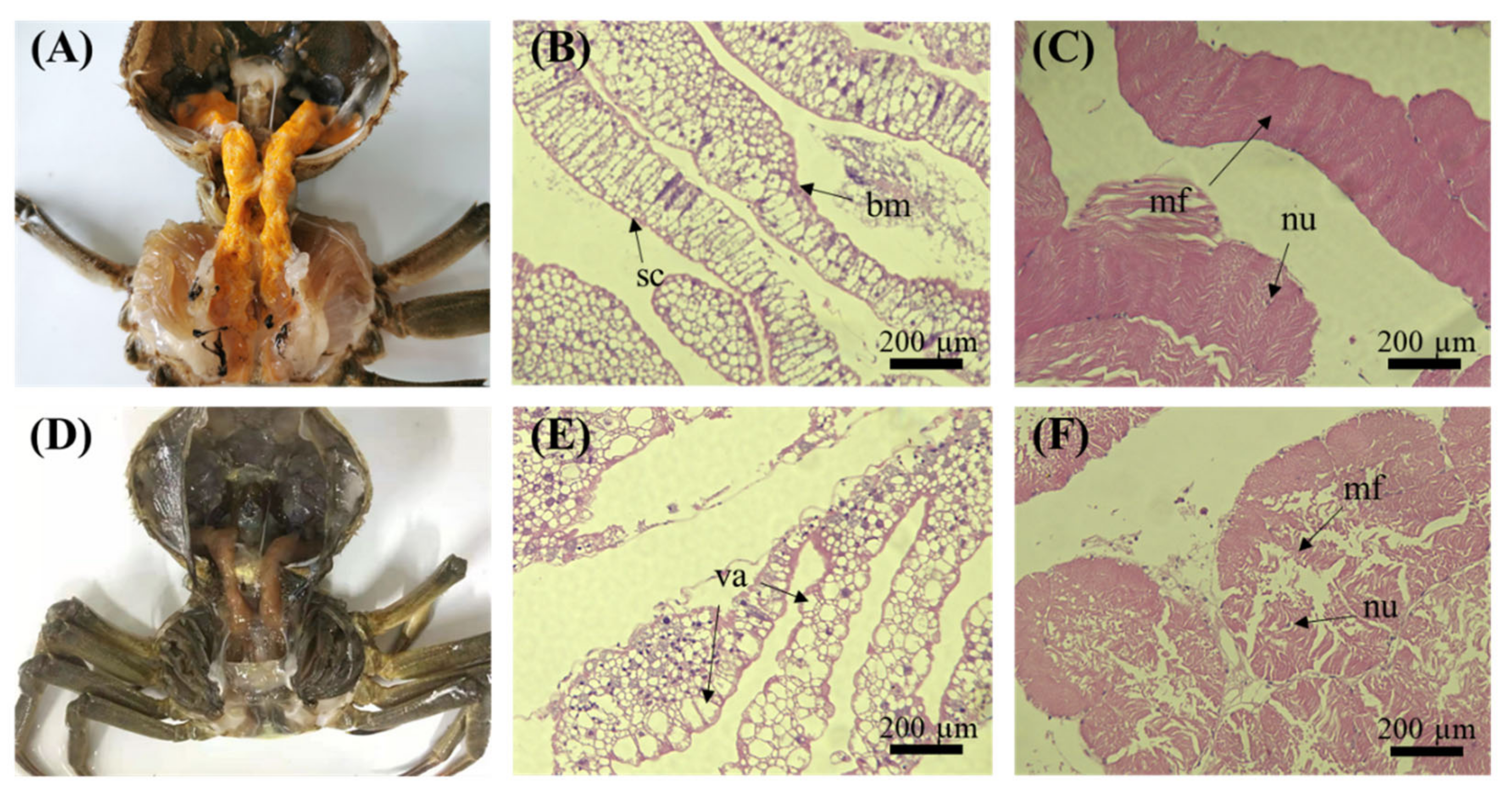
3.1. Histopathology of Crab Tissues

As shown in Figure 1, the hepatopancreas and muscles of crabs with HPND exhibited tissue damage compared with healthy tissues. In the hepatopancreas, the hepatic tubules were severely damaged, and a large number of vacuoles was observed. (Figure 1E). Additionally, muscle fibers appeared loose or broken and nuclei became smaller (Figure 1F).

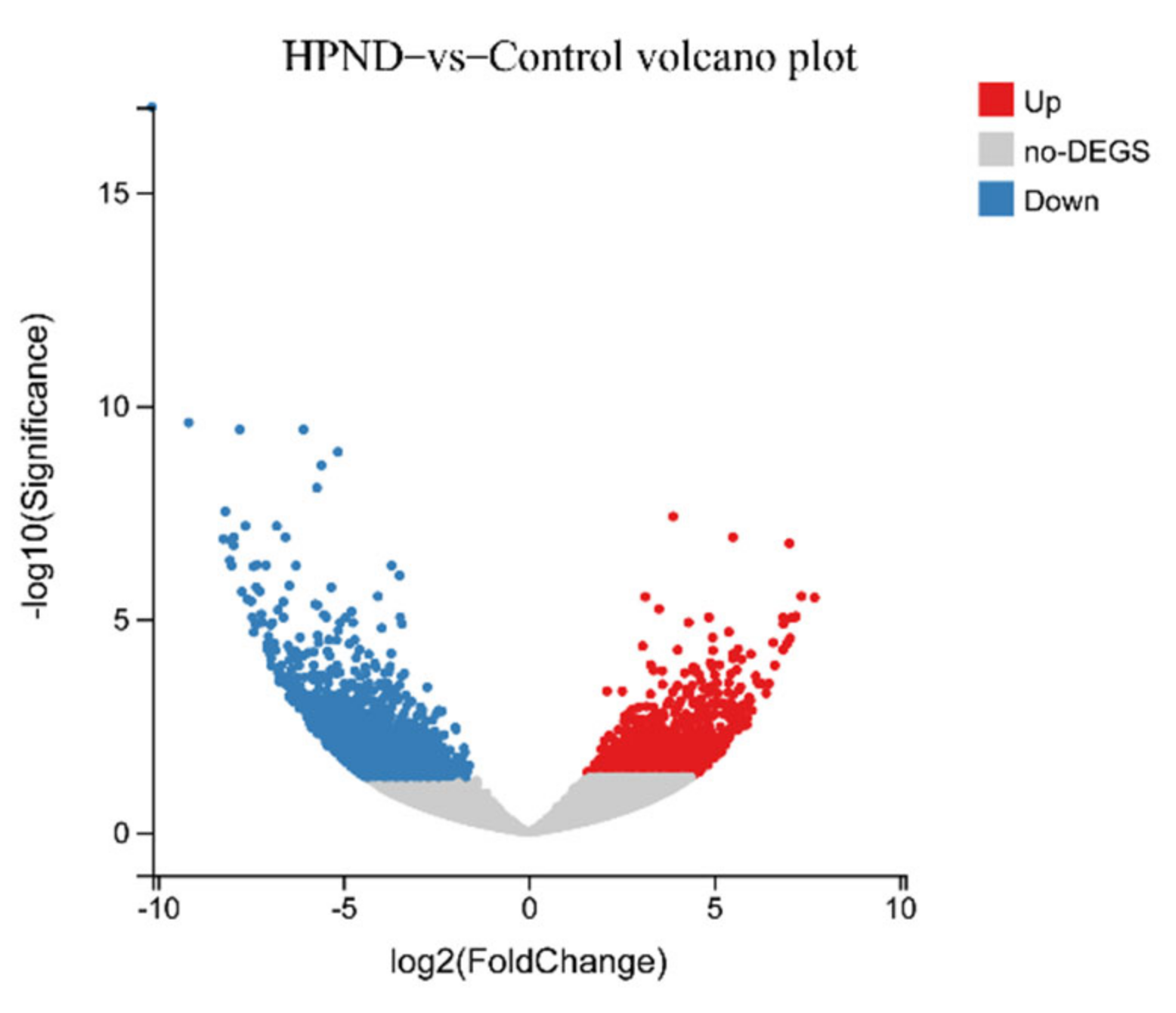
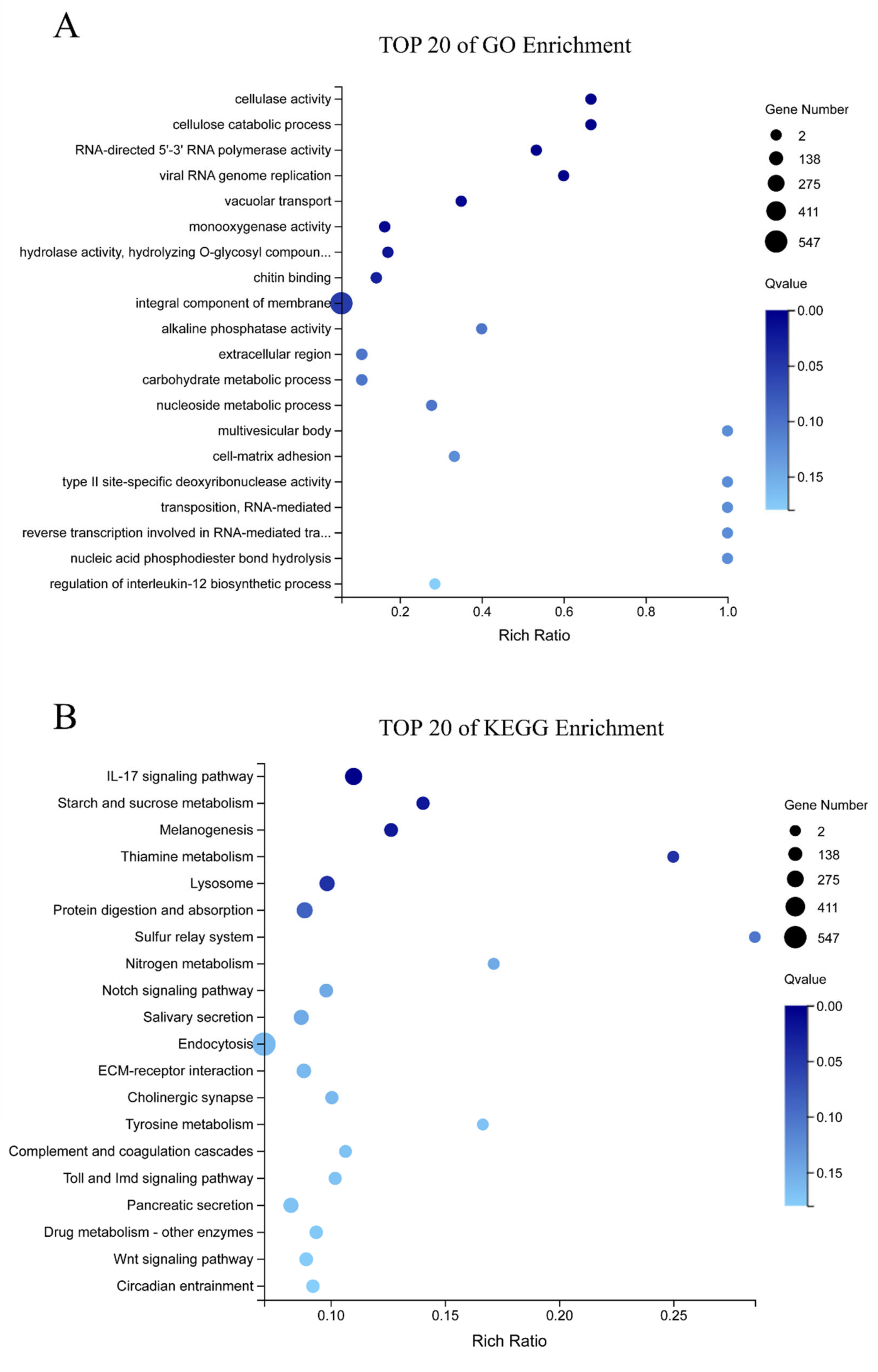
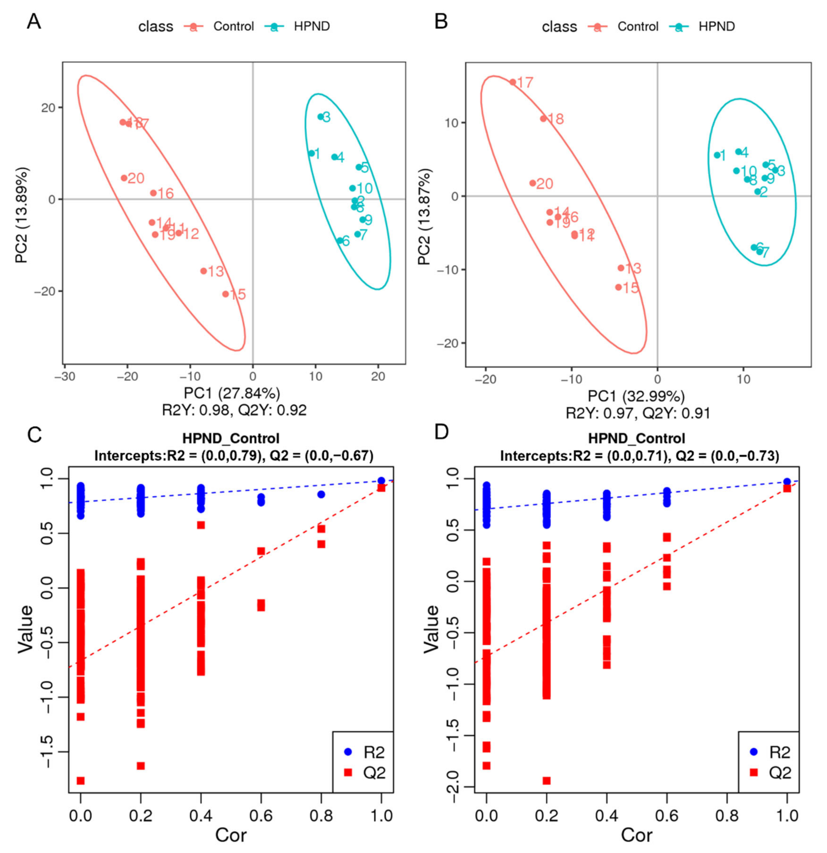
3.2. Transcriptomic Analysis of E. sinensis with HPND

After de-redundancy by clustering the assembled transcripts, 80,869 UniGenes were obtained. The total length, average length, N50, and GC content were 73,433,519 bp, 908 bp, 1668 bp, and 43.40%, respectively. The UniGenes were then compared to seven functional databases for annotation, and functional annotations were obtained for 33,762 (NR: 41.75%), 30,982 (NT: 38.31%), 21,769 (SwissProt: 26.92%), 20,610 (KOG: 25.49%), 23,392 (KEGG: 28.93%), 20,946 (GO: 25.90%), and 18,252 (Pfam: 22.57%) UniGene identifiers.
A total of 3332 DEGs were identified in the HPND vs. control group, of which 1491 DEGs were significantly up-regulated and 1841 DEGs were significantly down-regulated (Figure 2). These DEGs were subjected to GO and KEGG functional analysis to better understand the biological significance of these DEGs and the biochemical processes involved. The top 20 GO enrichment terms indicated that the DEGs were mainly involved in some important biological processes and molecular functions, such as cellulase activity, viral RNA genome replication, cellulose catabolic process, RNA-directed 5′-3′ RNA polymerase activity, and vacuolar transport. Moreover, the results of KEGG enrichment show that some pathways, such as the IL-17 signaling pathway, lysosome, melanogenesis, protein digestion and absorption, starch and sucrose metabolism, and thiamine metabolism underwent significant changes (Figure 3).

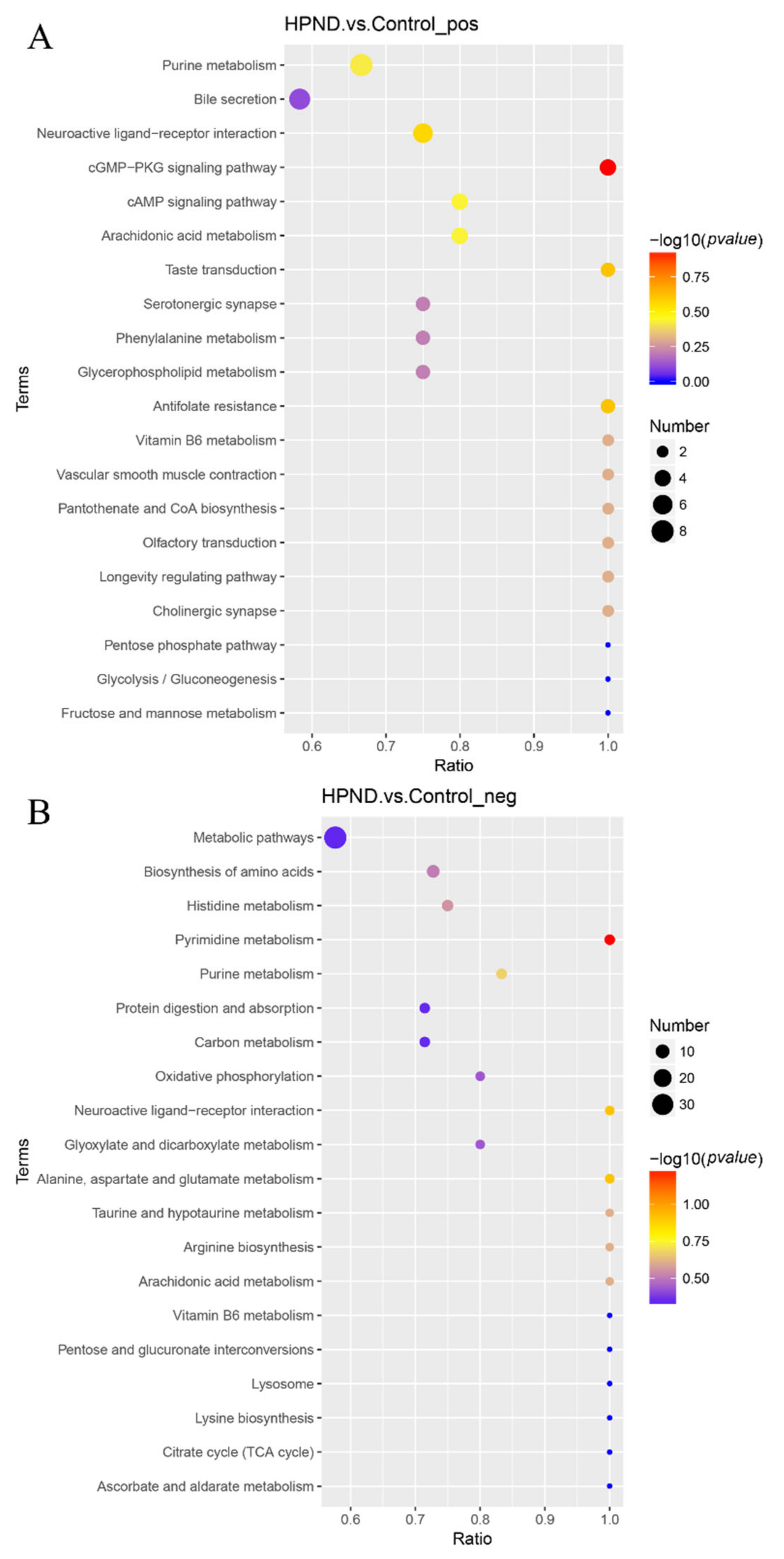
3.3. Metabolic Analysis of E. sinensis with HPND

LC-MS detections were performed in positive-ion (POS) modes and negative-ion (NEG) modes to maximize metabolite coverage. In the PCA analysis, the samples of different groups were grouped in distinct clusters, and the QC samples were clustered together, indicating that the samples of different groups were quite different, and the experiment was reproducible (Figure S1). Additionally, the PLS-DA model of each comparison group was established. The model evaluation parameters (POS: R2Y = 0.98, Q2Y = 0.92, NEG: R2Y = 0.97, Q2Y = 0.91) were obtained through 7-fold cross-validation, indicating that the model was well-established (Figure 4A,B). Next, the PLS-DA model was further validated by 200 permutation experiments. All R2 data were larger than the Q2 data in the positive and anion modes, and the Q2 regression line with Y-intercept was less than 0, indicating that the model was robust and reliable without overfitting (Figure 4C,D). Therefore, the experimental instrument analysis system was considered stable, and the data were reliable.
We screened for metabolites that were significantly different between the two groups based on the following criteria: VIP > 1.0, FC > 1.5 or FC < 0.667, and p-value < 0.05. In the POS and NEG mode, a total of 554 DMs were identified in the HPND vs. control group. Among them, 365 metabolites were in POS mode (210 up-regulated, 155 down-regulated) and 189 metabolites were in NEG mode (140 up-regulated, 49 down-regulated). In these DMs, a total of 87 metabolites were related to lipids, of which fatty acids (34) and glycerophospholipids (37) were the most abundant. KEGG enrichment analysis was performed on these DMs to explore the most relevant pathways. The results show that the top 20 enriched pathways were mainly related to the cGMP-PKG signaling pathway, purine metabolism, pyrimidine metabolism, arachidonic acid metabolism, amino acid biosynthesis, and neuroactive ligand–receptor interactions (Figure 5).

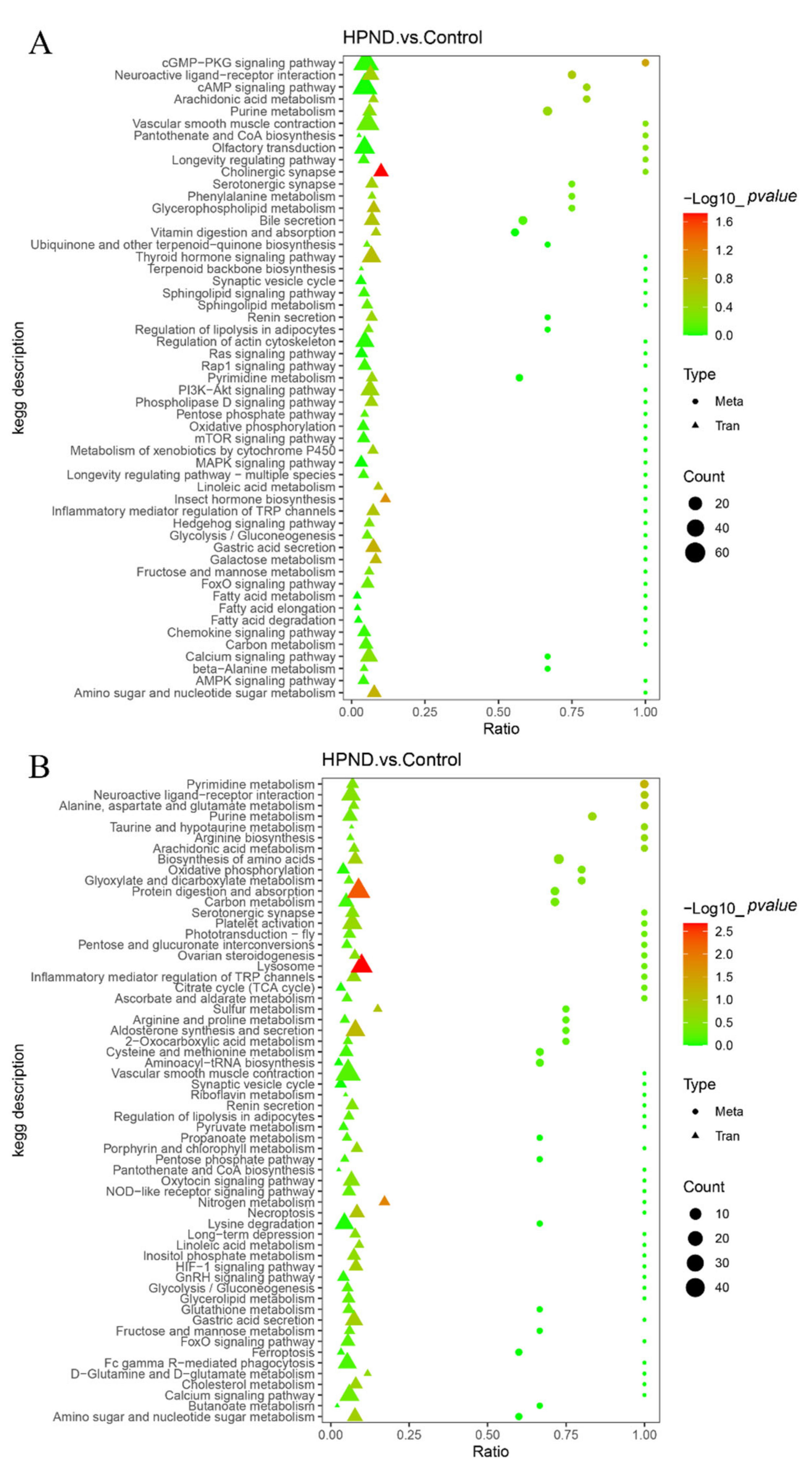
3.4. Integrated Analysis of Metabolome and Transcriptome

To further screen related genes and metabolites, as well as key metabolic pathways, we performed a comprehensive analysis of the transcriptome and metabolome. As shown in Figure S2, the top 50 differential metabolites had strong correlations with the top 100 differential genes. The KEGG enrichment results of DMs and DEGs identified several important response pathways in E. sinensis with HPND, including protein digestion and absorption, neuroactive ligand–receptor interactions, lysosome, purine metabolism, pyrimidine metabolism, oxidative phosphorylation, TCA cycle, and cholinergic synapse (Figure 6).

3.5. Molecular Responses of E. sinensis with HPND

Based on the results of comprehensive transcriptome and metabolome analysis, we found that the immune system, metabolic capacity, antioxidant system, detoxification capacity, and nervous system of Chinese mitten crabs with HPND were affected. Figure 7 shows hypothetical pathways for molecular responses in diseased crabs. Interestingly, many genes related to autophagy and apoptosis were significantly up-regulated in the HPND group, which may suggest that higher levels of cellular degradation occurred in the hepatopancreas of HPND-affected crabs. More details are provided in Supplementary Table S1. Some metabolites, such as taurine, arachidonic acid, uracil, xanthine, adenine, and uridine are associated with the immune system and were dysregulated in our study (Table 1). Additionally, we found that some genes and metabolites that participate in mitochondrial function were dysregulated, most of which were up-regulated. For example, in the mitochondrial respiratory chain complex I (NADH dehydrogenase), the ND4 and ND5 genes were significantly down-regulated, and Ndufab12 was significantly up-regulated; COX7A, COX11, and COX15 were significantly up-regulated in complex IV (cytochrome c oxidase); and ATP6F and ATP6C were significantly up-regulated in complex V (ATP synthase).

4. Discussion

Due to its high fatality rate (40–50%), the etiology and pathogenesis of HPND have attracted much attention [7]. Previous studies have shown that a single factor of pathogens, bacteria, and viruses does not directly cause HPND. After investigating the living environment, offspring seeds, and epidemiology of Chinese mitten crabs, recent studies have inferred that HPND is likely caused by abiotic factors. However, the real pathogenic factors still need to be explored further [3,4,5]. In this study, transcript and metabolite changes in diseased crabs were determined by transcriptomics and a metabolomics analysis of healthy and HPND-affected crabs.

4.1. Abnormalities of the Nervous System

In invertebrates, the nervous system plays a key role in coping with stress. Neurons can not only receive and process cues from environmental stressors but can also actively respond to promote survival [46,47]. Nonetheless, environmental stress can cause irreversible damage to neurons [48]. For example, hypoxia can cause neuronal dysfunction in C. elegans, leading to irreversible neuronal damage [47]. In this study, we found that some differentially expressed genes and metabolites were enriched in the calcium signaling pathway, neuroactive ligand–receptor interaction, and cholinergic synapse.
It has been reported that some pesticides (e.g., trichlorfon, deltamethrin, and chlorpyrifos) can cause neurotoxicity in Chinese mitten crab, which in turn affects the transmission of nerve impulses [12,49]. The systemic toxicity of these pesticides lies in their ability to inhibit acetylcholinesterase (AChE) [50,51]. The main biological role of AChE is to rapidly hydrolyze the neurotransmitter acetylcholine (ACh) to terminate impulse transmission at cholinergic synapses [44]. Nerve conduction becomes inhibited when ACh accumulates excessively, resulting in neuronal dysfunction and death [45]. Therefore, the inhibition of AChE activity is often used as a biomarker of neurotoxic substances [52]. In our study, the activity of AChE was inhibited, and the content of acetylcholine increased. This suggests that the diseased crabs may have been affected by a neurotoxin. Additionally, the content of L-glutamate was significantly up-regulated in the HPND group. L-glutamate is an important neurotransmitter and can activate several subtypes of receptors, resulting in increased intracellular calcium concentration [43,49]. When calcium levels rise in postsynaptic neurons, a cascade of signaling events is activated, which is critical for neuronal signal processing and survival [53]. Furthermore, the activation of neuroactive ligand–receptor interactions is important for neural repair [54]. Therefore, our findings suggest that failure to detect and adapt to environmental stressors can result in irreversible neuronal damage in diseased crabs.

4.2. Oxidative Stress and Impaired Detoxification

Studies have shown that intense environmental stress can disrupt the homeostasis of reactive oxygen species (ROS) in aquatic organisms, resulting in elevated levels of ROS and damage to cellular components, which is commonly referred to as “oxidative stress” [55,56]. Among the damaged cellular components are macromolecules, such as lipids and proteins [57]. Therefore, heat shock proteins (HSPs) can prevent cellular oxidative stress by renaturing denatured proteins [58]. In particular, HSP70 and HSP90 are often used as indicators of the health status of aquatic organisms [59,60]. Specifically, HSP70 can maintain cellular homeostasis, and HSP90 can prevent irreversible protein aggregation under stressful conditions [36,61]. Therefore, these proteins are typically up-regulated in Chinese mitten crabs under oxidative stress, as demonstrated in studies of crabs exposed to imidacloprid and avermectin [62,63]. Thus, the up-regulation of HSP70 and HSP90 in the HPND group in our study is likely a reflection of the body’s oxidative stress state.
Additionally, our study also found significant changes in two antioxidants PRDX5 and Glutathione S-transferase (GST). PRDX5 is a catalase that protects glial cells from oxidative stress [34]. The overexpression of PRDX5 further confirmed that the nervous system of the diseased crab may be affected. In contrast, the expression of GST was significantly inhibited, which may be related to the detoxification effect of GST. GST and carboxylesterase (CarEs) are two important detoxification enzymes in insects and play an important role in pesticide metabolism [64]. In crustaceans, these two enzymes are primarily active and metabolized in the hepatopancreas [65]. Among them, GST is a group of transferase enzymes involved in pesticide metabolism, and its activity is related to pesticide metabolism [38]. CarEs was identified as the main enzyme for decomposing pyrethroids and can be used as an indicator of pyrethroid toxicity [39]. The activities of CarEs and GST were found to be inhibited in many experiments on pesticide stress in Chinese mitten crabs [12,13]. Therefore, the expression of these two enzymes was down-regulated in the HPND group, indicating that the diseased crabs were likely to be stressed by pesticides such as pyrethroids, resulting in a decrease in their detoxification ability. In our metabolome data, the presence of some toxic phenylpropanoids and polyketides, such as 4-methoxycinnamic acid, aflatoxin G1, and 3-hydroxy-4-methoxycinnamic acid, further support this hypothesis.
However, we did not find significant changes in the activities of other antioxidants such as superoxide dismutase (SOD) and catalase (CAT), which are often used as indicators of oxidative stress in environmental pollution studies [66]. One possible explanation is that Chinese mitten crabs with HPND are exposed to lower concentrations of environmental pollutants. For example, Hong et al., found that when Chinese mitten crabs were exposed to less than 4.4 mg/L glyphosate, and the activities of SOD and peroxidase (POD) did not significantly change [67]. Collectively, our findings suggest that diseased crabs may be stressed by environmental pollutants, resulting in oxidative stress and impaired detoxification.

4.3. Increase in Autophagy and Apoptosis

Hepatopancreas damage is the most relevant pathological feature of E. sinensis with HPND (Figure 1). Previous studies found that hepatocytes from diseased crabs exhibit nuclear chromatin condensation, endoplasmic reticulum disintegration, and mitochondrial and lipid droplet cristae deformation. This is a pathological feature of apoptosis and autophagy [5]. Additionally, liver injury is reported to be closely related to autophagy and apoptosis [68,69]. In our study, many genes related to autophagy and apoptosis were found to be altered, and most of them were up-regulated.
To survive starvation and other forms of stress, eukaryotic cells can degrade the cytoplasm through lysosomes and this process is generally referred to as autophagy [70]. During the early stages of autophagy, Beclin-1 can activate downstream genes by recruiting other autophagy-related (Atg) proteins [71]. For example, the Beclin 1-Atg14L complex may participate in the early steps of autophagic biosynthesis by recruiting Atg5 and Atg12 [24]. Therefore, the overexpression of Atg14L can promote the formation of autophagosomes and up-regulate autophagy [24]. In our study, Atg14L was significantly up-regulated in the HPND group, as well as Atg1 and Atg7. The overexpression of Atg1 was shown to be sufficient to induce high levels of autophagy [26]. Atg7 plays an important role in starvation-induced autophagy [25]. Therefore, our study suggests that high levels of autophagy occur in diseased crabs. The overexpression of Atg7 may be associated with the emergence of the jejunum of diseased crabs. Rubicon is a Beclin 1-interacting protein whose forced expression results in abnormal lysosomal structure and impaired autophagosome maturation, thus inhibiting autophagy [24]. Interestingly, we found that Rubicon was significantly up-regulated and lysosomal-related genes and metabolites were significantly induced in the HPND group, which may be a manifestation of uncontrolled autophagy in the body.
Apoptosis is a form of programmed cell death that can be mediated by mitochondria, the endoplasmic reticulum, and death receptors [72,73]. In eukaryotes, an increase in misfolded or unfolded proteins causes a stress response in the endoplasmic reticulum, which reduces protein synthesis and increases protein folding [30]. However, excessive or prolonged ER stress can lead to apoptosis [74]. PERK and IP3R play important roles in endoplasmic reticulum stress-induced apoptosis. PERK is a protein kinase distributed on the endoplasmic reticulum membrane. When high-intensity or prolonged endoplasmic reticulum stress occurs, PERK will activate the transcriptional expression of the transcription factor ATF4, thereby promoting the expression of the apoptosis signaling molecule CHOP, which in turn promotes apoptosis [29,75]. On the other hand, a large number of calcium ions in the endoplasmic reticulum can enter cells through the activated IP3R channel and activate Caspase-4, thereby affecting apoptosis [76,77]. Therefore, the significant up-regulation of PERK, ATF4, CHOP, and Caspase-4 in our study suggested the activation of ER-mediated apoptosis in diseased crabs. Based on previous results, this may be related to oxidative damage in the diseased crabs. In mitochondria-mediated apoptosis, mitochondria can release cytochrome c by changing the outer membrane permeability (MOMP) and gradually activate Apaf-1 and Caspase-9 with the assistance of ATP and dATP. These components then form apoptotic bodies to activate caspase-3 and caspase-7, resulting in apoptosis [31,78]. Therefore, Bcl-2 family proteins can promote or inhibit apoptosis by changing MOMP and then affecting the release of cytochrome c [79]. In our study, ADP content was significantly increased and Caspase-7 expression was significantly up-regulated. However, cytochrome c and Caspase-9 did not significantly change, which may be related to the significant up-regulation of BNIP3 and BUFFY. BNIP3, a hypoxia-induced cell death protein, can cause mitochondrial dysfunction [27,80]. BUFFY is a Bcl-2 family protein with anti-apoptotic effects and its overexpression also induces cell death [28,81]. Therefore, our study indicated that diseased crabs presented high levels of apoptosis, as well as the possibility of abnormal mitochondrial function. It is worth mentioning that diseased crabs die within a short period of time out of the water. Previous studies have shown that hypoxia exacerbates the occurrence of HPND [11]. We speculate that this may be related to the overexpression of BNIP3 and mitochondrial dysfunction.
IL-17 is known to have pro-inflammatory effects and is associated with a variety of autoimmune diseases [82,83]. However, studies have shown that activation of the IL-17 signaling pathway is also closely related to autophagy and apoptosis [84,85]. For example, IL-17-induced apoptosis was alleviated after Act1 knockdown [86]. Furthermore, Zhao et al. found that fluoride could induce apoptosis and autophagy by activating the IL-17 signaling pathway and ultimately damage hepatocytes [87]. The present study also found that the IL-17 signaling pathway was activated in the diseased crabs. This may suggest that the hepatopancreas damage of the Chinese mitten crabs with HPND may be the result of excessive autophagy and apoptosis induced by environmental pollutants. Based on our previous results, we suspect that this may be related to the recent and widespread use of pyrethroids for pond cleaning in Xinghua city, Jiangsu province.

4.4. Impairment of Immune System

As an invertebrate, the Chinese mitten crab lacks adaptive immunity and only relies on innate immunity. In this study, we found that in addition to the lysosome and IL-17 signaling pathways, other pathways related to innate immunity were significantly induced in the diseased crabs, such as the CGMP-PKG signaling pathway, melanogenesis, and endocytosis. Crustacean innate defense systems include cellular and humoral immunity [88]. In humoral immunity, lysosomes can degrade substances through their own various hydrolases, thus playing an important role [89]. Alkaline phosphatase (AKP), acid phosphatase (ACP), lysozyme (LZM), cathepsin L, and cathepsin B are several important hydrolases in lysosomes. Among them, AKP, ACP, and LZM are considered to be the first line of defense within the humoral defense system [32]. Cathepsin L and cathepsin B are important proteases in lysosomes [90]. In our study, genes other than ACP were significantly induced. The insignificant changes in ACP may be related to the fact that a high pH is suspected to induce the occurrence of HPND [5]. Phagocytosis and apoptosis are important cellular immune response processes in invertebrates. Lectin and cathepsin L contribute to phagocytosis [33]. In our study, the C-type lectin, C-type lectin receptor protein, and cathepsin L genes were significantly down-regulated in diseased crabs, suggesting that the phagocytic activity of diseased crabs was reduced. Therefore, we concluded that the innate immunity of Chinese mitten crabs with HPND would be affected. It is worth mentioning that the high incidence of HPND is from May to July each year, during which time the weather changes greatly and the temperature is high. Dramatic changes in climate can further weaken a crustacean’s immune system and exacerbate the development of the disease [91]. Therefore, we suspect that unstable weather may exacerbate the occurrence of HPND.

4.5. Energy and Substance Metabolism Disorders

In response to environmental challenges, aquatic organisms can maintain homeostasis by increasing energy production, such as up-regulating the TCA cycle, oxidative phosphorylation, and glycolysis [15,48,92]. Pyruvate is the end product of glycolysis, and the resulting pyruvate can be converted into acetyl-CoA to enter the TCA cycle. In the HPND group, we found that some key metabolites (dihydroxyacetone phosphate, succinate, citrate) and key genes (hexokinase, acetyl-CoA synthetase, aconitate hydratase) involved in glycolysis and the TCA cycle were significantly up-regulated. The same result also occurs in oxidative phosphorylation (Figure 7). This indicates that Chinese mitten crabs with HPND appropriately increased their energy production. It is also worth mentioning that anaerobic metabolism is characterized by high levels of end products (e.g., alanine and succinate) in invertebrate species [40,93]. Therefore, the accumulation of succinate in the HPND group in our study is likely to indicate the occurrence of anaerobic metabolism in diseased crabs.
Lipids play an important role in energy storage and release in organisms. In our metabolomic data, many metabolites were associated with lipids, with fatty acids and glycerophospholipids accounting for the largest proportions of lipids. Studies have shown that fatty acid metabolism in crustaceans is closely related to immune responses [94]. As an essential fatty acid, arachidonic acid can affect the activity of cell membranes and membrane-associated enzymes, thereby altering immune function [41]. Abnormal arachidonic acid metabolism was also found in our results. Therefore, abnormal fatty acid metabolism may be related to immune dysregulation in diseased crabs. It is generally known that biological membranes are mainly composed of phospholipids. Most pesticides are highly lipophilic, and thus they primarily affect cell membranes [95]. Interestingly, almost all glycerophospholipid metabolites were downregulated in our results (Table S2).
Furthermore, some differentially expressed genes and metabolites were significantly enriched in pyrimidine and purine metabolism pathways in our results. Pyrimidines and purines are essential for life and perform many important cellular functions [96]. Viral infection can alter some metabolites involved in pyrimidine and purine metabolism [97,98]. Likewise, the apoptosis of hepatopancreatic cells can lead to disturbances in nucleotide metabolism [99]. Therefore, disturbances in pyrimidine and purine metabolism may lead to immunodeficiency [100]. In our study, some metabolites in purine and pyrimidine metabolic pathways, such as uracil, xanthine, adenine, and uridine, were significantly up-regulated in the diseased crabs. The accumulation of uracil and xanthine has been reported to induce apoptosis [42]. Therefore, we could deduce that the dysregulation of purine and pyrimidine metabolism in diseased Chinese mitten crabs is related to the high levels of autophagy and apoptosis in these organisms.
Additionally, we found that several other metabolic pathways were also altered, including starch and sucrose metabolism, protein digestion and absorption, vitamin digestion and absorption, and alanine, aspartate, and glutamate metabolism. Therefore, our findings suggest that the metabolic capacity of E. sinensis with HPND is adversely affected.

5. Conclusions

In conclusion, this study provide insights into metabolic changes and functional changes of Eriocheir sinensis with HPND by adopting a comprehensive metabolomic and transcriptomic analysis approach. The enriched pathways of DEGs and DMs may be related to the pathological mechanism of HPND and suggest that HPND could mainly be caused by abiotic factors. However, additional studies are required to identify the true pathogenic cause of HPND and whether it is caused by a single factor or a combination of factors.

Supplementary Materials

The following supporting information can be downloaded at: https://www.mdpi.com/article/10.3390/biology11091267/s1, Figure S1: PCA score plots in different groups (A POS mode, B NEG mode). Figure S2: The heat plot of the correlations between the top 50 differentially expressed metabolites (rows) and top 100 differentially expressed genes (columns). The red and blue colors show the positively and negative correlation between transcriptomics and metabolomics data (A POS mode, B NEG mode). Table S1: Differentially expressed genes associated with autophagy and apoptosis. Table S2: Differentially expressed metabolites associated with glycerophospholipid.

Author Contributions

Conceptualization, H.S.; Data curation, M.Z. (Ming Zhan) and L.W.; Formal analysis, M.Z. (Mengru Zhu), J.G., C.X., H.W. and G.X.; Investigation, M.Z. (Mengru Zhu) and C.X.; Methodology, H.S.; Project administration, H.S.; Resources, M.Z. (Mengru Zhu), J.G., C.X., H.W. and G.X.; Software, M.Z. (Ming Zhan) and L.W.; Supervision, G.X.; Validation, H.S.; Writing—original draft, M.Z. (Ming Zhan); Writing—review and editing, L.W. All authors have read and agreed to the published version of the manuscript.

Funding

This research was funded by the National Key Research and Development Program of China (2019YFC1605800), as well as the Jiangsu Natural Science Foundation Project (BK20181138).

Institutional Review Board Statement

Not applicable.

Informed Consent Statement

Not applicable.

Data Availability Statement

The transcriptome datasets generated by this study have been deposited into the NCBI SRA database (Accession number: SUB11342700; https://submit.ncbi.nlm.nih.gov/subs/sra/SUB11342700, accessed on 17 April 2022).

Conflicts of Interest

The authors declare no conflict of interest.

References

  1. Kong, L.; Cai, C.; Ye, Y.; Chen, D.; Wu, P.; Li, E.; Chen, L.; Song, L. Comparison of non-volatile compounds and sensory characteristics of Chinese mitten crabs (Eriocheir sinensis) reared in lakes and ponds: Potential environmental factors. Aquaculture 2012, 364, 96–102. [Google Scholar] [CrossRef]
  2. Yearbook CFS. China Fishery Statistical Yearbook; China Agriculture Press: Beijing, China, 2020; Volume 2020, pp. 24–34. [Google Scholar]
  3. Shen, H.; Zang, Y.; Song, K.; Ma, Y.; Dai, T.; Serwadda, A. A meta-transcriptomics survey reveals changes in the microbiota of the Chinese mitten crab Eriocheir sinensis infected with Hepatopancreatic necrosis disease. Front. Microbiol. 2017, 8, 732. [Google Scholar] [CrossRef] [PubMed]
  4. Cui, L.; Tang, S.; Qi, R.; Lei, Y.; Li, Y.; Wang, J. Pathological study on “Shuibiezi” disease of crab Eriocheir sinensis. J. Yantai Univ. 2017, 30, 313–316. [Google Scholar]
  5. Pan, Z.; Song, X.; Hu, X.; Xue, R.; Cao, G.; Zar, M.S.; Kumar, D.; Feng, Y.; Wei, Y.; Zhang, W. Pathological changes and risk factors of hepatopancreas necrosis disease of mitten crab, Eriocheir Sinensis. Fish. Aquac. J. 2017, 8, 3. [Google Scholar] [CrossRef]
  6. Zhan, M.; Xi, C.; Gong, J.; Zhu, M.; Sui, Y.; Xu, Z.; Xu, G.; Shen, H. 16S rRNA gene sequencing analysis reveals an imbalance in the intestinal flora of Eriocheir sinensis with hepatopancreatic necrosis disease. Comp. Biochem. Physiol. Part D Genom. Proteom. 2022, 42, 100988. [Google Scholar] [CrossRef] [PubMed]
  7. Ding, Z.; Meng, Q.; Liu, H.; Yuan, S.; Zhang, F.; Sun, M.; Zhao, Y.; Shen, M.; Zhou, G.; Pan, J. First case of hepatopancreatic necrosis disease in pond-reared Chinese mitten crab, Eriocheir sinensis, associated with microsporidian. J. Fish Dis. 2016, 39, 1043–1051. [Google Scholar] [CrossRef] [PubMed]
  8. Gao, T.; Xu, Y.; Wang, K.; Deng, Y.; Yang, Y.; Lu, Q.; Pan, J.; Xu, Z. Comparative LC-MS based non-targeted metabolite profiling of the Chinese mitten crab Eriocheir sinensis suffering from hepatopancreatic necrosis disease (HPND). Aquaculture 2018, 491, 338–345. [Google Scholar] [CrossRef]
  9. Yang, Z.; Zhang, Y.; Hu, K.; Liu, L.; Cai, H.; Zhang, F.; Yang, X. Etiological and histopathological study on hepatopancreatic necrosis syndrome in Eriocheir sinensis. Acta Hydrobiol. Sin. 2018, 42, 17–25. [Google Scholar]
  10. Zhu, J.; Wang, Z.; Cai, C.; Tang, X.; Shen, J.; Wu, D. Inducement and prevention technology of “Shuibiezi” disease of Chinese mitten crab Eriocheir sinensis. Sci. Fish Farming 2016, 6, 13–15. [Google Scholar]
  11. Gu, X.; Jiang, G.; Wei, B.; Niu, J.; Song, X. Investigation and analysis on the correlation of pesticides and hypoxia with “Shuibiezi” disease of Eriocheir sinensis. Sci. Fish Farming 2017, 4, 59–61. [Google Scholar]
  12. Zhu, M.; Gong, J.; Zhan, M.; Xi, C.; Shen, G.; Shen, H. Transcriptome analysis reveals the molecular mechanism of long-term exposure of Eriocheir sinensis to low concentration of trichlorfon.Comp. Biochem. Physiol. Part D Genom. Proteom. 2021, 40, 100916. [Google Scholar]
  13. Shen, G.; Shui, Y.; Zhang, X.; Song, K.; Wang, Y.; Xu, Z.; Shen, H. Hepatopancreatic necrosis disease (HPND) in Chinese mitten crab Eriocheir sinensis tightly linked to low concentration of two insecticides. Aquac. Res. 2021, 52, 2294–2304. [Google Scholar] [CrossRef]
  14. Shen, Z.; Kumar, D.; Liu, X.; Yan, B.; Fang, P.; Gu, Y.; Li, M.; Xie, M.; Yuan, R.; Feng, Y. Metatranscriptomic analysis reveals an imbalance of hepatopancreatic flora of Chinese mitten crab Eriocheir sinensis with hepatopancreatic necrosis disease. Biology 2021, 10, 462. [Google Scholar] [CrossRef]
  15. Wang, T.; Yang, C.; Zhang, S.; Rong, L.; Yang, X.; Wu, Z.; Sun, W. Metabolic changes and stress damage induced by ammonia exposure in juvenile Eriocheir sinensis. Ecotoxicol. Environ. Saf. 2021, 223, 112608. [Google Scholar] [CrossRef]
  16. Kong, T.; Ren, X.; Lin, S.; Li, S.; Gong, Y. Elucidation of metabolic responses in mud crab Scylla paramamosain challenged to WSSV infection by integration of metabolomics and transcriptomics. Dev. Comp. Immunol. 2020, 113, 103799. [Google Scholar] [CrossRef]
  17. Qiao, F.; Lei, K.; Li, Z.; Wei, Z.; Liu, Q.; Yang, L.; He, J.; An, L.; Qi, H.; Cui, S. Transcriptomic responses of the freshwater snail (Parafossarulus striatulus) following dietary exposure to cyanobacteria. Sci. Total Environ. 2018, 624, 153–161. [Google Scholar] [CrossRef]
  18. Mercuro, G.; Bassareo, P.P.; Deidda, M.; Cadeddu, C.; Barberini, L.; Atzori, L. Metabolomics: A new era in cardiology? J. Cardiovasc. Med. 2011, 12, 800–805. [Google Scholar] [CrossRef]
  19. Ren, S.; Shao, Y.; Zhao, X.; Hong, C.S.; Wang, F.; Lu, X.; Li, J.; Ye, G.; Yan, M.; Zhuang, Z. Integration of metabolomics and transcriptomics reveals major metabolic pathways and potential biomarker involved in prostate cancer. Mol. Cell. Proteom. 2016, 15, 154–163. [Google Scholar] [CrossRef]
  20. Want, E.J.; Masson, P.; Michopoulos, F.; Wilson, I.D.; Theodoridis, G.; Plumb, R.S.; Shockcor, J.; Loftus, N.; Holmes, E.; Nicholson, J.K. Global metabolic profiling of animal and human tissues via UPLC-MS. Nat. Protoc. 2013, 8, 17–32. [Google Scholar] [CrossRef]
  21. Wen, B.; Mei, Z.; Zeng, C.; Liu, S. metaX: A flexible and comprehensive software for processing metabolomics data. BMC Bioinform. 2017, 18, 183. [Google Scholar] [CrossRef]
  22. Langmead, B.; Salzberg, S.L. Fast gapped-read alignment with Bowtie 2. Nat. Methods 2012, 9, 357–359. [Google Scholar] [CrossRef]
  23. Li, B.; Dewey, C.N. RSEM: Accurate transcript quantification from RNA-Seq data with or without a reference genome. BMC Bioinform. 2011, 12, 323. [Google Scholar] [CrossRef]
  24. Zhong, Y.; Wang, Q.J.; Li, X.; Yan, Y.; Backer, J.M.; Chait, B.T.; Heintz, N.; Yue, Z. Distinct regulation of autophagic activity by Atg14L and Rubicon associated with Beclin 1–phosphatidylinositol-3-kinase complex. Nat. Cell Biol. 2009, 11, 468–476. [Google Scholar] [CrossRef]
  25. Komatsu, M.; Waguri, S.; Ueno, T.; Iwata, J.; Murata, S.; Tanida, I.; Ezaki, J.; Mizushima, N.; Ohsumi, Y.; Uchiyama, Y. Impairment of starvation-induced and constitutive autophagy in Atg7-deficient mice. J. Cell Biol. 2005, 169, 425–434. [Google Scholar] [CrossRef]
  26. Scott, R.C.; Juhász, G.; Neufeld, T.P. Direct induction of autophagy by Atg1 inhibits cell growth and induces apoptotic cell death. Curr. Biol. 2007, 17, 1–11. [Google Scholar] [CrossRef] [Green Version]
  27. Kothari, S.; Cizeau, J.; McMillan-Ward, E.; Israels, S.J.; Bailes, M.; Ens, K.; Kirshenbaum, L.A.; Gibson, S.B. BNIP3 plays a role in hypoxic cell death in human epithelial cells that is inhibited by growth factors EGF and IGF. Oncogene 2003, 22, 4734–4744. [Google Scholar] [CrossRef]
  28. Quinn, L.; Coombe, M.; Mills, K.; Daish, T.; Colussi, P.; Kumar, S.; Richardson, H. Buffy, a Drosophila Bcl-2 protein, has anti-apoptotic and cell cycle inhibitory functions. EMBO J. 2003, 22, 3568–3579. [Google Scholar] [CrossRef]
  29. Harding, H.P.; Zhang, Y.; Bertolotti, A.; Zeng, H.; Ron, D. Perk is essential for translational regulation and cell survival during the unfolded protein response. Mol. Cell 2000, 5, 897–904. [Google Scholar] [CrossRef]
  30. Hitomi, J.; Katayama, T.; Eguchi, Y.; Kudo, T.; Taniguchi, M.; Koyama, Y.; Manabe, T.; Yamagishi, S.; Bando, Y.; Imaizumi, K. Involvement of caspase-4 in endoplasmic reticulum stress-induced apoptosis and Aβ-induced cell death. J. Cell Biol. 2004, 165, 347–356. [Google Scholar] [CrossRef]
  31. Srinivasula, S.M.; Ahmad, M.; Fernandes-Alnemri, T.; Alnemri, E.S. Autoactivation of procaspase-9 by Apaf-1-mediated oligomerization. Mol. Cell 1998, 1, 949–957. [Google Scholar] [CrossRef]
  32. Rajalakshmi, S.; Mohandas, A. Copper-induced changes in tissue enzyme activity in a freshwater mussel. Ecotoxicol. Environ. Saf. 2005, 62, 140–143. [Google Scholar] [CrossRef] [PubMed]
  33. Martin, M.; Blom, A.M. Complement in removal of the dead–balancing inflammation. Immunol. Rev. 2016, 274, 218–232. [Google Scholar] [CrossRef] [PubMed]
  34. Walbrecq, G.; Wang, B.; Becker, S.; Hannotiau, A.; Fransen, M.; Knoops, B. Antioxidant cytoprotection by peroxisomal peroxiredoxin-5. Free Radic. Biol. Med. 2015, 84, 215–226. [Google Scholar] [CrossRef] [PubMed]
  35. Shengming, S.; Jian, Z.; Xianping, G.; Chengfeng, Z.; Linghong, M.; Wuxiao, Z.; Qiong, Z. Molecular cloning, characterization and mRNA expression of Mu-typ glutathione S-transferases from Megalobrama amblycephala. Asian J. Ecotoxicol. 2016, 11, 295–305. [Google Scholar]
  36. Parsell, D.; Lindquist, S. The function of heat-shock proteins in stress tolerance: Degradation and reactivation of damaged proteins. Ann. Rev. Genet. 1993, 27, 437–496. [Google Scholar] [CrossRef]
  37. Feder, M.E.; Hofmann, G.E. Heat-shock proteins, molecular chaperones, and the stress response: Evolutionary and ecological physiology. Annu. Rev. Physiol. 1999, 61, 243–282. [Google Scholar] [CrossRef] [Green Version]
  38. Hemingway, J.; Ranson, H. Insecticide resistance in insect vectors of human disease. Annu. Rev. Entomol. 2000, 45, 371–391. [Google Scholar] [CrossRef]
  39. Wheelock, C.E.; Miller, J.L.; Miller, M.J.; Phillips, B.M.; Huntley, S.A.; Gee, S.J.; Tjeerdema, R.S.; Hammock, B.D. Use of carboxylesterase activity to remove pyrethroid-associated toxicity to Ceriodaphnia dubia and Hyalella azteca in toxicity identification evaluations. Environ. Toxicol. Chem. 2006, 25, 973–984. [Google Scholar] [CrossRef]
  40. Grieshaber, M.; Hardewig, I.; Kreutzer, U.; Pörtner, H.-O. Physiological and metabolic responses to hypoxia in invertebrates. Rev. Physiol. Biochem. Pharm. 1993, 125, 43–147. [Google Scholar]
  41. Calder, P.C. The relationship between the fatty acid composition of immune cells and their function. Prostaglandins Leukot. Essent. Fat. Acids 2008, 79, 101–108. [Google Scholar] [CrossRef]
  42. Paone, A.; Marani, M.; Fiascarelli, A.; Rinaldo, S.; Giardina, G.; Contestabile, R.; Paiardini, A.; Cutruzzolà, F. SHMT1 knockdown induces apoptosis in lung cancer cells by causing uracil misincorporation. Cell Death Dis. 2014, 5, e1525. [Google Scholar] [CrossRef]
  43. Irving, A.; Collingridge, G.; Schofield, J. L-glutamate and acetylcholine mobilise Ca2+ from the same intracellular pool in cerebellar granule cells using transduction mechanisms with different Ca2+ sensitivities. Cell Calcium 1992, 13, 293–301. [Google Scholar] [CrossRef]
  44. Dvir, H.; Silman, I.; Harel, M.; Rosenberry, T.L.; Sussman, J.L. Acetylcholinesterase: From 3D structure to function. Chem. Interact. 2010, 187, 10–22. [Google Scholar] [CrossRef]
  45. Mileson, B.E.; Chambers, J.E.; Chen, W.; Dettbarn, W.; Ehrich, M.; Eldefrawi, A.T.; Gaylor, D.W.; Hamernik, K.; Hodgson, E.; Karczmar, A.G. Common mechanism of toxicity: A case study of organophosphorus pesticides. Toxicol. Sci. 1998, 41, 8–20. [Google Scholar]
  46. Kagias, K.; Nehammer, C.; Pocock, R. Neuronal responses to physiological stress. Front. Genet. 2012, 3, 222. [Google Scholar] [CrossRef]
  47. Kim, K.W.; Jin, Y. Neuronal responses to stress and injury in C. elegans. FEBS Lett. 2015, 589, 1644–1652. [Google Scholar] [CrossRef]
  48. Sun, X.; Tu, K.; Li, L.; Wu, B.; Wu, L.; Liu, Z.; Zhou, L.; Tian, J.; Yang, A. Integrated transcriptome and metabolome analysis reveals molecular responses of the clams to acute hypoxia. Mar. Environ. Res. 2021, 168, 105317. [Google Scholar] [CrossRef]
  49. Yang, Z.; Zhang, Y.; Jiang, Y.; Zhu, F.; Zeng, L.; Wang, Y.; Lei, X.; Yao, Y.; Hou, Y.; Xu, L. Transcriptional responses in the hepatopancreas of Eriocheir sinensis exposed to deltamethrin. PLoS ONE 2017, 12, e0184581. [Google Scholar] [CrossRef] [Green Version]
  50. Tu, H.T.; Silvestre, F.; De Meulder, B.; Thome, J.-P.; Phuong, N.T.; Kestemont, P. Combined effects of deltamethrin, temperature and salinity on oxidative stress biomarkers and acetylcholinesterase activity in the black tiger shrimp (Penaeus monodon). Chemosphere 2012, 86, 83–91. [Google Scholar] [CrossRef]
  51. Slotkin, T.A.; Brown, K.K.; Seidler, F.J. Developmental exposure of rats to chlorpyrifos elicits sex-selective hyperlipidemia and hyperinsulinemia in adulthood. Environ. Health Perspect. 2005, 113, 1291–1294. [Google Scholar] [CrossRef]
  52. Talesa, V.; Contenti, S.; Principato, G.; Pascolini, R.; Giovannini, E.; Rosi, G. Cholinesterases from Maia verrucosa and Palinurus vulgaris: A comparative study. Comp. Biochem. Physiol. Part C Comp. Pharmacol. 1992, 101, 499–503. [Google Scholar] [CrossRef]
  53. Redmond, L.; Ghosh, A. Regulation of dendritic development by calcium signaling. Cell Calcium 2005, 37, 411–416. [Google Scholar] [CrossRef] [PubMed]
  54. Cheng, Q.; Wang, Y.-X.; Yu, J.; Yi, S. Critical signaling pathways during Wallerian degeneration of peripheral nerve. Neural Regen. Res. 2017, 12, 995. [Google Scholar] [PubMed]
  55. Lushchak, V.I. Environmentally induced oxidative stress in aquatic animals. Aquat. Toxicol. 2011, 101, 13–30. [Google Scholar] [CrossRef]
  56. Marklund, S.; Marklund, G. Involvement of the superoxide anion radical in the autoxidation of pyrogallol and a convenient assay for superoxide dismutase. Eur. J. Biochem. 1974, 47, 469–474. [Google Scholar] [CrossRef]
  57. Meng, X.; Jayasundara, N.; Zhang, J.; Ren, X.; Gao, B.; Li, J.; Liu, P. Integrated physiological, transcriptome and metabolome analyses of the hepatopancreas of the female swimming crab Portunus trituberculatus under ammonia exposure. Ecotoxicol. Environ. Saf. 2021, 228, 113026. [Google Scholar] [CrossRef]
  58. Hendrick, J.P.; Hartl, F.-U. Molecular chaperone functions of heat-shock proteins. Annu. Rev. Biochem. 1993, 62, 349–384. [Google Scholar] [CrossRef]
  59. Downs, C.A.; Fauth, J.E.; Woodley, C.M. Assessing the health of grass shrimp (Palaeomonetes pugio) exposed to natural and anthropogenic stressors: A molecular biomarker system. Mar. Biotechnol. 2001, 3, 380–397. [Google Scholar] [CrossRef]
  60. Kim, B.-M.; Rhee, J.-S.; Jeong, C.-B.; Seo, J.S.; Park, G.S.; Lee, Y.-M.; Lee, J.-S. Heavy metals induce oxidative stress and trigger oxidative stress-mediated heat shock protein (hsp) modulation in the intertidal copepod Tigriopus japonicus. Comp. Biochem. Physiol. Part C Toxicol. Pharmacol. 2014, 166, 65–74. [Google Scholar] [CrossRef]
  61. Rutherford, S.L.; Lindquist, S. Hsp90 as a capacitor for morphological evolution. Nature 1998, 396, 336–342. [Google Scholar] [CrossRef]
  62. Hong, Y.; Huang, Y.; Huang, Z. Oxidative stress, immunological response, and heat shock proteins induction in the Chinese Mitten Crab, Eriocheir sinensis following avermectin exposure. Environ. Toxicol. 2020, 35, 213–222. [Google Scholar] [CrossRef]
  63. Hong, Y.; Huang, Y.; Wu, S.; Yang, X.; Dong, Y.; Xu, D.; Huang, Z. Effects of imidacloprid on the oxidative stress, detoxification and gut microbiota of Chinese mitten crab, Eriocheir sinensis. Sci. Total Environ. 2020, 729, 138276. [Google Scholar] [CrossRef]
  64. Li, X.; Schuler, M.A.; Berenbaum, M.R. Molecular mechanisms of metabolic resistance to synthetic and natural xenobiotics. Annu. Rev. Entomol. 2007, 52, 231–253. [Google Scholar] [CrossRef]
  65. Arrese, E.L.; Soulages, J.L. Insect fat body: Energy, metabolism, and regulation. Ann. Rev. Entomol. 2010, 55, 207–225. [Google Scholar] [CrossRef]
  66. Song, P.; Ping, L.; Gao, J.; Li, X.; Zhu, M.; Wang, J. Ecotoxicological effects of fertilizers made from pulping waste liquor on earthworm Eisenia fetida. Ecotoxicol. Environ. Saf. 2018, 166, 237–241. [Google Scholar] [CrossRef]
  67. Hong, Y.; Huang, Y.; Yan, G.; Pan, C.; Zhang, J. Antioxidative status, immunological responses, and heat shock protein expression in hepatopancreas of Chinese mitten crab, Eriocheir sinensis under the exposure of glyphosate. Fish Shellfish Immunol. 2019, 86, 840–845. [Google Scholar] [CrossRef]
  68. Cui, Y.-Q.; Liu, Y.-J.; Zhang, F. The suppressive effects of Britannin (Bri) on human liver cancer through inducing apoptosis and autophagy via AMPK activation regulated by ROS. Biochem. Biophys. Res. Commun. 2018, 497, 916–923. [Google Scholar] [CrossRef]
  69. Lv, S.-X.; Qiao, X. Isovitexin (IV) induces apoptosis and autophagy in liver cancer cells through endoplasmic reticulum stress. Biochem. Biophys. Res. Commun. 2018, 496, 1047–1054. [Google Scholar] [CrossRef]
  70. Kamada, Y.; Yoshino, K.-i.; Kondo, C.; Kawamata, T.; Oshiro, N.; Yonezawa, K.; Ohsumi, Y. Tor directly controls the Atg1 kinase complex to regulate autophagy. Mol. Cell. Biol. 2010, 30, 1049–1058. [Google Scholar] [CrossRef]
  71. Cheng, Z.; Zhu, Q.; Dee, R.; Opheim, Z.; Mack, C.P.; Cyr, D.M.; Taylor, J.M. Focal Adhesion Kinase-mediated Phosphorylation of Beclin1 Protein Suppresses Cardiomyocyte Autophagy and Initiates Hypertrophic Growth. J. Biol. Chem. 2017, 292, 2065–2079. [Google Scholar] [CrossRef]
  72. Cohen, G.M. Caspases: The executioners of apoptosis. Biochem. J. 1997, 326, 1–16. [Google Scholar] [CrossRef]
  73. Pinton, P.; Giorgi, C.; Siviero, R.; Zecchini, E.; Rizzuto, R. Calcium and apoptosis: ER-mitochondria Ca2+ transfer in the control of apoptosis. Oncogene 2008, 27, 6407–6418. [Google Scholar] [CrossRef]
  74. Imaizumi, K.; Miyoshi, K.; Katayama, T.; Yoneda, T.; Taniguchi, M.; Kudo, T.; Tohyama, M. The unfolded protein response and Alzheimer’s disease. Biochim. Biophys. Acta (BBA) Mol. Cell Res. 2001, 1536, 85–96. [Google Scholar] [CrossRef]
  75. Li, J.; Lee, B.; Lee, A.S. Endoplasmic reticulum stress-induced apoptosis: Multiple pathways and activation of p53-up-regulated modulator of apoptosis (PUMA) and NOXA by p53. J. Biolog. Chem. 2006, 281, 7260–7270. [Google Scholar] [CrossRef] [PubMed]
  76. Marchi, S.; Patergnani, S.; Missiroli, S.; Morciano, G.; Rimessi, A.; Wieckowski, M.R.; Giorgi, C.; Pinton, P. Mitochondrial and endoplasmic reticulum calcium homeostasis and cell death. Cell Calcium 2018, 69, 62–72. [Google Scholar] [CrossRef] [PubMed]
  77. Chukai, Y.; Ito, G.; Konno, M.; Sakata, Y.; Ozaki, T. Mitochondrial calpain-5 truncates caspase-4 during endoplasmic reticulum stress. Biochem. Biophys. Res. Commun. 2022, 608, 156–162. [Google Scholar] [CrossRef] [PubMed]
  78. Slee, E.A.; Harte, M.T.; Kluck, R.M.; Wolf, B.B.; Casiano, C.A.; Newmeyer, D.D.; Wang, H.-G.; Reed, J.C.; Nicholson, D.W.; Alnemri, E.S. Ordering the cytochrome c–initiated caspase cascade: Hierarchical activation of caspases-2,-3,-6,-7,-8, and-10 in a caspase-9–dependent manner. J. Cell Biol. 1999, 144, 281–292. [Google Scholar] [CrossRef] [PubMed]
  79. Mukhopadhyay, S.; Panda, P.K.; Sinha, N.; Das, D.N.; Bhutia, S.K. Autophagy and apoptosis: Where do they meet? Apoptosis 2014, 19, 555–566. [Google Scholar] [CrossRef]
  80. Rikka, S.; Quinsay, M.N.; Thomas, R.L.; Kubli, D.A.; Zhang, X.; Murphy, A.N.; Gustafsson, Å.B. Bnip3 impairs mitochondrial bioenergetics and stimulates mitochondrial turnover. Cell Death Differ. 2011, 18, 721–731. [Google Scholar] [CrossRef]
  81. Igaki, T.; Miura, M. Role of Bcl-2 family members in invertebrates. Biochim. Biophys. Acta (BBA) Mol. Cell Res. 2004, 1644, 73–81. [Google Scholar] [CrossRef]
  82. Gooderham, M.; Posso-De Los Rios, C.J.; Rubio-Gomez, G.A.; Papp, K. Interleukin-17 (IL-17) inhibitors in the treatment of plaque psoriasis: A review. Ski. Ther. Lett. 2015, 20, 1–5. [Google Scholar]
  83. Chenuet, P.; Fauconnier, L.; Madouri, F.; Marchiol, T.; Rouxel, N.; Ledru, A.; Mauny, P.; Lory, R.; Uttenhove, C.; van Snick, J. Neutralization of either IL-17A or IL-17F is sufficient to inhibit house dust mite induced allergic asthma in mice. Clin. Sci. 2017, 131, 2533–2548. [Google Scholar] [CrossRef]
  84. Yuan, J.; Yu, M.; Li, H.-H.; Long, Q.; Liang, W.; Wen, S.; Wang, M.; Guo, H.-P.; Cheng, X.; Liao, Y.-H. Autophagy contributes to IL-17-induced plasma cell differentiation in experimental autoimmune myocarditis. Int. Immunopharmacol. 2014, 18, 98–105. [Google Scholar] [CrossRef]
  85. Kim, E.K.; Kwon, J.-E.; Lee, S.-Y.; Lee, E.-J.; Kim, D.S.; Moon, S.-J.; Lee, J.; Kwok, S.-K.; Park, S.-H.; Cho, M.-L. IL-17-mediated mitochondrial dysfunction impairs apoptosis in rheumatoid arthritis synovial fibroblasts through activation of autophagy. Cell Death Dis. 2018, 8, e2565. [Google Scholar] [CrossRef]
  86. Qiu, A.-W.; Bian, Z.; Mao, P.-A.; Liu, Q.-H. IL-17A exacerbates diabetic retinopathy by impairing Müller cell function via Act1 signaling. Exp. Mol. Med. 2016, 48, e280. [Google Scholar] [CrossRef]
  87. Zhao, Y.; Li, Y.; Wang, J.; Manthari, R.K.; Wang, J. Fluoride induces apoptosis and autophagy through the IL-17 signaling pathway in mice hepatocytes. Arch. Toxicol. 2018, 92, 3277–3289. [Google Scholar] [CrossRef]
  88. Li, F.; Xiang, J. Recent advances in researches on the innate immunity of shrimp in China. Dev. Comp. Immunol. 2013, 39, 11–26. [Google Scholar] [CrossRef]
  89. Yu, F.; Chen, Z.; Wang, B.; Jin, Z.; Hou, Y.; Ma, S.; Liu, X. The role of lysosome in cell death regulation. Tumor Biol. 2016, 37, 1427–1436. [Google Scholar] [CrossRef]
  90. Cory, S.; Adams, J.M. The Bcl2 family: Regulators of the cellular life-or-death switch. Nat. Rev. Cancer 2002, 2, 647–656. [Google Scholar] [CrossRef]
  91. Shields, J.D. Climate change enhances disease processes in crustaceans: Case studies in lobsters, crabs, and shrimps. J. Crustac. Biol. 2019, 39, 673–683. [Google Scholar] [CrossRef]
  92. Yu, N.; Ding, Q.; Li, E.; Qin, J.G.; Chen, L.; Wang, X. Growth, energy metabolism and transcriptomic responses in Chinese mitten crab (Eriocheir sinensis) to benzo [α] pyrene (BaP) toxicity. Aquat. Toxicol. 2018, 203, 150–158. [Google Scholar] [CrossRef] [PubMed]
  93. Ali, F.; Nakamura, K. Metabolic characteristics of the Japanese clam Ruditapes philippinarum (Adams & Reeve) during aerial exposure. Aquac. Res. 2000, 31, 157–165. [Google Scholar]
  94. Shao, Y.; Li, C.; Xu, W.; Zhang, P.; Zhang, W.; Zhao, X. miR-31 links lipid metabolism and cell apoptosis in bacteria-challenged Apostichopus japonicus via targeting CTRP9. Front. Immunol. 2017, 8, 263. [Google Scholar] [CrossRef] [PubMed]
  95. Cengiz, E.I.; Kan, Y.; Kizmaz, V.; Başhan, M.; Yanar, M. The protective role of vitamin E on the fatty acid composition of phospholipid structure in gill and liver tissues of Oreochromis niloticus exposed to deltamethrin. Ecotoxicol. Environ. Saf. 2012, 80, 381–385. [Google Scholar] [CrossRef]
  96. Carter, N.S.; Yates, P.; Arendt, C.S.; Boitz, J.M.; Ullman, B. Purine and pyrimidine metabolism in Leishmania. Drug Targets Kinetoplastid Parasites 2008, 625, 141–154. [Google Scholar]
  97. Chandler, J.D.; Hu, X.; Ko, E.-J.; Park, S.; Lee, Y.-T.; Orr, M.; Fernandes, J.; Uppal, K.; Kang, S.-M.; Jones, D.P. Metabolic pathways of lung inflammation revealed by high-resolution metabolomics (HRM) of H1N1 influenza virus infection in mice. Am. J. Physiol. Integr. Comp. Physiol. 2016, 311, R906–R916. [Google Scholar] [CrossRef]
  98. Kong, T.; Lin, S.; Ren, X.; Li, S.; Gong, Y. Transcriptome and metabolome integration analysis of mud crab Scylla paramamosain challenged to Vibrio parahaemolyticus infection. Fish Shellfish Immunol. 2020, 103, 430–437. [Google Scholar] [CrossRef]
  99. Xiao, J.; Li, Q.-Y.; Tu, J.-P.; Chen, X.-L.; Chen, X.-H.; Liu, Q.-Y.; Liu, H.; Zhou, X.-Y.; Zhao, Y.-Z.; Wang, H.-L. Stress response and tolerance mechanisms of ammonia exposure based on transcriptomics and metabolomics in Litopenaeus vannamei. Ecotoxicol. Environ. Saf. 2019, 180, 491–500. [Google Scholar] [CrossRef]
  100. Nyhan, W.L. Disorders of purine and pyrimidine metabolism. Mol. Genet. Metab. 2005, 86, 25–33. [Google Scholar] [CrossRef]
Figure 1. Healthy (A) and HPND-affected (D) E. sinensis. (B) and (E) represent the hepatic tubules of healthy and diseased crabs, respectively. (C) and (F) represent the appendage muscle tissue of healthy and diseased crabs, respectively. bm: basement membrane; nu: nucleus; mf: muscle fiber; va: vacuole; sc: secretory cell.
Biology 11 01267 g001
Figure 2. Volcano plot of differentially expressed genes.
Biology 11 01267 g002
Figure 3. Top 20 GO and KEGG pathways significantly enriched with differentially expressed genes ((A) top 20 of GO enrichment; (B) top 20 of KEGG enrichment).
Biology 11 01267 g003
Figure 4. PLS-DA score plots in different groups ((A,C) POS mode; (B,D) NEG mode).
Biology 11 01267 g004
Figure 5. Top 20 KEGG pathways significantly enriched with differentially expressed metabolites ((A) POS mode; (B) NEG mode).
Biology 11 01267 g005
Figure 6. KEGG enrichment analysis for differentially expressed genes and metabolites ((A), POS mode; (B), NEG mode).
Biology 11 01267 g006
Figure 7. Hypothetical pathways for metabolome and transcriptome changes in E. sinensis with HPND. The boxes represent genes, and the dots represent metabolites. Red and blue represent up- and down-regulation, respectively, and both red and blue represents both up- and down-regulation. The solid arrows and dashed arrows indicate direct and indirect effects. Arrows with × indicate inhibition. PK: pyruvate kinase; ACSS: acetyl-CoA synthetase; ACO: aconitate hydratase; HK: hexokinase; UMPK: UMP-CMP kinase; APRT: adenine phosphoribosyltransferase; ANPEP: aminopeptidase N.
Biology 11 01267 g007
Table 1. Significantly dysregulated genes and metabolites in crabs with HPND.
NameGene/MetaboliteUp/DownFunctionReference
Immune system
beclin 1-associated autophagy-related key regulator (ATG14L)geneuppromote the formation of autophagosomes[24]
ubiquitin-like modifier-activating enzyme ATG7 (ATG7)geneupassociated with starvation-induced autophagy[25]
threonine-protein kinase ULK2 (ATG1)geneup induce high levels of autophagy[26]
BCL2/adenovirus E1B 19 kDa protein-interacting protein 3(BNIP3)geneupcell death protein[27]
run domain Beclin-1 interacting and cysteine-rich containing protein (RUBICON)geneupinhibit autophagy[24]
Bcl-2 family protein (BUFFY)geneupinhibit apoptosis[28]
ER protein kinase (PERK)geneuppromote apoptosis[29]
Inositol 1,4,5-Trisphosphate Receptor (IP3R)geneuprelease calcium ions[30]
caspase 7(CASP7)geneupmolecular triggers of apoptosis[31]
caspase 4 (CASP4)geneupmolecular triggers of apoptosis[30]
Lysozyme (LZM)genedownlysosomal hydrolase[32]
alkaline phosphatase (AKP)geneuplysosomal hydrolase[32]
C-type lectin (CTL)genedownaids in phagocytosis[33]
Antioxidant system
peroxiredoxin 5(PRDX5)geneupthioredoxin peroxidase[34]
glutathione S-transferase (GST)genedownremoves lipid peroxides and hydrogen peroxide[35]
heat shock protein 70 (HSP70)geneupmaintain cellular homeostasis[36]
heat shock protein 90 (HSP90)geneupprevents irreversible protein aggregation[37]
Detoxification
glutathione S-transferase (GST)genedowndecomposition of pesticides[38]
carboxylesterase (CarEs)genedownenzyme for decomposing pyrethroids[39]
metabolic system
succinatemetaboliteupalternative end product of anaerobic metabolism[40]
arachidonic acidmetaboliteupinvolved in immune response[41]
uracilmetaboliteuprelated to apoptosis[42]
xanthinemetaboliteuprelated to apoptosis[42]
nervous system
L-glutamatemetaboliteupneurotransmitter[43]
acetylcholinesterase (AChE)genedownhydrolyzed acetylcholine[44]
acetylcholine (ACh)metaboliteupneurotransmitters[45]
Publisher’s Note: MDPI stays neutral with regard to jurisdictional claims in published maps and institutional affiliations.

