Article

Phenotypic and Genotypic Detection of Biofilm-Forming Staphylococcus aureus from Different Food Sources in Bangladesh

Department of Microbiology and Hygiene, Faculty of Veterinary Science, Bangladesh Agricultural University, Mymensingh 2202, Bangladesh
*
Author to whom correspondence should be addressed.
Biology 2022, 11(7), 949; https://doi.org/10.3390/biology11070949
Submission received: 8 May 2022 / Revised: 30 May 2022 / Accepted: 20 June 2022 / Published: 22 June 2022
(This article belongs to the Special Issue Microbial Contamination and Food Safety)

Abstract

:

Simple Summary

Biofilm formation by Staphylococcus aureus in foods poses a potential concern for public health and food safety. Therefore, the present study was conducted to detect biofilm-producing S. aureus from foods and human hand swabs using phenotypic and genotypic assays. In this study, S. aureus was detected in 23.81% (100/420) of samples, and among them, 89 and 97 of the isolates were biofilm producers by qualitative and quantitative tests, respectively. At least one biofilm-forming gene was detected in 21 S. aureus isolates, of which four isolates harbored all five adhesion genes (icaA, icaB, icaC, icaD, and bap). In addition, the occurrence of adhesion genes in S. aureus isolates showed a strong significant correlation among themselves. This is the first report on detecting biofilm-forming S. aureus from foods and hand swabs in Bangladesh using the molecular technique. The findings from this study indicate a significant public health risk and suggest the necessity of maintaining food hygiene practices at every step of the food chain to prevent and control S. aureus foodborne illness.

Abstract

Staphylococcus aureus is a major foodborne pathogen. The ability of S. aureus to produce biofilm is a significant virulence factor, triggering its persistence in hostile environments. In this study, we screened a total of 420 different food samples and human hand swabs to detect S. aureus and to determine their biofilm formation ability. Samples analyzed were meat, milk, eggs, fish, fast foods, and hand swabs. S. aureus were detected by culturing, staining, biochemical, and PCR. Biofilm formation ability was determined by Congo Red Agar (CRA) plate and Crystal Violet Microtiter Plate (CVMP) tests. The icaA, icaB, icaC, icaD, and bap genes involved in the synthesis of biofilm-forming intracellular adhesion compounds were detected by PCR. About 23.81% (100/420; 95% CI: 14.17–29.98%) of the samples harbored S. aureus, as revealed by detection of the nuc gene. The CRA plate test revealed 20% of S. aureus isolates as strong biofilm producers and 69% and 11% as intermediate and non-biofilm producers, respectively. By the CVMP staining method, 20%, 77%, and 3% of the isolates were found to be strong, intermediate, and non-biofilm producers. Furthermore, 21% of S. aureus isolates carried at least one biofilm-forming gene, where icaA, icaB, icaC, icaD, and bap genes were detected in 15%, 20%, 7%, 20%, and 10% of the S. aureus isolates, respectively. Bivariate analysis showed highly significant correlations (p < 0.001) between any of the two adhesion genes of S. aureus isolates. To the best of our knowledge, this is the first study in Bangladesh describing the detection of biofilm-forming S. aureus from foods and hand swabs using molecular-based evidence. Our findings suggest that food samples should be deemed a potential reservoir of biofilm-forming S. aureus, which indicates a potential public health significance.

1. Introduction

Staphylococcus aureus is a food-borne pathogen ranked as the third most common bacterial cause of foodborne illness worldwide [1]. Consuming foods contaminated with heat-stable staphylococcal enterotoxins is a major cause of staphylococcal food poisoning, which leads to abdominal cramps, diarrhea, nausea, vomiting, endocarditis, pneumonia, toxic shock, and even skin infections in humans [2]. Food handlers, hand contact surfaces, and food contact surfaces are important sources of S. aureus transmission in food processing facilities, particularly during food processing and packaging [3].
Various pathogenic bacteria or other spoilage bacteria can be attached to hand and food contact surfaces as planktonic or adherent cells to form a biofilm. Biofilms are a common approach adopted by bacteria to endure various hostile environmental conditions by forming an aggregation of microbial cells surrounded by exopolymeric substances [4]. Biofilm-forming bacteria have several advantages over planktonic cells by showing more resistance to environmental stress conditions, sanitizers, and antimicrobials. Bacterial attachment and biofilm formation are affected by factors such as bacterial types, contact surface characteristics, growth conditions, and other environmental factors [5].
Staphylococcus aureus affects food quality and safety by persisting and developing biofilms in food processing environments. Biofilm development in S. aureus depends on five stages: initial attachment, unchangeable attachment, first maturation, second maturation, and finally detachment [6]. The extracellular matrix of staphylococcal biofilms includes exopolysaccharide, proteaceous, and extracellular DNA [7]. Exopolysaccharide, also termed Polysaccharide Intercellular Adhesin (PIA) or Poly-β-1,6-N-acetyl-D-glucosamine (PNAG), is the first widely researched matrix element. Its production and secretion are generated by a protein expressed in the icaADBC, an intercellular adhesion (ica) operon that includes icaA and icaB (both belong to N-acteylglucosamine transferase), icaC (belongs to an anticipated exporter), and icaD (belongs to a deacetylase) [8]. The staphylococcal biofilm formation significantly depends on the ica operon and environmental factors such as temperature, osmotic pressure, glucose, and low oxygen to induce its expression [9]. Certain species of S. aureus may also encode a microbial surface component named biofilm-associated protein (bap) that recognizes adhesive matrix substances and confers PIA production and biofilm development independently via cell-to-cell aggregation [10]. These bap and bap-associated proteins have the ability to be present on bacterial surfaces, show virulence properties, and control mobile elements [11].
Contamination of biofilm-forming S. aureus in food sources is a serious public health concern. Understanding staphylococcal biofilm formation is, therefore, pivotal to developing strategies for preventing biofilm-related contamination. As we know, there is no report on detecting biofilm-forming S. aureus phenotypically and genotypically from food sources in Bangladesh. The present study was therefore aimed: (1) to investigate biofilm production in S. aureus isolates by phenotypic and quantification approaches and (2) to evaluate different biofilm-related genes (icaADBC and bap) of S. aureus isolated from different food sources in Bangladesh.

2. Materials and Methods

2.1. Sample Size Calculation

Because no previous research had been conducted in Bangladesh, we could estimate the prevalence of biofilm-forming S. aureus to be 50%. Therefore, the sample size was enumerated following the formula described by Thrusfeld [12]:
n = Z 2 p q d 2
where n = desired sample size, Z = the standard normal deviation at 95% confidence level (1.96), p = assumed prevalence (50% = 0.5), q = 1 − p = (1 − 0.5) = 0.5, d = precision (it may be 5% or 10%, for the best accuracy we assume 5%, so d = 0.05). Therefore, n = (1.96)2 × 0.5 × 0.5/(0.05)2 = 384. To account for non-response, 10% more samples were calculated, and then the sample size was = (384 + 10% of 384) = (384 + 38.4) ≈ 422. However, 420 samples were collected related to foods and food products.

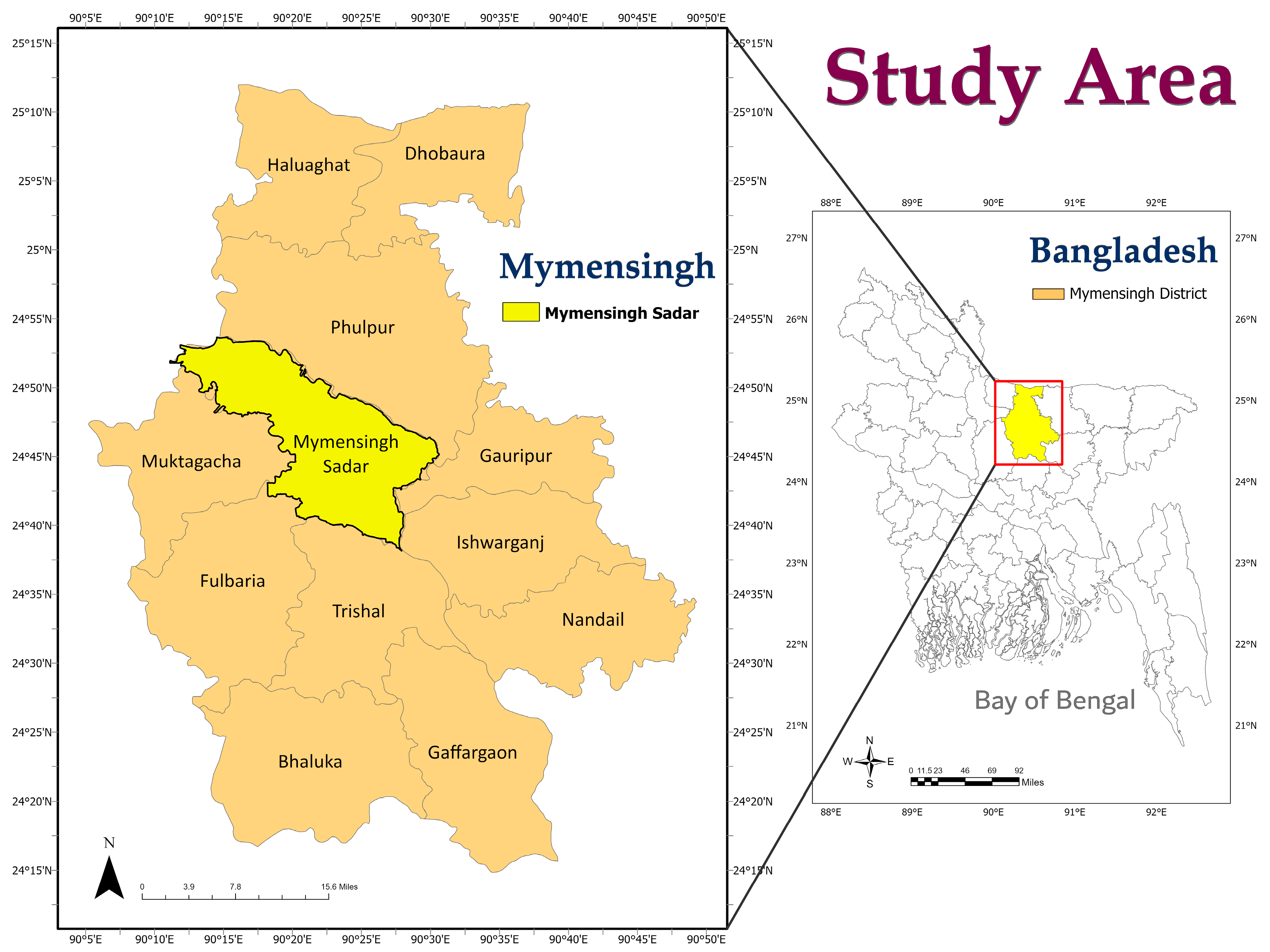
2.2. Study Area and Sampling

The present study was carried out in Mymensingh Sadar Upazila (24.7851° N, 90.3560° E), Mymensingh district of Bangladesh, between June 2021 and March 2022. The study area is featured in Figure 1.
A total of 420 samples associated with food and food products were collected aseptically, comprising human hand swab—60, raw milk—60, chicken muscle—60 (breast—30 and thigh—30), fish—60, egg surface—60, ready-to-eat foods—120 (fuchka—20, French fries—20, vegetable fries—20, puri—20, singara—20, and samosa—20).
Hand swabs were taken from vendors and dairy farms’ owners using sterile cotton buds, followed by immediately transferring into sterile test tubes containing 5 mL of nutrient broth (NB; HiMedia, India); 4 mL of raw milk (immediately after milking) was collected by sterile falcon tubes from different dairy farms; breast (25 g) and thigh (25 g) muscles from each chicken were collected by sterile zip-lock bags from poultry slaughterhouses; fresh fishes were collected from different local fish markets and eggs from egg markets using sterile zip-lock bags. In addition, ready-to-eat foods were collected from different food vendors and restaurants using sterile zip-lock bags. After collecting samples, particular tag numbers were given and transferred to the laboratory by maintaining a cold chain.

2.3. Sample Processing

After taking samples to the laboratory, each raw milk sample collected was vortexed, and 1 mL of sample was transferred into 5 mL of NB, contained in a sterile test tube. For chicken meat, 5 g of each sample was ground and transferred into 5 mL of NB, contained in a sterile test tube, followed by mixing homogenously. For ready-to-eat food samples, 20 mL of sterile phosphate buffer saline (PBS) was added to each sample and mixed by grinding with a sterile mortar and pestle. The mixture was then centrifuged at 4000 rotations per minute (rpm) for seven minutes in a centrifuge machine (KUBOTA 6500, Japan), followed by the collection of 100 µL of supernatant and transfer into 5 mL of sterile NB, contained in a sterile test tube. For egg samples, the surface of each egg was washed thoroughly using 5 mL of sterile PBS. Subsequently, 50 µL of egg-washed samples were taken into sterile test tubes containing 5 mL of NB. For fish samples, the gills were collected by an expert veterinarian, and swabs of the gills using sterile cotton buds were taken and transferred into 5 mL of sterile NB, contained in a sterile test tube. After transferring all samples into the sterile test tubes, they were incubated aerobically in an incubator overnight at 37 °C for microbial enrichment.

2.4. Isolation of S. aureus

S. aureus were initially isolated by culturing on Mannitol Salt Agar (MSA) (HiMedia, India) media. At first, one loopful of overnight-cultured broth was streaked on separate MSA plates and subsequently incubated aerobically under the appropriate time (24 h) and temperature (37 °C). Isolates having golden-yellow colonies on MSA agar plates were assumed to be S. aureus, and they were sub-cultured on MSA plates to obtain pure colonies. Moreover, the purity of the colonies was checked by culturing on 5% bovine blood agar plates, followed by incubating at 37 °C for 24 h. Presumptive S. aureus colonies were then isolated by Gram staining, Voges–Proskauer tests, glucose and mannitol utilization tests, catalase tests, and coagulase tests [13].

2.5. Molecular Detection of S. aureus

Presumptive staphylococcal isolates were subjected to polymerase chain reaction (PCR) to detect S. aureus molecularly by targeting the gene nuc (Table 1).
For the molecular detection of S. aureus, DNA was extracted by the boiling method, as previously described [19,20]. In brief, first, 1 mL of overnight growth S. aureus culture was centrifuged at 5000 rpm for 5 min in a centrifuge machine (KUBOTA 6500, Kubota Corporation, Tokyo, Japan). The supernatant was then discarded, and a similar process was followed by adding 1 mL of PBS. The supernatant was discarded again, and the remaining pellet was suspended with 200 µL of PBS. The suspension was then boiled and cooled for 10 min in each step before being centrifuged at 10,000 rpm for 10 min in a centrifuge machine (KUBOTA 6500, Kubota Corporation, Tokyo, Japan). In the final step, the supernatant was collected as genomic DNA and stored at −20 °C for future studies.
All the PCRs were performed with a final volume of 20 µL (nuclease-free water- 4 µL, master mix (2x, Promega, Madison, WI, USA)- 10 µL, forward and reverse primers- 1 µL each, genomic DNA- 4 µL). After the completion of amplification, the PCR products were electrophoresed on a 1.5% agarose gel, subsequently stained with ethidium bromide, and recorded on a UV transilluminator (Biometra, Göttingen, Germany). Note that a 100 bp (Promega) DNA ladder was used to check the expected band size of the amplified PCR products.

2.6. Biofilm Formation of S. aureus

2.6.1. Phenotypic Analysis of Biofilm Formation

Biofilm formation of S. aureus was phenotypically analyzed by the Congo Red (CR) test, as previously described [21]. In the CR test, the production of biofilm in the strains of S. aureus was studied by culturing the isolates on Congo Red Agar (CRA) plates. To prepare CRA plates, 0.8 g CR (HiMedia, India) and 36 g saccharose (HiMedia, India) were added to 1000 mL of blood agar (HiMedia, India) and subsequently incubated overnight at 37 °C to check their sterility. Then, overnight-growth S. aureus cultures were streaked on CRA plates, followed by incubation for 24 h and 48 h at 37 °C. The observable properties of the examined isolates were analyzed to check their biofilm-forming abilities. Isolates showing dry filamentous crusty black, pink with a dark center, and smooth pink colonies were interpreted as strong, intermediate/potential, and non-biofilm producers, respectively [22].

2.6.2. Quantitative Analysis of Biofilm Formation

The biofilm-forming ability of S. aureus was measured using 96-well flat-bottomed microtiter polystyrene plates, as previously described by Kouidhi et al. [23]. Briefly, single colonies from CRA plates were inoculated into 5 mL of sterile tryptic soy broth (TSB), followed by incubation at 37 °C for 18 h without shaking. The growth of the isolates was adjusted with the 0.5 McFarland concentration, corresponding to a cell concentration of approximately 108 colon-forming units (CFU)/mL for each strain [3]. The growth cultures were then diluted by a 10-fold dilution method in TSB supplemented with 10% glucose. An amount of 200 µL of the diluted culture was dispensed in three wells of the microtiter plate for each strain and incubated at 37 °C for 24 h. Negative controls were wells filled with broth medium (TSB + 10% glucose). Planktonic bacteria/cells were removed by washing each microtiter well 3–5 times with sterile PBS. The adherent cells were fixed with ethanol (95%) for 5 min, followed by emptying and drying the plates and subsequent staining for a few minutes with 100 µL of 1% crystal violet. The plates were air-dried after rinsing off the excess stain using sterile distilled water. The optical density (OD) value was evaluated at 570 nm (OD570) in an automatic spectrophotometer (VWR, part of Avantor, Radnor, PA, USA). The biofilm formation assay of each isolate was graded as strong biofilm (OD570 ≥ 1), moderate/intermediate biofilm (0.1 ≤ OD570 < 1), and non-biofilm (OD570 < 0.1) producers [23].

2.6.3. Genotypic Analysis of Biofilm Formation

The molecular detection of biofilm-forming S. aureus was performed by PCR-based amplification of adhesion genes of the icaADBC operon (icaA, icaB, icaC, and icaD) and biofilm-associated proteins (bap gene). Table 1 summarizes the primer sequences, the PCR product size, and the corresponding references. The PCR amplification of icaADBC genes in S. aureus was performed using the same method that was used to detect the nuc gene.

2.7. Statistical Analysis

Data obtained from the present study were entered into Excel 365 (Microsoft/Office 365, Redmond, DC, USA) and were subsequently transferred to the GraphPad Prism (Prism 8.4.2, San Diego, CA, USA) and the Statistical Package for Social Science (IBM SPSS 25.0, Chicago, IL, USA) for statistical analysis.

2.7.1. Descriptive Analysis

The prevalence of different variables was enumerated by descriptive analysis. The Wilson and Brown Hybrid method [24] was employed to calculate the binomial 95% confidence intervals for estimating the prevalence of different variables related to S. aureus-positive isolates. In addition, the chi-square test for goodness-of-fit was employed to determine the difference in the prevalence of S. aureus among different foods and hand swab samples. A p-value less than 0.05 (p < 0.05) was fixed to consider them statistically significant outcomes.

2.7.2. Bivariate Analysis

A bivariate analysis was undertaken to determine the correlation between pairs of different genes associated with biofilm-forming S. aureus isolates. The bivariate analysis to calculate Pearson correlation coefficients (ρ) was performed by the SPSS analysis tool. The correlation was statistically significant only when the p-value was less than 0.05 (p < 0.05).

2.7.3. Heatmap Analysis

A heatmap was generated to visualize the occurrence of staphylococcal biofilm-producing genes. The Origin Pro-2019b (Version 9.65, OriginLab Corporation, Northampton, MA, USA) was used to develop the heatmap. We considered the value “1” as positive and the value “0” as negative on the origin datasheet.

3. Results

3.1. Prevalence of S. aureus Isolates

Out of 420 samples analyzed, 151 (35.95%, 95% CI: 31.51–40.65%) samples were positive for S. aureus by observing the characteristic colonies of S. aureus on MS agar plates, Gram-staining, and biochemical tests. By PCR assay, 100 (23.81%; 95% CI: 19.99–28.11%) were found to be positive for S. aureus, as revealed by the detection of the nuc gene. Among the positive isolates, the highest prevalence was observed in raw milk, chicken muscle, fish, and egg surface samples, which showed equal prevalence (25%). Conversely, ready-to-eat food samples showed the lowest prevalence (21.67%). However, there was no statistically significant variation (p > 0.05) in the prevalence of S. aureus isolated from different food sources (Table 2).

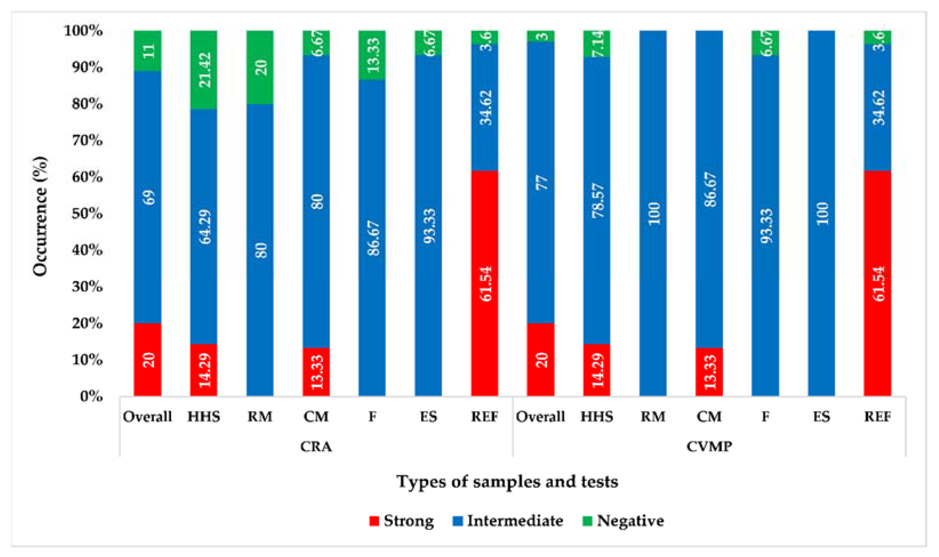
3.2. Phenotypic Biofilm Formation

On the CRA plates, 20% (20/100, 95% CI: 13.34–28.88%) of S. aureus isolates were strong biofilm producers, while 69% (69/100, 95% CI: 59.37–77.22%) and 11% (11/100, 95% CI: 6.25–18.63%) isolates were intermediate and non-biofilm producers, respectively (Figure 2). Sample-wise, the highest occurrence of strong biofilm-producing S. aureus was found in ready-to-eat food samples (61.54%, 16/26), but there was no strong biofilm-producing S. aureus in raw milk, fish, or egg surface samples (Figure 2).

3.3. Quantification of Biofilm Formation

By the CVMP test, 20 (20%, 95% CI: 13.34–28.88%), 77 (77%, 95% CI: 67.85–84.16%), and 3 (3%, 95% CI: 0.82–8.45%) of S. aureus isolates showed strong, intermediate, or non-biofilm-producing characteristics, respectively (Figure 2). Like the CRA plate test, the CVMP test also revealed that ready-to-eat food samples exhibited the highest occurrence (61.54%, 16/26) of strong biofilm-producing S. aureus isolates, while raw milk, fish, and egg surface samples did not harbor any strong biofilm-producing S. aureus isolates (Figure 2). In addition, all the 89 biofilm-producing isolates detected by the CRA test were also detected as biofilm producers by the CVMP test.

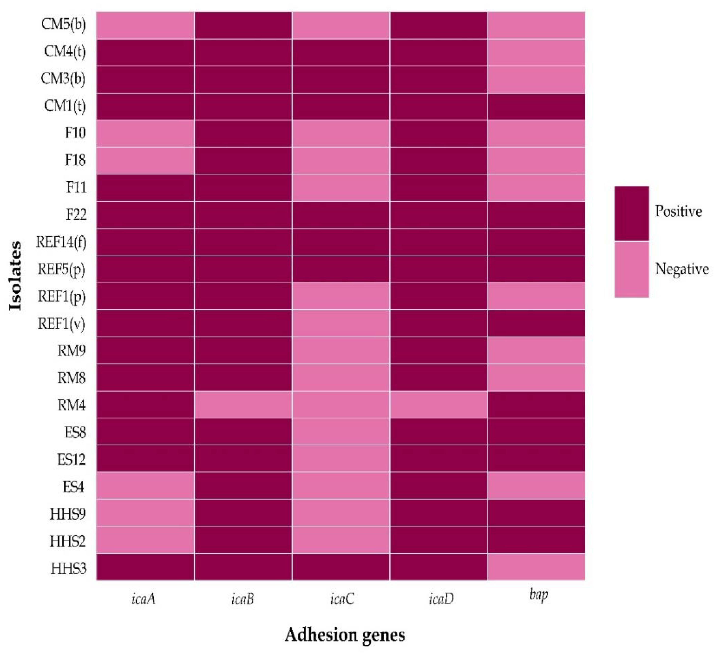
3.4. Genotypic Biofilm Formation

By PCR, out of 100 positive S. aureus isolates, 21 (21%, 95% CI: 14.17–29.98%) were found to carry at least one biofilm-forming gene. Among them, icaB and icaD were found to have the highest prevalence (20%, 95% CI: 13.34–28.89%) in the isolated S. aureus from different food samples and hand swabs, followed by icaA (15%, 95% CI: 9.31–23.28%), bap (10%, 95% CI: 5.52–17.44%), and icaC (7%, 95% CI: 3.43–13.75%) genes (Figure 3).
Out of 21 biofilm-forming S. aureus isolates, three and four adhesion genes were present in six isolates, two genes in five isolates, and all the selected genes were detected in four S. aureus isolates (Figure 3).
By bivariate analysis, strong positive significant correlations were observed between the biofilm-forming genes icaA and icaB (ρ = 0.770, p < 0.001), icaA and icaC (ρ = 0.653, p < 0.001), icaA and icaD (ρ = 0.770, p < 0.001), icaA and bap (ρ = 0.607, p < 0.001), icaB and icaC (ρ = 0.549, p < 0.001), icaB and icaD (ρ = 1.000, p < 0.001), icaB and bap (ρ = 0.583, p < 0.001), icaC and icaD (ρ = 0.549, p < 0.001), icaC and bap (ρ = 0.431, p < 0.001), icaD and bap (ρ = 0.583, p < 0.001) (Table 3).

4. Discussion

Staphylococcus aureus is a zoonotic pathogen that can cause a wide range of symptoms in humans [25]. The ability of S. aureus to form biofilm on food contact surfaces is considered to lead to food poisoning. Biofilm-producing S. aureus poses a serious health problem to humans. As we know, there is no previous report on the prevalence of biofilm-forming S. aureus from various food samples and hand swabs in Bangladesh.
In the current study, 23.81% of the food samples and hand swabs were found to be positive for S. aureus by the presence of the nuc gene. Previously in Bangladesh, Islam et al. [26] reported a similar occurrence of S. aureus (22%) in ready-to-eat foods, raw meat, raw milk, and fish samples, and a lower occurrence of S. aureus (6.67%) was detected in food samples by Urmi et al. [2]. In addition, Jahan et al. [27] and Pandit et al. [28] also detected S. aureus in raw milk (12/47) and on chicken egg surfaces (27/300) samples in Bangladesh. In addition, several previous studies showed diversified detection rates of S. aureus in different food samples abroad [29,30,31,32,33,34]. The observed variations between these studies and the present study could be attributed to differences in sample sources, size, and types; geographical locations; hygienic management; and other factors. Furthermore, ready-to-eat foods can be easily contaminated with S. aureus by food handlers, as most of them process and serve street foods with their bare hands in Bangladesh. In addition, proper hygiene practices are not guaranteed during food preparation; 23.33% of food-handler swab samples were found positive for S. aureus. Although there is no report in Bangladesh on the occurrence of S. aureus in food handlers’ hand swabs, Gadaga et al. [35] from Zimbabwe and Kasturwar and Shafee [36] from India recorded S. aureus in 32% and 36%, respectively, of samples from food handlers responsible for the food processing and serving. However, the presence of S. aureus in different food samples and hand swabs indicates a high health risk to consumers. In addition, the contamination level of food sources with S. aureus suggests that animal handling, food processing and handling, as well as cleaning and disinfecting food environments, must be improved.
The biofilm or slime formation on media is associated with the development of extracellular polysaccharides, which plays a significant role in bacterial adhesion [37,38]. Although the CRA test is not considered the most sensitive for determining biofilm development, this simple qualitative phenotypic test was used in this study because of its acceptable sensitivity and specificity [39]. In the present study, among 100 S. aureus isolates, 89% of the isolates showed characteristic colonies on CRA and were categorized as biofilm producers. The CVMP test revealed that 97 (97%) of the S. aureus isolates had the ability to produce biofilm. In addition, the same 20 S. aureus isolates were categorized as strong biofilm producers by both qualitative phenotypic and quantitative biofilm assays. The high and similar percentage of biofilm producers was in accordance with the previous study [10], which reported that 75% (63/84) and 97.62% (82/84) of the S. aureus isolated from food contact surfaces were biofilm producers by qualitative (CRA) and quantitative (crystal violet staining) assays, respectively. The variation in glucose concentration in the media used in the current study could explain the disparity in the results of the two tests. Rohde et al. [15] stated that adding 1% glucose to the TSB media could enhance the occurrence of biofilm formation in the S. aureus isolates by up to 83%. A previous study [40] reported that the expression of biofilm formation in S. epidermis isolates on CRA preparations with different amounts of glucose was significantly higher when the glucose concentration was higher.
This study recorded that 21% (21/100) of the S. aureus isolates harbored at least one biofilm-producing gene involved in the synthesis of PIA, where there was no difference in the distribution patterns of the icaB and icaD genes in the biofilm-forming S. aureus isolates. Chen et al. [3] also reported that the distribution of icaB and icaD genes was similar; however, the bap gene was not detected in the S. aureus isolates. In addition, the prevalence rates of other biofilm-related genes observed in the current study varied greatly, which can be supported by different studies [41,42,43]. Multifarious gene expression patterns can be recorded when S. aureus isolates are subjected to various temperatures and contact surfaces for varying lengths of time [3].
The present study revealed that the occurrence rates of biofilm-producing genes are much lower than the results observed in the CRA and CVMP tests. Similar findings were reported by previous studies [44,45], however, the high and similar prevalence rates of biofilm-producing S. aureus observed in the qualitative, quantitative, and molecular assays were recorded in previous studies [10,46,47]. This discrepancy could be explained by the different factors that are associated with biofilm formation in the S. aureus isolates. The interaction among different regulatory systems and the expression among different adhesion genes regulate biofilm formation, which can be triggered by various environmental factors such as the concentration of glucose in the used media, the temperature of the contact surfaces, the osmolality of the used media, and the growth conditions of the organisms [38,48,49]. In addition, the phenotypic characteristics of biofilm formation can be influenced by a discrepancy in the regulation of locus genes’ and putative adhesion genes’ expression [50]. Other credible mechanisms for the emergence of biofilm-negative strains of S. aureus include the disruption of genes by insertional inactivation and point mutations in the locus of adhesion genes [51]. The high variability of biofilm formation observed in the present study indicates that the determination of genes involved in the PIA or PNAG is not a complete determining factor of S. aureus’s ability to produce biofilm. Therefore, an integration of both phenotypic and genotypic tests could be used to identify biofilm-producing S. aureus isolates more accurately.

5. Conclusions

As we know, this is the first study in Bangladesh to detect biofilm-producing S. aureus phenotypically and genotypically from food. This study revealed that food samples and hand surfaces are prone to contamination with S. aureus, and most of the isolates could produce biofilm. Biofilm is a significant virulent substance in S. aureus infections, making their eradication difficult. The presence of biofilm-forming S. aureus in food samples and hand swabs indicates that food sources could act as a potential reservoir for pathogenic bacteria, posing serious public health significance. Our results are encouraging for further research focusing on the evaluation and exploration of the detailed genetic background of S. aureus isolated from food sources to assess their association with biofilm formation and infection development abilities. In addition, our findings emphasize the importance of good hygiene practices and installing strict food safety strategies at every stage of the food chain for preventing food-borne illnesses developed by S. aureus contamination and minimizing their cross-contamination hazards.

Author Contributions

Conceptualization, M.T.R.; methodology, M.R. and M.T.R.; software, M.S.I. and M.L.R.; validation, M.T.R.; formal analysis, M.S.I., F.M.B., M.L.R. and M.A.S.; sample collection, F.M.B., M.L.R., F.B.F., R.A., P.K.P. and J.K.; investigation, F.M.B., M.S.I., M.L.R., F.B.F., R.A. and S.I.; resources, M.T.R.; data curation, F.M.B., M.S.I. and M.L.R.; writing—original draft preparation, M.S.I., F.M.B. and M.T.R.; writing—review and editing, M.S.I., M.A.S., M.P.S., M.M.K., M.R. and M.T.R.; writing—critical revision, M.T.R.; visualization, F.M.B., M.L.R. and M.S.I.; supervision, M.R. and M.T.R.; project administration, M.T.R.; funding acquisition, M.T.R. All authors have read and agreed to the published version of the manuscript.

Funding

This research was supported by Tertiary Education Trust Fund, Nigeria (Grant No. TETFund/2020) and Bangladesh Agricultural University Research System (BAURES) project (Project No. 2019/8/BAU).

Institutional Review Board Statement

The methodologies and related protocols used in this study were approved by the Institutional Ethical Committee (AWEEC/BAU/2022 (20)). In addition, verbal permission from farm and vendor owners was taken during the sample collection.

Informed Consent Statement

Not applicable.

Data Availability Statement

Not applicable.

Acknowledgments

We are very grateful to all the farm owners and vendors for providing us with access to the samples. The authors would also like to express their gratitude to the Department of Microbiology and Hygiene, Faculty of Veterinary Science, Bangladesh Agricultural University, Mymensingh 2202, for their assistance during the research.

Conflicts of Interest

The authors declare no conflict of interest.

References

  1. Thiran, E.; Di Ciccio, P.; Graber, H.; Zanardi, E.; Ianieri, A.; Hummerjohann., J. Biofilm formation of Staphylococcus aureus dairy isolates representing different genotypes. J. Dairy Sci. 2018, 101, 1000–1012. [Google Scholar] [CrossRef] [PubMed] [Green Version]
  2. Urmi, M.R.; Ansari, W.K.; Islam, M.S.; Sobur, M.A.; Rahman, M.; Rahman, M.T. Antibiotic resistance patterns of Staphylococcus spp. isolated from fast foods sold in different restaurants of Mymensingh, Bangladesh. J. Adv. Vet. Anim. Res. 2021, 8, 274. [Google Scholar] [CrossRef]
  3. Chen, Q.; Xie, S.; Lou, X.; Cheng, S.; Liu, X.; Zheng, W.; Zheng, Z.; Wang, H. Biofilm formation and prevalence of adhesion genes among Staphylococcus aureus isolates from different food sources. Microbiologyopen 2020, 9, e00946. [Google Scholar] [CrossRef] [PubMed] [Green Version]
  4. Costa, O.Y.; Raaijmakers, J.M.; Kuramae, E.E. Microbial extracellular polymeric substances: Ecological function and impact on soil aggregation. Front. Microbiol. 2018, 9, 1636. [Google Scholar] [CrossRef] [PubMed] [Green Version]
  5. Souza, E.L.; Meira, Q.G.; Barbosa, I.D.; Athayde, A.J.; Conceição, M.L.; Siqueira Júnior, J.P. Biofilm formation by Staphylococcus aureus from food contact surfaces in a meat-based broth and sensitivity to sanitizers. Braz. J. Microbiol. 2014, 45, 67–75. [Google Scholar] [CrossRef] [PubMed] [Green Version]
  6. Karaguler, T.; Kahraman, H.; Tuter, M. Analyzing effects of ELF electromagnetic fields on removing bacterial biofilm. Biocybern. Biomed. Eng. 2017, 37, 336–340. [Google Scholar] [CrossRef]
  7. Payne, D.E.; Boles, B.R. Emerging interactions between matrix components during biofilm development. Curr. Genet. 2016, 62, 137–141. [Google Scholar] [CrossRef] [Green Version]
  8. Miao, J.; Lin, S.; Soteyome, T.; Peters, B.M.; Li, Y.; Chen, H.; Su, J.; Li, L.; Li, B.; Xu, Z. Biofilm formation of Staphylococcus aureus under food heat processing conditions: First report on CML production within biofilm. Sci. Rep. 2019, 9, 1–9. [Google Scholar] [CrossRef]
  9. Xie, J.; Peters, B.M.; Li, B.; Li, L.; Yu, G.; Xu, Z.; Shirtliff, M.E. Clinical features and antimicrobial resistance profiles of important Enterobacteriaceae pathogens in Guangzhou representative of Southern China, 2001–2015. Microb. Pathog. 2017, 107, 206–211. [Google Scholar] [CrossRef]
  10. Avila-Novoa, M.-G.; Iñíguez-Moreno, M.; Solís-Velázquez, O.-A.; González-Gomez, J.-P.; Guerrero-Medina, P.-J.; Gutiérrez-Lomelí, M.J. Biofilm formation by Staphylococcus aureus isolated from food contact surfaces in the dairy industry of Jalisco, Mexico. J. Food Qual. 2018, 2018, 1746139. [Google Scholar] [CrossRef] [Green Version]
  11. Archer, N.K.; Mazaitis, M.J.; Costerton, J.W.; Leid, J.G.; Powers, M.E.; Shirtliff, M.E. Staphylococcus aureus biofilms: Properties, regulation, and roles in human disease. Virulence 2011, 2, 445–459. [Google Scholar] [CrossRef] [PubMed] [Green Version]
  12. Thrusfeld, M. Veterinary Epidemiology, 2nd ed.; Blackwell Science Ltd.: London, UK, 1995; p. 83. [Google Scholar]
  13. Sobur, M.; Islam, M.; Haque, Z.F.; Orubu, E.S.F.; Toniolo, A.; Choudhury, M.; Rahman, M. Higher seasonal temperature enhances the occurrence of methicillin resistance of Staphylococcus aureus in house flies (Musca domestica) under hospital and environmental settings. Folia Microbiol. 2022, 67, 109–119. [Google Scholar] [CrossRef] [PubMed]
  14. Kalorey, D.R.; Shanmugam, Y.; Kurkure, N.V.; Chousalkar, K.K.; Barbuddhe, S.B. PCR-based detection of genes encoding virulence determinants in Staphylococcus aureus from bovine subclinical mastitis cases. J. Vet. Sci. 2007, 8, 151–154. [Google Scholar] [CrossRef] [Green Version]
  15. Rohde, H.; Knobloch, J.K.; Horstkotte, M.A.; Mack, D. Correlation of Staphylococcus aureus icaADBC genotype and biofilm expression phenotype. J. Clin. Microbiol. 2001, 39, 4595–4596. [Google Scholar] [CrossRef] [PubMed] [Green Version]
  16. Ma, Y.; Xu, Y.; Yestrepsky, B.D.; Sorenson, R.J.; Chen, M.; Larsen, S.D.; Sun, H. Novel inhibitors of Staphylococcus aureus virulence gene expression and biofilm formation. PLoS ONE 2012, 7, e47255. [Google Scholar] [CrossRef] [PubMed] [Green Version]
  17. Kiem, S.; Oh, W.S.; Peck, K.R.; Lee, N.Y.; Lee, J.-Y.; Song, J.-H.; Hwang, E.S.; Kim, E.-C.; Cha, C.Y.; Choe, K.-W. Phase variation of biofilm formation in Staphylococcus aureus by IS256 insertion and its impact on the capacity adhering to polyurethane surface. J. Korean Med. Sci. 2004, 19, 779–782. [Google Scholar] [CrossRef] [PubMed] [Green Version]
  18. Cucarella, C.; Tormo, M.A.; Ubeda, C.; Trotonda, M.P.; Monzón, M.; Peris, C.; Amorena, B.; Lasa, Í.; Penadés, J.R. Role of biofilm-associated protein bap in the pathogenesis of bovine Staphylococcus Aureus. Infect. Immun. 2004, 72, 2177–2185. [Google Scholar] [CrossRef] [PubMed] [Green Version]
  19. Islam, M.; Nayeem, M.; Hasan, M.; Sobur, M.; Ievy, S.; Rahman, S.; Kafi, M.; Ashour, H.M.; Rahman, M. Virulence determinants and multidrug resistance of Escherichia coli isolated from migratory birds. Antibiotics 2021, 10, 190. [Google Scholar] [CrossRef]
  20. Tawyabur, M.; Islam, M.; Sobur, M.; Hossain, M.; Mahmud, M.; Paul, S.; Hossain, M.T.; Ashour, H.M.; Rahman, M. Isolation and characterization of multidrug-resistant Escherichia coli and Salmonella spp. from healthy and diseased turkeys. Antibiotics 2020, 9, 770. [Google Scholar] [CrossRef]
  21. Arciola, C.R.; Baldassarri, L.; Montanaro, L. Presence of icaA and icaD genes and slime production in a collection of staphylococcal strains from catheter-associated infections. J. Clin. Microbiol. 2001, 39, 2151–2156. [Google Scholar] [CrossRef] [Green Version]
  22. Arciola, C.R.; Campoccia, D.; Gamberini, S.; Baldassarri, L.; Montanaro, L. Prevalence of can, fnbA and fnbB adhesin genes among Staphylococcus aureus isolates from orthopedic infections associated to different types of implant. FEMS Microbiol. Lett. 2005, 246, 81–86. [Google Scholar] [CrossRef] [PubMed] [Green Version]
  23. Kouidhi, B.; Zmantar, T.; Hentati, H.; Bakhrouf, A. Cell surface hydrophobicity, biofilm formation, adhesives properties and molecular detection of adhesins genes in Staphylococcus aureus associated to dental caries. Microb. Pathog. 2010, 49, 14–22. [Google Scholar] [CrossRef] [PubMed]
  24. Brown, L.D.; Cai, T.T.; DasGupta, A. Interval estimation for a binomial proportion. Stat. Sci. 2001, 16, 101–133. [Google Scholar] [CrossRef]
  25. Rahman, M.; Sobur, M.; Islam, M.; Ievy, S.; Hossain, M.; El Zowalaty, M.E.; Rahman, A.; Ashour, H.M. Zoonotic diseases: Etiology, impact, and control. Microorganisms 2020, 8, 1405. [Google Scholar] [CrossRef]
  26. Islam, M.A.; Parveen, S.; Rahman, M.; Huq, M.; Nabi, A.; Khan, Z.U.M.; Ahmed, N.; Wagenaar, J.A. Occurrence and characterization of methicillin resistant Staphylococcus aureus in processed raw foods and ready-to-eat foods in an urban setting of a developing country. Front. Microbiol. 2019, 10, 503. [Google Scholar] [CrossRef]
  27. Jahan, M.; Rahman, M.; Parvej, M.S.; Chowdhury, S.M.Z.H.; Haque, M.E.; Talukder, M.A.K.; Ahmed, S. Isolation and characterization of Staphylococcus aureus from raw cow milk in Bangladesh. J. Adv. Vet. Anim. Res. 2015, 2, 49–55. [Google Scholar] [CrossRef]
  28. Pondit, A.; Haque, Z.F.; Sabuj, A.A.M.; Khan, M.S.R.; Saha, S. Characterization of Staphylococcus aureus isolated from chicken and quail eggshell. J. Adv. Vet. Anim. Res. 2018, 5, 466. [Google Scholar] [CrossRef] [PubMed]
  29. Wu, S.; Huang, J.; Wu, Q.; Zhang, J.; Zhang, F.; Yang, X.; Wu, H.; Zeng, H.; Chen, M.; Ding, Y. Staphylococcus aureus isolated from retail meat and meat products in China: Incidence, antibiotic resistance and genetic diversity. Front. Microbiol. 2018, 9, 2767. [Google Scholar] [CrossRef]
  30. Wu, S.; Huang, J.; Zhang, F.; Wu, Q.; Zhang, J.; Pang, R.; Zeng, H.; Yang, X.; Chen, M.; Wang, J. Prevalence and characterization of food-related methicillin-resistant Staphylococcus aureus (MRSA) in China. Front. Microbiol. 2019, 10, 304. [Google Scholar] [CrossRef] [Green Version]
  31. Regasa, S.; Mengistu, S.; Abraha, A. Milk safety assessment, isolation, and antimicrobial susceptibility profile of Staphylococcus aureus in selected dairy farms of mukaturi and sululta town, oromia region, Ethiopia. Vet. Med. Int. 2019, 2019. [Google Scholar] [CrossRef] [Green Version]
  32. Kukułowicz, A.; Steinka, I.; Siwek, A. Presence of antibiotic-resistant Staphylococcus aureus in fish and seafood originating from points of sale in the tri-city area (Poland). J. Food Prot. 2021, 84, 1911–1914. [Google Scholar] [CrossRef] [PubMed]
  33. Shaltout, F.A.E.; Farouk, M.; Ibrahim, H.A.; Afifi, M.E. Incidence of coliform and Staphylococcus aureus in ready to eat fast foods. Benha Vet. Med. J. 2017, 32, 13–17. [Google Scholar] [CrossRef]
  34. Syed, M.A.; Shah, S.H.H.; Sherafzal, Y.; Shafi-ur-Rehman, S.; Khan, M.A.; Barrett, J.B.; Woodley, T.A.; Jamil, B.; Abbasi, S.A.; Jackson, C.R. Detection and molecular characterization of methicillin-resistant Staphylococcus aureus from table eggs in Haripur, Pakistan. Foodborne Pathog. Dis. 2018, 15, 86–93. [Google Scholar] [CrossRef] [PubMed]
  35. Gadaga, T.H.; Samende, B.K.; Musuna, C.; Chibanda, D. The microbiological quality of informally vended foods in Harare, Zimbabwe. Food Control. 2008, 19, 829–832. [Google Scholar] [CrossRef]
  36. Kasturwar, N.B.; Shafee, M. Knowledge, practices and prevalence of MRSA among food handlers. Int. J. Biol. Med. Res. 2011, 2, 889–894. [Google Scholar]
  37. O’Toole, G.; Kaplan, H.B.; Kolter, R. Biofilm formation as microbial development. Annu. Rev. Microbiol. 2000, 54, 49–79. [Google Scholar] [CrossRef]
  38. Torlak, E.; Korkut, E.; Uncu, A.T.; Şener, Y. Biofilm formation by Staphylococcus aureus isolates from a dental clinic in Konya, Turkey. J. Infect. Public Health 2017, 10, 809–813. [Google Scholar] [CrossRef]
  39. Hentzer, M.; Riedel, K.; Rasmussen, T.B.; Heydorn, A.; Andersen, J.B.; Parsek, M.R.; Rice, S.A.; Eberl, L.; Molin, S.; Høiby, N. Inhibition of quorum sensing in Pseudomonas aeruginosa biofilm bacteria by a halogenated furanone compound. Microbiology 2002, 148, 87–102. [Google Scholar] [CrossRef] [Green Version]
  40. Kaiser, T.D.L.; Pereira, E.M.; Dos Santos, K.R.N.; Maciel, E.L.N.; Schuenck, R.P.; Nunes, A.P.F. Modification of the Congo red agar method to detect biofilm production by Staphylococcus epidermidis. Diagn. Microbiol. Infect. Dis. 2013, 75, 235–239. [Google Scholar] [CrossRef] [Green Version]
  41. Kroning, I.S.; Iglesias, M.A.; Sehn, C.P.; Gandra, T.K.V.; Mata, M.M.; da Silva, W.P. Staphylococcus aureus isolated from handmade sweets: Biofilm formation, enterotoxigenicity and antimicrobial resistance. Food Microbiol. 2016, 58, 105–111. [Google Scholar] [CrossRef]
  42. Atshan, S.S.; Shamsudin, M.N.; Karunanidhi, A.; van Belkum, A.; Lung, L.T.T.; Sekawi, Z.; Nathan, J.J.; Ling, K.H.; Seng, J.S.C.; Ali, A.M. Quantitative PCR analysis of genes expressed during biofilm development of methicillin resistant Staphylococcus aureus (MRSA). Infect. Genet. Evol. 2013, 18, 106–112. [Google Scholar] [CrossRef] [PubMed]
  43. Stanley, N.R.; Lazazzera, B.A. Environmental signals and regulatory pathways that influence biofilm formation. Mol. Microbiol. 2004, 52, 917–924. [Google Scholar] [CrossRef]
  44. El-Nagdy, A.H.; Abdel-Fattah, G.M.; Emarah, Z. Detection and control of biofilm formation by Staphylococcus aureus from febrile neutropenic patient. Infect. Drug Resist. 2020, 13, 3091. [Google Scholar] [CrossRef] [PubMed]
  45. Szczuka, E.; Urbańska, K.; Pietryka, M.; Kaznowski, A. Biofilm density and detection of biofilm-producing genes in methicillin-resistant Staphylococcus aureus strains. Folia Microbiol. 2013, 58, 47–52. [Google Scholar] [CrossRef] [PubMed]
  46. Tang, J.; Chen, J.; Li, H.; Zeng, P.; Li, J. Characterization of adhesin genes, staphylococcal nuclease, hemolysis, and biofilm formation among Staphylococcus aureus strains isolated from different sources. Foodborne Pathog. Dis. 2013, 10, 757–763. [Google Scholar] [CrossRef]
  47. Gutiérrez, D.; Delgado, S.; Vázquez-Sánchez, D.; Martínez, B.; Cabo, M.L.; Rodríguez, A.; Herrera, J.J.; García, P. Incidence of Staphylococcus aureus and analysis of associated bacterial communities on food industry surfaces. Appl. Environ. Microbiol. 2012, 78, 8547–8554. [Google Scholar] [CrossRef] [Green Version]
  48. Beenken, K.E.; Dunman, P.M.; McAleese, F.; Macapagal, D.; Murphy, E.; Projan, S.J.; Blevins, J.S.; Smeltzer, M.S. Global gene expression in Staphylococcus aureus biofilms. J. Bacteriol. 2004, 186, 4665–4684. [Google Scholar] [CrossRef] [Green Version]
  49. Kim, J.-H.; Kim, C.-H.; Hacker, J.; Ziebuhr, W.; Lee, B.-K.; Cho, S.-h. Molecular characterization of regulatory genes associated with biofilm variation in a Staphylococcus aureus strain. J. Microbiol. Biotechnol. 2008, 18, 28–34. [Google Scholar]
  50. O’Gara, J.P. Ica and beyond: Biofilm mechanisms and regulation in Staphylococcus epidermidis and Staphylococcus aureus. FEMS Microbiol. Lett. 2007, 270, 179–188. [Google Scholar] [CrossRef] [Green Version]
  51. Ziebuhr, W.; Krimmer, V.; Rachid, S.; Lößner, I.; Götz, F.; Hacker, J. A novel mechanism of phase variation of virulence in Staphylococcus epidermidis: Evidence for control of the polysaccharide intercellular adhesin synthesis by alternating insertion and excision of the insertion sequence element IS256. Mol. Microbiol. 1999, 32, 345–356. [Google Scholar] [CrossRef]
Figure 1. Sample location map of the study area. The map was created by ArcMap 10.7 (ArcGIS Enterprise, ESRI, Redlands, CA, USA).
Biology 11 00949 g001
Figure 2. Occurrence of biofilm-producing S. aureus isolated from different foods and hand swabs, CRA = Congo Red Agar plating test, CVMP = Crystal Violet Microtiter Plate test, HHS = Human Hand Swab, RM = Raw Milk, CM = Chicken Muscle, F = Fish, ES = Egg Surface, REF = Ready-to-eat Food.
Biology 11 00949 g002
Figure 3. Heatmap showing the distribution of biofilm-forming genes in S. aureus isolated from different food samples and hand swabs, HHS = Human Hand Swab, ES = Egg Surface, RM = Raw Milk, REF = Ready-to-eat Food, v = Vegetable Fries, p = Puri, f = Fuchka, F = Fish, CM = Chicken Muscle, t = Thigh, b = Breast.
Biology 11 00949 g003
Table 1. List of primers used in the present study to detect biofilm-producing S. aureus from food sources and human hand swabs.
Targeted GenesPrimer Sequence (5′–3′)Annealing TemperatureAmplicon Size (bp)References
nucF: GCGATTGATGGTGATACGGT
R: AGCCAAGCCTTGACGAACTAAAGC
55279[14]
icaAF: GACCTCGAAGTCAATAGAGGT
R: CCCAGTATAACGTTGGATACC
56814[15]
icaDF: AGGCAATATCCAACGGTAA
R: GTCACGACCTTTCTTATATT
59526[16]
icaBF: ATCGCTTAAAGCACACGACGC
R: TATCGGCATCTGGTGTGACAG
59526[17]
icaCF: ATAAACTTGAATTAGTGTATT
R: ATATATAAAACTCTCTTAACA
45989[17]
bapF: CCCTATATCGAAGGTGTAGAATTGCAC
R: GCTGTTGAAGTTAATACTGTACCTGC
53971[18]
Table 2. Prevalence of Staphylococcus aureus isolated from different food sources and human hand swabs.
Name of SamplePositive Isolates (%)95% CIp-Value
Human hand swab (n = 60)14 (23.33)14.44–35.440.992
Raw milk (n = 60)15 (25)15.78–37.23
Chicken muscle (n = 60)15 (25)15.78–37.23
Fish (n = 60)15 (25)15.78–37.23
Egg surface (n = 60)15 (25)15.78–37.23
Ready-to-eat foods (n = 120)26 (21.67)15.24–29.85
Overall (n = 420)100 (23.81)19.99–28.11
Here, a p-value less or equal to 0.05 (p ≤ 0.05) was statistically significant, n = Number of samples, CI = Confidence interval.
Table 3. Pearson correlation coefficients (ρ) for pairs of adhesion genes to assess biofilm-forming S. aureus isolates from different food samples and hand swabs (n = 100).
icaAicaBicaCicaDbap
icaAPearson Correlation1
Sig. (2-tailed)-
icaBPearson Correlation0.770 **1
Sig. (2-tailed)0.000-
icaCPearson Correlation0.653 **0.549 **1
Sig. (2-tailed)0.0000.000-
icaDPearson Correlation0.770 **1.000 **0.549 **1
Sig. (2-tailed)0.0000.0000.000-
bapPearson Correlation0.607 **0.583 **0.431 **0.583 **1
Sig. (2-tailed)0.0000.0000.0000.000-
Here, a p-value less than 0.05 (p < 0.05) was considered statistically significant, **. Correlation is significant at the 0.01 level (2-tailed), Sig. = Significance.
Publisher’s Note: MDPI stays neutral with regard to jurisdictional claims in published maps and institutional affiliations.

