
Collagen Extraction from Animal Skin


Abstract
:Simple Summary
Abstract
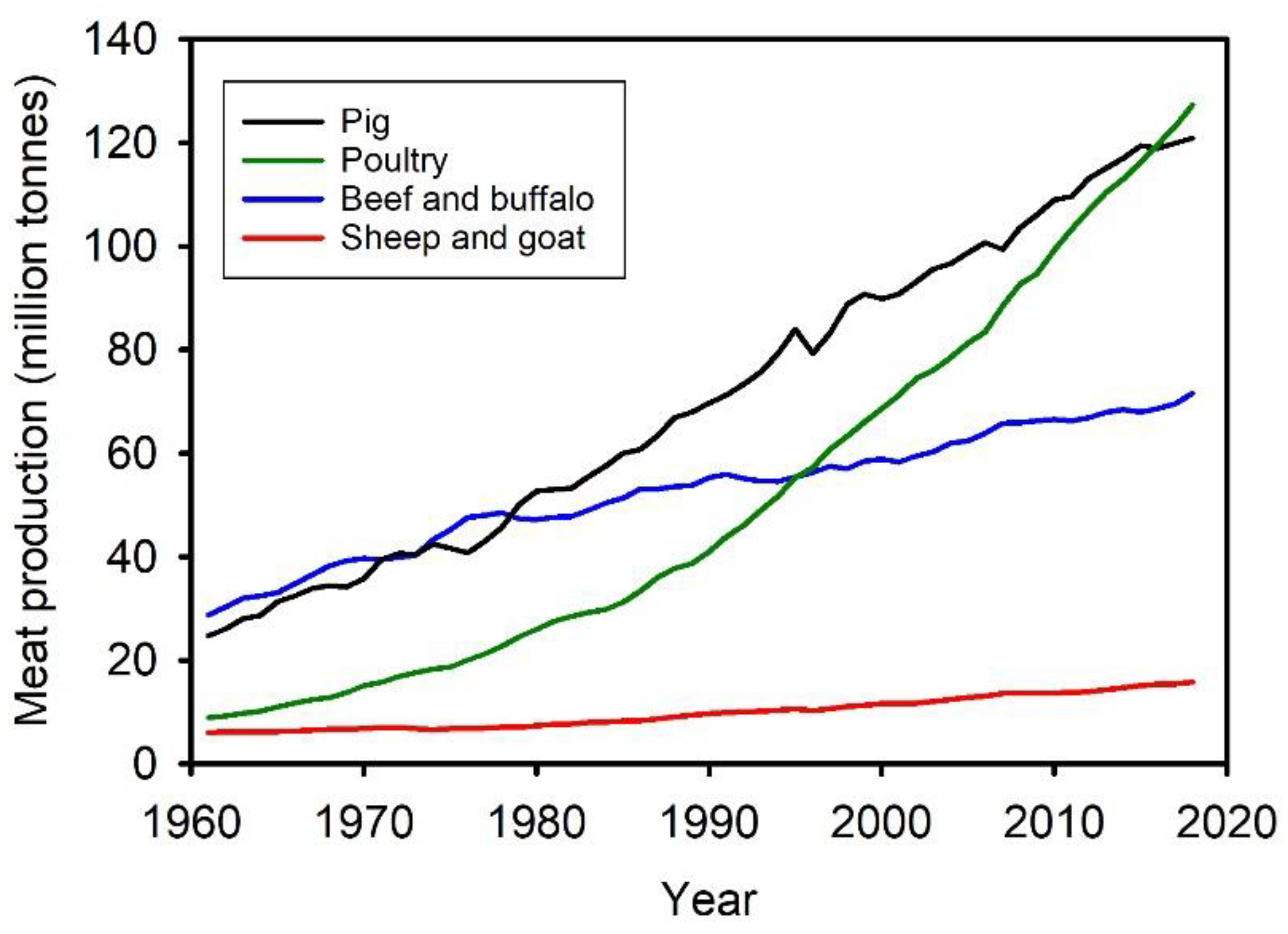
1. Introduction

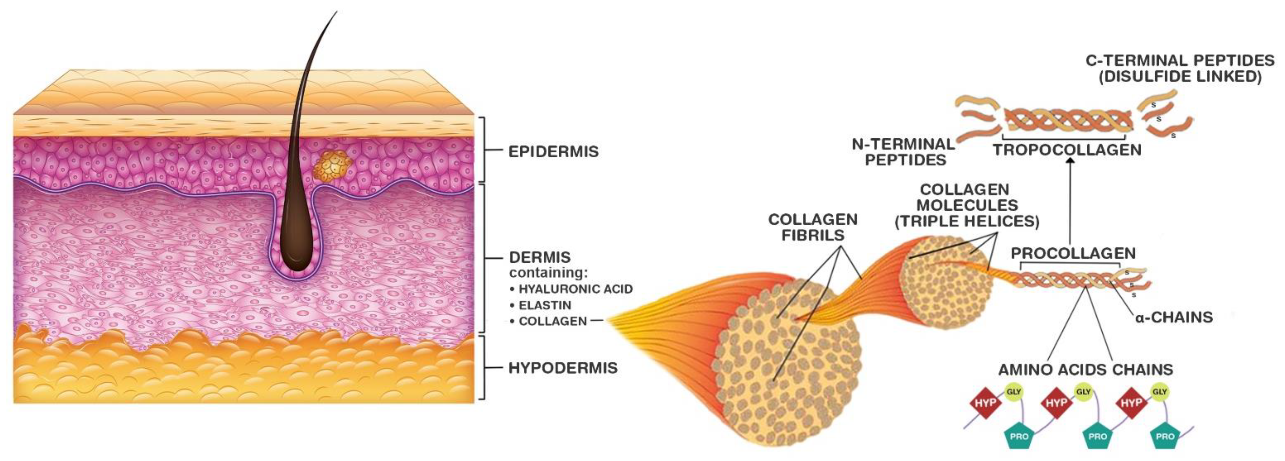
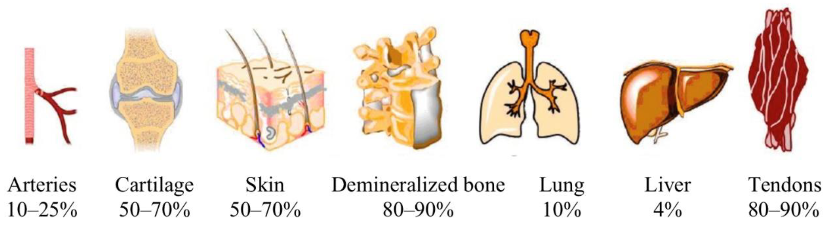
2. Collagen
Differences in Skin of Different Animals
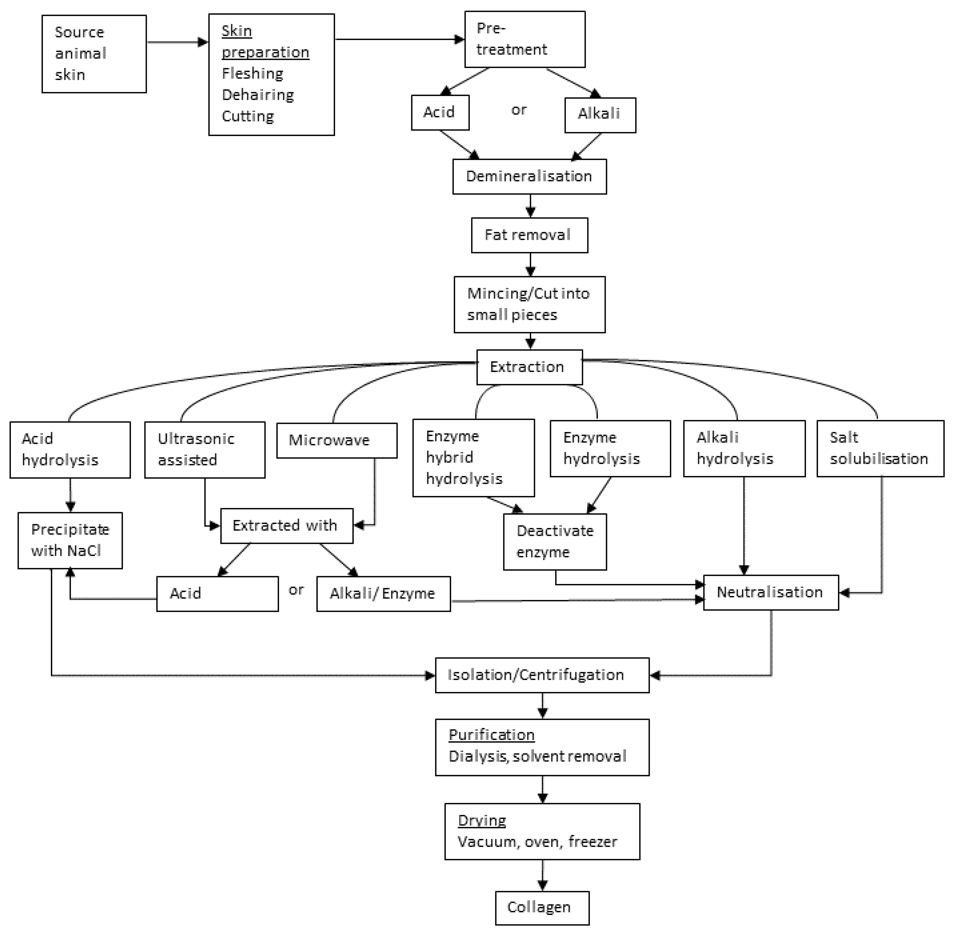
3. Collagen Extraction Process
3.1. Pretreatment
3.1.1. Acid Pretreatment
3.1.2. Alkaline Pretreatment
3.1.3. Skin-Specific Pretreatments
3.2. Extraction
3.2.1. Acid Hydrolysis
3.2.2. Alkali Hydrolysis
3.2.3. Salt Solubilization
3.2.4. Enzyme Hydrolysis
3.2.5. Ultrasound-Assisted Extraction
3.2.6. Other Extraction Methods
3.2.7. Precipitation of Solubilized Collagen
3.3. Post Extraction: Purification
4. Specific Extraction Methods
Future Considerations
5. Conclusions
Author Contributions
Funding
Institutional Review Board Statement
Informed Consent Statement
Data Availability Statement
Conflicts of Interest
References
- Jayathilakan, K.; Sultana, K.; Radhakrishna, K.; Bawa, A.S. Utilization of byproducts and waste materials from meat, poultry and fish processing industries: A review. J. Food Sci. Technol. 2012, 49, 278–293. [Google Scholar] [CrossRef] [PubMed] [Green Version]
- Bandaw, T.; Herago, T. Review on abattoir waste management. Glob. Vet. 2017, 19, 517–524. [Google Scholar] [CrossRef]
- Roberts, H.; de Jager, L.; Blight, G. Waste-handling practices at red meat abattoirs in South Africa. Waste Manag. Res. 2009, 27, 25–30. [Google Scholar] [CrossRef]
- Nwanta, J.A.; Onunkwo, J.I.; Ezenduka, V.; Phil-Eze, P.O.; Egege, S. Abattoir operations and waste management in Nigeria: A review of challenges and prospects. Sokoto J. Vet. Sci. 2008, 7, 61–67. [Google Scholar]
- Uddin, S.M.K.; Hossain, M.A.M.; Chowdhury, Z.Z.; Johan, M.R. Detection and discrimination of seven highly consumed meat species simultaneously in food products using heptaplex PCR-RFLP assay. J. Food Compos. Anal. 2021, 100, 103938. [Google Scholar] [CrossRef]
- Henchion, M.; McCarthy, M.; Resconi, V.C.; Troy, D. Meat consumption: Trends and quality matters. Meat Sci. 2014, 98, 561–568. [Google Scholar] [CrossRef] [Green Version]
- Ritchie, H.; Roser, M. Meat and dairy production. Our World in Data. 2017. Available online: https://ourworldindata.org/meat-production?fbclid=IwAR2I4y82fsZxHORHLWnsxcoeVKc9mSnMSURqynKD9AMtmttZ54a0GjXSYRU (accessed on 22 May 2022).
- Benjakul, S.; Oungbho, K.; Visessanguan, W.; Thiansilakul, Y.; Roytrakul, S. Characteristics of gelatin from the skins of bigeye snapper, Priacanthus tayenus and Priacanthus macracanthus. Food Chem. 2009, 116, 445–451. [Google Scholar] [CrossRef]
- Food and Agricultural Organization of the United Nations. Crops and Livestock Products. Available online: https://www.fao.org/faostat/en/#data/QCL/visualize (accessed on 25 January 2022).
- Kumar Kumawat, T.; Sharma, A.; Sharma, V.; Chandra, S. Keratin waste: The biodegradable polymers. In Keratin; Blumenberg, M., Ed.; IntechOpen: London, UK, 2018. [Google Scholar]
- Gaidau, C.; Stanca, M.; Niculescu, M.-D.; Alexe, C.-A.; Becheritu, M.; Horoias, R.; Cioineag, C.; Râpă, M.; Stanculescu, I.R. Wool keratin hydrolysates for bioactive additives preparation. Materials 2021, 14, 4696. [Google Scholar] [CrossRef]
- Sharma, S.; Gupta, A. Sustainable management of keratin waste biomass: Applications and future perspectives. Braz. Arch. Biol. Technol. 2016, 59, e16150684. [Google Scholar] [CrossRef] [Green Version]
- McLellan, J.; Thornhill, S.G.; Shelton, S.; Kumar, M. Keratin-based biofilms, hydrogels, and biofibers. In Keratin as a Protein Biopolymer; Sharma, S., Kumar, A., Eds.; Springer International Publishing: Cham, Switzerland, 2019; pp. 187–200. [Google Scholar]
- Nur Hanani, Z.A.; Roos, Y.H.; Kerry, J.P. Use and application of gelatin as potential biodegradable packaging materials for food products. Int. J. Biol. Macromol. 2014, 71, 94–102. [Google Scholar] [CrossRef]
- Almeida, P.F.; Lannes, S.C.D.S. Extraction and physicochemical characterization of gelatin from chicken by-product. J. Food Process. Eng. 2013, 36, 824–833. [Google Scholar] [CrossRef]
- Liu, D.; Nikoo, M.; Boran, G.; Zhou, P.; Regenstein, J.M. Collagen and gelatin. Annu. Rev. Food Sci. Technol. 2015, 6, 527–557. [Google Scholar] [CrossRef] [PubMed]
- Avila Rodríguez, M.I.; Rodríguez Barroso, L.G.; SánchezYang, H.; Guo, X.; Chen, X.; Shu, Z. Preparation and charact, M.L. Collagen: A review on its sources and potential cosmetic applications. J. Cosmet. Derm. 2018, 17, 20–26. [Google Scholar] [CrossRef] [PubMed]
- Nuñez, S.M.; Guzmán, F.; Valencia, P.; Almonacid, S.; Cárdenas, C. Collagen as a source of bioactive peptides: A bioinformatics approach. Electron. J. Biotechnol. 2020, 48, 101–108. [Google Scholar] [CrossRef]
- Patel, K.; Munir, D.; Santos, R.M. Beneficial use of animal hides for abattoir and tannery waste management: A review of unconventional, innovative, and sustainable approaches. Environ. Sci. Pollut. Res. 2021, 29, 1807–1823. [Google Scholar] [CrossRef]
- erization of collagen food packaging film. J. Chem. Pharm. Res. 2014, 6, 740–745.
- LaRoche, E.M.; Wu, W.J.; Garcia, P.; Song, B.; Chun, C.K.Y.; Jones, C.K.; Crane, A.R.; O’Quinn, T.G.; Chao, M.D. Evaluation of skin-on goat meat processing on processing efficiency, carcass yield, meat quality, and sensory attributes. Meat Sci. 2022, 184, 108675. [Google Scholar] [CrossRef]
- Fereidoun, H.; Bahram, A.; Soltanieh, K.; Abbass, S.; Pouria, H. Mean percentage of skin and visible fat in 10 chicken carcass weight. Int. J. Poult. Sci. 2007, 6, 43–47. [Google Scholar] [CrossRef] [Green Version]
- Shoulders, M.D.; Raines, R.T. Collagen structure and stability. Annu. Rev. Biochem. 2009, 78, 929–958. [Google Scholar] [CrossRef] [Green Version]
- Jafari, H.; Lista, A.; Siekapen, M.M.; Ghaffari-Bohlouli, P.; Nie, L.; Alimoradi, H.; Shavandi, A. Fish collagen: Extraction, characterization, and applications for biomaterials engineering. Polymers 2020, 12, 2230. [Google Scholar] [CrossRef] [PubMed]
- Holmes, D.F.; Lu, Y.; Starborg, T.; Kadler, K.E. Collagen fibril assembly and function. Curr. Top. Dev. Biol. 2018, 130, 107–142. [Google Scholar] [CrossRef] [PubMed]
- Litscher, E.; Wassarman, P.M. Extracellular Matrix and Egg Coats; Academic Press: Cambridge, MA, USA, 2018. [Google Scholar]
- Sorushanova, A.; Skoufos, I.; Tzora, A.; Mullen, A.M.; Zeugolis, D.I. The influence of animal species, gender and tissue on the structural, biophysical, biochemical and biological properties of collagen sponges. J. Mater. Sci. Mater. Med. 2021, 32, 12. [Google Scholar] [CrossRef]
- Muiznieks, L.D.; Keeley, F.W. Molecular assembly and mechanical properties of the extracellular matrix: A fibrous protein perspective. BBA Mol. Basis Dis. 2013, 1832, 866–875. [Google Scholar] [CrossRef] [PubMed] [Green Version]
- Tang, S.S.; Mohad, V.; Gowda, M.; Thibeault, S.L. Insights into the role of collagen in vocal fold health and disease. J. Voice 2017, 31, 520–527. [Google Scholar] [CrossRef] [PubMed]
- Reilly, D.M.; Lozano, J. Skin collagen through the lifestages: Importance for skin health and beauty. Plast. Aesthetic Res. 2021, 8, 2. [Google Scholar] [CrossRef]
- Sherman, V.R.; Yang, W.; Meyers, M.A. The materials science of collagen. J. Mech. Behav. Biomed. 2015, 52, 22–50. [Google Scholar] [CrossRef]
- Terzi, A.; Gallo, N.; Bettini, S.; Sibillano, T.; Altamura, D.; Madaghiele, M.; De Caro, L.; Valli, L.; Salvatore, L.; Sannino, A.; et al. Sub- and supramolecular X-Ray characterization of engineered tissues from equine tendon, bovine dermis, and fish skin type-I collagen. Macromol. Biosci. 2020, 20, 2000017. [Google Scholar] [CrossRef]
- Yamauchi, M.; Taga, Y.; Hattori, S.; Shiiba, M.; Terajima, M. Analysis of collagen and elastin cross-links. Methods Cell Biol. 2018, 143, 115–132. [Google Scholar] [CrossRef]
- Eyre, D.R.; Weis, M.; Rai, J. Analyses of lysine aldehyde cross-linking in collagen reveal that the mature cross-link histidinohydroxylysinonorleucine is an artifact. J. Biol. Chem. 2019, 294, 6578–6590. [Google Scholar] [CrossRef]
- Zahrani, A.R. Extraction and Isolation of Collagen Type I from Fish Skin. Master’s Thesis, The University of Otago, Dunedin, New Zealand, 2011. [Google Scholar]
- Gelse, K.; Pöschl, E.; Aigner, T. Collagens—Structure, function, and biosynthesis. Adv. Drug Deliv. Rev. 2003, 55, 1531–1546. [Google Scholar] [CrossRef] [Green Version]
- Schrieber, R.; Gareis, H. Gelatine Handbook: Theory and Industrial Practice; Wiley-VCH: Weinheim, Germany, 2007; 347p. [Google Scholar]
- Schmidt, M.M.; Dornelles, R.C.P.; Mello, R.O.; Kubota, E.H.; Mazutti, M.A.; Kempka, A.P.; Demiate, I.M. Collagen extraction process. Int. Food Res. J. 2016, 23, 913–922. [Google Scholar]
- Chuaychan, S. Production and characterization of collagen, gelatin and gelatin hydrolysate powder from scales of spotted golden goatfish. Ph.D. Thesis, Prince of Songkla University, Hat Yai, Thailand, 2016. [Google Scholar]
- Mayne, R.; Burgeson, R.E. Structure and Function of Collagen Types; Academic Press: London, UK, 1987. [Google Scholar]
- Tümerkan, E.T.A.; Cansu, Ü.; Boran, G.; Mac Regenstein, J.; Özoğul, F. Physiochemical and functional properties of gelatin obtained from tuna, frog and chicken skins. Food Chem. 2019, 287, 273–279. [Google Scholar] [CrossRef] [PubMed]
- Fourneau, M.; Canon, C.; Van Vlaender, D.; Collins, M.J.; Fiddyment, S.; Poumay, Y.; Deparis, O. Histological study of sheep skin transformation during the recreation of historical parchment manufacture. Herit. Sci. 2020, 8, 78. [Google Scholar] [CrossRef]
- Sizeland, K.H.; Basil-Jones, M.M.; Edmonds, R.L.; Cooper, S.M.; Kirby, N.; Hawley, A.; Haverkamp, R.G. Collagen orientation and leather strength for selected mammals. J. Agric. Food Chem. 2013, 61, 887–892. [Google Scholar] [CrossRef]
- Wells, H.C.; Sizeland, K.H.; Kirby, N.; Hawley, A.; Mudie, S.; Haverkamp, R.G. Collagen fibril structure and strength in acellular dermal matrix materials of bovine, porcine and human origin. ACS Biomat. Sci. Eng. 2015, 1, 1026–1038. [Google Scholar] [CrossRef]
- Wells, H.C.; Holmes, G.; Haverkamp, R.G. Looseness in bovine leather: Microstructural characterization. J. Sci. Food Agric. 2015, 96, 2731–2736. [Google Scholar] [CrossRef]
- Lin, H.; Fan, Y. Bovine Type I collagen: Preparation, characterization, and application in tissue regeneration. In Type I Collagen: Molecular Structure, Applications in Tissue Engineering and Role in Human Disorders; Rivera, G., Ed.; Nova Science Publishers: New York, NY, USA, 2015; pp. 1–71. [Google Scholar]
- El Blidi, O.; El Omari, N.; Balahabib, A.; Ghchime, R.; El Menyiy, N.; Ibrahimi, A.; Kaddour, K.B.; Bouyahya, A.; Chokairi, O.; Barkiyou, M. Extraction methods, characterization and biomedical applications of collagen: A review. Biointerface Res. Appl. Chem. 2021, 11, 13587–13613. [Google Scholar] [CrossRef]
- Prestesa, R.C. Collagen and its derivatives: Characteristics and applications in meat products. UNOPAR Cient. Ciênc. Biol. Saúde 2013, 15, 65–74. [Google Scholar]
- Ledward, D.A. Gelatin; Woodhead Publishing Ltd.: Cambridge, UK, 2000; pp. 67–86. [Google Scholar]
- Almeida, P.; Vanalle, R.; Carlos, J.; Santana, J. Produção de gelatina: Uma perspectiva competitiva para a cadeia produtiva de frango de corte. Prod. Produção 2012, 13, 22–39. [Google Scholar] [CrossRef]
- Liu, Y.; Ma, D.; Wang, Y.; Qin, W. A comparative study of the properties and self-aggregation behavior of collagens from the scales and skin of grass carp (Ctenopharyngodon idella). Int. J. Biol. Macromol. 2018, 106, 516–522. [Google Scholar] [CrossRef]
- Pal, G.K.; Suresh, P.V. Sustainable valorisation of seafood by-products: Recovery of collagen and development of collagen-based novel functional food ingredients. Innov. Food Sci. Emerg. Technol. 2016, 37, 201–215. [Google Scholar] [CrossRef]
- Silva, T.H.; Moreira-Silva, J.; Marques, A.L.P.; Domingues, A.; Bayon, Y.; Reis, R.L. Marine origin collagens and its potential applications. Mar. Drugs 2014, 12, 5881–5901. [Google Scholar] [CrossRef] [Green Version]
- Wahyuningsih, R.; Rusman; Nurliyani; Pertiwiningrum, A.; Rohman, A.; Fitriyanto, N.A.; Erwanto, Y. Optimization of acid soluble collagen extraction from Indonesian local “Kacang” goat skin and physico-chemical properties characterization. Chem. Eng. Trans. 2018, 63, 703–708. [Google Scholar] [CrossRef]
- Hakim, T.R.; Pratiwi, A.; Jamhari; Fitriyanto, N.A.; Rusman; Abidin, M.Z.; Matulessy, D.N.; Erwanto, Y. Extraction of collagen from the skin of Kacang goat and production of its hydrolysate as an inhibitor of angiotensin converting enzyme. Trop. Anim. Sci. J. 2021, 44, 222–228. [Google Scholar] [CrossRef]
- He, L.; Lan, W.; Zhao, Y.; Chen, S.; Liu, S.; Cen, L.; Cao, S.; Dong, L.; Jin, R.; Liu, Y. Characterization of biocompatible pig skin collagen and application of collagen-based films for enzyme immobilization. RSC Adv. 2020, 10, 7170–7180. [Google Scholar] [CrossRef] [PubMed] [Green Version]
- Zhang, X.; Xu, S.; Shen, L.; Li, G. Factors affecting thermal stability of collagen from the aspects of extraction, processing and modification. J. Soc. Leather Technol. Chem. 2020, 2, 19. [Google Scholar] [CrossRef]
- Andayani, A.A.; Harmita, H.; Maggadani, B.P. Isolation, purification, and characterization of porcine skin collagen: Analysis of the glycine, proline, and hydroxyproline components using high-performance liquid chromatography. Int. J. Appl. Pharm. 2018, 10, 294–298. [Google Scholar] [CrossRef] [Green Version]
- Noorzai, S.; Verbeek, C.J.R.; Lay, M.C.; Swan, J. Collagen extraction from various waste bovine hide sources. Waste Biomass Valori. 2020, 11, 5687–5698. [Google Scholar] [CrossRef]
- Staicu, T.; Cîrcu, V.; Ioniţə, G.; Ghica, C.; Popa, V.T.; Micutz, M. Analysis of bimodal thermally-induced denaturation of type I collagen extracted from calfskin. RSC Adv. 2015, 5, 38391–38406. [Google Scholar] [CrossRef]
- Vazquez, J.J.; Martínez, E.S.M. Collagen and elastin scaffold by electrospinning for skin tissue engineering applications. J. Mater. Res. 2019, 34, 2819–2827. [Google Scholar] [CrossRef]
- Cliche, S.; Amiot, J.; Avezard, C.; Gariépy, C. Extraction and characterization of collagen with or without telopeptides from chicken skin. Poult. Sci. 2003, 82, 503–509. [Google Scholar] [CrossRef] [PubMed]
- Gojkovic, Z.; Marova, I.; Matouskova, P.; Obruca, S.; Miloslav, P. Use of ultrasonic spectroscopy and viscosimetry for the characterization of chicken skin collagen in comparison with collagens from other animal tissues. Prep. Biochem. Biotechnol. 2014, 44, 761–771. [Google Scholar] [CrossRef] [PubMed]
- Zhang, Y.; Ma, L.; Cai, L.; Liu, Y.; Li, J. Effect of combined ultrasonic and alkali pretreatment on enzymatic preparation of angiotensin converting enzyme (ACE) inhibitory peptides from native collagenous materials. Ultrason. Sonochem. 2017, 36, 88–94. [Google Scholar] [CrossRef] [PubMed]
- Zhang, Y.; Zhang, Y.; Liu, X.; Huang, L.; Chen, Z.; Cheng, J. Influence of hydrolysis behaviour and microfluidisation on the functionality and structural properties of collagen hydrolysates. Food Chem. 2017, 227, 211–218. [Google Scholar] [CrossRef]
- Morimura, S.; Nagata, H.; Uemura, Y.; Fahmi, A.; Shigematsu, T.; Kida, K. Development of an effective process for utilization of collagen from livestock and fish waste. Process Biochem. 2002, 37, 1403–1412. [Google Scholar] [CrossRef]
- Hong, G.P.; Min, S.G.; Jo, Y.J. Anti-oxidative and anti-aging activities of porcine by-product collagen hydrolysates produced by commercial proteases: Effect of hydrolysis and ultrafiltration. Molecules 2019, 24, 1104. [Google Scholar] [CrossRef] [Green Version]
- Leon-Lopez, A.; Fuentes-Jimenez, L.; Hernandez-Fuentes, A.D.; Campos-Montiel, R.G.; Aguirre-Alvarez, G. Hydrolysed collagen from sheepskins as a source of functional peptides with antioxidant activity. Int. J. Mol. Sci. 2019, 20, 3931. [Google Scholar] [CrossRef] [Green Version]
- Chuaychan, S.; Benjakul, S.; Kishimura, H. Characteristics of acid- and pepsin-soluble collagens from scale of seabass (Lates calcarifer). LWT Food Sci. Technol. 2015, 63, 71–76. [Google Scholar] [CrossRef]
- Senadheera, T.R.L.; Dave, D.; Shahidi, F. Sea cucumber derived type I collagen: A comprehensive review. Mar. Drugs 2020, 18, 471. [Google Scholar] [CrossRef]
- Yang, H.; Shu, Z. The extraction of collagen protein from pigskin. J. Chem. Pharm. Res. 2014, 2, 683–687. [Google Scholar]
- Wang, L.; Yang, B.; Du, X.; Yang, Y.; Liu, J. Optimization of conditions for extraction of acid-soluble collagen from grass carp (Ctenopharyngodon idella) by response surface methodology. Innov. Food Sci. Emerg. Technol. 2008, 9, 604–607. [Google Scholar] [CrossRef]
- Liu, D.; Wei, G.; Li, T.; Hu, J.; Lu, N.; Regenstein, J.M.; Zhou, P. Effects of alkaline pretreatments and acid extraction conditions on the acid-soluble collagen from grass carp (Ctenopharyngodon idella) skin. Food Chem. 2015, 172, 836–843. [Google Scholar] [CrossRef] [PubMed]
- Skierka, E.; Sadowska, M. The influence of different acids and pepsin on the extractability of collagen from the skin of Baltic cod (Gadus morhua). Food Chem. 2007, 105, 1302–1306. [Google Scholar] [CrossRef]
- Ran, X.G.; Wang, L.Y. Use of ultrasonic and pepsin treatment in tandem for collagen extraction from meat industry by-products. J. Sci. Food Agric. 2014, 94, 585–590. [Google Scholar] [CrossRef]
- Wu, S.-L.; Kang, H.-B.; Li, D.-J. Technology for extracting effective components from fish scale. J. Food Sci. Eng. 2017, 7, 351–358. [Google Scholar] [CrossRef]
- Naomi, R.; Ridzuan, P.M.; Bahari, H. Current insights into collagen type I. Polymers 2021, 13, 2642. [Google Scholar] [CrossRef]
- Nabijon, N.; Ahmed, M.D.R.; Adkham, R.; Heng, Q. Extraction of collagen from cattle skin and synthesis of collagen based flame retardant composition and introduction into cellulosic textile material by graft copolymerization. Asian J. Chem. 2017, 29, 2470–2474. [Google Scholar] [CrossRef]
- Fernandez Hervas, F.; Celma, P.; Punti, I.; Cisa, J.; Cot, J.; Marsal, A.; Manich, A. The enzyme activity of trypsin on sheepskin trimmings in a two-step collagen extraction process. J. Am. Leather Chem. Assoc. 2007, 102, 1–9. [Google Scholar]
- Hong, H.; Chaplot, S.; Chalamaiah, M.; Roy, B.C.; Bruce, H.L.; Wu, J. Removing cross-linked telopeptides enhances the production of low-molecular-weight collagen peptides from spent hens. J. Agric. Food Chem. 2017, 65, 7491–7499. [Google Scholar] [CrossRef]
- Wahyuningsih, R.; Rusman; Nurliyani; Pertiwiningrum, A.; Rohman, A.; Fitriyanto, N.A.; Erwanto, Y. Optimization of conditions for extraction of pepsin-soluble collagen from Indonesian local “Kacang” goatskin by response surface methodology. Am. J. Anim. Vet. Sci. 2018, 13, 70–75. [Google Scholar] [CrossRef] [Green Version]
- Liu, X.; Dan, N.; Dan, W. Preparation and characterization of an advanced collagen aggregate from porcine acellular dermal matrix. Int. J. Biol. Macromol. 2016, 88, 179–188. [Google Scholar] [CrossRef]
- Feng, M.; Betti, M. Transepithelial transport efficiency of bovine collagen hydrolysates in a human Caco-2 cell line model. Food Chem. 2017, 224, 242–250. [Google Scholar] [CrossRef]
- Zhang, Z.; Li, G.; Shi, B. Physicochemical properties of collagen, gelatin and collagen hydrolysate derived from bovine limed split wastes. J. Soc. Leather Technol. Chem. 2006, 90, 23–28. [Google Scholar]
- Kim, H.K.; Kim, Y.H.; Kim, Y.J.; Park, H.J.; Lee, N.H. Effects of ultrasonic treatment on collagen extraction from skins of the sea bass Lateolabrax japonicus. Fish Sci. 2012, 78, 485–490. [Google Scholar] [CrossRef]
- Kadam, S.U.; Tiwari, B.K.; Álvarez, C.; O’Donnell, C.P. Ultrasound applications for the extraction, identification and delivery of food proteins and bioactive peptides. Trends Food Sci. Technol. 2015, 46, 60–67. [Google Scholar] [CrossRef]
- Kim, H.K.; Kim, Y.H.; Park, H.J.; Lee, N.H. Application of ultrasonic treatment to extraction of collagen from the skins of sea bass Lateolabrax japonicus. Fish Sci. 2013, 79, 849–856. [Google Scholar] [CrossRef]
- Li, D.; Mu, C.; Cai, S.; Lin, W. Ultrasonic irradiation in the enzymatic extraction of collagen. Ultrason. Sonochem. 2009, 16, 605–609. [Google Scholar] [CrossRef] [PubMed]
- Lin, Y.-J.; Le, G.-W.; Wang, J.-Y.; Li, Y.-X.; Shi, Y.-H.; Sun, J. Antioxidative peptides derived from enzyme hydrolysis of bone collagen after microwave assisted acid pre-treatment and nitrogen protection. Int. J. Mol. Sci. 2010, 11, 4297–4308. [Google Scholar] [CrossRef] [Green Version]
- Jin, H.-X.; Xu, H.-P.; Li, Y.; Zhang, Q.-W.; Xie, H. Preparation and evaluation of peptides with potential antioxidant activity by microwave assisted enzymatic hydrolysis of collagen from sea cucumber Acaudina molpadioides obtained from Zhejiang Province in China. Mar. Drugs 2019, 17, 169. [Google Scholar] [CrossRef] [Green Version]
- Ahmed, M.; Verma, A.K.; Patel, R. Collagen extraction and recent biological activities of collagen peptides derived from sea-food waste: A review. Sustain. Chem. Pharm. 2020, 18, 100315. [Google Scholar] [CrossRef]
- Khong, N.M.H.; Yusoff, F.M.; Jamilah, B.; Basri, M.; Maznah, I.; Chan, K.W.; Armania, N.; Nishikawa, J. Improved collagen extraction from jellyfish (Acromitus hardenbergi) with increased physical-induced solubilization processes. Food Chem. 2018, 251, 41–50. [Google Scholar] [CrossRef]
- Du, L.; Betti, M. Chicken collagen hydrolysate cryoprotection of natural actomyosin: Mechanism studies during freeze-thaw cycles and simulated digestion. Food Chem. 2016, 211, 791–802. [Google Scholar] [CrossRef] [PubMed]
- Gorlov, I.F.; Titov, E.I.; Semenov, G.V.; Slozhenkina, M.I.; Sokolov, A.Y.; Omarov, R.S.; Goncharov, A.I.; Zlobina, E.Y.; Litvinova, E.V.; Karpenko, E.V. Collagen from porcine skin: A method of extraction and structural properties. Int. J. Food Prop. 2018, 21, 1031–1042. [Google Scholar] [CrossRef]
- Naffa, R.; Edwards, P.J.B.; Norris, G. Isolation and characterization of collagen type I crosslink from skin: High-resolution NMR reveals diastereomers of hydroxylysinonorleucine crosslink. Amino Acids 2019, 51, 705–715. [Google Scholar] [CrossRef] [PubMed]
- Cao, C.; Xiao, Z.; Ge, C.; Wu, Y. Animal by-products collagen and derived peptide, as important components of innovative sustainable food systems—A comprehensive review. Crit. Rev. Food Sci. Nutr. 2021, in press. [Google Scholar] [CrossRef] [PubMed]
- Hyde, A.M.; Zultanski, S.L.; Waldman, J.H.; Zhong, Y.-L.; Shevlin, M.; Peng, F. General principles and strategies for salting-out informed by the Hofmeister series. Org. Process Res. Dev. 2017, 21, 1355–1370. [Google Scholar] [CrossRef] [Green Version]
- Picot, L.; Ravallec, R.; Fouchereau-Péron, M.; Vandanjon, L.; Jaouen, P.; Chaplain-Derouiniot, M.; Guérard, F.; Chabeaud, A.; LeGal, Y.; Alvarez, O.M.; et al. Impact of ultrafiltration and nanofiltration of an industrial fish protein hydrolysate on its bioactive properties. J. Sci. Food. Agric. 2010, 90, 1819–1826. [Google Scholar] [CrossRef] [Green Version]
- Suurs, P.; van den Brand, H.; Daamen, W.F.; Barbut, S. Properties of different poultry skins sources in relation to co-extruded sausage casings. Food Hydrocoll. 2022, 125, 107434. [Google Scholar] [CrossRef]
- Wahyuningsih, R.; Rusman; Nurliyani; Rohman, A.; Erwanto, Y. Potency of pepsin soluble collagen from Indonesian local goat skin as an antioxidant. Am. J. Anim. Vet. Sci. 2021, 16, 144–151. [Google Scholar] [CrossRef]
- Hu, Y.; Liu, L.; Dan, W.; Dan, N.; Gu, Z.; Yu, X. Synergistic effect of carbodiimide and dehydrothermal crosslinking on acellular dermal matrix. Int. J. Biol. Macromol. 2013, 55, 221–230. [Google Scholar] [CrossRef]
- Hsieh, D.J.; Srinivasan, P. Protocols for accelerated production and purification of collagen scaffold and atelocollagen from animal tissues. BioTechniques 2020, 69, 221–225. [Google Scholar] [CrossRef] [PubMed]
- Fu, R.; Yao, K.; Zhang, Q.; Jia, D.; Zhao, J.; Chi, Y. Collagen hydrolysates of skin shavings prepared by enzymatic hydrolysis as a natural flocculant and their flocculating property. Appl. Biochem. Biotechnol. 2017, 182, 55–66. [Google Scholar] [CrossRef]
- Cabeza, L.F.; Taylor, M.M.; DiMaio, G.L.; Brown, E.M.; Marmer, W.N.; Carrió, R.; Celma, P.J.; Cot, J. Processing of leather waste: Pilot scale studies on chrome shavings. Isolation of potentially valuable protein products and chromium. Waste Manag. 1998, 18, 211–218. [Google Scholar] [CrossRef]
- Marmer, W.N.; Dudley, R.L. Oxidative dehairing by sodium percarbonate. J. Am. Leather Chem. Assoc. 2005, 100, 427–431. [Google Scholar]
- Brown, E.M.; Latona, R.J.; Taylor, M.M.; Garcia, R.A. Effects of pretanning processes on bovine hide collagen structure. J. Am. Leather Chem. Assoc. 2012, 107, 1–7. [Google Scholar]
- Said, M.I.; Burhan, T.; Haerati. Synthesis of collagen from Bali cattle’s hide using a combination of acid and alkali on the extracting process. J. Indones. Trop. Anim. Agric. 2018, 43, 247–256. [Google Scholar] [CrossRef]


| Type | Molecule | Source |
|---|---|---|
| I | Skin, tendon, bone, ligaments, interstitial tissues | |
| II | Intervertebral disc, cartilage, vitreous humor | |
| III | Cardiovascular vessel, uterus, skin, muscle | |
| V | Similar to type I, also cell cultures, fetal tissues; associates with type I | |
| XI | Cartilage, in vertebral cartilage and bone enamel |
| Pretreatment | Extraction and Isolation | Reference |
|---|---|---|
| Chicken | ||
| Hot-water bath (40 and 60 °C, 1 h) | Multi-step extraction (4 °C): (1) Protease inhibitors solution: 1 kmol m−3 NaCl and 50 × 10−3 kmol m−3 Tris-HCl with PhCH2SO2F (1 × 10⁻³ kmol m−3), MalNEt (10 × 10−3 kmol m−3) and EDTA (20 × 10−3 kmol m−3) for 24 h; (2) Ethylene diamine dihydrochloride, 24 h; (3) 0.5 kmol m−3 acetic acid; (4) 0.5 kmol m−3 acetic acid with pepsin (1 mg/mL). Followed by precipitation steps: Ammonium sulfate (25% saturation) for precipitation in between extraction rounds. NaCl (crystals) for collagen type-specific precipitation. | [62] |
| Non-collagen removal (0.1 kmol m−3 NaOH, 6 h); fat removal (10% v/v butyl alcohol, 24 h, 4 °C) | Acid hydrolysis (0.5 kmol m−3 acetic acid, 42 h, 4 °C); precipitation (2 kmol m−3 NaCl); dialysis (water); centrifugation | [93] |
| Non-collagen removal (0.1 kmol m−3 NaOH, 6 h); fat removal (4% detergent, Triton X-100 and 5% KCl, 12 h) | Acid hydrolysis (0.5 kmol m−3 acetic acid, 3 days at <10 °C); precipitation (2.5 kmol m−3 NaCl and 0.05 kmol m−3 Tris-(hydroxymethyl)-aminomethane); centrifugation | [63] |
| Fat and pigment removal (centrifugation) | Acid hydrolysis (0.5 kmol m−3 acetic acid, 72 h at 4 °C); centrifugation; dialysis (distilled water) | [61] |
| Non-collagen removal (0.1 kmol m−3 NaOH, 24 h at 4 °C); fat removal (10% butyl alcohol, 24 h at 4 °C) | Acid hydrolysis (0.5 kmol m−3 acetic acid, 24 h at 4 °C); vacuum (varies depending on breed) | [99] |
| Fat and pigment removal (centrifugation); non-collagen removal (2 kmol m−3 NaOH, 12 h) | Acid hydrolysis (0.5 kmol m−3 acetic acid, 24 h at 4 °C); precipitation (0.9 kmol m−3 NaCl); for acid insoluble collagen: (1) heat soluble collagen (95 °C), (2) enzyme hydrolysis (1% w/w pepsin; centrifugation; dialysis (deionized water) | [80] |
| Sheep | ||
| Conducted at 4 °C. Non-collagen removal (0.1 kmol m−3 NaOH, 6 h); demineralize (0.5 kmol m−3 EDTA-2Na, 48 h) | Acid hydrolysis (0.5 kmol m−3 acetic acid, 3 h at 20 °C); enzyme addition (pepsin 1 g L−1, 48 h); precipitation (2.6 kmol m−3 NaCl); centrifugation; second hydrolysis (1 kmol m−3 NaCO3, trypsin 1:50 w/v at 60 °C from 10 min to 4 h) | [68] |
| Wash; dehair (deionized water) | Acid-enzyme hybrid (0.5 kmol m−3 acetic acid, 0.01 and 0.001 g g−1 trypsin, at 20 and 35 °C, pH 7 and 9 for 30–360 min); filtration and centrifugation | [79] |
| Goat | ||
| Non-collagen removal (0.1 kmol m−3 NaOH, 0–48 h at 4 °C) | Conducted at 4 °C. Acid-enzyme hybrid (0.5 kmol m−3 acetic acid with 0.1% w/v pepsin, 24 h); precipitation (2.6 kmol m−3 NaCl, 12 h); centrifugation (4500× g, 30 min) | [55] |
| Non-collagen removal (0.1 kmol m−3 NaOH, 24 h at 4 °C) | Acid hydrolysis (0.5 kmol m−3 acetic acid 24–72 h); precipitation (2.6 kmol m−3 NaCl); centrifugation (7000× g); redissolution and dialysis (acetic acid) | [54] |
| Non-collagen removal (0.1 kmol m−3 NaOH, 0–48 h at 4 °C) | Acid-enzyme hybrid (0.5 kmol m−3 acetic acid with 0.1% pepsin, 24–72h at 38 °C); precipitation (2.6 kmol m−3 NaCl); centrifugation (7000× g, 30 min at 4 °C); redissolution and dialysis (acetic acid) | [81] |
| No pretreatment detailed | Enzyme hydrolysis (1 g pepsin in 100 mL buffer (pH 2.0) at 37 °C for 15 min); second enzyme addition (0.1 U pepsin, 1–120 min); neutralization (1 kmol m−3 NaOH); centrifugation (1000× g, 15 min) | [100] |
| Pig | ||
| Degrease in ultrasonic bath (75% sodium dodecylbenzene (SDBS)), skin to SDBS 1:2.5 volume ratio at 25 °C, 120 W); non-collagen removal (1% NaCl for 6 h) | Conducted at 4 °C. Acid-enzyme hybrid (2000 U g−1 pepsin in 0.5 kmol m−3 acetic acid, 18 h); precipitation (NaCl, 8–12 h); centrifugation; redissolution and dialysis (both acetic acid, and water for second dialysis) | [56] |
| No pretreatment detailed | Alkaline treatment (3–7% NaOH in 6% NaCl solution, 24 h); neutral salt wash (NaCl); neutralization (7% acetic acid); vacuum freeze dry (75–90% moisture removal) | [94] |
| Fat removal (1:3 w/v petroleum ether at 30 °C, 1 h) | Enzyme hydrolysis (microfluidizer) (2400 U g−1 pepsin (pH 7, 50 °C) and 3000 U g−1 Alcalase (pH 8.5, 60 °C)); centrifugation (10,000× g, 4 °C, 20 min); dialysis and freeze dry | [65] |
| Fat removal (hexane, 60 g skin per 400 mL); drying (24 h at 60 °C, −76 mm Hg) | Hydrolysis with rotary evaporator (125 g L−1 of acid with pH 3 or alkali with pH 12 at 60 °C for 1 h at rotary speed scale 6); centrifuge (8000× g, 10 min); freeze dry | [66] |
| Fat removal (petroleum ether); non-collagen removal (1% w/v NaCl, 6 h); alkali pretreatment (2% g g−1 NaOH with 2:5 g skin mL−1 solution ratio, 1 h); ultrasound (in alkali solution, at 25 kHz at 290 W, 40 min) | Maintain basic pH (0.1 kmol m−3 phosphate buffer with pH 8.0); enzyme hydrolysis (Alcalase 1:100 w/v at 55 °C, varying hydrolysis times); centrifugation | [64] |
| Fat removal (10% Na2CO3; hot water bath 45 °C) | pH adjustment (pH 8); enzyme hydrolysis (2 h at 40 °C, enzyme not mentioned); filtration and centrifugation; freeze dry (supernatant) | [20] |
| In a rotating drum: Wash (0.3% peregal at 30 °C for 3 h); fat removal (manual defleshing; 300% float with 2.5% Na2CO3 and 0.5% peregal at 30 °C for 3 h); dehair (2.5% trypsin (250 µ mg−1) coated on flesh side at 25 °C overnight) | Alkali-enzyme hydrolysis (not detailed); freeze dry (−5 °C for 5 h) (stirred at 4 °C); acid hydrolysis (2 kmol m−3 acetic acid with 1:50 w/v, 6 h) centrifugation (20,000× g, 30 min); neutralization (NaOH addition until pH 7.5); precipitation (1.5 kmol m−3 NaCl, refrigerated desiccator for 12 h); centrifugation; dialysis(water until neutral pH, 30 kDa molecular weight cutoff membrane; freeze dry; acid-enzyme hybrid (0.5 kmol m−3 acetic acid solution containing 1.5% pepsin with 1:20 w/v ratio for 3 days); centrifugation and precipitation; dialysis (2×); freeze dry | [82] Based on [101] and patent CN1569260 |
| Fat and flesh removal (mechanical removal); wash (phosphate-buffered saline) | Decellularized collagen: Supercritical CO2 vessel system (75% ethanol, 30–50 °C and 200–350 bar for 40min); neutralization (0.1–1 kmol m−3 NaOH); drying and sterilization (γ-irradiation 25 kGy). Atelocollagen: Milling (freeze milled with liquid nitrogen, 50–200 µm); acid and enzyme digestion (0.01 kmol m−3 HCl containing 1 g L−1 pepsin, stirred for 16–18 h at 25 °C); filtration (1) 0.1–0.4 µm, (2) >150 kDa, (3) 0.2 µm; fibrillogenesis (3 mg mL−1 acidic atelocollagen solution with 0.2 kmol m−3 phosphate buffer at a ratio of 9:1 v/v); centrifugation (7000× g, 30 min at 4 °C); freeze dry (precipitate) | [102] |
| Hot water bath (1:9 w/v at 60 °C, 30 min) | Acid/alkali-enzyme hydrolysis ((1) 762 U g−1 pepsin, (2) trypsin and (3) Alcalase with 1 kmol m−3 HCl or NaOH at their respective optimal temperature and pH, 4 h); inactivate enzyme, neutralization, filtration, dialysis and freeze dry | [103] |
| Alkali treatment (0.1 kmol m−3 NaOH at 1:5 volume ratio, 3 days) | Acid hydrolysis and filtration (0.5 kmol m−3 acetic acid for 3 days, 2×. Filtrate was collected separately); freeze and precipitated (frozen 24 h; added in 0.9 kmol m−3 NaCl for 12 h). Centrifugation; dissolution and dialysis (acetic acid; dialysis at 1:10 for 2 days); freeze dry | [58] |
| Hot water bath (90 °C, 1 min) | Control mixture (5:100 volume ratio skin to solution (water)); enzyme hydrolysis (1) Alcalase, (2) Flavorzyme, (3) Neutrase, (4) bromeline, (5) Protamex, (6) papain-at 1:100 enzyme substrate ratio with hydrolysis times 1–24 h at optimal temperature; inactivation and cooling; centrifugation (4000× g, 15 min); filtration (3 kDa, 60 psi nitrogen gas at 20 °C); freeze dry | [67] |
| Cattle | ||
| No pretreatment detailed | Alkali hydrolysis (3–5% NaOH, stirred at 60 °C); filtration (filtrate collected); neutralization (acetic acid); dialysis | [78] |
| Neutral salt wash (10 volumes (v/w) 0.5 kmol m−3 NaCl for 2 h); lime treatment (dehair) (40 g L−1 lime suspension at 1:10 w/v for 4 days); non-collagen removal (2% NaOH at 1:10 w/v for 5 days) | Conducted at 4 °C, stirred. Acid-enzyme hybrid (0.5 kmol m−3 acetic acid at 1:100 w/v with pepsin addition at 1:20 w/w, 2 days); centrifugation (20,000× g, 20 min), precipitation (2 kmol m−3 NaCl, 6 h); centrifugation (15 min); dialysis (14 kDa MW, deionized water for 4 days); freeze dry | [83] |
| Hair removal (1) sulfide solution [104]; (2) oxidative process [105]; relime [104]; acid pretreatment (0.5% acetic acid first at pH 7.1 for 3 h and then at pH 5.4 for 4 h). Neutral salt wash (400% water containing 4.35% NaCl for 2 h, twice); lime treatment (400% water containing 0.3% lime for 2 h, thrice); acetone treatment (400% acetone, 12 h repeated 5–7 times); air dry and frozen; (pulverized) | Conducted at 4 °C. Acid hydrolysis (200–250 mg hide powder in 100 mL 0.5 kmol m−3 acetic acid, 3 days); centrifugation and freeze dry | [106] |
| No pretreatment detailed | Skin-buffer mixture (100 g L−1 skin to solution, 0.1 kmol m−3 phosphate buffer and 0.15 kmol m−3 NaCl with pH 7.5); alkali addition (0.001 kmol m−3 NaOH and 3.5g NaBH4, incubated for 24 h at 25 °C); acid addition (glacial acetic acid, adjusting pH to 3); centrifugation (23,000× g for 30 min, pellet collected); freeze dry | [95] |
| Hair removal (0.5 kmol m−3 NaOH, 24 h); non-collagen removal (3 different samples prepared: (1) 1:10 w/v 0.1 kmol m−3 NaOH, 6 h; (2) 20 vol 0.1 kmol m−3 NaOH and 0.1 kmol m−3 NaCl, 24 h; (3) 1:20 w/v 0.1 kmol m−3 NaOH and 0.1 kmol m−3 NaCl, 6h); fat removal/demineralization ((1) 1:10 w/v 10% butyl alcohol, 10 h; (2) 0.1 kmol m−3 HCl and 0.1 kmol m−3 NaCl 1:20 w/v, 24 h; (MAES) 0.1 kmol m−3 HCl and 0.1 kmol m−3 NaCl 1:20 w/v, 24 h) | Acid/acid-enzyme hydrolysis: (1) 30 vol. 0.5 kmol m−3 acetic acid, 24 h, (2) 20 vol 0.5 kmol m−3 acetic acid with 1% w/w pepsin, 24 h, (3) 20 vol 0.7 M acetic acid with 1% w/w pepsin, 48 h; filtration (4 mm and 250 µm filter); precipitation: (1) 2.5 kmol m−3 NaCl, (2) 2.5 kmol m−3 NaCl, (3) 2.5 kmol m−3 NaCl; centrifugation (various times); dialysis (14 kDa MW, acetic acid and water); freeze dry (−52 °C, 48–72 h) | [59] |
| Lime treatment (100 g hide in 1.5 g Na2S and 5 g CaO solution, soaked for 2 days); de-lime treatment (2 g NH4Cl and 4 mL concentrated HCl); wash (rinsed with water until neutral pH); fat removal (10 volumes 10% butanol for 24 h) | Acid hydrolysis (30 volumes 0.5 kmol m−3 acetic acid solution containing 1% pepsin, 2 days stirred periodically); filtration and centrifugation (2-layer cheesecloth; 10,000 g for 20 min). Precipitation (3 kmol m−3 NaCl); centrifugation and dissolution (acetic acid); dialysis (tris buffer) | [60] |
| Hot-water bath (70 °C for 15 min, dehair) | Solutions (500 g hide each sample, calcium hydroxide and acetic acid solutions were added to each beaker). Samples and concentrations (1) control, not pretreated; (2) 5% w/v Ca(OH)2; (3) 15% w/v Ca(OH)2; (4) 5% w/v Ca(OH)2 and 5% v/v CH3COOH; (5) 15% w/v Ca(OH)2 and 5% v/v CH3COOH. Hydrolyzed for 4 days); hot water bath (60–70 °C for 24 h); filtration and dehydration (filtrate collected, 60 °C oven for 24 h) | [107] |
| De-lime treatment (2% NH4Cl and 0.5% HCl, 60 min); neutralization (0.5% HCl until pH 6–7; rinsed with distilled water | Acid-enzyme hybrid (30 volumes 0.5 kmol m−3 acetic acid containing 1% pepsin, 48 h, 4 °C); centrifugation (10,000× g at 4 °C, 15 min); precipitation (3 kmol m−3 NaCl); centrifugation; dissolution and precipitation (0.5 kmol m−3 acetic acid and 0.7 kmol m−3 NaCl); dissolution and dialysis (0.5 kmol m−3 and 0.1 kmol m−3 acetic acid, respectively) | [84] |
Publisher’s Note: MDPI stays neutral with regard to jurisdictional claims in published maps and institutional affiliations. |
© 2022 by the authors. Licensee MDPI, Basel, Switzerland. This article is an open access article distributed under the terms and conditions of the Creative Commons Attribution (CC BY) license (https://creativecommons.org/licenses/by/4.0/).
Share and Cite
Matinong, A.M.E.; Chisti, Y.; Pickering, K.L.; Haverkamp, R.G. Collagen Extraction from Animal Skin. Biology 2022, 11, 905. https://doi.org/10.3390/biology11060905
Matinong AME, Chisti Y, Pickering KL, Haverkamp RG. Collagen Extraction from Animal Skin. Biology. 2022; 11(6):905. https://doi.org/10.3390/biology11060905
Chicago/Turabian StyleMatinong, Andrea Marie E., Yusuf Chisti, Kim L. Pickering, and Richard G. Haverkamp. 2022. "Collagen Extraction from Animal Skin" Biology 11, no. 6: 905. https://doi.org/10.3390/biology11060905