Article

Antibacterial Activity of TiO2 Photocatalyst Alone or in Coatings on E. coli: The Influence of Methodological Aspects

1
Université de Toulouse, UPS, INSA, LMDC (Laboratoire Matériaux et Durabilité des Constructions), 135 Avenue de Rangueil, F-31077 Toulouse Cedex 04, France
2
Université de Toulouse, UPS, LGC (Laboratoire de Génie Chimique), Dép. BioSyM, UFR Pharmacie–35 rue des Maraîchers, 31062 Toulouse Cedex 09, France
*
Author to whom correspondence should be addressed.
Coatings 2014, 4(3), 670-686; https://doi.org/10.3390/coatings4030670
Submission received: 1 July 2014 / Revised: 4 August 2014 / Accepted: 12 August 2014 / Published: 22 August 2014

Abstract

:
In damp environments, indoor building materials are among the main proliferation substrates for microorganisms. Photocatalytic coatings, including nanoparticles of TiO2, could be a way to prevent microbial proliferation or, at least, to significantly reduce the amount of microorganisms that grow on indoor building materials. Previous works involving TiO2 have already shown the inactivation of bacteria by the photocatalysis process. This paper studies the inactivation of Escherichia coli bacteria by photocatalysis involving TiO2 nanoparticles alone or in transparent coatings (varnishes) and investigates different parameters that significantly influence the antibacterial activity. The antibacterial activity of TiO2 was evaluated through two types of experiments under UV irradiation: (I) in slurry with physiological water (stirred suspension); and (II) in a drop deposited on a glass plate. The results confirmed the difference in antibacterial activity between simple drop-deposited inoculum and inoculum spread under a plastic film, which increased the probability of contact between TiO2 and bacteria (forced contact). In addition, the major effect of the nature of the suspension on the photocatalytic disinfection ability was highlighted. Experiments were also carried out at the surface of transparent coatings formulated using nanoparticles of TiO2. The results showed significant antibacterial activities after 2 h and 4 h and suggested that improving the formulation would increase efficiency.

1. Introduction

Indoor air pollution is a serious public health concern and a major cause of morbidity and mortality worldwide. In Europe, the total disease burden due to indoor air is about two million DALY (disability-adjusted life year) a year [1]. In 2006, the World Health Organization (Regional Office for Europe) started to draw up guidelines for indoor air quality [2] and addressed the three causes of indoor pollution that were most relevant for public health [3]:
-
Biological indoor air pollutants (damp and mold) [4];
-
Chemical indoor air pollutants (selected products) [5];
-
Pollutants from indoor combustion of fuels (in progress).
The presence of microbial populations in damp indoor environments is one of the main causes of the degradation of indoor air quality and contributes to Sick Building Syndrome [6,7]. In Northern Europe and North America, the prevalence of mold contamination in buildings is estimated at between 20% and 40% [8]. Among the hundreds of microbial species that can be found in indoor environments [9,10,11], some are listed as potentially pathogenic species by the French High Council for Public Health and the France Environment Health Association [8,12,13]. Various studies have reported associations of mold growth with respiratory diseases in buildings, especially damp and water-damaged buildings [14]. Microorganisms may produce contaminants, i.e., aerial particles, such as spores, allergens, toxins and other metabolites, that can be serious health hazards to occupants [15,16,17,18,19,20,21,22,23]. Frequent exposure to these contaminants can lead to various health troubles, including irritations and toxic effects, superficial and systemic infections, allergies and other respiratory or skin diseases [13,23,24,25,26]. Sick Building Syndrome has extensive economic and social impact [27,28,29]. A number of researchers have already pointed out that indoor building materials can become major sites of microbial growth when promoting conditions, such as high humidity and nutrient content, are present [30]. These conditions are easily satisfied in water-damaged buildings, damp buildings and badly-insulated buildings. Results from earlier studies have revealed that various microorganisms, including potentially pathogenic species, are detected on building materials [30].
A substantial amount of literature has been published on the effect of photocatalytic TiO2 nanoparticles on microorganisms [31,32,33,34]. These studies show that the photocatalytic process in water is effective against a wide range of organisms, such as algae, viruses, fungi and bacteria. It should be noted that the different tests were carried out in aqueous slurry or with aqueous inoculum (sprayed or dropped), emphasizing the major role of water in the microorganism photo-killing process. In addition, TiO2 nanoparticles can be used as (I) powder, usually dispersed in aqueous slurry or (II) film/coating applied to various substrates. Several works have highlighted very high bactericidal efficiency on different microorganisms: around 3 log after 30 min [35] and 6 log after 90 min [36] on E. coli, approximately 8 log after 90 min on mutans streptococci [37], etc. However, studies reporting such efficiencies used relatively strong light intensity, close to 10 W/m2, and sometimes even beyond intensities in everyday use, up to 500 W/m2, with photon wavelengths usually between 300 and 400 nm [38,39,40]. To our knowledge, no study reports such inactivation values with weaker light intensity, closer to a passive photocatalytic device. The efficiency of photocatalytic disinfection is attributed to the oxidative damage mainly induced by reactive oxygen species (ROS), such as O2-−, H2O2 and HO-. These reactive oxygen species are produced by redox reactions between adsorbed species (such as water and oxygen) and electrons and holes photo-generated by the illumination of TiO2. On the basis of studies on Escherichia coli, OH radicals were assumed to be the major cause of the bactericidal effect [41,42], although direct oxidation by “holes” (h+) from the valence band on the TiO2 surface is also highlighted in some works [43,44]. Regarding the process of degradation, the authors agree that the outer membrane, if present (Gram-negative bacteria), is the first barrier and, once it is damaged, the cytoplasmic membrane is attacked. The loss of cytoplasmic membrane integrity, which is involved in the process of cellular respiration, leads to the death of the cell.
This work is a preliminary study on transparent coatings formulated using TiO2 nanoparticles to fight against microbial proliferation in indoor conditions. As such, the first step of our work was to explore the different parameters influencing the efficiency of TiO2 nanoparticles when used alone for disinfection, i.e., before being included in coatings. The aim of the paper was to emphasize the different factors determining disinfection efficiency and to show that the various performances reported in the literature should be correlated with experimental parameters. Passive devices in the form of semi-transparent photocatalytic coatings, easy to apply to the building material surfaces, are also considered.
Our previous investigations have already shown the efficiency of semi-transparent coatings on the abatement of NOx and VOC in air under various environmental conditions (Relative Humidity—RH, concentration of polluting gas, etc.) [45,46]. Such coatings consisted of ultra-light varnishes formulated using nanoparticles of TiO2, acrylic resin and silicates as the inorganic binder. The results obtained in air purification point out the interest of testing these transparent coatings for the photocatalytic disinfection of microorganisms. However, the coatings were found to be inefficient against green algae colonization in accelerated tests [47]. Regarding TiO2 nanoparticles alone, very good antibacterial performance is sometimes reported for photocatalytic TiO2, but may be related to very specific experimental conditions that are not representative of the natural conditions to be considered for passive devices. Three sets of experiments were carried out to highlight different factors determining the extent to which Escherichia coli, a Gram-negative bacterium, was inactivated by TiO2 photocatalysis: (1) the activity of TiO2 in the dark allowed the photocatalytic effect to be dissociated from the physical effect; (2) the deposited drop experiment was carried out to evaluate the influence of forced conditions between bacteria and particles; and (3) the stirring experiment, which was easier to carry out for the kinetics evaluation, enabled the effect of the suspension to be estimated.
We also highlight some of the issues to be faced in the formulation of such a product, for example the inclusion of nanoparticles within a binder matrix (acrylic resin here), which can act as a mask against UV absorption and/or can react with photogenerated radicals.

2. Materials and Methods

2.1. Cultivation of Bacteria

Escherichia coli CIP 53126 was obtained from Institut Pasteur Collection, Paris, France. The strain was preserved at −80 °C in Eugon medium supplemented with 10% glycerol. Before each experiment, bacterial cells were pre-cultured on a nutrient agar slant. They were then transferred to a trypticase soy agar and incubated at a temperature of 36 °C ± 1 °C for 16 to 24 h. In addition, one plastic loop of bacteria was transferred to a fresh trypticase soy agar and incubated at a temperature of 36 °C ± 1 °C for 16 to 20 h prior to the test. For testing, one plastic loop of bacteria was dispersed evenly in a small amount of 1/500 nutrient broth (NB) [48] or of sterile distilled water, depending on the test, and the bacterial cell content of the suspension for inoculation was adjusted to about 108 cells/mL with a spectrophotometer (640 nm). The cell suspension was then 10-fold steps diluted, and 1 mL of each dilution was incorporated in trypticase soy agar to determine the number of CFU/mL. The test suspensions were prepared by 10-fold dilutions.

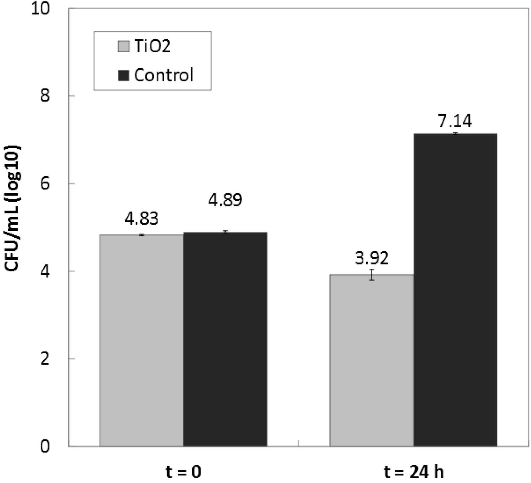
2.2. Antibacterial Activity of TiO2 in the Dark

TiO2 nanoparticles (KRONOClean 7050) were suspended in 1/500 NB [48] at the concentration of 13.9 g/L. Eleven milliliters of the suspension were then deposited onto a sterile Petri dish, so that the total area of the inside part of the dish was covered. The Petri dishes were placed in a sterile flow hood for air drying until the water had totally evaporated. A film of TiO2 was visible at the bottom. Then, 11 mL of the inoculum (between 8 × 10⁴ and 2 × 10⁵ cells/mL) were deposited on the TiO2 film, and the Petri dishes were covered with a lid [48]. After a fixed time (0 and 24 h), the lid was removed, the bottoms of the Petri dishes were gently scraped with a plastic loop in order to remove any adhered cells and 1 mL of the suspension was collected and diluted in phosphate buffer. Control samples were studied in Petri dishes without TiO2.
One-mL quantities of the appropriate dilutions were then dropped into distilled sterile water and filtered on cellulose ester filters (ϕ = 0.45 μm) in order to separate bacterial cells from nanoparticles. The filters were then deposited on trypticase soy agar and incubated at a temperature of 36 °C ± 1 °C for 40 to 48 h. After incubation, the number of viable cells was estimated in CFU/mL.

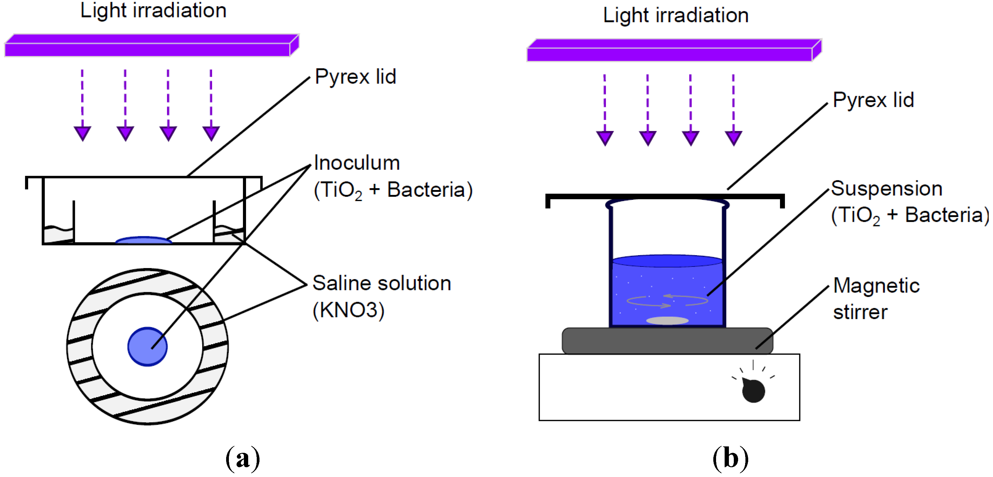
2.3. Deposited-Drop Experiment

To avoid damage by UV irradiation alone [49], the maximum UV intensity was maintained at 2.5 W/m2. Previous tests with higher UV intensity had shown total drying of the inoculum during the experiment and led to the inactivation of bacteria in control samples. The light intensity was measured on the samples using a UV-A radiometer (Gigahertz-Optik, GmbH Türkenfeld, Germany) in the 310–400 nm range.
Various configurations were studied: samples under UV irradiation (TiO2-bearing samples and control specimen without TiO2) and samples kept in the dark (TiO2-bearing samples and control specimen without TiO2). All tests were carried out in triplicate. The data shown are the average of triplicates, with the corresponding standard errors.

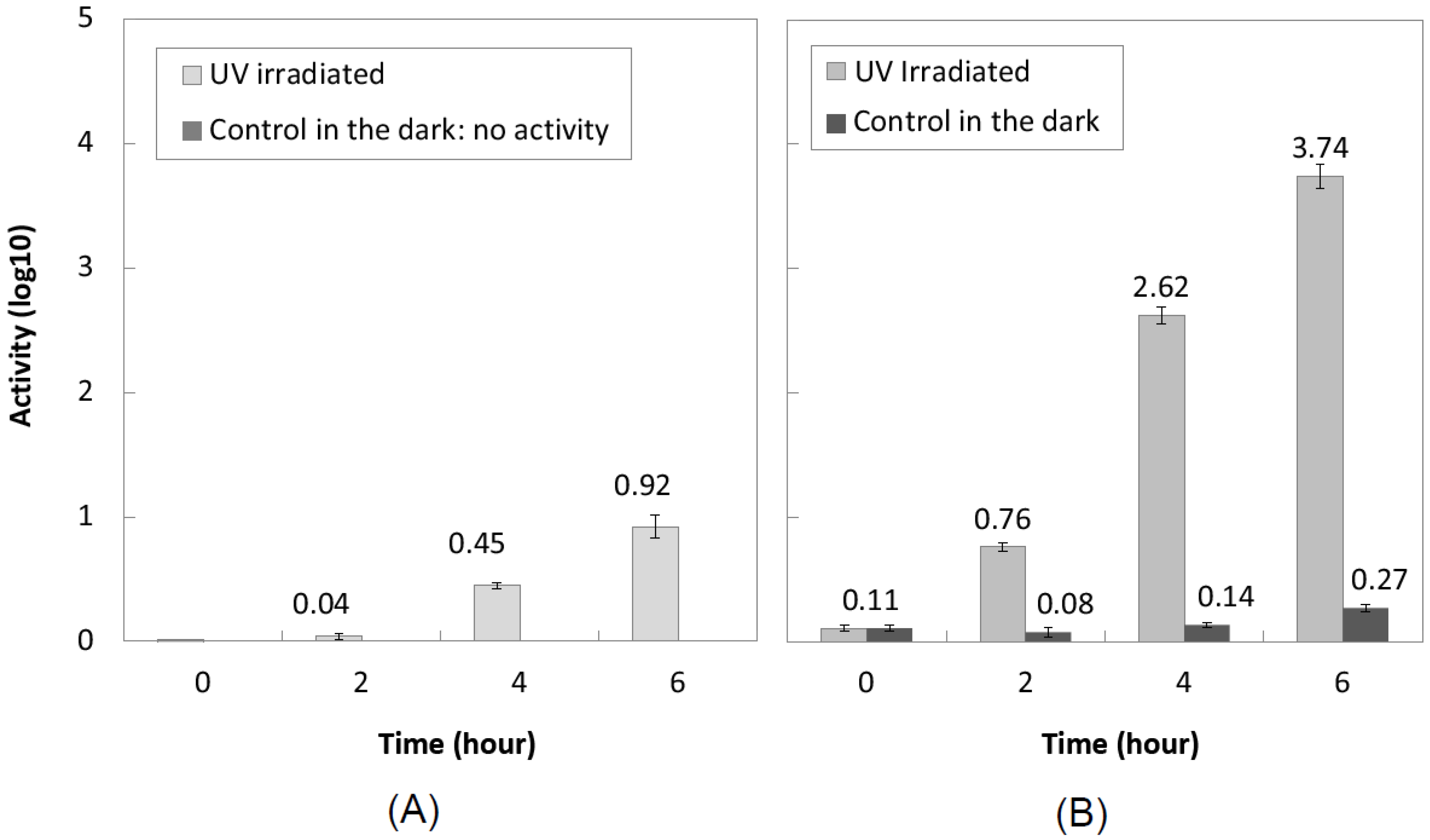
2.3.1. With TiO2 Powder

The experiment was based on the standards JIS Z 2801 (Japanese Industrial Standard) and ISO 27447 [48,49]. TiO2 nanoparticle powder (KRONOClean7050–anatase) was suspended in 9 mL of 1/500 NB [48], and 1 mL of the bacterial suspension (Section 2.1) was added. Final concentrations were 1 g/L for TiO2 and between 8 × 10⁴ and 2 × 10⁵ CFU/mL for bacteria. The bacterial suspension (Section 2.1) without TiO2 was used as a control. Then, 0.4 mL of the inoculum were instilled onto a Pyrex Petri dish designed so that an external ring could receive 2 mL of a supersaturated saline solution (KNO3) to maintain 90% RH and was covered with a Pyrex lid (Figure 1). The Petri dishes were placed in a sterile flow hood and illuminated with an 8-W black-light bulb. After a few minutes, the TiO2 nanoparticles were observed to have sedimented at the bottom of the drop.
Figure 1. Schematic illustration of the deposited-drop experiments with TiO2 powder and TiO2 semi-transparent coatings (a) and stirring experiment (b).
Coatings 04 00670 g001
A Soybean Casein Lecithin Polysorbate 80 Medium, also known as SCDLP broth, was prepared in sterile distilled water as recommended in standard JIS Z 2801 [48], using casein peptone, soybean peptone, sodium chloride, disodium hydrogen phosphate, glucose, lecithin and Tween 80.
After different contact times (2 h, 4 h, 6 h), the suspension was washed out with the appropriate amount of SCDLP broth and with sterile glass beads (d = 4 mm). When necessary, the washed-out suspension was diluted X times in a phosphate buffer, so that it contained 30 to 300 cells per mL. For each sample, 1 mL of the appropriate dilution was dispensed into two sterilized Petri dishes with 15 to 20 mL of trypticase soy agar (TSA) and incubated at a temperature of 36 °C ± 1 °C for 40 to 48 h. After incubation, the number of viable cells was estimated in terms of CFU. The overall procedure was also systematically carried out immediately after inoculation (t = 0 h) to validate the test. The antibacterial activity was then calculated as the difference between the average logarithm of the number of viable bacteria on the control without TiO2 and the average logarithm of the number of viable bacteria on the TiO2 sample:
Coatings 04 00670 i001
where, A: antibacterial activity; NTiO2: average number of CFU on TiO2 sample at time t; Ncontrol: average number of CFU on control sample at time t.
The test was then repeated with a transparent film (9–10 cm2) gently placed on the inoculum before irradiation in order to increase the probability of contact between bacteria cells and TiO2 nanoparticles (forced contact).

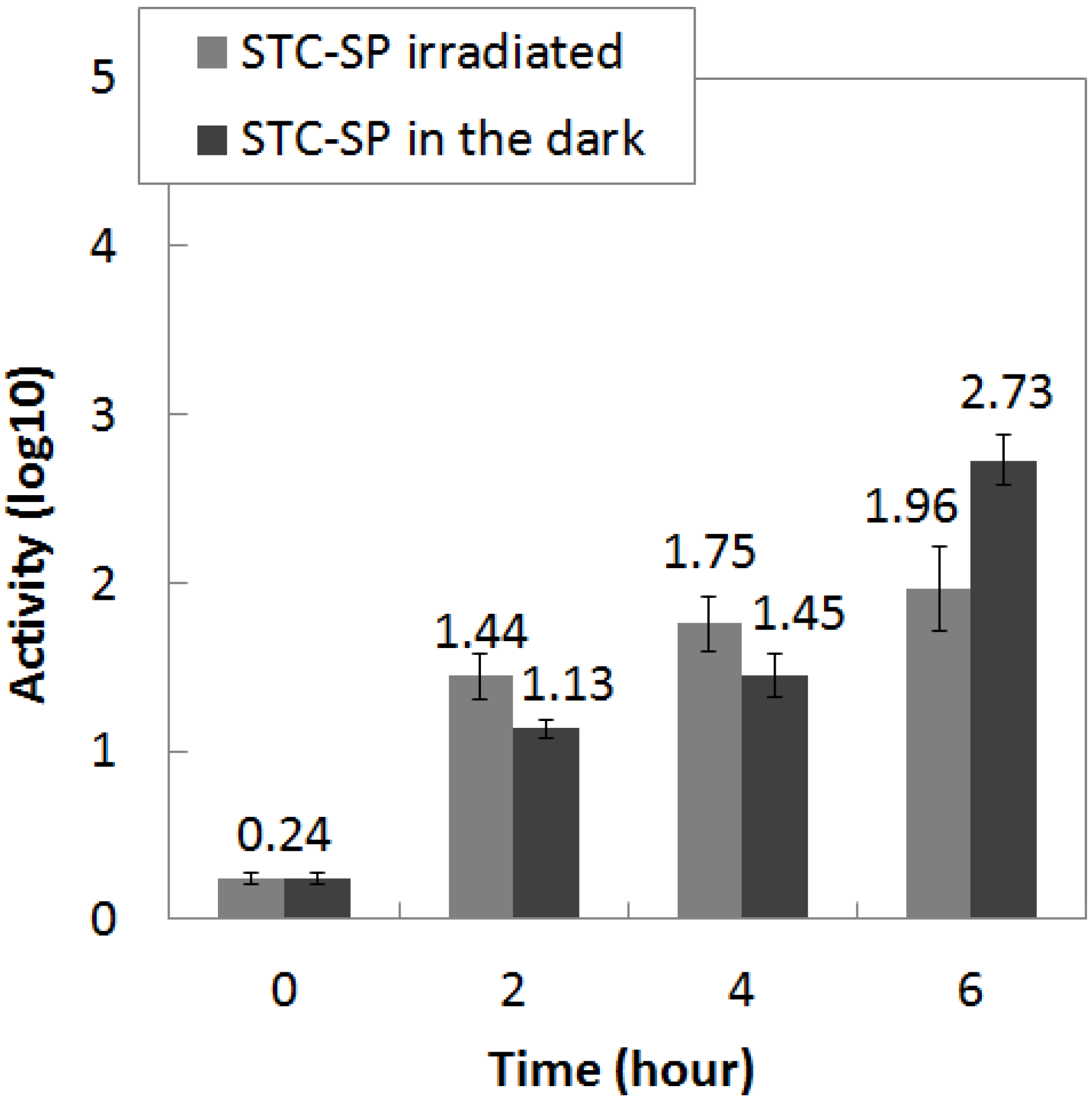
2.3.2. With TiO2 Semi-Transparent Coating

The deposited-drop experiment was repeated with semi-transparent coating formulated using TiO2 nanoparticles as an antibacterial product: TiO2 powder (KronoClean 7050, KRONOS/Société Industrielle du Titane, Paris, France) and TiO2 dispersion (Kronos type 7454, trial product, KRONOS/Société Industrielle du Titane, Paris, France). The coating formulation included water and acrylic based on the work of Martinez et al. [46], as shown in Table 1. Sterilized cover-glasses (26 × 76 mm2) were covered with the coatings by instilling 1 mL, so that the total area of each glass was coated. The cover-glasses were then placed under a sterile flow hood for air drying. After drying, the semi-transparent coatings with TiO2 powder (STC-SP (with silicates), STC-P) were gently sanded with fine sandpaper in order to prevent the possible inclusion of nanoparticles in the binder. The semi-transparent coatings with TiO2 in aqueous suspension (STC-A) were pre-aged by irradiating them with UV light (2.5 W/m2) for 80 h. The amount of TiO2 was estimated at 2.5 mg/cm2 for samples coated with TiO2 powder (STC-SP, STC-P) and 0.63 mg/cm2 for samples coated with TiO2 aqueous suspension (STC-A). In order to evaluate the possible inclusion of nanoparticles in the binder of STC-A, samples were also prepared with water and TiO2 aqueous suspension, without acrylic resin (STC-A2). For each test sample, corresponding controls were prepared in the same way with water and acrylic resin, but without TiO2.
Table 1. Formulation of semi-transparent coatings. STC: semi-transparent coating.
STC-SPSTC-PSTC-ASTC-A2
WaterWaterWaterWater
Acrylic resin (7.5 wt%)Acrylic resin (12 wt%)Acrylic resin (2 wt%)
TiO2 powder (KronoClean 7050)TiO2 powder (KronoClean 7050)TiO2 in aqueous suspension (Kronos trial product 7454)TiO2 in aqueous suspension (Kronos trial product 7454)
Silicates (12.5 wt%)
The inoculation suspension (of Section 2.1) was diluted to make the concentration of inoculum 8 × 10⁴ to 2 × 10⁵ CFU/mL. The coated cover-glasses were placed over the internal ring of the Pyrex Petri dishes shown in Figure 1. Relative humidity was maintained with 2 mL of the supersaturated saline solution (KNO3) deposited in the external ring of each dish. Then, 0.4 mL of the inoculum were instilled on each coated cover-glass, and a transparent plastic film was applied, spreading the inoculum over a surface area of 10 cm2. The Petri dishes were then covered with a Pyrex lid (Figure 1), placed in a sterile flow hood and illuminated with an 8-W black-light bulb.
After different contact times (2 h, 4 h, 6 h), the cover-glasses were recovered with sterile pliers and placed in plastic Petri dishes for wash-out. The wash-out of bacteria cells and the following procedures for CFU counting were repeated as in Section 2.3.1.

2.4. Stirring Experiment

For the stirring experiment; TiO2 nanoparticles (KronoClean7050) were suspended in a sterile beaker in 27 mL of 1/500 NB or sterile distilled water; depending on the test; and 3 mL of the cell suspension were added to make the final test suspension. Final concentrations of the suspension were 1 g/L for TiO2 and 7 × 10⁴ to 1 × 10⁵ CFU/mL for bacteria. A suspension of bacterial cells without TiO2 was prepared as a control. The beakers (test sample and control) were placed in a sterile flow hood; covered with a Pyrex lid and illuminated with an 8-W black-light bulb at a light intensity of 5 W/m2.
An aliquot of 1 mL was taken from each beaker every 30 min during 4 h and, when necessary, diluted in phosphate buffer before inclusion in trypticase soy agar as in the deposited-drop experiment. Controls were also carried out without TiO2 and in the dark, with/without TiO2. The data presented are the average of three experiments with the corresponding standard errors.
To assess the influence of the nature of the water during the experiment, two solutions were used: 1/500 nutrient broth and sterile distilled water. The two conductivities of the solutions were compared using a conductivity meter before the test. At room temperature (~21 °C), the conductivities were 4.437 mS/m for 1/500 NB and 1.1 mS/m for distilled water.

3. Results and Discussion

3.1. Effect of TiO2 in the Dark

Figure 2 shows the bacterial concentration of E. coli cells after 0 and 24 h of contact with TiO2 nanoparticles (11 mL inoculation of air-dried TiO2 film in Petri dishes). From these data, an increase can be seen in the number of CFUs for control samples (+2.25 ± 0.06 log) and a decrease in the number of CFUs for TiO2 samples (−0.91 ± 0.14 log). The corresponding antibacterial activity, calculated from Equation (1), is 3.22 ± 0.14 log. It is therefore likely that the activity of TiO2 on E. coli in the dark is correlated with a growth inhibitory effect as a major pathway and a bactericidal effect as a minor pathway. These results highlight the physical impact on E. coli cells induced by contact with TiO2 nanoparticles, without regard to the photocatalytic process. This also agrees with earlier observations by Liu et al. [50] and Gogniat et al. [38], which showed a loss of bacterial culturability after contact with TiO2 nanoparticles in the dark. A study by de Niederhãusen and Bondi [51] on the self-cleaning of Ag-TiO2-coated ceramic tiles also showed significant antibacterial activity for 24 h in the dark.
Interestingly, we detected no difference in the CFU counts between bacterial suspensions with TiO2 (1 g/L and 10 g/L) and a control bacterial suspension (without TiO2) after direct plating of 2 × 1 mL on TSA and 48 hours’ incubation (data not shown). It seems possible that the physical damage sustained is not sufficient to kill bacterial cells when they are growing in a nutrient-rich culture medium. Such a “neutralizing” effect of culture media is current with antiseptic and disinfectant molecules, which highlights the impact of test conditions on efficiency evaluation.
Figure 2. Bacterial concentration after 24 h with TiO2 nanoparticles in the dark. Mean ± SE, n = 3.
Coatings 04 00670 g002

3.2. Free Surface Drop Deposit vs. Forced Contact between Bacteria and Nanoparticles

The antibacterial activities of TiO2 nanoparticles on E. coli during the deposited-drop experiment are presented in Figure 3. No activity was detected for samples kept in the dark for 6 h under normal testing conditions (A). The bacterial reduction reached 0.92 ± 0.09 log after 6 h of irradiation (A).
Figure 3. Antibacterial activity of TiO2 as a support under UV irradiation (≈2.5 W/m2): (A) standard conditions; (B) after application of a transparent plastic film on the inoculum. Mean ± SE, n = 3.
Coatings 04 00670 g003
Since the bactericidal effect induced by photocatalysis of TiO2 nanoparticles depends on many factors, such as the amount [35,37,52,53,54,55] and the crystalline nature [39,55,56,57] of TiO2, the irradiation time and intensity [35,52,55,58] and the inoculum concentration [42,53,59], it is reasonable to assume that the dispersion of TiO2 particles and bacteria (low probability of contact) and the very low light intensity used in our experiment (2.5 W/m2 in order to avoid any UV-damage) could be major factors explaining the low activity observed after 6 h of irradiation. Various studies from the literature show intensities of over 10 W/m2 and antibacterial activities on E. coli easily greater than 3 log after 90 min of irradiation [39,40,42,60].
Following the application of a transparent film (9 cm2) onto the inoculum, a significant increase in the antibacterial activity was observed. As shown in Figure 3B, the activity was 3.74 ± 0.1 log after 6 h under UV irradiation and 0.27 ± 0.03 log after 6 h in the dark.
The present findings seem to support the idea that reducing the distance between bacterial cells and TiO2 nanoparticles enhances the photocatalytic disinfection process. This also agrees with earlier research highlighting the importance of the contact between bacterial cells and the surface of TiO2 [39,40,41,43,44,50,61,62,63]. In addition to the oxidative stress induced by reactive oxygen species (ROS) on bacterial cells, contact with the TiO2 surface leads to the direct oxidation of cells by photogenerated holes, which also reduces the recombination of charges inside the photocatalyst [38,43,44,64]. Moreover, it has been suggested that direct contact and adsorption on TiO2 nanoparticles cause (I) a loss of membrane integrity [38,50,61] and possibly (II) a process of phagocytosis of the nanoparticles by the cells (the findings of Cai et al. [64] must be interpreted with caution in this paper, because they focused on HeLa cells and not bacterial cells.) [64], both leading to the reduction of the number of cultivable cells, if not to cell death. These results also agree with the findings of other studies that have highlighted the major role of surface radicals compared to free radicals in photocatalytic disinfection [40,41,62].

3.3. Influence of the Nature of the Solution for Suspension

The results obtained from the stirring experiment are presented in Figure 4. For TiO2 samples, both inactivation curves consist of two steps: the first with a very low inactivation rate followed by the second with a higher inactivation rate. In addition, the second rate appears to be the same for both distilled water and 1/500 NB. It is likely that 1/500 NB acts as a retarding agent of the photocatalytic disinfection process.
Figure 4. Survival of E. coli cells vs. irradiation time at ~5 W/m2 with the standard error of three experiments. Mean ± SE, n = 3.
Coatings 04 00670 g004
The present finding is in full agreement with the inhibitory effect of various ions and organic compounds on photocatalytic disinfection, which is widely reported in the literature [34,37,56,65,66]. The presence of ions and organic compounds can reduce the efficiency in different ways:
-
Competition between ions, compounds and bacteria for the adsorption on the TiO2 surface [37,38,56,65,67];
-
The ROS mobilized by ions and compounds cannot oxidize bacterial cells [65];
-
Aggregates of organic compounds could create a barrier filtering UV.
According to Dunlop et al. [56] and Sunada et al. [42], the low rate of inactivation in the first step may be due to the preliminary attack of the outer membrane of cells by ROS.
During this first step, the damage sustained by the outer membrane may be insufficient to kill bacteria: they can recover from the damage and re-grow once they are plated in agar media [42,56]. After some time, degradation of the outer membrane enables reactive species to penetrate, which induces damage, leading to the death of the bacterial cells (second, higher rates on the curves of Figure 4). This hypothesis has also been considered by other researchers [58,68]. Mitoraj et al. [68] explained this “incubation period” as the time for the concentration of photogenerated ROS to increase to a level that is harmful to bacteria.
Another possible explanation for the first step with the low inactivation rate is proposed by Gogniat et al. [38]. In their works, they observed the two-stage curve only in a sodium phosphate solution and not in a NaCl-KCl solution. They hypothesized that the change of adsorption properties of TiO2 when illuminated led to a photo-desorption of ions previously adsorbed on its surface. Thus, the time taken for the photo-desorption process explains the low inactivation rate observed during the first minutes of the experiment [38].
Interestingly, the third step observed in earlier studies [58,68,69,70] and consisting of strong attenuation of the bacterial inactivation was not observed here. One of the hypotheses suggested is that photocatalytic inactivation is built up by bacterial growth after a certain period of time [69]. It can be supposed that bacterial growth in pure water is slowed down or stopped. Further investigations in 1/500 NB after longer times could show similar attenuation of the inactivation rate.
Some authors have compared efficiencies between scattered and fixed TiO2 [71,72,73]. Pablos et al. [72] observed a higher inactivation rate at the beginning of the reaction with fixed TiO2. They suggested that damage was uniformly distributed over the whole cell wall in slurries, whereas it was more concentrated on small areas with fixed TiO2, requiring smaller amounts of radicals to achieve inactivation. However, they observed similar times for total inactivation of bacteria (E. coli) for both implementations (fixed and scattered). On the other hand, Gumy et al. [40] found higher inactivation efficiency with suspended TiO2 than with TiO2 coated on a fibrous web and suggested that particles dispersed in slurry would provide more surfaces for the adsorption of bacteria. In addition, inactivation of bacteria has been observed in the presence of TiO2 nanoparticles in the dark, suggesting that phenomena other than photocatalytic processes can explain inactivation [50,61]. Although the complete process is not perfectly understood yet, the overall literature points to the importance of the contact between bacteria and TiO2 for improving disinfection efficiency, suggesting both chemical and physical influences.
In their work, Gomes et al. [71] also reported a higher inactivation rate in slurry than with TiO2 supported on Ahlstrom paper. They suggested that such results could be explained by competitive reactions of TiO2 with the organic matter released by the paper during the experiment. Accordingly, the presence of ions and/or organic compounds in the slurry/inoculum considerably reduced the efficiency by reacting with ROS and being adsorbed on TiO2 in place of bacterial cells [38,65]. These works also raised the problem of TiO2 coatings in which the organic matter from the binder can monopolize photogenerated radicals and, thus, lead to a decrease in disinfection efficiency.

3.4. Semi-Transparent Coating

Figure 5 presents the experimental data on the antibacterial activity of semi-transparent coatings formulated with silicate and TiO2 powder (STC-SP). Surprisingly, antibacterial activity was also observed on samples kept in the dark. A quick estimation of the pH of the inoculum with indicator paper showed a pH around 11–12, far too high for E. coli survival. It is then reasonable to assume that the antibacterial activity detected on STC-SP samples was not induced by the photocatalytic process, but by the silicates, making the inoculum strongly basic. Results from sample coatings without silicate (STC-P) showed no activity after eight hours’ irradiation and no activity after 8 h in the dark. Possible explanations are that photogenerated radicals may have reacted with the binder instead of with bacteria or that the inclusion of nanoparticles within the binder may have prevented UV absorption and physical damage by contact. Moreover, the use of TiO2 powder without a dispersing agent may lead to the formation of aggregates, reducing the surface available for reaction.
Figure 5. Antibacterial activity of SCT-SP coatings under UV irradiation (≈2.5 W/m2) and in the dark. Mean ± SE, n = 3.
Coatings 04 00670 g005
Figure 6 presents the antibacterial activity obtained with STC-A and STC-A2. The activity reaches 1.49 ± 0.47 log for STC-A and 1.54 ± 0.13 log for STC-A2 after four hours’ irradiation. The observed increase of antibacterial activity, compared to SCT-P in which no activity was detected, could be attributed to the use of the TiO2 dispersion. Nanoparticles, stabilized by the dispersing agent, may have provided more active sites for the photocatalytic process. Moreover, the smaller amount of acrylic resin within STC-A and STC-A2 (2%) may have reduced the inclusion of TiO2 nanoparticles compared to SCT-P. The similar activities observed on STC-A (with acrylic resin) and on STC-A2 (without acrylic resin) seem to confirm this hypothesis. Further investigations on the formulation of these coatings along with observations of nanoparticle distribution in the binder will be helpful in the development of antibacterial products for building materials.
Figure 6. Antibacterial activity of STC-A and STC-A2 coatings under UV irradiation (≈2.5 W/m2). Mean ± SE, n = 3.
Coatings 04 00670 g006

4. Conclusions

This paper has examined the effect of TiO2 photocatalyst on E. coli in terms of antibacterial activity by carrying out two different tests (a drop deposited on a photocatalytic substrate and a stirring experiment in a TiO2-bearing suspension).
Some general effects reported in the literature concerning the photocatalytic disinfection process have been observed.
-
Prolonged contact (24 h) in the dark leads to significant antibacterial activities, potentially explained by a combination of the direct contact (I) bactericidal effect and the (II) growth inhibiting effect.
-
Reducing the distance between nanoparticles and bacteria significantly increases the inactivation of E. coli by non-photocatalytic effects (direct contact) and the photocatalysis disinfection process.
-
The presence of ions and organic compounds in the suspension during the test delays the inactivation.
In addition, the transparent coatings tested showed significant antibacterial activities under low UV irradiation. The results suggest that improving the formulation, i.e., varying the proportions of the components, could increase the efficiency of coatings. In a broader framework, further experimental investigations will be conducted on the resistance of this coating to fungal proliferation and on the protection it affords against the formation of microbial biofilms.

Acknowledgments

The authors would like to thank Université Paul Sabatier Toulouse III for its financial support.

Author Contributions

Experimental measurements have been conducted by Thomas Verdier. Analysis and interpretation of the results as well as conclusions have been conducted by all the co-authors. The manuscript has been written by Thomas Verdier with the revision and approval by the others co-authors.

Conflicts of Interest

The authors declare no conflict of interest.

References

  1. Déoux, S. Les Enjeux Sanitaires de la Qualité de l’air Intérieur; Cité des Sciences et de l’industrie: Paris, France, 2013. (In French) [Google Scholar]
  2. Development of WHO Guidelines for Indoor Air Quality. Available online: http://www.euro.who.int/en/health-topics/environment-and-health/air-quality/publications/pre2009/development-of-who-guidelines-for-indoor-air-quality (accessed on 10 June 2014).
  3. WHO Guidelines for Indoor Air Quality. Available online: http://www.euro.who.int/en/health-topics/environment-and-health/air-quality/policy/who-guidelines-for-indoor-air-quality (accessed on 10 June 2014).
  4. World Health Organization. WHO Guidelines for Indoor Air Quality: Dampness and Mould; WHO: Geneva, Switzerland, 2009. [Google Scholar]
  5. World Health Organization. WHO Guidelines for Indoor Air Quality: Selected Pollutants; WHO: Geneva, Switzerland, 2010. [Google Scholar]
  6. Cooley, J.D.; Wong, W.C.; Jumper, C.A.; Straus, D.C. Correlation between the prevalence of certain fungi and sick building syndrome. Occup. Environ. Med. 1998, 55, 579–584. [Google Scholar] [CrossRef] [PubMed]
  7. Li, C.S.; Hsu, C.W.; Tai, M.L. Indoor pollution and sick building syndrome symptoms among workers in day-care centers. Arch. Environ. Health 1997, 52, 200–207. [Google Scholar] [CrossRef] [PubMed]
  8. Conseil Supérieur d’Hygiène Publique de France (CSHPF). Contaminations Fongiques en Milieux Intérieurs. Diagnostic, Effet sur la Santé Respiratoire, Conduite à Tenir; CSHPF: Paris, France, 2006. (In French) [Google Scholar]
  9. Nolard, N.; Beguin, H. Moisissures. In Traité D’Allergologie Paris Médecine-Sci.; Flammarion: Paris, France, 2003; pp. 441–461. (In French) [Google Scholar]
  10. Gutarowska, B.; Żakowska, Z. Elaboration and application of mathematical model for estimation of mould contamination of some building materials based on ergosterol content determination. Int. Biodeterior. Biodegrad. 2002, 49, 299–305. [Google Scholar]
  11. Minnesota Department of Health (MDH). Recommended Best Practices for Mold Investigations in Minnesota Schools; MDH, Environmental Health Division, Indoor Air Unit: Saint Paul, MN, USA, 2001. [Google Scholar]
  12. Reboux, G.; Bellanger, A.-P.; Roussel, S.; Grenouillet, F.; Millon, L. Moisissures et Habitat: Risques Pour la Santé et Espèces Impliquées. Rev. Fr. Allergol. 2010, 50, 611–620. (In French) [Google Scholar]
  13. ASEF Pollution de l’air intérieur de l’habitat. Available online: http://www.asef-asso.fr/attachments/1141_Guide_air%20int%C3%A9rieur.pdf (accessed on 30 October 2013). (In French)
  14. Fung, F.; Hughson, W.G. Health Effects of Indoor Fungal Bioaerosol Exposure. Appl. Occup. Environ. Hyg. 2003, 18, 535–544. [Google Scholar] [CrossRef]
  15. Santucci, R.; Meunier, O.; Ott, M.; Herrmann, F.; Freyd, A.; de Blay, F. Contamination fongique des habitations: Bilan de 10 années d’analyses. Rev. Fr. Allergol. Immunol. Clin. 2007, 47, 402–408. (In French) [Google Scholar]
  16. Nielsen, K.F.; Holm, G.; Uttrup, L.P.; Nielsen, P.A. Mould growth on building materials under low water activities. Influence of humidity and temperature on fungal growth and secondary metabolism. Influence of humidity and temperature on fungal growth and secondary metabolism. Int. Biodeterior. Biodegrad. 2004, 54, 325–336. [Google Scholar]
  17. Spengler, J.D.; Chen, Q. Indoor Air Quality Factors in Designing a Healthy Building. Annu. Rev. Energy Environ. 2000, 25, 567–600. [Google Scholar] [CrossRef]
  18. Tuomi, T.; Reijula, K.; Johnsson, T.; Hemminki, K.; Hintikka, E.-L.; Lindroos, O.; Kalso, S.; Koukila-Kähkölä, P.; Mussalo-Rauhamaa, H.; Haahtela, T. Mycotoxins in Crude Building Materials from Water-Damaged Buildings. Appl. Environ. Microbiol. 2000, 66, 1899–1904. [Google Scholar] [CrossRef] [PubMed]
  19. Bellanger, A.-P.; Reboux, G.; Roussel, S.; Grenouillet, F.; Didier-Scherer, E.; Dalphin, J.-C.; Millon, L. Indoor fungal contamination of moisture-damaged and allergic patient housing analysed using real-time PCR. Lett. Appl. Microbiol. 2009, 49, 260–266. [Google Scholar] [CrossRef] [PubMed]
  20. Andersson, M.A.; Nikulin, M.; Köljalg, U.; Andersson, M.C.; Rainey, F.; Reijula, K.; Hintikka, E.L.; Salkinoja-Salonen, M. Bacteria, molds, and toxins in water-damaged building materials. Appl. Environ. Microbiol. 1997, 63, 387–393. [Google Scholar]
  21. Dillon, H.K.; Miller, J.D.; Sorenson, W.G.; Douwes, J.; Jacobs, R.R. Review of methods applicable to the assessment of mold exposure to children. Environ. Health Perspect. 1999, 107, 473–480. [Google Scholar] [CrossRef]
  22. Torvinen, E.; Meklin, T.; Torkko, P.; Suomalainen, S.; Reiman, M.; Katila, M.-L.; Paulin, L.; Nevalainen, A. Mycobacteria and Fungi in Moisture-Damaged Building Materials. Appl. Environ. Microbiol. 2006, 72, 6822–6824. [Google Scholar] [CrossRef] [PubMed]
  23. Health Implications of Fungi in Indoor Environments; Samson, R.A.; Flannigan, B.; Flannigan, M.E.; Verhoeff, A.P.; Adan, O.C.G.; Hoekstra, E.S. (Eds.) Elsevier Science Ltd.: Kidlington, UK, 1994.
  24. Flannigan, B.F.; Samson, R.A.; Miller, J.D. Microorganisms in Home and Indoor Work Environments: Diversity, Health Impacts, Investigation and Control; Taylor & Francis Group: Abingdon, UK, 2001. [Google Scholar]
  25. Parat, S.; Perdrix, A.; Mann, S.; Cochet, C. A Study of the Relationship between Airborne Microbiological Concentrations and Symptoms in Office in Buildings. In Proceedings of the Healthy Building, Milan, Italy, 10–15 September 1995.
  26. Williamson, I.J.; Martin, C.J.; McGill, G.; Monie, R.D.; Fennerty, A.G. Damp housing and asthma: A case-control study. Thorax 1997, 52, 229–234. [Google Scholar] [CrossRef] [PubMed]
  27. Mudarri, D.; Fisk, W.J. Public health and economic impact of dampness and mold. Indoor Air 2007, 17, 226–235. [Google Scholar] [CrossRef] [PubMed]
  28. Johanning, E. Mycotoxin and Indoor Health. In Proceedings of the Sixth VI International Conference on Mycotoxins in the Environment of People and Animals, Bydgoszcz, Poland, 25–27 September 2002.
  29. Gutarowska, B.; Piotrowska, M. Methods of mycological analysis in buildings. Build. Environ. 2007, 42, 1843–1850. [Google Scholar] [CrossRef]
  30. Verdier, T.; Coutand, M.; Bertron, A.; Roques, C. A review of indoor microbial growth across building materials and sampling and analysis methods. Build. Environ. 2014, 80, 136–149. [Google Scholar] [CrossRef]
  31. Chong, M.N.; Jin, B.; Chow, C.W.K.; Saint, C. Recent developments in photocatalytic water treatment technology: A review. Water Res. 2010, 44, 2997–3027. [Google Scholar] [CrossRef] [PubMed]
  32. Gamage, J.; Zhang, Z.S. Applications of Photocatalytic Disinfection. Int. J. Photoenergy 2010, 2010. [Google Scholar] [CrossRef]
  33. Dalrymple, O.K.; Stefanakos, E.; Trotz, M.A.; Goswami, D.Y. A review of the mechanisms and modeling of photocatalytic disinfection. Appl. Catal. B Environ. 2010, 98, 27–38. [Google Scholar] [CrossRef]
  34. Foster, H.A.; Ditta, I.B.; Varghese, S.; Steele, A. Photocatalytic disinfection using titanium dioxide: Spectrum and mechanism of antimicrobial activity. Appl. Microbiol. Biotechnol. 2011, 90, 1847–1868. [Google Scholar] [CrossRef] [PubMed]
  35. Wei, C.; Lin, W.Y.; Zainal, Z.; Williams, N.E.; Hemminki, K.; Kruzic, A.P.; Smith, R.L.; Rajeshwar, K. Bactericidal Activity of TiO2 Photocatalyst in Aqueous Media: Toward a Solar-Assisted Water Disinfection System. Environ. Sci. Technol. 1994, 28, 934–938. [Google Scholar] [CrossRef]
  36. Sunada, K.; Watanabe, T.; Hashimoto, K. Bactericidal Activity of Copper-Deposited TiO2 Thin Film under Weak UV Light Illumination. Environ. Sci. Technol. 2003, 37, 4785–4789. [Google Scholar] [CrossRef]
  37. Saito, T.; Iwase, T.; Horie, J.; Morioka, T. Mode of photocatalytic bactericidal action of powdered semiconductor TiO2 on mutans streptococci. J. Photochem. Photobiol. B 1992, 14, 369–379. [Google Scholar] [CrossRef]
  38. Gogniat, G.; Thyssen, M.; Denis, M.; Pulgarin, C.; Dukan, S. The bactericidal effect of TiO2 photocatalysis involves adsorption onto catalyst and the loss of membrane integrity. FEMS Microbiol. Lett. 2006, 258, 18–24. [Google Scholar] [CrossRef] [PubMed]
  39. Gumy, D.; Morais, C.; Bowen, P.; Pulgarin, C.; Giraldo, S.; Hajdu, R.; Kiwi, J. Catalytic activity of commercial of TiO2 powders for the abatement of the bacteria (E. coli) under solar simulated light: Influence of the isoelectric point. Appl. Catal. B Environ. 2006, 63, 76–84. [Google Scholar]
  40. Gumy, D.; Rincon, A.G.; Hajdu, R.; Pulgarin, C. Solar photocatalysis for detoxification and disinfection of water: Different types of suspended and fixed TiO2 catalysts study. Sol. Energy 2005, 80, 1376–1381. [Google Scholar] [CrossRef]
  41. Kikuchi, Y.; Sunada, K.; Iyoda, T.; Hashimoto, K.; Fujishima, A. Photocatalytic bactericidal effect of TiO2 thin films: Dynamic view of the active oxygen species responsible for the effect. J. Photochem. Photobiol. Chem. 1997, 106, 51–56. [Google Scholar] [CrossRef]
  42. Sunada, K.; Watanabe, T.; Hashimoto, K. Studies on photokilling of bacteria on TiO2 thin film. J. Photochem. Photobiol. Chem. 2003, 156, 227–233. [Google Scholar] [CrossRef]
  43. Nadtochenko, V.; Denisov, N.; Sarkisov, O.; Gumy, D.; Pulgarin, C.; Kiwi, J. Laser kinetic spectroscopy of the interfacial charge transfer between membrane cell walls of E. coli and TiO2. J. Photochem. Photobiol. Chem. 2006, 181, 401–407. [Google Scholar] [CrossRef]
  44. Nadtochenko, V.A.; Sarkisov, O.M.; Nikandrov, V.V.; Chubukov, P.A.; Denisov, N.N. Inactivation of Pathogenic Microorganisms in the Photocatalytic Process on Nanosized TiO2 Crystals. Russ. J. Phys. Chem. B Focus Phys. 2008, 2, 105–114. [Google Scholar]
  45. Martinez, T. Revêtements Photocatalytiques Pour les Matériaux de Construction: Formulation, Évaluation de L’efficacité de la Dépollution de l’air et Écotoxicité; Génie Civil, Toulouse III–Paul Sabatier: Toulouse, France, 2012. (In French) [Google Scholar]
  46. Martinez, T.; Bertron, A.; Ringot, E.; Escadeillas, G. Degradation of NO using photocatalytic coatings applied to different substrates. Build. Environ. 2011, 46, 1808–1816. [Google Scholar] [CrossRef]
  47. Martinez, T.; Bertron, A.; Escadeillas, G.; Ringot, E. Algal growth inhibition on cement mortar: Efficiency of water repellent and photocatalytic treatments under UV/VIS illumination. Int. Biodeterior. Biodegrad. 2014, 89, 115–125. [Google Scholar] [CrossRef]
  48. Japanese Industrial Standard. In Antibacterial Products—Test for Antibacterial Activity and Efficacy; JIS Z 2801; Japanese Standards Association: Tokyo, Japan, 2010.
  49. International Organization for Standardizaiton (ISO). ISO 27447 Fine Ceramics (Advanced Ceramics, Advanced Technical Ceramics)—Test Method for Antibacterial Activity of Semiconducting Photocatalytic Materials; ISO: Berlin, Germany, 2009. [Google Scholar]
  50. Liu, L.; John, B.; Yeung, K.L.; Si, G. Non-UV based germicidal activity of metal-doped TiO2 coating on solid surfaces. J. Environ. Sci. 2007, 19, 745–750. [Google Scholar] [CrossRef]
  51. De Niederhãusern, S.; Bondi, M.; Bondioli, F. Self-Cleaning and Antibacteric Ceramic Tile Surface. Int. J. Appl. Ceram. Technol. 2013, 10, 949–956. [Google Scholar]
  52. Horie, Y.; David, D.A.; Taya, M.; Tone, S. Effects of Light Intensity and Titanium Dioxide Concentration on Photocatalytic Sterilization Rates of Microbial Cells. Ind. Eng. Chem. Res. 1996, 35, 3920–3926. [Google Scholar] [CrossRef]
  53. Maness, P.-C.; Smolinski, S.; Blake, D.M.; Huang, Z.; Wolfrum, E.J.; Jacoby, W.A. Bactericidal Activity of Photocatalytic TiO2 Reaction: Toward an Understanding of Its Killing Mechanism. Appl. Environ. Microbiol. 1999, 65, 4094–4098. [Google Scholar] [PubMed]
  54. Huang, Z.; Maness, P.-C.; Blake, D.M.; Wolfrum, E.J.; Smolinski, S.L.; Jacoby, W.A. Bactericidal mode of titanium dioxide photocatalysis. J. Photochem. Photobiol. Chem. 2000, 130, 163–170. [Google Scholar] [CrossRef]
  55. Rincón, A.G.; Pulgarin, C. Photocatalytical inactivation of E. coli: Effect of (continuous–intermittent) light intensity and of (suspended–fixed) TiO2 concentration. Appl. Catal. B Environ. 2003, 44, 263–284. [Google Scholar]
  56. Dunlop, P.S.M.; Byrne, J.A.; Manga, N.; Eggins, B.R. The photocatalytic removal of bacterial pollutants from drinking water. J. Photochem. Photobiol. Chem. 2001, 148, 355–363. [Google Scholar] [CrossRef]
  57. Caratto, V.; Aliakbarian, B.; Casazza, A.A.; Setti, L.; Bernini, C.; Perego, P.; Ferretti, M. Inactivation of Escherichia coli on anatase and rutile nanoparticles using UV and fluorescent light. Mater. Res. Bull. 2013, 48, 2095–2101. [Google Scholar] [CrossRef]
  58. Benabbou, A.K.; Derriche, Z.; Felix, C.; Lejeune, P.; Guillard, C. Photocatalytic inactivation of Escherischia coli: Effect of concentration of TiO2 and microorganism, nature, and intensity of UV irradiation. Appl. Catal. B Environ. 2007, 76, 257–263. [Google Scholar] [CrossRef]
  59. Matsunaga, T.; Tomoda, R.; Nakajima, T.; Nakamura, N.; Komine, T. Continuous-sterilization system that uses photosemiconductor powders. Appl. Environ. Microbiol. 1988, 54, 1330–1333. [Google Scholar]
  60. Kim, D.S.; Kwak, S.-Y. Photocatalytic Inactivation of E. coli with a Mesoporous TiO2 Coated Film Using the Film Adhesion Method. Environ. Sci. Technol. 2009, 43, 148–151. [Google Scholar]
  61. Caballero, L.; Whitehead, K.A.; Allen, N.S.; Verran, J. Inactivation of Escherichia coli on immobilized TiO2 using fluorescent light. J. Photochem. Photobiol. Chem. 2009, 202, 92–98. [Google Scholar] [CrossRef]
  62. Pryor, W.A. Oxy-Radicals and Related Species: Their Formation, Lifetimes, and Reactions. Annu. Rev. Physiol. 1986, 48, 657–667. [Google Scholar] [CrossRef]
  63. Guillard, C.; Bui, T.-H.; Felix, C.; Moules, V.; Lina, B.; Lejeune, P. Microbiological disinfection of water and air by photocatalysis. Comptes Rendus Chim. 2008, 11, 107–113. [Google Scholar] [CrossRef]
  64. Cai, R.; Hashimoto, K.; Itoh, K.; Kubota, Y.; Fujishima, A. Photokilling of malignant cells with ultrafine TiO2 powder. Bull. Chem. Soc. Jpn. 1991, 64, 1268–1273. [Google Scholar] [CrossRef]
  65. Rincón, A.-G.; Pulgarin, C. Effect of pH, inorganic ions, organic matter and H2O2 on E. coli K12 photocatalytic inactivation by TiO2: Implications in solar water disinfection. Appl. Catal. B Environ. 2004, 51, 283–302. [Google Scholar]
  66. Carp, O.; Huisman, C.L.; Reller, A. Photoinduced reactivity of titanium dioxide. Prog. Solid State Chem. 2004, 32, 33–177. [Google Scholar] [CrossRef]
  67. Okazaki, S.; Aoki, T.; Tani, K. The Adsorption of Basic α-Amino Acids in an Aqueous Solution by Titanium(IV) Oxide. Bull. Chem. Soc. Jpn. 1981, 54, 1595–1599. [Google Scholar] [CrossRef]
  68. Mitoraj, D.; Jańczyk, A.; Strus, M.; Kisch, H.; Stochel, G.; Heczko, P.B.; Macyk, W. Visible light inactivation of bacteria and fungi by modified titanium dioxide. Photochem. Photobiol. Sci. 2007, 6, 642–648. [Google Scholar] [CrossRef]
  69. Rincón, A.-G.; Pulgarin, C.  Use of coaxial photocatalytic reactor (CAPHORE) in the TiO2 photo-assisted treatment of mixed E. coli and Bacillus sp. and bacterial community present in wastewater. Catal. Today 2005, 101, 331–344. [Google Scholar]
  70. Muranyi, P.; Schraml, C.; Wunderlich, J. Antimicrobial efficiency of titanium dioxide-coated surfaces. J. Appl. Microbiol. 2010, 108, 1966–1973. [Google Scholar]
  71. Gomes, A.I.; Santos, J.C.; Vilar, V.J.P.; Boaventura, R.A.R. Inactivation of Bacteria E. coli and photodegradation of humic acids using natural sunlight. Appl. Catal. B Environ. 2009, 88, 283–291. [Google Scholar]
  72. Pablos, C.; van Grieken, R.; Marugán, J.; Moreno, B. Photocatalytic inactivation of bacteria in a fixed-bed reactor: Mechanistic insights by epifluorescence microscopy. Catal. Today 2011, 161, 133–139. [Google Scholar] [CrossRef]
  73. Van Grieken, R.; Marugán, J.; Sordo, C.; Pablos, C. Comparison of the photocatalytic disinfection of E. coli suspensions in slurry, wall and fixed-bed reactors. Catal. Today 2009, 144, 48–54. [Google Scholar]

