
A Mini-Review of Synthetic Organic and Nanoparticle Antimicrobial Agents for Coatings in Textile Applications
by
 ,
,
Michail Karypidis
1,*,
Evangelos Karanikas
2,
Aikaterini Papadaki
1 and
Eleftherios G. Andriotis
1 1
Faculty of Creative Design and Clothing, School of Design Sciences, International Hellenic University, 61100 Kilkis, Greece
2
Department of Industrial Chemistry and Chemical Technology, School of Chemistry, Aristotle University of Thessaloniki, 54124 Thessaloniki, Greece
*
Author to whom correspondence should be addressed.
Coatings 2023, 13(4), 693; https://doi.org/10.3390/coatings13040693
Submission received: 12 December 2022
/
Revised: 8 March 2023
/
Accepted: 24 March 2023
/
Published: 28 March 2023
(This article belongs to the Special Issue Coatings for Antimicrobial Textiles)
Abstract
:Many synthetic compounds have been applied to impart antimicrobial properties to fabrics. In this review, the types of bacteria are described. Furthermore, synthetic antimicrobial agents, namely quaternary ammonium compounds (QACs), polyhexamethylene biguanide (PHMB), triclosan, and nitrogen-halamines (N-halamines), are discussed along with their properties, their advantages and disadvantages. Although synthetic antimicrobial agents neutralise microorganisms, some adversely affect the environment, safety and health. These problems led to a novel generation of antimicrobial coating treatments on textiles, such as copper nanoparticles (CNPs) and silver nanoparticles (AgNPs) formed on plant extracts, chitosan and green synthesis, with a lower environmental impact but unaltered premium antimicrobial performance and improved durability.
1. Introduction
This article intends to review the types of the most commonly known synthetic antimicrobial agents for functional applications in the textile sector against the main types of microbes and reveals their environmental friendliness. Its rationale is to present advances of novel antimicrobial coatings, which have a lower environmental impact; many of those are based on natural sources. This work is of a significant importance in highlighting the recent concerns on environmental impacts.
Microorganisms have a dual purpose in human life. They exhibit beneficial and harmful effects. The positive effects include oxygen production via photosynthesis, nitrogen fixation, circulation of carbon by decomposition of dead organic matter and the formation of crude oil. Microorganisms are commonly used to make bread, beer, cheese and antibiotics. Harmful effects are caused by the virulence of pathogenic microorganisms, i.e., infection-causing bacteria such as Staphylococcus aureus (S. aureus), Escherichia coli (E. coli) and Enterococcus faecalis (E. faecalis) [1,2,3]. Textiles provide the media transfer of microorganisms and are responsible for the growth of microorganisms, such as bacteria and fungi, depending on various factors such as the food available, pH, exposed temperature, time of exposure, oxygen and moisture availability [4]. When bacteria come in contact with the fibres, undesirable effects take place on the textile material, such as the generation of unwanted odour and discolouration of the fabric with an overall drop in the fabric’s mechanical strength as a result of the fibre damage [5] and possible contamination [1,2,3]. Natural fibres such as cotton are the ideal fabrics for the growth of pathogens. Thermoplastics such as polyester [6] tend to be more resistant to microorganisms’ attacks. This is partly due to their hydrophobicity compared to natural fibres. In addition, perspiration, dust and soil can become feeding sources for microorganisms [1,3]. The most frequent microorganisms found on the surface of cotton fabrics include E. coli, Klebsiella pneumonaie (K. pneumonaie), Pseudomonas aeruginosa (P. aeruginosa), S. aureus and Acinetobacter baumannii. These can cause pathogenic effects on human beings because of user contamination and cross-infection [7,8]. Hospitals and pathology labs are the primary places for the growth of microbes due to blood, body fluids, stools, urine, etc. Health workers can be vulnerable to garment contamination with these germs [9,10,11,12].
In the last few decades, newly developed antibacterial textiles have been gaining increasing attention [13,14]. The use of antimicrobial textiles can help control the growth of or kill pathogens mentioned previously and limit the spread of infections through textiles. Various methods can be used to apply antimicrobial agents to textiles. The bonding depends on the chemistry between the antimicrobial agent and the textile [1,3]. In consequence, there has been extensive research in recent years in this area. Statistics demonstrated an increasing demand, with approximately 30,000 tons of antimicrobial textiles being produced in Western Europe and 100,000 tons worldwide in the year 2000. Specifically, between 2001 and 2005, the production increased by over 15% annually in Western Europe, revealing the rapidly developing sector of this textile market [1,2]. Socks, shoe linings, sportswear and lingerie account for approximately 85% of the total antimicrobial textile production. Furthermore, a large market for antibacterial fabrics in air filters, wallpapers, outdoor fabrics and medical fabrics has recently emerged [1,2].
Antibacterial fabrics prevent the growth and dissemination of such bacteria, reassuring both doctors and patients that they stay safe from infections in hospitals, where sterilisation is important. Freshness and personal hygiene are of the greatest importance in the hospitality sector, making antibacterial fabrics of particular importance [10,11,12,15]. Textiles, such as underwear, home textiles, children’s clothing, medical textiles and activewear, are in close contact with human skin and are likely to be contaminated with dead human skin. Dirt from the environment, perspiration and other human skin, are nutrient sources for microorganisms. Therefore, antimicrobial fabrics ensure freshness and improved performance for users [9,15,16].
2. Short Introduction to the Types of Microbes
A microbe is an organism that cannot be observed by the naked eye and has to be observed under a microscope. A variety of microbes can be found in nature and some are described below.
2.1. Bacteria
These unicellular organisms grow very fast under conditions of warmth and moisture. Bacteria feed off of fabric and skin. Their waste products are odour and stains. The familiar odour in the socks, after a few hours of wear, is a classic example of bacterial growth in clothing [17].
According to scientist Hans Christian Gram’s method, there are two different types of bacteria, namely Gram-positive and the Gram-negative bacteria, which are based on the structural differences in their cell walls. In the test, the Gram-positive bacteria are coloured dark blue or with crystal violet dye due to a high concentration of peptidoglycan in the cell wall. The Gram-negative bacteria do not retain the violet dye and present a red or pink hue. Their peptidoglycan layer is either thicker or thinner, respectively, but is additionally protected by an outer membrane. Comparing the two, the Gram-negative bacteria exhibit higher resistance to antibodies because of their impenetrable cell wall [9,18,19].
The most known bacterial enemies for textile products are K. pneumonaie, P. aeruginosa and E. coli [20]. K. pneumonaie causes urinary tract infections, septicaemia, surgical wound infections, pneumonia, endocarditis, pyogenic liver abscess cystitis and endogenous endophthalmitis [21]. P. aeruginosa is associated with respiratory infections and is resistant to multiple classes of antibiotics [21,22]. P. aeruginosa grows and colonises in moist environments, especially in healthcare settings for chronic wounds, respiratory support, or in urinary tract devices, causing immune evasion and antimicrobial resistance [21,23]. E. coli is the bacteria that causes bloodstream and urinary tract infections (UTI). E. coli is the most common Gram-negative bacterial species isolated from blood and urine cultures [21,24].
2.2. Fungi
These organisms grow at a slower rate than bacteria. Fungi generally propagate by the movement of spores, which travel through the air. In the textile industry, many types of outdoor fabrics are affected by fungal infestations. Awnings and tents made of canvas are susceptible to fungal attacks. Fungal infestation damages the fabric and creates stains which can be challenging to remove [17]. Fungi causing the deterioration of fabric are Aspergillus niger, Aspergillus fumigatus, Trichoderma viride, Curvularia lunota and Penicillium [10].
2.3. Algae
Algae are a special class of microorganisms whose nutrition is obtained by photosynthesis along with a constant source of water and sunlight. A common problem that often arises is that surfaces in constant contact with water favour the growth of algae. Bright green or blue-coloured stains can be caused by algae [17]. Some harmful species of algae are Oscillatoria borneti, Selenastrum gracile, Schenedesmus quadricauda, Volvox sp., Anabaena cylindrical, Pleurococcus sp., Gonium sp. and Chlorella vulgaris [11].
2.4. Dust Mites
2.5. Mechanisms of Action against Microorganisms
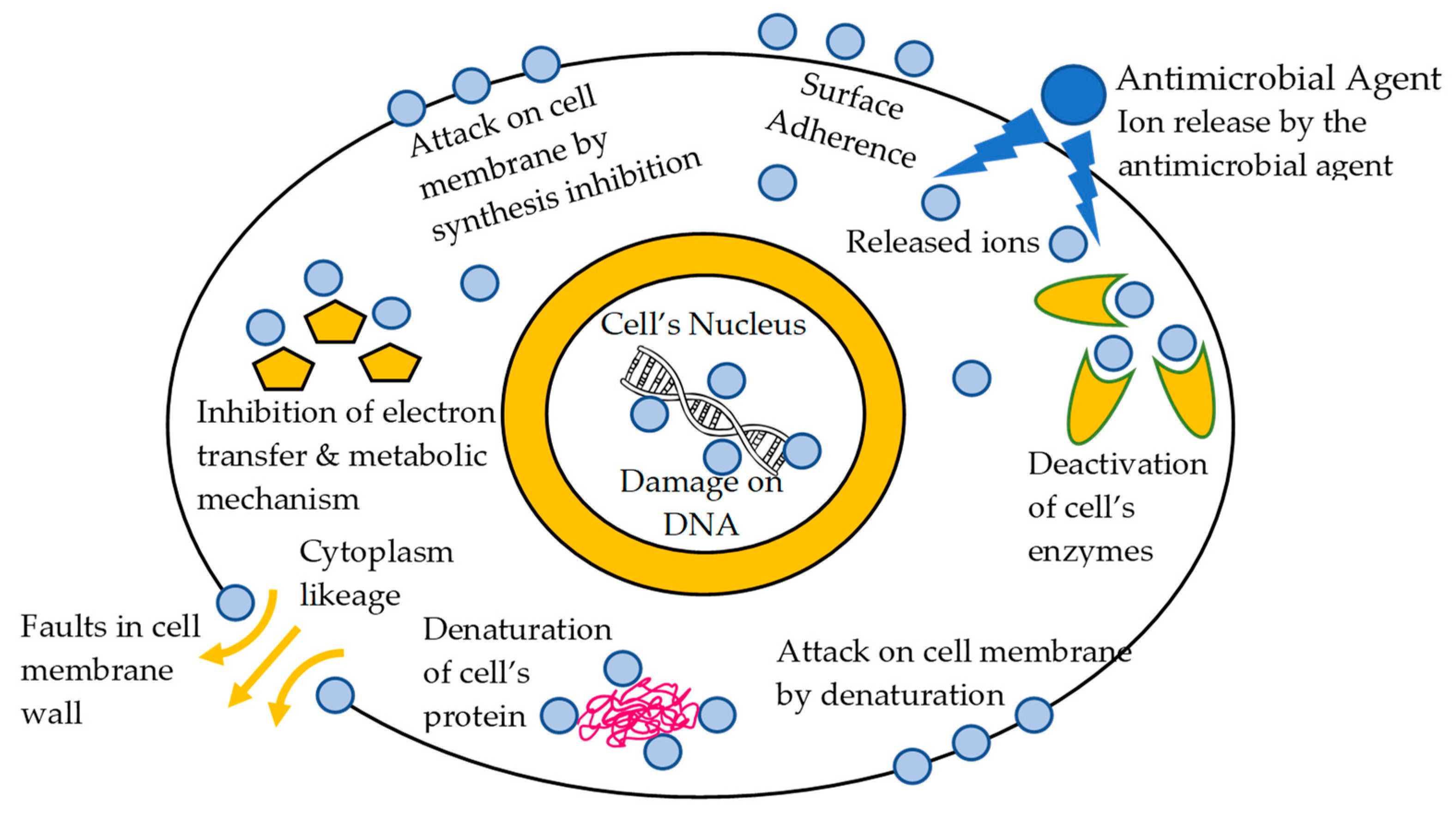
Various methods of action against microorganisms are used for bacterial elimination. The principal method is to attack the proteins of the bacteria, in which the bacteria’s protein structure is denatured or altered. The denaturation disrupts the hydrogen and disulfide bonds. The denaturation can be classified as permanent or temporary, depending on the disruption of their formal structure. When the denaturation is permanent, the mechanism of action is called bactericidal, fungicidal, etc., and when it is temporary, the mechanism of action is called bacteriostatic, fungistatic, etc. Another mechanism is the attack on their cell membrane proteins or membrane lipids. In the latter, the mechanism of action consists of denaturation, while a surfactant dissolves the lipids, damaging their cell membrane. Another known mode of action is obtained by the inhibition of its synthesis, causing an impairment of the formation of the cell wall. Similarly, the inhibition of the replication, transcription and translation of the nucleic acid structure is another distinct mechanism, and finally, the mechanism of the metabolic disturbance is also used [19]; see Scheme 1.
3. Synthetic Organic Antimicrobial Agents
Synthetic organic antimicrobial agents are organic compounds and polymers that exhibit antimicrobial activity, which is self-activated through the aforementioned mode of action [25]. Their chemical structure is critical for their categorisation. Recently, a huge number of antimicrobial polymers were synthesized. These species could be quaternary ammonium compounds (QACs), halogen-containing compounds (molecules containing fluorine or chlorine, N-halamines or triclosan), guanidine-containing polymers (polyhexamethylene vinyguanide), polymers containing phospho- and sulpho-derivatives, polymers of phenol and benzoic acid derivatives, nitro compounds, urea, amines, formaldehyde, organometallic polymers and others [19,26].
3.1. Quaternary Ammonium Compounds (QACs)
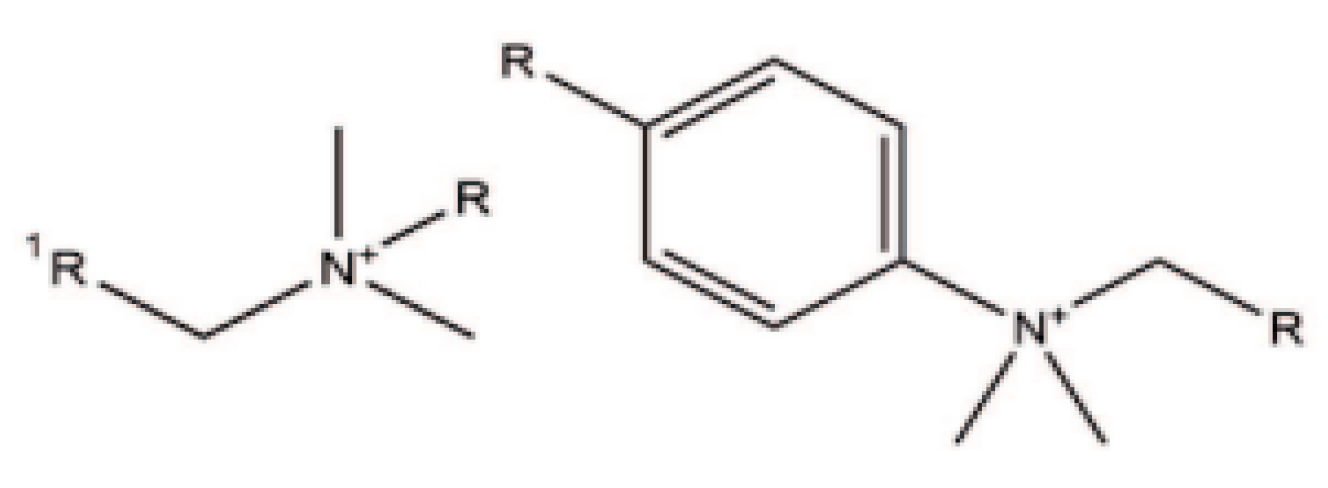
Quaternary ammonium compounds (QACs) are cationic surface active agents known as cationic surfactants, which bear a positive charge on the nitrogen (N) atom and typically adsorb to the surface of an anionic fibre through ionic interaction [13,27,28]. They consist of a nitrogen (N) atom attached to four different moieties through a covalent bond. QACs refer to a subgroup of linear alkylammonium compounds, which consist of a hydrophobic alkyl chain and a hydrophilic part. In textiles, compounds containing long alkyl chains (12–18 carbon atoms) are mainly used for cellulosic substrates, polyester, nylon and wool [13,27,29,30]. The general formula of QAC is N+R1R2R3R4X−, where R can be a hydrogen atom, a plain alkyl group or an alkyl group substituted with other functional groups, and X represents an anion; see Figure 1. They are commonly used in textiles, and they had an essential role as biocides for many years, being characterised as effective antiseptic and disinfectant agents [31]. Generally, long-chain QACs with 8–18 carbon atoms possess good germicidal activity. Important representatives of this class are benzalkonium chloride, stearalkonium chloride and cetrimonium chloride [32]. These compounds can react with both Gram-positive and Gram-negative bacteria, fungi and certain types of viruses [13]. The antimicrobial activity of QACs depends on several factors such as the length of the alkyl chain, the presence of the halogenated group and the number of cationic ammonium groups in the molecule. The antimicrobial action starts from electrostatic interactions between the positive charge of the N+ atom and the microbe’s negatively charged cell membrane, creating surfactant–microbe complexes interrupting its essential functions and protein activity, as described in the antimicrobial action section [31].
Carbon atoms with alkyl chains between 11 and 16 have been widely used as disinfectants; these compounds exert a positive charge on the N atom, which in reaction with the bacteria causes various adverse effects, resulting in microbial death [33,34]. When the length of the alkyl chain contains between 12–14 carbon atoms, optimal antibacterial activity against Gram-positive bacteria is achieved, while the alkyl group with 14–16 carbon atoms is superior in the attack against Gram-negative bacteria [32]. A type of QAC frequently applied to apparel textiles is 3-(trihydroxysilyl) propyldimethyl-octadecyl ammonium chloride, where a silane group is bonded to the long non-polar chain and the positively charged ammonium group and is often referred to as Si-QAC [29].
QACs have many favourable antimicrobial properties; however, they have no reactive functional groups in their structure to create a chemical bond with the fibres. This causes a gradual detachment from the textile due to the lack of physical bonding, called leaching. As a consequence, a fast concentration decrease in the QACs in the textile is observed. New studies report the synthesis of polymerisable QACs [13,31]. This is achieved by the incorporation of acrylate or methacrylate groups in their structure which is capable of forming permanent bonds, which are known as non-leaching QACs’ biocidal. The QACs monomers are named surfactant monomers or “surfmers”. The “surfmers” can polymerise into a bulk polymer network. They exhibit a polycationic chemical structure under the appropriate conditions, which include groups chemically side-bonded to the main polyacrylate chain. The outcome is that the QAC groups can function as a biological barrier and kill microorganisms when they come into contact. The formation of the polymer network on the surface of the fibres improves the coating’s wash fastness and durability of antimicrobial agents in general [31].
Sol–gel technology has also been used in antimicrobial textiles, where QACs regulate textile fibre behaviour. A nanocomposite polymer network with an organic–inorganic hybrid structure is formatted by this method. Colloidal solutions (sols) have been formulated for this purpose by incorporating mixtures of tetraalkoxysilane (Si(OR)4) with different structures of QACs or organic–inorganic hybrids, including alkyl- trialkoxysilanes (Rx-Si(OR)3). The increased durability and wash fastness on the finished fibres are provided by the formation of covalent bonds between sol–gel –SiOH groups and –OH groups of the fibres [31].
Novel cationic antimicrobial dyes are obtained by attaching QACs to the chromophore structure. In this manner, one or two QAC groups of different structures are in the molecule of mono-, diazo and anthraquinone dyes. Antimicrobial activity increases with the number of chemical substitutions and QAC groups. Moreover, the chain length of the hydrocarbons of the QACs’ component in the dye structure also promotes antimicrobial activity [31]. Consequently, when used on acrylic fibres, cationic anthraquinone dyes bear a double role; firstly, they confer colouration and simultaneously exert an antimicrobial function [35]. Similarly, n anthraquinone cationic reactive dye has been synthesised to exhibit antimicrobial characteristics to improve the wash fastness of the antimicrobial when applying dye on the cellulosic substrate [36] where high exhaustion and fixation values were recorded, even if applied without the electrolyte addition in the dyebath. However, repeated laundering seems to decrease the antimicrobial activity of both these dyes.
QAC antimicrobial agents are used on both natural fibres, such as cotton and wool, as well as man-made fibres, such as polyester and polyamide [13,21]. The QAC antimicrobial activity has been tested in the above substrates, where the concentration of 10–100 mg/L presented good reproducibility, as well as adequate wash fastness [37]. Due to its high solubility in water, its usage as an antimicrobial agent in textile finishes is, however, limited [38].
3.2. The N-Halamines
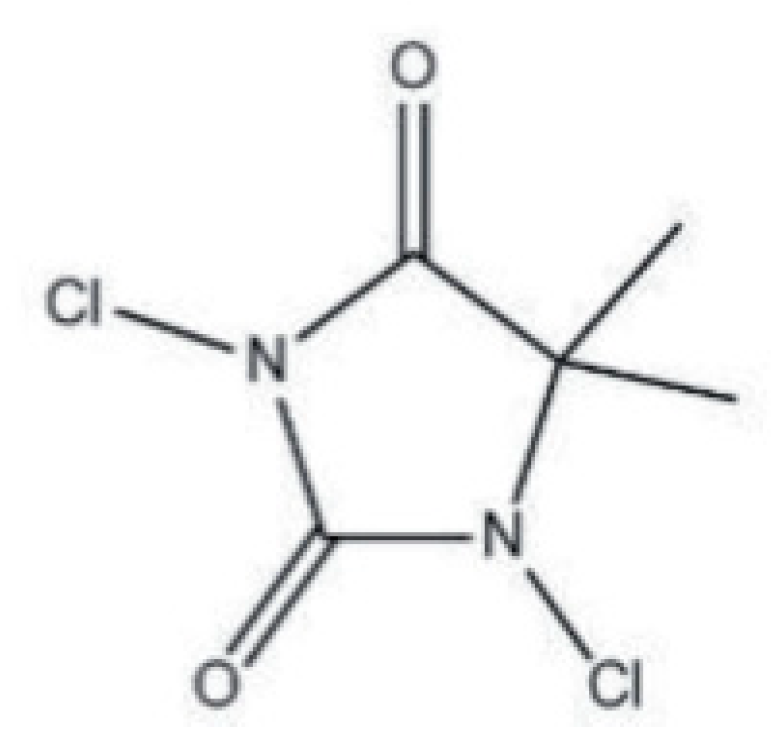
N-halamines are heterocyclic organic compounds bearing at least one or more nitrogen–halogen (N–X) covalent bonds, as shown in Figure 2. They are typically formed by the halogenation of amine, amide or imide groups, which is responsible for the stability of the structure and shows a controlled release of free active halogen species into the environment [31,39]. Chlorine is the most frequently used halogen in this category, but the activity of other halogens, such as bromine and iodine, is not unusual. N-halamines are environmentally friendlier and healthier, with enhanced antimicrobial efficacy against a broad spectrum of microorganisms [32]. While the bonded N–H, formed by a substitution reaction, exerts no antimicrobial properties, the further exposure of the reagent to dilute sodium hypochlorite develops its antimicrobial activity [31]. The last takes place with the release of chlorine by its electrophilic substitution with H in the N–Cl bond and is effective against a vast range of bacteria, fungi and viruses. The reaction occurs once water is present, allowing chlorine free-cations to be released, thus allowing them to bind to the acceptor regions of the bacteria. As a consequence, the enzymes and metabolism of the microorganism are hindered, leading to its gradual deterioration [37]. The application procedure composes the pad-dry method followed by exposure to chlorine bleach for the formation of antimicrobial cotton fabric. The chlorinated substrate exhibits strong antimicrobial properties against Gram-positive and Gram-negative pathogens. The chlorinated fabric can be recharged to the extent of 85% after being stored for fifteen days. This proves the strong efficacy of N-halamine compounds as antibacterial agents for medical textile finishes [40,41].
A wide range of textile surfaces, such as cellulose, wool, cotton, polyamide and polyester fibres, can be treated by the above process with N-amines [13,21,31]. The effectiveness and durability of the antimicrobial finishing can be further enhanced by the synthesis of N-halamide monomers with the incorporation of a reactive vinyl group.
A variation to the above reagent is the N-halamide monomer, which possesses enhanced durability and antimicrobial action. The molecule is obtained by the addition of the vinyl reactive group that, under the proper conditions, can polymerise cellulose fibres to form a coating with excellent fastness to washing. In addition, the preparation of the N-halamine precursors with two hydroxyl groups promotes covalent bonding to the cellulose surface. The species can be chemically bonded to hydroxyl groups in cellulose fibres in the presence of 1,2,3,4-butantetracarboxylic acid as a crosslinking agent. Alternatively, bonding can also be achieved by synthesising an N-halamine siloxane monomer precursor, which allows the silanol groups to react with hydroxyl groups of cellulose, forming a nanocomposite coating. Consequently, combining N-halamines with N-halamine siloxane and QACs siloxane confers a synergistic effect of antimicrobial action [31].
3.3. Triclosan
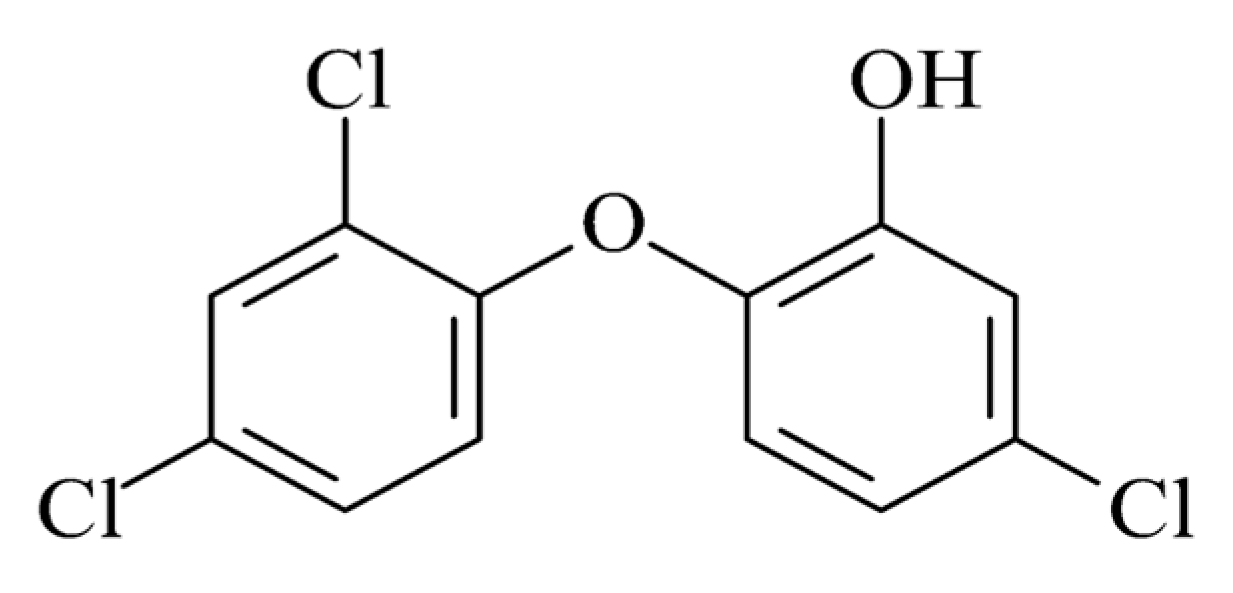
Triclosan 5-chloro-2-(2.4-dichlorophenoxy) phenol (C12H7Cl3O2) is an odourless synthetic chlorinated bisphenol, as shown in Figure 3. Triclosan differs from most cationic biocides, and it is not ionised in solutions, which improves its wash fastness. It has a reliable antimicrobial activity against Gram-negative and Gram-positive bacteria, but it also has antifungal and antiviral characteristics. The mechanism of action of this biocide agent works by blocking lipid biosynthesis, such as phospholipids, lipopolysaccharides and lipoproteins, affecting the integrity of cell membranes, as explained in the respective section. Triclosan, throughout the last 30 years, has become the most efficient and widely used biocide. It is included in many consumer and professional healthcare products, such as soaps, lotions and creams, toothpastes, mouthwashes and underarm deodorants, and it is also incorporated into textile fabrics and plastics. It is also mainly used in synthetic fibres such as polyester, nylon, polypropylene, cellulose acetate and acrylic fibres. Several products are available on the market, either as isolated antimicrobial agents for a finishing option or to incorporate into fibres, such as Microban® Cannock, United Kingdom and Irgaguard® (Ludwigshafen, Germany) 1000. Some are already incorporated into fibre or fabric, such as BiofresH™ (Salem, MA, USA) and Silfresh® (Magenta, Italy) [13].
Triclosan has been applied to cellulose fibres in combination with polycarboxylic acids as crosslinking agents. The pretreatment application of polycarboxylic acid to fibres followed by after-treatment with triclosan enhances the washing durability of the coating. Furthermore, another mode of the application consists of adding triclosan molecules to novel host–guest complexes with pretreatment with cationic β-cyclodextrins. β-cyclodextrins are cyclic oligosaccharides which contain six to eight glucose units linked by β-1,4 bonds. Antimicrobial activity, water solubility and stability have been examined for the host–guest complexes. The complexes are nearly all adsorbed to the surface of cellulose fibres because of the strong electrostatic attraction. Finally, triclosan can be used for finishing non-woven textiles when encapsulated in biodegradable polylactide as a carrier [31].
However, in recent times, the phenomenon of the extensive use of triclosan in non-healthcare settings has been observed. This is a major concern because bacterial microorganisms develop a resistance to triclosan [13]. Another significant concern is that the photochemical exposure of triclosan leads to the formation of 2,8-dichlorodibenzo-p-dioxin in aqueous solutions, which is highly toxic [42,43]. Triclosan is used as an antimicrobial agent on cellulose acetate, polyester, nylon and polypropylene fibres [13,21].
3.4. Polybiguanidines
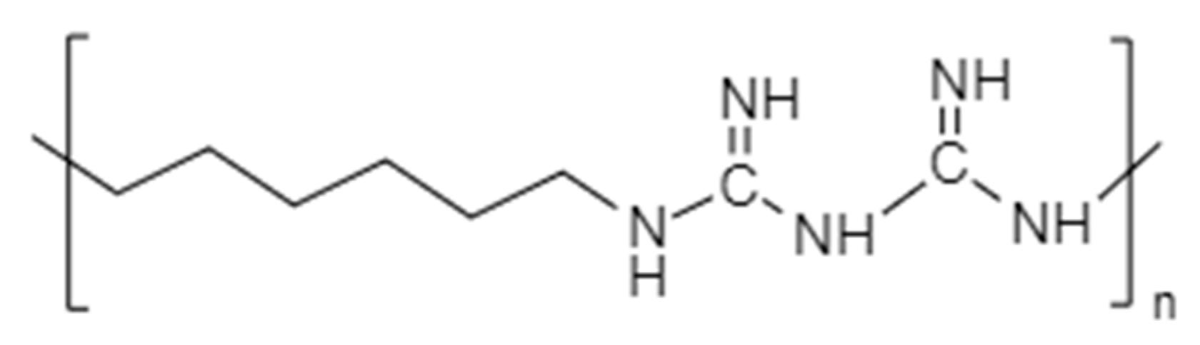
Polybiguanides are polymeric polycationic amines that contain cationic biguanide repeat units separated by aliphatic chain linkers. This can be of identical or dissimilar lengths; see Figure 4. The main representative antimicrobial is poly(hexamethylenebiguanide) (PHMB) ((C8H17N5)n), with an average of 11 biguanide units [31]. It consists of a hydrophobic backbone, in which the cationic biguanide groups are interspersed between hydrophobic hexamethylene groups [44,45]. The cationic and hydrophobic character of PHMB reassures the interactivity with microbial cell membranes through electrostatic and hydrophobic interactions [46]. This mechanism causes a cell membrane disruption and a lethal leakage of cytoplasmic materials, and its activity increases the levels of polymerisation [13,40,44,45].
Guanidine-based polymers can be obtained from guanidine salt, biguanidine salt, cyanamide, dicyandiamide and other similar small molecules. These act as monomers to confer the antimicrobial guanidio group to the final guanidine polymer. The guanidio group possesses a strong antimicrobial activity caused by its cationic charge. The negatively charged membrane of bacteria attracts and binds with the positive polymer, which causes the displacement of Mg2+ and Ca2+ ions. The lipopolysaccharide and peptidoglycan components of the cell wall are also bound, causing a major change to the phospholipid environment of the membrane, thus destroying the cytoplasmic membrane and resulting in the bacteria’s death [47].
Both monomeric and dimeric biguanides exert antimicrobial activity, while the latter is more effective against all types of bacteria. The properties of PHMB can be summarised as highly water soluble, chemically stable, less toxic, cost-effective with high antimicrobial activity and good wash fastness, provoking less skin irritation [33]. PHMB is found in health products (mouthwash and wound dressings), clothing, pharmacy and food industries, household and water treatment textiles [13,45]. It is applied during the finishing process of the products, such as underwear and towels, to stop microbial growth. In addition, it is commonly used in medicine as an antiseptic agent for the prevention of wound infection by antibiotic-resistant bacteria. For the aforementioned reasons of biocidal activity and low toxicity, it is used to protect sensitive textile fibres such as cotton [31], but it is also used as an antimicrobial agent to polyester and nylon fibres [13,21]. The highest antibacterial inhibition effect of PHMB is observed at a slightly acidic area of pH 5–6 [33,37,40].
Antimicrobial tests on polyester samples with 2% and 4% of the weight of the fibre (o.w.f.) of PHMB demonstrated that the bacterial-free area was >99.9% in both cases of the treated area for K. pneumonaie. The same test was repeated again after 20 washings. The results remained excellent, with a 99.9% reduction of K. pneumonaie, for polyester treated with 2% and then >99.9% for polyester treated with 4% o.w.f. PHMB. Consequently, this indicates that PHMB remained in the substrate after repeated wet treatments, making it a valuable antibacterial additive [48]. A similar test, performed on cotton fabric with 2% o.w.f. PHMB, demonstrated excellent results (>99.9% and 99.9% reduction after 20 washings) [49].
4. Advantages and Disadvantages of Synthetic Antimicrobial Agents
According to the research associated with the use of antimicrobial agents and their environmental impact, scientists concluded that an ideal antimicrobial agent should combine different salient properties [50]. These include friendliness to humans and the environment, exhibiting a strong antimicrobial activity to a wide spectrum of bacteria, while it should not alter the fabric tactile or mechanical properties. Moreover, it should be able to withstand fabric production procedures, and lastly, the antimicrobial action should not decrease throughout the fabric’s use [51]. A brief review of the benefits and risks associated with the use of any antimicrobial agent or antimicrobial fabric is given below [29,50].
Antimicrobial agents are used in textiles to prevent and limit infections caused by pathogenic microorganisms [50,52], especially in hospitals and other public health-related places where the transmission of infectious diseases is the most prominent. The use of antimicrobial fabrics controls the proliferation of bacteria [53]. The dual role of antimicrobial agents is to confer user protection against pathogens while helping to maintain the quality of the fabric [29,54].
Antimicrobial tests conducted on fabrics treated with various synthetic antimicrobial agents demonstrated excellent results owing to their strong antimicrobial properties and durability.
The main advantage of QACs is the broad spectrum of antimicrobial activity they possess. N-halamine compounds are also effective against a wide range of microorganisms with a rapid bactericidal action. Moreover, they can be used for long-term disinfection. Triclosan, as an antimicrobial agent, has an immediate, persistent, broad-spectrum antimicrobial effect against bacteria, fungi and viruses [50]. PHMB has a highly effective antimicrobial and ecological character through a low toxicity to human cells and the environment [54,55].
In textiles, several antimicrobial agents have been applied to fabrics [31,50,52], and their effectiveness depends on their chemical composition [56]. These products are organometallics, slow iodine-releasing substances (iodo-phors), inorganic salts, anionic-group-bearing heterocyclics, phenols, thiophenols, urea and its compounds, nitro compounds, amines, derivatives of formaldehyde and several more [50,57,58,59]. Unfortunately, scientific research has demonstrated that many of these compounds do not always biodegrade rapidly and can be highly toxic to humans [52,57]. Conversely, those that degrade easily can be used without further ecological damage. Consequently, although synthetic antimicrobial agents can be very effective, they generally raise concerns due to health risks and environmental pollution [60,61,62], owing to their high toxicity and resilience to biodegradation [8,50,57].
Comparing the aforementioned types of antimicrobial agents, PHMB and N-halamines are mild and their impact on humans is possibly negligible, as well as on the environment. A summary of the main disadvantages of the two other classes (triclosan and QACs) is highlighted in the following paragraphs.
In ecotoxicity and environmental effects, studies have demonstrated that triclosan is a widely used antimicrobial agent, and its residues are found in rivers and lakes as a contaminant [51]. The exposure of triclosan to sunlight leads to its photodegradation and the formation of toxic polychlorinated dioxins [27,50]. QAC products also raise environmental concerns; whereas, their effects are limited due to their slow rate of diffusion [63,64].
Regarding skin irritation and the effects of exposure to antimicrobial finishes, triclosan-treated fabrics may cause skin sensitisation and irritation, such as itching [27,50]. In addition, a major concern arises from the fact that triclosan has been detected in human breast milk, at levels ranging from 0 to 2100 mg/kg lipid in serum and urine [65,66,67,68,69]. Finally, laboratory studies have demonstrated that thyroid systems in rats and frogs are disturbed by triclosan [35,70,71].
A major problem also emerges from the overuse of treated fabrics with certain synthetic antimicrobial agents, which can cause bacterial resistance [27]. The release of QAC components from fabrics into the water, i.e., rivers and lakes, leads to catastrophic effects on organisms living in water, as they affect vulnerable bacteria and thus lead to the formation of potentially resistant bacteria [72].
Furthermore, recent studies have demonstrated that the presence of QACs in QAC-polluted environments leads to the antibiotic resistance of bacteria, which is presently a potential threat to human life [73]. Additionally, the molecules of triclosan stop the biosynthesis of bacterial lipids, which causes a possible development of bacterial resistance [74,75].
5. Potential Applications for Textiles of Other Polymers
This last section briefly reports information about some synthetic polymers with antimicrobial properties that have been synthesised but have yet to be applied to textiles. The potential of these polymers has been examined for their deposition on textile materials, as well as the process for producing antimicrobial fabrics and garments. Two new polymers with different molecular weights have been synthesised: poly(p-vinylbenzyl tetramethylenesulfonium tetrafluoroborate) and poly(pethylbenzyl tetramethylenesulfonium tetrafluoroborate), as reported by Kanazawa et al. [76] and referenced in [77].
The antimicrobial activity of these two polymers was high against Gram-positive S. aureus, whereas it was less active against Gram-negative E. coli. Polymeric sulfonium salts had much higher antimicrobial activity than the corresponding monomers. Additionally, with the increase in molecular weight, their efficacy was also increased.
The polymerisation of (benzofuran-2-yl) (3-mesityl-3-methylcyclobutyl)-O- methacrylketoxime monomer was described by Erol [78]. The polymer produced impeded the proliferation of micro-organisms, such as P. aeruginasa, E. coli, Candida albicans (C. albicans) and S. aureus. The proposed mechanism resides in the oxime esters and carbonyl groups, which possibly have hindered the production of necessary enzymes.
Dizman et al. [79] had synthesised methacrylate monomers containing pendant quaternary ammonium moieties based on 1,4-diazabicyclo-[2.2.2]-octane (DABCO). Antimicrobial activities against the S. aureus and E. coli were found to be satisfactory, which appeared to improve as the N-alkyl chain length increased from four to six carbons.
A Friedel–Craft alkylation between guaiacol and N-hydroxymethylacrylamide was conducted to obtain a novel acrylamide-type monomer (N-(4-hydroxy-3-methoxy-benzyl)-acrylamide). Liu et al. [80] polymerised the monomer successfully, conducting the conventional radical polymerisation technique.
Antibacterial tests of polymers were performed using Gram-positive Bacillus subtilis (B. subtilis) (ATCC 6633) bacteria exhibiting encouraging antibacterial results.
Mohsin et al. [81] achieved water and oil repellency and antimicrobial properties by using a C6-based fluorocarbon, which was cross-linked with maleic acid and catalysed with sodium dihydrogen phosphate. They reported that the lower cover factor of the fabric samples entailed a higher antimicrobial effect since a less dense fabric allows for a better penetration of the finish in between its threads.
Cross-linked chitosan to cotton fabrics using 1,2,3,4 butane tetracarboxylic acid (BTCA) was also reported by Hebeish et al. [82] as an effective antimicrobial coating. A low molecular weight and high amount of fixed chitosan on fabrics was the definitive factor for the optimal antimicrobial activity against both Gram-positive (Staphylococcus aureus, Bacillus subtilis and Bacillus cereus) and Gram-negative (E. coli) bacteria. However, the fabric samples demonstrated zero antimicrobial activity against fungi (Aspergillus niger (A. niger), Maccrophomina phaseoli (M. phaseolina), Trichoderma viride (T. viride) and Fusarium oxysporium (F. oxysporum)).
Apart from synthetic polymers that are used as antimicrobials, scientists have focused in the fabrication of antimicrobial agents in a very small scale, where the known matter properties differ in the bulk due to the excessive surface area to volume ratio, which is commonly known as the “nano effect”. Nanotechnology is a new promising science with several applications in biological, pharmaceutical, photocatalysts, information technology, electrocatalysts, chemical science, physics, wastewater treatment, infection control and textiles [83,84,85,86,87]. Nanoparticles (NPs) are tiny molecules whose diameter ranges between 1 to 100 nm [87,88,89]. NPs can be produced by several methods, including physical, chemical and biological methods [85,90,91]. Recently, NPs have a direct application in hospitals due to the large number of infections among both patients and medical staff; this is through medical textiles providing a resistance to pathogenic microbes as well as UV protection [84,90,91].
Metal NPs exhibit a very different activity from the corresponding bulk materials, owing to their different sizes and shapes, crystal structure, thermal stability, charge and zeta potential, which give rise to distinctive quantum properties. Metal NPs offer many advantages such as biocompatibility, safe and easy handling, large-scale production potential, rapid development and avoidance of hazard by-products, and they are friendly to the environment [85].
6. Copper in Polymeric Matrices
Copper (Cu) is an essential trace element found inside the human body; it promotes the formation of healthy red blood cells and is a structural constituent of various enzymes. Furthermore, copper constituents play a separate role in cell wall metabolism, mitochondrial respiration, photosynthetic electron transport, oxidative stress responses and hormone signalling. One of the many properties of copper is that it can also cause the enhanced production of bioactive compounds [85]. During the course of evolution, the human organism has developed complex mechanisms to regulate copper levels in the system, whether they exceed or are below natural levels [92,93]. This mechanism renders the human body more resistant to copper toxicity, while for microorganisms lacking this mechanism, copper is extremely toxic. Therefore, copper was known from antiquity as a safe metal and was frequently used to sanitise and decontaminate provisions, and even wounds and skin diseases. According to the OEKO-TEX® STANDARD 100 (annex 4) certification, the safe limit value of cooper content is 25 mg/kg for baby textiles (class I) and 50 mg/kg for products in direct contact to the skin and other textiles (class II, III and IV), while the ECO PASSPORT by OEKOTEX® mentions a threshold value of 250 mg/kg for the copper content for textile producers [94,95,96].
Up to this day, copper is used to hinder bacterial growth in many processes, such as water purification, and was proven to be effective against a wide variety of pathogens, including S. aureus, E. coli, B. subtilis and P. aeruginosa.
This both experiential and scientific understanding of the bactericidal effects of copper is leading scientists to study copper nanoparticles (CNPs) and their antimicrobic applications.
Ligand is a molecule that produces a signal by binding to a site of a target protein to form a complex for a biological purpose [97]. Copper ions can displace crucial metals from their native binding sites or interfere with biological functions via ligand interactions. Redox cycling reactions between Cu2+ and Cu+ can trigger Fenton-like reactions, producing highly reactive hydroxyl radicals that attack the microbes’ biomolecules [98,99]. This results in a similar antimicrobial action as that described earlier.
Copper oxide nanoparticles (CuO-NPs) play an important role in many biological activities, such as antibacterial and antifungal activity, antioxidant properties, drug delivery, and cytotoxic efficacy against tumour and cancer cells, due to the highly interactive characteristics displayed [100,101]. CuO-NPs have been manufactured through different biological entities, such as bacterial, fungal, actinomycetes, algae and plants. The utilization of fungal species as decreasing, covering and stabilizing agents to fabricate NPs is most interesting because of the presence of many secreted metabolites, high metal accumulation and scalability [85].
CuO-NPs are considered in applications such as antimicrobial activity against different pathogenic microbes, antioxidant activity, anticancer activity, antifungal against phyto-pathogenic fungi [102]. CuO-NPs can be produced in many ways, including through physical, chemical and biological processes [103,104,105,106]. Different methods have been proposed for the synthesis of CuO-NPs, such as the sol–gel method, microwave irradiations, thermal decomposition, electrochemical technique and alkoxide-supported method. The main disadvantages of these methods are the use of harmful chemicals, as well as thehigh energy consumption and high impurities in synthesized NPs. On the other hand, the synthesis of CuO-NPs with green methods using multicellular and unicellular organisms such as actinomycetes, fungi, bacteria, plant or algae is gaining ground for pharmaceutical applications because of the biosafety of biosynthesized CuO-NPs [102].
Cu NPs play an important role in the textile industry because of the high natural abundance, low cost, practical, straightforward and multiple ways of preparing them. Although bulk Cu can be used in many applications of various fields, such as optics, electronics, etc., the usage of Cu NPs is restricted. This is because of Cu’s inherent instability under atmospheric conditions that lead to oxidation. Several attempts have been performed to increase the stability of Cu NPs by altering their sensitivity to oxygen, water and other chemical entities. These efforts have encouraged the exploration of alternative Cu-based NPs with more complex structures, such as core/shell Cu NPs or systems based on copper oxides. Cu, as a metal, has interesting physical and chemical properties. Firstly, it possesses a 3D transition; this gives Cu a wide range of accessible oxidation states (Cu0, CuI, CuII and CuIII), which can promote and undergo a variety of reactions. Cu-based Nano catalysts have many applications in nanotechnology, which are, mainly, catalytic organic transformations, electrocatalysis and photocatalysis. The preparation of nanomaterials that are inexpensive, selective, stable, robust and highly active is the main purpose in the development of catalytic NPs. A cost-effective way of producing advanced Cu-based nanomaterials for catalysis is by coupling Cu NPs (e.g., Cu, CuO or Cu2O) with agents such as iron oxides, SiO2, carbon-based materials or polymers. Finally, the high boiling point of Cu NPs makes them compatible with high-temperature and high-pressure chemical reactions, such as continuous flow reactions, microwave-assisted reactions, vapor phase reactions and various organic transformations. Subsequently, it is obvious that Cu NP and its alloys will continue to play an important role in the future because of its unique properties [107].
Polymer matrices are a suitable base for antimicrobic nanocomposites, and CNPs can be efficiently implemented as a filler. When placed in an aquatic environment, water that contains dissolved oxygen allows the release of cupric ions (Cu2+), which diffuse through the nanocomposite and are released [108].
The matrix selection is very important, as different polymers can yield different properties. Poly (vinyl methyl ketone) (PVMK) and polyvinyl chloride (PVC) yield more cupric ions than poly (vinylidene fluoride) (PVDF). Polyethylene (CNP) displays exceptional bioactive properties. It is noticed that the production of (Cu2+) is much stronger during the first month of incubation, and it eventually ebbs away. Another polymer that was used is polypropylene for antimicrobial plastic production. Two different nanocomposites were created using the melt blending method. One is with copper metal nanoparticles and the other is with copper oxide nanoparticles. It was concluded that copper oxide nanoparticles had a better antimicrobial effect against E. coli than CNPs [109].
A different method of nanocomposite formulation is to add the CNPs into the filler. Such a filler was used to create a poly (methyl methacrylate) (PMMA) latex nanocomposite with bentonite-supported copper nanoparticles, which exhibited antimicrobial activity against S. aureus [110].
The biggest drawback of using CNPs in polymers is that they can cluster together, thus reducing their antibacterial properties. The solution to that is creating silica nanospheres that will contain the CNPs inside them.
This approach can be applied in antimicrobic textile development, where nanocomposites can be used as coatings [19].
Although nanoparticles (NPs) can be synthesised by both chemical and physical methods, the biological method of using living cells is favourable, owing to its easier, safer, cleaner, eco-friendly and cost-effective results, as no high temperature or pressure is needed. Additionally, no hazardous or toxic materials are required, and there is no need for external reducing, capping and stabilising agents. Species as yeast, bacteria, fungi, algae, actinomycetes and plant extracts are often used to produce green NPs with variable shape, size and stability and can be applied in the field of food and textile industries [88,111].
CuO nanoparticles have also been used via biosynthesis for the purpose of creating antibacterial-active textiles as cotton fabric. Researchers took advantage of the active ingredients, as enzymes, and the protein secreted by fungi, as the Aspergillus terreus strain AF-1, to cap CuO-NPs/proteins, taking into consideration their cytotoxicity [112].
CuO-NPs were also fabricated by utilising metabolites of the Aspergillus niger strain (G3-1), where functional groups of metabolites serve to cap and reduce, and are agents to stabilise the CuO-NP formation for an insecticidal purpose against wheat grain insects Sitophilus granarius and Rhyzopertha dominica [85].
7. Green Methods of Creating and Applying AgNPs onto Fabrics
Conventional ways of creating antimicrobial fabrics have a high environmental impact that has raised multiple concerns during the past few years. The synthetic and organic antimicrobials, triclosan and QACs, should be avoided due to environmental and health concerns, whereas PHMB and N-halamine are preferable because of their lower toxicity. Considering the previous facts and the harmful effects caused by triclosan and QACs, further research is necessary to find novel alternatives with non-toxic ingredients in manufacturing fabric products with potent antimicrobial properties. Due to this, recently, more and more researchers are focusing their attention on greener routes in order to create antimicrobial fabrics.
One of these paths appears to be the use of silver nanoparticles. In multiple studies, their effectiveness against pathogens is proven as well as the inability of microorganisms to develop an immunity to them, unlike with many antibiotics. Nevertheless, factors such as cytotoxicity or the eco-friendliness of the methods used in implementing AgNPs onto fabrics must be taken into consideration.
Cotton is inexpensive and has a lot of benefits, such as softness, breathability, comfort when in contact with the skin and biodegradability [113,114]. As a consequence, the fabrics used in hospitals are often made of cotton. However, due to its nature, it provides optimal conditions for the growth and spread of pathogenic microorganisms such as the heat from our bodies and humidity from sweat absorption [115].
7.1. Synthesis of AgNPs
7.1.1. Plant Extracts
The Curcuma species exhibit multiple pharmaceutical properties, including antimicrobial activity, rendering them a suitable choice for researchers to use in fabricating AgNPs. The research team of Maghimaa and Alharbi [116] prepared an aqueous extract using a fine order to formulate the AgNPs. After the evaluation of AgNPs through HR-TEM (high-resolution transmission electron microscopy) and FT-IR (Fourier-transform infrared) spectroscopy, the leaf extract was applied to the cotton fabrics. Their antimicrobial potency was tested against S. aureus, Streptococcus pyogenes, E. coli, P. aeruginosa and C. albicans—pathogens that are responsible for skin infections. It was determined that a concentration of 35 mg of the synthesized AgNPs demonstrated apparent antimicrobial properties against the tested pathogens, and thus, when embedded in cotton fabrics, they also displayed significant antimicrobial attributes. When tested for cytotoxicity, it was confirmed that the AgNPs developed from Curcuma longa L. not only demonstrated insignificant levels of toxicity against fibroblast (L929) cells but also promoted their rapid generation and cell migration to wounds [116].
Cotton fabric treated with AgNPs prepared by the green synthesis of an endophytic strain of Streptomyces antimycoticus L-1 from Mentha longifolia L. leaves through their secreted enzymes and proteins exhibited broad-spectrum antibacterial activity at different concentrations (6.25–100 ppm). The AgNP concentration of 100 ppm was selected as a safe level of application against normal cells with the distribution of AgNPs as 2% of the total cotton fabric elements. The antimicrobial activity was against pathogenic Gram negative and positive bacteria; it remained even after 5 and 10 washing cycles for 45 min at 40–60 °C with 2% sodium carbonate, and it was dried at 80 °C [86,91].
Using food waste that has no other purpose and plants that are invasive to an ecosystem, possibly causing irreversible damage, is an inexpensive, feasible and eco-friendly method of AgNP phytosynthesis. In this research, plant extracts used as reducing agents were prepared by drying and pulverizing food waste such as green tea leaves, avocado seeds and pomegranate peels. They also included the invasive Japanese knotweed rhizome plant, the staghorn sumac fruit and goldenrod flowers. A total of 20 g/L of each powder was used to create a reducing agent bath. For the in situ composition of the AgNPs, cotton fabrics were soaked in a silver nitrate solution; then, they were squeezed and dipped in the reducing agent bath. The fabrics exhibited remarkable antimicrobial effects against S. aureus and E. coli. They also demonstrated exceptional UV protection even after many washing cycles [117].
Oryza sativa L., more commonly known as black rice, was used by the research team of Yu et al. [114] as a reducing agent for the synthesis of AgNPs due to its ample content in anthocyanins, which have variable pharmaceutical properties. Cotton fabrics were treated with a carboxymethyl chitosan bath before they were soaked into solutions of assorted silver nitrate concentrations and the anthocyanin extract from the black rice. The samples exhibited arresting antibacterial properties against S. aureus and E.coli. They reduced the E. coli population by an average of 97.99% and the S. aureus population by 96.75%. The fabrics also displayed superhydrophobicity and high resistance to UV radiation [114].
7.1.2. Fungi
Fungi are often preferred over bacteria in the green synthesis of metal nanoparticles due to their larger surface area and higher secretion of protein and enzymes, but also their ability to grow even in harsh conditions. Therefore, metal salts are converted to metal nanoparticles at a much faster pace and in much larger quantities [118,119].
Bionectria ochroleuca is an epiphytic fungus that has been used before in the biosynthesis of AgNPs by the Rodrigues et al. [120] research group, yielding noticeable antibacterial and antifungal abilities against P. aeruginosa, E. coli, S. aureus, Micrococcusluteus and Candida sp. On this occasion, the same research team implemented AgNPs into cotton and polyester fabrics. Initially, they soaked the fabrics into the AgNP solution, and afterwards, they compressed them. Both fabrics proved particularly efficient in hindering the S. aureus and E. coli growth by 100% and suppressing the growth of the C. albicans, Candida parapsilosis (C. parapsilosis) and Candida glabrata (C. glabrata) fungi. In addition, the biologically produced AgNPs impeded the creation of biofilm of the bacteria P. aeruginosa. The use of the Galleria mellonella (G. mellonella) larvae model to determine the toxicity of the AgNPs demonstrated a considerably high rate of the survival of larvae at various concentrations of AgNPs at either 28 or 37 [118,120].
Using the in situ procedure, this time, Shaheen and Abd El Aty [119] isolated fungi from the sprouts of Arctostaphylos uva-ursi, Anabasisarticulate, Mentha and Cornulaca, which are all medicinal plants. Of the fifteen identified fungi, five were chosen and were each placed in culture media to enhance their enzymes and protein production. The produced biomass of each fungi was filtered and added to distilled water. Afterwards, silver nitrate was added to the five different solutions, into which cotton fabrics were submerged for 5 min. They were dried afterwards. This procedure lets the AgNPs grow directly onto the fabrics. However, the remaining solutions were still producing AgNPs even after the cotton fabrics were removed. As such, these AgNPs were synthesised using the ex situ method. The results demonstrated that while the in-situ-synthesised AgNPs had a slightly lower antimicrobial activity than the ones produced ex situ, their durability when the cotton fabrics were washed was much higher [119].
7.2. Securing AgNPs on Cotton
7.2.1. Chitosan
In the past few years, researchers have raised concerns about the toxicity of AgNPs caused by their release of Ag ions. A solution to this appears to be the addition of an agent that will strengthen the bonding of AgNPs on the fabrics. Chitosan is an ideal option due to its natural origin and abundance.
For the needs of their research, Xu et al. [121] misted a cotton fabric sample primarily with a carboxymethyl chitosan solution, followed by a silver nitrate solution and finishing with a trisodium citrate solution. Another sample was prepared for comparison purposes by simply dipping the fabric into the carboxymethyl chitosan solution and then misting it with the silver nitrate and the trisodium citrate solutions. It was concluded that although both fabrics demonstrated significant antimicrobial properties against E. coli and S. aureus and maintained an over 95% antimicrobial rate even after 50 washing cycles, the samples that were only subjected to misting, lost only 11.7% of AgNPs, while the sample that was immersed in the carboxymethyl chitosan solution lost 23.5% of AgNPs. This shows that the misting method creates more durable bonds between the AgNPs and the cotton fibres. It was also proven that the misting method affects neither the physical nor the mechanical properties of the cotton. Misting also minimises contact with the skin as it segregates the AgNPs on one side of the fabric, effectively diminishing the cytotoxicity [121].
Another research group, comprising Rehan et al. [122], utilised a compound of three ingredients: chitosan, AgNPs and clay. Apart from the antimicrobic properties of the AgNPs, the addition of clay serves as a natural flame-retardant factor and provides thermal isolation. For the generation of AgNPs, UV radiation served as an environmentally friendly method to diminish silver salt amalgamation and promote AgNP synthesis. For comparison purposes, two different solutions were created. The first was composed of chitosan (Cs) and AgNPs, while the other was of chitosan, AgNPs and clay. Bleached cotton fabrics were immersed into the solutions for 1 h, then squeezed, dried and cured. Fabrics treated with the Cs/AgNPs/clay solution proved to be more effective in prohibiting the growth of E. coli, S. aureus and Candida albican by 99.95%, 98.3% and 91%, correspondingly, while fabrics coated with the Cs/AgNPs solution demonstrated 97.4%, 96.4% and 88% antimicrobic abilities. Fabrics coated with the Cs/AgNPs/clay solution demonstrated the highest scores for UV protection, durability through washing cycles and flame resistance. To test the levels of cytotoxicity, the human cells Hep2, HepG2 and BGM were used. It was reported that the cell livability for both Cs/AgNPs/clay and Cs/AgNPs decreased within a 72-h period, albeit a lot more for the Cs/AgNPs/clay-treated fabrics. It was deduced that these samples exhibited a slight cytotoxicity [122].
7.2.2. Aloe vera (AV)
In their study, researchers Verma et al. [123] developed an eco-friendly nanogel in order to encompass and secure the AgNPs inside its mass. This nanogel can be used as a coating to create antimicrobial textiles and, meanwhile, secure the adhesion of AgNPs inside it and on the fabric. The highly aqueous gel is composed of Aloe vera gel, which is used as a reductant and polyvinyl alcohol. Aloe vera was chosen due to the fact that it demonstrates antimicrobial effects and promotes wound healing. Once again, a silver nitrate solution was added to the gel to proceed with the synthesis of the AgNPs. Khadi fabrics were dipped into the gel for 12 h, then squeezed and dried for 24 h. While assessing the antimicrobial properties of the nanogel, it was reported that samples with a higher concentration of AgNPs prohibited the growth of E. coli and S. aureus by just about 100%. For the fabrics, in the case of an untreated sample, it was noticed that the bacteria colonies latched and grew on it, while the fabric that was coated not only prevented the propagation of said colonies but also hindered their attachment with the nanogel. This is attributed to the demonstrated increased hydrophilicity that was caused by the nanogel. The treated fabrics retained high reduction levels of bacteria even after 25 washing cycles, rendering the nanogel as an efficient and long-lasting method of creating antimicrobial textiles. To test the possible toxicity on skin, Swiss albino mice were used. Results indicated no skin irritations and thus it was confirmed that the nanogel-coated fabrics are congruous with human skin [123].
7.2.3. Gum tragacanth (GT)
Gum tragacanth can be used as a different alternative to chitosan, as it is, likewise, a naturally resourced material but is also inexpensive and easier to obtain. The Ranjbar-Mohammadi [124] research group formulated solutions of the Gum tragacanth with added AgNPs and then submerged bleached and cleansed cotton fabric samples in them. The samples were padded, dried and washed 5 times to remove the excess GT solution. Results demonstrated a significant resistance against the E. coli and S. aureus bacteria with relatively low cytotoxicity against fibroblast cells [124].
8. Multifunctional Fabrics
Within the different processes of creating antimicrobial fabrics, additional properties can be incorporated, which are equally sought out and are as beneficial. As has been already mentioned, the teams of Čuk et al. [117] and Yu et al. [114] developed antimicrobial fabrics that also encompass UV protection.
Soroh et al. [125], developed a microemulsion composed of essential oils from litsea and lemon, water, chitosan, sodium alginate and calcium chloride. Fabrics that were submerged into the microemulsion not only displayed antimicrobial properties, successfully inhibiting the growth of S. aureus, Staphylococcus epidermidis (S. epidermidis), E. coli, P. aeruginosa and Trichophyton rubrum (T. rubrum), but also demonstrated a repelling action against Aedes aegypti mosquitoes [125]. Thermal isolation and flame-retardant properties were demonstrated in the work of Rehan et al. [122] with the addition of clay in the solution used for the AgNPs’ creation.
Similar work to AgNPs has been presented using the bio-synthesis of ZnO nanoparticles for multifunctional textiles with consideration of the cytotoxicity limitation. The protein secreted by the isolated fungus, Aspergillus terreus, successfully capped ZnO-NPs. The antibacterial activity of the ZnO-NPs applied on cotton fabrics was reasonable against both Gram negative and positive bacteria, while offering good UV-protection with reasonable UVA and UVB blocking [84].
Gao et al. [113] achieved antibacterial, superhydrophobic and microwave shielding properties, by initially treating cotton fabrics with a solution of HCl, ethanol and dopamine hydrochloride for 24 h at 23 ± 2 °C, followed by soaking the treated fabrics in a silver nitrate and ammonia solution for 30 min in order to formulate the AgNPs. The Ag remnants were reduced by adding glucose to the solution. To achieve superhydrophobicity, polydimethylsiloxane (PDMS) and polyamide (PI) were both used. All fabric samples demonstrated an increased electric conductivity equivalent to the thickness of the Ag layer, and high EMI-shielding properties of almost 99.9% efficiency.
9. Conclusions
Antimicrobial agents applied on fabrics developed a significant role because they protect from fungi and other pathogenic bacteria, which cause infections and occasionally can lead to death. However, the synthetic organic antimicrobials, triclosan and QACs, should be avoided due to environmental and health concerns, whereas PHMB and N-halamine are preferable because of their generally lower toxicity. Copper-based antimicrobial agents are promising sources, as they confer an extremely low probability of toxicity while offering outstanding effectiveness against a wide range of bacteria. Cooper is an essential micronutrient required to sustain life, and living systems possess a sophisticated mechanism to maintain and regulate an optimal copper concentration, which can directly eliminate the excess from the human body.
Recent developments in antimicrobial agents stress the importance of environmental impacts. The fabrication of antimicrobial agents has demonstrated an equally or even enhanced biocidal performance, with a control release manner and lower concentration needed. This improvement is additionally noted in their leaching performance, where the antimicrobial activity demonstrates negligible or no reduction after several laboratorial washes [44,49,86,91]. Antimicrobial agents’ nanoparticles (NPs) can be synthesized by chemical and physical methods; however, recently, the biological method using living cells was demonstrated to be favourable for several reasons, such as being cleaner and eco-friendly, as no hazardous or toxic materials is involved. A technique frequently applied to improve the durability of common textile finishes on cotton and other substrates utilises the synergistic effect of chemical crosslinking used in the easy-care of other textile finishes, such as hydrophobicity or flame retardancy [81,126]. Similarly, results have been reported for chitosan but the technique needs to be further explored [82].
Considering the previous facts and the harmful effects caused by triclosan and QACs, further research is necessary to find novel alternatives with non-toxic ingredients in manufacturing fabric products with potent antimicrobial properties. Consequently, new antimicrobial agents should be made either from plant sources or from microorganisms. Although these natural products have a lower efficiency compared to synthetic antimicrobial agents at the moment, there is room for improvement. In recent years, as sustainability is of the outmost importance, the next generation of antimicrobial agents should be developed with human and environmental friendliness as the top priority, along with performance, ease of application, durability and cost. Although the optimum recipe has not yet been discovered, significant advances light the way.
Author Contributions
Conceptualization, M.K. and E.G.A.; writing—original draft preparation, M.K., A.P. and E.K.; writing—review and editing, M.K., A.P. and E.K.; visualization, M.K. and E.G.A.; supervision, M.K.; project administration, M.K. funding acquisition, M.K. All authors have read and agreed to the published version of the manuscript.
Funding
This research received no external funding.
Institutional Review Board Statement
Not applicable.
Informed Consent Statement
Not applicable.
Data Availability Statement
Not applicable.
Acknowledgments
The authors would like to express his gratitude to Anastasia Chatzinikolaou for her support and contribution in the editing stage.
Conflicts of Interest
The authors declare no conflict of interest. No external funders were present to take any role in the design of the study; in the collection, analyses, or interpretation of data; in the writing of the manuscript; or in the decision to publish the results.
Abbreviations
| QACs | Quaternary ammonium compounds |
| Si-QAC | 3-(trimethoxysilyl)-propyldimethyloctadecyl ammonium chloride |
| Cs | Chitosan |
| N-halamines | Nitrogen-halamines |
| Sols | Colloidal solutions |
| PHMB | Polyhexamethylene biguanide |
| PVMK | Poly (vinyl methyl ketone) |
| PVC | Polyvinyl chloride |
| PVDF | Poly (vinylidene fluoride) |
| PMMA | Poly (methyl methacrylate) |
| DABCO | 1,4-diazabicyclo-[2.2.2]-octane |
| NPs | Nanoparticles |
| CNPs | Copper nanoparticles |
| CuO-NPs | Copper oxide nanoparticles |
| AgNPs | Silver nanoparticles |
| GT | Gum tragacanth |
| AV | Aloe vera |
| S. aureus | Staphylococcus aureus |
| E. coli | Escherichia coli |
| E. faecalis | Enterococcus faecalis |
| K. pneumonaie | Klebsiella pneumonaie |
| P. aeruginosa | Pseudomonas aeruginosa |
| C. albicans | Candida albicans |
| C. parapsilosis | Candida parapsilosis |
| C. glabrata | Candida glabrata |
| B. subtilis | Bacillus subtilis |
| G. mellonella | Galleria mellonella |
| S. epidermidis | Staphylococcus epidermidis |
| T. rubrum | Trichophyton rubrum |
| A. niger | Aspergillus niger |
| M. phaseolina | Maccrophomina phaseoli |
| T. viride | Trichoderma viride |
| F. oxysporum | Fusarium oxysporium |
| o.w.f. | Of the weight of the fibre |
| UTI | Urinary tract infections |
| HR-TEM | High-resolution transmission electron microscopy |
| FT-IR | Fourier-transform infrared spectroscopy |
References
- Ibrahim, A.; Laquerre, J.-É.; Forcier, P.; Deregnaucourt, V.; Decaens, J.; Vermeersch, O. Antimicrobial Agents for Textiles: Types, Mechanisms and Analysis Standards. In Textiles for Functional Applications; Kumar, B., Ed.; IntechOpen: London, UK, 2021; ISBN 978-1-83968-629-0. [Google Scholar]
- Kumar, M.N.V.R. A Review of Chitin and Chitosan Applications. React. Funct. Polym. 2000, 46, 1–27. [Google Scholar] [CrossRef]
- Gupta, D.; Haile, A. Multifunctional Properties of Cotton Fabric Treated with Chitosan and Carboxymethyl Chitosan. Carbohydr. Polym. 2007, 69, 164–171. [Google Scholar] [CrossRef]
- Dube, P.; Meyer, S.; Marnewick, J.L. Antimicrobial and Antioxidant Activities of Different Solvent Extracts from Fermented and Green Honeybush (Cyclopia intermedia) Plant Material. S. Afr. J. Bot. 2017, 110, 184–193. [Google Scholar] [CrossRef]
- Alaribe, F.N.; Maepa, M.J.; Mkhumbeni, N.; Motaung, S.C. Possible Roles of Eucomis autumnalis in Bone and Cartilage Regeneration: A Review. Trop. J. Pharm. Res. 2018, 17, 741–749. [Google Scholar] [CrossRef] [Green Version]
- Neely, A.N.; Maley, M.P. Survival of Enterococci and Staphylococci on Hospital Fabrics and Plastic in: Survival of Enterococci and Staphylococci on Hospital Fabrics and Plastic. J. Clin. Microbiol. 2000, 38, 724–726. [Google Scholar] [CrossRef] [PubMed] [Green Version]
- Huang, T.; Chen, C.; Li, D.; Ek, M. Hydrophobic and Antibacterial Textile Fibres Prepared by Covalently Attaching Betulin to Cellulose. Cellulose 2019, 26, 665–677. [Google Scholar] [CrossRef] [Green Version]
- Dastjerdi, R.; Montazer, M. A Review on the Application of Inorganic Nano-Structured Materials in the Modification of Textiles: Focus on Anti-Microbial Properties. Colloid Surf. B 2010, 79, 5–18. [Google Scholar] [CrossRef] [PubMed]
- Taylor, J. Microorganisms and Biotechnology, 2nd ed.; Bath Advanced Science; Nelson Thornes: Cheltenham, UK, 2001; ISBN 978-0-17-448255-0. [Google Scholar]
- Renaud, F.N.; Doré, J.; Freney, H.J.; Coronel, B.; Dusseau, J.Y. Evaluation of Antimicrobial Properties of a Textile Product with Antimicrobial Finish in a Hospital Environment. J. Ind. Text. 2006, 36, 89–94. [Google Scholar] [CrossRef]
- Abdollahi, A.; Mahmoudzadeh, S. Microbial Profile of Air Contamination in Hospital Wards. Iran. J. Pathol. 2012, 7, 177–182. [Google Scholar]
- Koca, O.; Altoparlak, U.; Ayyildiz, A.; Kaynar, H. Persistence of Nosocomial Pathogens on Various Fabrics. Eurasian J. Emerg. Med. 2012, 44, 28–31. [Google Scholar] [CrossRef] [PubMed]
- Morais, D.S.; Guedes, R.M.; Lopes, M.A. Antimicrobial Approaches for Textiles: From Research to Market. Materials 2016, 9, 498. [Google Scholar] [CrossRef]
- Gargoubi, S.; Tolouei, R.; Chevallier, P.; Levesque, L.; Ladhari, N.; Boudokhane, C.; Mantovani, D. Enhancing the Functionality of Cotton Fabric by Physical and Chemical Pre-Treatments: A Comparative Study. Carbohydr. Polym. 2016, 147, 28–36. [Google Scholar] [CrossRef] [PubMed]
- Patel, K.J.; Patel, B.H.; Naik, J.A.; Bhavsar, A.M. Eco Friendly Dyeing with Extracts of Tulsi Leaves. In Natural Dyes: Scopes and Challenges; M.S. University: Gujarat, India, 2006; Volume 45, pp. 185–195. ISBN 8172334451/9788172334451. [Google Scholar]
- Holme, I. Antimicrobials Impact Durable Finishes. Int. Dyer 2002, 12, 9–11. [Google Scholar]
- Tandel, M.G.; Patel, B.H. Antimicrobial Finishing for Textiles; An Overview. Asian Dyer 2005, 5–6, 31–36. [Google Scholar]
- Diffen, Gram Negative Bacteria vs. Gram Positive Bacteria. Available online: http://www.diffen.com/difference/Gram-negative_Bacteria_vs_Gram-positive_Bacteria (accessed on 9 September 2022).
- Muñoz-Bonilla, A.; María, L.; Fernández-García, M. Polymeric Materials with Antimicrobial Activity from Synthesis to Applications, Chapter 1; The Royal Society of Chemistry: Cambridge, UK, 2014. [Google Scholar]
- Gettings, R.L.; Triplett, B.L. A New, Durable Antimicrobial Finish for Textiles. In Book of papers; Fibre2fashion: Ahmedabad, India, 1978. [Google Scholar]
- Nortjie, E.; Basitere, M.; Moyo, D.; Nyamukamba, P. Extraction Methods, Quantitative and Qualitative Phytochemical Screening of Medicinal Plants for Antimicrobial Textiles: A Review. Plants 2022, 11, 2011. [Google Scholar] [CrossRef] [PubMed]
- Magill, S.S.; Edwards, J.R.; Bamberg, W.; Beldavs, Z.G.; Dumyati, G.; Kainer, M.A.; Lynfield, R.; Maloney, M.; McAllister-Hollod, L.; Nadle, J.; et al. Multistate Point-Prevalence Survey of Health Care–Associated Infections. N. Engl. J. Med. 2014, 370, 1198–1208. [Google Scholar] [CrossRef] [Green Version]
- Yayan, J.; Ghebremedhin, B.; Rasche, K. Antibiotic Resistance of Pseudomonas Aeruginosa in Pneumonia at a Single University Hospital Center in Germany over a 10-Year Period. PLoS ONE 2015, 10, e0139836. [Google Scholar] [CrossRef] [PubMed] [Green Version]
- Reza, A.; Sutton, J.M.; Rahman, K.M. Effectiveness of Efflux Pump Inhibitors as Biofilm Disruptors and Resistance Breakers in Gram-Negative (ESKAPEE) Bacteria. Antibiotics 2019, 8, 229. [Google Scholar] [CrossRef] [PubMed] [Green Version]
- Simoes, J.A.; Citron, D.M.; Aroutcheva, A.; Anderson, R.A.; Chany, C.J.; Waller, D.P.; Faro, S.; Zaneveld, L.J.D. Two Novel Vaginal Microbicides (Polystyrene Sulfonate and Cellulose Sulfate) Inhibit Gardnerella vaginalis and Anaerobes Commonly Associated with Bacterial Vaginosis. Antimicrob. Agents Chemother. 2002, 46, 2692–2695. [Google Scholar] [CrossRef] [Green Version]
- Candan, A. The Waste Problem of Antimicrobial Finishing. In Waste in Textile and Leather Sectors; IntechOpen: London, UK, 2020. [Google Scholar]
- Gao, Y.; Cranston, R. Recent Advances in Antimicrobial Treatments of Textiles. Text. Res. J. 2008, 78, 60–72. [Google Scholar] [CrossRef]
- McDonnell, G.; Russell, A.D. Antiseptics and Disinfectants: Activity, Action, and Resistance. Clin. Microbiol. Rev. 1999, 12, 147–179. [Google Scholar] [CrossRef] [Green Version]
- Windler, L.; Height, M.; Nowack, B. Comparative Evaluation of Antimicrobials for Textile Applications. Environ. Int. 2013, 53, 62–73. [Google Scholar] [CrossRef]
- Kegley, S.E.; Hill, B.R.; Orme, S.; Choi, A.H. Pan Pesticide Database, Pesticide Action Network, North America; Pesticide Action Network: Oakland, CA, USA; San Francisco, CA, USA, 2010. [Google Scholar]
- Simoncic, B.; Tomsic, B. Structures of Novel Antimicrobial Agents for Textiles—A Review. Text. Res. J. 2010, 80, 1721–1737. [Google Scholar] [CrossRef]
- Jain, A.; Duvvuri, L.S.; Farah, S.; Beyth, N.; Domb, A.J.; Khan, W. Antimicrobial Polymers. Adv. Healthcare Mater. 2014, 3, 1969–1985. [Google Scholar] [CrossRef]
- Kamel, M.Y.; Hassabo, A.G. Anti-Microbial Finishing for Natural Textile Fabrics. J. Text. Color. Polym. Sci. 2021, 18, 83–95. [Google Scholar]
- El-Ola, S.M.A. Recent Developments in Finishing of Synthetic Fibres for Medical Applications. Des Monomers Polym. 2008, 11, 483–533. [Google Scholar] [CrossRef] [Green Version]
- Ma, M.; Sun, G. Antimicrobial Cationic Dyes. Part 3: Simultaneous Dyeing and Antimicrobial Finishing of Acrylic Fabrics. Dye Pigments 2005, 66, 33–41. [Google Scholar] [CrossRef]
- Zhao, T.; Sun, G.; Song, X. An Antimicrobial Cationic Reactive Dye: Synthesis and Applications on Cellulosic Fibres. J. Appl. Polym. Sci. 2008, 108, 1917–1923. [Google Scholar] [CrossRef]
- Zanoaga, M.; Tanasa, F. Antimicrobial Reagents as Functional Finishing for Textiles Intended for Biomedical Applications. I. Synthetic Organic Compounds. Chem. J. Mold. 2014, 9, 14–32. [Google Scholar] [CrossRef] [PubMed]
- Choudhury, A.K.R. Finishes for Protection against Microbial, Insect and Uv Radiation. In Principles of Textile Finishing; Woodhead Publishing: Sawston, UK, 2017; Chapter 11. [Google Scholar]
- Hui, F.; Debiemme-Chouvy, C. Antimicrobial N-Halamine Polymers and Coatings: A Review of Their Synthesis, Characterization, and Applications. Biomacromolecules 2013, 14, 585–601. [Google Scholar] [CrossRef] [PubMed]
- Afraz, N.; Uddin, F.; Syed, U.; Mahmood, A. Antimicrobial Finishes for Textiles. Curr. Trends Fash. Technol. Text. Eng. 2019, 4, 87–94. [Google Scholar] [CrossRef]
- Ramadan, A.M.; Gawish, S.M. Review on Recent Applications of Antimicrobial Agents for Polyamide and Polypropylene. Al-Azhar J. Dent. Sci. 2012, 23, 1–28. [Google Scholar]
- Latch, D.E.; Packer, J.L.; Arnold, W.A.; McNeill, K. Photochemical Conversion of Triclosan to 2,8-Dichlorodibenzo-p-Dioxin in Aqueous Solution. J. Photochem. Photobiol. A Chem. 2003, 158, 63–66. [Google Scholar] [CrossRef]
- Buth, J.M.; Steen, P.O.; Sueper, C.; Blumentritt, D.; Vikesland, P.J.; Arnold, W.A.; McNeill, K. Dioxin Photoproducts of Triclosan and Its Chlorinated Derivatives in Sediment Cores. Environ. Sci. Technol. 2010, 44, 4545–4551. [Google Scholar] [CrossRef] [PubMed]
- Wang, W.Y.; Chiou, J.C.; Yip, J.; Yung, K.F.; Kan, C.W. Development of Durable Antibacterial Textile Fabrics for Potential Application in Healthcare Environment. Coatings 2020, 10, 520. [Google Scholar] [CrossRef]
- Chadeau, E.; Brunon, C.; Degraeve, P.; Léonard, D.; Grossiord, C.; Bessueille, F.; Oulahal, N. Evaluation of Antimicrobial Activity of a Polyhexamethylene Biguanide-Coated Textile by Monitoring Both Bacterial Growth (Iso 20743/2005 Standard) and Viability (Live/Dead Baclight Kit). J. Food Saf. 2012, 32, 141–151. [Google Scholar] [CrossRef]
- Gulati, R.; Sharma, S.; Sharma, R.K. Antimicrobial Textile: Recent Developments and Functional Perspective. Polym. Bull. 2021, 79, 5747–5771. [Google Scholar] [CrossRef] [PubMed]
- Liu, R.; Chen, X.; Hayouka, Z.; Chakraborty, S.; Falk, S.P.; Weisblum, B.; Gellman, S.H. Nylon-3 Polymers with Selective Antifungal Activity. J. Am. Chem. Soc. 2013, 135, 5270–5273. [Google Scholar] [CrossRef] [PubMed] [Green Version]
- Karanikas, E.; Nikolaidis, N.; Tsatsaroni, E. Disperse Inkjet Inks with a New Antibacterial Agent as Additive: Properties and Application to Polyester and Polyamide Fibres. Fibers Polym. 2016, 17, 248–256. [Google Scholar] [CrossRef]
- Tsatsaroni, E.; Karanikas, E.; Nikolaidis, N. Polyhexamethylene Bisguanidine: A New Antimicrobial Agent for Textile Applications. Int. J. Curr. Res. 2016, 8, 41705–41710. [Google Scholar]
- Zain, N.B.M.; Akindoyo, J.O.; Beg, M.D.H. Synthetic Antimicrobial Agent and Antimicrobial Fabrics: Progress and Challenges. IIUM Eng. J. 2018, 19, 10–19. [Google Scholar] [CrossRef] [Green Version]
- Zhang, Y.; Xu, Q.; Fu, F.; Liu, X. Durable Antimicrobial Cotton Textiles Modified with Inorganic Nanoparticles. Cellulose 2016, 23, 2791–2808. [Google Scholar] [CrossRef]
- Cheng, X.; Ma, K.; Li, R.; Ren, X.; Huang, T.S. Antimicrobial Coating of Modified Chitosan onto Cotton Fabrics. Appl. Surf. Sci. 2014, 309, 138–143. [Google Scholar] [CrossRef]
- Ren, X.; Kocer, H.B.; Worley, S.D.; Broughton, R.M.; Huang, T.S. Rechargeable Biocidal Cellulose: Synthesis and Application of 3-(2,3-Dihydroxypropyl)-5,5-Dimethylimidazolidine2,4-Dione. Carbohydr. Polym. 2009, 75, 683–687. [Google Scholar] [CrossRef]
- Müller, G.; Kramer, A. Biocompatibility Index of Antiseptic Agents by Parallel Assessment of Antimicrobial Activity and Cellular Cytotoxicity. J. Antimicrob. Chemother. 2008, 61, 1281–1287. [Google Scholar] [CrossRef] [Green Version]
- Wiegand, C.; Abel, M.; Ruth, P.; Hipler, U.C. HaCaT Keratinocytes in Co-Culture with Staphylococcus Aureus Can Be Protected from Bacterial Damage by Polihexanide. Wound Repair Regen. 2009, 17, 730–738. [Google Scholar] [CrossRef] [PubMed]
- Li, R.; Hu, P.; Ren, X.; Worley, S.D.; Huang, T.S. Antimicrobial N-Halamine Modified Chitosan Films. Carbohydr. Polym. 2013, 92, 534–539. [Google Scholar] [CrossRef] [PubMed]
- Fu, X.; Shen, Y.; Jiang, X.; Huang, D.; Yan, Y. Chitosan Derivatives with Dual-Antibacterial Functional Groups for Antimicrobial Finishing of Cotton Fabrics. Carbohydr. Polym. 2011, 85, 221–227. [Google Scholar] [CrossRef]
- Zhang, S.; Yang, X.; Tang, B.; Yuan, L.; Wang, K.; Liu, X.; Zhu, X.; Li, J.; Ge, Z.; Chen, S. New Insights into Synergistic Antimicrobial and Antifouling Cotton Fabrics via Dually Finished with Quaternary Ammonium Salt and Zwitterionic Sulfobetaine. Chem. Eng. J. 2018, 336, 123–132. [Google Scholar] [CrossRef]
- Iyigundogdu, Z.U.; Demir, O.; Asutay, A.B.; Sahin, F. Developing Novel Antimicrobial and Antiviral Textile Products. Appl. Biochem. Biotechnol. 2017, 181, 1155–1166. [Google Scholar] [CrossRef]
- Joshi, M.; Ali, S.W.; Purwar, R.; Rajendran, S. Ecofriendly Antimicrobial Finishing of Textiles Using Bioactive Agents Based on Natural Products. Indian J. Fibre Text. Res. 2009, 34, 295–304. [Google Scholar]
- Grillitsch, B.; Gans, O.; Kreuzinger, N.; Scharf, S.; Uhl, M.; Fürhacker, M. Environmental Risk Assessment for Quaternary Ammonium Compounds: A Case Study from Austria. Water Sci. Technol. 2006, 54, 111–118. [Google Scholar] [CrossRef]
- Singh, R.; Jain, A.; Panwar, S.; Gupta, D.; Khare, S.K. Antimicrobial Activity of Some Natural Dyes. Dye Pigments 2005, 66, 99–102. [Google Scholar] [CrossRef]
- Kenawy, E.R.; Abdel-Hay, F.I.; El-Shanshoury, A.E.R.R.; El-Newehy, M.H. Biologically Active Polymers: Synthesis and Antimicrobial Activity of Modified Glycidyl Methacrylate Polymers Having a Quaternary Ammonium and Phosphonium Groups. J. Control. Release 1998, 50, 145–152. [Google Scholar] [CrossRef] [PubMed]
- Cakmak, I.; Ulukanli, Z.; Tuzcu, M.; Karabuga, S.; Genctav, K. Synthesis and Characterization of Novel Antimicrobial Cationic Polyelectrolytes. Eur. Polym. J. 2004, 40, 2373–2379. [Google Scholar] [CrossRef]
- Adolfsson-Erici, M.; Pettersson, M.; Parkkonen, J.; Sturve, J. Triclosan, a Commonly Used Bactericide Found in Human Milk and in the Aquatic Environment in Sweden. Chemosphere 2002, 46, 1485–1489. [Google Scholar] [CrossRef]
- Dayan, A.D. Risk Assessment of Triclosan [Irgasan®] in Human Breast Milk. Food Chem. Toxicol. 2007, 45, 125–129. [Google Scholar] [CrossRef] [PubMed]
- Hovander, L.; Malmberg, T.; Athanasiadou, M.; Athanassiadis, I.; Rahm, S.; Wehler, E.K. Identification of Hydroxylated PCB Metabolites and Other Phenolic Halogenated Pollutants in Human Blood Plasma. Arch. Environ. Contam. Toxicol. 2002, 42, 105–117. [Google Scholar] [CrossRef] [PubMed]
- Calafat, A.M.; Ye, X.; Wong, L.Y.; Reidy, J.A.; Needham, L.L. Urinary Concentrations of Triclosan in the U.S. Population: 2003–2004. Environ. Health Perspect. 2008, 116, 303–307. [Google Scholar] [CrossRef] [PubMed]
- Wolff, M.S.; Teitelbaum, S.L.; Windham, G.; Pinney, S.M.; Britton, J.A.; Chelimo, C.; Godbold, J.; Biro, F.; Kushi, L.; Pfeiffer, C.M.; et al. Pilot Study of Urinary Biomarkers of Phytoestrogens, Phthalates, and Phenols in Girls. Environ. Health Perspect. 2007, 115, 116–121. [Google Scholar] [CrossRef] [PubMed] [Green Version]
- Crofton, K.M.; Paul, K.B.; DeVito, M.; Hedge, J.M. Short-Term in Vivo Exposure to the Water Contaminant Triclosan: Evidence for Disruption of Thyroxine. Environ. Toxicol. Pharmacol. 2007, 24, 194–197. [Google Scholar] [CrossRef] [PubMed]
- Veldhoen, N.; Skirrow, R.C.; Osachoff, H.; Wigmore, H.; Clapson, D.J.; Gunderson, M.P.; Van Aggelen, G.; Helbing, C.C. The Bactericidal Agent Triclosan Modulates Thyroid Hormone-Associated Gene Expression and Disrupts Postembryonic Anuran Development. Aquat. Toxicol. 2006, 80, 217–227. [Google Scholar] [CrossRef] [PubMed]
- Ioannou, C.J.; Hanlon, G.W.; Denyer, S.P. Action of Disinfectant Quaternary Ammonium Compounds against Staphylococcus Aureus. Antimicrob. Agents Chemother. 2007, 51, 296–306. [Google Scholar] [CrossRef] [PubMed] [Green Version]
- Gaze, W.H.; Abdouslam, N.; Hawkey, P.M.; Wellington, E.M.H. Incidence of Class 1 Integrons in a Quaternary Ammonium Compound-Polluted Environment. Antimicrob. Agents Chemother. 2005, 49, 1802–1807. [Google Scholar] [CrossRef] [Green Version]
- McMurry, L.M.; Oethinger, M.; Levy, S.B. Triclosan Targets Lipid Synthesis. Nature 1998, 394, 531–532. [Google Scholar] [CrossRef] [PubMed]
- Levy, C.W.; Roujeinikova, A.; Sedelnikova, S.; Baker, P.J.; Stuitje, A.R.; Slabas, A.R.; Rice, D.W.; Rafferty, J.B. Molecular Basis of Triclosan Activity. Nature 1999, 398, 383–384. [Google Scholar] [CrossRef]
- Kanazawa, A.; Ikeda, T.; Endo, T. Antibacterial Activity of Polymeric Sulfonium Salts. J. Polym. Sci. Part A 1993, 31, 2873–2876. [Google Scholar] [CrossRef]
- Varesano, A.; Vineis, C.; Aluigi, A.; Rombaldoni, F. Antimicrobial Polymers for Textile Products; Formatex Research Center: Badajoz, Spain, 2011; pp. 99–110. [Google Scholar]
- Erol, I. Synthesis and Characterization of a New Methacrylate Polymer with Side Chain Benzofurane and Cyclobutane Ring: Thermal Properties and Antimicrobial Activity. High Perform. Polym. 2009, 21, 411–423. [Google Scholar] [CrossRef]
- Dizman, B.; Elasri, M.O.; Mathias, L.J. Synthesis and Antimicrobial Activities of New Water-Soluble Bis-Quaternary Ammonium Methacrylate Polymers. J. Applied Pol. Sci. 2004, 94, 635–642. [Google Scholar] [CrossRef]
- Liu, H.; Lepoittevin, B.; Roddier, C.; Guerineau, V.; Bech, L.; Herry, J.M.; Roger, P. Facile Synthesis and Promising Antibacterial Properties of a New Guaiacol-Based Polymer. Polymer 2011, 52, 1908–1916. [Google Scholar] [CrossRef]
- Abdelrahman, M.S.; Khattab, T.A. Development of One-Step Water-Repellent and Flame-Retardant Finishes for Cotton. ChemistrySelect 2019, 4, 3811–3816. [Google Scholar] [CrossRef]
- Hebeish, A.; Abdel-Mohdy, F.A.; Fouda, M.M.G.; Elsaid, Z.; Essam, S.; Tammam, G.H.; Drees, E.A. Green Synthesis of Easy Care and Antimicrobial Cotton Fabrics. Carbohydr. Polym. 2011, 86, 1684–1691. [Google Scholar] [CrossRef]
- Al-Rajhi, A.M.H.; Salem, S.S.; Alharbi, A.A.; Abdelghany, T.M. Ecofriendly Synthesis of Silver Nanoparticles Using Kei-Apple (Dovyalis Caffra) Fruit and Their Efficacy against Cancer Cells and Clinical Pathogenic Microorganisms. Arab. J. Chem. 2022, 15, 103927. [Google Scholar] [CrossRef]
- Fouda, A.; Hassan, S.E.-D.; Salem, S.S.; Shaheen, T.I. In-Vitro Cytotoxicity, Antibacterial, and UV Protection Properties of the Biosynthesized Zinc Oxide Nanoparticles for Medical Textile Applications. Microb. Pathog. 2018, 125, 252–261. [Google Scholar] [CrossRef] [PubMed]
- Badawy, A.A.; Abdelfattah, N.A.H.; Salem, S.S.; Awad, M.F.; Fouda, A. Efficacy Assessment of Biosynthesized Copper Oxide Nanoparticles (CuO-NPs) on Stored Grain Insects and Their Impacts on Morphological and Physiological Traits of Wheat (Triticum aestivum, L.) Plant. Biology 2021, 10, 233. [Google Scholar] [CrossRef] [PubMed]
- Eid, A.M.; Fouda, A.; Niedbała, G.; Hassan, S.E.-D.; Salem, S.S.; Abdo, A.M.; Hetta, H.F.; Shaheen, T.I. Endophytic Streptomyces Laurentii Mediated Green Synthesis of Ag-NPs with Antibacterial and Anticancer Properties for Developing Functional Textile Fabric Properties. Antibiotics 2020, 9, 641. [Google Scholar] [CrossRef] [PubMed]
- Soliman, M.K.Y.; Abu-Elghait, M.; Salem, S.S.; Azab, M.S. Multifunctional Properties of Silver and Gold Nanoparticles Synthesis by Fusarium Pseudonygamai. Biomass Convers. Biorefin. 2022. [Google Scholar] [CrossRef]
- Salem, S.S.; Fouda, A. Green Synthesis of Metallic Nanoparticles and Their Prospective Biotechnological Applications: An Overview. Biol. Trace Elem. Res. 2021, 199, 344–370. [Google Scholar] [CrossRef] [PubMed]
- Al-Zahrani, F.A.M.; Salem, S.S.; Al-Ghamdi, H.A.; Nhari, L.M.; Lin, L.; El-Shishtawy, R.M. Green Synthesis and Antibacterial Activity of Ag/Fe2O3 Nanocomposite Using Buddleja Lindleyana Extract. Bioengineering 2022, 9, 452. [Google Scholar] [CrossRef]
- Mohamed, A.A.; Fouda, A.; Abdel-Rahman, M.A.; Hassan, S.E.-D.; El-Gamal, M.S.; Salem, S.S.; Shaheen, T.I. Fungal Strain Impacts the Shape, Bioactivity and Multifunctional Properties of Green Synthesized Zinc Oxide Nanoparticles. Biocatal. Agric. Biotechnol. 2019, 19, 101103. [Google Scholar] [CrossRef]
- Salem, S.S.; EL-Belely, E.F.; Niedbała, G.; Alnoman, M.M.; Hassan, S.E.-D.; Eid, A.M.; Shaheen, T.I.; Elkelish, A.; Fouda, A. Bactericidal and In-Vitro Cytotoxic Efficacy of Silver Nanoparticles (Ag-NPs) Fabricated by Endophytic Actinomycetes and Their Use as Coating for the Textile Fabrics. Nanomaterials 2020, 10, 2082. [Google Scholar] [CrossRef]
- Cockell, K.A.; Bertinato, J.; L’Abbé, M.R. Regulatory Frameworks for Copper Considering Chronic Exposures of the Population. Am. J. Clin. Nutr. 2008, 88, 863S–866S. [Google Scholar] [CrossRef] [PubMed] [Green Version]
- Morrell, A.; Tallino, S.; Yu, L.; Burkhead, J.L. The Role of Insufficient Copper in Lipid Synthesis and Fatty-Liver Disease: Insufficient Copper in Liver Disease. IUBMB Life 2017, 69, 263–270. [Google Scholar] [CrossRef] [PubMed] [Green Version]
- OEKO-TEX® OEKO-TEX® Standard 100. Available online: https://www.oeko-tex.com/fileadmin/user_upload/Marketing_Materialien/STANDARD_100/MLD_Standard/OTS100Standard_01.2023_en_de.pdf (accessed on 3 July 2022).
- OEKO-TEX® OEKO-TEX® Standard 100, Annex 4. Available online: https://www.oeko-tex.com/importedmedia/downloadfiles/STANDARD_100_by_OEKO-TEX_R__-_Limit_Values_and_Individual_Substances_According_to_Appendices_4___5_ja.pdf (accessed on 3 July 2022).
- OEKO-TEX® ECO PASSPORT Standard. Available online: https://www.oeko-tex.com/fileadmin/user_upload/Marketing_Materialien/STANDARD_100/MLD_Standard/OTS100Standard_01.2023_en_de.pdf (accessed on 3 July 2022).
- Teif, V.B. Ligand-Induced DNA Condensation: Choosing the Model. Biophys. J. 2005, 89, 2574–2587. [Google Scholar] [CrossRef] [PubMed] [Green Version]
- Borkow, G.; Gabbay, J. Copper as a Biocidal Tool. Curr. Med. Chem. 2005, 12, 2163–2175. [Google Scholar] [CrossRef] [Green Version]
- Teitzel, G.M.; Geddie, A.; de Long, S.K.; Kirisits, M.J.; Whiteley, M.; Parsek, M.R. Survival and Growth in the Presence of Elevated Copper: Transcriptional Profiling of Copper-Stressed Pseudomonas aeruginosa. J. Bacteriol. 2006, 188, 7242–7256. [Google Scholar] [CrossRef] [PubMed] [Green Version]
- Majumdar, T.D.; Singh, M.; Thapa, M.; Dutta, M.; Mukherjee, A.; Ghosh, C.K. Size-Dependent Antibacterial Activity of Copper Nanoparticles against Xanthomonas Oryzae Pv. Oryzae—A Synthetic and Mechanistic Approach. Colloid Interface Sci. Commun. 2019, 32, 100190. [Google Scholar] [CrossRef]
- Fouda, A.; Salem, S.S.; Wassel, A.R.; Hamza, M.F.; Shaheen, T.I. Optimization of Green Biosynthesized Visible Light Active CuO/ZnO Nano-Photocatalysts for the Degradation of Organic Methylene Blue Dye. Heliyon 2020, 6, e04896. [Google Scholar] [CrossRef] [PubMed]
- Hassan, S.E.-D.; Fouda, A.; Radwan, A.A.; Salem, S.S.; Barghoth, M.G.; Awad, M.A.; Abdo, A.M.; El-Gamal, M.S. Endophytic Actinomycetes Streptomyces Spp Mediated Biosynthesis of Copper Oxide Nanoparticles as a Promising Tool for Biotechnological Applications. J. Biol. Inorg. Chem. 2019, 24, 377–393. [Google Scholar] [CrossRef] [PubMed]
- Marouzi, S.; Sabouri, Z.; Darroudi, M. Greener Synthesis and Medical Applications of Metal Oxide Nanoparticles. Ceram. Int. 2021, 47, 19632–19650. [Google Scholar] [CrossRef]
- Hasanin, M.; Hashem, A.H.; Lashin, I.; Hassan, S.A.M. In Vitro Improvement and Rooting of Banana Plantlets Using Antifungal Nanocomposite Based on Myco-Synthesized Copper Oxide Nanoparticles and Starch. Biomass Convers. Biorefinery 2021. [Google Scholar] [CrossRef]
- Hasanin, M.; Al Abboud, M.A.; Alawlaqi, M.M.; Abdelghany, T.M.; Hashem, A.H. Ecofriendly Synthesis of Biosynthesized Copper Nanoparticles with Starch-Based Nanocomposite: Antimicrobial, Antioxidant, and Anticancer Activities. Biol. Trace Elem. Res. 2022, 200, 2099–2112. [Google Scholar] [CrossRef] [PubMed]
- Shehabeldine, A.M.; Amin, B.H.; Hagras, F.A.; Ramadan, A.A.; Kamel, M.R.; Ahmed, M.A.; Atia, K.H.; Salem, S.S. Potential Antimicrobial and Antibiofilm Properties of Copper Oxide Nanoparticles: Time-Kill Kinetic Essay and Ultrastructure of Pathogenic Bacterial Cells. Appl. Biochem. Biotechnol. 2023, 195, 467–485. [Google Scholar] [CrossRef] [PubMed]
- Gawande, M.B.; Goswami, A.; Felpin, F.-X.; Asefa, T.; Huang, X.; Silva, R.; Zou, X.; Zboril, R.; Varma, R.S. Cu and Cu-Based Nanoparticles: Synthesis and Applications in Catalysis. Chem. Rev. 2016, 116, 3722–3811. [Google Scholar] [CrossRef] [PubMed] [Green Version]
- Paul, D.R.; Robeson, L.M. Polymer Nanotechnology: Nanocomposites. Polymer 2008, 49, 3187–3204. [Google Scholar] [CrossRef] [Green Version]
- Cioffi, N.; Torsi, L.; Ditaranto, N.; Tantillo, G.; Ghibelli, L.; Sabbatini, L.; Bleve-Zacheo, T.; D’Alessio, M.; Zambonin, P.G.; Traversa, E. Copper Nanoparticle/Polymer Composites with Antifungal and Bacteriostatic Properties. Chem. Mater. 2005, 17, 5255–5262. [Google Scholar] [CrossRef]
- Weickmann, H.; Tiller, J.C.; Thomann, R.; Mülhaupt, R. Metallized Organoclays as New Intermediates for Aqueous Nanohybrid Dispersions, Nanohybrid Catalysts and Antimicrobial Polymer Hybrid Nanocomposites. Macromol. Mater. Eng. 2005, 290, 875–883. [Google Scholar] [CrossRef]
- Salem, S.S. Baker’s Yeast-Mediated Silver Nanoparticles: Characterisation and Antimicrobial Biogenic Tool for Suppressing Pathogenic Microbes. BioNanoScience 2022, 12, 1220–1229. [Google Scholar] [CrossRef]
- Shaheen, T.I.; Fouda, A.; Salem, S.S. Integration of Cotton Fabrics with Biosynthesized CuO Nanoparticles for Bactericidal Activity in the Terms of Their Cytotoxicity Assessment. Ind. Eng. Chem. Res. 2021, 60, 1553–1563. [Google Scholar] [CrossRef]
- Gao, Y.-N.; Wang, Y.; Yue, T.-N.; Weng, Y.-X.; Wang, M. Multifunctional Cotton Non-Woven Fabrics Coated with Silver Nanoparticles and Polymers for Antibacterial, Superhydrophobic and High Performance Microwave Shielding. J. Colloid Interface Sci. 2021, 582, 112–123. [Google Scholar] [CrossRef]
- Yu, Z.; Liu, J.; He, H.; Wang, Y.; Zhao, Y.; Lu, Q.; Qin, Y.; Ke, Y.; Peng, Y. Green Synthesis of Silver Nanoparticles with Black Rice (Oryza Sativa, L.) Extract Endowing Carboxymethyl Chitosan Modified Cotton with High Anti-Microbial and Durable Properties. Cellulose 2021, 28, 1827–1842. [Google Scholar] [CrossRef]
- Li, L.; Duan, P.; Xu, Q.; Zhang, X.; Chen, J.; Fu, F.; Diao, H.; Liu, X. The Oligomer Polyacrylic Acid Effect on Immobilization of Silver Nanoparticles onto Cotton Fabric to Achieve a Durably Antibacterial Function. Fibers Polym. 2020, 21, 1965–1974. [Google Scholar] [CrossRef]
- Maghimaa, M.; Alharbi, S.A. Green Synthesis of Silver Nanoparticles from Curcuma Longa, L. and Coating on the Cotton Fabrics for Antimicrobial Applications and Wound Healing Activity. J. Photochem. Photobiol. B Biol. 2020, 204, 111806. [Google Scholar] [CrossRef] [PubMed]
- Čuk, N.; Šala, M.; Gorjanc, M. Development of Antibacterial and UV Protective Cotton Fabrics Using Plant Food Waste and Alien Invasive Plant Extracts as Reducing Agents for the In-Situ Synthesis of Silver Nanoparticles. Cellulose 2021, 28, 3215–3233. [Google Scholar] [CrossRef]
- Rodrigues, A.G.; Gonçalves, P.J.R.D.O.; Ottoni, C.A.; Ruiz, R.D.C.; Morgano, M.A.; de Araújo, W.L.; de Melo, I.S.; De Souza, A.O. Functional Textiles Impregnated with Biogenic Silver Nanoparticles from Bionectria Ochroleuca and Its Antimicrobial Activity. Biomed. Microdevices 2019, 21, 56. [Google Scholar] [CrossRef]
- Shaheen, T.I.; El Aty, A.A.A. In-Situ Green Myco-Synthesis of Silver Nanoparticles onto Cotton Fabrics for Broad Spectrum Antimicrobial Activity. Int. J. Biol. Macromol. 2018, 118, 2121–2130. [Google Scholar] [CrossRef]
- Rodrigues, A.G.; Ping, L.Y.; Marcato, P.D.; Alves, O.L.; Silva, M.C.P.; Ruiz, R.C.; Melo, I.S.; Tasic, L.; De Souza, A.O. Biogenic Antimicrobial Silver Nanoparticles Produced by Fungi. Appl. Microbiol. Biotechnol. 2013, 97, 775–782. [Google Scholar] [CrossRef] [PubMed]
- Xu, Q.; Xie, L.; Diao, H.; Li, F.; Zhang, Y.; Fu, F.; Liu, X. Antibacterial Cotton Fabric with Enhanced Durability Prepared Using Silver Nanoparticles and Carboxymethyl Chitosan. Carbohydr. Polym. 2017, 177, 187–193. [Google Scholar] [CrossRef]
- Rehan, M.; El-Naggar, M.E.; Mashaly, H.M.; Wilken, R. Nanocomposites Based on Chitosan/Silver/Clay for Durable Multi-Functional Properties of Cotton Fabrics. Carbohydr. Polym. 2018, 182, 29–41. [Google Scholar] [CrossRef] [PubMed]
- Verma, C.; Gupta, A.; Singh, S.; Somani, M.; Sharma, A.; Singh, P.; Bhan, S.; Dey, A.; Rymbai, R.; Lyngdoh, A.; et al. Bioactive Khadi Cotton Fabric by Functional Designing and Immobilization of Nanosilver Nanogels. ACS Appl. Bio Mater. 2021, 4, 5449–5460. [Google Scholar] [CrossRef] [PubMed]
- Ranjbar-Mohammadi, M. Production of Cotton Fabrics with Durable Antibacterial Property by Using Gum Tragacanth and Silver. Int. J. Biol. Macromol. 2018, 109, 476–482. [Google Scholar] [CrossRef] [PubMed]
- Soroh, A.; Owen, L.; Rahim, N.; Masania, J.; Abioye, A.; Qutachi, O.; Goodyer, L.; Shen, J.; Laird, K. Microemulsification of Essential Oils for the Development of Antimicrobial and Mosquito Repellent Functional Coatings for Textiles. J. Appl. Microbiol. 2021, 131, 2808–2820. [Google Scholar] [CrossRef] [PubMed]
- Mohsin, M.; Sarwar, N.; Ahmad, S.; Rasheed, A.; Ahmad, F.; Afzal, A.; Zafar, S. Maleic Acid Crosslinking of C-6 Fluorocarbon as Oil and Water Repellent Finish on Cellulosic Fabrics. J. Clean. Prod. 2016, 112, 3525–3530. [Google Scholar] [CrossRef]
Scheme 1.
General chemical structure of quaternary ammonium compounds (QACs).

Figure 1.
General chemical structure of quaternary ammonium compounds (QACs).

Figure 2.
Molecular structure of N-halamines.

Figure 3.
Molecular structure of triclosan.

Figure 4.
Molecular Structure of poly(hexamethylenebiguanide) (PHMB).

Disclaimer/Publisher’s Note: The statements, opinions and data contained in all publications are solely those of the individual author(s) and contributor(s) and not of MDPI and/or the editor(s). MDPI and/or the editor(s) disclaim responsibility for any injury to people or property resulting from any ideas, methods, instructions or products referred to in the content. |
© 2023 by the authors. Licensee MDPI, Basel, Switzerland. This article is an open access article distributed under the terms and conditions of the Creative Commons Attribution (CC BY) license (https://creativecommons.org/licenses/by/4.0/).
Share and Cite
MDPI and ACS Style
Karypidis, M.; Karanikas, E.; Papadaki, A.; Andriotis, E.G. A Mini-Review of Synthetic Organic and Nanoparticle Antimicrobial Agents for Coatings in Textile Applications. Coatings 2023, 13, 693. https://doi.org/10.3390/coatings13040693
AMA Style
Karypidis M, Karanikas E, Papadaki A, Andriotis EG. A Mini-Review of Synthetic Organic and Nanoparticle Antimicrobial Agents for Coatings in Textile Applications. Coatings. 2023; 13(4):693. https://doi.org/10.3390/coatings13040693
Chicago/Turabian StyleKarypidis, Michail, Evangelos Karanikas, Aikaterini Papadaki, and Eleftherios G. Andriotis. 2023. "A Mini-Review of Synthetic Organic and Nanoparticle Antimicrobial Agents for Coatings in Textile Applications" Coatings 13, no. 4: 693. https://doi.org/10.3390/coatings13040693
Note that from the first issue of 2016, this journal uses article numbers instead of page numbers. See further details here.