
Enterosorbents Based on Rhubarb Biomass with a Hybrid Polymer-Inorganic Coating for the Immobilization of Azaheterocyclic Mycotoxins
 , , , and
Abstract
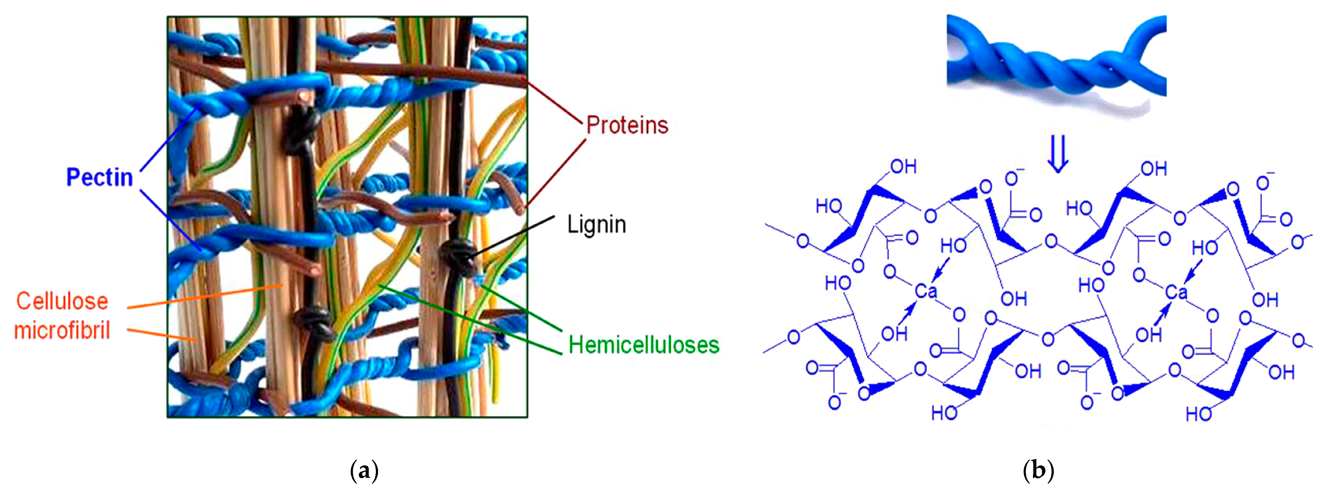
:1. Introduction
2. Materials and Methods
2.1. Materials
2.2. Rhéum Rhabarbarum Preparation
2.3. PRh–Mt and PCt–Mt Preparation
2.4. Rh–Mt and Rhbio–Mt Preparation
2.5. Evaluation of the Rh–Mt and Rhbio–Mt Structure
2.6. Evaluation of the Pectin Structure
2.7. Evaluation of the PRh–Mt and PCt–Mt Structure
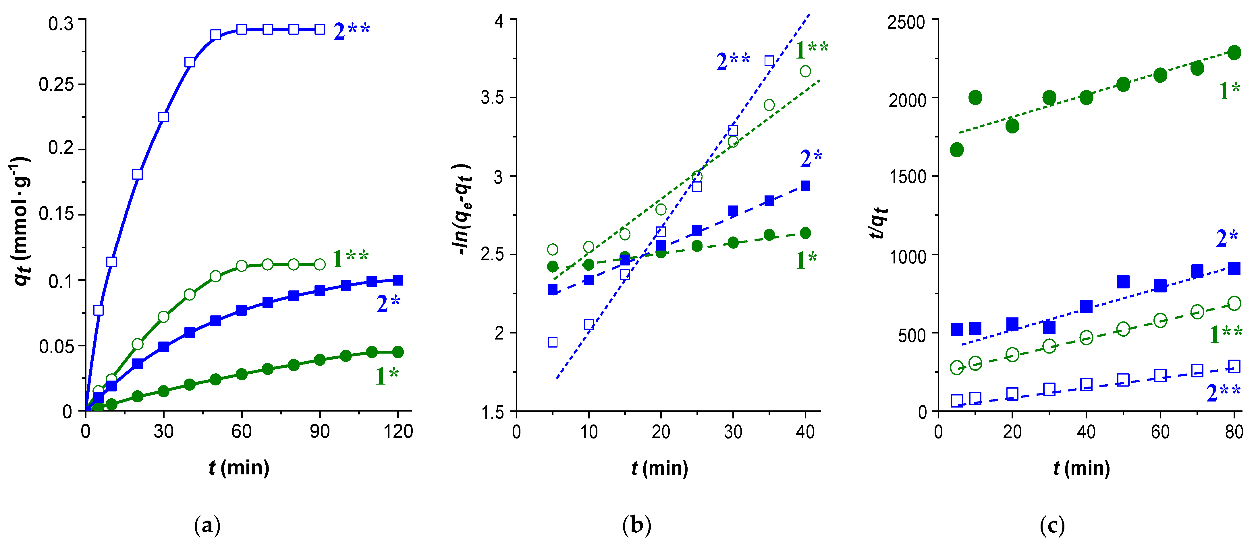
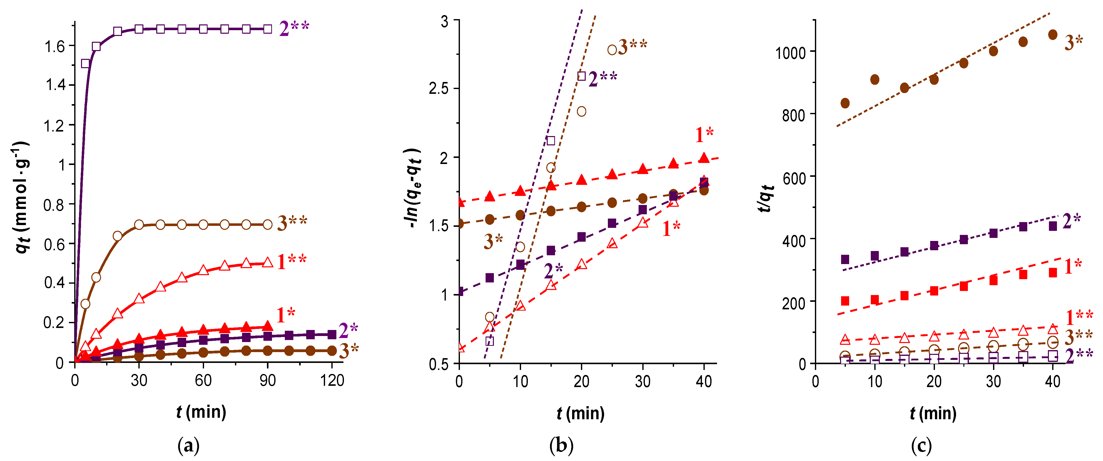
2.8. Kinetics of Theophylline Sorption
3. Results and Discussion
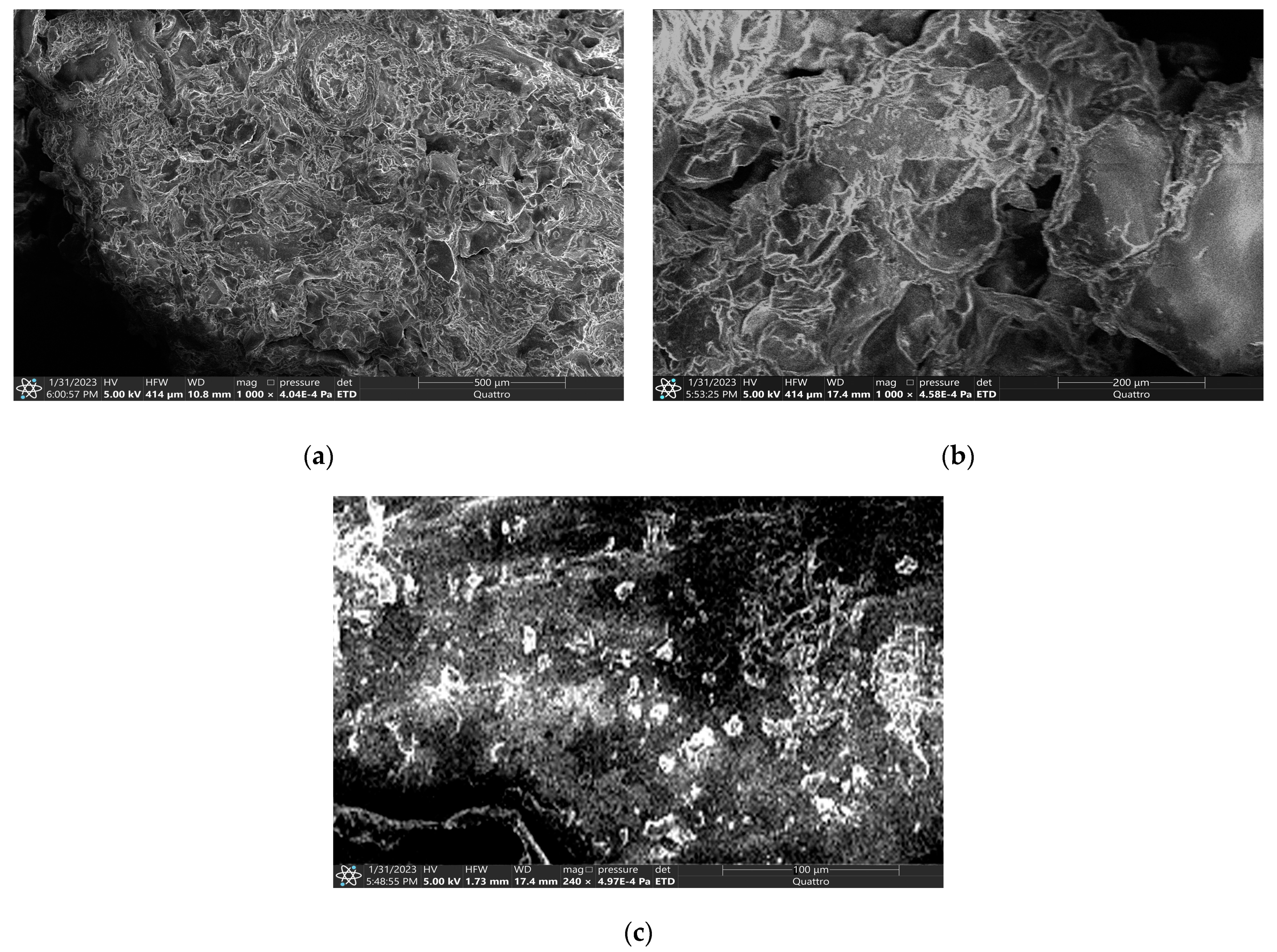
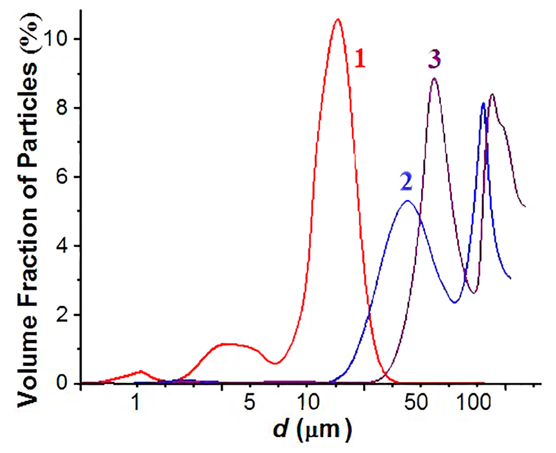
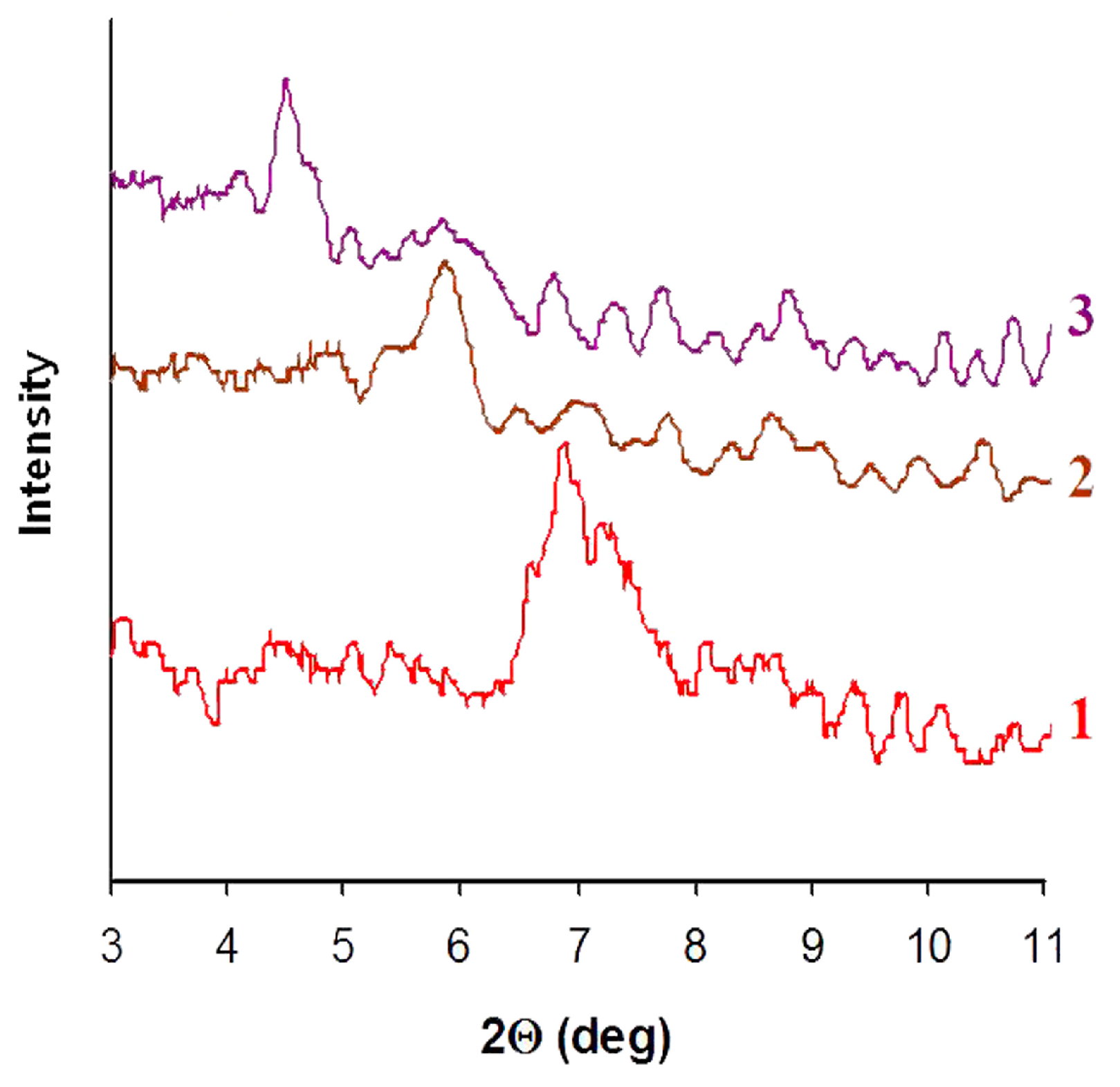
3.1. Structure and Sorption Properties of the Modified Phytocomposite
3.2. Structure and Sorption Properties of the Hybrid Polymer-Inorganic PRh–Mt Coating
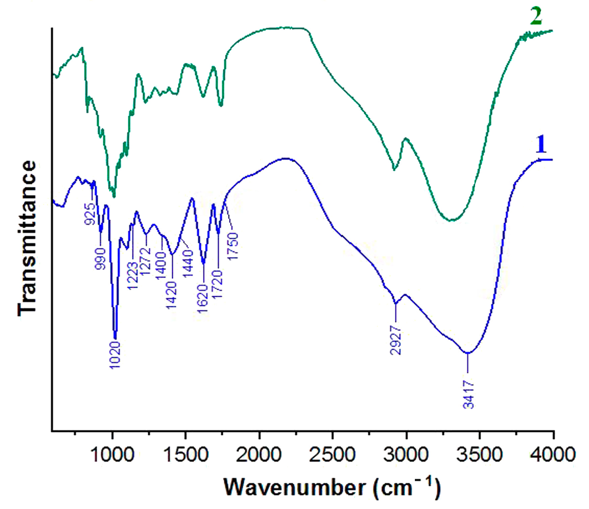
- 1750 cm−1—the valence vibrations of carbonyls in ester groups νas(C=O);
- 1720 cm−1—the vibrations of C=O bonds in non-esterified carboxyl groups;
- 1420 and 1615 cm−1—the symmetric and asymmetric valence vibrations of carboxyl with metal cations νs, νas(C–OMe);
- 1440 cm−1—the deformation asymmetric vibrations in the methoxyl group δas(O–CH3);
- 1400 cm−1—the valence vibrations of the C-OH bond in carboxyl ν(C–OH);
- 1272 and 1223 cm−1—the valence vibrations of ester bond ν(C–O–C);
- 1020–1010 cm−1—the valence vibrations of pyranose rings ν(C–C)(C–O);
- 990 cm−1—the deformation vibrations of carboxyl δ(C–OMe);
- 920 cm−1—the pendular vibrations of methyl in the ester group ρ(O–CH3).
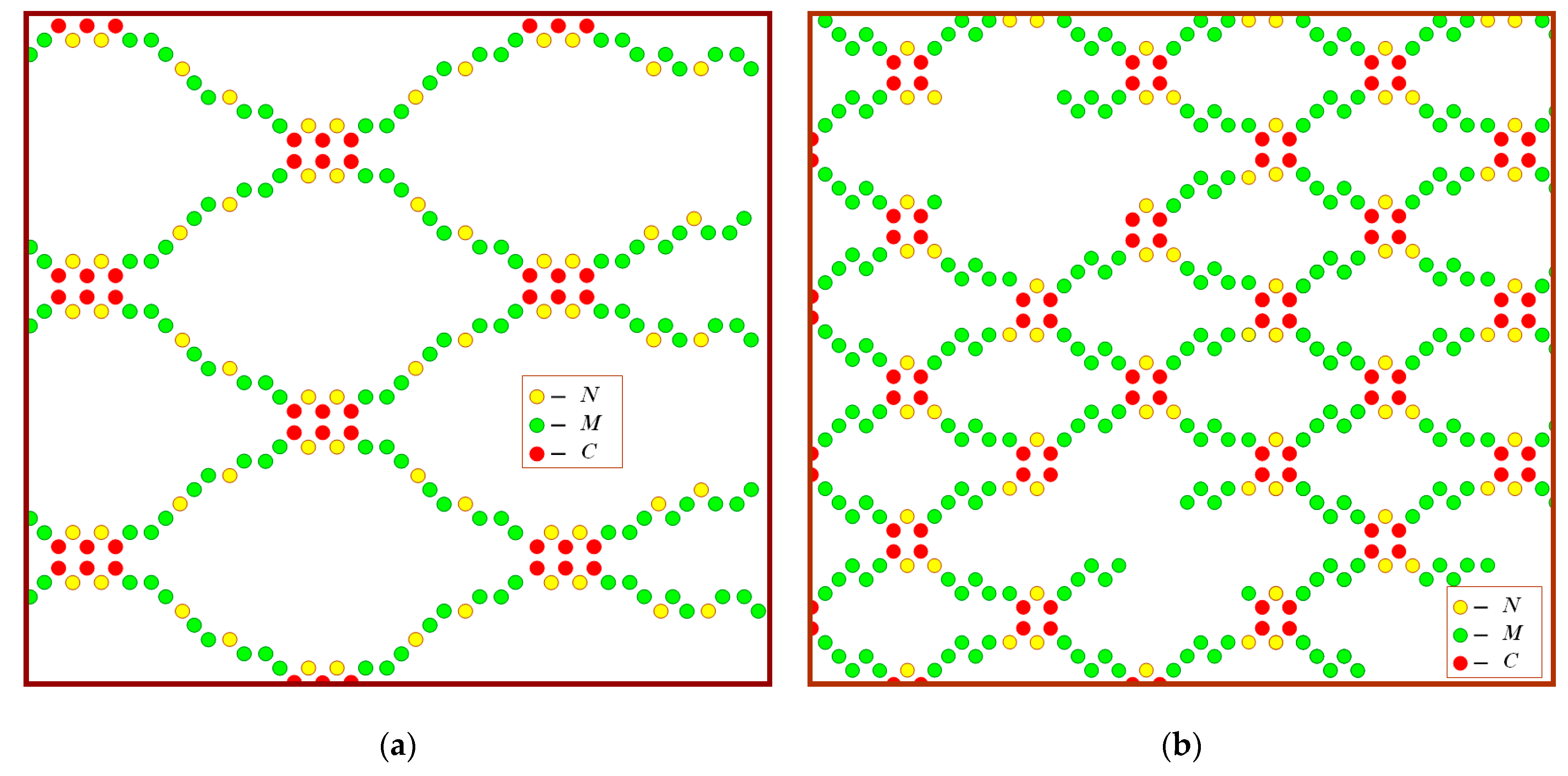
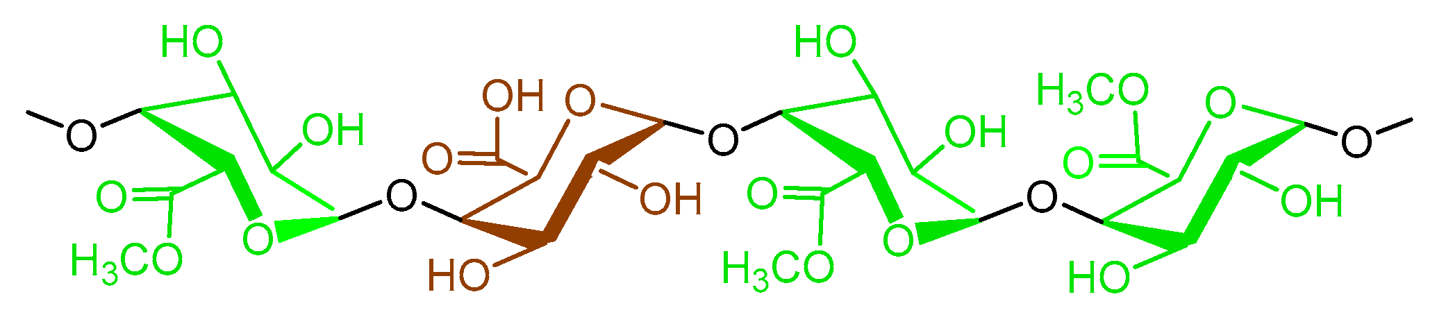
- The total composition of units for the PRh sample at DP = 120 is C24N32M64 and has the distribution [(C3N2)(M8N2)]8. Each macromolecule passes through eight blocks, which are formed by three C-links and between them two links in the N-form. Blocks are separated by branches consisting of eight M-units with two imbedded N-units.
- The total composition of units for the PC sample at DP = 68 is C16N12M40 and the distribution of subunits is [(C2N2)M5+(C2N1)M5)]4. Each macromolecule passes a paired combination of blocks four times, which collectively contain four units in C-form and three units in N-form; that is, they are formed with the participation of a double alternation of C–N units from one chain and a fragmentation of C–N–C from an adjacent chain. The branches between the blocks contain five units in M-form.
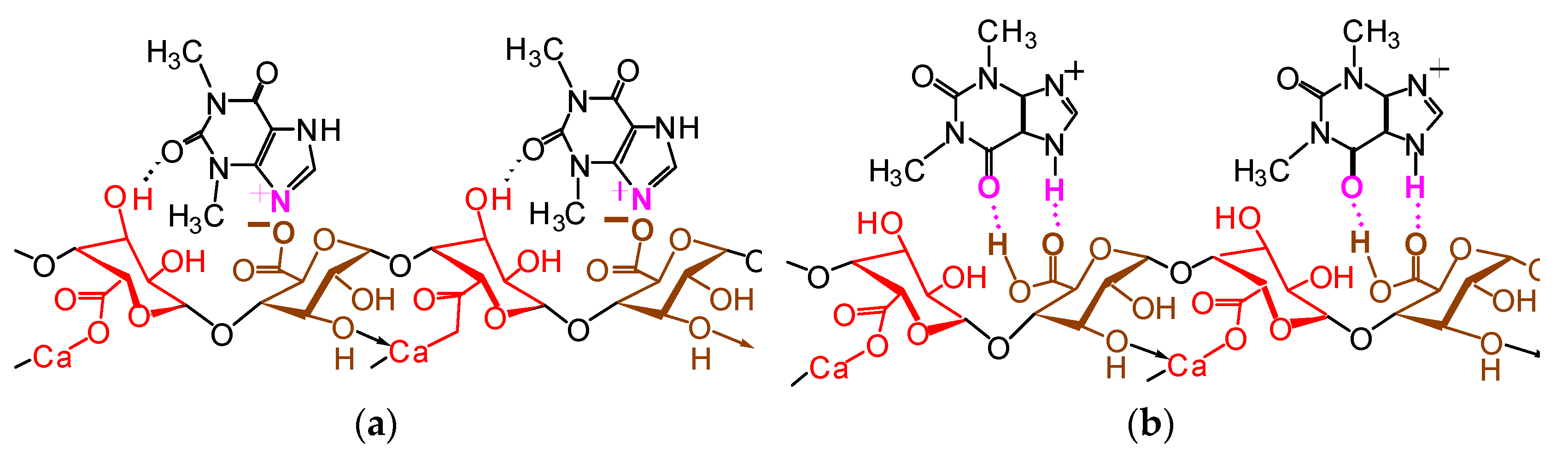
- (a)
- During the passage of food in the human duodenum:
- (b)
- During the passage of feed mass in the body of farm animals:
4. Conclusions
5. Patents
Author Contributions
Funding
Institutional Review Board Statement
Informed Consent Statement
Data Availability Statement
Acknowledgments
Conflicts of Interest
References
- Chatterjee, S.; Dhole, A.; Krishnan, A.A.; Banerjee, K. Mycotoxin Monitoring, Regulation and Analysis in India: A Success Story. Foods 2023, 12, 705. [Google Scholar] [CrossRef] [PubMed]
- Moretti, A.; Pascale, M.; Logrieco, A.F. Mycotoxin risks under a climate change scenario in Europe. Trends Food Sci. Technol. 2019, 84, 38–40. [Google Scholar] [CrossRef]
- Milicevic, D.; Nesic, K.; Jaksic, S. Mycotoxin contamination of the food supply chain—Implications for one health programme. Procedia Food Sci. 2015, 5, 187–190. [Google Scholar] [CrossRef] [Green Version]
- Alshannaq, A.; Yu, J.-K. Occurrence, toxicity and analysis of major mycotoxins in food. Int. J. Environ. Res. Public Health 2017, 14, 632. [Google Scholar] [CrossRef] [PubMed] [Green Version]
- Wielogórska, E.; MacDonald, S.; Elliot, C.T. A review of the efficacy of mycotoxin detoxifying agents used in feed in light of changing global environment and legislation. World Mycotoxin J. 2016, 9, 419–433. [Google Scholar] [CrossRef]
- Xu, R.; Kiarie, E.; Yiannikouris, A.; Sun, L.; Karrow, N. Nutritional impact of mycotoxins in food animal production and strategies for mitigation. J. Anim. Sci. Biotechnol. 2022, 13, 69. [Google Scholar] [CrossRef]
- Van Egmond, H.P.; Schothorst, R.C.; Jonker, M.A. Regulations relating to mycotoxins in food. Anal. Bioanal. Chem. 2007, 389, 147–157. [Google Scholar] [CrossRef] [Green Version]
- Tutelyan, V.A. Deoxynivalenol in cereals in Russia. Toxicol. Lett. 2004, 153, 173–179. [Google Scholar] [CrossRef]
- Rodrigues, I.; Naehrer, K. A three-year survey on the worldwide occurrence of mycotoxins in feedstuffs and feed. Toxins 2012, 4, 663–675. [Google Scholar] [CrossRef]
- Gruber-Dorninger, C.; Jenkins, T.; Schatzmayr, G. Global Mycotoxin Occurrence in Feed: A Ten-Year Survey. Toxins 2019, 11, 375. [Google Scholar] [CrossRef] [Green Version]
- Xu, W.; Han, X.; Li, F. Co-occurrence of multi-mycotoxins in wheat grains harvested in Anhui province, China. Food Control. 2019, 96, 180–185. [Google Scholar] [CrossRef]
- Feng, S.; Hua, M.Z.; Roopesh, M.S.; Lu, X. Rapid detection of three mycotoxins in animal feed materials using competitive ELISA-based origami microfluidic paper analytical device (μPAD). Anal. Bioanal. Chem. 2023, 14, 127. [Google Scholar] [CrossRef] [PubMed]
- Wang, M.; Hearon, S.E.; Phillips, T.D. Development of enterosorbents that can be added to food and water to reduce toxin exposures during disasters. J. Environ. Sci. Health B 2019, 54, 514–524. [Google Scholar] [CrossRef] [PubMed]
- Wang, M.; Safe, S.; Hearon, S.E.; Phillips, T.D. Strong adsorption of Polychlorinated Biphenyls by processed montmorillonite clays: Potential applications as toxin enterosorbents during disasters and floods. Environ. Pollut. 2019, 255, 113210. [Google Scholar] [CrossRef] [PubMed]
- Luo, Y.; Liu, X.; Li, J. Updating techniques on controlling mycotoxins—A review. Food Control 2018, 89, 123–132. [Google Scholar] [CrossRef]
- Hamad, G.; Mehany, T.; Simal-Gandara, J.; Hafez, E.E. A review of recent innovative strategies for controlling mycotoxins in foods. Food Control 2023, 144, 109350. [Google Scholar] [CrossRef]
- Adebo, O.A.; Molelekoa, T.; Makhuvele, R.; Adebiyi, J.A.; Oyedeji, A.B.; Gbashi, S. A review on novel non-thermal food processing techniques for mycotoxin reduction. Int. J. Food Sci. Technol. 2020, 56, 13–27. [Google Scholar] [CrossRef]
- Riseh, R.S.; Hassanisaadi, M.; Vatankhah, M.; Kennedy, J.F. Encapsulating biocontrol bacteria with starch as a safe and edible biopolymer to alleviate plant diseases: A review. Carbohydr. Polym. 2022, 302, 120384. [Google Scholar] [CrossRef]
- Vila-Donat, P.; Marin, S.; Sanchis, V.; Ramos, A.J. A review of the mycotoxin adsorbing agents, with an emphasis on their multi-binding capacity, for animal feed decontamination. Food Chem. Toxicol. 2018, 114, 246–259. [Google Scholar] [CrossRef] [Green Version]
- Sharma, V.; Patial, V. Food Mycotoxins: Dietary interventions implicated in the prevention of mycotoxicosis. ACS Food Sci. Technol. 2021, 1, 1717–1739. [Google Scholar] [CrossRef]
- Kozak, L.; Szilagyi, Z.; Toth, L.; Pócsi, I.; Molnár, I. Tremorgenic and neurotoxic paspaline-derived indole-diterpenes: Biosynthetic diversity, threats and applications. Appl. Microbiol. Biotechnol. 2019, 103, 1599–1616. [Google Scholar] [CrossRef] [PubMed]
- Miller, T.R.; Beversdorf, L.J.; Weirich, C.A.; Bartlett, S.L. Cyanobacterial toxins of the laurentian great lakes, their toxicological effects, and numerical limits in drinking water. Mar. Drugs 2017, 15, 160. [Google Scholar] [CrossRef] [PubMed] [Green Version]
- Dittmann, E.; Fewer, D.P.; Neilan, B.A. Cyanobacterial toxins: Biosynthetic routes and evolutionary roots. FEMS Microbiol. Rev. 2013, 37, 23–43. [Google Scholar] [CrossRef]
- Papadimitriou, T.; Kagalou, I.; Stalikas, C.; Pilidis, G.; Leonardos, I.D. Assessment of microcystin distribution and biomagnification in tissues of aquatic food web compartments from a shallow lake and evaluation of potential risks to public health. Ecotoxicology 2012, 21, 1155–1166. [Google Scholar] [CrossRef] [PubMed]
- Tomczak, E.; Tosik, P. Sorption equilibrium of azo dyes direct orange 26 and reactive blue 81 onto a cheap plant sorbent. Ecol. Chem. Eng. 2014, S21, 435–445. [Google Scholar] [CrossRef] [Green Version]
- Koksharov, S.A.; Aleeva, S.V.; Lepilova, O.V. Biomodification of flax fibrous materials for increase of sorption to organic compounds. Intern. J. Chem. Eng. 2019, 2019, 4137593. [Google Scholar] [CrossRef]
- Adunphatcharaphon, S.; Petchkongkaew, A.; Greco, D.; D’Ascanio, V.; Visessanguan, W.; Avantaggiato, G. The Effectiveness of Durian Peel as a Multi-Mycotoxin Adsorbent. Toxins 2020, 12, 108. [Google Scholar] [CrossRef] [PubMed] [Green Version]
- Shanab, O.; Abdeen, A.; Abdelkader, A. Arabic Gum Could Alleviate the Aflatoxin B1-provoked Hepatic Injury in Rat: The Involvement of Oxidative Stress, Inflammatory, and Apoptotic Pathways. Toxins 2022, 14, 605. [Google Scholar] [CrossRef]
- Koksharov, S.A.; Aleeva, S.V.; Lepilova, O.V. Description of adsorption interactions of lead ions with functional groups of pectin-containing substances. J. Mol. Liq. 2019, 283, 606–616. [Google Scholar] [CrossRef]
- Aleeva, S.V.; Lepilova, O.V.; Koksharov, S.A. Revealing the regularities of sorption binding of cadmium ions by pectin substances from aqueous solutions. Prot. Met. Phys. Chem. Surf. 2021, 57, 37–44. [Google Scholar] [CrossRef]
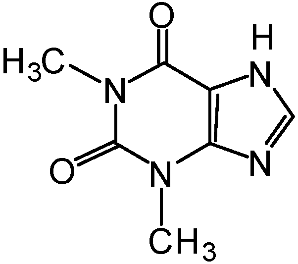
- Koksharov, S.A.; Aleeva, S.V.; Lepilova, O.V. The Influence of the structure of pectin substances of flax fodder supplements on absorption binding of azaheterocyclic mycotoxins. Russ. J. Gen. Chem. 2021, 91, S60–S83. [Google Scholar] [CrossRef]
- Koksharov, S.A.; Aleeva, S.V.; Lepilova, O.V. Kinetics of the sorption of theophylline in pectin hydrogels with different structural properties. Russ. J. Phys. Chem. A 2022, 96, 773–780. [Google Scholar] [CrossRef]
- Wang, X.-M.; Hou, X.-Q.; Zhang, Y.-Q.; Yang, R.; Feng, S.-F.; Li, Y.; Ren, Y. Genetic diversity of the endemic and medicinally important plant Rheum officinale as revealed by Inter-Simpe Sequence Repeat (ISSR) markers. Int. J. Mol. Sci. 2012, 13, 3900–3915. [Google Scholar] [CrossRef] [Green Version]
- Aleeva, S.V.; Lepilova, O.V.; Koksharov, S.A. Laws of Cresol-Vapor Sorption on Highly Porous Materials of Biomodified Flax Shive. Prot. Met. Phys. Chem. Surf. 2022, 58, 13–21. [Google Scholar] [CrossRef]
- Wang, M.; Hearon, S.; Phillips, T.D. A high capacity bentonite clay for the sorption of aflatoxins. Food Addit. Contam. Part A 2019, 37, 1–10. [Google Scholar] [CrossRef]
- Lepilova, O.V.; Aleeva, S.V.; Koksharov, S.A. Role of pectin substances in the structural organization of the flax fiber-montmorillonite hybrid sorbent. Russ. J. Appl. Chem. 2018, 91, 90–95. [Google Scholar] [CrossRef]
- Koksharov, S.A.; Aleeva, S.V.; Lepilova, O.V. Preparation of hybrid polymer-inorganic chelators based on pectin and montmorillonite. Key Eng. Mater. 2019, 816, 333–338. [Google Scholar] [CrossRef]
- Kochkina, N.E.; Skobeleva, O.A.; Khokhlova, Y.V. Investigation of cationic starch/Na-monmorillonite bionanocomposite adsorbent prepared by vibration milling for acid dye removal. Part. Sci Technol. 2017, 35, 259–264. [Google Scholar] [CrossRef]
- Aleeva, S.V.; Chistyakova, G.V.; Lepilova, O.V.; Koksharov, S.A. Effect of the state of carboxyl groups of pectin on the sorption binding of copper ions. Russ. J. Phys. Chem. A 2018, 92, 1583–1589. [Google Scholar] [CrossRef]
- Pretsch, E.; Buhlmann, P.; Badertscher, M. Structure Determination of Organic Compounds; Springer: Berlin/Heidelberg, Germany, 2009. [Google Scholar] [CrossRef]
- Morris, E.R.; Powell, D.A.; Gidley, M.J.; Rees, D.A. Conformations and interactions of pectins: I. Polymorphism between gel and solid states of calcium polygalacturonate. J. Mol. Biol. 1982, 155, 517–531. [Google Scholar] [CrossRef]
- Gharibzahedi, S.M.T.; Smith, B.; Guo, Y. Pectin extraction from common fig skin by different methods: The physicochemical, rheological, functional, and structural evaluations. Int. J. Biol. Macromol. 2019, 136, 275–283. [Google Scholar] [CrossRef] [PubMed]
- Yang, J.; Ching, Y.C.; Chuah, C.H. Applications of lignocellulosic fibers and lignin in bioplastics: A Review. Polymers 2019, 11, 751. [Google Scholar] [CrossRef] [PubMed] [Green Version]
- Noguchi, M.; Hasegawa, Y.; Suzuki, S.; Nakazawa, M.; Ueda, M.; Sakamoto, T. Determination of chemical structure of pea pectin by using pectinolytic enzymes. Carbohydr. Polym. 2020, 231, 115738. [Google Scholar] [CrossRef]
- Rahmani, Z.; Khodaiyan, F.; Kazemi, M.; Sharifan, A. Optimization of microwave-assisted extraction and structural characterization of pectin from sweet lemon peel. Int. J. Biol. Macromol. 2020, 147, 1107–1115. [Google Scholar] [CrossRef]
- Chistyakova, G.V.; Koksharov, S.A. Features of pectin biodegradation in the presence of sodium ethylendiamintetraacetate. Russ. J. Gen. Chem. 2014, 84, 763–766. [Google Scholar] [CrossRef]
- Assifaoui, A.; Lerbret, A.; Uyen, H.T.D.; Neiers, F.; Chambin, O.; Loupiac, C.; Cousin, F. Structural behaviour differences in low methoxy pectin solutions in the presence of divalent cations (Ca2+ and Zn2+): A process driven by the binding mechanism of the cation with the galacturonate unit. Soft Matter 2015, 11, 551–560. [Google Scholar] [CrossRef] [PubMed]
- Gawkowska, D.; Cybulska, J.; Zdunek, A. Structure-related gelling of pectins and linking with other natural compounds: A review. Polymers 2018, 10, 762. [Google Scholar] [CrossRef] [Green Version]
- Doğan, H.; Meral, R. The effects of locust bean gum and rubarum on the physical and functional properties of the gluten-free biscuits. Ital. J. Food Sci. 2019, 31, 542–555. [Google Scholar] [CrossRef]
- Moor, E.; Reynol, R.C. X-ray Diffraction and the Identification and Analysis of Clay Minerals; Oxford University Press: Oxford, UK, 1989. [Google Scholar] [CrossRef]
- Gerasin, V.A.; Antipov, E.M.; Karbushev, V.V.; Kulichikhin, V.G.; Karpacheva, G.P.; Talroze, R.V.; Kudryavtsev, Y.V. New approaches to the development of hybrid nanocomposites: From structural materials to high-tech applications. Russ. Chem. Rev. 2013, 82, 303–332. [Google Scholar] [CrossRef]
- Ho, Y.S.; Ng, J.C.Y.; McKay, G. Kinetics of pollutant sorption by biosorbents: Review. Sep. Purif. Methods 2000, 2, 189–232. [Google Scholar] [CrossRef]
- Ho, Y.S. Citation review of Lagergren kinetic rate equation on adsorption reactions. Scientometrics 2004, 1, 171–177. [Google Scholar] [CrossRef]
- Douven, S.; Paez, C.A.; Gommes, C.J. The Range of Validity of Sorption Kinetic Models. J. Colloid Interface Sci. 2015, 448, 437–450. [Google Scholar] [CrossRef] [PubMed]
- Van de Graaff, K.M. Anatomy and physiology of the gastrointestinal tract. Pediatr. Infect. Dis. 1986, 5, 11–16. [Google Scholar] [CrossRef] [PubMed]


| Symbol | Preparation |
|---|---|
| Rh–Mt | hybrid rhubarb–montmorillonite phytocomposites based on the original Rh biomass |
| Rhbio–Mt | hybrid rhubarb–montmorillonite phytocomposites based on the biomodified Rh biomass |
| PRh | pectin preparation isolated from biomodified Rh biomass |
| PCt | commercial citrus pectin preparation (reference sample) |
| PRh–Mt | hybrid nanocomposites of the compared pectins from Rh biomass with montmorillonite |
| PCt–Mt | hybrid nanocomposites of the compared citrus pectin with montmorillonite |
| Ratio of Rh:Mt in the Sample | VP (cm3·g−1) | SA (m2·g−1) | ||
|---|---|---|---|---|
| Rh–Mt | Rhbio–Mt | Rh–Mt | Rhbio–Mt | |
| 100:0 | 0.019 | 0.070 | 14.0 | 57.2 |
| 97.5:2.5 | 0.027 | 0.078 | 32.8 | 75.0 |
| 95:5 | 0.034 | 0.089 | 49.7 | 93.8 |
| 90:10 | 0.045 | 0.102 | 83.6 | 132.6 |
| 0:100 | 0.270 | 690.8 | ||
| Sorption Time, t (min) | Quantity of Sorbed Theophylline, qt (mg·g−1) | |||
|---|---|---|---|---|
| Rh | Rhbio | Rh–Mt (90:10) | Rhbio–Mt (90:10) | |
| 20 | 0.90 | 18.38 | 8.83 | 39.24 |
| 30 | 1.44 | 18.92 | 9.91 | 39.52 |
| 60; (180) | (5.59) | 18.92 | 10.45 | 39.91 |
| Pectin | Fractional Content of Galacturonate Units Forms (Units) | ||
|---|---|---|---|
 |  |  | |
| PRh | 0.28 | 0.53 | 0.19 |
| PCt | 0.15 | 0.65 | 0.20 |
| Pectin | Decalcification | [η] ± 0.05 (cm3·g−1) | MP ± 0.1 (kDa) | DP ± 5 |
|---|---|---|---|---|
| PRh | − | 23.7 | 155.6 | 802 |
| + | 2.3 | 23.3 | 120 | |
| PCt | − | 7.4 | 60.7 | 313 |
| + | 1.15 | 13.0 | 68 |
| Pectin | qe (mmol·g−1) | pH | Pseudo-First-Order Model | Pseudo-Second-Order Model | ||||
|---|---|---|---|---|---|---|---|---|
| qe* (mmol·g−1) | k1 (min−1) | R2 | qe* (mmol g−1) | k2 (g·mmol−1·min −1) | R2 | |||
| PCt | 0.045 | 2 | 0.049 | 0.006 | 0.997 | 0.030 | 0.067 | 0.663 |
| 0.112 | 5 | 0.118 | 0.052 | 0.803 | 0.116 | 0.119 | 0.992 | |
| PRh | 0.100 | 2 | 0.103 | 0.019 | 0.994 | 0.106 | 0.096 | 0.769 |
| 0.292 | 5 | 0.364 | 0.071 | 0.887 | 0.309 | 0.171 | 0.995 | |
| Sorbent | qe (mmol·g−1) | pH | Pseudo-First-Order Model | Pseudo-Second-Order Model | ||||
|---|---|---|---|---|---|---|---|---|
| qe* (mmol·g−1) | k1 (min−1) | R2 | qe* (mmol·g−1) | k2 (g·mmol−1·min−1) | R2 | |||
| Mt | 0.177 | 2 | 0.181 | 0.008 | 0.996 | 0.285 | 0.008 | 0.824 |
| 0.498 | 5 | 0.504 | 0.030 | 0.992 | 0.862 | 0.022 | 0.841 | |
| PCt–Mt (90:10) | 0.058 | 2 | 0.062 | 0.009 | 0.992 | – | – | – |
| 0.695 | 5 | – | – | – | 0.704 | 0.137 | 0.997 | |
| PRh–Mt (90:10) | 0.139 | 2 | 0.141 | 0.036 | 0.995 | – | – | – |
| 1.685 | 5 | – | – | – | 1.690 | 0.480 | 0.994 | |
| Sorbent | qe (mmol·g−1) | Pseudo-Second-Order Model | ||
|---|---|---|---|---|
| qe* (mmol·g−1) | k2 (g·mmol−1·min−1) | R2 | ||
| PCt | 0.115 | 0.116 | 0.150 | 0.995 |
| PRh | 0.305 | 0.309 | 0.315 | 0.991 |
| PCt–Mt (90:10) | 0.701 | 0.704 | 0.137 | 0.997 |
| PRh–Mt (90:10) | 1.689 | 1.690 | 0.965 | 0.994 |
Disclaimer/Publisher’s Note: The statements, opinions and data contained in all publications are solely those of the individual author(s) and contributor(s) and not of MDPI and/or the editor(s). MDPI and/or the editor(s) disclaim responsibility for any injury to people or property resulting from any ideas, methods, instructions or products referred to in the content. |
© 2023 by the authors. Licensee MDPI, Basel, Switzerland. This article is an open access article distributed under the terms and conditions of the Creative Commons Attribution (CC BY) license (https://creativecommons.org/licenses/by/4.0/).
Share and Cite
Kornilova, N.; Koksharov, S.; Aleeva, S.; Lepilova, O.; Bikbulatova, A.; Nikiforova, E. Enterosorbents Based on Rhubarb Biomass with a Hybrid Polymer-Inorganic Coating for the Immobilization of Azaheterocyclic Mycotoxins. Coatings 2023, 13, 684. https://doi.org/10.3390/coatings13040684
Kornilova N, Koksharov S, Aleeva S, Lepilova O, Bikbulatova A, Nikiforova E. Enterosorbents Based on Rhubarb Biomass with a Hybrid Polymer-Inorganic Coating for the Immobilization of Azaheterocyclic Mycotoxins. Coatings. 2023; 13(4):684. https://doi.org/10.3390/coatings13040684
Chicago/Turabian StyleKornilova, Nadezhda, Sergey Koksharov, Svetlana Aleeva, Olga Lepilova, Albina Bikbulatova, and Elena Nikiforova. 2023. "Enterosorbents Based on Rhubarb Biomass with a Hybrid Polymer-Inorganic Coating for the Immobilization of Azaheterocyclic Mycotoxins" Coatings 13, no. 4: 684. https://doi.org/10.3390/coatings13040684