
Copper-Treated Environmentally Friendly Antipathogenic Cotton Fabric with Modified Reactive Blue 4 Dye to Improve Its Antibacterial and Aesthetic Properties
 , , , ,
  and
and
Abstract
:1. Introduction
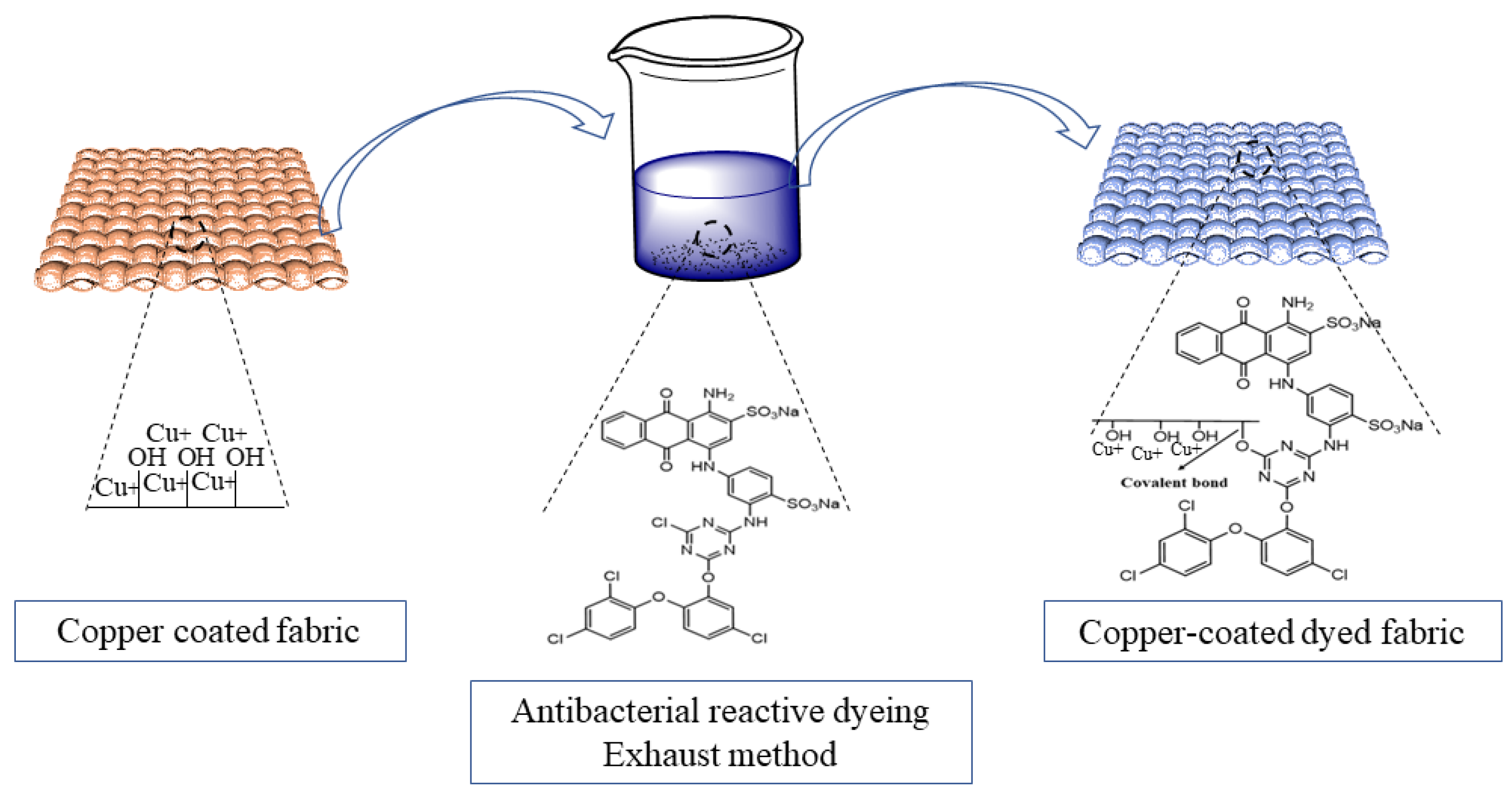
- The synthesis of cuprous oxide nanoparticles by using three different reducing agents.
- Application of the synthesized Cu-NPs on cotton fabrics to impart antibacterial functionality.
- Application of antibacterial dye on copper-coated fabrics to improve their aesthetic appearance.
2. Experimental Section
2.1. Materials
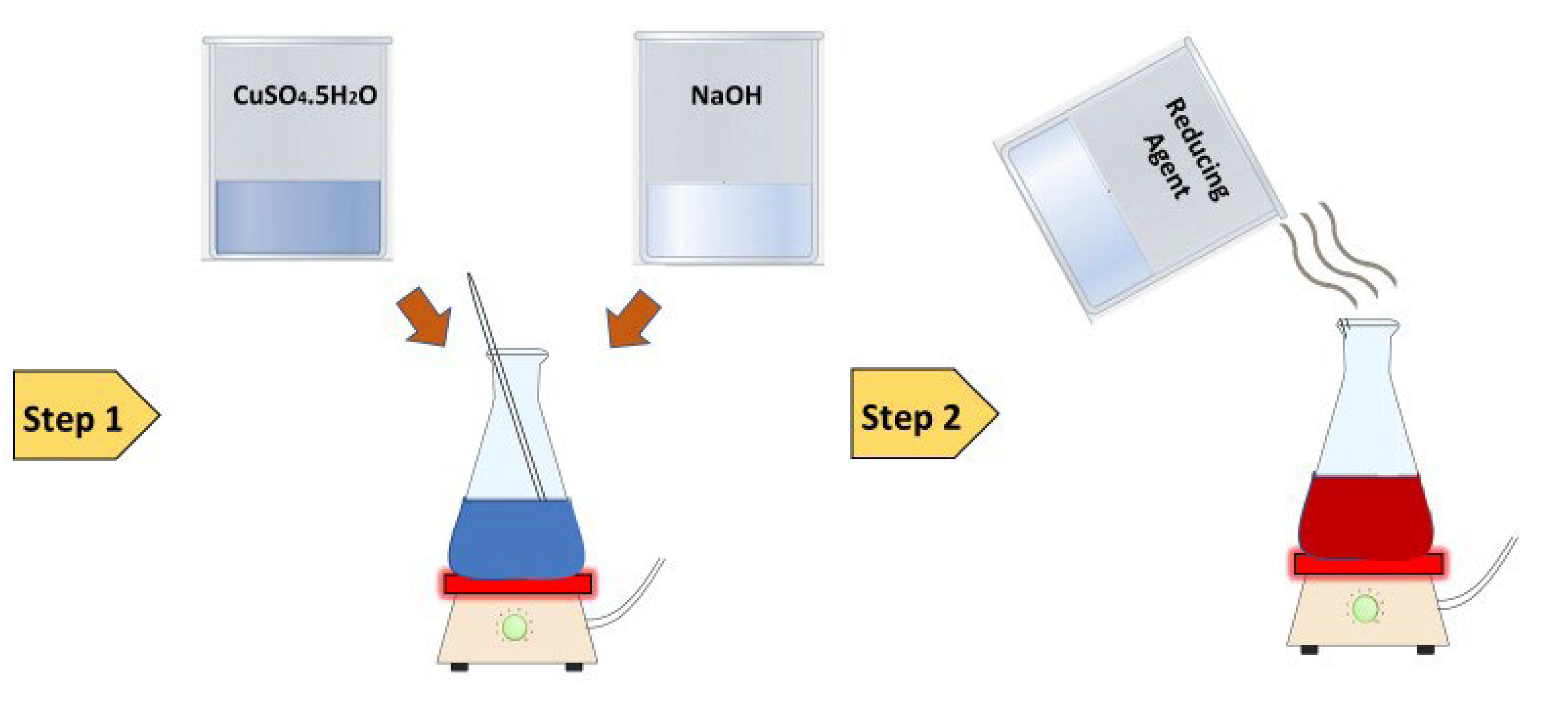
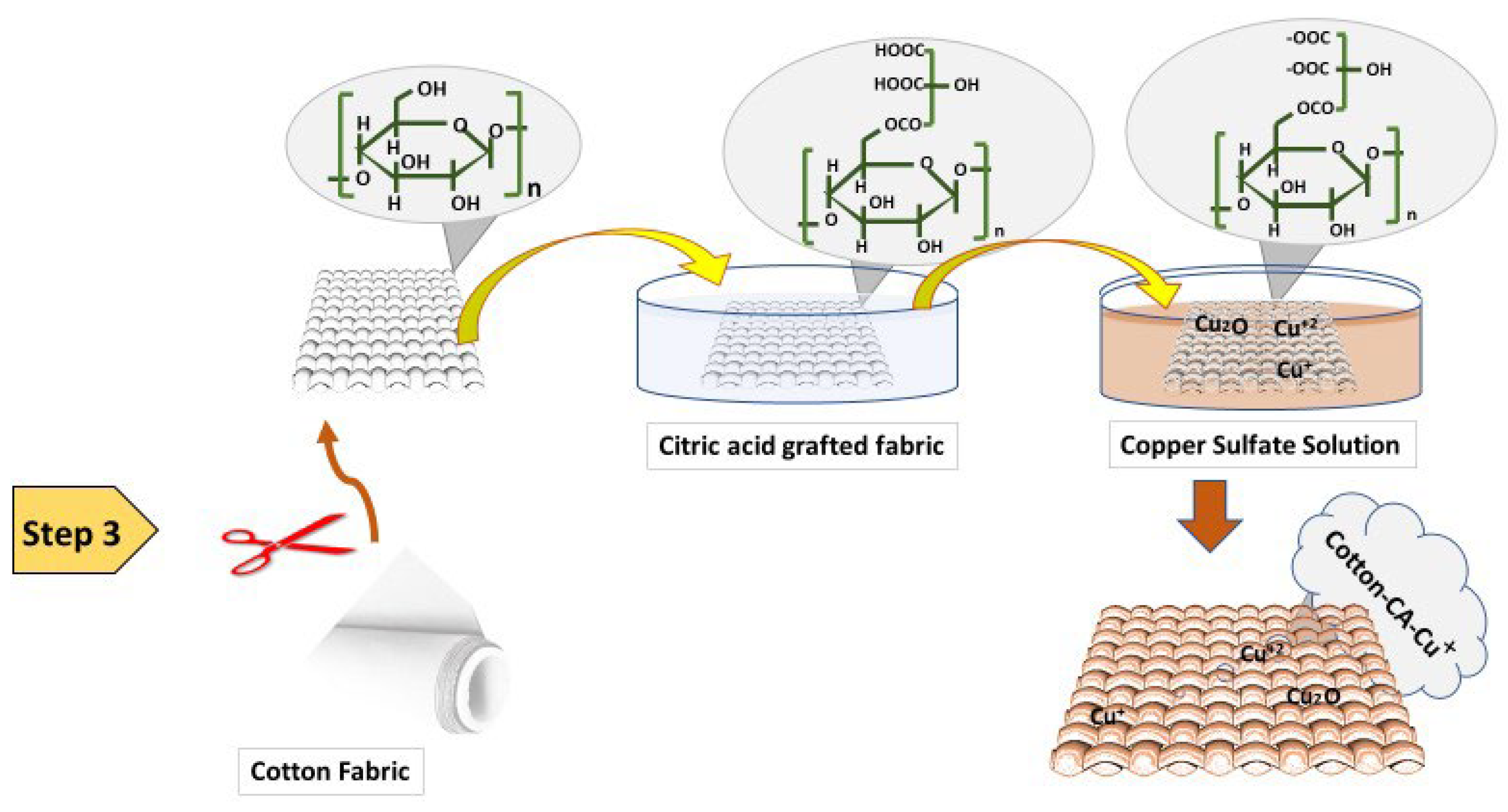
2.2. Preparation of Cuprous Oxide Particles and Deposition on Cotton
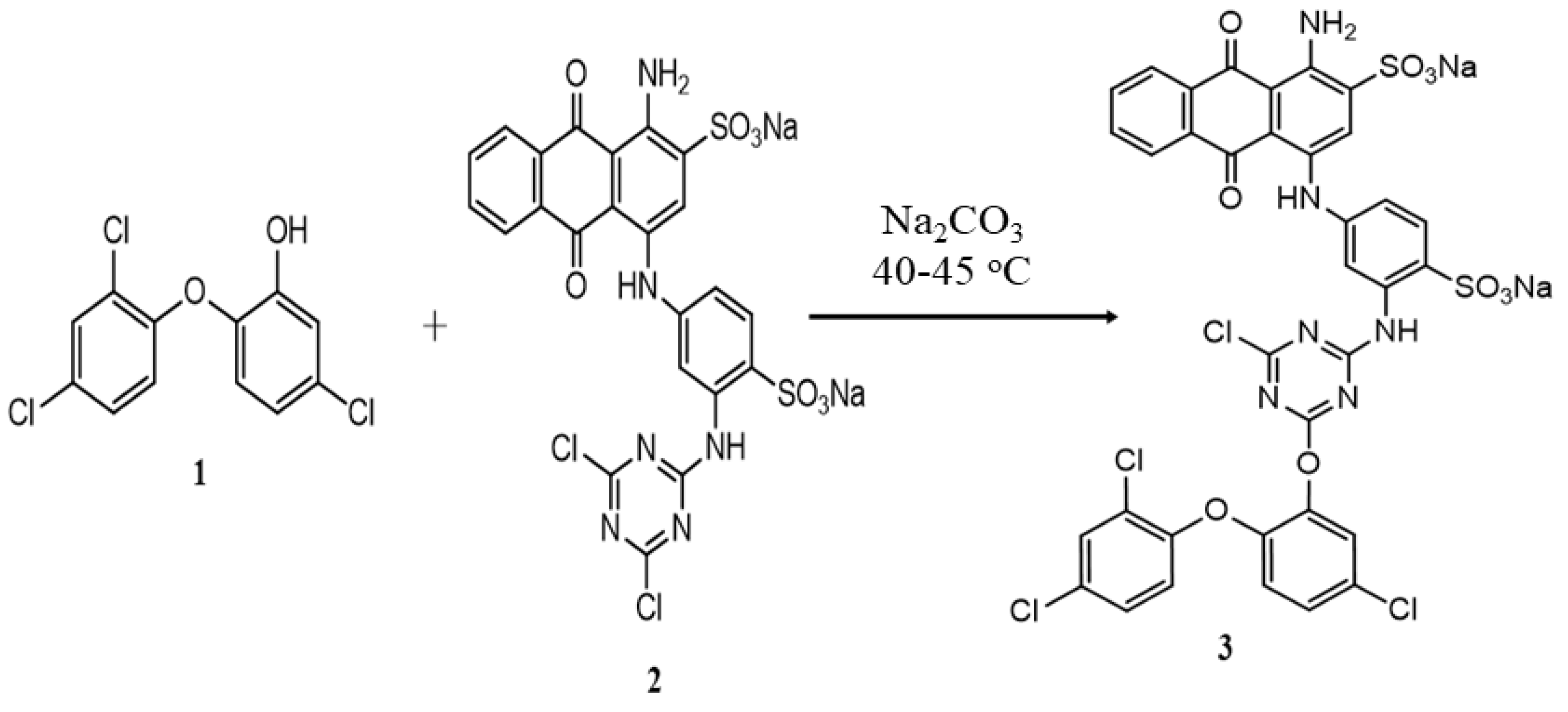
2.3. Functionalization of Reactive Blue 4 Dye with Triclosan
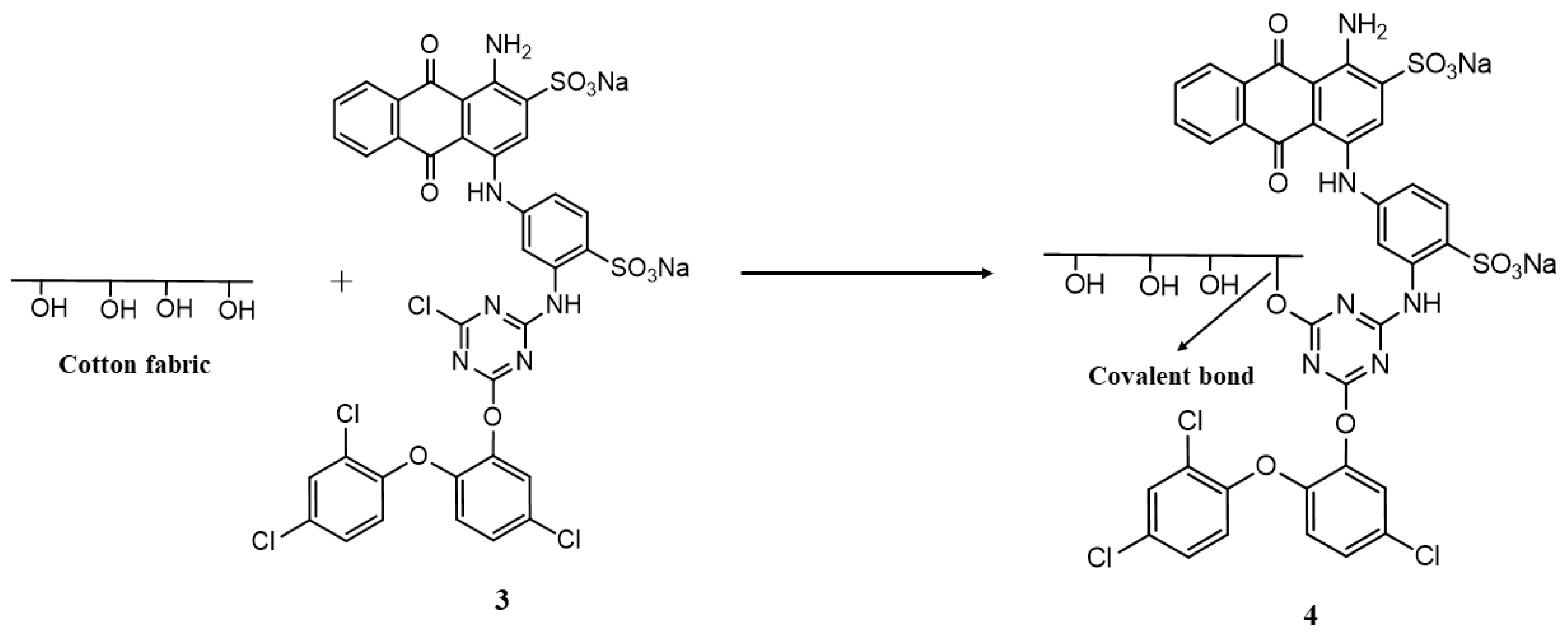
2.4. Application of Functionalized Dye on Fabric
2.5. Characterization
2.5.1. Surface Characterizations
2.5.2. Dye Exhaustion (E%), Fixation (F%), and Total Fixation (T%) Analysis
2.5.3. Fastness Properties
2.5.4. Colorimetric (CIELAB) values analysis
2.5.5. Assessment of Dye Levelness
2.6. Antimicrobial Properties
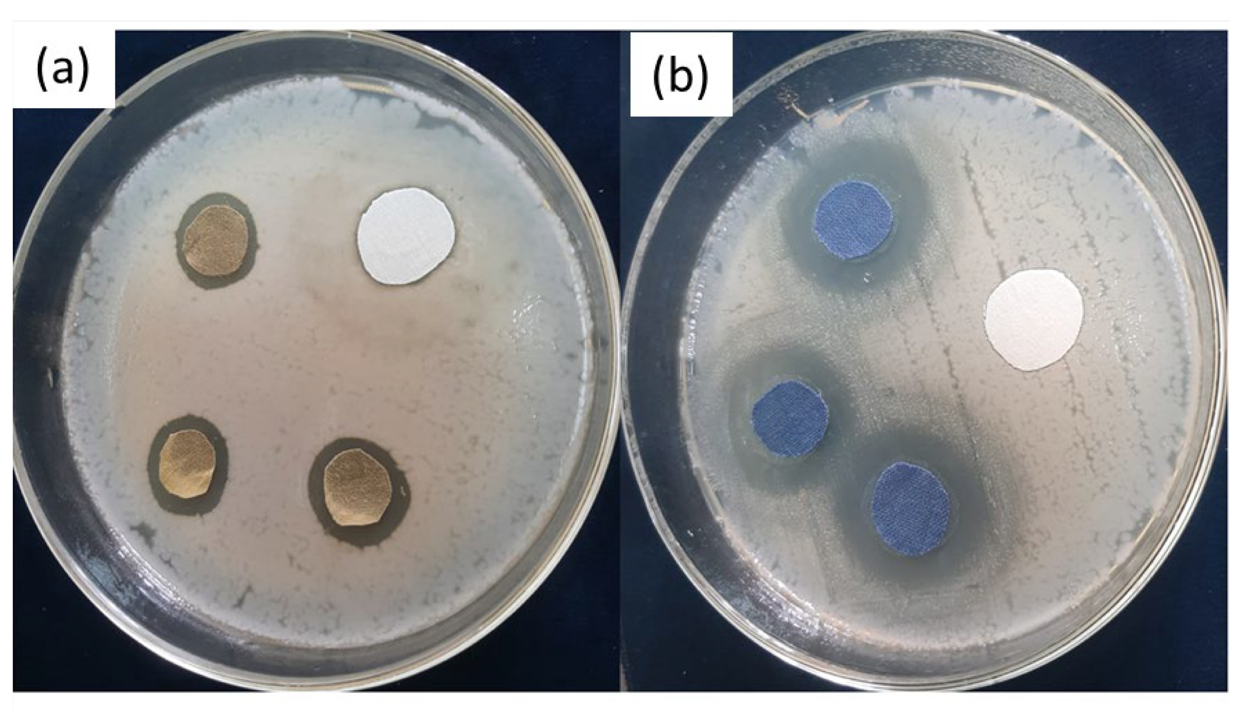
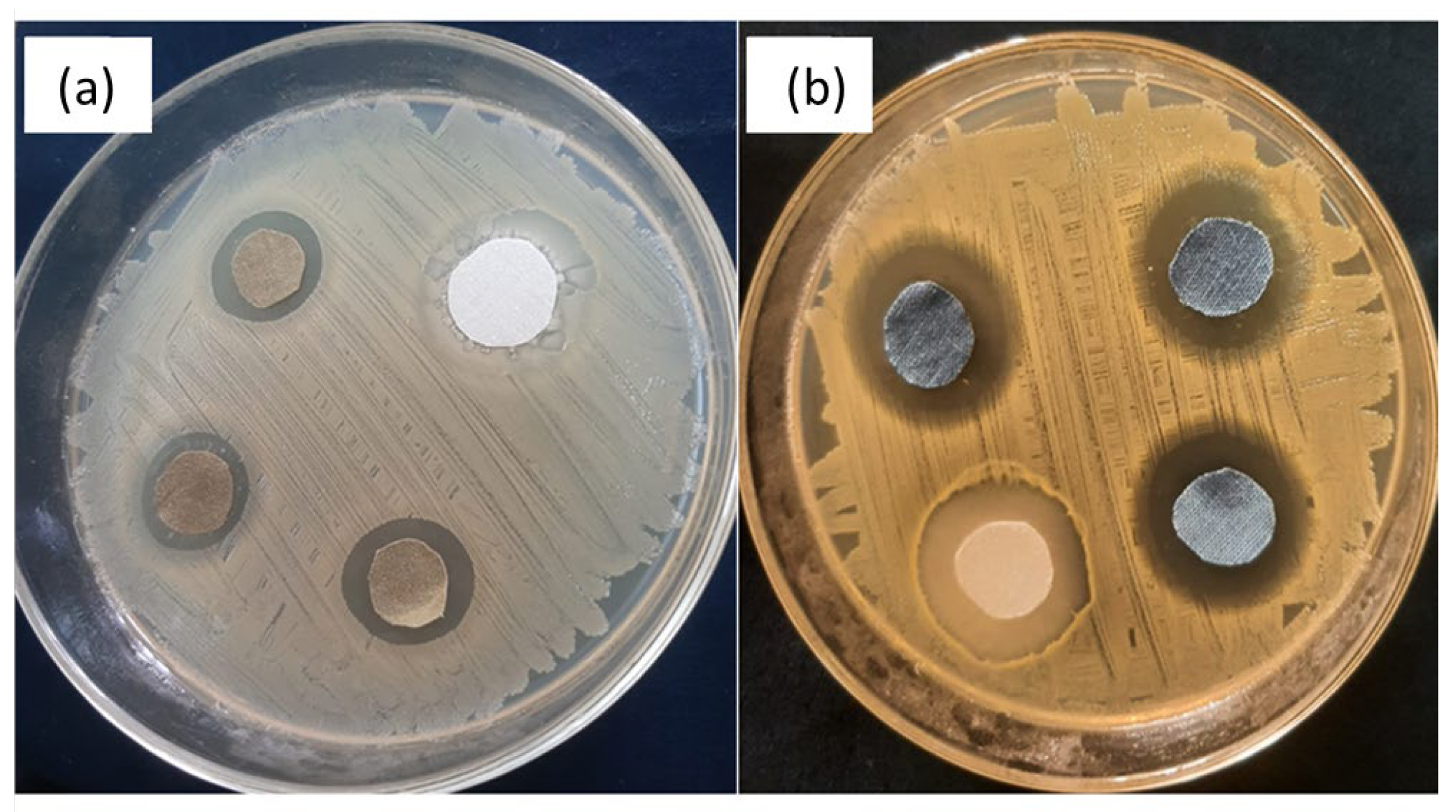
2.6.1. Zone of Inhibition Test
Preparation of Bacterial Strain
Determining Zone of Inhibition
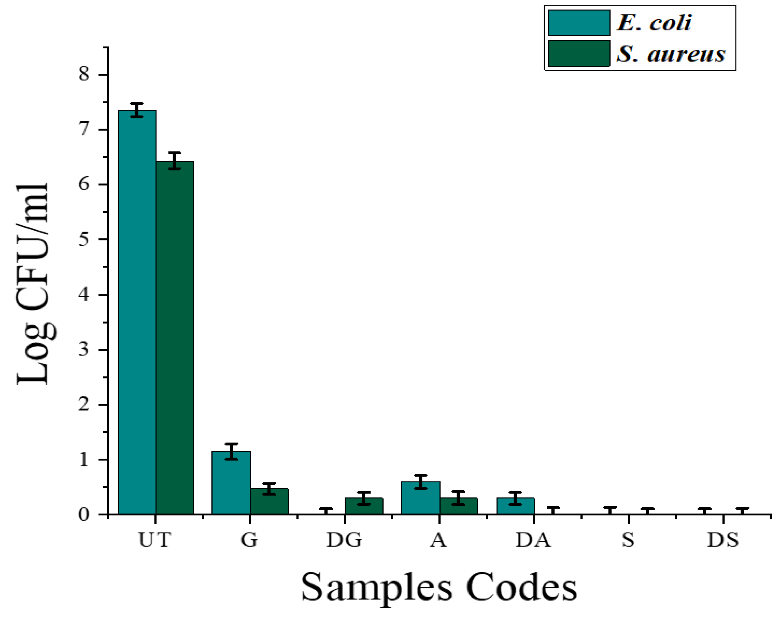
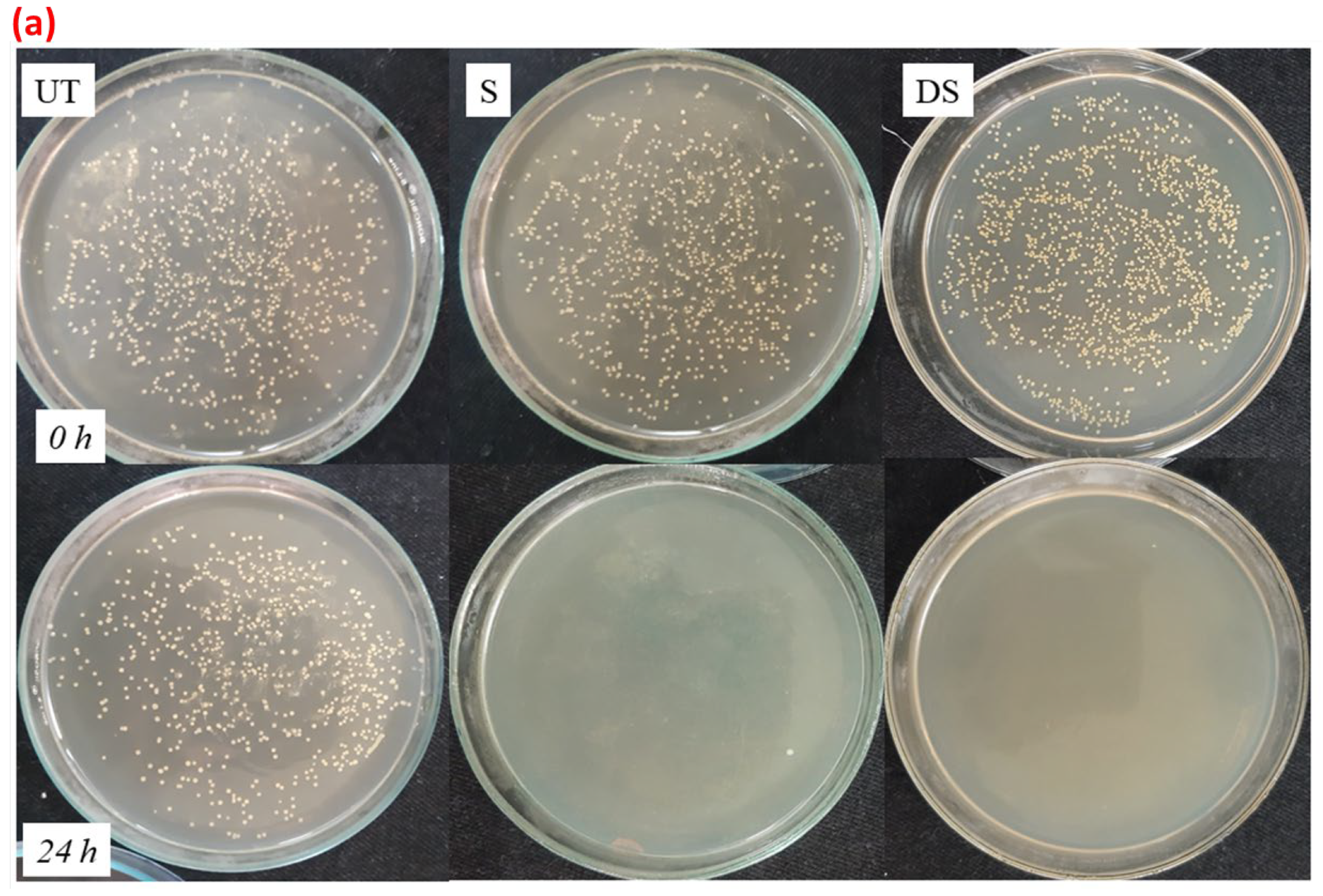
2.6.2. Reduction Factor (Quantitative Test)
2.6.3. Antifungal Analysis
2.6.4. Antiviral Activity
2.7. Durability of Bioactive Fabrics
3. Results and Discussion
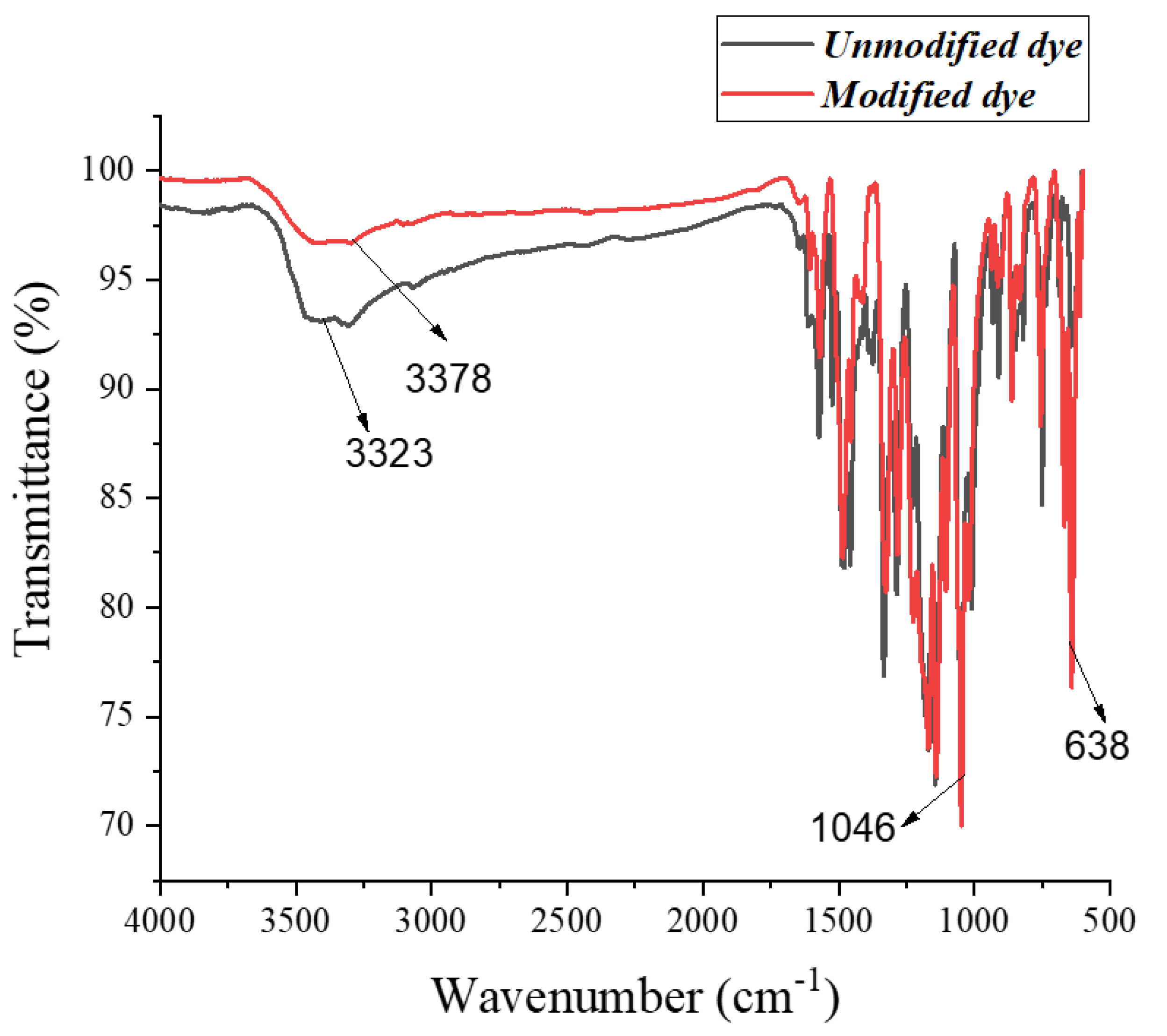
3.1. FTIR Analysis
3.2. Colorimetric Data Measurement
3.3. Levelness of Copper-Treated Undyed and Dyed Fabric
3.4. Fastness Properties of Copper-Coated Dyed Fabric
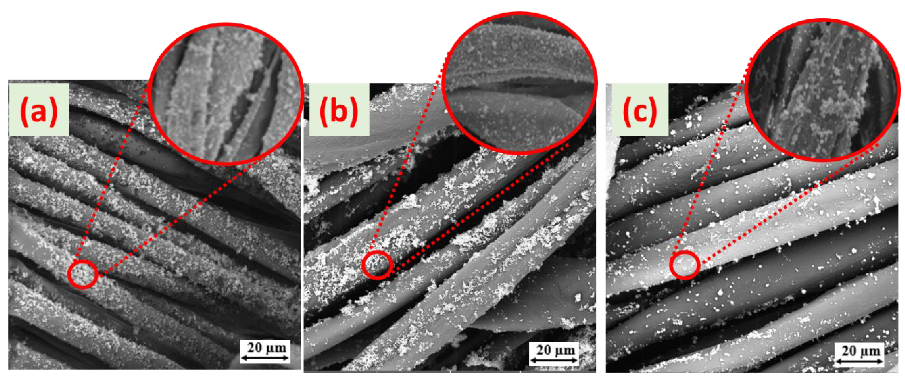
3.5. Morphology of Copper-Coated Dyed Cotton Fabrics
SEM microstructure
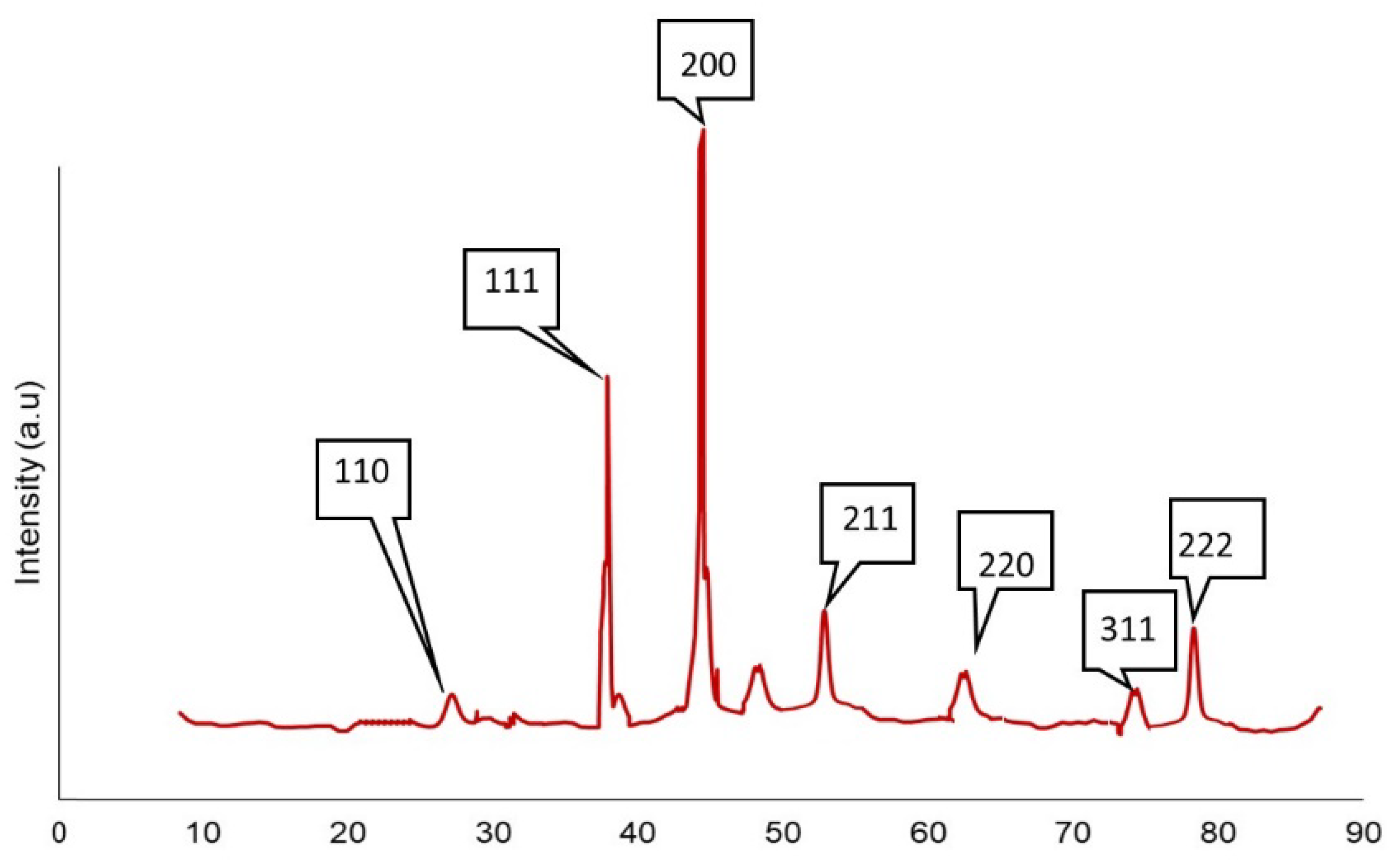
3.6. XRD Analysis
3.7. Antibacterial Analysis
3.7.1. Zone of Inhibition Tests
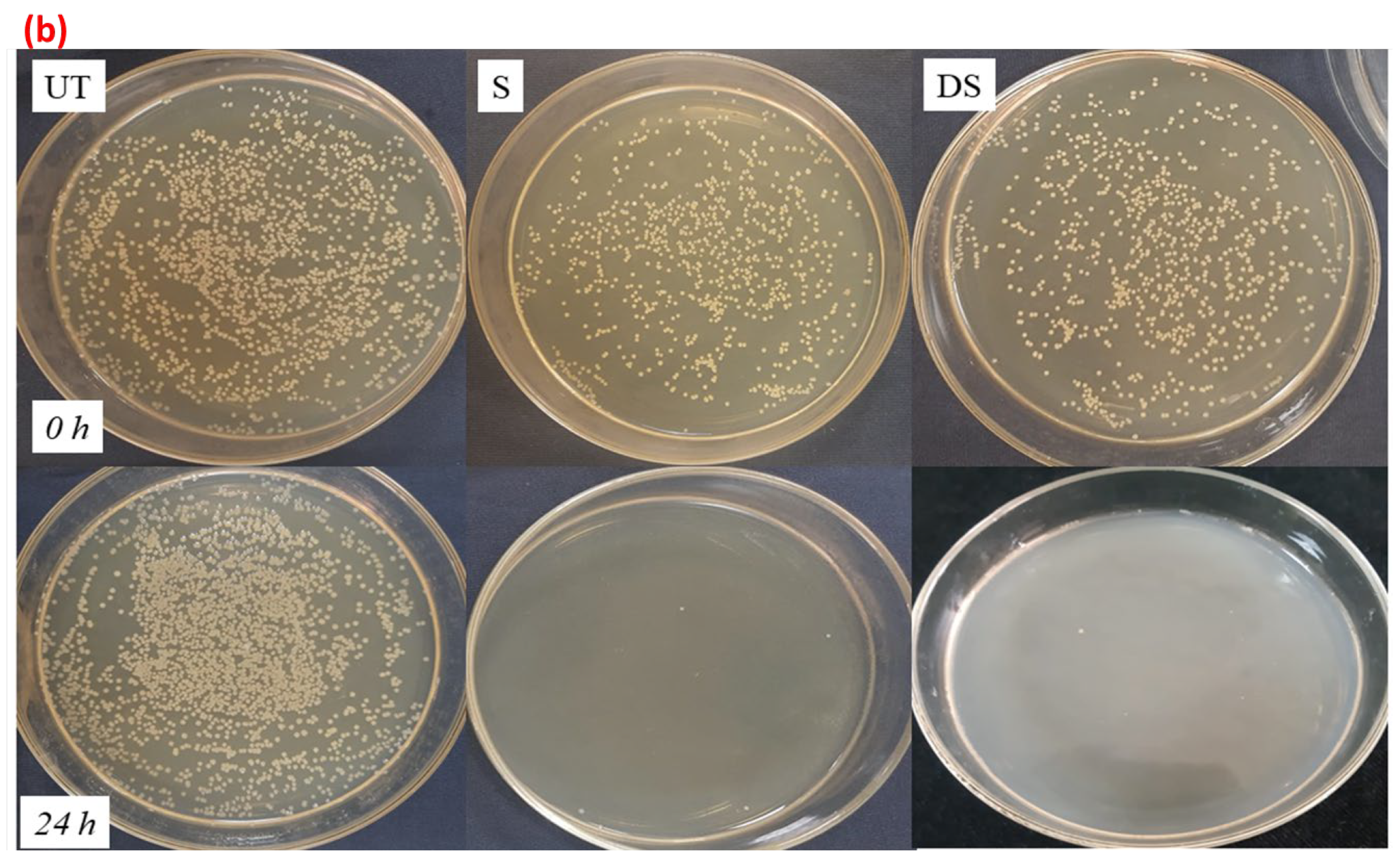
3.7.2. Reduction Factor (Quantitative Test)
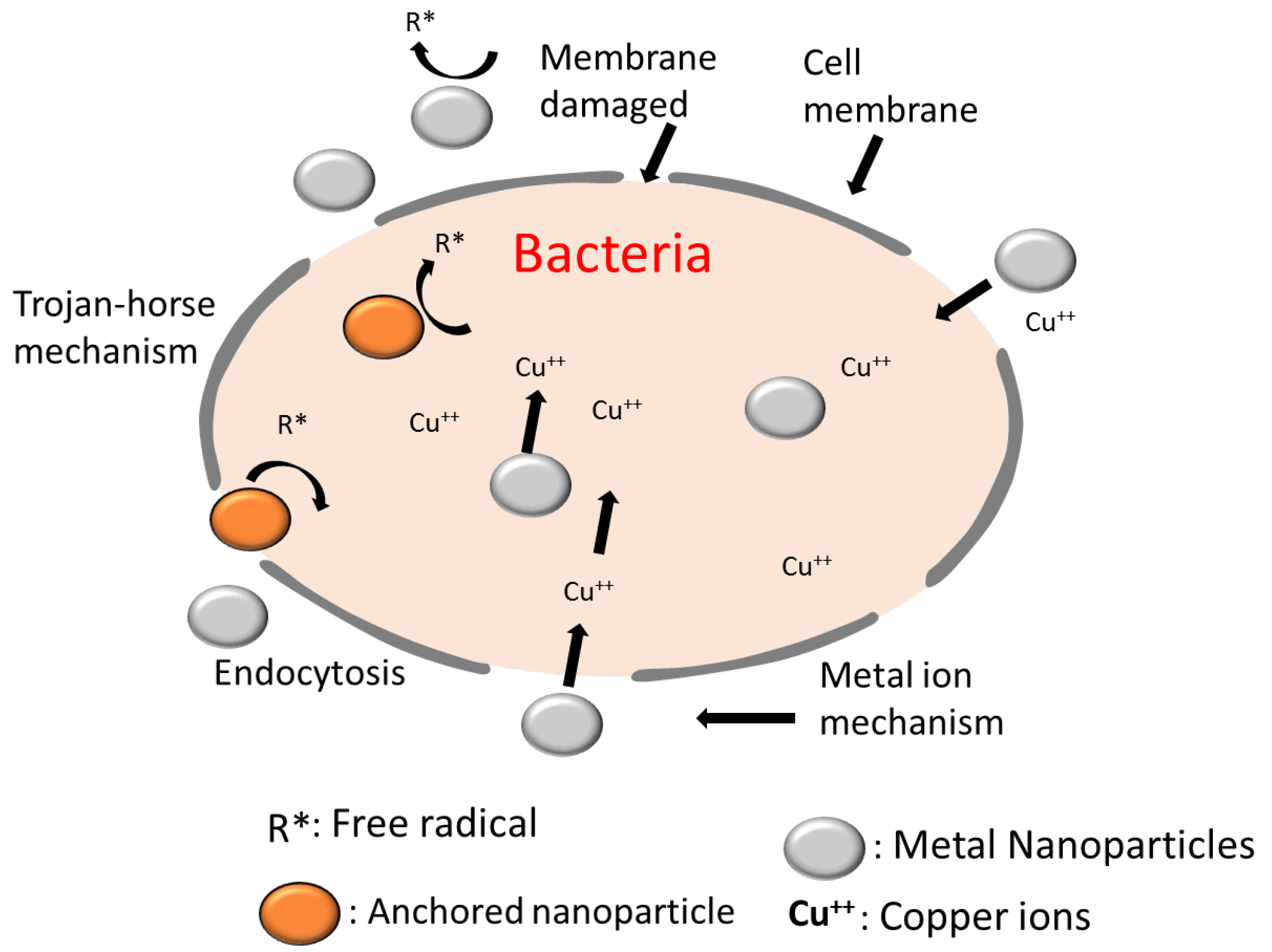
3.7.3. Mechanism of Antibacterial Action
3.8. Antifungal Activity of Treated Samples
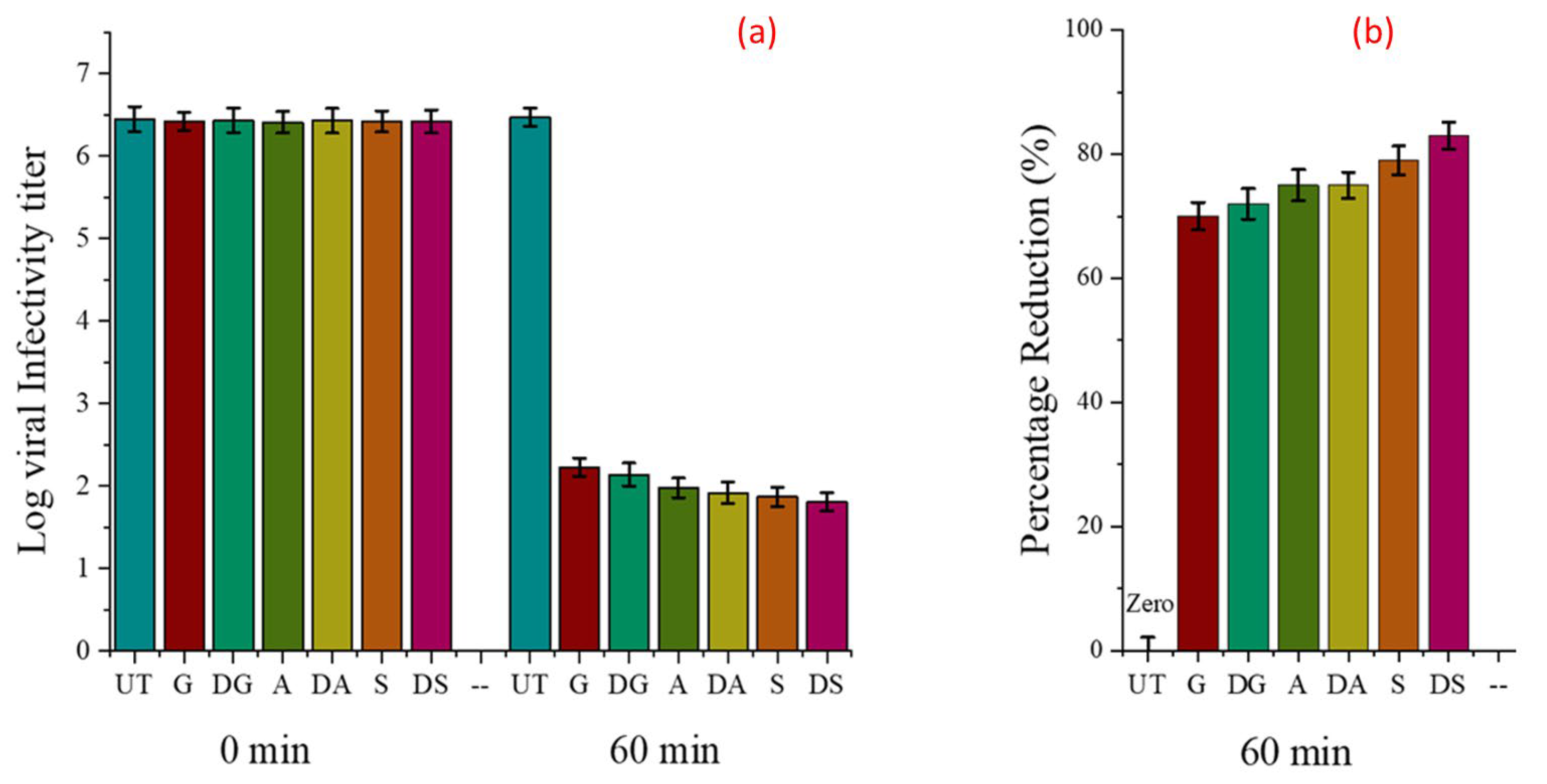
3.9. Antiviral Effectiveness
3.10. Durability of Cuprous-oxide-Coated Fabrics
4. Conclusions
Author Contributions
Funding
Institutional Review Board Statement
Informed Consent Statement
Data Availability Statement
Conflicts of Interest
References
- Mallakpour, S.; Azadi, E.; Hussain, C.M. Recent breakthroughs of antibacterial and antiviral protective polymeric materials during COVID-19 pandemic and after pandemic: Coating, packaging, and textile applications. Curr. Opin. Colloid Interface Sci. 2021, 55, 101480. [Google Scholar] [CrossRef]
- Benbow, M. Exploring the concept of moist wound healing and its application in practice. Br. J. Nurs. 2008, 17, S4–S16. [Google Scholar] [CrossRef]
- Baino, F.; Potestio, I. Orbital implants: State-of-the-art review with emphasis on biomaterials and recent advances. Mater. Sci. Eng. C 2016, 69, 1410–1428. [Google Scholar] [CrossRef]
- Fatima, A.; Yasir, S.; Khan, M.S.; Manan, S.; Ullah, M.W.; Ul-Islam, M. Plant extract-loaded bacterial cellulose composite membrane for potential biomedical applications. J. Bioresour. Bioprod. 2021, 6, 26–32. [Google Scholar] [CrossRef]
- Wang, Y.; Zhao, M.; Liu, T. Superhydrophobic modification of cellulose and cotton textiles: Method-ologies and applications. J. Bioresour. Bioprod. 2020, 5, 286–293. [Google Scholar]
- Zhu, G.; Jin, Y.; Ge, M. Simple and green method for preparing copper nanoparticles supported on carbonized cotton as a heterogeneous Fenton-like catalyst. Colloids Surf. A Physicochem. Eng. Asp. 2022, 647, 128978. [Google Scholar] [CrossRef]
- Bhandari, V.; Jose, S.; Badanayak, P.; Sankaran, A.; Anandan, V. Antimicrobial Finishing of Metals, Metal Oxides, and Metal Composites on Textiles: A Systematic Review. Ind. Eng. Chem. Res. 2022, 61, 86–101. [Google Scholar] [CrossRef]
- Hassabo, A.G.; El-Naggar, M.E.; Mohamed, A.L.; Hebeish, A.A. Development of multifunctional modified cotton fabric with tri-component nanoparticles of silver, copper and zinc oxide. Carbohydr. Polym. 2019, 210, 144–156. [Google Scholar] [CrossRef] [PubMed]
- Laime-Oviedo, L.A.; Soncco-Ccahui, A.A.; Peralta-Alarcon, G.; Arenas-Chávez, C.A.; Pineda-Tapia, J.L.; Díaz-Rosado, J.C.; Alvarez-Risco, A.; Del-Aguila-Arcentales, S.; Davies, N.M.; Yáñez, J.A.; et al. Optimization of Synthesis of Silver Nanoparticles Conjugated with Lepechinia meyenii (Salvia) Using Plackett-Burman Design and Response Surface Methodology—Preliminary Antibacterial Activity. Processes 2022, 10, 1727. [Google Scholar] [CrossRef]
- Vera-Nuñez, L.D.C.; Cornejo-Ruiz, J.O.; Arenas-Chávez, C.A.; de Hollanda, L.M.; Alvarez-Risco, A.; Del-Aguila-Arcentales, S.; Davies, N.M.; Yáñez, J.A.; Vera-Gonzales, C. Green synthesis of a novel silver nanoparticle conjugated with Thelypteris Glandulosolanosa (Raqui-Raqui): Preliminary characterization and anticancer activity. Processes 2022, 10, 1308. [Google Scholar] [CrossRef]
- Nikolova, M.P.; Chavali, M.S. Metal Oxide Nanoparticles as Biomedical Materials. Biomimetics 2020, 5, 27. [Google Scholar] [CrossRef]
- Quispe-Quispe, L.G.; Limpe-Ramos, P.; Arenas-Chávez, C.A.; Gomez, M.M.; Mejia, C.R.; Alvarez-Risco, A.; Del-Aguila-Arcentales, S.; Yáñez, J.A.; Vera-Gonzales, C. Physical and Mechanical Characterization of a Functionalized Cotton Fabric with Nanocomposite Based on Silver Nanoparticles and Carboxymethyl Chitosan Using Green Chemistry. Processes 2022, 10, 1207. [Google Scholar] [CrossRef]
- Gawande, M.B.; Goswami, A.; Felpin, F.-X.; Asefa, T.; Huang, X.; Silva, R.; Zou, X.; Zboril, R.; Varma, R.S. Cu and Cu-Based Nanoparticles: Synthesis and Applications in Catalysis. Chem. Rev. 2016, 116, 3722–3811. [Google Scholar] [CrossRef] [PubMed] [Green Version]
- Kuchur, O.; Tsymbal, S.; Shestovskaya, M.; Serov, N.; Dukhinova, M.; Shtil, A. Metal-derived nanoparticles in tumor theranostics: Potential and limitations. J. Inorg. Biochem. 2020, 209, 111117. [Google Scholar] [CrossRef] [PubMed]
- Shahid, M.; Ali, A.; Khaleeq, H.; Tahir, M.F.; Militky, J.; Wiener, J. Development of Antimicrobial Multifunctional Textiles to Avoid from Hospital-Acquired Infections. Fibers Polym. 2021, 22, 3055–3067. [Google Scholar] [CrossRef]
- Jillek, W.; Yung, W.K.C. Embedded components in printed circuit boards: A processing technology review. Int. J. Adv. Manuf. Technol. 2005, 25, 350–360. [Google Scholar] [CrossRef]
- Wu, J.; Wu, Y.; Yuan, Y.; Xia, C.; Saravanan, M.; Shanmugam, S.; Sabour, A.; Alshiekheid, M.; Brindhadevi, K.; Chi, N.T.L.; et al. Eco-friendly, green synthesized copper oxide nanoparticle (CuNPs) from an important medicinal plant Turnera subulata Sm. and its biological evaluation. Food Chem. Toxicol. 2022, 168, 113366. [Google Scholar] [CrossRef]
- Ran, J.; He, M.; Li, W.; Cheng, D.; Wang, X. Growing ZnO Nanoparticles on Polydopamine-Templated Cotton Fabrics for Durable Antimicrobial Activity and UV Protection. Polymers 2018, 10, 495. [Google Scholar] [CrossRef] [Green Version]
- Javed, A.; Wiener, J.; Tamulevičienė, A.; Tamulevičius, T.; Lazauskas, A.; Saskova, J.; Račkauskas, S. One Step In-Situ Synthesis of Zinc Oxide Nanoparticles for Multifunctional Cotton Fabrics. Materials 2021, 14, 3956. [Google Scholar] [CrossRef]
- Ali, A.; Baheti, V.; Vik, M.; Militky, J. Copper electroless plating of cotton fabrics after surface activation with deposition of silver and copper nanoparticles. J. Phys. Chem. Solids 2020, 137, 109181. [Google Scholar] [CrossRef]
- Ali, A.; Baheti, V.; Militky, J.; Khan, Z.; Gilani, S.Q.Z. Comparative Performance of Copper and Silver Coated Stretchable Fabrics. Fibers Polym. 2018, 19, 607–619. [Google Scholar] [CrossRef]
- Shahid, M.; Shahid-ul-Islam; Rather, L.J.; Manzoor, N.; Mohammad, F. Simultaneous shade development, antibacterial, and antifungal functionalization of wool using Punica granatum L. Peel extract as a source of textile dye. J. Nat. Fibers 2019, 16, 555–566. [Google Scholar] [CrossRef]
- El-Shishtawy, R.M.; Youssef, Y.; Ahmed, N.S.; Mousa, A. The use of sodium edate in dyeing: II. Union dyeing of cotton/wool blend with hetero bi-functional reactive dyes. Dye. Pigment. 2007, 72, 57–65. [Google Scholar] [CrossRef]
- Nikolic, G.; Zlatkovic, S.; Cakic, M.; Cakic, S.; Lacnjevac, C.; Rajic, Z. Fast Fourier Transform IR Characterization of Epoxy GY Systems Crosslinked with Aliphatic and Cycloaliphatic EH Polyamine Adducts. Sensors 2010, 10, 684–696. [Google Scholar] [CrossRef] [Green Version]
- Mondal, T.; Bhowmick, A.K.; Krishnamoorti, R. Chlorophenyl pendant decorated graphene sheet as a potential antimicrobial agent: Synthesis and characterization. J. Mater. Chem. 2012, 22, 22481–22487. [Google Scholar] [CrossRef]
- Hoop, C.L.; Iuliucci, R.J. 13C chemical-shift anisotropy of alkyl-substituted aromatic carbon in anthracene derivatives. Solid State Nucl. Magn. Reson. 2013, 53, 1–12. [Google Scholar] [CrossRef]
- Horsley, W.J.; Sternlicht, H.; Cohen, J.S. Carbon-13 magnetic resonance studies of amino acids and peptides. II. J. Am. Chem. Soc. 1970, 92, 680–686. [Google Scholar] [CrossRef]
- Dewan, S.K. Organic Spectroscopy(Nmr, Ir, Mass And Uv); CBS Publishers: New Delhi, India, 2011; Volume 22, p. 565. [Google Scholar]
- Fan, Y.; Zhang, Y.-Q.; Yan, K.; Long, J.-J. Synthesis of a Novel Disperse Reactive Dye Involving a Versatile Bridge Group for the Sustainable Coloration of Natural Fibers in Supercritical Carbon Dioxide. Adv. Sci. 2019, 6, 1801368. [Google Scholar] [CrossRef]
- Ali, A.; Baheti, V.; Javaid, M.U.; Militky, J. Enhancement in ageing and functional properties of copper-coated fabrics by subsequent electroplating. Appl. Phys. A 2018, 124, 651. [Google Scholar] [CrossRef]
- Gan, X.; Wu, Y.; Liu, L.; Shen, B.; Hu, W. Electroless plating of Cu–Ni–P alloy on PET fabrics and effect of plating parameters on the properties of conductive fabrics. J. Alloys Compd. 2008, 455, 308–313. [Google Scholar] [CrossRef]
- He, Q.; Tian, Y.; Wu, Y.; Liu, J.; Li, G.; Deng, P.; Chen, D. Electrochemical Sensor for Rapid and Sensitive Detection of Tryptophan by a Cu2O Nanoparticles-Coated Reduced Graphene Oxide Nanocomposite. Biomolecules 2019, 9, 176. [Google Scholar] [CrossRef] [PubMed]
- Zhao, Y.; Cai, Z.; Fu, X.; Song, B.; Zhu, H. Electrochemical deposition and characterization of copper crystals on polyaniline/poly(ethylene terephthalate) conductive textiles. Synth. Met. 2013, 175, 1–8. [Google Scholar] [CrossRef]
- Fu, P.P.; Xia, Q.; Hwang, H.-M.; Ray, P.C.; Yu, H. Mechanisms of nanotoxicity: Generation of reactive oxygen species. J. Food Drug Anal. 2014, 22, 64–75. [Google Scholar] [CrossRef] [PubMed]


| Material Description | Source |
|---|---|
| Plain-woven 100% cotton bleached fabric, areal density 220 g/m2 | Licolor, a.s., Liberec, Czech Republic |
| Sodium potassium tartrate | ACS reagent, Prague, Czech Republic |
| Copper sulfate pentahydrate | ACS reagent, Prague, Czech Republic |
| Ascorbic acid | ACS reagent, Prague, Czech Republic |
| Glucose | Aldrich Reagent-plus, Lahore, Pakistan |
| Na2S2O4 (sodium dithionite) | ACS reagent, Prague, Czech Republic |
| Reactive Blue 4 | Sigma-Aldrich, Lahore, Pakistan |
| Triclosan | TCI Japan, Tokyo, Japan |
| No. of Samples | Reducing Agent | Applicant of Dye | Sample Code |
|---|---|---|---|
| 1 | Glucose | No | G |
| 2 | Glucose | Yes | DG |
| 3 | Ascorbic acid | No | A |
| 4 | Ascorbic acid | Yes | DA |
| 5 | Sodium hydrosulfite | No | S |
| 6 | Sodium hydrosulfite | Yes | DS |
| Sr.# | Properties | Copper-Coated Fabrics | Copper-Coated Dyed Fabrics |
|---|---|---|---|
| 1. | Fabric color |  |  |
| 2. | K/S | 7.08 | 12.13 |
| 3. | L* | 52.43 | 39.14 |
| 4. | a* | 5.15 | −9.34 |
| 5. | b* | 31.56 | −16.13 |
| 6. | C* | 32.87 | 19.14 |
| 7. | H* | 80.15 | 241.13 |
| Number of Scans | K/S Values Undyed Sample | Standard Deviation (S.D) | K/S Values Dyed Sample | Standard Deviation (S.D) |
|---|---|---|---|---|
| Reading 1 | 12.97 | 0.19 | 15.24 | 2.16 |
| Reading 2 | 12.97 | 18.67 | ||
| Reading 3 | 12.95 | 11.55 | ||
| Reading 4 | 12.97 | 8.73 | ||
| Reading 5 | 12.98 | 16.81 | ||
| Reading 6 | 12.97 | 10.78 | ||
| Reading 7 | 12.96 | 17.78 | ||
| Reading 8 | 12.97 | 9.77 | ||
| Reading 9 | 12.98 | 14.65 | ||
| Reading 10 | 12.96 | 7.63 |
| Sr. # | Sample Code | Exhaustion % | Fixation % | Washing Fastness | Rubbing Fastness | Light Fastness |
|---|---|---|---|---|---|---|
| 1. | DG | 91 | 84 | 4–5 | 4 | 4–5 |
| 2. | DA AG | 93 | 85 | 4–5 | 4 | 4–5 |
| 3. | DS SG | 93 | 84 | 4–5 | 4 | 4–5 |
| Sr.# | Sample Code | ZOI (mm) | |
|---|---|---|---|
| S. aureus | E. coli | ||
| 1. | UT | 0 | 0 |
| 2. | G | 5 | 2 |
| 3. | DG | 7 | 3 |
| 4. | A | 4 | 3 |
| 5. | DA | 7 | 3 |
| 6. | S | 5 | 3 |
| 7. | DS | 8 | 4 |
| Sr.# | Reducing Agent | Application of Dye | Sample Code | E. coli | S. aureus |
|---|---|---|---|---|---|
| 1. | Untreated cotton | No | UT | 0% | 0% |
| 2. | Glucose | No | G | 87% | 91% |
| 3. | Glucose | Yes | DG | 99.99% | 97.99% |
| 4. | Ascorbic acid | No | A | 90.99% | 95.99% |
| 5. | Ascorbic acid | Yes | DA | 99.9% | 99.99% |
| 6. | Sodium hydrosulfite | No | S | 98.99% | 99.99% |
| 7. | Sodium hydrosulfite | Yes | DS | 99.99% | 99.99% |
| Sr.# | Reducing Agent | Application of Dye | Sample Code | A. Niger |
|---|---|---|---|---|
| 1. | Untreated cotton | No | UT | 0% |
| 2. | Glucose | No | G | 75% |
| 3. | Glucose | Yes | DG | 79% |
| 4. | Ascorbic acid | No | A | 83% |
| 5. | Ascorbic acid | Yes | DA | 85% |
| 6. | Sodium hydrosulfite | No | S | 89% |
| 7. | Sodium hydrosulfite | Yes | DS | 91% |
| Sr.# | Reducing Agent | Application of Dye | Sample Code | Coronavirus |
|---|---|---|---|---|
| 1. | Untreated cotton | No | UT | 0% |
| 2. | Glucose | No | G | 70% |
| 3. | Glucose | Yes | DG | 72% |
| 4. | Ascorbic acid | No | A | 75% |
| 5. | Ascorbic acid | Yes | DA | 75% |
| 6. | Sodium hydrosulfite | No | S | 79% |
| 7. | Sodium hydrosulfite | Yes | DS | 83% |
| Sr.# | Sample Code | ZOI (mm) | ZOI (mm) Washed Samples | ||||
|---|---|---|---|---|---|---|---|
| Unwashed Samples | |||||||
| S. aureus | E. coli | S. aureus | E. coli | ||||
| 20 Washes | 40 Washes | 20 Washes | 40 Washes | ||||
| 1. | G | 5 | 2 | 5 | 4 | 2 | 2 |
| 2. | DG | 7 | 3 | 6 | 5 | 3 | 2 |
| 3. | A | 4 | 3 | 4 | 3 | 2 | 1 |
| 4. | DA | 7 | 3 | 7 | 5 | 3 | 2 |
| 5. | S | 5 | 3 | 5 | 4 | 2 | 2 |
| 6. | DS | 8 | 4 | 7 | 6 | 4 | 3 |
Disclaimer/Publisher’s Note: The statements, opinions and data contained in all publications are solely those of the individual author(s) and contributor(s) and not of MDPI and/or the editor(s). MDPI and/or the editor(s) disclaim responsibility for any injury to people or property resulting from any ideas, methods, instructions or products referred to in the content. |
© 2023 by the authors. Licensee MDPI, Basel, Switzerland. This article is an open access article distributed under the terms and conditions of the Creative Commons Attribution (CC BY) license (https://creativecommons.org/licenses/by/4.0/).
Share and Cite
Shahid, M.; Ali, A.; Zahid, N.; Anjam, M.S.; Militky, J.; Wiener, J.; Palanisamy, S.; Tomkova, B. Copper-Treated Environmentally Friendly Antipathogenic Cotton Fabric with Modified Reactive Blue 4 Dye to Improve Its Antibacterial and Aesthetic Properties. Coatings 2023, 13, 133. https://doi.org/10.3390/coatings13010133
Shahid M, Ali A, Zahid N, Anjam MS, Militky J, Wiener J, Palanisamy S, Tomkova B. Copper-Treated Environmentally Friendly Antipathogenic Cotton Fabric with Modified Reactive Blue 4 Dye to Improve Its Antibacterial and Aesthetic Properties. Coatings. 2023; 13(1):133. https://doi.org/10.3390/coatings13010133
Chicago/Turabian StyleShahid, Muhammad, Azam Ali, Nageena Zahid, Muhammad Shahzad Anjam, Jiri Militky, Jakub Wiener, Sundaramoorthy Palanisamy, and Blanka Tomkova. 2023. "Copper-Treated Environmentally Friendly Antipathogenic Cotton Fabric with Modified Reactive Blue 4 Dye to Improve Its Antibacterial and Aesthetic Properties" Coatings 13, no. 1: 133. https://doi.org/10.3390/coatings13010133