Article

Advancement of Marginal Bone and Soft Tissue Aesthetics for Slope-Configured Implants

by
Małgorzata Pietruska
1,2,* and
Jan Krzysztof Pietruski
2
1
Department of Periodontal and Oral Mucosa Diseases, Medical University of Białystok, ul. Waszyngtona 13, 15-269 Białystok, Poland
2
Praktyka Stomatologiczna, Małgorzata i Jan Pietruscy, ul. Waszyngtona 1/34, 15-269 Białystok, Poland
*
Author to whom correspondence should be addressed.
Coatings 2022, 12(9), 1295; https://doi.org/10.3390/coatings12091295
Submission received: 3 August 2022 / Revised: 26 August 2022 / Accepted: 29 August 2022 / Published: 2 September 2022

Abstract

:
The aim of the study was to examine changes within the marginal bone and soft tissue aesthetics following placement of implants with a sloped shoulder configuration. Thirty patients with a single missing tooth who showed a palatal/lingual–buccal bone height discrepancy of 2.0–3.0 mm on CBCT were enrolled in the study. The thickness of buccal and palatal/lingual bone plates 1 and 3 mm apically from the platform; Pink Aesthetic Score and Papilla Index were evaluated. After the implant insertion the mean thickness of the buccal bone plate when measured 1 mm and 3 mm from the shoulder was 1.85 ± 0.68 mm and 1.99 ± 1.05 mm. Six months after the definitive crown delivery, the value of this parameter decreased by 0.32 ± 0.53 mm and 0.15 ± 1.05 mm, respectively. After the temporary crown delivery, the median Pink Aesthetic Score was 5, and it increased to 7.75 six months after the definitive crown delivery. Likewise, the Papilla Index median improved from 1 to 2. After the use of implants with a sloped shoulder configuration, a slight decrease in buccal bone plate thickness can be expected. However, the reduction in the thickness of this bone plate does not have a negative impact on soft tissues, as evidenced by the improvement in indices assessing aesthetics.

1. Introduction

The ultimate goal of implant treatment is to achieve the optimum aesthetics of the prosthetic reconstruction of the tooth and the surrounding tissues using minimally invasive surgical techniques. The prerequisites to achieve this are precise implant positioning and very good quality and quantity of bone and soft tissues [1,2]. In many cases these two prerequisites cannot be met due to loss of tissues through various pathologies; most commonly, however, it is due to reduction of the buccolingual and apicocoronal dimensions of the alveolar ridge after extraction [3,4].
Bone augmentation has become a standard way to help optimize 3D implant positioning, although not without drawbacks. Augmentative procedures are costly, require expert skills and increase trauma and risk of complications [5,6,7]. In cases where aesthetics are not a priority, bone augmentation may be omitted in the awareness that the appearance of the prosthetic restoration and the surrounding tissues will differ from the original appearance of the treated region [8].
A recent alternative to the above solutions can be an implant with a slope-configured shoulder that matches a discrepancy in bone height between the buccal and lingual aspects [9]. Its advantage constitutes its drawback: while it eliminates a need of additional bone augmentation, it is to be placed within the existing inadequate bone structure. The relevant studies published so far have reported promising results, although few concern a marginal bone preservation and soft tissue assessment [10,11,12]. In addition, these studies focused on the position of the bone margin on 2D radiographs, which meant that the thickness of the buccal and palatal/lingual bone plate could not be examined. On the other hand, the soft tissue evaluation was only concerned with parameters such as probing depth or the thickness and width of the keratinized gingiva, with no evaluation of aesthetic parameters.
It is for this reason that the purpose of the study was to investigate three-dimensional changes of marginal bone and soft tissue aesthetics with regards to implants with slope-configured shoulder.

2. Materials and Methods

2.1. Study Design

This prospective, single-center, case series study was caried out collectively by the Department of Periodontal and Oral Mucosa Diseases (Medical University of Białystok) and Dental Practice in Białystok between 2017 and 2020. All patients signed a consent form to participate in the study. The study was performed in accordance with Helsinki Declaration of 1975, as revised in 2000, and was reviewed and approved by the local ethics committee (R-I-002/6/2016). The study was registered with ClinicalTrials.gov (NCT04243421).
The following inclusion criteria had to be fulfilled: lack of tooth in position from the canine to the first molar in the maxilla and from the canine the second premolar in the mandible; the presence of the healed site (tooth extraction at least 3 months before the examination) demonstrating a palatal/lingual–buccal bone height discrepancy of 2.0–3.0 mm visible in CBCT; and a minimum of 18 years of age. The definitive inclusion was made at the surgery stage, after final osteotomy—if there was palatal/lingual–buccal bone height discrepancy of 1.5–2.0 mm measured with a periodontal probe PCP-15 (Hu-Friedy, Chicago, IL, USA). The exclusion criteria were: smoking, uncontrolled diabetes, pregnancy or lactation or poor oral hygiene.
A single surgeon and prosthodontist conducted all relevant procedures: MP—surgery, JKP—prosthodontics. Likewise, one specialist took CBCT measurements (JKP), and the other one evaluated the aesthetics (MP). Both were calibrated through examining 10 patients from outside the study prior to examining the target group.

2.2. Treatment

The surgical phase of treatment was planned as a two-stage procedure. All patients were subject to a presurgical antibiotic regimen of 1 g Amoxicillin twice daily, starting one h before the surgery, for 5 consecutive days. Those allergic to penicillin took 600 mg Clindamycin, administered likewise. An optional postsurgical analgesic was either 400 mg Ibuprofen up to 4 doses per day or, for those with gastric problems, 1 g Paracetamol, up to 4 g daily. All were instructed to rinse their mouth with 0.12% chlorhexidine solution for 1 min twice a day, for 3 weeks.
Surgical procedures were conducted under local anesthesia (Ubistesin, 3M ESPE, St. Paul, MN, USA). Once mucoperiosteal flap was elevated, removal of granulation tissue and debridement of neighboring roots with hand instruments followed. The osteotomy was sequenced according to the implant producer’s guidance. After the ᴓ3.2 mm drill was used, measurements were taken with a periodontal probe PCP-15 (Hu-Friedy, Chicago, IL, USA) to confirm the presence of palatal/lingual–buccal discrepancy.
The implant bed preparation was completed with a conical drill. Therefore the implant (OsseoSpeed™ Profile EVᴓ4.2 mm C, Astra Tech Implant System, Dentsly Sirona Implants, Mölndal, Sweden) was placed in special manner—so that its sloped part matches the most apical margin of the osteotomy preparation. The opposite side of the implant shoulder was positioned either at the level of the bone crest or 0.5 mm below.
The flap was repositioned and fixed with 5/0 or 6/0 non-absorbable mattress and single sutures (Ethilon, Ethicon Johnson & Johnson Company, New Brunswick, NY, USA) and removed 2 weeks after the surgery. Treatment site healing was checked at 1, 2, 3 and 6 weeks.
Implants were uncovered 8 weeks after the operation (post-op). A crestal incision was made to gently elevate the flap in order to assess the position of the bone margin around the implant’s shoulder and measure the height of denudated implant surface, if present, with a periodontal probe. After screwing on the healing abutment single absorbable, 5/0 sutures were placed (Biosyn®, Medtronic, Mounds View, MN, USA), if necessary, to be removed 7 days later. The implants were loaded with temporary crowns 3 months after insertion. Open tray impression was taken with PVS (Honigum-Putty and Honigum-Light, DMG Chemisch-Pharmazeutische Fabrik GmbH, Hamburg, Germany). Provisional crowns were made from PMMA on titanium Temp Abutment Profile EV (Astra Tech Implant System, Dentsly Sirona Implants, Mölndal, Sweden) and tightened with the 15 Ncm torque. The definitive prosthetic reconstruction, delivered 6 months post-op, employed the above impression technique, used full-contour zirconia crowns (Prettau Zirkonia, Zirkonzahn Gais, Gais BZ, Italy) cemented to customized titanium abutments (Atlantis Custom Base, Dentsply Sirona Implants, Mölndal, Sweden) and was tightened with the 25 Ncm torque.

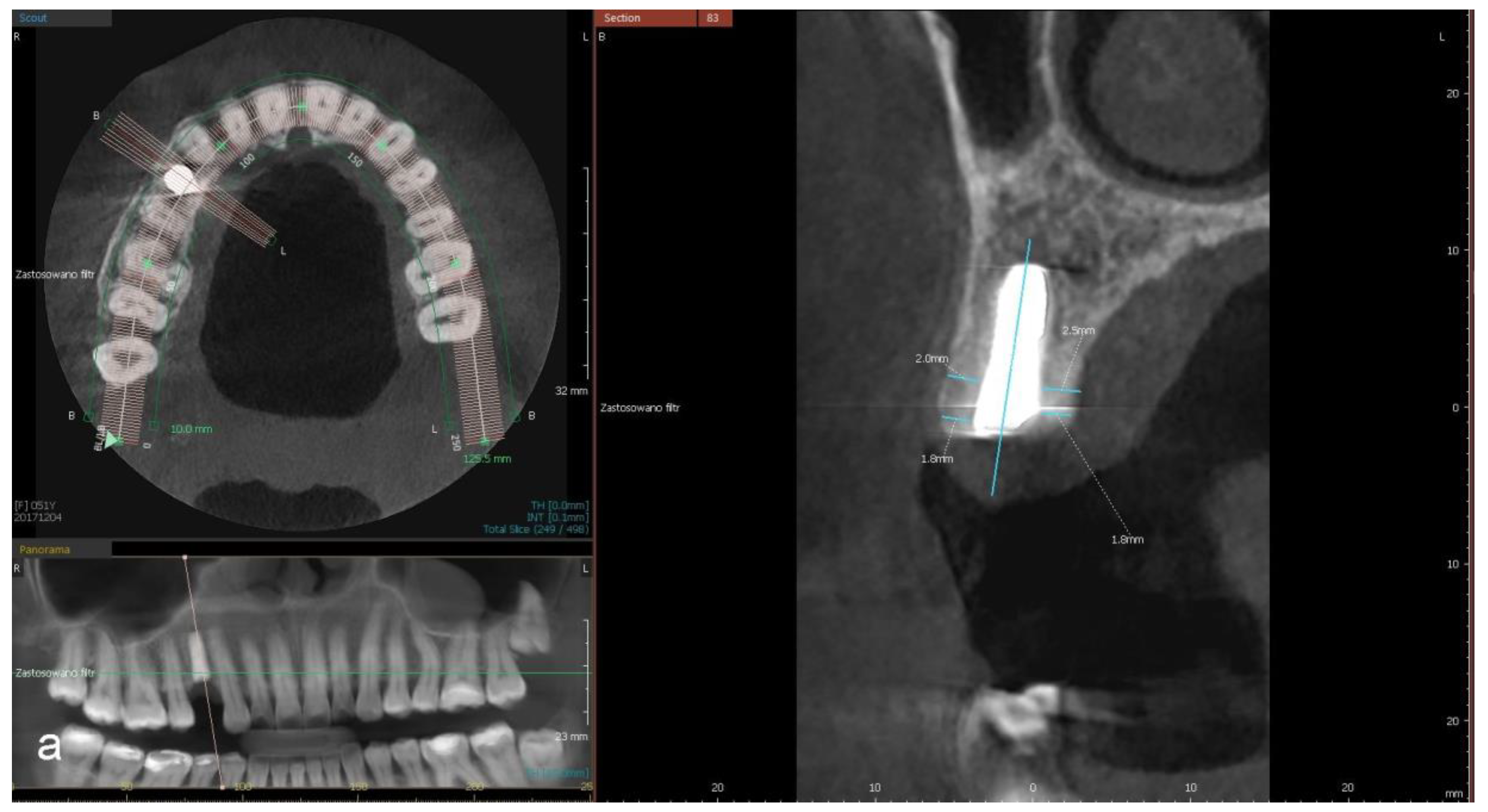
2.3. Radiographic Examination

The CBCT examination was performed three times—immediately after the implantation (IMP), after the temporary crown delivery (TCD) and one year after implantation (1YIMP); i.e., six months after the definitive crown delivery (DCD). Technical parameters for each scanning session were set automatically by CBCT machine’s internal software (Pax-i3D, Vatech, Hwaseong, Korea) according to each patient’s characteristics. The scans were taken within the field of 8 cm × 5 cm, then all images exported and computed by EzDent-I Software (Vatech, Gyeonggi-do, Korea), with a slice interval of 0.1 mm. Every image of each implant was adjusted in such a way that the implant would be visible in one sagittal plane. To avoid bias, CBCT results were re-examined 48 h later, and a mean value was derived from the two consecutive assessments.
The following radiographic parameters were determined for buccal and palatal/lingual aspects:
  • a distance between the implant’s platform and buccal and palatal/lingual bone margin (dehiscence)
  • thickness of buccal and palatal/lingual plates, 1 mm and 3 mm apically from the implant’s platform (perpendicular to the long axis of the implant).

2.4. Aesthetic Evaluation

Aesthetic outcome was evaluated via Pink Aesthetic Score (PES) [13] as modified by Belser et al. and Papilla Index (PI) [14] on three occasions—after the temporary crown delivery, then after the definitive crown delivery and six months after its delivery. All parameters were expressed as a mean value calculated from two separate measurements taken 48 h apart.

2.5. Statistical Analysis

The variables were described by the parameters of descriptive statistics, i.e., an arithmetic mean with standard deviation, 95% confidence interval and median as well as the minimum and maximum value.
The normality of the distribution for each parameter was verified with the Shapiro–Wilk test. The comparisons were performed by the Analysis of Variance (ANOVA) test for related variables and Fisher’s least significant difference (LSD) test, or the Friedman test along with Dunn’s multiple comparisons test with Bonferroni correction, depending on whether the assumptions of normality of distribution and sphericity were met. Additionally, the trend analysis was done with the Page test.
The intra-examiner reproducibility for radiographic and aesthetic parameters was determined through Kendall’s W test.
The results with p < 0.05 were considered statistically significant. All calculations were computed by PQStat version 1.8.0.392.

3. Results

The study involved 30 patients aged 25 to 81 years—19 women aged 28–81 and 11 men aged 25–64—needing a single tooth replacement. Each patient received one implant; the group—30 implants in total: 16 premolars in the maxilla, 11 maxillary first molars and 3 mandibular premolars. All implants had a conical profile (4.2 mm/3.6 mm in diameter), while their length was either 9 or 11 mm (21 and 9 implants, respectively). In 16 cases, the sloped part of the implant was located in the buccal direction, while in the remaining 14 cases, in palatal direction.
Healing was uneventful and the survival rate measured after a year equaled 100%. At the point of uncovering, bone completely surrounded the collars of all implants—without either dehiscence of buccal and palatal/lingual plates nor crestal bone loss at mesial/distal areas. In a few cases bone that grew over the cover screw crevice had to be removed. In addition, consecutive CBCT examinations did not show dehiscence in buccal or palatal/lingual bone plates. Due to numerous artifacts at the implant shoulder making it difficult to reliably evaluate the CBCT scans, measurements of buccal and palatal/lingual plate thickness at a measurement point 1 mm from the implant shoulder were taken only with 16 implants. As the measurement point moved away from the implant’s shoulder, the reliability of the CBCT image improved; therefore, at the level of 3 mm, the measurement was made for all 30 implants.
Right after implant insertion, the mean thickness of the buccal bone plate measured 1 mm from the shoulder was 1.85 ± 0.68 mm. After placement of the temporary crown, the value of this parameter decreased by 0.13 ± 0.644 mm, and 6 months after placement of the definitive crown, it decreased by another 0.12 ± 0.54 mm (p = 0.04). In addition, the Page test showed a significant trend—reduction of buccal plate thickness during the 1-year follow-up (p = 0.048). The mean value of palatal/lingual plate thickness was 1.87 ± 0.75 mm after implantation and remained comparable during 1-year observation (p = 0.22).
At the time of implantation, a mean thickness of the buccal plate at the 3 mm measurement point was 1.99 ± 1.05 mm. CBCT examination at TCD revealed it to be significantly thinner by 0.23 ± 0.42 mm, whereas six months after DCD by 0.08 ± 0.27, which reduced the difference by 0.15 ± 0.39 (p = 0.39) as compared with the baseline value. The Page trend test did not show a significant tendency in changes of buccal plate thickness over time (p = 0.131). A mean palatal/lingual plate thickness was 2.55 ± 1.01mm and remained such throughout the annual period (p = 0.39). See Table 1 and Table 2 and Figure 1a–c for CBCT details.
The Pink Aesthetic Score ranged between 3 implant insertions (Median = 5) at TCD. The Page trend analysis showed a significant tendency for PES to improve (p < 0.0001), including related indices, over the annual observation (Table 3). At DCD, an increase in PES was noted in 14 patients as compared with its values at TCD. However, six months after DCD, an increase in PES was observed in 16 patients and a decrease in 2, as compared with the TCD score. The number of patients displaying an acceptable PES results (>6) rose from 5 (16.7%) at TCD, to 13 (43.3%) at DCD, and to 19 (63.3%) six months later.
Equally beneficial changes concerned PI. Both PIm and PId ranged within 0–2 (Median = 1) at TCD. The trend analysis proved a significant tendency for PIm and PId to improve (p < 0.0001) over the annual observation (Table 4). PIm improved at DCD in 14 patients, while regressed only in 1. The improved PId values concerned 10 patients while the regressed ones 1 patient. Overall, 6 months after DCD, an improvement in both parameters was evident in 15 patients against their TCD values. Figure 2a–d present the clinical status (PES, PI) of one patient before and after treatment.
The intra-examiner reproducibility for radiographic and aesthetic parameters was high. See Table 5 for details.

4. Discussion

The principal aim of the study was to evaluate the changes in buccal and palatal/lingual cortical bone plates to meet about placing a prosthetic crown on a slope-configured implant. A CBCT examination, carried out directly after the temporary crown delivery, showed that the thickness of the buccal plate decreased by 0.13 ± 0.44 mm and 0.23 ± 0.42 mm in both measurement points in relation to the figure indicated immediately after implantation—1.85 ± 0.68 mm and 1.99 ± 1.05 mm. The study’s conclusive examination, a year after implant placement, showed that the actual decrease was greater by 0.32 ± 0.53 mm at the measurement point located 1 mm from the shoulder (p = 0.04). However, at the measurement point located 3 mm from the shoulder, reduction in buccal bone thickness was slighter, by 0.15 ± 0.39 mm, a fact which made overall changes of this parameter insignificant (p = 0.39). The simultaneously examined thickness of the palatal/lingual plate in both measurement points remained comparable over 12 months. Analysis of the results showed that only buccal plate thickness measured 1 mm from the implant shoulder decreased during the one-year follow-up. In contrast, buccal plate thickness measured more apically and palatal/lingual plate thickness, regardless of the measurement point location, did not change significantly.
Available literature does not embrace studies on the thickness of bone adjacent to the buccal and palatal/lingual surfaces of implants with slope-configured shoulder. The articles examining this parameter with regards to regular implant shoulder are also scarce [15,16]. Moreover, most researchers focused on the distance between implant’s platform and marginal bone level in contact areas [10,11,12,17]. They observed the distance increase insignificantly between the implantation and uncovering procedures and decrease from the point of uncovering till a year after implantation. Such a pattern was similar for implants with both regular and sloped shoulder, although for the latter type an increasing distance was less pronounced [11]. According to Noelken et al. [12], in case of sloped shoulders, a loss of bone around contact surfaces was 0.38 ± 0.82 mm between the implantation and uncovering and 0.69 ± 0.91 mm between the implantation and final prosthetic procedure. However, in an examination performed one year after implantation, the bone loss had decreased to 0.54 ± 1.29 mm.
A favorable time-related progress in radiographic parameters observed in our own and other authors’ studies can be explained by changes within the bone structure caused by post-surgical remodeling; a process taking at least 6 months, during which bone mineralization reaches its baseline values. Therefore, any CBCT evaluation carried out at longer intervals appears to be more reliable. However, the absence of grey-level calibration further lessens chances for well-founded comparisons of scans over time, making a CBCT evaluation, even by an experienced clinician, subjective to a degree [18,19].
One should also be aware of the difficulty in examining the bone surrounding a metal implant and the resulting potential errors in CBCT measurements (artifacts) [20,21,22]. Therefore, the precision of radiographic assessments can also be affected by the location of measurement points. This study chose to examine thickness of the buccal and palatal/lingual bone plates at a 1- and 3-millimetre distances from the implant’s platform.
However, it was not until 3 mm from the implant platform that the bone was clearly visible on all scans and the measurement of its thickness appeared to be more reliable, although in three patients the buccal plate thickness was less than 1 mm (0.2 mm, 0.85 mm, and 0.9 mm). The more coronally-wise taken measurements implied a presence of bone loss on contact surfaces and dehiscence within the buccal and palatal/lingual plates, which was clinically unnoticeable during the uncovering. Previous studies reported such overemphasized detection of dehiscence-type bone loss by CBCT in the case of a thin bone plate adjacent to the implant [23,24]. Therefore, there would be a need to exclude some radiographic measurements from the analysis, which, on the one hand, would improve the reliability of the study, but, on the other hand, would limit the size of the study groups [12]. This was the situation in the presented study. Due to the inability to properly measure bone thickness at a distance of 1 mm from the implant neck, only 16 of 30 scans were used for analysis. This fact can be seen as a limitation of the study.
The secondary aim of the study was to evaluate the aesthetics of the tissues surrounding single implant-based reconstructions via PES and PI; the two indicators considered to be the most evidence-based ways of aesthetic evaluation in implant dentistry [25,26]. For a majority of patients—63.3%—PES results fell within the aesthetically pleasing range. Such a meaningful proportion corresponds with those met in literature—42%–65%, and relevant to implants with a regular shoulder, i.e., placed in the alveolar bone with better characteristics [13,27,28,29,30]. What is more, even bone augmentation does not bring about better results—60% and 66% of patients benefited from acceptable PES scores after augmentative procedures and insertion of implants with a regular shoulder [27,31]. On account of comparable aesthetic outcomes of both approaches, trauma and costs involved, an implant with a slope-configured shoulder seems to be a reasonable solution when bone loss is the case.
The study observed that the soft tissue aesthetics progressively improved since functional loading (TCD). At that point, a satisfactory PES indication embraced 16.7% of patients, and the proportion rose significantly to 63.3% until six months after DCD.
With none of the papillae fully filling the interdental space, baseline papillary height was comparatively low—a minimal PI value equaled 0. Subsequent examinations saw a rise in papillary, notably mesial, height. No papillae matched PIM = 0; 12 mesial papillae (40%) and 5 distal ones (16.7%) corresponded to PIM = 3. A median of 2 was comparable with other authors’ results [27,28]. As to papillae filling up the interdental space, proportions in different studies are remarkably discrepant: 18%–75% [32,33,34,35].
The beneficial changes of soft tissue contour that followed prosthetic loading are likely to stem from the process of periodontal tissue maturation, particularly evident within the first year of function [32,33,35,36,37,38]. It was its dynamics that in numerous cases lessened the potential of some factors to affect outcome, i.e., vertical and horizontal loss of alveolar bone and periodontal status of the tooth adjacent to implant [39,40,41,42]. Both factors could have influenced the treatment’s results; however, implants with slope-configured shoulder are to be applied essentially in compromised bone conditions, which often co-exist with attachment loss in the neighboring teeth and such was the case for this study—most teeth adjacent to the implant suffered attachment loss at contact areas and in many sites soft tissues were impaired by past pathologies or traumatic extractions. Some researchers maintain that the height of the papillae may be affected by the implant-tooth distance, whose optimal range is 2.5–4 mm [39,40,43,44]. Its role, however, is still disputable [45,46,47].
In summation, when used in anatomically compromised regions, implants with slope-configured shoulder contribute to satisfactory results of treatment, without additional bone and/or soft tissue augmentation and can be an alternative to implants with regular shoulders. Few available studies lacking long-term reports, however, necessitate more research in that area.

5. Conclusions

After the use of implants with a sloped shoulder configuration, a slight decrease in buccal bone plate thickness can be expected. However, the reduction in the thickness of this bone plate does not have a negative impact on soft tissues, as evidenced by the improvement in indices assessing aesthetics.
However, the results of our study would need to be confirmed in multicenter studies involving much larger groups of patients.

Author Contributions

M.P. contributed to: the concept and design of the study; collection, analysis and interpretation of data; drafting and critical revision of the manuscript. J.K.P. contributed to: the concept and design of the study; collection, analysis and interpretation of data; drafting and critical revision of the manuscript. All authors have read and agreed to the published version of the manuscript.

Funding

Dentsply Sirona Implants (No D-2013-010) and Medical University of Białystok (N/ST/ZB/17/004/1164) provided support to the study.

Institutional Review Board Statement

The study was compliant with Helsinki Declaration of 1975 as revised in 2000. Local ethics committee approved this study under R-I-002/6/2016.

Informed Consent Statement

Each patient gave their informed consent prior to the study.

Data Availability Statement

Not applicable.

Conflicts of Interest

The authors declare no conflict of interest in connection with this article.

Abbreviations

CBCTcone-beam computed tomography
IMPimmediately after the implantation
PESPink Aesthetic Score
PIPapilla Index
PIdPapilla Index for distal papilla
PImPapilla Index for mesial papilla
TCDafter the temporary crown delivery
1YIMPone year after implantation
DCDafter the definitive crown delivery

References

  1. D’haese, J.; Ackhurst, J.; Wismeijer, D.; De Bruyn, H.; Tahmaseb, A. Current state of the art of computer-guided implant surgery. Periodontology 2000 2017, 73, 121–133. [Google Scholar] [CrossRef]
  2. Jung, R.E.; Heitz-Mayfield, L.; Schwarz, F. Evidence-based knowledge on the aesthetics and maintenance of peri-implant soft tissues: Osteology Foundation Consensus Report Part 3-Aesthetics of peri-implant soft tissues. Clin. Oral. Implant Res. 2018, 29, 14–17. [Google Scholar] [CrossRef]
  3. Schropp, L.; Wenzel, A.; Kostopoulos, L.; Karring, T. Bone healing and soft tissue contour changes following single tooth extraction: A clinical and radiographic 12 month prospective study. Int. J. Periodontics Rest. Dent. 2003, 23, 313–323. [Google Scholar]
  4. Araújo, M.G.; Silva, C.O.; Misawa, M.; Sukekava, F. Alveolar socket healing: What can we learn? Periodontology 2000 2015, 68, 122–134. [Google Scholar] [CrossRef] [PubMed]
  5. Troeltzsch, M.; Troeltzsch, M.; Kauffmann, P.; Gruber, R.; Brockmeyer, P.; Moser, N.; Rau, A.; Schliephake, H. Clinical efficacy of grafting materials in alveolar ridge augmentation: A systematic review. J. Craniomaxillofac. Surg. 2016, 44, 1618–1629. [Google Scholar] [CrossRef]
  6. Elnayef, B.; Monje, A.; Gargallo-Albiol, J.; Galindo-Moreno, P.; Wang, H.-L.; Hernández-Alfaro, F. Vertical ridge augmentation in the atrophic mandible: A systematic review and meta-analysis. Int. J. Oral. Maxillofac. Implant 2017, 32, 291–312. [Google Scholar] [CrossRef]
  7. Cucchi, A.; Vignudelli, E.; Napolitano, A.; Marchetti, C.; Corinaldesi, G. Evaluation of complication rates and vertical bone gain after guided bone regeneration with non-resorbable membranes versus titanium meshes and resorbable membranes. A randomized clinical trial. Clin. Implant Dent. Relat.Res. 2017, 19, 821–832. [Google Scholar] [CrossRef]
  8. Felice, P.; Barausse, C.; Pistilli, R.; Ippolito, D.R.; Esposito, M. Five-year results from a randomised controlled trial comparing prostheses supported by 5-mm long implants or by longer implants in augmented bone in posterior atrophic edentulous jaws. Int. J. Oral. Implant 2019, 12, 25–37. [Google Scholar]
  9. Abrahamsson, I.; Welander, M.; Linder, E.; Berglundh, T. Healing at implants placed in an alveolar ridge with a sloped configuration: An experimental study in dogs. Clin. Implant Dent. Relat. Res. 2014, 16, 62–69. [Google Scholar] [CrossRef]
  10. Schiegnitz, E.; Noelken, R.; Moergel, M.; Berres, M.; Wagner, W. Survival and tissue maintenance of an implant with a sloped configurated shoulder in the posterior mandible—A prospective multicenter study. Clin. Oral. Implants Res. 2017, 28, 721–726. [Google Scholar] [CrossRef]
  11. Tu, C.-C.; Wang, S.-H.; Hu, C.-Y.; Chang, P.-C. Comparison of regular- and slope-configurated dental implants placed in the edentulous ridge with height discrepancy: A pilot randomized controlled trial. ScienceDirect 2020, 120, 1386–1393. [Google Scholar] [CrossRef]
  12. Noelken, R.; Donati, M.; Fiorellini, J.; Gellrich, N.-C.; Parker, W.; Wada, K.; Berglundh, T. Soft and hard tissue alterations around implant placed in an alveolar ridge with a sloped configuration. Clin. Oral. Implants Res. 2014, 25, 3–9. [Google Scholar] [CrossRef]
  13. Fürhauser, R.; Florescu, D.; Benesch, T.; Haas, R.; Mailath, G.; Watzek, G. Evaluation of soft tissue around single-tooth implant crowns: The pink esthetic score in the anterior mandible. Clin. Oral. Implants Res. 2005, 16, 639–644. [Google Scholar] [CrossRef] [PubMed]
  14. Jemt, T. Regeneration of gingival papillae after single-implant treatment. Int. J. Periodontics Res. Dent. 1997, 17, 326–333. [Google Scholar]
  15. Veltri, M.; Ekestubbe, A.; Abrahamsson, I.; Wennström, J.L. Three-Dimensional buccal bone anatomy and aesthetic outcome of single dental implants replacing maxillary incisors. Clin. Oral. Implants Res. 2016, 27, 956–963. [Google Scholar] [CrossRef]
  16. Meijer, H.J.A.; Slagter, K.W.; Vissink, A.; Raghoebar, G.M. Buccal bone thickness at dental implants in the maxillary anterior region with large bony defects at time of immediate implant placement: A 1-year cohort study. Clin. Implant Dent. Relat. Res. 2019, 21, 73–79. [Google Scholar] [CrossRef]
  17. Temmerman, A.; Keestra, J.A.J.; Coucke, W.; Teughels, W.; Quirynen, M. The outcome of oral implants placed in bone with limited bucco-oral dimensions: A 3-year follow-up study. J. Clin. Periodontol. 2015, 42, 311–318. [Google Scholar] [CrossRef]
  18. Bornstein, M.M.; Horner, K.; Jacobs, R. Use of cone beam computed tomography in implant dentistry: Current concepts, indications and limitations for clinical practice and research. Periodontol. 2000 2017, 73, 51–72. [Google Scholar] [CrossRef]
  19. Pauwels, R.; Jacobs, R.; Singer, S.R.; Mupparapu, M. CBCT-based bone quality assessment: Are Hounsfield units applicable? Dentomaxillofacial 2015, 44, 20140238. [Google Scholar] [CrossRef]
  20. Hatcher, D.C.; Aboudara, C.L. Diagnosis goes digital. Am. J. Orthod. Dentofac. Orthop. 2004, 125, 512–515. [Google Scholar] [CrossRef]
  21. Ballrick, J.W.; Martin Palomo, J.; Ruch, E.; Amberman, B.D.; Hans, M.G. Image distortion and spatial resolution of a commercially available cone-beam computed tomography machine. Am. J. Orthod Dentofac. Orthop. 2008, 134, 573–582. [Google Scholar] [CrossRef]
  22. Stratemann, S.A.; Huang, J.C.; Maki, K.; Miller, A.J.; Hatcher, D.C. Comparison of cone beam computed tomography imaging with physical measures. Dentomaxillofac. Radiol. 2008, 37, 80–93. [Google Scholar] [CrossRef]
  23. Razavi, T.; Palmer, R.M.; Davies, J.; Wilson, R.; Palme, P.J. Accuracy of measuring the cortical bone thickness adjacent to dental implants using cone beam computed tomography. Clin. Oral. Implants Res. 2010, 21, 718–725. [Google Scholar] [CrossRef]
  24. Naitoh, M.; Hayashi, H.; Tsukamoto, N.; Ariji, N. Labial bone assessment surrounding dental implant using cone-beam computed tomography: An in vitro study. Clin. Oral. Implants Res. 2012, 23, 970–974. [Google Scholar] [CrossRef]
  25. Tettamanti, S.; Millen, C.; Gavric, J.; Buser, D.; Belser, U.C.; Brägger, U.; Wittneben, J.-G. Esthetic evaluation of implant crowns and peri-implant soft tissue in the anterior maxilla: Comparison and reproducibility of three different indices. Clin. Implant Dent. Relat Res. 2016, 18, 517–526. [Google Scholar] [CrossRef]
  26. Hof, M.; Umar, N.; Budas, N.; Seemann, R.; Pommer, B.; Zechner, W. Evaluation of implant esthetics using eight objective indices-Comparative analysis of reliability and validity. Clin. Oral. Implants Res. 2018, 29, 697–706. [Google Scholar] [CrossRef]
  27. Hof, M.; Pommer, B.; Strbac, G.D.; Sütö, D.; Watzek, G.; Zechner, W. Esthetic evaluation of single-tooth implants in the anterior maxilla following autologous bone augmentation. Clin. Oral. Implants Res. 2011, 24, 88–93. [Google Scholar] [CrossRef]
  28. Hof, M.; Tepper, G.; Koller, B.; Krainhöfner, M.; Watzek, G.; Pommer, B. Esthetic evaluation of single-tooth implants in the anterior mandible. Clin. Oral. Implants 2014, 25, 1022–1026. [Google Scholar] [CrossRef]
  29. Cosyn, J.; Eghbali, A.; De Bruyn, H.; Dierens, M.; De Rouck, T. Single implant treatment in healing versus healed sites of the anterior maxilla: An aesthetic evaluation. Clin. Implant Dent. Relat Res. 2012, 14, 517–526. [Google Scholar] [CrossRef]
  30. Lai, H.-C.; Zhang, Z.-Y.; Wang, F.; Zhuang, L.-F.; Liu, X.; Pu, Y.P. Evaluation of soft-tissue alteration around implant- supported single-tooth restoration in anterior maxilla: The pink esthetic score. Clin. Oral. Implants Res. 2008, 19, 560–564. [Google Scholar] [CrossRef]
  31. Meijndert, L.; Meijer, H.J.A.; Stellingsma, K.; Stegenga, B.; Raghoebar, G.M. Evaluation of aesthetics of implant-supported single-tooth replacements using different bone augmentation procedures: A prospective randomized clinical study. Clin. Oral. Implants Res. 2007, 18, 715–719. [Google Scholar] [CrossRef]
  32. Agabiti, I.; Alí Apaza Alccayhuaman, K.; Nakajima, Y.; Botticelli, D. An enigmatic soft tissue creeping phenomenon: The spontaneous peri-implant mucosa margin and papilla growth. A retrospective clinical study. Clin. Exp. Dent. Res. 2021, 7, 474–483. [Google Scholar] [CrossRef]
  33. Cardaropoli, G.; Lekholm, U.; Wennström, J.L. Tissue alterations at implant-supported single-tooth replacements: A 1-year prospective clinical study. Clin. Implant Dent. Relat Res. 2006, 17, 165–171. [Google Scholar] [CrossRef]
  34. Cosyn, J.; Raes, M.; Packet, M.; Cleymaet, R.; De Bruyn, H. Disparity in embrasure fill and papilla height between tooth- and implant-borne fixed restorations in the anterior maxilla: A cross-sectional study. J. Clin. Periodontol. 2013, 40, 728–733. [Google Scholar] [CrossRef]
  35. Priest, G. Predictability of soft tissue form around single-tooth implant restorations. Int. J. Periodontics Res. Dent. 2003, 23, 19–27. [Google Scholar]
  36. Barwacz, C.A.; Stanford, C.M.; Diehl, U.A.; Cooper, L.F.; Feine, J.; McGuire, M.; Scheyer, T. Pink Esthetic Score outcomes around three implant-abutment configurations: 3-year results. Int. J. Oral. Maxillofac. Implants 2018, 33, 1126–1135. [Google Scholar] [CrossRef]
  37. Schropp, L.; Isidor, F.; Kostopoulos, L.; Wenzel, A. Interproximal papilla levels following early versus delayed placement of single-tooth implants: A controlled clinical trial. Int. Oral. Maxillofac. Impl. 2005, 20, 753–761. [Google Scholar] [CrossRef]
  38. Schropp, L.; Isidor, F. Papilla dimension and soft tissue level after early vs. delayed placement of single-tooth implants: 10-year results from a randomized controlled clinical trial. Clin. Oral. Implants Res. 2015, 26, 278–286. [Google Scholar] [CrossRef]
  39. Grunder, U. Stability of the mucosal topography around single-tooth implants and adjacent teeth: 1-year results. Int. J. Periodontics Res. Dent. 2000, 20, 11–17. [Google Scholar]
  40. Choquet, V.; Hermans, M.; Adriaenssens, P.; Daelemans, P.; Tarnow, D.P.; Malevez, C. Clinical and radiographic evaluation of the papilla level adjacent to single-tooth dental implants. A retrospective study in the maxillary anterior region. J. Periodontol. 2001, 72, 1364–1371. [Google Scholar] [CrossRef]
  41. Benic, G.I.; Wolleb, K.; Sancho-Puchades, M.; Hammerle, C.H. Systematic review of parameters and methods for the professional assessment of aesthetic in dental implant research. J. Clin. Periodontol. 2012, 39, 160–192. [Google Scholar] [CrossRef] [PubMed]
  42. Roccuzzo, M.; Roccuzzo, A.; Ramanuskaite, A. Papilla height in relation to the distance between bone crest and interproximal contact point at single-tooth implants: A systematic review. Clin. Oral. Implants Res. 2017, 29, 50–61. [Google Scholar] [CrossRef] [PubMed]
  43. Lops, D.; Chiapasco, M.; Rossi, A.; Bressan, E.; Romeo, E. Incidence of inter-proximal papilla between a tooth and an adjacent immediate implant placed into a fresh extraction socket: 1-year prospective study. Clin. Oral. Implants Res. 2008, 19, 1135–1140. [Google Scholar] [CrossRef]
  44. Romeo, E.; Lops, D.; Rossi, A.; Storelli, S.; Rozza, R.; Chiapasco, M. Surgical and prosthetic management of interproximal region with single-implant restorations: 1-year prospective study. J. Periodontol. 2018, 79, 1048–1055. [Google Scholar] [CrossRef] [PubMed]
  45. Cooper, L.; Felton, D.A.; Kugelberg, C.F.; Ellner, S.; Chaffee, N.; Molina, A.L.; Moriarty, J.D.; Paquette, D.; Palmqvist, U. A multicenter 12-month evaluation of single-tooth implants restored 3 weeks after 1-stage surgery. Int. J. Oral. Maxillofac. Implant 2001, 16, 182–192. [Google Scholar]
  46. Palmer, R.M.; Farkondeh, N.; Palmer, P.J.; Wilson, R.F. Astra Tech single-tooth implants: An audit of patient satisfaction and soft tissue form. J. Clin. Periodontol. 2007, 34, 633–638. [Google Scholar] [CrossRef] [PubMed]
  47. Wang, T.; De Kok, I.J.; Zhong, S.; Vo, C.; Mendonça, G.; Nares, S.; Cooper, L. The role of implant-tooth distance on marginal bone levels and esthetics. Int. J. Oral. Maxillofac. Implants 2019, 34, 499–505. [Google Scholar] [CrossRef]
Figure 1. (a) CBCT scan taken immediately after implant insertion at position 14. At the measurement point located 1 mm from the implant shoulder: BP = 1.8 mm, PP = 1.8 mm. At the measurement point located 3 mm away from the implant shoulder: BP = 2 mm, PP = 2.6 mm. (b) Scan of the implant after the provisional crown delivery. At the measurement point located 1 mm from the implant shoulder: BP = 1.4 mm, PP = 2.1 mm. At the measurement point located 3 mm away from the implant shoulder: BP = 1.6 mm, PP = 2.7 mm. (c) Scan of the implant after placement of the definitive crown. At the measurement point located 1 mm from the implant shoulder: BP = 1.4 mm, PP = 2.1 mm. At the measurement point located 3 mm away from the implant shoulder: BP = 1.9 mm, PP = 2.8 mm.
Coatings 12 01295 g001aCoatings 12 01295 g001b
Figure 2. (a). Clinical status before treatment. Lack of tooth 14—tissue deformation, loss of attachment on adjacent tooth 13 and 15. (b). Soft tissue status immediately after the temporary crown delivery on the implant in position 14. PES = 6; PIM = 2; PID = 1. (c) Soft tissue status immediately after the definitive crown delivery. PES = 6.5; PIM = 2; PID = 1. (d) Soft tissue status six months after the definitive crown delivery (one year after implant insertion). PES = 9; PIM = 3; PID = 1.
Coatings 12 01295 g002aCoatings 12 01295 g002b
Table 1. Results of CBCT examination at relevant stages of study.
Parameter Mean ± SDMinimumMaximumMedian−95%Cl95%ClDifference
± SD
p (Post-Hock)
1 mm
(n = 16)
BP01.85 ± 0.680.93.21.771.532.17
PP01.87 ± 0.750.53.21.81.522.23
BPtc1.83 ± 0.660.653.051.651.482.18−0.13 ± 0.440.27
PPtc1.9 ± 0.760.53.652.11.52.29−0.1 ± 0.680.50
BP1y1.53 ± 0.770.13.551.41.161.89−0.32 ± 0.530.04
PP1y1.67 ± 0.690.73.351.721.341.99−0.21 ± 0.710.22
3 mm
(n = 30)
BP01.99 ± 1.050.24.651.721.592.38
PP02.55 ± 1.010.74.42.622.172.93
BPtc1.67 ± 1.040.24.71.41.272.07−0.23 ± 0.420.03
PPtc2.6 ± 1.070.74.82.652.193.010.06 ± 0.430.43
BP1y1.84 ± 1.20.34.91.621.392.29−0.15 ± 0.390.39
PP1y2.6 ± 0.950.854.252.652.242/950.05 ± 0.330.39
BP0—buccal plate thickness at implant installation; PP0—palatal plate thickness at implant installation; BPtc—buccal plate thickness at temporary crown delivery; PPtc—palatal plate thickness at temporary crown delivery; BP1y—buccal plate thickness one year after implant insertion; PP1y—palatal plate thickness one year after implant insertion.
Table 2. Changes in thickness of buccal and palatal/lingual bony plates at relevant CBCT examinations.
Parameter Thickness IncreaseThickness ReductionNo Change
1 mm (n = 16)Buccal plateIMP—tc6100
tc—1y4102
Palatal/lingual plateIMP—tc790
tc—1y583
3 mm (n = 30)Buccal plateIMP—tc6195
tc—1y17121
Palatal/lingual
plate
IMP—tc16122
tc—1y15123
IMP—implantation; tc—temporary crown delivery; 1y—one year after implantation.
Table 3. Pink aesthetic score at relevant stages of study.
PESMedianMinimumMaximump (tc-dc)p (tc-dc 1y)
MPtc1.00.01.0
DPtc1.00.01.0
Ctc1.00.52.0
LMtc1.00.52.0
CCtc1.01.02.0
PEStc5.03.08.0
MPdc1.01.02.00.006
DPdc1.00.02.00.031
Cdc1.01.02.00.03
LMdc1.50.52.00.51
CCdc1.01.02.00.0305
PESdc6.04.08.00.0007
MPdc 1y1.012 0.009
DPdc 1y1.002 0.13
Cdc 1y1.512 0.135
LMdc 1y2.012 0.27
CCdc 1y1.2512 0.13
PESdc 1y7.75510 <0.0001
PES—pink aesthetic score; MP—mesial papilla; DP—distal papilla; C—curvature of the facial mucosa; LM—level of the facial mucosa; CC—root convexity/soft tissue color and texture. tc—temporary crown delivery; dc—definitive crown delivery; dc 1y—one year after implantation.
Table 4. Papilla index at relevant stages of study.
PIMedianMinimumMaximump (tc-dc)p (tc-dc 1y)
PIMtc1.0002
PIDtc1.0002
PIMdc1.25130.0355
PIDdc1.00030.1836
PIMdc 1y213 <0.0001
PIDdc 1y203 <0.0001
M—mesial papilla; D—distal papilla. tc—temporary crown delivery; dc—definitive crow delivery; dc 1y—one.
Table 5. Kendall W for radiographic parameters, PES and PI.
ParameterKendall’s WP
BP0.9940.0012
PP0.9890.0013
PES0.9360.0030
PIM0.9470.0025
PID0.9380.0029
BP—buccal plate thickness; PP—palatal plate thickness; PES—Pink Aesthetic Score; PIM—Papilla Index for mesial papilla; PID—Papilla Index for distal papilla.
Publisher’s Note: MDPI stays neutral with regard to jurisdictional claims in published maps and institutional affiliations.

