
Application of Metal Nanoparticles for Production of Self-Sterilizing Coatings


1
Department of Biological Bases of Food and Feed Technologies, Faculty of Production Engineering, University of Life Sciences in Lublin, 28 Głęboka Str., 20-612 Lublin, Poland
2
Department of Agricultural Forestry and Transport Machines, Faculty of Production Engineering, University of Life Sciences in Lublin, 28 Głęboka Str., 20-612 Lublin, Poland
*
Author to whom correspondence should be addressed.
Coatings 2022, 12(4), 480; https://doi.org/10.3390/coatings12040480
Submission received: 5 March 2022
/
Revised: 28 March 2022
/
Accepted: 31 March 2022
/
Published: 1 April 2022
(This article belongs to the Special Issue Advances in Biodegradable Packaging, Bioedible Films, and Coatings for Food)
Abstract
:Metal nanoparticles (NPs) are increasingly being used in many areas, e.g., industry, pharmacy, and biomedical engineering. NPs can be obtained through chemical and biological synthesis or using physical methods. AgNPs, AuNPs, CuNPs, FeNPs, MgNPs, SnO2NPs, TiO2NPs, and ZnONPs are the most commonly synthesized metal nanoparticles. Many of them have anti-microbial properties and documented activity supported by many tests against some species of pathogenic bacteria, viruses, and fungi. AgNPs, which are used for the production of commercial self-sterilizing packages, are one of the best-explored nanoparticles. Moreover, the EFSA has approved the use of small doses of silver nanoparticles (0.05 mg Ag·kg−1) to food products. Recent studies have shown that metal NPs can be used for the production of coatings to prevent the spread of the SARS-CoV-2 virus, which has caused the global pandemic. Some nanoparticles (e.g., ZnONPs and MgONPs) have the Generally Recognized As Safe (GRAS) status, i.e., they are considered safe for consumption and can be used for the production of edible coatings, protecting food against spoilage. Promising results have been obtained in research on the use of more than one type of nanometals, which prevents the development of pathogen resistance through various mechanisms of inactivation thereof.
1. Introduction
Microbiological stability is particularly important in the case of food with a high water content (e.g., fruit and vegetables). Various physical and chemical methods are used to ensure the microbiological purity of food. There is a trend towards minimization of the use of chemical preservatives and replacement thereof with natural substances. However, this does not always yield a fully satisfactory solution. The great interest in nanoparticles can be attributed to their physicochemical properties and wide application in many industries [1,2,3,4,5]. In agricultural and production practice, cheap and easy protection measures against microorganisms are still a big challenge. New protection strategies against harmful microorganisms are continuously being developed. A promising approach may be the use of nanoparticles with antimicrobial and antiviral properties for the production of face masks, textiles, and other coatings [6,7,8]. The use of metal nanoparticles for surface functionalization has been particularly successful in obtaining unique textiles with desirable properties, such as self-cleaning, antimicrobial, antifungal, flame retardant, ultraviolet blocking, and superhydrophobicity [9]. The development of novel coatings that are safe, intelligent, and active is an important factor contributing to a reduction in food spoilage. The task of food coatings is to maintain the quality and properties of food products for as long as possible. Various additives are used to obtain intelligent and active food coatings. Nanoparticles occupy a special place among the huge variety of additives [10,11]. The term “nanoparticles” refers to solid particles with a size between 1 and 100 nm [12,13]. Nanoparticles usually have different properties than their macroscopic counterparts. In biological applications, the reduction in the size of metal particles to the nanometer scale is associated with an increase in their cytotoxicity. This is associated with their larger active surface and thus a higher reactivity than that of conventional materials and interactions with other compounds present in the environment. They also exhibit a higher bioavailability, which makes them easier to adsorb in specific organs, tissues, and cells [14]. Another special active property of nanoparticles is their ability to absorb ultraviolet light, which is considered important for the prevention of photochemical reactions leading to the spoilage of food products. Nanoparticles also exert an antioxidant effect, which is a highly desirable property [10]. Moreover, their most noticeable trait is the lower light scattering degree accompanying a reduction in the particle size. Sufficiently small particles can form transparent coatings. Nanoparticles do not directly induce transparency, but their light scattering effect declines with the decreasing particle size. At the same time, light scattering depends on the difference in the refractive index between the particle and the surrounding medium. A close match of refractive indices promotes the formation of transparent mixtures [12]. Particles must be very well dispersed to achieve transparency in the nanoscale system, as agglomerated nanoparticles have the same optical properties as particles with the size of the agglomerate [12].
Depending on their shape, nanoparticles are classified as zero-dimensional (0D), one-dimensional (1D), two-dimensional (2D), or three-dimensional (3D). This classification is associated with the movement of electrons in different planes. Electrons in 0D are trapped in a dimensionless space, whereas the electrons of 1D nanomaterials can move along the x-axis. 2D and 3D nanomaterials have electrons moving along the x-y axis and the x-y-z axis, respectively [15]. One-dimensional nanomaterials form long nanostructures with thick membranes, i.e., nanotubes, nanofibers, nanowires, nanorods, and nanofilaments. Their length is greater than their width [16]. In turn, 2D nanomaterials have sheet-like structures. These nanomaterials have the largest specific surface area of all known nanoparticles [17]. Due to their high surface-to-volume ratio and the anisotropic physical and chemical properties, 2D nanomaterials are widely used against microorganisms and are attractive for food packaging applications. Large-area 2D nanomaterials directly interact with bacterial membranes, thereby enhancing the antibacterial effect [18,19].
2. Aim of the Application of Nanoparticles in Packaging
Nanotechnological inventions are used in computers, nanodiodes, nanotransistors, etc. This technology provides innovative solutions for the production of batteries, fuel cells, and solar cells, which are lighter, more durable, and stronger than those produced with other methods [20]. Nanomaterials exhibit increased catalytic activity, thermal conductivity, and chemical stability [21]. They are also used in various fields of science; in particular, biology and biomedicine. The size of nanorods, nanofibers, and other nanomaterials is comparable to the size of biological particles [22]. Metal-based nanoparticles effectively inactivate microorganisms. Hence, some of them, e.g., silver, titanium dioxide, and zinc oxide nanoparticles, are used as disinfectants and additives in food, cosmetic, and pharmaceutical packaging [14]. Another example is the use of nanoparticles for surface protection and the conservation of antiquities made of marble, stone, glass, bronze, and even objects made of bread [23].
One of the very promising approaches to fighting bacteria is the use of nanomaterial-based antimicrobials. Antimicrobial coatings can be divided into two main groups, germicidal (active) and bacteriostatic (passive). Bactericidal active coatings are able to disrupt the structural integrity of the bacterial cell or membrane. In contrast, bacteriostatic coatings can prevent or reduce the attachment of microorganisms to their surface [24]. Nanoparticles have been well documented to kill bacteria via several different mechanisms [25]. They have the ability to damage the cell membrane physically. They can also generate reactive oxygen species (ROS) and free radicals. This process intensifies oxidative stress, which enhances the fragmentation of genomic DNA and damages the structural integrity of cells [26]. Furthermore, nanoparticles penetrate cell membranes much more easily than antibiotics [27,28]. Recent reports also indicate that nanoparticle-based antimicrobials act as efficient inhibitors of efflux pumps (proteins present in cell membranes) [29,30]. This proves that nanometals may have multiple applications as antimicrobial agents. Moreover, they are more effective than other antimicrobial agents and are less prone to induce bacterial resistance than antibiotics available on the market.
3. Methods for Production of Metal Nanoparticles
Metal nanoparticles can be produced using chemical, physical, and biological methods (Figure 1).
There are two basic strategies used in the production of nanoscale structures: bottom-up and top-down. The former approach is based on the atom-by-atom construction of a nanoparticle. Depending on the required properties of the end product, atoms, molecules, or colloidal particles can be used as substrates. The control of the synthesis conditions and the initial size of the building material can determine the traits of the final structure. A reverse mechanism is employed in the top-down process based on dispersion methods, which are based on the fragmentation of materials, yielding a final size of the particles in the range of 1–100 nm [31].
3.1. Chemical and Physicochemical Methods
Chemical methods are currently regarded as the most popular approaches, although they are not always safe and environmentally friendly. The synthesis of metal nanoparticles using chemical methods is based primarily on chemical reduction. The reaction consists of three steps: reduction of metal salts and formation of free atoms, nucleation, i.e., formation of stable 1–2 nm nuclei, and addition of stabilizing agents to complete the reaction. Currently, this is the cheapest method. Borohydride, citrate, ascorbate, and compounds with hydroxyl or carboxyl groups, i.e., alcohols, aldehydes, carbohydrates, and their derivatives, are the most frequently used reducing agents [32,33,34,35,36]. The process of chemical reduction requires the use of a stabilizing substance whose main task is to protect newly formed nanoparticles against aggregation into larger aggregates [31,37,38]. Such polymers as poly(vinylpyrrolidone) (PVP) [39], poly(methylacrylic acid) (PMAA) [40], poly(methyl methacrylate) (PMMA) [41], and poly(ethylene glycol) (PEG) are used most frequently as stabilizing agents [42]. The greatest advantage of this method is the possible impact on the properties of the final product through regulation of the process conditions. For example, the shape, size, aggregation state, and stability of nanoparticles can be controlled by changing the concentration of the stabilizer or salt, the molar ratio of the reducing and stabilizing agent, temperature, and medium pH [43].
3.2. Physical Methods
Physical methods are based on the top-down strategy. Particularly popular are the methods of physical vapor deposition (PVD), in which, a solid material is converted into gas in physical processes and then deposited on the substrate [44]; chemical vapor deposition (CVD), in which, products of chemical reactions are deposited on the substrate [45]; the use of the electromagnetic microwave field, in which, a substantially faster heating rate of the reaction mixture is achieved than in the convection heating process [46]; and the method based on γ or UV radiation used as a catalyst for a chemical reaction [47,48]. Another method for the production of various types of nanoparticles is the application of laser radiation. In this approach, nanoparticles are generated by the nucleation and growth of laser-vaporized metals in background gas [49] (Figure 1).
3.3. Green Synthesis
Green synthesis is gaining increasing support as a sustainable, reliable, and environmentally friendly approach to the synthesis of various nanoparticles. It does not require the use of solvents that pose a threat to humans and the environment or additional reducing agents. This technique of producing nanoparticles is simple and cost-effective [50,51,52,53]. It is based on the production of nanoparticles with the use of plant parts or microorganisms (Figure 1, Table 1) [54]. As in the chemical and physical methods, two approaches in the generation of nanoparticles from living organisms are used, i.e., the top-down and bottom-up strategies. In the top-down methods, the material is fragmented with the use of various techniques, whereas the bottom-up methods are based on the self-organization of atoms leading to the formation of new cell nuclei in chemical or biological reactions [55]. Nanoparticles produced via green synthesis are less toxic to living organisms [56].
The most common metal nanoparticles produced via green synthesis are oxide and dioxide, including polymer-coated iron [85], zinc [86], copper [87], gold [88], silver, and their oxides [27,89,90]. Many plant species are used in the production of metal nanoparticles (Table 1). The fruits of langsat (Lansium domesticum) and amla (Phyllanthus emblica) are used for the production of gold nanoparticles with antimicrobial activity. In turn, AgNPs are rapidly synthesized with the use of Calendula officinalis and Capsicum annuum seeds. Tea plant (Camellia sinensis) leaves serve as a catalytic agent in the synthesis of ZnONPs [3]. Younis et al. [91] used dried Rosa floribunda petals for the green synthesis of MgONPs. Asiya et al. [92] used lemon fruit (Citrus limon) to produce gold nanoparticles. Another promising trend in the production of nanoparticles is the use of biowaste, contributing to a reduction in its amounts. This approach has been reported by Jain et al. [93], who used the skin of jackfruit (Artocarpus heterophyllus) to isolate iron nanoparticles.
4. Antimicrobial Properties of Metal Nanoparticles
The direct toxic effects of metal nanoparticles on organisms depend primarily on the type and surface reactivity. Indirect effects are due to physical confinement, the release of toxic ions, or from the generation of reactive oxygen species (ROS). ROS are thought to lead to additional cellular responses classified as defense, proinflammatory effects (in plant and animal cells), and cytotoxicity [94,95]. Indirect toxicological effects of metal nanoparticles may include: oxidative stress and inflammation associated with ROS production, glutathione depletion and accumulation of oxidized glutathione in response to ROS production, protein denaturation, membrane damage, DNA damage, immune system reactivity and foreign body granuloma formation, and a reduction or loss of photosynthetic activity in algae and plants [95,96]. Much recent research has been directed toward developing self-responsive systems, e.g., triggered by specific physiological environments (local acidity, presence of substances secreted by bacteria) or external stimuli (photothermal, electrical, magnetic) [23,24,97,98].
4.1. Antibacterial Activity
The antibacterial activity of metal nanoparticles depends on their size, chemical composition, surface structure, solubility, shape, and tendency to aggregate, as well as environmental conditions, e.g., pH, salinity, and the presence of organic matter. The cell wall of Gram-positive bacteria is a complex mixture of glycopolymers and proteins. It consists of a peptidoglycan sacculus composed of long polysaccharide chains linked via peptide bridges. The wall surrounds the cytoplasmic membrane. It plays an important role in bacterial physiology, as it maintains the shape and integrity of the cell during growth and division. Additionally, it acts as an interface between the bacterium and its environment [99]. The bacterial cell wall is mostly composed of murein, which is multilayered in Gram-positive bacteria. In turn, Gram-negative bacteria have a single layer of murein additionally surrounded by an outer membrane. The potential mechanisms of the antibacterial effect of metal nanoparticles consist of their impact on the structure and function of cell membranes, disturbances in metabolic processes caused by the generation of reactive oxygen species, and a negative influence on the activity of enzymes and DNA synthesis [100]. As suggested by Wong et al. [101], nanoparticles should be smaller than 50 nm to become effective antimicrobial agents. Table 2 shows examples of the antimicrobial activity of metal nanoparticles.
The antiseptic capacity against eukaryotic cells is different. The feature that distinguishes eukaryotic cells from prokaryotic cells (bacteria, archaeons) is a very complex internal structure. Among other things, eukaryotic cells have a cytoskeleton. Nanoparticles, immobilized in a gel matrix, can exert antimicrobial activity by contacting the bacterial membrane, while they cannot be taken up and internalized by eukaryotic cells [102]. Gold and zinc nanoparticles in the form of colloidal solutions that allow penetration into cells may be effective against eukaryotic cells [103,104]. In the case of fungi, which belong to the group of eukaryotic organisms, the same interactions between nanometals and cells occur as in the case of bacteria [87].
Another important consideration is the long-term stability of nanoparticle surfaces and the ability to maintain antibacterial properties over time. A promising strategy is so-called smart antimicrobial surfaces, which can kill bacteria attached to their surface and then release dead bacteria and other contaminants on demand to reveal a clean surface under the right stimulus [105].
4.2. Antifungal Activity
Metal nanoparticles can be used as potential inhibitors of fungal microorganisms (Table 2). To achieve the antifungal effect, nanoparticles are most often produced with the use of physical and chemical methods and less often through biosynthesis. It has been found that the mechanisms of action of nanometals against fungi are similar to those involved in their antibacterial properties, e.g., the induction of oxidative stress through ROS generation, damage to the cell wall and membrane through direct contact with nanoparticles, and cellular uptake of nanoparticles and subsequent harmful interactions with the cell interior [172]. In some cases, nanoparticles induce the deformation of fungal hyphae. Fungicidal activity is exhibited by the nano-oxides of some metals as well [173].
4.3. Antiviral Activity
Due to their unique properties, metal and metal-based nanoparticles have a number of medical applications in the treatment of diseases or as antiviral coatings of biological materials. Nanoscale metals, e.g., copper, silver, and gold, exhibit potent biocidal properties [174,175]. Their antiviral activity described in the available literature [128,176,177,178,179] consists of:
- Modification of the viral surface to prevent penetration into cells;
- Generation of virus-degrading reactive oxygen species (ROS);
- Interactions with structures present on the virus surface, proteins, and nucleic acids to destroy the integrity of the virus and inhibit its vital functions (genome replication, protein synthesis);
- Cleavage of disulfide bonds between cysteine amino acid residues to denature and inactivate viral glycoproteins.
The mechanisms through which certain nanoparticles act on different viruses are highly variable and depend both on the reactivity of the nanoparticles and on the structure of the virus [178]. Nanoparticles can be designed to contain conventional antiviral properties and traits that are unique to nanosystems, i.e., a small size, a high surface-to-volume ratio, and multi-functionality. This makes them an ideal component for the creation of anti-viral surfaces. Poorly water-soluble and unstable compounds can be modified with nanocarriers to ensure a better solubility and stability in physiological conditions [7].
5. Antimicrobial Properties and Applications of Some Metal NPs
5.1. Silver Nanoparticles (AgNPs)
Nanosilver is one of the most widely used biocidal nanometals. In fact, ionic silver has been eliminated from use by silver nanoparticles. This is mainly related to the susceptibility of silver ions to the formation of precipitation complexes inactivating the metal. Moreover, nanosilver is potentially safer, and its bactericidal properties can be additionally enhanced by, e.g., the addition of stabilizers [180]. A new technique to exploit the biocidal capacity of nanosilver is to produce biological cells with functionalized surfaces. Nanoparticles stabilized with cationic polymers adhere electrostatically to microbial cells, forming a uniform monolayer on the cell walls. “Cyborg” cells are then used to deliver the nanoparticles to the pathogenic microorganisms [181].
One of the bactericidal mechanisms of action is the binding of silver ions released from the surface of nanoparticles to the cell wall. It has been demonstrated that silver nanostructures ranging in size from several to a few dozen nanometers induce perforations in the bacterial wall, leading to bacterial death [182]. Silver nanoparticles can also cause the detachment of the wall from the cell membrane in Staphylococcus aureus and Escherichia coli, causing annihilation of these bacteria [183]. In comparison with Gram-negative bacteria, Gram-positive bacteria exhibit a greater resistance to nanosilver, as they contain greater amounts of negatively charged murein, which binds silver cations in the cell wall, thus limiting the penetration of the metal into the bacterial interior. Through damage to the cell wall, nanoparticles disrupt the electron transport chain, which results in disturbances in metabolism. This is related to the ability of nanosilver to absorb oxygen and catalyze the oxidation reaction. Oxygen absorbed on the surface of silver ions removes the hydrogen atom from cysteine thiol (–SH) groups. The removal of hydrogen yields a –S–S– bond between the amino acids. When this reaction takes place inside the electron transfer channel, the channel is closed and the bacterium loses the cellular respiration function and, consequently, dies [184]. Silver nanoparticles penetrating the cytoplasm are able to catalyze intracellular reactions and interact with cellular structures. Through reactions with functional groups of sulfur amino acids, they are responsible for damage to receptor and transport proteins, as well as enzymes, which disrupts the proper cell functioning. The bactericidal properties of silver nanoparticles are also associated with the generation of reactive oxygen species (ROS). These reactive chemical entities contain oxygen atoms with an unpaired electron. They exert a detrimental effect on microbial metabolism [185]. It was shown that 15 nm silver nanostructures were responsible for an increase in the concentration of ROS inside the cells of nitrifying bacteria. The high concentration of reactive oxygen species exhibited a strong correlation with the degree of microbial growth inhibition.
Raczkowska et al. [186] demonstrated the temperature dependence of biocidal intensity for POEGMA188 and P4VP coatings containing nanosilver. Siriwardana et al. [187] reported the ability of silver nanoparticles to bind and cleave disulfide bonds. This mechanism can potentially be used to combat the SARS-CoV-2 virus. In the case of enveloped viruses, AgNPs inhibit the process of viral nucleotide copying in infected cells, which, in turn, prevents virus replication [188]. With its biocidal properties, Ag excellently disinfects and protects contact surfaces [113,189]. In addition to pure silver nanoparticles, functional antiviral agents containing nanoparticles of silver compounds can be successfully produced, e.g., to combat SARS-CoV-2 [190]. Silver nanoparticles can also be used to produce self-sterilizing composite polymers, custom packaging, and contact surface sterilizers [191,192]. Silver nanoparticles, with their well-explored properties, are part of commercial packaging (e.g., Duretham, DuPont, Sumitomo, Metal Ferro, NovaCentrix, US Research Nanomaterials, NANOMAS, Heraeus, and Kodak) extending the shelf life of food products [193] (Table 3).
5.2. Titanium Dioxide Nanoparticles (TiO2NPs)
TiO2 is a well-known and relatively cheap metal oxide used in the photocatalysis process. It has a number of desirable properties, e.g., it is an excellent inhibitor of UV-activated viruses [201]. In industry, TiO2 nanoparticles are used in filters, cosmetics, toothpaste, and soaps; they have strong bactericidal properties, remove odors, and, in combination with silver, serve as antibacterial agents. Titanium oxide nanoparticles generated through green synthesis exhibit very good antibacterial activity [81]. However, in 2016, the EFSA outlined the need for further research into the safety of TiO2. Since that year, TiO2 has no longer been regarded as a safe food additive. Its genotoxicity and accumulation in the organism after ingestion cannot be ruled out. Nevertheless, there is no ban on the use of TiO2 nanoparticles in the food industry [202]. There is ample evidence in the literature showing the effects of TiO2NPs on such viruses as human norovirus [161], human influenza virus (A/PR8/H1N1) [162], and herpes simplex virus [163]. However, TiO2 is activated only by UVA photons (λ < 387 nm), which may limit its use in rooms with dominant visible-range lighting. Nakano et al. [162] confirmed that TiO2-coated glass exhibited virucidal activity. In turn, the reduction in influenza virus titers below the limit of detection at typical indoor lighting intensity (~0.01 mW·cm−2) required an almost 24 h exposure. Park et al. [161] showed that TiO2 combined with fluorine exhibited better virucidal activity. The authors suggest that the surface fluorination of TiO2 reduces UV reflection and generates additional free OH– and O2, which can damage viruses. The TiO2 photodegradation mechanism is effective in the inactivation of both non-enveloped viruses (noroviruses) and enveloped viruses, such as HSV-1. The SARS-CoV-2 virus is an enveloped virus, similar to HSV-1, with a similar lipid bilayer shell, and may be susceptible to the effects of TiO2 [202]. As reported by Srivastava et al. [187], titanium oxide nanoparticles are effective in killing Newcastle disease viruses (NDV). Similar studies were conducted by Mazurkova et al. [203], who demonstrated the antiviral effect of these nanoparticles against the influenza virus, even when no UV radiation was used. As suggested in their study, TiO2NPs can inactivate viruses through the penetration and disintegration of their envelope. It has been shown that TiO2 nanoparticle coatings are a promising solution for the production of sterile surfaces and prevention of virus transmission.
5.3. Magnesium Oxide Nanoparticles (MgONps)
MgONPs have many properties, e.g., antimicrobial activity, chemical inertness, thermal stability, photostability, electrical insulation, non-toxicity, good biocompatibility and biodegradability, high surface area-to-volume ratio, low production cost, UV-blocking ability, and photocatalytic activity [204]. Hence, they are widely applied in such areas as food packaging, pharmacy, and medical sciences [46]. Magnesium oxide nanoparticles exert an extremely potent antibacterial effect, and their safety has been confirmed by the FDA (Food and Drug Administration) [205]. Mg is an essential element for living organisms; nevertheless, its nanoxide form makes magnesium highly active. Its antimicrobial activity is based on ROS production and lipid peroxidation [206]. MgO nanoparticles can also adhere to cell walls and break peptide bonds [207]. As reported by Huang et al. [208], nano-MgO used both directly and as an additive to interior wall paint has more potent bactericidal activity than TiO2. Moreover, nano-MgO is active even in the absence of radiation.
5.4. Zinc Oxide Nanoparticles (ZnONps)
Due to their unique properties, ZnO nanoparticles are used in medicine. There are many methods for ZnONPs synthesis and each yields nanoparticles with different morphological structures determining their properties. The nanoparticles have been shown to have antibacterial activity against a wide range of microorganisms, e.g., Escherichia coli, Pseudomonas aeruginosa, Klebsiella pneumoniae, Pseudomonas vulgaris, and Campylobacter jejuni [209]. Additionally, they are used as drug transporters, in cancer therapy, and as strong antibacterial agents [210]. The US FDA has registered ZnO nanoparticles as GRAS [21]; hence, they are regarded as safe for the skin and can be used as dietary supplements [211]. The high toxicity of ZnO to pathogens and its minimal impact on human health facilitates the use of its nanoscale form for the production of food packaging and plastics with antimicrobial properties. Zinc oxide can be blended with petroleum-based biodegradable PHBV polymers to improve food stability and safety [192]. The bactericidal activity of ZnONPs consists of destructive diffusion through the cell membrane leading to bacterial cell lysis [212]. Moreover, ZnONPs have similar properties to TiO and can act as an effective, low-cost photocatalyst in the presence of water and sunlight or UV radiation [213]. The highest antibacterial activity against E. coli and S. aureus was achieved through the synthesis of ZnO nanospheres, with an average diameter of approx. 30 nm and largest specific surface area of 25.70 mg [214]. In addition, in the case of viruses, ZnO nanoparticles induce the inhibition of protease and viral RNA and DNA polymerase [169,215]. As demonstrated by AbouAitah et al. [215], a nanoformulation with zinc nanoparticles is a potentially safe and low-cost hybrid agent that can be used as a new alternative strategy against the SARS-CoV-2 virus. This has been confirmed in the study conducted by El-Megharbel [216] showing virucidal properties of low concentrations of zinc nanoparticles (IC50 526 ng/mL) against the SARS-CoV-2 virus. Biosynthesized ZnONPs have been found to exhibit antimicrobial activity against a wide variety of microorganisms (Table 2). This activity is associated with ROS production, cell membrane damage, and interactions of ZnONPs with intracellular components [217,218]. Schwartz et al. [219] have developed an antimicrobial hydrogel composite coating by mixing poly(N-isopropylacrylamide) with zinc oxide nanoparticles. The coating was effective in the complete inactivation of E. coli bacteria at a low zinc oxide concentration of 1.33 mM. Other studies have also shown that the process of coating the surface of ZnO nanoparticles with polyethylene glycol may increase their antiviral efficacy and reduce their in vitro cytotoxicity [169].
5.5. Gold Nanoparticles (AuNPs)
Gold is known for its stability and ability to form complexes with biomolecules that support its antiviral and antibacterial properties. One of the properties of gold-based nanoparticles is the ability to block viral surface receptors, preventing viruses from the attachment and penetration of host cells. With their virucidal activity, AuNPs may be used to prevent infections with MEV and other enveloped viruses, such as SARS-CoV-2 [128]. Many researchers have observed that the size of gold nanoparticles is important for their antiviral activity. Gold nanoparticles that are equal to or greater than viruses can act as effective cross-linkers, binding multivalently and entrapping many virions. In contrast, smaller gold nanoparticles are less effective in covering the surface of individual viruses [130,131,220]. AuNPs have bacteriostatic activity against various bacterial strains through two of the most important mechanisms: the inhibition of ATPase activity, which blocks energy metabolism, and the inhibition of transcription via the prevention of tRNA ribosome interactions [221]. There are few literature reports on the use of AuNPs for the production of biocidal surface coatings or personal protective equipment. This is most probably related to the relatively high cost of gold nanoparticles compared with the cost of other metals.
5.6. Iron Oxide Nanoparticles (FeONPs)
Iron oxide nanoparticles have many biomedical applications, from nanotherapy to imaging contrast agents [222]. They are characterized by a high biocompatibility and magnetic properties, which are used to induce local hyperthermia, e.g., in cancer therapy [223]. With their properties, FeONPs have been approved by the American Food and Drug Administration (FDA) and other European Union agencies [224]. These nanoparticles have antibacterial properties against both Gram-positive and Gram-negative pathogenic bacterial strains [225,226] and against such viruses as influenza virus [145], rotavirus [147], and dengue virus [146]. The main mechanism of the antimicrobial activity of iron nanoparticles is the oxidative stress generated by ROS. Superoxide radicals (O2−), hydroxyl radicals (-OH), hydrogen peroxide (H2O2), and singlet oxygen (1O2) can damage bacterial proteins and DNA [225]. Researchers are investigating the potential use of iron nanoparticles for the inactivation of the SARS-CoV-2 virus. Abo-Zeid and Williams [226] carried out detailed theoretical analyses of the possibility of binding Fe3O4 (magnetite) and Fe2O3 (hematite) nanoparticles with the binding domain of SARS-CoV-2 protein receptors and the E1 and E2 glycoproteins of hepatitis C virus (HCV). Coating nanoparticles with, e.g., polyvinylpyrrolidone (PVP), polyethylene glycol (PEG), or glycine polymers can enhance their antiviral activity, increase their stability, and reduce their toxicity [227,228]. The degradation of rotavirus virion capsids has been induced by the mechanism of electrostatic adsorption to large-surface nanoparticles [147]. This phenomenon can be employed in the design of surface coatings [229].
5.7. Copper-Based Nanoparticles
In contrast to some other nanoparticles (gold, silver), copper is an essential trace element for the human organism and can be used safely in small doses [230]. Due to their properties, copper nanoparticles and compounds have been applied as part of biocidal preparations. CuNPs have been found to exert numerous toxic effects on bacteria, e.g., reactive oxygen generation, lipid peroxidation, protein oxidation, and DNA degradation in E. coli cells [231]. The antiviral properties of cuprous iodide nanoparticles, which are devoid of brown color in contrast to other copper compounds, have been investigated. CuI nanoparticles have been shown to dissociate into Cu+ and generate highly reactive hydroxyl (OH−) radicals in phosphate-buffered saline [138]. It is well known that ROS cause oxidative damage to biological molecules by inducing nucleic acid mutations and random amino acid modifications [232]. CuI nanoparticles may exert such an effect against SARS-CoV-2 and other similar viruses. Both Cu(I) and Cu(II) oxides have been shown to be effective antiviral agents and to have antibacterial activity against both Gram-positive and Gram-negative bacteria [233]. Cu2ONPs are effective in the fight against the highly mutagenic and drug-resistant hepatitis C virus (HCV). These nanoparticles block HCV attachment to host cells, thus inhibiting viral entry [141]. There are many studies on the use of nanoparticles for the production of packaging and personal protective equipment. For instance, biodegradable polymers and filtering masks containing CuNPs oxides have been investigated in this respect [198,199]. Some personal protective equipment has been approved by the FDA or the US Environmental Protection Agency (EPA) and is now available on the market [234,235].
5.8. Tin(IV) Oxide Nanoparticles (SnO2NPs)
Tin(IV) oxide nanoparticles (SnO2NPs) are inexpensive, non-toxic, and exhibit a high electron mobility, high photosensitivity, and good stability [236]. They exert potent antifungal effects on C. albicans, as confirmed in numerous studies [237]. Moreover, promising fungicidal effects were achieved with the use of various concentrations of tin-doped indium oxide nanoparticles [158]. Rehman et al. [159] investigated the antifungal activity of SnO2 nanoparticles. They reported minimal fungicidal activity against C. albicans at a concentration >16 mg/mL. Maximum effects were achieved on Escherichia coli as well.
6. Hybrid Surfaces with More Than One Type of Nanoparticles
Given the different mechanisms of the biocidal activity of nanoparticles, a promising approach is to create hybrid surfaces with more than one type of particles to increase the self-sterilization efficiency. Hodek et al. [238] developed a hybrid polymethyl methacrylate (PMMA) surface coating containing silver, copper, and zinc cations to combat viruses present on surfaces. The HIV-1 titer was reduced by 99.5–100% after only a 20 min exposure to the coating. Other viruses were reduced by 97% (dengue virus), 100% (herpes simplex virus), and 77% (influenza virus). The researchers evidenced that the coating stably released ions at a sufficient rate to elicit virucidal activity but was safe for human cells. Nangmenyi et al. [144] developed a hybrid material composed of Ag-modified Fe2O3 nanoparticles embedded in fiberglass. Within the first minute of contact with the Ag/Fe2O3 fiberglass, a 99% inactivation of the E. coli cultures was already observed. The combined use of metal nanoparticles may reduce the risk of the development of pathogen resistance via many different inactivation mechanisms [144].
7. Biocompatibility of Metal Nanoparticles
Biocompatibility is the property of a material or substance functioning properly in a living organism. This means that, in addition to being present in living tissues, it must fulfill certain functions [239]. Biocompatibility depends on many factors: the targeted delivery mechanism, coating, biodegradability, chemistry of surface phenomena, structure, stability of nanoparticle colloidal solutions, and their ability to integrate into a cell or tissue without causing adverse effects (Figure 2). A lack of biocompatibility for nanoparticles means a disruption of cellular and tissue metabolism leading to toxic effects in the body [240].
Nanoparticles show a higher biocompatibility than metal ions, which can attack both normal mammalian cells and pathogenic bacteria. The biocompatibility of antimicrobial agents with metal ions is often a major clinical problem. Pure metal nanoparticles with the highest biocompatibility include AuNPS. Compared with Ag and Cu nanoparticles, Au nanoparticles are much more biocompatible and therefore represent a potentially more therapeutically promising antimicrobial agent. In addition to pure metals, nanoparticles also exist in the form of metal oxides. Among the most biocompatible ones is ZnONPs. It is characterized by a low toxicity to living cells and can be successfully used for drug delivery, edible coatings, and, supportively, as a cosmetic additive [241]. Minimizing the toxicity of metal-based nanoparticles to normal eukaryotic cells without compromising their antimicrobial activity remains a major challenge. A future direction leading to an improved biocompatibility of nanoparticles may be the modification of their surface with polymers and peptides or polysaccharides [242,243].
8. Use of Metal Nanoparticles in Protective Edible Coatings
The appropriate packaging and coating of fresh fruit and vegetables is essential to prevent quick drying, loss of firmness, and exposure to pathogens. Coatings have an impact on water vapor permeability, which should be low in order to reduce the drying rate. Additionally, the oxygen permeability should be reduced to slow down respiration without the creation of anaerobic conditions promoting the emergence of bad taste and production of ethanol. Ideal coatings ensure resistance and durability during the distribution and transport of food products and high antimicrobial properties to minimize microbial growth [244]. Various active packaging systems are used to extend the shelf life of fruits and vegetables, e.g., oxygen and moisture absorbers, CO2 emitters, and antioxidant/antimicrobial composites. In recent years, an increase in the use of bio/polymer nanocomposites based on nanoparticles (NPs) has been reported. The use of NPs in bio/polymers offers many advantages over other packaging systems. Submicron-sized metal nanoparticles exhibit various possibilities for incorporation into food-grade polymer matrices serving as support systems. Metal nanosystems help to modify the properties of edible coatings. These properties are useful in solving food safety problems associated with pathogenic microorganisms, as they can limit the growth of yeasts, molds, and bacteria degrading the quality of food during storage and reducing the food shelf life [10]. Table 4 presents some studies on bio-nanocomposite films and edible coatings used to improve physicochemical properties of food and extend its shelf life.
The use of metal nanoparticles in edible coatings poses several problems that have to be solved. One of them is associated with the achievement of uniform dispersion of NPs and the problem of the agglomeration of nanoparticles [261]. Edible coatings must be appropriately applied to the raw material. The coating process consists of wetting the food surface with the coating solution followed by an adhesion step, in which, the coating solution penetrates the fruit or vegetable peel. Therefore, a high affinity between the coating solution and the food surface is necessary to facilitate the spread of the coating over the surface. Dipping, brushing, spraying, pan-coating, and fluidized the bed coating are well-known techniques, while electrostatic spraying has been used more recently [262].
Another issue is the undesirable effect of metal nanoparticles on the human organism [263]. Some nanoparticles have been approved for consumption, e.g., ZnONPs, which have been classified as GRAS by the FDA (21CFR73.1991) and as a safe additive (E6) by the EFSA [264]. Silver is on the EFSA list of the Scientific Committee for Food (SCF), i.e., a group of additives with no established daily intake recommendations but with an accepted level limited to 0.05 mg Ag/kg food, based on a maximum concentration that does not cause any detectable adverse changes in morphology, functional capacity, growth performance, or survival in humans [265].
Recently, due to the suspicion of some metal nanoparticles as carcinogens, many studies on non-toxicity and biocompatibility with the human body have been generated [266]. Tan et al. [267] investigated a polysaccharide coating based on metallic nanoparticles that was used as a packaging material to encapsulate curcumin. According to the authors, excellent biocompatibility properties were obtained. Pandey et al. [268] tested a protein-based silver nanoparticle coating and found its low toxicity, effective application as a coating material for food products, and its high biocompatibility. Gnanasekar et al. [269] developed a material containing palladium nanoparticles that was safe for use in food and did not interact with red blood cells. It also destroyed bacterial cells and showed anti-cancer properties. Li et al. [270] used AuNPs to produce a β-glucan-based coating that was applied to edible mushrooms. This coating had beneficial effects in the gastrointestinal tract by causing the growth and activity of gut microbiota.
One of the trends in the latest research is the use of mixed nanomaterials, such as Zn-MgO, which is formed through association of zinc with highly biocompatible magnesium oxide (MgO) nanoparticles. The new material has been shown to retain strong antibacterial activity against Gram-positive bacteria, similar to ZnONPs, and to be safe for mammalian cells, similar to nano-MgO [259].
When analyzing the suitability of metal nanoparticles for commercial application in the production of self-sterilizing surfaces, several issues must be considered. Modified surfaces or new biocidal formulations in general face some serious challenges before they reach the market. Problems such as manufacturing costs, production scalability, intellectual property issues, or regulatory aspects are just some of the major hurdles that these products will have to overcome in order to be commercialized [271]. Some products, even with great lab results, fail when introduced on a larger scale due to high production costs, making them unattractive from a commercial standpoint. On the other hand, studies on the effects of a new product on human health are very time- and cost-intensive. However, the benefits of such materials may influence attempts to commercialize them [272].
9. Conclusions
- Self-sterilizing coatings are widely used in many areas where pathogenic bacteria, viruses, and fungi are present. Their use seems to be one of the potential solutions to the problem of increasingly frequent global epidemics;
- Zinc oxide nanoparticles are currently most widely used for surface coating due to their unique low cytotoxicity towards human cells, ROS-generating photocatalysis, and efficacy in damaging lipid membranes through diffusion and accumulation;
- Similar to Ag and Cu, gold nanoparticles exhibit antiviral activity through binding to viral surface structures and thus inhibiting their function;
- Iron oxide nanoparticles are characterized by a potent virus inhibition capability and biocompatibility, which can be increased via the modification of the polymer surface or hybridization with other particles. Moreover, they exhibit magnetic activity. Encapsulated iron and zinc nanoparticles have a greater antiviral activity and lower toxicity than non-encapsulated nanoparticles;
- Given the evidenced effectiveness, low cytotoxicity, and relatively low cost of production, it is advisable to incorporate copper oxide NPs into the structures of self-sterilizing materials, including biodegradable polymers. Cu2O-NPs are a promising option for the design of a new antiviral agent, and prompt further research into their practical application;
- Some non-toxic nanometal particles are used for the production of edible coatings to prevent food spoilage.
Author Contributions
Conceptualization, D.G. and M.G.-K.; writing—original draft preparation, M.G.-K.; writing—review and editing, D.G.; visualization, M.G.-K.; supervision, D.G. All authors have read and agreed to the published version of the manuscript.
Funding
This research received no external funding.
Institutional Review Board Statement
Not applicable.
Informed Consent Statement
Not applicable.
Data Availability Statement
Not applicable.
Conflicts of Interest
The authors declare no conflict of interest.
References
- Jung, S.; Myung, Y.; Das, G.S.; Bhatnagar, A.; Park, J.W.; Tripathi, K.M.; Kim, T. Carbon nano-onions from waste oil for application in energy storage devices. New J. Chem. 2020, 44, 7369–7375. [Google Scholar] [CrossRef]
- Tripathi, K.M.; Tyagi, A.; Ashfaq, M.; Gupta, R.K. Temperature dependent, shape variant synthesis of photoluminescent and biocompatible carbon nanostructures from almond husk for applications in dye removal. RSC Adv. 2016, 6, 29545–29553. [Google Scholar] [CrossRef]
- El-Seedi, H.R.; El-Shabasy, R.M.; Khalifa, S.A.M.; Saeed, A.; Shah, A.; Shah, R.; Iftikhar, F.J.; Abdel-Daim, M.M.; Omri, A.; Hajrahand, N.H.; et al. Metal nanoparticles fabricated by green chemistry using natural extracts: Biosynthesis, mechanisms, and applications. RSC Adv. 2019, 9, 24539–24559. [Google Scholar] [CrossRef] [Green Version]
- Lyagin, I.; Stepanov, N.; Frolov, G.; Efremenko, E. Combined Modification of Fiber Materials by Enzymes and Metal Nanoparticles for Chemical and Biological Protection. Int. J. Mol. Sci. 2022, 23, 1359. [Google Scholar] [CrossRef]
- Chandrakala, V.; Aruna, V.; Angajala, G. Review on metal nanoparticles as nanocarriers: Current challenges and perspectives in drug delivery systems. Emergent Mater. 2022, 1, 1–23. [Google Scholar] [CrossRef]
- Milovanovic, M.; Arsenijevic, A.; Milovanovic, J.; Kanjevac, T.; Arsenijevic, N. Nanoparticles in Antiviral Therapy. In Antimicrobial Nanoarchitectonics: From Synthesis to Applications; Elsevier: Amsterdam, The Netherlands, 2017; pp. 383–410. ISBN 9780323527347. [Google Scholar]
- Singh, L.; Kruger, H.G.; Maguire, G.E.M.; Govender, T.; Parboosing, R. The role of nanotechnology in the treatment of viral infections. Ther. Adv. Infect. Dis. 2017, 4, 105–131. [Google Scholar] [CrossRef]
- Stewart, J.M.; Keselowsky, B.G. Combinatorial drug delivery approaches for immunomodulation. Adv. Drug Deliv. Rev. 2017, 114, 161–174. [Google Scholar] [CrossRef] [PubMed] [Green Version]
- Jafari-Kiyan, A.; Karimi, L.; Davodiroknabadi, A. Producing colored cotton fabrics with functional properties by combining silver nanoparticles with nano titanium dioxide. Cellulose 2017, 24, 3083–3094. [Google Scholar] [CrossRef]
- Kritchenkov, A.S.; Egorov, A.R.; Dubashynskaya, N.V.; Volkova, O.V.; Zabodalova, L.A.; Suchkova, E.P.; Kurliuk, A.V.; Shakola, T.V.; Dysin, A.P. Natural polysaccharide-based smart (temperature sensing) and active (antibacterial, antioxidant and photoprotective) nanoparticles with potential application in biocompatible food coatings. Int. J. Biol. Macromol. 2019, 134, 480–486. [Google Scholar] [CrossRef]
- Moradi, M.; Razavi, R.; Omer, A.K.; Farhangfar, A.; McClements, D.J. Interactions between nanoparticle-based food additives and other food ingredients: A review of current knowledge. Trends Food Sci. Technol. 2022, 120, 75–87. [Google Scholar] [CrossRef]
- Pilotek, S.; Tabellion, F. Nanoparticles in coatings. Eur. Coat. J. 2005, 4, 170. [Google Scholar]
- Hansjosten, I.; Takamiya, M.; Rapp, J.; Reiner, L.; Fritsch-Decker, S.; Mattern, D.; Andraschko, S.; Anders, C.; Pace, G.; Dickmeis, T.; et al. Surface functionalisation-dependent adverse effects of metal nanoparticles and nanoplastics in zebrafish embryos. Environ. Sci. Nano 2022, 9, 375–392. [Google Scholar] [CrossRef]
- Swaminathan, M.; Sharma, N.K. Antimicrobial Activity of the Engineered Nanoparticles Used as Coating Agents. In Handbook of Ecomaterials; Springer: Cham, Switzerland, 2018; pp. 1–15. [Google Scholar]
- Jeevanandam, J.; Barhoum, A.; Chan, Y.S.; Dufresne, A.; Danquah, M.K. Review on nanoparticles and nanostructured materials: History, sources, toxicity and regulations. Beilstein J. Nanotechnol. 2018, 9, 1050–1074. [Google Scholar] [CrossRef] [Green Version]
- Liu, J.L.; Bashir, S. Advanced Nanomaterials and Their Applications in Renewable Energy; Elsevier Inc.: Amsterdam, The Netherlands, 2015; ISBN 9780128017081. [Google Scholar]
- Tan, C.; Cao, X.; Wu, X.J.; He, Q.; Yang, J.; Zhang, X.; Chen, J.; Zhao, W.; Han, S.; Nam, G.H.; et al. Recent Advances in Ultrathin Two-Dimensional Nanomaterials. Chem. Rev. 2017, 117, 6225–6331. [Google Scholar] [CrossRef] [PubMed]
- Hu, T.; Mei, X.; Wang, Y.; Weng, X.; Liang, R.; Wei, M. Two-dimensional nanomaterials: Fascinating materials in biomedical field. Sci. Bull. 2019, 64, 1707–1727. [Google Scholar] [CrossRef] [Green Version]
- Mei, L.; Zhu, S.; Yin, W.; Chen, C.; Nie, G.; Gu, Z.; Zhao, Y. Two-dimensional nanomaterials beyond graphene for antibacterial applications: Current progress and future perspectives. Theranostics 2020, 10, 757–781. [Google Scholar] [CrossRef]
- Parveen, K.; Banse, V.; Ledwani, L. Green synthesis of nanoparticles: Their advantages and disadvantages. In Proceedings of the AIP Conference Proceedings; AIP Publishing LLC: Melville, NY, USA, 2016; Volume 1724, p. 020048. [Google Scholar]
- Agarwal, H.; Venkat Kumar, S.; Rajeshkumar, S. A review on green synthesis of zinc oxide nanoparticles—An eco-friendly approach. Resour. Technol. 2017, 3, 406–413. [Google Scholar] [CrossRef]
- Bhat, S.S.; Qurashi, A.; Khanday, F.A. ZnO nanostructures based biosensors for cancer and infectious disease applications: Perspectives, prospects and promises. TrAC Trends Anal. Chem. 2017, 86, 1–13. [Google Scholar] [CrossRef]
- Shevtsova, T.; Cavallaro, G.; Lazzara, G.; Milioto, S.; Donchak, V.; Harhay, K.; Korolko, S.; Budkowski, A.; Stetsyshyn, Y. Temperature-responsive hybrid nanomaterials based on modified halloysite nanotubes uploaded with silver nanoparticles. Colloids Surfaces A Physicochem. Eng. Asp. 2022, 641, 128525. [Google Scholar] [CrossRef]
- Hadzhieva, Z.; Boccaccini, A.R. Recent developments in electrophoretic deposition (EPD) of antibacterial coatings for biomedical applications—A review. Curr. Opin. Biomed. Eng. 2022, 21, 100367. [Google Scholar] [CrossRef]
- Begum, S.; Pramanik, A.; Davis, D.; Patibandla, S.; Gates, K.; Gao, Y.; Ray, P.C. 2D and Heterostructure Nanomaterial Based Strategies for Combating Drug-Resistant Bacteria. ACS Omega 2020, 5, 3116–3130. [Google Scholar] [CrossRef] [PubMed]
- Alimohammadi, F.; Sharifian, M.; Attanayake, N.H.; Thenuwara, A.C.; Gogotsi, Y.; Anasori, B.; Strongin, D.R. Antimicrobial Properties of 2D MnO2 and MoS2 Nanomaterials Vertically Aligned on Graphene Materials and Ti3C2 MXene. Langmuir 2018, 34, 7192–7200. [Google Scholar] [CrossRef] [PubMed]
- Salem, S.S.; Fouda, A. Green Synthesis of Metallic Nanoparticles and Their Prospective Biotechnological Applications: An Overview. Biol. Trace Elem. Res. 2021, 199, 344–370. [Google Scholar] [CrossRef] [PubMed]
- Viraka Nellore, B.P.; Kanchanapally, R.; Pedraza, F.; Sinha, S.S.; Pramanik, A.; Hamme, A.T.; Arslan, Z.; Sardar, D.; Ray, P.C. Bio-Conjugated CNT-Bridged 3D Porous Graphene Oxide Membrane for Highly Efficient Disinfection of Pathogenic Bacteria and Removal of Toxic Metals from Water. ACS Appl. Mater. Interfaces 2015, 7, 19210–19218. [Google Scholar] [CrossRef] [Green Version]
- Zucker, I.; Werber, J.R.; Fishman, Z.S.; Hashmi, S.M.; Gabinet, U.R.; Lu, X.; Osuji, C.O.; Pfefferle, L.D.; Elimelech, M. Loss of Phospholipid Membrane Integrity Induced by Two-Dimensional Nanomaterials. Environ. Sci. Technol. Lett. 2017, 4, 404–409. [Google Scholar] [CrossRef] [Green Version]
- El Meouche, I.; Dunlop, M.J. Heterogeneity in efflux pump expression predisposes antibiotic-resistant cells to mutation. Science 2018, 362, 686–690. [Google Scholar] [CrossRef] [Green Version]
- Iravani, S.; Korbekandi, H.; Mirmohammadi, S.V.; Zolfaghari, B. Synthesis of silver nanoparticles: Chemical, physical and biological methods. Res. Pharm. Sci. 2014, 9, 385–406. [Google Scholar]
- Saravanan, B.; Manivannan, V. Synthesis and charaterization of copper nanoparticles and evaluation of antibacterial activity. Res. J. Life Sci. Bioinform. Pharm. Chem. Sci. 2018, 11, 1451–1457. [Google Scholar] [CrossRef]
- Vaid, K.; Dhiman, J.; Sarawagi, N.; Kumar, V. Experimental and Computational Study on the Selective Interaction of Functionalized Gold Nanoparticles with Metal Ions: Sensing Prospects. Langmuir 2020, 36, 12319–12326. [Google Scholar] [CrossRef]
- Khan, S.B. Metal nanoparticles containing chitosan wrapped cellulose nanocomposites for catalytic hydrogen production and reduction of environmental pollutants. Carbohydr. Polym. 2020, 242, 116286. [Google Scholar] [CrossRef]
- Ahmad, R.; Bedük, T.; Majhi, S.M.; Salama, K.N. One-step synthesis and decoration of nickel oxide nanosheets with gold nanoparticles by reduction method for hydrazine sensing application. Sens. Actuators B Chem. 2019, 286, 139–147. [Google Scholar] [CrossRef]
- Zhang, S.; Tang, Y.; Vlahovic, B. A Review on Preparation and Applications of Silver-Containing Nanofibers. Nanoscale Res. Lett. 2016, 11, 80. [Google Scholar] [CrossRef] [PubMed] [Green Version]
- Syafiuddin, A.; Salmiati; Salim, M.R.; Beng Hong Kueh, A.; Hadibarata, T.; Nur, H. A Review of Silver Nanoparticles: Research Trends, Global Consumption, Synthesis, Properties, and Future Challenges. J. Chin. Chem. Soc. 2017, 64, 732–756. [Google Scholar] [CrossRef]
- Rhim, J.W.; Wang, L.F.; Lee, Y.; Hong, S.I. Preparation and characterization of bio-nanocomposite films of agar and silver nanoparticles: Laser ablation method. Carbohydr. Polym. 2014, 103, 456–465. [Google Scholar] [CrossRef] [PubMed]
- Hirai, H.; Yakura, N. Protecting polymers in suspension of metal nanoparticles. Polym. Adv. Technol. 2001, 12, 724–733. [Google Scholar] [CrossRef]
- Mahltig, B.; Cheval, N.; Gohy, J.F.; Fahmi, A. Preparation of gold nanoparticles under presence of the diblock polyampholyte PMAA-b-PDMAEMA. J. Polym. Res. 2010, 17, 579–588. [Google Scholar] [CrossRef]
- Guo, Z.; Henry, L.L.; Palshin, V.; Podlaha, E.J. Synthesis of poly(methyl methacrylate) stabilized colloidal zero-valence metallic nanoparticles. J. Mater. Chem. 2006, 16, 1772–1777. [Google Scholar] [CrossRef]
- Wang, W.; Wei, Q.Q.; Wang, J.; Wang, B.C.; Zhang, S.H.; Yuan, Z. Role of thiol-containing polyethylene glycol (thiol-PEG) in the modification process of gold nanoparticles (AuNPs): Stabilizer or coagulant? J. Colloid Interface Sci. 2013, 404, 223–229. [Google Scholar] [CrossRef]
- Zhang, W.; Qiao, X.; Chen, J. Synthesis of silver nanoparticles-Effects of concerned parameters in water/oil microemulsion. Mater. Sci. Eng. B Solid-State Mater. Adv. Technol. 2007, 142, 1–15. [Google Scholar] [CrossRef]
- Jimenez-Cadena, G.; Comini, E.; Ferroni, M.; Vomiero, A.; Sberveglieri, G. Synthesis of different ZnO nanostructures by modified PVD process and potential use for dye-sensitized solar cells. Mater. Chem. Phys. 2010, 124, 694–698. [Google Scholar] [CrossRef] [Green Version]
- Gutés, A.; Carraro, C.; Maboudian, R. Single-layer CVD-grown graphene decorated with metal nanoparticles as a promising biosensing platform. Biosens. Bioelectron. 2012, 33, 56–59. [Google Scholar] [CrossRef] [PubMed]
- Marquardt, D.; Xie, Z.; Taubert, A.; Thomann, R.; Janiak, C. Microwave synthesis and inherent stabilization of metal nanoparticles in 1-methyl-3-(3-carboxyethyl)-imidazolium tetrafluoroborate. Dalt. Trans. 2011, 40, 8290–8293. [Google Scholar] [CrossRef]
- Tamai, T.; Watanabe, M.; Hatanaka, Y.; Tsujiwaki, H.; Nishioka, N.; Matsukawa, K. Formation of metal nanoparticles on the surface of polymer particles incorporating. Polysilane by UV irradiation. Langmuir 2008, 24, 14203–14208. [Google Scholar] [CrossRef] [PubMed]
- Flores-Rojas, G.G.; López-Saucedo, F.; Bucio, E. Gamma-irradiation applied in the synthesis of metallic and organic nanoparticles: A short review. Radiat. Phys. Chem. 2020, 169, 107962. [Google Scholar] [CrossRef]
- Semaltianos, N.G. Nanoparticles by laser ablation. Crit. Rev. Solid State Mater. Sci. 2010, 35, 105–124. [Google Scholar] [CrossRef]
- Celik, C.; Ildiz, N.; Ocsoy, I. Building block and rapid synthesis of catecholamines-inorganic nanoflowers with their peroxidase-mimicking and antimicrobial activities. Sci. Rep. 2020, 10, 2903. [Google Scholar] [CrossRef] [PubMed] [Green Version]
- Koca, F.D.; Demirezen Yilmaz, D.; Ertas Onmaz, N.; Yilmaz, E.; Ocsoy, I. Green synthesis of allicin based hybrid nanoflowers with evaluation of their catalytic and antimicrobial activities. Biotechnol. Lett. 2020, 42, 1683–1690. [Google Scholar] [CrossRef]
- Koca, F.D.; Demirezen Yilmaz, D.; Ertas Onmaz, N.; Ocsoy, I. Peroxidase-like activity and antimicrobial properties of curcumin-inorganic hybrid nanostructure. Saudi J. Biol. Sci. 2020, 27, 2574–2579. [Google Scholar] [CrossRef]
- Some, S.; Sarkar, B.; Biswas, K.; Jana, T.K.; Bhattacharjya, D.; Dam, P.; Mondal, R.; Kumar, A.; Deb, A.K.; Sadat, A.; et al. Bio-molecule functionalized rapid one-pot green synthesis of silver nanoparticles and their efficacy toward the multidrug resistant (MDR) gut bacteria of silkworms (Bombyx mori). RSC Adv. 2020, 10, 22742–22757. [Google Scholar] [CrossRef]
- Naikoo, G.A.; Mustaqeem, M.; Hassan, I.U.; Awan, T.; Arshad, F.; Salim, H.; Qurashi, A. Bioinspired and green synthesis of nanoparticles from plant extracts with antiviral and antimicrobial properties: A critical review. J. Saudi Chem. Soc. 2021, 25, 101304. [Google Scholar] [CrossRef]
- Lu, J.; Guo, J.; Song, S.; Yu, G.; Liu, H.; Yang, X.; Lu, Z. Preparation of Ag nanoparticles by spark ablation in gas as catalysts for electrocatalytic hydrogen production. RSC Adv. 2020, 10, 38583–38587. [Google Scholar] [CrossRef]
- Maťátková, O.; Michailidu, J.; Miškovská, A.; Kolouchová, I.; Masák, J.; Čejková, A. Antimicrobial properties and applications of metal nanoparticles biosynthesized by green methods. Biotechnol. Adv. 2022, 11, 107905. [Google Scholar] [CrossRef] [PubMed]
- Aljelehawy, Q.; Karimi, N.; Alavi, M. Comparison of antibacterial and cytotoxic activities of phytosynthesized ZnONPs by leaves extract of Daphne mucronata at different salt sources. Mater. Technol. 2021, 36, 747–759. [Google Scholar] [CrossRef]
- Mohamed, A.A.; Fouda, A.; Abdel-Rahman, M.A.; Hassan, S.E.D.; El-Gamal, M.S.; Salem, S.S.; Shaheen, T.I. Fungal strain impacts the shape, bioactivity and multifunctional properties of green synthesized zinc oxide nanoparticles. Biocatal. Agric. Biotechnol. 2019, 19, 101103. [Google Scholar] [CrossRef]
- Abdelhakim, H.K.; El-Sayed, E.R.; Rashidi, F.B. Biosynthesis of zinc oxide nanoparticles with antimicrobial, anticancer, antioxidant and photocatalytic activities by the endophytic Alternaria tenuissima. J. Appl. Microbiol. 2020, 128, 1634–1646. [Google Scholar] [CrossRef] [PubMed]
- Ganesan, V.; Hariram, M.; Vivekanandhan, S.; Muthuramkumar, S. Periconium sp. (endophytic fungi) extract mediated sol-gel synthesis of ZnO nanoparticles for antimicrobial and antioxidant applications. Mater. Sci. Semicond. Process. 2020, 105, 104739. [Google Scholar] [CrossRef]
- Ebadi, M.; Zolfaghari, M.R.; Aghaei, S.S.; Zargar, M.; Shafiei, M.; Zahiri, H.S.; Noghabi, K.A. A bio-inspired strategy for the synthesis of zinc oxide nanoparticles (ZnO NPs) using the cell extract of cyanobacterium: Nostoc sp. EA03: From biological function to toxicity evaluation. RSC Adv. 2019, 9, 23508–23525. [Google Scholar] [CrossRef] [Green Version]
- Chowdhury, N.R.; MacGregor-Ramiasa, M.; Zilm, P.; Majewski, P.; Vasilev, K. ‘Chocolate’ silver nanoparticles: Synthesis, antibacterial activity and cytotoxicity. J. Colloid Interface Sci. 2016, 482, 151–158. [Google Scholar] [CrossRef]
- Ibrahim, E.H.; Kilany, M.; Ghramh, H.A.; Khan, K.A.; ul Islam, S. Cellular proliferation/cytotoxicity and antimicrobial potentials of green synthesized silver nanoparticles (AgNPs) using Juniperus procera. Saudi J. Biol. Sci. 2019, 26, 1689–1694. [Google Scholar] [CrossRef]
- Zhang, W.; Jiang, W. Antioxidant and antibacterial chitosan film with tea polyphenols-mediated green synthesis silver nanoparticle via a novel one-pot method. Int. J. Biol. Macromol. 2020, 155, 1252–1261. [Google Scholar] [CrossRef]
- Gond, S.K.; Mishra, A.; Verma, S.K.; Sharma, V.K.; Kharwar, R.N. Synthesis and Characterization of Antimicrobial Silver Nanoparticles by an Endophytic Fungus Isolated from Nyctanthes arbor-tristis. Proc. Natl. Acad. Sci. India Sect. B Biol. Sci. 2020, 90, 641–645. [Google Scholar] [CrossRef]
- Manjunath Hulikere, M.; Joshi, C.G. Characterization, antioxidant and antimicrobial activity of silver nanoparticles synthesized using marine endophytic fungus- Cladosporium cladosporioides. Process Biochem. 2019, 82, 199–204. [Google Scholar] [CrossRef]
- Hu, X.; Saravanakumar, K.; Jin, T.; Wang, M.H. Mycosynthesis, characterization, anticancer and antibacterial activity of silver nanoparticles from endophytic fungus Talaromyces purpureogenus. Int. J. Nanomed. 2019, 14, 3427–3438. [Google Scholar] [CrossRef] [PubMed] [Green Version]
- Divya, M.; Kiran, G.S.; Hassan, S.; Selvin, J. Biogenic synthesis and effect of silver nanoparticles (AgNPs) to combat catheter-related urinary tract infections. Biocatal. Agric. Biotechnol. 2019, 18, 101037. [Google Scholar] [CrossRef]
- Aromal, S.A.; Vidhu, V.K.; Philip, D. Green synthesis of well-dispersed gold nanoparticles using Macrotyloma uniflorum. Spectrochim. Acta Part A Mol. Biomol. Spectrosc. 2012, 85, 99–104. [Google Scholar] [CrossRef]
- Babu, P.J.; Sharma, P.; Saranya, S.; Bora, U. Synthesis of gold nanoparticles using ethonolic leaf extract of Bacopa monnieri and UV irradiation. Mater. Lett. 2013, 93, 431–434. [Google Scholar] [CrossRef]
- Radha, R.; Murugalakshmi, M.; Kokila, S. Eco friendly synthesis and characterization of Gold Nanoparticles from Bauhinia purpurea flower extract. Imp. J. Interdiscip. Res. 2016, 2, 306–310. [Google Scholar]
- Abbasi, T.; Anuradha, J.; Ganaie, S.U.; Abbasi, S.A. Gainful utilization of the highly intransigent weed ipomoea in the synthesis of gold nanoparticles. J. King Saud Univ. Sci. 2015, 27, 15–22. [Google Scholar] [CrossRef] [Green Version]
- Gonnelli, C.; Cacioppo, F.; Giordano, C.; Capozzoli, L.; Salvatici, M.C.; Colzi, I.; del Bubba, M.; Ancillotti, C.; Ristori, S. Cucurbita pepo L. extracts as a versatile hydrotropic source for the synthesis of gold nanoparticles with different shapes. Green Chem. Lett. Rev. 2015, 8, 39–47. Available online: http://mc.manuscriptcentral.com/tgcl (accessed on 2 February 2022). [CrossRef] [Green Version]
- Jayaseelan, C.; Ramkumar, R.; Rahuman, A.A.; Perumal, P. Green synthesis of gold nanoparticles using seed aqueous extract of Abelmoschus esculentus and its antifungal activity. Ind. Crops Prod. 2013, 45, 423–429. [Google Scholar] [CrossRef]
- Murthy, H.C.A.; Desalegn, T.; Kassa, M.; Abebe, B.; Assefa, T. Synthesis of Green Copper Nanoparticles Using Medicinal Plant Hagenia abyssinica (Brace) JF. Gmel. Leaf Extract: Antimicrobial Properties. J. Nanomater. 2020, 2020, 1–12. [Google Scholar] [CrossRef]
- Lee, H.J.; Song, J.Y.; Kim, B.S. Biological synthesis of copper nanoparticles using Magnolia kobus leaf extract and their antibacterial activity. J. Chem. Technol. Biotechnol. 2013, 88, 1971–1977. [Google Scholar] [CrossRef]
- Mali, S.C.; Dhaka, A.; Githala, C.K.; Trivedi, R. Green synthesis of copper nanoparticles using Celastrus paniculatus Willd. leaf extract and their photocatalytic and antifungal properties. Biotechnol. Rep. 2020, 27, e00518. [Google Scholar] [CrossRef] [PubMed]
- Manikandan, V.; Velmurugan, P.; Park, J.H.; Lovanh, N.; Seo, S.K.; Jayanthi, P.; Park, Y.J.; Cho, M.; Oh, B.T. Synthesis and antimicrobial activity of palladium nanoparticles from Prunus × yedoensis leaf extract. Mater. Lett. 2016, 185, 335–338. [Google Scholar] [CrossRef]
- Srinivasan, M.; Venkatesan, M.; Arumugam, V.; Natesan, G.; Saravanan, N.; Murugesan, S.; Ramachandran, S.; Ayyasamy, R.; Pugazhendhi, A. Green synthesis and characterization of titanium dioxide nanoparticles (TiO2 NPs) using Sesbania grandiflora and evaluation of toxicity in zebrafish embryos. Process Biochem. 2019, 80, 197–202. [Google Scholar] [CrossRef]
- Atique Ullah, A.K.M.; Tamanna, A.N.; Hossain, A.; Akter, M.; Kabir, M.F.; Tareq, A.R.M.; Fazle Kibria, A.K.M.; Kurasaki, M.; Rahman, M.M.; Khan, M.N.I. In vitro cytotoxicity and antibiotic application of green route surface modified ferromagnetic TiO2 nanoparticles. RSC Adv. 2019, 9, 13254–13262. [Google Scholar] [CrossRef] [Green Version]
- Subhapriya, S.; Gomathipriya, P. Green synthesis of titanium dioxide (TiO2) nanoparticles by Trigonella foenum-graecum extract and its antimicrobial properties. Microb. Pathog. 2018, 116, 215–220. [Google Scholar] [CrossRef]
- Ali, R.; Shanan, Z.J.; Saleh, G.M.; Abass, Q. Green synthesis and the study of some physical properties of MgO nanoparticles and their antibacterial activity. Iraqi J. Sci. 2020, 61, 266–276. [Google Scholar] [CrossRef]
- Karpagavinayagam, P.; Vedhi, C. Green synthesis of iron oxide nanoparticles using Avicennia marina flower extract. Vacuum 2019, 160, 286–292. [Google Scholar] [CrossRef]
- Muthukumar, H.; Matheswaran, M. Amaranthus spinosus Leaf Extract Mediated FeO Nanoparticles: Physicochemical Traits, Photocatalytic and Antioxidant Activity. ACS Sustain. Chem. Eng. 2015, 3, 3149–3156. [Google Scholar] [CrossRef]
- Ulbrich, K.; Holá, K.; Šubr, V.; Bakandritsos, A.; Tuček, J.; Zbořil, R. Targeted Drug Delivery with Polymers and Magnetic Nanoparticles: Covalent and Noncovalent Approaches, Release Control, and Clinical Studies. Chem. Rev. 2016, 116, 5338–5431. [Google Scholar] [CrossRef] [PubMed]
- Fouda, A.; EL-Din Hassan, S.; Salem, S.S.; Shaheen, T.I. In-Vitro cytotoxicity, antibacterial, and UV protection properties of the biosynthesized Zinc oxide nanoparticles for medical textile applications. Microb. Pathog. 2018, 125, 252–261. [Google Scholar] [CrossRef] [PubMed]
- Hassan, S.; Salem, S.S.; Fouda, A.; El-Gamal, M.S.; Abdo, A.M.; Awad, M.A. New approach for antimicrobial activity and bio-control of various pathogens by biosynthesized copper nanoparticles using endophytic actinomycetes. J. Radiat. Res. Appl. Sci. 2018, 11, 262–270. [Google Scholar] [CrossRef] [Green Version]
- Keijok, W.J.; Pereira, R.H.A.; Alvarez, L.A.C.; Prado, A.R.; da Silva, A.R.; Ribeiro, J.; de Oliveira, J.P.; Guimarães, M.C.C. Controlled biosynthesis of gold nanoparticles with Coffea arabica using factorial design. Sci. Rep. 2019, 9, 16019. [Google Scholar] [CrossRef]
- Cai, Y.; Tan, F.; Qiao, X.; Wang, W.; Chen, J.; Qiu, X. Room-temperature synthesis of silica supported silver nanoparticles in basic ethanol solution and their antibacterial activity. RSC Adv. 2016, 6, 18407–18412. [Google Scholar] [CrossRef]
- Fouda, A.; Hassan, S.E.D.; Abdo, A.M.; El-Gamal, M.S. Antimicrobial, Antioxidant and Larvicidal Activities of Spherical Silver Nanoparticles Synthesized by Endophytic streptomyces spp. Biol. Trace Elem. Res. 2020, 195, 707–724. [Google Scholar] [CrossRef]
- Younis, I.Y.; El-Hawary, S.S.; Eldahshan, O.A.; Abdel-Aziz, M.M.; Ali, Z.Y. Green synthesis of magnesium nanoparticles mediated from Rosa floribunda charisma extract and its antioxidant, antiaging and antibiofilm activities. Sci. Rep. 2021, 11, 16868. [Google Scholar] [CrossRef]
- SI, A.; Pal, K.; Kralj, S.; El-Sayyad, G.S.; de Souza, F.G.; Narayanan, T. Sustainable preparation of gold nanoparticles via green chemistry approach for biogenic applications. Mater. Today Chem. 2020, 17, 100327. [Google Scholar] [CrossRef]
- Jain, R.; Mendiratta, S.; Kumar, L.; Srivastava, A. Green synthesis of iron nanoparticles using Artocarpus heterophyllus peel extract and their application as a heterogeneous Fenton-like catalyst for the degradation of Fuchsin Basic dye. Curr. Res. Green Sustain. Chem. 2021, 4, 100086. [Google Scholar] [CrossRef]
- Quigg, A.; Chin, W.C.; Chen, C.S.; Zhang, S.; Jiang, Y.; Miao, A.J.; Schwehr, K.A.; Xu, C.; Santschi, P.H. Direct and indirect toxic effects of engineered nanoparticles on algae: Role of natural organic matter. ACS Sustain. Chem. Eng. 2013, 1, 686–702. [Google Scholar] [CrossRef]
- Nel, A.E.; Mädler, L.; Velegol, D.; Xia, T.; Hoek, E.M.V.; Somasundaran, P.; Klaessig, F.; Castranova, V.; Thompson, M. Understanding biophysicochemical interactions at the nano-bio interface. Nat. Mater. 2009, 8, 543–557. [Google Scholar] [CrossRef] [PubMed]
- Clément, L.; Hurel, C.; Marmier, N. Toxicity of TiO2 nanoparticles to cladocerans, algae, rotifers and plants—Effects of size and crystalline structure. Chemosphere 2013, 90, 1083–1090. [Google Scholar] [CrossRef] [PubMed]
- Wei, T.; Yu, Q.; Chen, H. Responsive and Synergistic Antibacterial Coatings: Fighting against Bacteria in a Smart and Effective Way. Adv. Healthc. Mater. 2019, 8, 1801381. [Google Scholar] [CrossRef] [PubMed]
- Zhang, S.; Liang, R.; Xu, K.; Zheng, S.; Mukherjee, S.; Liu, P.; Wang, C.; Chen, Y. Construction of multifunctional micro-patterned PALNMA/PDADMAC/PEGDA hydrogel and intelligently responsive antibacterial coating HA/BBR on Mg alloy surface for orthopedic application. Mater. Sci. Eng. C 2022, 132, 112636. [Google Scholar] [CrossRef] [PubMed]
- Chapot-Chartier, M.P.; Kulakauskas, S. Cell wall structure and function in lactic acid bacteria. Microb. Cell Fact. 2014, 13, S9. [Google Scholar] [CrossRef] [Green Version]
- Slavin, Y.N.; Asnis, J.; Häfeli, U.O.; Bach, H. Metal nanoparticles: Understanding the mechanisms behind antibacterial activity. J. Nanobiotechnol. 2017, 15, 65. [Google Scholar] [CrossRef]
- Wong, C.W.; Chan, Y.S.; Jeevanandam, J.; Pal, K.; Bechelany, M.; Abd Elkodous, M.; El-Sayyad, G.S. Response Surface Methodology Optimization of Mono-dispersed MgO Nanoparticles Fabricated by Ultrasonic-Assisted Sol–Gel Method for Outstanding Antimicrobial and Antibiofilm Activities. J. Clust. Sci. 2020, 31, 367–389. [Google Scholar] [CrossRef]
- Travan, A.; Pelillo, C.; Donati, I.; Marsich, E.; Benincasa, M.; Scarpa, T.; Semeraro, S.; Turco, G.; Gennaro, R.; Paoletti, S. Non-cytotoxic silver nanoparticle-polysaccharide nanocomposites with antimicrobial activity. Biomacromolecules 2009, 10, 1429–1435. [Google Scholar] [CrossRef]
- Marsich, E.; Travan, A.; Donati, I.; Di Luca, A.; Benincasa, M.; Crosera, M.; Paoletti, S. Biological response of hydrogels embedding gold nanoparticles. Colloids Surf. B Biointerfaces 2011, 83, 331–339. [Google Scholar] [CrossRef]
- Canta, M.; Cauda, V. The investigation of the parameters affecting the ZnO nanoparticle cytotoxicity behaviour: A tutorial review. Biomater. Sci. 2020, 8, 6157–6174. [Google Scholar] [CrossRef]
- Wei, T.; Tang, Z.; Yu, Q.; Chen, H. Smart Antibacterial Surfaces with Switchable Bacteria-Killing and Bacteria-Releasing Capabilities. ACS Appl. Mater. Interfaces 2017, 9, 37511–37523. [Google Scholar] [CrossRef] [PubMed]
- Mirzajani, F.; Ghassempour, A.; Aliahmadi, A.; Esmaeili, M.A. Antibacterial effect of silver nanoparticles on Staphylococcus aureus. Res. Microbiol. 2011, 162, 542–549. [Google Scholar] [CrossRef] [PubMed]
- Tamayo, L.A.; Zapata, P.A.; Vejar, N.D.; Azócar, M.I.; Gulppi, M.A.; Zhou, X.; Thompson, G.E.; Rabagliati, F.M.; Páez, M.A. Release of silver and copper nanoparticles from polyethylene nanocomposites and their penetration into Listeria monocytogenes. Mater. Sci. Eng. C 2014, 40, 24–31. [Google Scholar] [CrossRef] [PubMed]
- Siddique, M.H.; Aslam, B.; Imran, M.; Ashraf, A.; Nadeem, H.; Hayat, S.; Khurshid, M.; Afzal, M.; Malik, I.R.; Shahzad, M.; et al. Effect of Silver Nanoparticles on Biofilm Formation and EPS Production of Multidrug-Resistant Klebsiella pneumoniae. Biomed Res. Int. 2020, 2020, 1–9. [Google Scholar] [CrossRef] [Green Version]
- Guzman, M.; Dille, J.; Godet, S. Synthesis and antibacterial activity of silver nanoparticles against gram-positive and gram-negative bacteria. Nanomed. Nanotechnol. Biol. Med. 2012, 8, 37–45. [Google Scholar] [CrossRef]
- Banerjee, A.; Das, D.; Rudra, S.G.; Mazumder, K.; Andler, R.; Bandopadhyay, R. Characterization of Exopolysaccharide Produced by Pseudomonas sp. PFAB4 for Synthesis of EPS-Coated AgNPs with Antimicrobial Properties. J. Polym. Environ. 2020, 28, 242–256. [Google Scholar] [CrossRef]
- Shariq Ahmed, M.; Soundhararajan, R.; Akther, T.; Kashif, M.; Khan, J.; Waseem, M.; Srinivasan, H. Biogenic AgNPs synthesized via endophytic bacteria and its biological applications. Environ. Sci. Pollut. Res. 2019, 26, 26939–26946. [Google Scholar] [CrossRef]
- Lu, L.; Sun, R.W.Y.; Chen, R.; Hui, C.K.; Ho, C.M.; Luk, J.M.; Lau, G.K.K.; Che, C.M. Silver nanoparticles inhibit hepatitis B virus replication. Antivir. Ther. 2008, 13, 252–262. [Google Scholar] [CrossRef]
- Lara, H.H.; Garza-Treviño, E.N.; Ixtepan-Turrent, L.; Singh, D.K. Silver nanoparticles are broad-spectrum bactericidal and virucidal compounds. J. Nanobiotechnol. 2011, 9, 30. [Google Scholar] [CrossRef] [Green Version]
- Baram-Pinto, D.; Shukla, S.; Perkas, N.; Gedanken, A.; Sarid, R. Inhibition of herpes simplex virus type 1 infection by silver nanoparticles capped with mercaptoethane sulfonate. Bioconjug. Chem. 2009, 20, 1497–1502. [Google Scholar] [CrossRef]
- Sharma, V.; Kaushik, S.; Pandit, P.; Dhull, D.; Yadav, J.P.; Kaushik, S. Green synthesis of silver nanoparticles from medicinal plants and evaluation of their antiviral potential against chikungunya virus. Appl. Microbiol. Biotechnol. 2019, 103, 881–891. [Google Scholar] [CrossRef] [PubMed]
- Xiang, D.; Zheng, Y.; Duan, W.; Li, X.; Yin, J.; Shigdar, S.; O’Connor, M.L.; Marappan, M.; Zhao, X.; Miao, Y.; et al. Inhibition of A/Human/Hubei/3/2005 (H3N2) influenza virus infection by silver nanoparticles in vitro and in vivo. Int. J. Nanomed. 2013, 8, 4103–4114. [Google Scholar] [CrossRef] [PubMed] [Green Version]
- Huy, T.Q.; Hien Thanh, N.T.; Thuy, N.T.; Van Chung, P.; Hung, P.N.; Le, A.T.; Hong Hanh, N.T. Cytotoxicity and antiviral activity of electrochemical—synthesized silver nanoparticles against poliovirus. J. Virol. Methods 2017, 241, 52–57. [Google Scholar] [CrossRef] [PubMed]
- Morris, D.; Ansar, M.; Speshock, J.; Ivanciuc, T.; Qu, Y.; Casola, A.; Garofalo, R. Antiviral and immunomodulatory activity of silver nanoparticles in experimental rsv infection. Viruses 2019, 11, 732. [Google Scholar] [CrossRef] [PubMed] [Green Version]
- Chen, N.; Zheng, Y.; Yin, J.; Li, X.; Zheng, C. Inhibitory effects of silver nanoparticles against adenovirus type 3 in vitro. J. Virol. Methods 2013, 193, 470–477. [Google Scholar] [CrossRef]
- Haggag, E.G.; Elshamy, A.M.; Rabeh, M.A.; Gabr, N.M.; Salem, M.; Youssif, K.A.; Samir, A.; Bin Muhsinah, A.; Alsayari, A.; Abdelmohsen, U.R. Antiviral potential of green synthesized silver nanoparticles of lampranthus coccineus and malephora lutea. Int. J. Nanomed. 2019, 14, 6217–6229. [Google Scholar] [CrossRef] [Green Version]
- Paulkumar, K.; Rajeshkumar, S.; Gnanajobitha, G.; Vanaja, M.; Malarkodi, C.; Annadurai, G. Eco-friendly Synthesis of Silver Chloride Nanoparticles using Klebsiella planticola (MTCC 2277). Int. J. Green Chem. Bioprocess 2013, 2013, 12–16. [Google Scholar]
- Rajeshkumar, S.; Malarkodi, C.; Paulkumar, K.; Vanaja, M.; Gnanajobitha, G.; Annadurai, G. Algae Mediated Green Fabrication of Silver Nanoparticles and Examination of Its Antifungal Activity against Clinical Pathogens. Int. J. Met. 2014, 2014, 692643. [Google Scholar] [CrossRef]
- Panáček, A.; Kolář, M.; Večeřová, R.; Prucek, R.; Soukupová, J.; Kryštof, V.; Hamal, P.; Zbořil, R.; Kvítek, L. Antifungal activity of silver nanoparticles against Candida spp. Biomaterials 2009, 30, 6333–6340. [Google Scholar] [CrossRef]
- Mohamed, M.M.; Fouad, S.A.; Elshoky, H.A.; Mohammed, G.M.; Salaheldin, T.A. Antibacterial effect of gold nanoparticles against Corynebacterium pseudotuberculosis. Int. J. Vet. Sci. Med. 2017, 5, 23–29. [Google Scholar] [CrossRef] [Green Version]
- Purbowati, R.; Sugiharto, S.; Listyawati, A.F.; Masfufatun, M.; Tjandra, L.; Indahsari, N.K. Antibacterial and antibiofilm effect of silver and gold nanoparticles in Uropathogenic Escherichia coli. Berk. Penelit. Hayati 2022, 27, 67–72. [Google Scholar] [CrossRef]
- Ortiz-Benítez, E.A.; Velázquez-Guadarrama, N.; Durán Figueroa, N.V.; Quezada, H.; De Jesús Olivares-Trejo, J. Antibacterial mechanism of gold nanoparticles on: Streptococcus pneumoniae. Metallomics 2019, 11, 1265–1276. [Google Scholar] [CrossRef] [PubMed]
- Mohamady Hussein, M.A.; Baños, F.G.D.; Grinholc, M.; Abo Dena, A.S.; El-Sherbiny, I.M.; Megahed, M. Exploring the physicochemical and antimicrobial properties of gold-chitosan hybrid nanoparticles composed of varying chitosan amounts. Int. J. Biol. Macromol. 2020, 162, 1760–1769. [Google Scholar] [CrossRef]
- Meléndez-Villanueva, M.A.; Morán-Santibañez, K.; Martínez-Sanmiguel, J.J.; Rangel-López, R.; Garza-Navarro, M.A.; Rodríguez-Padilla, C.; Zarate-Triviño, D.G.; Trejo-Ávila, L.M. Virucidal activity of gold nanoparticles synthesized by green chemistry using garlic extract. Viruses 2019, 11, 1111. [Google Scholar] [CrossRef] [PubMed] [Green Version]
- Cagno, V.; Andreozzi, P.; D’Alicarnasso, M.; Silva, P.J.; Mueller, M.; Galloux, M.; Goffic, R.L.; Jones, S.T.; Vallino, M.; Hodek, J.; et al. Broad-spectrum non-toxic antiviral nanoparticles with a virucidal inhibition mechanism. Nat. Mater. 2018, 17, 195–203. [Google Scholar] [CrossRef] [PubMed]
- Papp, I.; Sieben, C.; Ludwig, K.; Roskamp, M.; Böttcher, C.; Schlecht, S.; Herrmann, A.; Haag, R. Inhibition of influenza virus infection by multivalent sialic-acid- functionalized gold nanoparticles. Small 2010, 6, 2900–2906. [Google Scholar] [CrossRef]
- Vonnemann, J.; Sieben, C.; Wolff, C.; Ludwig, K.; Böttcher, C.; Herrmann, A.; Haag, R. Virus inhibition induced by polyvalent nanoparticles of different sizes. Nanoscale 2014, 6, 2353–2360. [Google Scholar] [CrossRef]
- Vinay Gopal, J.; Thenmozhi, M.; Kannabiran, K.; Rajakumar, G.; Velayutham, K.; Rahuman, A.A. Actinobacteria mediated synthesis of gold nanoparticles using Streptomyces sp. VITDDK3 and its antifungal activity. Mater. Lett. 2013, 93, 360–362. [Google Scholar] [CrossRef]
- Azad, A.; Rostamifar, S.; Modaresi, F.; Bazrafkan, A.; Rezaie, Z. Assessment of the antibacterial effects of bismuth nanoparticles against enterococcus faecalis. BioMed Res. Int. 2020, 2020, 1–7. [Google Scholar] [CrossRef]
- Salehi, B.; Mehrabian, S.; Ahmadi, M. Investigation of antibacterial effect of Cadmium Oxide nanoparticles on Staphylococcus Aureus bacteria. J. Nanobiotechnol. 2014, 12, 26. [Google Scholar] [CrossRef] [Green Version]
- Sampath Kumar, V.; Raja, C.; Jayakumar, C. A review on solubility enhancement using hydrotropic phenomena. Int. J. Pharm. Pharm. Sci. 2014, 6, 1–7. [Google Scholar]
- Ruparelia, J.P.; Chatterjee, A.K.; Duttagupta, S.P.; Mukherji, S. Strain specificity in antimicrobial activity of silver and copper nanoparticles. Acta Biomater. 2008, 4, 707–716. [Google Scholar] [CrossRef] [PubMed]
- Azam, A.; Ahmed, A.S.; Oves, M.; Khan, M.S.; Memic, A. Size-dependent antimicrobial properties of CuO nanoparticles against Gram-positive and -negative bacterial strains. Int. J. Nanomed. 2012, 7, 3527–3535. [Google Scholar] [CrossRef] [PubMed] [Green Version]
- Fujimori, Y.; Sato, T.; Hayata, T.; Nagao, T.; Nakayam, M.; Nakayam, T.; Sugamat, R.; Suzuki, K. Novel antiviral characteristics of nanosized copper(i) iodide particles showing inactivation activity against 2009 pandemic H1N1 influenza virus. Appl. Environ. Microbiol. 2012, 78, 951–955. [Google Scholar] [CrossRef] [PubMed] [Green Version]
- Shionoiri, N.; Sato, T.; Fujimori, Y.; Nakayama, T.; Nemoto, M.; Matsunaga, T.; Tanaka, T. Investigation of the antiviral properties of copper iodide nanoparticles against feline calicivirus. J. Biosci. Bioeng. 2012, 113, 580–586. [Google Scholar] [CrossRef]
- Broglie, J.J.; Alston, B.; Yang, C.; Ma, L.; Adcock, A.F.; Chen, W.; Yang, L. Antiviral activity of gold/copper sulfide core/shell nanoparticles against human norovirus virus-like particles. PLoS ONE 2015, 10, e0141050. [Google Scholar] [CrossRef]
- Hang, X.; Peng, H.; Song, H.; Qi, Z.; Miao, X.; Xu, W. Antiviral activity of cuprous oxide nanoparticles against Hepatitis C Virus in vitro. J. Virol. Methods 2015, 222, 150–157. [Google Scholar] [CrossRef]
- Tavakoli, A.; Hashemzadeh, M.S. Inhibition of herpes simplex virus type 1 by copper oxide nanoparticles. J. Virol. Methods 2020, 275, 113688. [Google Scholar] [CrossRef]
- Tran, N.; Mir, A.; Mallik, D.; Sinha, A.; Nayar, S.; Webster, T.J. Bactericidal effect of iron oxide nanoparticles on Staphylococcus aureus. Int. J. Nanomed. 2010, 5, 277–283. [Google Scholar] [CrossRef] [Green Version]
- Nangmenyi, G.; Li, X.; Mehrabi, S.; Mintz, E.; Economy, J. Silver-modified iron oxide nanoparticle impregnated fiberglass for disinfection of bacteria and viruses in water. Mater. Lett. 2011, 65, 1191–1193. [Google Scholar] [CrossRef]
- Qin, T.; Ma, R.; Yin, Y.; Miao, X.; Chen, S.; Fan, K.; Xi, J.; Liu, Q.; Gu, Y.; Yin, Y.; et al. Catalytic inactivation of influenza virus by iron oxide nanozyme. Theranostics 2019, 9, 6920–6935. [Google Scholar] [CrossRef]
- Murugan, K.; Wei, J.; Alsalhi, M.S.; Nicoletti, M.; Paulpandi, M.; Samidoss, C.M.; Dinesh, D.; Chandramohan, B.; Paneerselvam, C.; Subramaniam, J.; et al. Magnetic nanoparticles are highly toxic to chloroquine-resistant Plasmodium falciparum, dengue virus (DEN-2), and their mosquito vectors. Parasitol. Res. 2017, 116, 495–502. [Google Scholar] [CrossRef] [PubMed]
- Gutierrez, L.; Li, X.; Wang, J.; Nangmenyi, G.; Economy, J.; Kuhlenschmidt, T.B.; Kuhlenschmidt, M.S.; Nguyen, T.H. Adsorption of rotavirus and bacteriophage MS2 using glass fiber coated with hematite nanoparticles. Water Res. 2009, 43, 5198–5208. [Google Scholar] [CrossRef] [PubMed]
- Krishnamoorthy, K.; Manivannan, G.; Kim, S.J.; Jeyasubramanian, K.; Premanathan, M. Antibacterial activity of MgO nanoparticles based on lipid peroxidation by oxygen vacancy. J. Nanopart. Res. 2012, 14, 1063. [Google Scholar] [CrossRef]
- Saied, E.; Eid, A.M.; Hassan, S.E.D.; Salem, S.S.; Radwan, A.A.; Halawa, M.; Saleh, F.M.; Saad, H.A.; Saied, E.M.; Fouda, A. The catalytic activity of biosynthesized magnesium oxide nanoparticles (Mgo-nps) for inhibiting the growth of pathogenic microbes, tanning effluent treatment, and chromium ion removal. Catalysts 2021, 11, 821. [Google Scholar] [CrossRef]
- El-Sayyad, G.S.; Mosallam, F.M.; El-Batal, A.I. One-pot green synthesis of magnesium oxide nanoparticles using Penicillium chrysogenum melanin pigment and gamma rays with antimicrobial activity against multidrug-resistant microbes. Adv. Powder Technol. 2018, 29, 2616–2625. [Google Scholar] [CrossRef]
- Kumar, P.V.; Jelastin Kala, S.M.; Prakash, K.S. Green synthesis derived Pt-nanoparticles using Xanthium strumarium leaf extract and their biological studies. J. Environ. Chem. Eng. 2019, 7, 103146. [Google Scholar] [CrossRef]
- Bhosale, S.V.; Ekambe, P.S.; Bhoraskar, S.V.; Mathe, V.L. Effect of surface properties of NiFe2O4 nanoparticles synthesized by dc thermal plasma route on antimicrobial activity. Appl. Surf. Sci. 2018, 441, 724–733. [Google Scholar] [CrossRef]
- Angel Ezhilarasi, A.; Judith Vijaya, J.; Kaviyarasu, K.; John Kennedy, L.; Ramalingam, R.J.; Al-Lohedan, H.A. Green synthesis of NiO nanoparticles using Aegle marmelos leaf extract for the evaluation of in-vitro cytotoxicity, antibacterial and photocatalytic properties. J. Photochem. Photobiol. B Biol. 2018, 180, 39–50. [Google Scholar] [CrossRef]
- Khan, A.; Shkir, M.; Ibrahim, E.H.; Kilany, M.; AlFaify, S.; Sayed, M.A.; El-Toni, A.M.; Aldalbahi, A.; Rahaman, H.; Siddiquei, M.M. Effect of Bi contents on key physical properties of NiO NPs synthesized by flash combustion process and their cytotoxicity studies for biomedical applications. Ceram. Int. 2020, 46, 19691–19700. [Google Scholar] [CrossRef]
- da Silva Ferreira, V.; ConzFerreira, M.E.; Lima, L.M.T.R.; Frasés, S.; de Souza, W.; Sant’Anna, C. Green production of microalgae-based silver chloride nanoparticles with antimicrobial activity against pathogenic bacteria. Enzyme Microb. Technol. 2017, 97, 114–121. [Google Scholar] [CrossRef] [PubMed]
- Ayeshamariam, A.; Begam, M.; Jayachandran, M.; Kumar, G.P.; Bououdina, M. Green Synthesis of Nanostructured Materials for Antibacterial and Antifungal Activities. Int. J. Bioassays 2012, 2, 304–311. [Google Scholar]
- Kamaraj, P.; Vennila, R.; Arthanareeswari, M. Biological Activities Of Tin Oxide Nanoparticles Synthesized Using Plant Extract. World J. Pharm. Pharm. Sci. 2014, 3, 382–388. [Google Scholar]
- Rehman, S.; Asiri, S.M.; Khan, F.A.; Jermy, B.R.; Ravinayagam, V.; Alsalem, Z.; Al Jindan, R.; Qurashi, A. Anticandidal and In vitro Anti-Proliferative Activity of Sonochemically synthesized Indium Tin Oxide Nanoparticles. Sci. Rep. 2020, 10, 3228. [Google Scholar] [CrossRef]
- Rehman, S.; Asiri, S.M.; Khan, F.A.; Jermy, B.R.; Khan, H.; Akhtar, S.; Al Jindan, R.; Khan, K.M.; Qurashi, A. Biocompatible Tin Oxide Nanoparticles: Synthesis, Antibacterial, Anticandidal and Cytotoxic Activities. ChemistrySelect 2019, 4, 4013–4017. [Google Scholar] [CrossRef]
- Besinis, A.; De Peralta, T.; Handy, R.D. The antibacterial effects of silver, titanium dioxide and silica dioxide nanoparticles compared to the dental disinfectant chlorhexidine on Streptococcus mutans using a suite of bioassays. Nanotoxicology 2014, 8, 1–16. [Google Scholar] [CrossRef] [Green Version]
- Park, G.W.; Cho, M.; Cates, E.L.; Lee, D.; Oh, B.T.; Vinjé, J.; Kim, J.H. Fluorinated TiO2as an ambient light-activated virucidal surface coating material for the control of human norovirus. J. Photochem. Photobiol. B Biol. 2014, 140, 315–320. [Google Scholar] [CrossRef]
- Nakano, R.; Ishiguro, H.; Yao, Y.; Kajioka, J.; Fujishima, A.; Sunada, K.; Minoshima, M.; Hashimoto, K.; Kubota, Y. Photocatalytic inactivation of influenza virus by titanium dioxide thin film. Photochem. Photobiol. Sci. 2012, 11, 1293–1298. [Google Scholar] [CrossRef]
- Hajkova, P.; Spatenka, P.; Horsky, J.; Horska, I.; Kolouch, A. Photocatalytic effect of TiO2 films on viruses and bacteria. In Proceedings of the Plasma Processes and Polymers; Wiley-VCH: Weinheim, Germany, 2007; Volume 4. [Google Scholar]
- Wang, Z.L.; Gao, R.P.; Nikoobakht, B.; El-Sayed, M.A. Surface reconstruction of the unstable {110} surface in gold nanorods. J. Phys. Chem. B 2000, 104, 5417–5420. [Google Scholar] [CrossRef]
- Haghighi, F.; Mohammadi, S.R.; Mohammadi, P.; Hosseinkhani, S.; Shidpour, R. Antifungal Activity of TiO 2 nanoparticles and EDTA on Candida albicans Biofilms. Infect. Epidemiol. Med. 2013, 1, 33–38. [Google Scholar]
- Bueloni, B.; Sanna, D.; Garribba, E.; Castro, G.R.; León, I.E.; Islan, G.A. Design of nalidixic acid-vanadium complex loaded into chitosan hybrid nanoparticles as smart strategy to inhibit bacterial growth and quorum sensing. Int. J. Biol. Macromol. 2020, 161, 1568–1580. [Google Scholar] [CrossRef] [PubMed]
- Basak, G.; Das, D.; Das, N. Dual role of acidic diacetate sophorolipid as biostabilizer for ZnO nanoparticle synthesis and biofunctionalizing agent against Salmonella enterica and Candida albicans. J. Microbiol. Biotechnol. 2014, 24, 87–96. [Google Scholar] [CrossRef] [PubMed] [Green Version]
- O’Donnell, C.D.; Shukla, D. The importance of heparan sulfate in herpesvirus infection. Virol. Sin. 2008, 23, 383–393. [Google Scholar] [CrossRef] [PubMed] [Green Version]
- Ghaffari, H.; Tavakoli, A.; Moradi, A.; Tabarraei, A.; Bokharaei-Salim, F.; Zahmatkeshan, M.; Farahmand, M.; Javanmard, D.; Kiani, S.J.; Esghaei, M.; et al. Inhibition of H1N1 influenza virus infection by zinc oxide nanoparticles: Another emerging application of nanomedicine. J. Biomed. Sci. 2019, 26, 70. [Google Scholar] [CrossRef] [PubMed]
- Jamdagni, P.; Khatri, P.; Rana, J.S. Green synthesis of zinc oxide nanoparticles using flower extract of Nyctanthes arbor-tristis and their antifungal activity. J. King Saud Univ. Sci. 2018, 30, 168–175. [Google Scholar] [CrossRef] [Green Version]
- Fathima, J.B.; Pugazhendhi, A.; Venis, R. Synthesis and characterization of ZrO2 nanoparticles-antimicrobial activity and their prospective role in dental care. Microb. Pathog. 2017, 110, 245–251. [Google Scholar] [CrossRef]
- Sousa, F.; Ferreira, D.; Reis, S.; Costa, P. Current insights on antifungal therapy: Novel nanotechnology approaches for drug delivery systems and new drugs from natural sources. Pharmaceuticals 2020, 13, 248. [Google Scholar] [CrossRef]
- Sharmin, S.; Rahaman, M.M.; Sarkar, C.; Atolani, O.; Islam, M.T.; Adeyemi, O.S. Nanoparticles as antimicrobial and antiviral agents: A literature-based perspective study. Heliyon 2021, 7, e06456. [Google Scholar] [CrossRef]
- Rai, M.; Deshmukh, S.D.; Ingle, A.P.; Gupta, I.R.; Galdiero, M.; Galdiero, S. Metal nanoparticles: The protective nanoshield against virus infection. Crit. Rev. Microbiol. 2016, 42, 46–56. [Google Scholar] [CrossRef]
- Prasher, P.; Singh, M.; Mudila, H. Oligodynamic Effect of Silver Nanoparticles: A Review. Bionanoscience 2018, 8, 951–962. [Google Scholar] [CrossRef]
- Bawage, S.S.; Tiwari, P.M.; Singh, A.; Dixit, S.; Pillai, S.R.; Dennis, V.A.; Singh, S.R. Gold nanorods inhibit respiratory syncytial virus by stimulating the innate immune response. Nanomed. Nanotechnol. Biol. Med. 2016, 12, 2299–2310. [Google Scholar] [CrossRef] [PubMed] [Green Version]
- Watanabe, Y.; Allen, J.D.; Wrapp, D.; McLellan, J.S.; Crispin, M. Site-specific glycan analysis of the SARS-CoV-2 spike. Science 2020, 369, 330–333. [Google Scholar] [CrossRef] [PubMed]
- Lin, N.; Verma, D.; Saini, N.; Arbi, R.; Munir, M.; Jovic, M.; Turak, A. Antiviral nanoparticles for sanitizing surfaces: A roadmap to self-sterilizing against COVID-19. Nano Today 2021, 40, 101267. [Google Scholar] [CrossRef] [PubMed]
- Demchenko, V.; Kobylinskyi, S.; Iurzhenko, M.; Riabov, S.; Vashchuk, A.; Rybalchenko, N.; Zahorodnia, S.; Naumenko, K.; Demchenko, O.; Adamus, G.; et al. Nanocomposites based on polylactide and silver nanoparticles and their antimicrobial and antiviral applications. React. Funct. Polym. 2022, 170, 105096. [Google Scholar] [CrossRef]
- Wolska, K.I.; Markowska, K.; Wypij, M.; Golińska, P.; Dahm, H. Nanocząstki srebra, synteza i biologiczna aktywność. Kosmos 2017, 66, 125–138. [Google Scholar]
- Konnova, S.A.; Danilushkina, A.A.; Fakhrullina, G.I.; Akhatova, F.S.; Badrutdinov, A.R.; Fakhrullin, R.F. Silver nanoparticle-coated “cyborg” microorganisms: Rapid assembly of polymer-stabilised nanoparticles on microbial cells. RSC Adv. 2015, 5, 13530–13537. [Google Scholar] [CrossRef]
- Gogoi, S.K.; Gopinath, P.; Paul, A.; Ramesh, A.; Ghosh, S.S.; Chattopadhyay, A. Green fluorescent protein-expressing Escherichia coli as a model system for investigating the antimicrobial activities of silver nanoparticles. Langmuir 2006, 22, 9322–9328. [Google Scholar] [CrossRef]
- Feng, Q.L.; Wu, J.; Chen, G.Q.; Cui, F.Z.; Kim, T.N.; Kim, J.O. A mechanistic study of the antibacterial effect of silver ions on Escherichia coli and Staphylococcus aureus. J. Biomed. Mater. Res. 2000, 52, 662–668. [Google Scholar] [CrossRef]
- Wzorek, Z.; Konopka, M. Nanosrebro—nowy środek bakteriobójczy. Czas. Tech. Chem. 2007, 104, 175–181. [Google Scholar]
- Marambio-Jones, C.; Hoek, E.M.V. A review of the antibacterial effects of silver nanomaterials and potential implications for human health and the environment. J. Nanoparticle Res. 2010, 12, 1531–1551. [Google Scholar] [CrossRef]
- Raczkowska, J.; Stetsyshyn, Y.; Awsiuk, K.; Brzychczy-Włoch, M.; Gosiewski, T.; Jany, B.; Lishchynskyi, O.; Shymborska, Y.; Nastyshyn, S.; Bernasik, A.; et al. “Command” surfaces with thermo-switchable antibacterial activity. Mater. Sci. Eng. C 2019, 103, 109806. [Google Scholar] [CrossRef] [PubMed]
- Srivastava, D.N.; Perkas, N.; Gedanken, A.; Felner, I. Sonochemical synthesis of mesoporous iron oxide and accounts of its magnetic and catalytic properties. J. Phys. Chem. B 2002, 106, 1878–1883. [Google Scholar] [CrossRef]
- Galdiero, S.; Falanga, A.; Vitiello, M.; Cantisani, M.; Marra, V.; Galdiero, M. Silver nanoparticles as potential antiviral agents. Molecules 2011, 16, 8894–8918. [Google Scholar] [CrossRef] [PubMed] [Green Version]
- Morones, J.R.; Elechiguerra, J.L.; Camacho, A.; Holt, K.; Kouri, J.B.; Ramírez, J.T.; Yacaman, M.J. The bactericidal effect of silver nanoparticles. Nanotechnology 2005, 16, 2346–2353. [Google Scholar] [CrossRef] [PubMed] [Green Version]
- Du, T.; Liang, J.; Dong, N.; Lu, J.; Fu, Y.; Fang, L.; Xiao, S.; Han, H. Glutathione-Capped Ag2S Nanoclusters Inhibit Coronavirus Proliferation through Blockage of Viral RNA Synthesis and Budding. ACS Appl. Mater. Interfaces 2018, 10, 4369–4378. [Google Scholar] [CrossRef]
- Martínez-Abad, A.; Ocio, M.J.; Lagarón, J.M.; Sánchez, G. Evaluation of silver-infused polylactide films for inactivation of Salmonella and feline calicivirus in vitro and on fresh-cut vegetables. Int. J. Food Microbiol. 2013, 162, 89–94. [Google Scholar] [CrossRef] [PubMed]
- Castro-Mayorga, J.L.; Fabra, M.J.; Pourrahimi, A.M.; Olsson, R.T.; Lagaron, J.M. The impact of zinc oxide particle morphology as an antimicrobial and when incorporated in poly(3-hydroxybutyrate-co-3-hydroxyvalerate) films for food packaging and food contact surfaces applications. Food Bioprod. Process. 2017, 101, 32–44. [Google Scholar] [CrossRef] [Green Version]
- Priyadarshi, R.; Roy, S.; Ghosh, T.; Biswas, D.; Rhim, J.-W. Antimicrobial nanofillers reinforced biopolymer composite films for active food packaging applications—A review. Sustain. Mater. Technol. 2021, e00353. [Google Scholar] [CrossRef]
- Chen, Y.N.; Hsueh, Y.H.; Te Hsieh, C.; Tzou, D.Y.; Chang, P.L. Antiviral activity of graphene–silver nanocomposites against non-enveloped and enveloped viruses. Int. J. Environ. Res. Public Health 2016, 13, 430. [Google Scholar] [CrossRef] [Green Version]
- Busolo, M.A.; Fernandez, P.; Ocio, M.J.; Lagaron, J.M. Novel silver-based nanoclay as an antimicrobial in polylactic acid food packaging coatings. Food Addit. Contam. Part A Chem. Anal. Control. Expo. Risk Assess. 2010, 27, 1617–1626. [Google Scholar] [CrossRef]
- Ghosh, S.; Vandana, V. Nano-structured mesoporous silica/silver composite: Synthesis, characterization and targeted application towards water purification. Mater. Res. Bull. 2017, 88, 291–300. [Google Scholar] [CrossRef]
- Yang, H.; Li, G.; Stansbury, J.W.; Zhu, X.; Wang, X.; Nie, J. Smart Antibacterial Surface Made by Photopolymerization. ACS Appl. Mater. Interfaces 2016, 8, 28047–28054. [Google Scholar] [CrossRef] [PubMed]
- Castro Mayorga, J.L.; Fabra Rovira, M.J.; Cabedo Mas, L.; Sánchez Moragas, G.; Lagarón Cabello, J.M. Antimicrobial nanocomposites and electrospun coatings based on poly(3-hydroxybutyrate-co-3-hydroxyvalerate) and copper oxide nanoparticles for active packaging and coating applications. J. Appl. Polym. Sci. 2018, 135, 45673. [Google Scholar] [CrossRef] [Green Version]
- Borkow, G.; Zhou, S.S.; Page, T.; Gabbay, J. A novel anti-influenza copper oxide containing respiratory face mask. PLoS ONE 2010, 5, e11295. [Google Scholar] [CrossRef] [Green Version]
- Sun, C.; Li, Y.; Li, Z.; Su, Q.; Wang, Y.; Liu, X. Durable and Washable Antibacterial Copper Nanoparticles Bridged by Surface Grafting Polymer Brushes on Cotton and Polymeric Materials. J. Nanomater. 2018, 2018, 1–7. [Google Scholar] [CrossRef] [Green Version]
- Shakeel, M.; Jabeen, F.; Shabbir, S.; Asghar, M.S.; Khan, M.S.; Chaudhry, A.S. Toxicity of Nano-Titanium Dioxide (TiO2-NP) Through Various Routes of Exposure: A Review. Biol. Trace Elem. Res. 2016, 172, 1–36. [Google Scholar] [CrossRef] [PubMed]
- Nikolic, M.V.; Vasiljevic, Z.Z.; Auger, S.; Vidic, J. Metal oxide nanoparticles for safe active and intelligent food packaging. Trends Food Sci. Technol. 2021, 116, 655–668. [Google Scholar] [CrossRef]
- Mazurkova, N.A.; Spitsyna, Y.E.; Shikina, N.V.; Ismagilov, Z.R.; Zagrebel’nyi, S.N.; Ryabchikova, E.I. Interaction of titanium dioxide nanoparticles with influenza virus. Nanotechnologies Russ. 2010, 5, 417–420. [Google Scholar] [CrossRef]
- Shahbazi, Y.; Shavisi, N. Current advancements in applications of chitosan based nano-metal oxides as food preservative materials. Nanomed. Res. J. 2019, 4, 122–129. [Google Scholar] [CrossRef]
- Kumar, H.; Bhardwaj, K.; Kuča, K.; Kalia, A.; Nepovimova, E.; Verma, R.; Kumar, D. Flower-based green synthesis of metallic nanoparticles: Applications beyond fragrance. Nanomaterials 2020, 10, 766. [Google Scholar] [CrossRef] [Green Version]
- Nguyen, N.Y.T.; Grelling, N.; Wetteland, C.L.; Rosario, R.; Liu, H. Antimicrobial Activities and Mechanisms of Magnesium Oxide Nanoparticles (nMgO) against Pathogenic Bacteria, Yeasts, and Biofilms. Sci. Rep. 2018, 8, 16260. [Google Scholar] [CrossRef] [PubMed] [Green Version]
- Mirtalebi, S.S.; Almasi, H.; Alizadeh Khaledabad, M. Physical, morphological, antimicrobial and release properties of novel MgO-bacterial cellulose nanohybrids prepared by in-situ and ex-situ methods. Int. J. Biol. Macromol. 2019, 128, 848–857. [Google Scholar] [CrossRef]
- Huang, L.; Li, D.Q.; Lin, Y.J.; Wei, M.; Evans, D.G.; Duan, X. Controllable preparation of Nano-MgO and investigation of its bactericidal properties. J. Inorg. Biochem. 2005, 99, 986–993. [Google Scholar] [CrossRef] [PubMed]
- Stoimenov, P.K.; Klinger, R.L.; Marchin, G.L.; Klabunde, K.J. Metal oxide nanoparticles as bactericidal agents. Langmuir 2002, 18, 6679–6686. [Google Scholar] [CrossRef]
- Hameed, A.S.H.; Karthikeyan, C.; Ahamed, A.P.; Thajuddin, N.; Alharbi, N.S.; Alharbi, S.A.; Ravi, G. In vitro antibacterial activity of ZnO and Nd doped ZnO nanoparticles against ESBL producing Escherichia coli and Klebsiella pneumoniae. Sci. Rep. 2016, 6, 24312. [Google Scholar] [CrossRef] [Green Version]
- Gupta, M.; Mahajan, V.K.; Mehta, K.S.; Chauhan, P.S. Zinc therapy in dermatology: A review. Dermatol. Res. Pract. 2014, 2014, 709152. [Google Scholar] [CrossRef]
- Siddiqi, K.S.; ur Rahman, A.; Tajuddin; Husen, A. Properties of Zinc Oxide Nanoparticles and Their Activity Against Microbes. Nanoscale Res. Lett. 2018, 13, 141. [Google Scholar] [CrossRef]
- Premanathan, M.; Karthikeyan, K.; Jeyasubramanian, K.; Manivannan, G. Selective toxicity of ZnO nanoparticles toward Gram-positive bacteria and cancer cells by apoptosis through lipid peroxidation. Nanomed. Nanotechnol. Biol. Med. 2011, 7, 184–192. [Google Scholar] [CrossRef]
- Talebian, N.; Amininezhad, S.M.; Doudi, M. Controllable synthesis of ZnO nanoparticles and their morphology-dependent antibacterial and optical properties. J. Photochem. Photobiol. B Biol. 2013, 120, 66–73. [Google Scholar] [CrossRef]
- Abouaitah, K.; Allayh, A.K.; Wojnarowicz, J.; Shaker, Y.M.; Swiderska-Sroda, A.; Lojkowski, W. Nanoformulation Composed of Ellagic Acid and Functionalized Zinc Oxide Nanoparticles Inactivates DNA and RNA Viruses. Pharmaceutics 2021, 13, 2174. [Google Scholar] [CrossRef]
- El-Megharbel, S.M.; Alsawat, M.; Al-Salmi, F.A.; Hamza, R.Z. Utilizing of (Zinc oxide nano-spray) for disinfection against “sars-cov-2” and testing its biological effectiveness on some biochemical parameters during (covid-19 pandemic)—”zno nanoparticles have antiviral activity against (sars-cov-2)”. Coatings 2021, 11, 388. [Google Scholar] [CrossRef]
- Singh, P.; Garg, A.; Pandit, S.; Mokkapati, V.R.S.S.; Mijakovic, I. Antimicrobial effects of biogenic nanoparticles. Nanomaterials 2018, 8, 1009. [Google Scholar] [CrossRef] [PubMed] [Green Version]
- Saravanan, M.; Gopinath, V.; Chaurasia, M.K.; Syed, A.; Ameen, F.; Purushothaman, N. Green synthesis of anisotropic zinc oxide nanoparticles with antibacterial and cytofriendly properties. Microb. Pathog. 2018, 115, 57–63. [Google Scholar] [CrossRef] [PubMed]
- Schwartz, V.B.; Thétiot, F.; Ritz, S.; Pütz, S.; Choritz, L.; Lappas, A.; Förch, R.; Landfester, K.; Jonas, U. Antibacterial surface coatings from zinc oxide nanoparticles embedded in poly(N-isopropylacrylamide) hydrogel surface layers. Adv. Funct. Mater. 2012, 22, 2376–2386. [Google Scholar] [CrossRef]
- Bowman, M.C.; Ballard, T.E.; Ackerson, C.J.; Feldheim, D.L.; Margolis, D.M.; Melander, C. Inhibition of HIV fusion with multivalent gold nanoparticles. J. Am. Chem. Soc. 2008, 130, 6896–6897. [Google Scholar] [CrossRef] [PubMed] [Green Version]
- Singh, A.K.; Tiwari, R.; Singh, V.K.; Singh, P.; Khadim, S.R.; Singh, U.; Laxmi; Srivastava, V.; Hasan, S.H.; Asthana, R.K. Green synthesis of gold nanoparticles from Dunaliella salina, its characterization and in vitro anticancer activity on breast cancer cell line. J. Drug Deliv. Sci. Technol. 2019, 51, 164–176. [Google Scholar] [CrossRef]
- Vallabani, N.V.S.; Singh, S. Recent advances and future prospects of iron oxide nanoparticles in biomedicine and diagnostics. 3 Biotech 2018, 8, 279. [Google Scholar] [CrossRef] [Green Version]
- Winiarczyk, K.; Gac, W.; Góral-Kowalczyk, M.; Surowiec, Z. Magnetic properties of iron oxide nanoparticles with a DMSA-modified surface. Hyperfine Interact. 2021, 242, 48. [Google Scholar] [CrossRef]
- Bobo, D.; Robinson, K.J.; Islam, J.; Thurecht, K.J.; Corrie, S.R. Nanoparticle-Based Medicines: A Review of FDA-Approved Materials and Clinical Trials to Date. Pharm. Res. 2016, 33, 2373–2387. [Google Scholar] [CrossRef]
- Behera, S.S.; Patra, J.K.; Pramanik, K.; Panda, N.; Thatoi, H. Characterization and Evaluation of Antibacterial Activities of Chemically Synthesized Iron Oxide Nanoparticles. World J. Nano Sci. Eng. 2012, 02, 196–200. [Google Scholar] [CrossRef] [Green Version]
- Abo-zeid, Y.; Williams, G.R. The potential anti-infective applications of metal oxide nanoparticles: A systematic review. Wiley Interdiscip. Rev. Nanomed. Nanobiotechnol. 2020, 12, e1592. [Google Scholar] [CrossRef] [PubMed]
- Kumar, S.R.; Paulpandi, M.; Manivelraja, M.; Mangalaraj, D.; Viswanathan, C.; Kannan, S.; Ponpandian, N. An in vitro analysis of H1N1 viral inhibition using polymer coated superparamagnetic Fe3O4 nanoparticles. RSC Adv. 2014, 4, 13409–13418. [Google Scholar] [CrossRef]
- Kumar, R.; Nayak, M.; Sahoo, G.C.; Pandey, K.; Sarkar, M.C.; Ansari, Y.; Das, V.N.R.; Topno, R.K.; Bhawna; Madhukar, M.; et al. Iron oxide nanoparticles based antiviral activity of H1N1 influenza A virus. J. Infect. Chemother. 2019, 25, 325–329. [Google Scholar] [CrossRef] [PubMed]
- Ryan, J.N.; Harvey, R.W.; Metge, D.; Elimelech, M.; Navigato, T.; Pieper, A.P. Field and laboratory investigations of inactivation of viruses (PRD1 and MS2) attached to iron oxide-coated quartz sand. Environ. Sci. Technol. 2002, 36, 2403–2413. [Google Scholar] [CrossRef] [PubMed]
- Arredondo, M.; Núñez, M.T. Iron and copper metabolism. Mol. Aspects Med. 2005, 26, 313–327. [Google Scholar] [CrossRef]
- Chatterjee, A.K.; Chakraborty, R.; Basu, T. Mechanism of antibacterial activity of copper nanoparticles. Nanotechnology 2014, 25, 135101. [Google Scholar] [CrossRef]
- Rattanakul, S.; Oguma, K. Analysis of Hydroxyl Radicals and Inactivation Mechanisms of Bacteriophage MS2 in Response to a Simultaneous Application of UV and Chlorine. Environ. Sci. Technol. 2017, 51, 455–462. [Google Scholar] [CrossRef]
- Dealba-Montero, I.; Guajardo-Pacheco, J.; Morales-Sánchez, E.; Araujo-Martínez, R.; Loredo-Becerra, G.M.; Martínez-Castañón, G.A.; Ruiz, F.; Jasso, M.E.C. Antimicrobial Properties of Copper Nanoparticles and Amino Acid Chelated Copper Nanoparticles Produced by Using a Soya Extract. Bioinorg. Chem. Appl. 2017, 2017, 1–6. [Google Scholar] [CrossRef]
- Ready, T. FDA Clears First Antiviral Surgical Mask. Available online: https://www.medscape.com/viewarticle/745801 (accessed on 17 February 2022).
- Mitovski, A.; Grekulović, V.; Štrbac, N.; Jovanović, S.M.; Božinović, K.; Zdravković, M. Antimicrobial properties of copper and its alloys through the prism of the current SARS CoV-2 pandemic. Mater. Prot. 2021, 62, 297–303. [Google Scholar] [CrossRef]
- Luque, P.A.; Garrafa-Gálvez, H.E.; Nava, O.; Olivas, A.; Martínez-Rosas, M.E.; Vilchis-Nestor, A.R.; Villegas-Fuentes, A.; Chinchillas-Chinchillas, M.J. Efficient sunlight and UV photocatalytic degradation of Methyl Orange, Methylene Blue and Rhodamine B, using Citrus×paradisi synthesized SnO2 semiconductor nanoparticles. Ceram. Int. 2021, 47, 23861–23874. [Google Scholar] [CrossRef]
- Matussin, S.; Harunsani, M.H.; Tan, A.L.; Khan, M.M. Plant-Extract-Mediated SnO2 Nanoparticles: Synthesis and Applications. ACS Sustain. Chem. Eng. 2020, 8, 3040–3054. [Google Scholar] [CrossRef]
- Hodek, J.; Zajícová, V.; Lovetinská-Šlamborová, I.; Stibor, I.; Müllerová, J.; Weber, J. Protective hybrid coating containing silver, copper and zinc cations effective against human immunodeficiency virus and other enveloped viruses. BMC Microbiol. 2016, 16, 56. [Google Scholar] [CrossRef] [PubMed] [Green Version]
- Naahidi, S.; Jafari, M.; Edalat, F.; Raymond, K.; Khademhosseini, A.; Chen, P. Biocompatibility of engineered nanoparticles for drug delivery. J. Control. Release 2013, 166, 182–194. [Google Scholar] [CrossRef]
- Zamay, G.S.; Zamay, T.N.; Lukyanenko, K.A.; Kichkailo, A.S. Aptamers increase biocompatibility and reduce the toxicity of magnetic nanoparticles used in biomedicine. Biomedicines 2020, 8, 59. [Google Scholar] [CrossRef] [Green Version]
- Yuan, P.; Ding, X.; Yang, Y.Y.; Xu, Q.H. Metal Nanoparticles for Diagnosis and Therapy of Bacterial Infection. Adv. Healthc. Mater. 2018, 7, 1701392. [Google Scholar] [CrossRef] [PubMed]
- Schärtl, W. Current directions in core-shell nanoparticle design. Nanoscale 2010, 2, 829–843. [Google Scholar] [CrossRef] [PubMed]
- Sanità, G.; Carrese, B.; Lamberti, A. Nanoparticle Surface Functionalization: How to Improve Biocompatibility and Cellular Internalization. Front. Mol. Biosci. 2020, 7, 381. [Google Scholar] [CrossRef]
- Fierascu, I.; Fierascu, I.C.; Dinu-Pirvu, C.E.; Fierascu, R.C.; Anuta, V.; Velescu, B.S.; Jinga, M.; Jinga, V. A short overview of recent developments on antimicrobial coatings based on phytosynthesized metal nanoparticles. Coatings 2019, 9, 787. [Google Scholar] [CrossRef] [Green Version]
- Li, J.; Sun, Q.; Sun, Y.; Chen, B.; Wu, X.; Le, T. Improvement of banana postharvest quality using a novel soybean protein isolate/cinnamaldehyde/zinc oxide bionanocomposite coating strategy. Sci. Hortic. 2019, 258, 108786. [Google Scholar] [CrossRef]
- La, D.D.; Nguyen-Tri, P.; Le, K.H.; Nguyen, P.T.M.; Nguyen, M.D.B.; Vo, A.T.K.; Nguyen, M.T.H.; Chang, S.W.; Tran, L.D.; Chung, W.J.; et al. Effects of antibacterial ZnO nanoparticles on the performance of a chitosan/gum arabic edible coating for post-harvest banana preservation. Prog. Org. Coat. 2021, 151, 106057. [Google Scholar] [CrossRef]
- Lavinia, M.; Hibaturrahman, S.N.; Harinata, H.; Wardana, A.A. Antimicrobial activity and application of nanocomposite coating from chitosan and ZnO nanoparticle to inhibit microbial growth on fresh-cut papaya. Food Res. 2020, 4, 307–311. [Google Scholar] [CrossRef]
- Koushesh Saba, M.; Amini, R. Nano-ZnO/carboxymethyl cellulose-based active coating impact on ready-to-use pomegranate during cold storage. Food Chem. 2017, 232, 721–726. [Google Scholar] [CrossRef] [PubMed]
- Saekow, M.; Naradisorn, M.; Tongdeesoontorn, W.; Hamauzu, Y. Effect of carboxymethyl cellulose coating containing ZnO-nanoparticles for prolonging shelf life of persimmon and tomato fruit. J. Food Sci. Agric. Technol. 2019, 5, 41–48. [Google Scholar]
- Le, K.H.; Nguyen, M.D.B.; Tran, L.D.; Nguyen Thi, H.P.; Van Tran, C.; Van Tran, K.; Nguyen Thi, H.P.; Dinh Thi, N.; Yoon, Y.S.; Nguyen, D.D.; et al. A novel antimicrobial ZnO nanoparticles-added polysaccharide edible coating for the preservation of postharvest avocado under ambient conditions. Prog. Org. Coat. 2021, 158, 106339. [Google Scholar] [CrossRef]
- Joshy, K.S.; Jose, J.; Li, T.; Thomas, M.; Shankregowda, A.M.; Sreekumaran, S.; Kalarikkal, N.; Thomas, S. Application of novel zinc oxide reinforced xanthan gum hybrid system for edible coatings. Int. J. Biol. Macromol. 2020, 151, 806–813. [Google Scholar] [CrossRef]
- Arroyo, B.J.; Bezerra, A.C.; Oliveira, L.L.; Arroyo, S.J.; de Melo, E.A.; Santos, A.M.P. Antimicrobial active edible coating of alginate and chitosan add ZnO nanoparticles applied in guavas (Psidium guajava L.). Food Chem. 2020, 309, 125566. [Google Scholar] [CrossRef]
- Chi, H.; Song, S.; Luo, M.; Zhang, C.; Li, W.; Li, L.; Qin, Y. Effect of PLA nanocomposite films containing bergamot essential oil, TiO2 nanoparticles, and Ag nanoparticles on shelf life of mangoes. Sci. Hortic. 2019, 249, 192–198. [Google Scholar] [CrossRef]
- An, J.; Zhang, M.; Wang, S.; Tang, J. Physical, chemical and microbiological changes in stored green asparagus spears as affected by coating of silver nanoparticles-PVP. LWT Food Sci. Technol. 2008, 41, 1100–1107. [Google Scholar] [CrossRef]
- Kanikireddy, V.; Varaprasad, K.; Rani, M.S.; Venkataswamy, P.; Mohan Reddy, B.J.; Vithal, M. Biosynthesis of CMC-Guar gum-Ag0 nanocomposites for inactivation of food pathogenic microbes and its effect on the shelf life of strawberries. Carbohydr. Polym. 2020, 236, 116053. [Google Scholar] [CrossRef]
- Kanikireddy, V.; Kanny, K.; Padma, Y.; Velchuri, R.; Ravi, G.; Jagan Mohan Reddy, B.; Vithal, M. Development of alginate-gum acacia-Ag0 nanocomposites via green process for inactivation of foodborne bacteria and impact on shelf life of black grapes (Vitis vinifera). J. Appl. Polym. Sci. 2019, 136, 47331. [Google Scholar] [CrossRef]
- Youssef, A.M.; Assem, F.M.; Abdel-Aziz, M.E.; Elaaser, M.; Ibrahim, O.A.; Mahmoud, M.; Abd El-Salam, M.H. Development of bionanocomposite materials and its use in coating of Ras cheese. Food Chem. 2019, 270, 467–475. [Google Scholar] [CrossRef] [PubMed]
- Kaewklin, P.; Siripatrawan, U.; Suwanagul, A.; Lee, Y.S. Active packaging from chitosan-titanium dioxide nanocomposite film for prolonging storage life of tomato fruit. Int. J. Biol. Macromol. 2018, 112, 523–529. [Google Scholar] [CrossRef] [PubMed]
- Vizzini, P.; Beltrame, E.; Zanet, V.; Vidic, J.; Manzano, M. Development and evaluation of qpcr detection method and zn-mgo/alginate active packaging for controlling listeria monocytogenes contamination in cold-smoked salmon. Foods 2020, 9, 1353. [Google Scholar] [CrossRef] [PubMed]
- Lin, B.; Luo, Y.; Teng, Z.; Zhang, B.; Zhou, B.; Wang, Q. Development of silver/titanium dioxide/chitosan adipate nanocomposite as an antibacterial coating for fruit storage. LWT 2015, 63, 1206–1213. [Google Scholar] [CrossRef]
- Xing, Y.; Li, W.; Wang, Q.; Li, X.; Xu, Q.; Guo, X.; Bi, X.; Liu, X.; Shui, Y.; Lin, H.; et al. Antimicrobial nanoparticles incorporated in edible coatings and films for the preservation of fruits and vegetables. Molecules 2019, 24, 1695. [Google Scholar] [CrossRef] [PubMed] [Green Version]
- Zambrano-Zaragoza, M.L.; González-Reza, R.; Mendoza-Muñoz, N.; Miranda-Linares, V.; Bernal-Couoh, T.F.; Mendoza-Elvira, S.; Quintanar-Guerrero, D. Nanosystems in edible coatings: A novel strategy for food preservation. Int. J. Mol. Sci. 2018, 19, 705. [Google Scholar] [CrossRef] [PubMed] [Green Version]
- Ojha, P.K.; Kar, S.; Roy, K.; Leszczynski, J. Toward comprehension of multiple human cells uptake of engineered nano metal oxides: Quantitative inter cell line uptake specificity (QICLUS) modeling. Nanotoxicology 2019, 13, 14–34. [Google Scholar] [CrossRef] [PubMed]
- Chang, H.J.; Choi, S.W.; Ko, S.; Chun, H.S. Effect of Particle Size of Zinc Oxides on Cytotoxicity and Cell Permeability. in Caco-2 Cells Research Note. J. Food Sci. Nutr. 2011, 16, 174–178. [Google Scholar] [CrossRef]
- EFSA Food Contact Materials, Enzymes and Processing Aids | EFSA. EFSA J. 2013, 11, 3155.
- Qari, S.H.; Alrefaei, A.F.; Ashoor, A.B.; Soliman, M.H. Genotoxicity and Carcinogenicity of Medicinal Herbs and Their Nanoparticles. Nutraceuticals 2021, 1, 31–41. [Google Scholar] [CrossRef]
- Tan, C.; Xie, J.; Zhang, X.; Cai, J.; Xia, S. Polysaccharide-based nanoparticles by chitosan and gum arabic polyelectrolyte complexation as carriers for curcumin. Food Hydrocoll. 2016, 57, 236–245. [Google Scholar] [CrossRef]
- Pandey, S.; De Klerk, C.; Kim, J.; Kang, M.; Fosso-Kankeu, E. Eco friendly approach for synthesis, characterization and biological activities of milk protein stabilized silver nanoparticles. Polymers 2020, 12, 1418. [Google Scholar] [CrossRef]
- Gnanasekar, S.; Murugaraj, J.; Dhivyabharathi, B.; Krishnamoorthy, V.; Jha, P.K.; Seetharaman, P.; Vilwanathan, R.; Sivaperumal, S. Antibacterial and cytotoxicity effects of biogenic palladium nanoparticles synthesized using fruit extract of Couroupita guianensis Aubl. J. Appl. Biomed. 2018, 16, 59–65. [Google Scholar] [CrossRef]
- Li, X.; Cheung, P.C.K. Application of natural β-glucans as biocompatible functional nanomaterials. Food Sci. Hum. Wellness 2019, 8, 315–319. [Google Scholar] [CrossRef]
- Serban, M.A. Translational biomaterials—The journey from the bench to the market—Think “product”. Curr. Opin. Biotechnol. 2016, 40, 31–34. [Google Scholar] [CrossRef] [PubMed]
- Querido, M.M.; Aguiar, L.; Neves, P.; Pereira, C.C.; Teixeira, J.P. Self-disinfecting surfaces and infection control. Colloids Surf. B Biointerfaces 2019, 178, 8–21. [Google Scholar] [CrossRef]
Figure 1.
Methods for production of metal nanoparticles.

Figure 2.
Chosen properties determining the biocompatibility of nanoparticles.


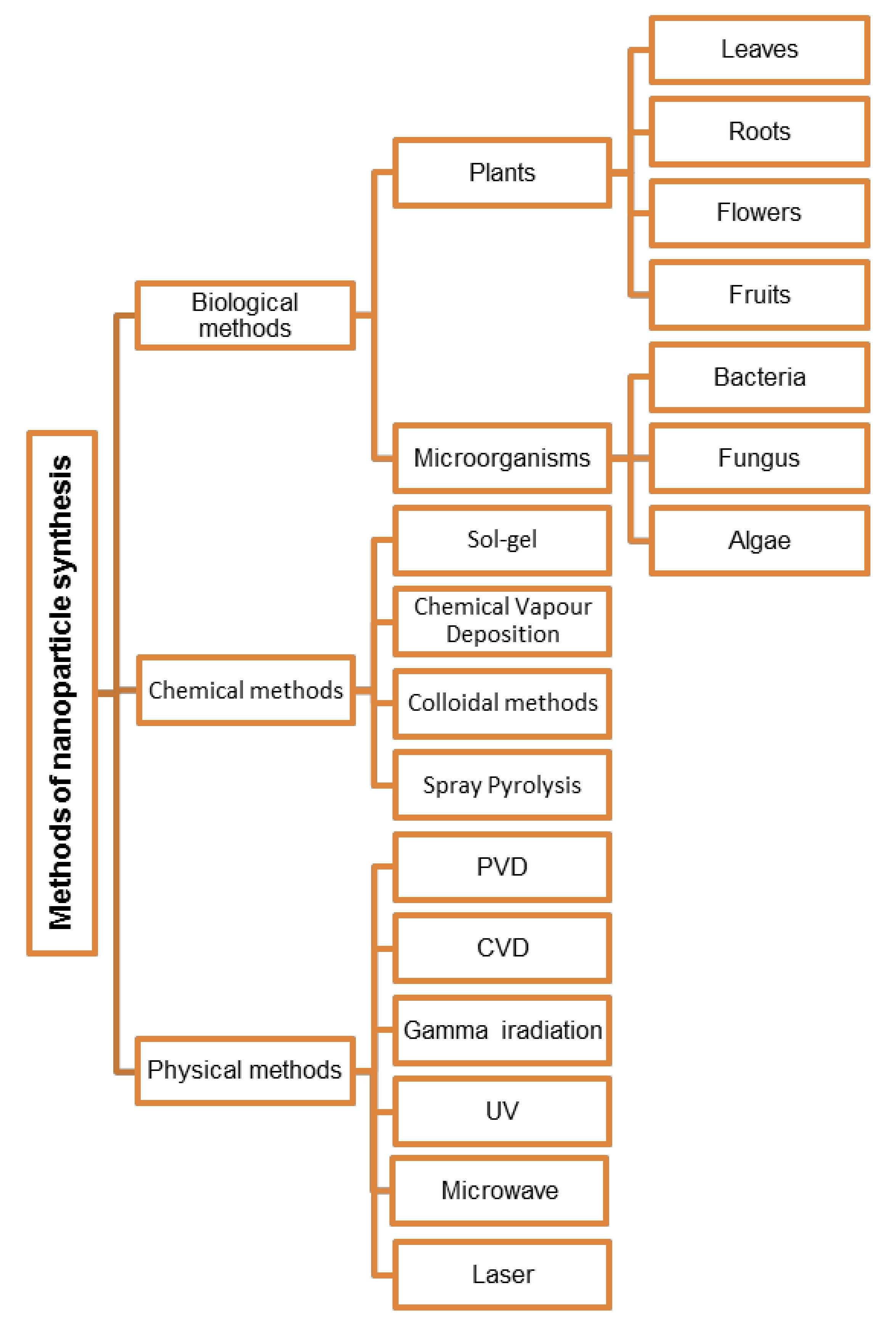
Table 1.
Shape and size of nanoparticles obtained by green synthesis.
| Nanoparticles | Shape | Size, nm | Source | References |
|---|---|---|---|---|
| ZnONPs | spherical | 129 | Daphne leaves | [57] |
| plate | 87 | |||
| hexagonal | 10–42 | Fusarium keratoplasticum | [58] | |
| rod | 8–38 | Aspergillus niger | ||
| hexagonal | 15.45 | Alternaria tenuissima | [59] | |
| quasi-spherical | 16–78 | Periconium sp. | [60] | |
| star | 50–80 | cyanobacterium Nostoc sp. | [61] | |
| AgNPs | spherical | 35–42.5 | cacao powder | [62] |
| cubic and spherical | 30–90 | leaves of Juniperus procera | [63] | |
| spherical | 28 | tea polyphenol | [64] | |
| 35 | Nyctanthes arbor-tristis | [65] | ||
| circular | 100 | C. cladosporiodes | [66] | |
| triangular | 25 | Talaromyces purpureogenus | [67] | |
| spherical | 30–50 | coral | [68] | |
| AuNPs | spherical | 14–17 | Macrotyloma uniflorum | [69] |
| spherical | 3–45 | leaves of Bacopa monnieri | [70] | |
| cubic | 20–50 | flower of Butterfly tree | [71] | |
| triangular, hexagonal, pentagonal, rod, and truncated | 25–100 | Ipomoea carnea | [72] | |
| triangular | 600–800 | pumpkin | [73] | |
| spherical and narrow | 45–75 | Abelmoschus esculentus | [74] | |
| CuNPs | spherical, hexagonal, cylindrical, triangular, and prismatic shapes | 10–50 | leaves of Hagenia abyssinica | [75] |
| spherical | 50–250 | leaves of Magnolia kobus | [76] | |
| spherical | 2–10 | Celastrus paniculatus | [77] | |
| PdNPs | spherical | 50–150 | Prunus × yedoensis | [78] |
| TiO2 NPs | different | 200 | Sesbania grandiflora | [79] |
| spherical | 15–20 | jackfruit leaves | [80] | |
| spherical | 20–90 | Trigonella foenum-graecum | [81] | |
| MgO NPs | spherical and cylindrical | 67.70 | Persimmon | [82] |
| FeO NPs | honeycomb | 30–100 | flowers of Avecinnia marina | [83] |
| spherical | 58 to 530 | Amaranthus spinosus leaf | [84] |
Table 2.
Antimicrobial activity of metal nanoparticles.
| Metal | Pathogens | Nanoparticles | Genus or Species | References |
|---|---|---|---|---|
| Ag | Bacteria | AgNPs | Acinetobacter baumannii, Coliform bacteria, Escherichia Coli, Klebsiella pneumoniae, Listeria monocytogenes, Salmonella enterica, Salmonella typhi, Staphylococcus aureus, Vibrio cholerae, Pseudomonas aeruginosa | [62,106,107,108,109,110,111] |
| Viruses | Adenovirus type 3 (Ad3), Chikungunya virus (CHIKV), CoxB4, HAV-10, HSV-1, Hepatitis B virus (HBV), Herpes simplex virus (HSV), Human immunodeficiency virus (HIV), Influenza virus, Poliovirus, Respiratory syncytial virus (RSV), SARS-CoV-2 | [112,113,114,115,116,117,118,119,120] | ||
| Fungi | Candida albicans, Candida parapsilosis, Candida tropicalis, Microsporum canis, Trichophyton mentagrophytes, | [121,122] | ||
| AgNPs, AgClNPs | Aspergillus flavus, Aspergillus niger, Candida albicans, Cladosporium cladosporioides | [66,68,123] | ||
| Au | Bacteria | AuNPs | Corynebacterium pseudotuberculosis, Escherichia coli, Streptococcus pneumoniae | [124,125,126] |
| Gold-chitosan hybrid nanoparticles | Pseudomonas aeruginosa, Staphylococcus aureus | [127] | ||
| Viruses | AuNps | Dengue, Herpes simplex virus (HSV), Human papillomavirus, Influenza, Lentivirus, Measles virus (MeV), Respiratory syncytial virus (RSV), Vesicular stomatitis virus (VSV) | [128,129,130,131] | |
| Fungi | Aspergillus flavus, Aspergillus niger, Candida albicans, Trichophyton rubrum | [132] | ||
| Bi | Bacteria | BiNPs | Enterococcus faecalis | [133] |
| Cd | Bacteria | CdONPs | Staphylococcus aureus | [134] |
| Cu | Bacteria | CuNPs | Bacillus subtilis, Escherichia coli, Listeria monocytogenes, Pseudomonas aeruginosa, Staphylococcus aureus, | [75,107,135] |
| CuONps | Bacillus subtilis, Escherichia coli, Psedomonas aeruginosa, Staphylococcus aureus, Klebsiella pneumoniae | [136,137] | ||
| Viruses | CuINPs | Feline calicivirus (FCV), H1N1 influenza | [138] | |
| CuSNPs | Human norovirus | [139] | ||
| Cu2ONPs | Hepatitis C virus (HCV) | [140] | ||
| CuONPs | Herpes simplex virus type 1 (HSV-1) | [141,142] | ||
| Fe | Bacteria | FeONPs | Staphylococcus aureus | [143] |
| Fe2O3 modified by Ag | Escherichia coli | [144] | ||
| Viruses | FeONPs | Dengue virus, Influenza A subtypes: H1N1, H5N1, and H7N9, Rotavirus | [145,146,147] | |
| Fungi | FeONPs | Alternaria alternate, Aspergillus niger | [20] | |
| Mg | Bacteria | MgONps | Acinetobacter baumannii, Enterobacter cloacae, Enterococcus faecalis, Escherichia coli, Bacillus megaterium, Bacillus subtilis, Staphylococcus aureus, Staphylococcus epidermidis | [148,149,150] |
| Mn | Bacteria | MnNPs | Escherichia coli, Staphylococcus aureus | [151] |
| Ni | Bacteria | NiFe2O4NPs | Escherichia coli NCIM 2345, Salmonella typhimurium NCIM 2501, Staphylococcus aureus NCIM 5021, Streptococcus pyogenes NCIM 5280 | [152] |
| NiONPs | Bacillus subtilis, Escherichia coli, Protius mirabilis, Staphylococcus aureu, Streptococcus pneumoniae | [153,154] | ||
| Pd | Bacteria | PdNPs | Bacillus subtilis, Escherichia coli, Psedomonas aeruginosa, Staphylococcus aureus | [78,155] |
| Pt | Bacteria | PtNPs | Bacillus subtilis, Escherichia coli, Klebsiella pneumoniae, Psedomonas aeruginosa, Staphylococcus aureus | [151] |
| Fungi | Aspergillus sp., Candida sp. | |||
| Sn | Bacteria | SnO2NPs | Escherichia coli, Staphylococcus aureus | [156,157,158] |
| Fungi | Candida albicans | [159] | ||
| Ti | Bacteria | TiO2NPs | Bacillus subtilis, Enterococcus faecalis, Escherichia coli, Klebsiella pneumoniae, Proteus vulgaris, Pseudomonas aeruginosa, Salmonella typhimurium, Staphylococcus aureus, Streptococcus faecalis, Yersinia enterocolitica | [80,81,160] |
| Viruses | Herpes simplex virus (HSV), Human norovirus, Influenza virus (A/PR8/H1N1) | [161,162,163] | ||
| Fungi | Aspergillus fumigatus, Arthrographis cuboid, Aspergillus niger, Candida albicans | [81,164,165] | ||
| V | Bacteria | Nalidixic acid-vanadium (V-NA) nanoparticles | Bacillus cereus, Escherichia coli, Psedomonas aeruginosa, Staphylococcus aureus | [166] |
| Zn | Bacteria | ZnONPs | Bacillus cereus, Bacillus subtilis, Campylobacter jejuni, Escherichia coli, Klebsiella pneumonia, Listeria monocytogenes, Pseudomonas aeruginos, Pseudomonas vulgaris, Salmonella spp., Staphylococcus aureus | [58,108,167] |
| Viruses | H1N1 influenza virus, Herpes simplex virus type-1 (HSV-1) | [168,169] | ||
| Fungi | Alternaria alternata (ITCC 6531), Aspergillus niger (ITCC 7122), Candida albicans | [60,170] | ||
| Zr | Bacteria | ZrO | Bacillus subtilis MTCC 1305, Escherichia coli, Pseudomonas aeruginosa MTCC 2453, Staphylococcus aureus MTCC 3160 | [171] |
Table 3.
Application of silver and copper nanoparticles for production of self-sterilizing materials.
Table 3.
Application of silver and copper nanoparticles for production of self-sterilizing materials.
| NPs | Concentration | Material | Patogens | References |
|---|---|---|---|---|
| AgNPs | 0.27 ppm | Polyhydroxyalkanoate bioplastic film | Surrogates, murine norovirus (MNV), Feline calicivirus (FCV) | [192] |
| 1% (complete inactivation) | Polylactide plastic | Salmonella spp., Feline calicivirus FCV | [191] | |
| AgNO3 (0.5 M, 35 mL) and ethylene glycol, 25% inhibition of infection | Graphene oxide sheets | Feline coronavirus (FCoV) | [194] | |
| Less than 1 mg per 10 mL | PLA/silver-based nanoclay (organomodified MMT) | Salmonella spp. | [195] | |
| 20 mg AgNPs per 150 mL | Agar nanocomposite films | Escherichia coli, Listeria monocytogenes | [38] | |
| MSF-Ag at 110 μg per mL of S. aureus and 70 μg per mL of E. coli | Mesoporous silica flakes (MSF) | Escherichia coli, Staphylococcus aureus | [196] | |
| 25 mg L−1 (silver content 1.5 mg L−1) | Monodispersed silica spheres | Escherichia coli | [89] | |
| 0.005 M AgNO3 solution | Temperature-responsive POEGMA188 and P4VP grafted polymer brush coatings attached to a glass surface | Escherichia coli ATCC 25922, Staphylococcus aureus ATCC 25923 | [186] | |
| 0.04–1.18 silver nitrate (mol L−1) | Poly(N-isopropylacrylamide) (PNIPAAm) glass surfaces | Escherichia coli | [197] | |
| CuONPs | 0.1% or 0.05% | Electrospun poly(3-hydroxybutyrate-co-hydroxyvalerate) PHBV polymers | Murine norovirus | [198] |
| Cu2ONPs | 2% | Face mask | Influenza A viruses (H1N1) | [199] |
| CuNPs | 0.01–0.15 g L−1 Cu2+ | Cotton and polyester substrates | Staphylococcus aureus ATCC 25923 | [200] |
Table 4.
Examples of the use of metal nanoparticles in edible coatings.
| NPs | Coating | Food | References |
|---|---|---|---|
| ZnONps | soy protein isolate | banana | [245] |
| chitosan/gum arabic | [246] | ||
| chitosan | papaya | [247] | |
| carboxymethyl-cellulose | pomegranate fruit | [248] | |
| tomato | [249] | ||
| chitosan/gum arabic | avocado | [250] | |
| xanthan gum | tomato and apple | [251] | |
| chitosan/alginate | guava | [252] | |
| AgNps | polylactic acid film | mango | [253] |
| polyvinylpyrrolidone | asparagus | [254] | |
| carboxymethyl cellulose (CMC)-guar gum | strawberry | [255] | |
| sodium alginate-gum | black grapes | [256] | |
| TiO2NPs | chitosan/polyvinyl alcohol | cheese | [257] |
| chitosan | tomato | [258] | |
| Zn-MgONps | alginate solution | cold-smoked salmon | [259] |
| AgTiO2NPs | chitosan | fruit | [260] |
Publisher’s Note: MDPI stays neutral with regard to jurisdictional claims in published maps and institutional affiliations. |
© 2022 by the authors. Licensee MDPI, Basel, Switzerland. This article is an open access article distributed under the terms and conditions of the Creative Commons Attribution (CC BY) license (https://creativecommons.org/licenses/by/4.0/).
Share and Cite
MDPI and ACS Style
Góral, D.; Góral-Kowalczyk, M. Application of Metal Nanoparticles for Production of Self-Sterilizing Coatings. Coatings 2022, 12, 480. https://doi.org/10.3390/coatings12040480
AMA Style
Góral D, Góral-Kowalczyk M. Application of Metal Nanoparticles for Production of Self-Sterilizing Coatings. Coatings. 2022; 12(4):480. https://doi.org/10.3390/coatings12040480
Chicago/Turabian StyleGóral, Dariusz, and Małgorzata Góral-Kowalczyk. 2022. "Application of Metal Nanoparticles for Production of Self-Sterilizing Coatings" Coatings 12, no. 4: 480. https://doi.org/10.3390/coatings12040480
Note that from the first issue of 2016, this journal uses article numbers instead of page numbers. See further details here.