
Biomaterials in Orthopedic Devices: Current Issues and Future Perspectives
 , , ,
, , ,
Abstract
:1. Introduction
2. Requirements for Materials Used to Fix Bones
2.1. General Requirements of Materials for Orthopedic Implant
2.2. Specific Requirements of Materials for Orthopedic Implant
2.2.1. Mechanical Requirements
2.2.2. Biological Requirements
3. Specific Orthopedic Implant Materials
3.1. Metal Materials
3.1.1. Stainless Steel
3.1.2. Cobalt-Based Alloys
3.1.3. Titanium-Based Alloys
3.2. Non-Metal Materials
3.2.1. Polymeric Materials
Polyethylene
Polymethylmethacrylate
Polyurethanes
3.2.2. Ceramics
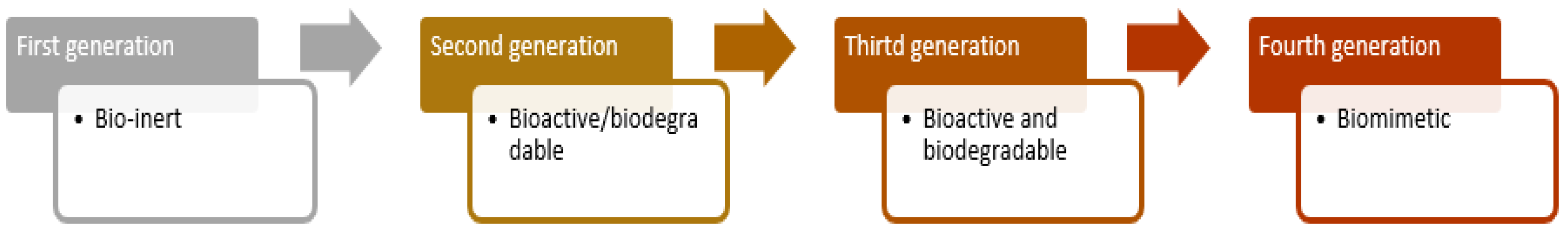
4. Future Perspectives for the Materials Used in Orthopedics
5. Conclusions
Author Contributions
Funding
Institutional Review Board Statement
Informed Consent Statement
Data Availability Statement
Conflicts of Interest
References
- Marin, E.; Boschetto, F.; Pezzotti, G. Biomaterials and biocompatibility: An historical overview. J. Biomed. Mater. Res. Part A 2020, 108, 1617–1633. [Google Scholar] [CrossRef] [PubMed]
- Chaudhari, A.A.; Vig, K.; Baganizi, D.R.; Sahu, R.; Dixit, S.; Dennis, V.; Singh, S.R.; Pillai, S.R. Future prospects for scaffolding methods and biomaterials in skin tissue engineering: A review. Int. J. Mol. Sci. 2016, 17, 1974. [Google Scholar] [CrossRef] [PubMed]
- Fan, J.; Abedi-Dorcheh, K.; Sadat Vaziri, A.; Kazemi-Aghdam, F.; Rafieyan, S.; Sohrabinejad, M.; Ghorbani, M.; Rastegar Adib, F.; Ghasemi, Z.; Klavins, K.; et al. A Review of Recent Advances in Natural Polymer-Based Scaffolds for Musculoskeletal Tissue Engineering. Polymers 2022, 14, 2097. [Google Scholar] [CrossRef] [PubMed]
- Tsegay, F.; Elsherif, M.; Butt, H. Smart 3D Printed Hydrogel Skin Wound Bandages: A Review. Polymers 2022, 14, 1012. [Google Scholar] [CrossRef] [PubMed]
- Ramakrishna, S.; Mayer, J.; Wintermantel, E.; Leong, K.W. Biomedical applications of polymer-composite materials: A review. Compos. Sci. Technol. 2001, 61, 1189–1224. [Google Scholar] [CrossRef]
- Francis, A. Biological evaluation of preceramic organosilicon polymers for various healthcare and biomedical engineering applications: A review. J. Biomed. Mater. Res. Part B Appl. Biomater. 2021, 109, 744–764. [Google Scholar] [CrossRef] [PubMed]
- Oladapo, B.I.; Zahedi, S.A.; Ismail, S.O.; Olawade, D.B. Recent advances in biopolymeric composite materials: Future sustainability of bone-implant. Renew. Sustain. Energy Rev. 2021, 150, 111505. [Google Scholar] [CrossRef]
- Ghasemi-Mobarakeh, L.; Kolahreez, D.; Ramakrishna, S.; Williams, D. Key terminology in biomaterials and biocompatibility. Curr. Opin. Biomed. Eng. 2019, 10, 45–50. [Google Scholar] [CrossRef]
- Heimann, R.B. (Ed.) Materials for Medical Application; Walter de Gruyter GmbH & Co KG: Görlitz, Germany, 2020. [Google Scholar]
- Love, B. Biomaterials: A Systems Approach to Engineering Concepts; Academic Press: Cambridge, MA, USA, 2017. [Google Scholar]
- Hacker, M.C.; Krieghoff, J.; Mikos, A.G. Synthetic polymers. In Principles of Regenerative Medicine; Academic Press: Cambridge, MA, USA, 2019; pp. 559–590. [Google Scholar]
- Xu, X.; Song, J. Segmental long bone regeneration guided by degradable synthetic polymeric scaffolds. Biomater. Transl. 2020, 1, 33. [Google Scholar]
- Mir, M.; Ali, M.N.; Barakullah, A.; Gulzar, A.; Arshad, M.; Fatima, S.; Asad, M. Synthetic polymeric biomaterials for wound healing: A review. Prog. Biomater. 2018, 7, 1–21. [Google Scholar] [CrossRef] [Green Version]
- Donnaloja, F.; Jacchetti, E.; Soncini, M.; Raimondi, M.T. Natural and Synthetic Polymers for Bone Scaffolds Optimization. Polymers 2020, 12, 905. [Google Scholar] [CrossRef]
- Tian, L.; Tang, N.; Ngai, T.; Wu, C.; Ruan, Y.; Huang, L.; Qin, L. Hybrid fracture fixation systems developed for orthopaedic applications: A general review. J. Orthop. Transl. 2019, 16, 1–13. [Google Scholar] [CrossRef]
- Pal, S. Mechanical properties of biological materials. In Design of Artificial Human Joints & Organs; Springer: Boston, MA, USA, 2014; pp. 23–40. [Google Scholar]
- Silver, F.H.; Shah, R. Measurement of mechanical properties of natural and engineered implants. Adv. Tissue Eng. Regen. Med. Open Access 2016, 1, 20–25. [Google Scholar] [CrossRef]
- Li, J.; Zhao, Z.; Yin, P.; Zhang, L.; Tang, P. Comparison of three different internal fixation implants in treatment of femoral neck fracture—A finite element analysis. J. Orthop. Surg. Res. 2019, 14, 76. [Google Scholar] [CrossRef]
- Bankoff, A.D.P. Biomechanical characteristics of the bone. Hum. Musculoskelet. Biomech. 2012, 61, 86. [Google Scholar]
- Basso, T. Internal fixation of fragility fractures of the femoral neck: Ex vivo biomechanical studies. Acta Orthop. 2015, 86 (Suppl. S361), S1–S36. [Google Scholar] [CrossRef] [Green Version]
- Todros, S.; Todesco, M.; Bagno, A. Biomaterials and Their Biomedical Applications: From Replacement to Regeneration. Processes 2021, 9, 1949. [Google Scholar] [CrossRef]
- Thanigaivel, S.; Priya, A.K.; Balakrishnan, D.; Dutta, K.; Rajendran, S.; Soto-Moscoso, M. Insight on recent development in metallic biomaterials: Strategies involving synthesis, types and surface modification for advanced therapeutic and biomedical applications. Biochem. Eng. J. 2022, 187, 108522. [Google Scholar] [CrossRef]
- Dolcimascolo, A.; Calabrese, G.; Conoci, S.; Parenti, R. Innovative biomaterials for tissue engineering. In Biomaterial-Supported Tissue Reconstruction or Regeneration; IntechOpen: London, UK, 2019. [Google Scholar] [CrossRef] [Green Version]
- Festas, A.J.; Ramos, A.; Davim, J.P. Medical devices biomaterials—A review. Proc. Inst. Mech. Eng. Part L J. Mater. Des. Appl. 2020, 234, 218–228. [Google Scholar] [CrossRef]
- Bhaskar, B.; Nagarjuna, V. Biomaterials, Tissue Engineering, and Regenerative Medicine: A Brief Outline. In Biomaterials in Tissue Engineering and Regenerative Medicine; Springer: Singapore, 2021; pp. 3–17. [Google Scholar]
- Basu, B.; Gowtham, N.H.; Xiao, Y.; Kalidindi, S.R.; Leong, K.W. Biomaterialomics: Data science-driven pathways to develop fourth-generation biomaterials. Acta Biomater. 2022, 143, 1–25. [Google Scholar] [CrossRef]
- Allo, B.A.; Costa, D.O.; Dixon, S.J.; Mequanint, K.; Rizkalla, A.S. Bioactive and Biodegradable Nanocomposites and Hybrid Biomaterials for Bone Regeneration. J. Funct. Biomater. 2012, 3, 432–463. [Google Scholar] [CrossRef] [PubMed]
- Gautam, G.; Kumar, S.; Kumar, K. Processing of biomaterials for bone tissue engineering: State of the art. Mater. Today Proc. 2022, 50, 2206–2217. [Google Scholar] [CrossRef]
- Saad, M.; Akhtar, S.; Srivastava, S. Composite polymer in orthopedic implants: A review. Mater. Today Proc. 2018, 5, 20224–20231. [Google Scholar] [CrossRef]
- Sheikh, Z.; Najeeb, S.; Khurshid, Z.; Verma, V.; Rashid, H.; Glogauer, M. Biodegradable Materials for Bone Repair and Tissue Engineering Applications. Materials 2015, 8, 5744–5794. [Google Scholar] [CrossRef] [PubMed]
- Arifin, A.; Sulong, A.B.; Muhamad, N.; Syarif, J.; Ramli, M.I. Material processing of hydroxyapatite and titanium alloy (HA/Ti) composite as implant materials using powder metallurgy: A review. Mater. Des. 2014, 55, 165–175. [Google Scholar] [CrossRef]
- Zakaria, S.M.; Sharif Zein, S.H.; Othman, M.R.; Yang, F.; Jansen, J.A. Nanophase hydroxyapatite as a biomaterial in advanced hard tissue engineering: A review. Tissue Eng. Part B Rev. 2013, 19, 431–441. [Google Scholar] [CrossRef] [PubMed]
- Ning, C.; Zhou, L.; Tan, G. Fourth-generation biomedical materials. Mater. Today 2016, 19, 2–3. [Google Scholar] [CrossRef]
- Serrano, M.C.; Ameer, G.A. Recent insights into the biomedical applications of shape-memory polymers. Macromol. Biosci. 2012, 12, 1156–1171. [Google Scholar] [CrossRef]
- Montoya, C.; Du, Y.; Gianforcaro, A.L.; Orrego, S.; Yang, M.; Lelkes, P.I. On the road to smart biomaterials for bone research: Definitions, concepts, advances, and outlook. Bone Res. 2021, 9, 12. [Google Scholar] [CrossRef]
- Shekhawat, D.; Singh, A.; Bhardwaj, A.; Patnaik, A. A short review on polymer, metal and ceramic based implant materials. IOP Conf. Ser. Mater. Sci. Eng. 2021, 1017, 012038. [Google Scholar] [CrossRef]
- Wall, E.J.; Jain, V.; Vora, V.; Mehlman, C.T.; Crawford, A.H. Complications of titanium and stainless steel elastic nail fixation of pediatric femoral fractures. J. Bone Jt. Surg. 2008, 90, 1305–1313. [Google Scholar] [CrossRef]
- Ghunawat, S.; Relhan, V.; Garg, V.K. Hypersensitivity Reactions to Metal Implants: Clinical, Diagnostic and Treatment Overview. Indian J. Clin. Dermatol. 2019, 2, 29–34. [Google Scholar]
- Chen, Q.; Thouas, G.A. Metallic implant biomaterials. Mater. Sci. Eng. R Rep. 2015, 87, 1–57. [Google Scholar] [CrossRef]
- Brogini, S.; Sartori, M.; Giavaresi, G.; Cremascoli, P.; Alemani, F.; Bellini, D.; Martini, L.; Maglio, M.; Pagani, S.; Fini, M. Osseointegration of additive manufacturing Ti–6Al–4V and Co–Cr–Mo alloys, with and without surface functionalization with hydroxyapatite and type I collagen. J. Mech. Behav. Biomed. Mater. 2021, 115, 104262. [Google Scholar] [CrossRef]
- Solanke, S.; Gaval, V.; Sanghavi, S. In vitro tribological investigation and osseointegration assessment for metallic orthopedic bioimplant materials. Mater. Today Proc. 2021, 44, 4173–4178. [Google Scholar] [CrossRef]
- Aherwar, A.; Singh, A.K.; Patnaik, A. Cobalt Based Alloy: A Better Choice Biomaterial for Hip Implants. Trends Biomater. Artif. Organs 2016, 30, 50–55. [Google Scholar]
- Anene, F.A.; Aiza Jaafar, C.N.; Zainol, I.; Azmah Hanim, M.A.; Suraya, M.T. Biomedical materials: A review of titanium based alloys. Proc. Inst. Mech. Eng. Part C J. Mech. Eng. Sci. 2021, 235, 3792–3805. [Google Scholar] [CrossRef]
- Thomas, P.; Thomas, M.; Summer, B.; Dietrich, K.; Zauzig, M.; Steinhauser, E.; Krenn, V.; Arnholdt, H.; Flaig, M.J. Impaired wound-healing, local eczema, and chronic inflammation following titanium osteosynthesis in a nickel and cobalt-allergic patient: A case report and review of the literature. J. Bone Jt. Surg. 2011, 93, e61. [Google Scholar] [CrossRef]
- Nasibi, S.; Alimohammadi, K.; Bazli, L.; Eskandarinezhad, S.; Mohammadi, A.; Sheysi, N. TZNT alloy for surgical implant applications: A systematic review. J. Compos. Compd. 2020, 2, 62–68. [Google Scholar] [CrossRef]
- Xu, W.; Lu, X.; Tian, J.; Huang, C.; Chen, M.; Yan, Y.; Wang, L.; Qu, X.; Wen, C. Microstructure, wear resistance, and corrosion performance of Ti35Zr28Nb alloy fabricated by powder metallurgy for orthopedic applications. J. Mater. Sci. Technol. 2020, 41, 191–198. [Google Scholar] [CrossRef]
- Findik, F. Recent developments of metallic implants for biomedical applications. Period. Eng. Nat. Sci. 2020, 8, 33–57. [Google Scholar]
- Lin, X.; Saijilafu; Wu, X.; Wu, K.; Chen, J.; Tan, L.; Witte, F.; Yang, H.; Mantovani, D.; Zhou, H.; et al. Biodegradable Mg-based alloys: Biological implications and restorative opportunities. Int. Mater. Rev. 2022, 67, 1–39. [Google Scholar] [CrossRef]
- Barber, C.C.; Burnham, M.; Ojameruaye, O.; McKee, M.D. A systematic review of the use of titanium versus stainless steel implants for fracture fixation. OTA Int. 2021, 4, e138. [Google Scholar] [CrossRef]
- Mahyudin, F.; Widhiyanto, L.; Hermawan, H. Biomaterials in orthopaedics. In Biomaterials and Medical Devices; Springer: Cham, Switzerland, 2016; pp. 161–181. [Google Scholar]
- Ghosh, S.; Sanghavi, S.; Sancheti, P. Metallic biomaterial for bone support and replacement. In Fundamental Biomaterials: Metals; Woodhead Publishing: Cambridge, UK, 2018; pp. 139–165. [Google Scholar]
- Mehta, H.; Kaur, G.; Chaudhary, G.R.; Prabhakar, N.; Kaul, S.; Singhal, N.K. Evaluation of corrosion resistant, antimicrobial and cytocompatible behaviour of cobalt based metallosurfactants self-assembled monolayers on 316L stainless steel surface. Surf. Coat. Technol. 2022, 444, 128657. [Google Scholar] [CrossRef]
- Orlov, A.A.; Sarychev, S.M.; Orlov, A.A. The Effect of Surface Conditions on Corrosion Resistance of a Cobalt-Chromium Alloy. In Key Engineering Materials; Trans Tech Publications Ltd.: Bäch, Switzerland; Volume 887, pp. 358–363.
- Nagay, B.E.; Cordeiro, J.M.; Barão, V.A.R. Alloy materials for biomedical applications. In Alloy Materials and Their Allied Applications; Scrivener Publishing LLC: Austin, TX, USA, 2020; pp. 159–189. [Google Scholar]
- Baltatu, M.S.; Burduhos-Nergis, D.D.; Burduhos-Nergis, D.P.; Vizureanu, P. Advanced Metallic Biomaterials; Materials Research Forum LLC: Millersville, PA, USA, 2022. [Google Scholar]
- Kaur, M.; Singh, K. Review on titanium and titanium based alloys as biomaterials for orthopaedic applications. Mater. Sci. Eng. C 2019, 102, 844–862. [Google Scholar] [CrossRef]
- Baltatu, M.S.; Tugui, C.A.; Perju, M.C.; Benchea, M.; Spataru, M.C.; Sandu, A.V.; Vizureanu, P. Biocompatible titanium alloys used in medical applications. Rev. Chim. 2019, 70, 1302–1306. [Google Scholar] [CrossRef]
- Rony, L.; Lancigu, R.; Hubert, L. Intraosseous metal implants in orthopedics: A review. Morphologie 2018, 102, 231–242. [Google Scholar] [CrossRef]
- İzmir, M.; Ercan, B. Anodization of titanium alloys for orthopedic applications. Front. Chem. Sci. Eng. 2019, 13, 28–45. [Google Scholar] [CrossRef]
- Jin, W.; Chu, P.K. Orthopedic implants. Encycl. Biomed. Eng. 2019, 1, 3. [Google Scholar]
- Maitz, M.F. Applications of synthetic polymers in clinical medicine. Biosurface Biotribology 2015, 1, 161–176. [Google Scholar] [CrossRef] [Green Version]
- Singh, D.K.; Verma, R.K. Contemporary Development on the Performance and Functionalization of Ultra High Molecular Weight Polyethylene (UHMWPE) for Biomedical Implants. Nano LIFE 2021, 11, 2130009. [Google Scholar] [CrossRef]
- Zivic, F.; Affatato, S.; Trajanovic, M.; Schnabelrauch, M.; Grujovic, N.; Choy, K.L. (Eds.) Biomaterials in Clinical Practice: Advances in Clinical Research and Medical Devices; Springer: Cham, Switzerland, 2017. [Google Scholar]
- Jefferies, C.; Al-Malaika, S.; Sheena, H.H. New and novel stabilisation approach for radiation-crosslinked Ultrahigh Molecular Weight Polyethylene (XL-UHMWPE) targeted for use in orthopeadic implants. Polym. Degrad. Stab. 2021, 183, 109462. [Google Scholar] [CrossRef]
- Boschetto, F.; Ngoc Doan, H.; Phong Vo, P.; Zanocco, M.; Zhu, W.; Sakai, W.; Adachi, T.; Ohgitani, E.; Tsutsumi, N.; Mazda, O.; et al. Antibacterial and Osteoconductive Effects of Chitosan/Polyethylene Oxide (PEO)/Bioactive Glass Nanofibers for Orthopedic Applications. Appl. Sci. 2020, 10, 2360. [Google Scholar] [CrossRef] [Green Version]
- Bistolfi, A.; Giustra, F.; Bosco, F.; Sabatini, L.; Aprato, A.; Bracco, P.; Bellare, A. Ultra-high molecular weight polyethylene (UHMWPE) for hip and knee arthroplasty: The present and the future. J. Orthop. 2021, 25, 98–106. [Google Scholar] [CrossRef]
- Wahed, S.B.; Dunstan, C.R.; Boughton, P.C.; Ruys, A.J.; Faisal, S.N.; Wahed, T.B.; Salahuddin, B.; Cheng, X.; Zhou, Y.; Wang, C.H.; et al. Functional Ultra-High Molecular Weight Polyethylene Composites for Ligament Reconstructions and Their Targeted Applications in the Restoration of the Anterior Cruciate Ligament. Polymers 2022, 14, 2189. [Google Scholar] [CrossRef]
- Said, A. Ultra-High-Molecular-Weight-Polyethylene (UHMWPE) as Desired Polymer Material for Biomedical. Khalij-Libya J. Dent. Med. Res. 2022, 6, 11–16. [Google Scholar] [CrossRef]
- Hinz, N.; Dehoust, J.; Schroeter, J.; Schulz, A.P.; Hartel, M.J.; Lutz, C.; Frosch, K.H.; Wendlandt, R. Biomechanical in vitro analysis of a novel flexible implant for pubic symphysis disruption using an ultra-high molecular weight polyethylene fiber cord. Clin. Biomech. 2022, 95, 105652. [Google Scholar] [CrossRef]
- Kasser, M.J. Regulation of UHMWPE biomaterials in total hip arthroplasty. J. Biomed. Mater. Res. Part B Appl. Biomater. 2013, 101, 400–406. [Google Scholar] [CrossRef]
- Kammerlander, C.; Neuerburg, C.; Verlaan, J.J.; Schmoelz, W.; Miclau, T.; Larsson, S. The use of augmentation techniques in osteoporotic fracture fixation. Injury 2016, 47, S36–S43. [Google Scholar] [CrossRef]
- Scolaro, J.A.; Lackman, R.D. Surgical management of metastatic long bone fractures: Principles and techniques. JAAOS-J. Am. Acad. Orthop. Surg. 2014, 22, 90–100. [Google Scholar]
- Grechenig, S.; Gänsslen, A.; Gueorguiev, B.; Berner, A.; Müller, M.; Nerlich, M.; Schmitz, P. PMMA-augmented SI screw: A biomechanical analysis of stiffness and pull-out force in a matched paired human cadaveric model. Injury 2015, 46, S125–S128. [Google Scholar] [CrossRef]
- Yaacobi, E.; Sanchez, D.; Maniar, H.; Horwitz, D.S. Surgical treatment of osteoporotic fractures: An update on the principles of management. Injury 2017, 48, S34–S40. [Google Scholar] [CrossRef]
- Moazen, M.; Mak, J.H.; Etchels, L.W.; Jin, Z.; Wilcox, R.K.; Jones, A.C.; Tsiridis, E. The effect of fracture stability on the performance of locking plate fixation in periprosthetic femoral fractures. J. Arthroplast. 2013, 28, 1589–1595. [Google Scholar] [CrossRef] [Green Version]
- Kumar, P.A.; Irudhayam, J.S.; Naviin, D. A review on importance and recent applications of polymer composites in orthopaedics. Int. J. Eng. Res. Dev. 2012, 5, 40–43. [Google Scholar]
- Nixon, A.J.; Auer, J.A.; Watkins, J.P. Principles of fracture fixation. In Equine Fracture Repair; John Wiley & Sons, Inc.: Hoboken, NJ, USA, 2019; pp. 127–155. [Google Scholar]
- Corró, S.; García-Albó, E.; Andrés-Peiró, J.V.; Teixidor, J.; Tomás, J. Bone Defect Management and Augumentation of Distal Femoral Fractures with Polymethylmethacrylate Bone Cement. J. Musculoskelet. Res. 2022, 25, 2250013. [Google Scholar] [CrossRef]
- Grant, K.D.; Busse, E.C.; Park, D.K.; Baker, K.C. Internal fixation of osteoporotic bone. JAAOS-J. Am. Acad. Orthop. Surg. 2018, 26, 166–174. [Google Scholar] [CrossRef]
- Hsu, R.Y.; Ramirez, J.M.; Blankenhorn, B.D. Surgical considerations for osteoporosis in ankle fracture fixation. Orthop. Clin. 2019, 50, 245–258. [Google Scholar] [CrossRef]
- Allizond, V.; Comini, S.; Cuffini, A.M.; Banche, G. Current Knowledge on Biomaterials for Orthopedic Applications Modified to Reduce Bacterial Adhesive Ability. Antibiotics 2022, 11, 529. [Google Scholar] [CrossRef]
- Sa, Y.; Yang, F.; Wang, Y.; Wolke, J.G.; Jansen, J.A. Modifications of poly (methyl methacrylate) cement for application in orthopedic surgery. In Cutting-Edge Enabling Technologies for Regenerative Medicine; Springer: Singapore, 2018; Volume 1078, pp. 119–134. [Google Scholar]
- Shirvan, A.R.; Nouri, A.; Wen, C. Structural polymer biomaterials. In Structural Biomaterials; Woodhead Publishing: Cambridge, UK, 2021; pp. 395–439. [Google Scholar]
- Hamdy, T.M. Polymers and ceramics biomaterials in orthopedics and dentistry: A review article. Egypt. J. Chem. 2018, 61, 723–730. [Google Scholar] [CrossRef]
- Kenny, S.M.; Buggy, M. Bone cements and fillers: A review. J. Mater. Sci. Mater. Med. 2003, 14, 923–938. [Google Scholar] [CrossRef]
- Soles, G.L.; Ferguson, T.A. Fragility fractures of the pelvis. Curr. Rev. Musculoskelet. Med. 2012, 5, 222–228. [Google Scholar] [CrossRef] [Green Version]
- Davis, F.J.; Mitchell, G.R. Polyurethane based materials with applications in medical devices. In Bio-Materials and Prototyping Applications in Medicine; Springer: Boston, MA, USA, 2008; pp. 27–48. [Google Scholar]
- Mathew, E.; Domínguez-Robles, J.; Larrañeta, E.; Lamprou, D.A. Fused Deposition Modelling as a Potential Tool for Antimicrobial Dialysis Catheters Manufacturing: New Trends vs. Conventional Approaches. Coatings 2019, 9, 515. [Google Scholar] [CrossRef] [Green Version]
- Pruitt, L.; Furmanski, J. Polymeric biomaterials for load-bearing medical devices. JOM 2009, 61, 14–20. [Google Scholar] [CrossRef]
- Mayet, N.; Choonara, Y.E.; Kumar, P.; Tomar, L.K.; Tyagi, C.; Du Toit, L.C.; Pillay, V. A comprehensive review of advanced biopolymeric wound healing systems. J. Pharm. Sci. 2014, 103, 2211–2230. [Google Scholar] [CrossRef]
- Bezuidenhout, D.; Williams, D.F.; Zilla, P. Polymeric heart valves for surgical implantation, catheter-based technologies and heart assist devices. Biomaterials 2015, 36, 6–25. [Google Scholar] [CrossRef]
- Blaheta, R.A.; Oertl, A.; Freisleben, H.J.; Nelson, K.; Ackermann, H.; Haferkamp, A.; Engl, T. Detection of early DJ-stent encrustation by sonographic twinkling-artifacts–A pilot study. Cent. Eur. J. Urol. 2017, 70, 107. [Google Scholar]
- Ong, K.L.; Yun, B.M.; White, J.B. New biomaterials for orthopedic implants. Orthop. Res. Rev. 2015, 7, 107–130. [Google Scholar] [CrossRef] [Green Version]
- Giza, E.; Frizzell, L.; Farac, R.; Williams, J.; Kim, S. Augmented tendon Achilles repair using a tissue reinforcement scaffold: A biomechanical study. Foot Ankle Int. 2011, 32, 545–549. [Google Scholar] [CrossRef]
- Faroni, A.; Mobasseri, S.A.; Kingham, P.J.; Reid, A.J. Peripheral nerve regeneration: Experimental strategies and future perspectives. Adv. Drug Deliv. Rev. 2015, 82, 160–167. [Google Scholar] [CrossRef] [PubMed]
- Zafar, F.; Sharmin, E. (Eds.) Polyurethane; BoD–Books on Demand, IntechOpen: Rijeka, Croatia, 2012. [Google Scholar]
- Thompson, M.S.; McCarthy, I.D.; Lidgren, L.; Ryd, L. Compressive and shear properties of commercially available polyurethane foams. J. Biomech. Eng. 2003, 125, 732–734. [Google Scholar] [CrossRef]
- Calvert, K.L.; Trumble, K.P.; Webster, T.J.; Kirkpatrick, L.A. Characterization of commercial rigid polyurethane foams used as bone analogs for implant testing. J. Mater. Sci. Mater. Med. 2010, 21, 1453–1461. [Google Scholar] [CrossRef] [PubMed]
- Bredbenner, T.L.; Haug, R.H. Substitutes for human cadaveric bone in maxillofacial rigid fixation research. Oral Surg. Oral Med. Oral Pathol. Oral Radiol. Endodontol. 2000, 90, 574–580. [Google Scholar] [CrossRef] [PubMed]
- Piconi, C. Ceramics for joint replacement: Design and application of commercial bearings. In Advances in Ceramic Biomaterials; Woodhead Publishing: Cambridge, UK, 2017; pp. 129–179. [Google Scholar]
- Piconi, C.; Sprio, S. Oxide Bioceramic Composites in Orthopedics and Dentistry. J. Compos. Sci. 2021, 5, 206. [Google Scholar] [CrossRef]
- Wang, W.; Ouyang, Y.; Poh, C.K. Orthopaedic implant technology: Biomaterials from past to future. Ann. Acad. Med. 2011, 40, 237. [Google Scholar]
- Ma, H.; Suonan, A.; Zhou, J.; Yuan, Q.; Liu, L.; Zhao, X.; Zhang, Y.G. PEEK (Polyether-ether-ketone) and its composite materials in orthopedic implantation. Arab. J. Chem. 2021, 14, 102977. [Google Scholar] [CrossRef]
- Badulescu, O.V.; Filip, N.; Sirbu, P.D.; Bararu-Bojan, I.; Vladeanu, M.; Bojan, A.; Ciocoiu, M. Current practices in haemophilic patients undergoing orthopedic surgery-a systematic review. Exp. Ther. Med. 2020, 20, 1–4. [Google Scholar] [CrossRef]
- Tsakiris, V.; Tardei, C.; Clicinschi, F.M. Biodegradable Mg alloys for orthopedic implants—A review. J. Magnes. Alloy. 2021, 9, 1884–1905. [Google Scholar] [CrossRef]
- Pattanayak, S.; Sahoo, S.K. Micro engraving on 316L stainless steel orthopedic implant using fiber laser. Opt. Fiber Technol. 2021, 63, 102479. [Google Scholar] [CrossRef]
- Badulescu, O.V.; Sirbu, P.D.; Ungureanu, C.; Pȋnzariu, A.; Cojocaru, E.; Filip, N.; Bararu-Bojan, I.; Vladeanu, M.; Ciocoiu, M. Orthopedic surgery in hemophilic patients with musculoskeletal disorders: A systematic review. Exp. Ther. Med. 2021, 22, 995. [Google Scholar] [CrossRef]
- Badulescu, O.V.; Ciocoiu, M.; Filip, N.; Veringa, V. Tranexamic acid-major antifibrinolytic agent used to achieve hemostasis in hemophilic patients with anti-factor VIII antibodies who must undergo total joint replacement. Rev. Chim. 2019, 70, 638–641. [Google Scholar] [CrossRef]
- Filip, A.; Alexa, O.; Sirbu, P.D.; Filip, C.; Andrusca, L.; Pascal, I.A.; Oprea, S.; Badulescu, O.V. Assessment of the Mechanical Properties of Orthopedic Screws Coated with Polyurethane Acrylate Containing Hydroxyapatite, Intended to Fix the Fragility Fractures. Mater. Plast. 2019, 56, 1028. [Google Scholar] [CrossRef]
- Filip, A.; Badulescu, O.V.; Sirbu, P.D.; Veliceasa, B.; Puha, B.; Pascal, I.A.; Andrusca, L.; Oprea, D.; Filip, C.; Alexa, O. Preliminary investigation on mechanical properties of polymer coating screws for the future fragility fracture fixation. Mater. Plast. 2019, 56, 559. [Google Scholar] [CrossRef]
- Li, H.; Xia, P.; Pan, S.; Qi, Z.; Fu, C.; Yu, Z.; Kong, W.; Chang, Y.; Wang, K.; Wu, D.; et al. The advances of ceria nanoparticles for biomedical applications in orthopaedics. Int. J. Nanomed. 2020, 15, 7199. [Google Scholar] [CrossRef]
- Kalyanaraman, V.; Naveen, S.V.; Mohana, N.; Balaje, R.M.; Navaneethakrishnan, K.R.; Brabu, B.; Murugan, D.S.; Kumaravel, T.S. Biocompatibility studies on cerium oxide nanoparticles–Combined study for local effects, systemic toxicity and genotoxicity via implantation route. Toxicol. Res. 2019, 8, 25–37. [Google Scholar] [CrossRef] [Green Version]
- Soni, M.; Mehta, P.; Soni, A.; Goswami, G.K. Green nanoparticles: Synthesis and applications. IOSR J. Biotechnol. Biochem. 2018, 4, 78–83. [Google Scholar]
- Nikolova, M.P.; Chavali, M.S. Metal oxide nanoparticles as biomedical materials. Biomimetics 2020, 5, 27. [Google Scholar] [CrossRef]
- Luo, J.; Zhu, S.; Tong, Y.; Zhang, Y.; Li, Y.; Cao, L.; Kong, M.; Luo, M.; Bi, Q.; Zhang, Q. Cerium Oxide Nanoparticles Promote Osteoplastic Precursor Differentiation by Activating the Wnt Pathway. Biol. Trace Elem. Res. 2022, 201, 1–9. [Google Scholar] [CrossRef]
- Castiglioni, S.; Cazzaniga, A.; Locatelli, L.; Maier, J.A.M. Silver Nanoparticles in Orthopedic Applications: New Insights on Their Effects on Osteogenic Cells. Nanomaterials 2017, 7, 124. [Google Scholar] [CrossRef] [Green Version]
- Samanta, A.; Podder, S.; Kumarasamy, M.; Ghosh, C.K.; Lahiri, D.; Roy, P.; Bhattacharjee, S.; Ghosh, J.; Mukhopadhyay, A.K. Au nanoparticle-decorated aragonite microdumbbells for enhanced antibacterial and anticancer activities. Mater. Sci. Eng. C 2019, 103, 109734. [Google Scholar] [CrossRef]
- Holweg, P.; Labmayr, V.; Schwarze, U.; Soomer, N.; Ornig, M.; Leithner, A. Osteotomy after medial malleolus fracture fixed with magnesium screws ZX00—A case report. Trauma Case Rep. 2022, 42, 100706. [Google Scholar] [CrossRef]
- Antoniac, I.; Miculescu, M.; Manescu, V.; Stere, A.; Quan, P.H.; Paltânea, G.; Robu, A.; Earar, K. Magnesium-Based Alloys Used in Orthopedic Surgery. Materials 2022, 15, 1148. [Google Scholar] [CrossRef]
- Memarian, P.; Pishavar, E.; Zanotti, F.; Trentini, M.; Camponogara, F.; Soliani, E.; Gargiulo, P.; Isola, M.; Zavan, B. Active Materials for 3D Printing in Small Animals: Current Modalities and Future Directions for Orthopedic Applications. Int. J. Mol. Sci. 2022, 23, 1045. [Google Scholar] [CrossRef]
- Feltz, K.P.; MacFadden, L.N.; Gieg, S.D.; Lough, C.P.; Bezold, W.A.; Skelley, N.W. Mechanical properties of 3D-printed orthopedic one-third tubular plates and cortical screws. J. 3D Print. Med. 2022, 6, 129–145. [Google Scholar] [CrossRef]
- Li, B.; Zhang, M.; Lu, Q.; Zhang, B.; Miao, Z.; Li, L.; Zheng, T.; Liu, P. Application and Development of Modern 3D Printing Technology in the Field of Orthopedics. BioMed Res. Int. 2022, 2022, 8759060. [Google Scholar] [CrossRef]


| Metal Materials | Advantages | Disadvantages | References |
|---|---|---|---|
| Stainless steel (316L) | high resistance, less expensive, easy fabrication | allergic reaction, stress shielding effect | [37,38,39] |
| Cobalt-chromium-based alloys (Co-Cr-Mo, Cr-Ni-Cr-Mo) | high corrosion resistance, wear resistance | early implant loosening rate, difficult fabrication | [36,40,41,42] |
| Titanium –based alloys (Ti-4Al-4V, Ti-6Al-7Nb, Ti-13Nb-13Zr) | biocompatibility, Young modulus close to bone, excellent corrosion resistance, good osteointegration | expensive, intoxication, bone resorption, allergy | [43,44,45,46] |
| Mg based alloys | biodegradability in vivo, biocompatibility | low mechanical strength, fast degradation | [36,47,48] |
| Non-Metal Materials | Advantages | Disadvantages | References |
|---|---|---|---|
| Polyethylene (PE)/ultrahigh molecular weight polyethylene (UHMWPE) | low resistance to friction, resistance to wear, biocompatibility | debris generation along time as a result of wear, osteolysis | [63,64,65,66,67,68,69,70] |
| Polymethylmethacrylate (PMMA) | good tensile properties and tensile strength | long-term usage can produce cement fragmentation | [71,72,73,74,75,76,77,78,79,80] |
Publisher’s Note: MDPI stays neutral with regard to jurisdictional claims in published maps and institutional affiliations. |
© 2022 by the authors. Licensee MDPI, Basel, Switzerland. This article is an open access article distributed under the terms and conditions of the Creative Commons Attribution (CC BY) license (https://creativecommons.org/licenses/by/4.0/).
Share and Cite
Filip, N.; Radu, I.; Veliceasa, B.; Filip, C.; Pertea, M.; Clim, A.; Pinzariu, A.C.; Drochioi, I.C.; Hilitanu, R.L.; Serban, I.L. Biomaterials in Orthopedic Devices: Current Issues and Future Perspectives. Coatings 2022, 12, 1544. https://doi.org/10.3390/coatings12101544
Filip N, Radu I, Veliceasa B, Filip C, Pertea M, Clim A, Pinzariu AC, Drochioi IC, Hilitanu RL, Serban IL. Biomaterials in Orthopedic Devices: Current Issues and Future Perspectives. Coatings. 2022; 12(10):1544. https://doi.org/10.3390/coatings12101544
Chicago/Turabian StyleFilip, Nina, Iulian Radu, Bogdan Veliceasa, Cristiana Filip, Mihaela Pertea, Andreea Clim, Alin Constantin Pinzariu, Ilie Cristian Drochioi, Remus Lucian Hilitanu, and Ionela Lacramioara Serban. 2022. "Biomaterials in Orthopedic Devices: Current Issues and Future Perspectives" Coatings 12, no. 10: 1544. https://doi.org/10.3390/coatings12101544