Review

Extrusion-Based 3D Printing Applications of PLA Composites: A Review

by
Eda Hazal Tümer
and
Husnu Yildirim Erbil
*
Department of Chemical Engineering, Gebze Technical University, 41400 Gebze, Kocaeli, Turkey
*
Author to whom correspondence should be addressed.
Coatings 2021, 11(4), 390; https://doi.org/10.3390/coatings11040390
Submission received: 1 March 2021 / Revised: 19 March 2021 / Accepted: 24 March 2021 / Published: 29 March 2021

Abstract

:
Polylactic acid (PLA) is the most widely used raw material in extrusion-based three-dimensional (3D) printing (fused deposition modeling, FDM approach) in many areas since it is biodegradable and environmentally friendly, however its utilization is limited due to some of its disadvantages such as mechanical weakness, water solubility rate, etc. FDM is a simple and more cost-effective fabrication process compared to other 3D printing techniques. Unfortunately, there are deficiencies of the FDM approach, such as mechanical weakness of the FDM parts compared to the parts produced by the conventional injection and compression molding methods. Preparation of PLA composites with suitable additives is the most useful technique to improve the properties of the 3D-printed PLA parts obtained by the FDM method. In the last decade, newly developed PLA composites find large usage areas both in academic and industrial circles. This review focuses on the chemistry and properties of pure PLA and also the preparation methods of the PLA composites which will be used as a raw material in 3D printers. The main drawbacks of the pure PLA filaments and the necessity for the preparation of PLA composites which will be employed in the FDM-based 3D printing applications is also discussed in the first part. The current methods to obtain PLA composites as raw materials to be used as filaments in the extrusion-based 3D printing are given in the second part. The applications of the novel PLA composites by utilizing the FDM-based 3D printing technology in the fields of biomedical, tissue engineering, human bone repair, antibacterial, bioprinting, electrical conductivity, electromagnetic, sensor, battery, automotive, aviation, four-dimensional (4D) printing, smart textile, environmental, and luminescence applications are presented and critically discussed in the third part of this review.

1. Introduction

Fused deposition modeling (FDM) is one of the additive manufacturing (AM) processes to fabricate objects from three-dimensional (3D) models. The use of FDM has been increasing rapidly in the last decade and it was proposed that this technology can revolutionize the manufacturing practices in many sectors [1,2,3,4,5]. Polylactic acid (PLA) is the most widely used raw material in the FDM-based 3D printing process due to its biodegradability and environmentally friendly properties, however the use of pure PLA polymer is limited in the FDM approach due to its disadvantages such as mechanical weakness, water solubility rate, etc. [6,7,8]. Thus, the preparation of PLA composites with suitable additives is suggested to be a feasible method to improve the properties of the 3D-printed PLA parts which are obtained by the FDM method.
Many good review articles were published on the applications of the FDM approach and also on the use of pure PLA polymer in 3D printing [1,2,3,4,5,6,7,8]. However, there is lack of a review article on the use of novel PLA composite filaments in the FDM-based 3D printing technology and also on the main preparation methods to obtain the PLA composite filaments which will specifically be used as a raw material in 3D printers. Moreover, the improvements of the properties of the 3D-printed objects by using the novel PLA composites in the fields of biomedical, tissue engineering, bioprinting, electrical conductivity, automotive, aviation, sensor, battery, robotic, and smart textile industries have not been reviewed before and this review will also fill this gap.
We will focus on the chemistry, physical properties, and drawbacks of homopolymer PLA in the second section of this review. The main preparation methods of the PLA composites which can be utilized as a raw material in the FDM-based 3D printers will be presented in the third section. Finally, the main applications of the novel PLA composites by using the extrusion-based 3D printing technology will be reviewed in the fields of biomedical, tissue engineering, antibacterial, bioprinting, electrical conductivity, electromagnetic, sensor, battery, automotive, aviation, four-dimensional (4D) printing, smart textile, environmental, luminescence, and fluorescence in the fourth section.

2. Chemistry of PLA and its Use and Limitations in FDM-Based 3D Printing

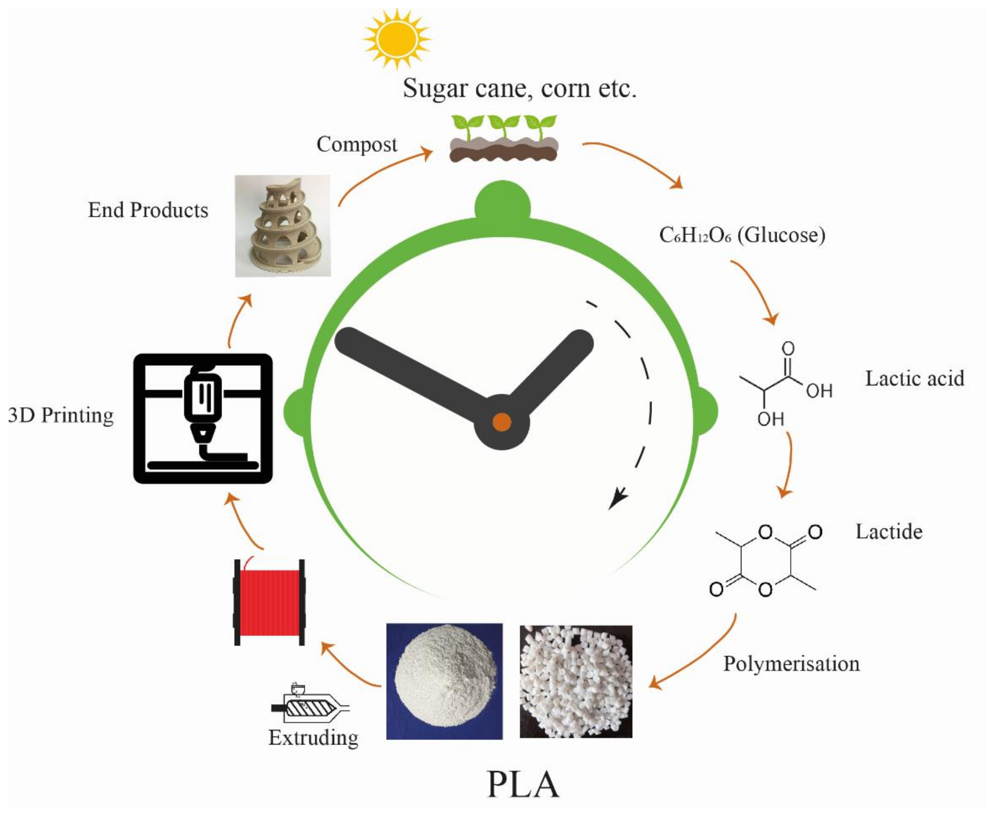
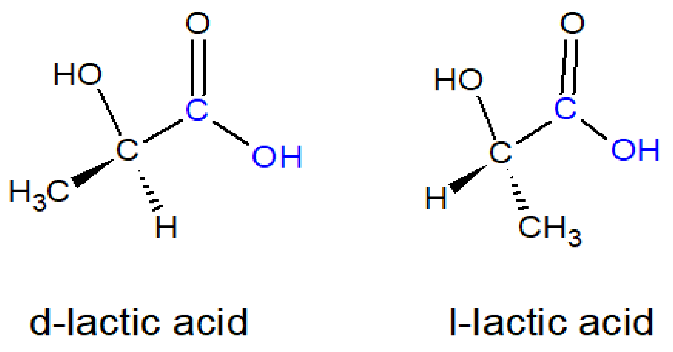
2.1. Outline of the Chemistry of Lactic Acid and PLA

Lactic acid (2-hydroxypropanoic acid)-based polymers are called either poly (lactic acid) or polylactide and they are both abbreviated as PLA. In the last two decades, pure PLA polymer filaments which are used in the FDM approach became the most important thermoplastic source in the three-dimensional (3D) printing field [1,2,3,4,5,6,7,8]. The production of PLA polymer is environmentally preferred since it is generally obtained from renewable sources (e.g., corn, sugar cane, wheat, and rice) and also large amounts of carbon dioxide gas is consumed during its production [9,10,11,12,13,14,15]. PLA has many advantages, such as it is biodegradable, recyclable, and can easily be processed thermally, as schematically shown in Figure 1.
The use of PLA polymers in the injection molding, blow molding, thermoforming, fiber spinning, film forming, and biomedical industries is expanded in the last decade [9,10,11,12].
Lactic acid (LA) is the monomer of PLA polymer and chemically synthesized LA gives the racemic mixture (50% D and 50% L). LA is generally manufactured by bacterial fermentation mainly from corn starch by using a strain of Lactobacillus giving essentially one major stereoisomer, i.e., LA consists of about 99.5% of L-isomer and 0.5% of D-isomer and the presence of 99.5% of L-isomer introduces the high mechanical strength into the PLA polymer after polymerization [15,16,17].
Lactic acid exists in two enantiomeric forms, L- and D-isomer, differing in their effect on polarized light, where the L-isomer rotates the plane of polarized light clockwise, and the D-isomer rotates it counterclockwise, as seen in Figure 2. LA has a hydroxyl and an acid functional group in it and can give both intermolecular and intramolecular esterification reactions. The cyclic dimer (lactide) can be formed by intramolecular esterification of lactoyl, lactic acid or alternatively by the breakdown of higher oligomers. The dehydrated, cyclic dimer of LA is called “lactide” (3,6-dimethyl-1,4-dioxane- 2,5-dione) and exists in three different forms due to the two asymmetric carbon atoms in the molecule: D,D-lactide (called D-lactide), L,L lactide (called L-lactide), and L,D- or D,L-lactide racemate (called meso-lactide), which is optically inactive [9,11,12,13,14,15]. Apart from its use as a monomer in the PLA polymerization, LA is generally used in cosmetics, food and beverages, pharmaceutical, chemical, and medical industries as a pH regulator, flavoring agent, and an inhibitor of residual bacteria in the food and beverages sectors [15,17].
PLA is industrially produced via the polymerization of LA or by the ring opening polymerization of lactide (the cyclic dimer of lactic acid, as an intermediate). In the first one, the direct polycondensation of LA using organic solvents under high vacuum is applied, where only low to intermediate molecular weight PLA can be produced. In the second method, the formation and later the ring-opening polymerization of lactide is carried out, where no solvent is used [9,10,11,12,13,14,15]. The second method is usually preferred since the catalytic ring-opening polymerization of the purified lactide gives high molecular weight (>100,000 Daltons) PLA [18,19]. In the first step of the lactide production process by the ring-opening polymerization, the produced water is distilled out in a continuous condensation reaction giving low molecular weight oligomers or prepolymers. Later, the produced prepolymer is catalytically converted through an internal transesterification reaction to the cyclic lactide and then purified by distillation under vacuum. Then, the catalytic ring-opening polymerization of the purified lactide is carried out by using a stannous octoate-based catalyst, or other organo-tin catalysts. After polymerization, the residual lactide monomer is removed and recycled within the process. The selection of the type and concentration of the catalysts and the residence time and temperature during the polymerization enables to control the molecular weight, and also the ratio of D- and L-lactic acid units in the final PLA polymer [9,15,18,19]. If PLA consists entirely of L-LA acid monomer, then it is called PLLA, and if it is composed of D-LA monomer, it is called PDLA.
Many PLA types having different isomer ratios can be formed and the thermal, mechanical, and biodegradation characteristics of PLA are dependent on the choice and distribution of stereoisomers within the polymer chains. For example, when D-content is larger than 20%, then a fully amorphous polymer can be obtained, whereas highly crystalline PLA can only be obtained when the L-content is larger than 90% [9,15,17]. PLA has a slow crystallization rate. Commercial PLA is mostly a blend of PLLA and PDLA or copolymer PDLLA, which is obtained by the polymerization of L-LA and DL-LA. The melting temperature (Tm), glass transition temperature (Tg), and crystallinity % of PLA decrease with the decrease of L- and increase of the D-isomer content. PLA granules have a certain degree of crystallinity which was introduced during the production stage. In general, commercially available PLA polymer has a glass transition temperature in the range 60–65 °C and melting temperature of 160–180 °C [15]. Antioxidants, heat stabilizers, light stabilizers, impact modifiers, and several other additives are used to improve the properties of commercial PLA [10].
PLA polymer is biodegradable and recyclable since it degrades by the hydrolysis of the backbone ester groups and sometimes by microbial attack. The rate of PLA degradation depends on its properties such as crystallinity, molecular weight, morphology, water diffusion rate, and stereoisomeric content. In general, PLA has a slow degradation rate, which is useful in some biomedical applications but is a problem with the disposal of the consumer commodities such as packaging films [9,10,11,12,13,14,15,20]. PLA is biocompatible with the body fluids and thus can be used in biomedical applications. It does not produce toxic or carcinogenic effects in local tissues. PLA hydrolyzes to its constituent hydroxyl acid when used in the interior of a human body. The resultant metabolites are incorporated into the tricarboxylic acid cycle after hydrolysis, and excreted from the body [16,20,21]. The use of PLA as a suture material is confirmed by the Food and Drug Administration (FDA) in cases of direct contact with biological fluids [22]. However, PLA is a relatively hydrophobic polymer with an apparent water contact angle of 75°–85° and its cell affinity is low, and sometimes an inflammatory response happens from the contacting surrounding tissue [23,24].

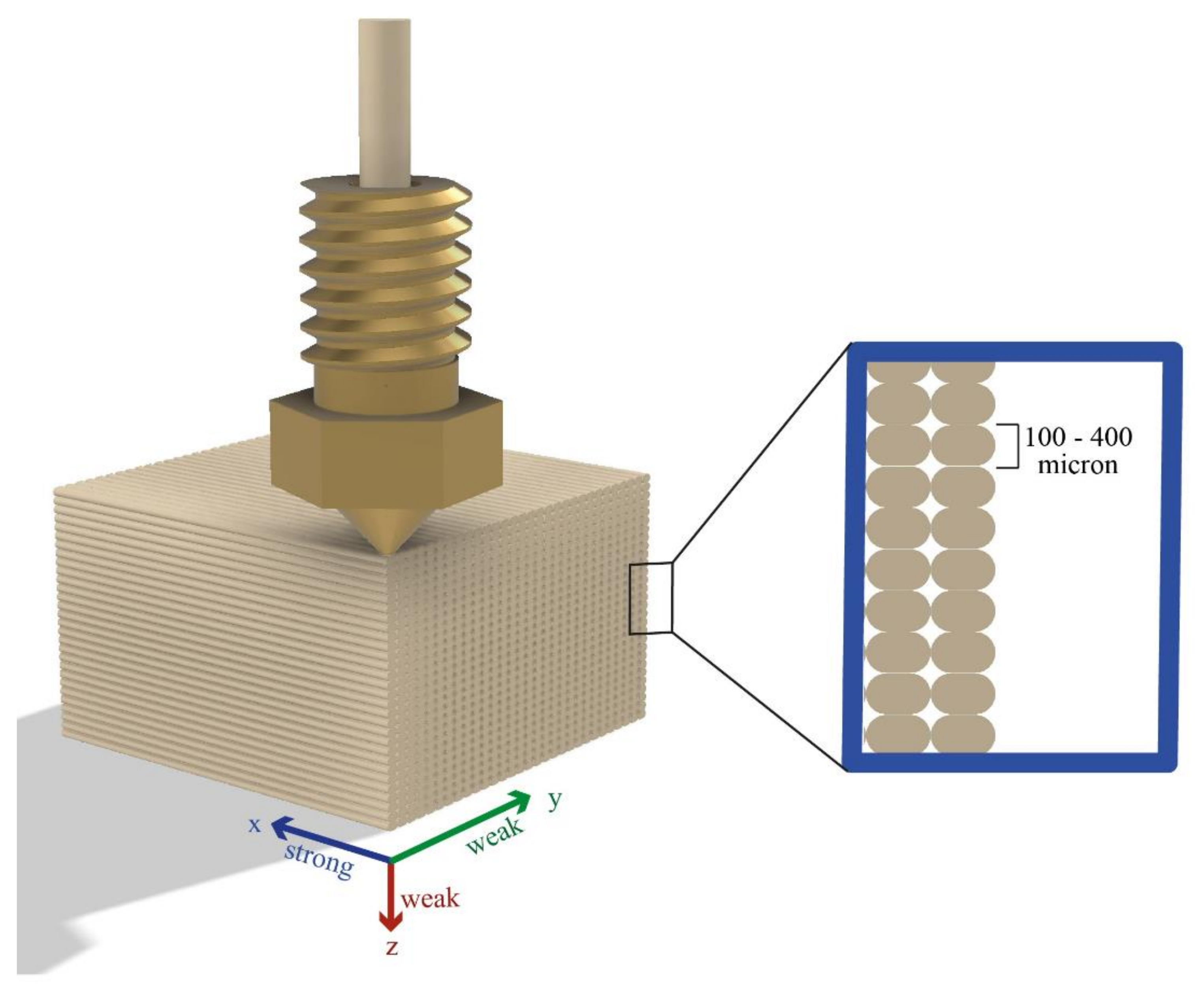
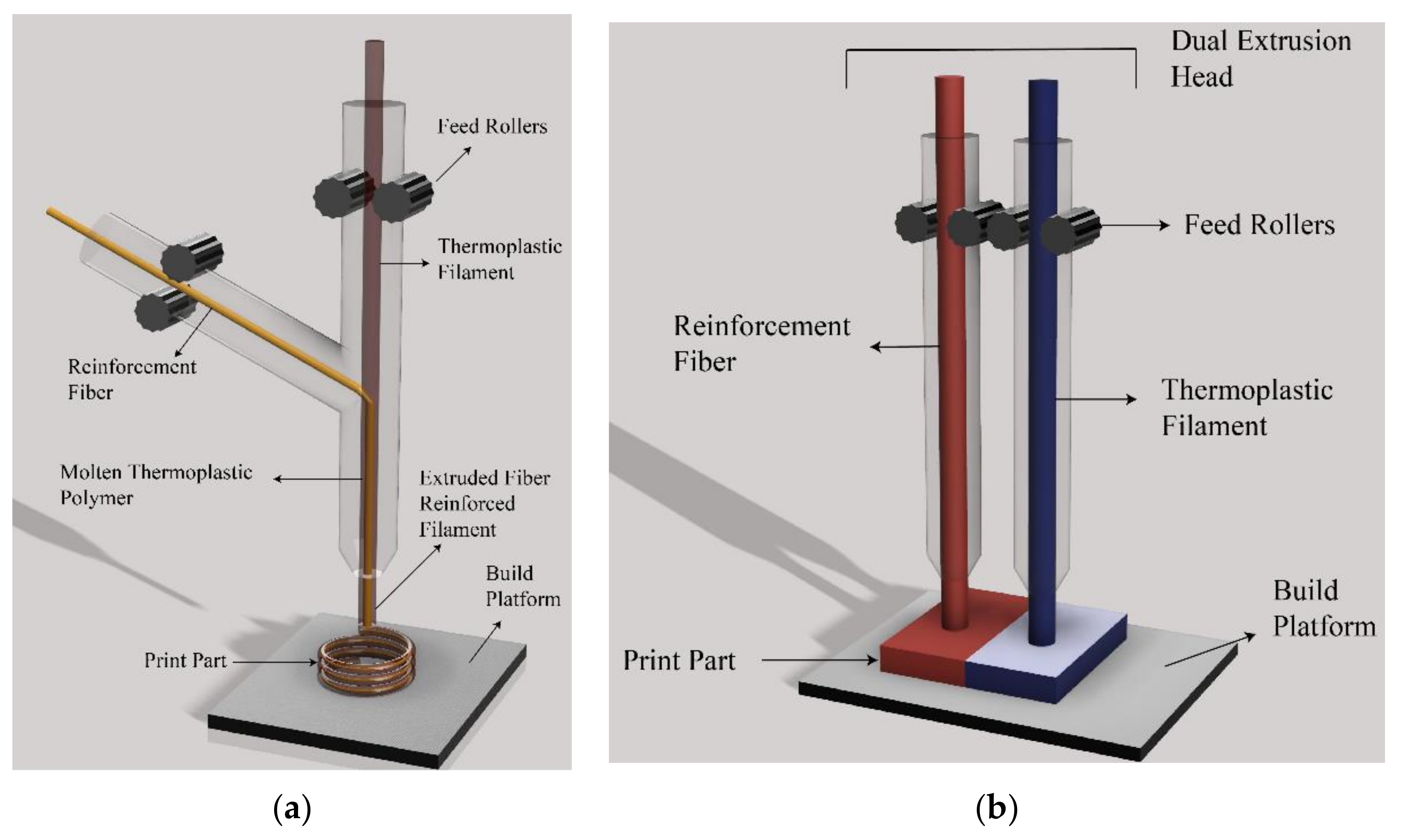
2.2. Use of PLA Filaments in 3D Printers

3D printing (also known as “additive manufacturing” (AM) and “rapid prototyping” (RP)) is a process to create objects from 3D model data drawn with the aid of computer-aided design (CAD) or a similar program. There are many 3D printing methods with different working principles. FDM, selective laser sintering, inkjet 3D printing, stereolithography, and 3D printing are the main AM methods. At present, FDM is the most budget-friendly and popular 3D printing method due to its easy use and available cheap raw materials [6,7,8,25,26,27,28,29,30,31,32,33].
A 3D printer working with the FDM method extrudes thermoplastic filaments, while the temperature and the rate of polymer flow are precisely controlled, as shown in Figure 3. There is one (or sometimes two) small extruder head nozzle in a printer. Polymeric filaments melt into a semi-liquid state at the nozzle to create successive object layers. The fused filaments are extruded onto a build platform layer by layer, where the extruded filament cools, solidifies, and adheres with the adjoining material which was previously deposited. The print nozzle or the base plate moves along the horizontal xy plane and the molten material is placed on the build surface during this motion. The base plate moves down (or the nozzle moves up) in the vertical z-direction by an increment which is equal to the height of the filament during the deposition process. After a whole layer is placed, another layer is deposited again until all the layers solidify into a final 3D object whose shape and dimensions are previously determined. In the FDM approach, the mechanical strength and surface finish quality of the printed parts can be controlled by altering the type (properties) of the polymeric filaments and also the printing parameters of the 3D printer by varying the temperature of the nozzle, polymer flow, layer thickness, orientation, raster width, and raster angle, etc. [33]. The temperature history of the interfaces plays an important role in determining the adhesion quality between the deposited layers. In fact, the adhesion between these extruded filaments depends on the molecular diffusion and randomization of the PLA polymer chains across the interfaces of the filaments [2,3,8,25,26,27].
The popular polymers used in FDM-type printers are as follows: PLA, acrylonitrile butadiene styrene (ABS), polyamide (PA), polypropylene (PP), polyethylene (PE), and polycarbonate (PC) [1,2,3,4,5,6,7,8,26]. At present, PLA filaments are going to be the most used material with an annual growth rate of around 20% [7]. The ease of printing, glossiness, and multicolor appearance are the reasons for the choice of PLA. The accuracy for the dimensional parts of the PLA objects is high since it poses less warp behavior than the ABS filaments.
3D printers which are used in the FDM process are small in size and PLA or composites of PLA flow through single or multiple nozzles (sometimes through multi-channels). PLA has a melting point of 150–160 °C, which is low when compared with other polymers which are used in the FDM printing industry and requires less energy consumption during printing [34]. PLA has high tensile, flexural strength, and Young’s modulus when compared with other feedstock polymers such as PS, PP, and PE [35].

2.3. Main Drawbacks of the PLA Filaments and the Necessity for the PLA Composite Preparation to Be Used in the Extrusion-Based 3D Printing Applications

Unfortunately, PLA polymer has some disadvantages, such as low toughness, high brittleness (with less than 10% elongation at break), low melt strength, poor heat bending temperature, poor thermal stability, narrow processing window, and non-conductivity, which limit its use in many industrial applications [6,7,8,25,26,27,28,29,30,31,32,36]. The mechanical strength of PLA is low in comparison with ABS and PC polymers, since PLA has a more linear molecular chain structure preventing chain entanglement and imparting mechanical strength. Moreover, the dimensional stability of PLA objects after printing is not good due to the volume changes occurring and stress forming during 3D printing, which is caused by the variation of PLA crystallinity.
The adhesion between two fused PLA filaments during 3D printing occurs via a three-step process, as seen schematically in Figure 4. Initially, the surfaces of two independent PLA filaments contact with each other, and later a neck grows between them, and finally, the molecular diffusion of the PLA chains occurs at the interphase region between two adjacent filaments in order to remove the presence of the solid surface meniscus [37]. In general, the weak interfacial adhesion between the fused filaments is the mean cause for the poor mechanical properties of the 3D-printed PLA objects. It was proposed that the reason for the weak interfacial adhesion is due to the short contact time of the extruded PLA filaments which are kept around their melting points, since the temperature of the filaments decreases rapidly to the PLA melting point (or sometimes lower) in a short time due to the rapid heat transfer towards the environment (usually around the room temperature) [37,38,39]. When this happens, the linear PLA macromolecules which are present in the interfacial region of two neighboring filaments cannot be diffused into each other completely, and usually some cavities occur in the interphase and the presence of these cavities decreases the mechanical strength of the printed object.
When the cooling speed of the injected filaments is slow, then some disordered parts and poor surface quality occur on the object. Conversely, when the cooling speed of injected filaments is rapid, then poor PLA macromolecular diffusivity happens in the interface between adjacent filaments, resulting in the consequent weak interfacial bonding, which is the main reason for the poor mechanical properties of the final 3D-printed object. Thus, the selection of the nozzle temperature, polymer flow rate, and also the local printing temperature is important to control the state of PLA macromolecules and the corresponding mechanical properties of the final object [40,41].
Another problem is the mechanical strength variation of the 3D-printed PLA objects according to the printing direction of the fused filaments, as seen schematically in Figure 4. The direction of the filaments should be the same with the direction of main stretching load and this property was investigated and reported in many articles [25,26,27,28,29,30,33,42,43].
There are limitations of improving the mechanical and other properties of homopolymer PLA printed objects using the FDM approach. The use of PLA composites instead of pure PLA polymer was proposed to increase the mechanical strength of PLA homopolymer. PLA composites were formed by the incorporation of metal, ceramic, organic, inorganic, and nanomaterials into the PLA matrix. The objective is to obtain better-performing filaments which will be used as feedstock in FDM-based 3D printers instead of homopolymer PLA filaments. It was suggested that the development of such successful PLA composite filaments as raw materials would expand the use of the 3D printing technology [25,26,27].

3. Methods to Synthesize Pla Composites as Raw Materials to Be Used in the Extrusion-Based 3d Printing

There are many techniques to prepare the PLA composites, such as melt mixing, injection molding, thermoforming, compression molding, etc. However, a continuous PLA composite filament is needed as a raw material for the FDM-based 3D printing applications and the most appropriate method to prepare such a PLA composite is the extrusion melt compounding by using single (or preferably) twin screw extruders [15,25]. Apart from obtaining better mechanical properties, the incorporation of various optical, thermal, and electrical functionalities that cannot be attained by the pure PLA polymer is another objective of the PLA composite preparation [44,45]. A composite consists of two general parts, the matrix and the reinforcing agent. Mainly, micro- or nano-particles of inorganic, carbon, and organic origin, short or long continuous fibers, and some specific metal particles are used to obtain the reinforced PLA composites [8,26,27,46,47,48,49,50,51,52]. Another method is the incorporation of continuous fibers into PLA by using special FDM-based 3D printers to obtain mechanically strong printed parts. The schematic description of the co- and dual-extrusion 3D printing of the PLA with continuous fibers is given in Figure 5.
The nature of dispersed phases, blend composition, and processing conditions (shear, temperatures, mixing time, etc.) in the melt compounding in an extruder are all important. However, the absence of water in the produced PLA composite is the most important requirement. The content of water in the PLA composite filament should be less than 50–250 ppm, otherwise the presence of larger amounts of water can cause swelling and also the hydrolytic degradation. Moreover, if a water-swollen filament is fed into a 3D printer, then it will block the hot nozzle. The moisture trapped in the PLA filament can convert into steam if the printer heats it rapidly and the tiny pockets of steam will interfere with the flow of the printing filament while passing through the hot nozzle. Thus, all precautions should be applied to prevent the presence of water in a PLA composite filament [10,15]. On the other hand, the morphology of the blend and the interactions between polymer—polymer and polymer—nanoparticles also affect the final properties of the 3D-printed product and should be considered [53].
In the present literature, there are many articles reporting the addition of cellulose based natural fibers such as wood, hemp, kenaf, bamboo, sugarcane bagasse and flax to PLA using extrusion blending to form PLA composites. It was determined that the application of fiber chemical treatments improved the stiffness and mechanical strength of the resultant PLA composites depending on the ratio of the filler and also processing conditions during compounding [54,55,56,57,58,59,60,61,62,63,64,65,66,67]. However, it is known that cellulose based fibers are highly hygroscopic and must be dried before or after compounding with PLA polymer. Moreover, these PLA composites should be kept in a dry store in order to prevent water vapor take-up. The effect of the incorporation of suitable plasticizers to cellulose based additives [68,69,70] and also to the toughening agents [71,72] was also investigated.

4. 3D Printing Applications of the PLA Composites

In this section, 3D printing applications of the PLA composites will be presented in nine separate parts to enable the readers to find their selections easily. Biomedical, tissue engineering, biodegradability, and bio-printing applications are related to the health industry. Electrical, microelectronic, electromagnetic, sensor, battery, and photocatalytic cell applications are related to the electromechanical system industries. Automotive, aviation, and space applications are related mostly with the mechanical engineering industries. Finally, the recently developed 4D printing applications were also discussed in this section.

4.1. Biomedical, Tissue Engineering, and Antibacterial Applications

3D-printed PLA composites were used to produce artificial tissues, bones, some organs, and cellular structures [30,73,74,75]. These composites were applied for the reconstruction of skeletal defects and damaged bone structures to renew their functions. Micro- or nano-bioactive ceramic powders like hydroxyapatite (HA) were incorporated to increase the mechanical strength and osseointegration properties of the printed PLA composites [76]. HA is a source of minerals for bone cells and similar to the bone tissue, and thus is suitable for the bone regeneration. HA powders having the average particle size between 90 nm and 1 micron were generally used for this purpose. In a study, mixing of PLA and 15% weight HA powder was carried out in a screw extruder to obtain the composite filaments, and then PLA-HA porous scaffolds with porosity of 30 vol.% for bone implants were prepared using a 3D printer, where the architecture of porous scaffolds was similar to the structure of a trabecular bone [76]. Compression and three-point bending tests were conducted to examine the mechanical properties of the synthetic scaffolds, and it was found that the use of PLA-HA composite improved the mechanical strength of the scaffolds, which can withstand up to 10 MPa pressure without deformation and loss of mechanical strength. The mechanical properties of these 3D-printed biocompatible, biodegradable scaffolds were in the range of targeted properties of the bone tissue and thus they could be used for the treatment of small bone defects [76].
PLA-chitosan-hydroxyapatite load-bearing porous hydrogel scaffolds were obtained by applying 3D printing in order to increase the compressive strength of the synthetic bone tissue. The large pores which were present in the structure of the 3D-printed scaffold provided enough free space to form a composite hydrogel and a higher human stem cell osteogenesis occurred in 3 weeks [77]. In another study, a PLA composite with poly-ε-caprolactone and small amounts of titanium oxide were prepared and then bone replacement objects were 3D-printed using these composite filaments [78]. It was found that the addition of titanium oxide increased the tensile strength and the fracture strain, resulting in values of approximately 45 MPa and 5.5% elongation. Moreover, these 3D-printed composites showed good in vitro biocompatibility, including cell proliferation and adhesion [78].
In another study, composites of PLA and hydroxyapatite (HA) were obtained by extrusion mixing and then porous scaffolds which would be used in the replacement of the trabecular bone defect were obtained by 3D printing [79]. Long-term creep and Charpy impact tests of the samples have shown that PLA-HA scaffolds could be operated under a load of up to 10 MPa, at 119 N impact without deformation and weakening of the mechanical strength. In vivo tests have been performed to investigate the biocompatibility of scaffolds and it was found that these scaffolds can be used as implants for the unloaded small bone-defect replacements [79]. The same group investigated the effect of the cultural environment on the properties of the scaffolds which were obtained by using PLA and micro- and nano-particulate HA composites after extrusion mixing [80]. Compression tests were conducted on the samples and a decrease in the mechanical properties was observed after incubation. These PLA-HA scaffolds were found to support the growth of mesenchymal stromal cells and also stimulated their active proliferation [80].
Homogenous PLA/HA starting composite materials were pre-mixed using a rotomolding machine which produced a PLA composite in the form of pellets, and then these pellets were processed to obtain a continuous wire which was suitable to feed a 3D printer in another investigation [81]. It was found in the composite filament that the HA content was homogeneously distributed within the PLA matrix, as confirmed by SEM and EDX analyses. Despite adding a small amount of HA content, the flexural modulus increased slightly when compared with the pure PLA. This filament was successfully used to fabricate a 3D-printed osteogenic hydroxyapatite PLA bone graft using the clinical images of a maxillary sinus which were obtained by tomography [81].
The production of bone screws is an important objective of the 3D printing approach. For example, PLA bone screws containing iron oxide (Fe3O4) nanoparticles were fabricated by loading 20% Fe3O4 [82]. The mechanical properties of the PLA-Fe3O4 composite screws were evaluated by applying anti-bending and anti-torque strength tests. Histologic and computer tomographic (CT) imaging studies using an animal model were applied to assess the tissue response and radiopacity of these composite screws. It was found that the addition of Fe3O4 increased the crystallization and thermal stability of the 3D-printed composite bone screws, which provided very good local tissue response [82].
An interesting study was the production of 3D-printed porous PLA-HA composite scaffolds which have shape memory effect, to be used for self-fitting implants [83]. The incorporation of HA nanoparticles to the PLA matrix affected the alignment of polymer molecular chains, which resulted in a consequent change in the friction between molecular chains and served as a stationary phase center to govern the shape memory effects. It was found that the 3D-printed porous PLA-HA skeletons supported the survival of mesenchymal stromal cells and stimulated the active proliferation of these cells to help vascularization of the implant, which was essential for the successful bone prosthesis [83].
PLA-HA composite scaffolds were also used to repair large bone defects having complex geometries [84]. Rabbit bone was used as a model and the fabrication of a vascularized tissue-engineered bone was carried out with the aid of an in vivo bioreactor. A tibial periosteum capsule was seeded with the bone marrow stromal cells and filled with 3D-printed PLA-HA composite scaffolds which crossed with a vascular bundle. The tissue-engineered bone was found to be medically successful after four and eight weeks [84].
PLA-HA scaffolds having three different pore sizes were produced and it was found that the 3D printing decreased both PLA molecular weight and degradation temperatures, but there was no change in the crystallinity. No effect of the pore size was seen on the mechanical properties of the scaffolds. The scaffold samples did not show any cytotoxicity towards human bone marrow stromal cells after the sterilization by γ irradiation, indicating that they can be used for the tissue engineering [85]. Later, one-step fabrication of high loaded PLA/HA composite filament for 3D printing was carried out to increase the bioactivity of the scaffold material. The properties of the filaments remained the same after the 3D printing process and HA was found to be well-distributed in the polymer matrix for the viscosity range around the melting point [86].
In general, fixing of multiple fragmented bony fractures is difficult using internal fixation devices, and to drill in the bone to install such devices may result in secondary fractures. 3D-printed PLA–HA–silk composite bone clip internal fixation devices were produced based on the CT scan of a femur in a rat [87]. The novel PLA–HA–silk composite bone clip exhibited similar mechanical stability and better biocompatibility in comparison with the performance of the original PLA–HA clips. PLA–HA–silk clip was found to be relatively noninvasive since there is no requirement to drill holes in the bone [87].
Composites which were formed by mixing PLA, thermoplastic polyurethane (TPU), and graphene oxide (GO) were found to be suitable to construct tissue engineering scaffolds [88]. PLA-TPU-GO composites were prepared using a solvent-based method, where dichloromethane and dimethylformamide solvents were used to dissolve the above polymers, and later, the mixed polymer solutions were co-precipitated in alcohol and finally dried. Next, the composite filaments were prepared by the melt extrusion of the dried composite. The use of TPU polymer improved the elasticity and toughness of the composite. FTIR and SEM images indicated a good dispersion of GO nanoparticles in the polymer matrix. The addition of GO enhanced the thermal stability, and the novel composites can be easily printed into complex shapes. It was found that the addition of GO improved the mechanical properties of the TPU-PLA-GO composite by 167% in compression modulus and 75.5% in tensile modulus after 3D printing, as seen in Figure 6. Degradation temperature increased (90 °C) and better crystallinity was obtained. These composites showed very good cell viability with the NIH3T3 cells, indicating that the addition of GO had no toxicity to the cell growth in the used ratios and PLA-TPU-GO scaffolds could be successfully used in tissue engineering applications [88].
In vivo and in vitro analysis of two-layer 3D-printed scaffolds containing PLA and a biphasic PLA-bioglass (G5) layer were carried out in another study [89]. It was found that PLA-G5 scaffolds had a higher-pressure modulus than the untreated PLA scaffolds, however, a decrease in other mechanical properties was also observed. The structural integrity of the scaffold was preserved with some changes in morphology. The addition of G5 reduced the weight loss of the PLA scaffold. The use of a two-layer scaffold resulted in a different in vivo response even though the PLA and PLA-G5 layers were physically close. [89].
PLA-Nano-b-tricalcium phosphate (b-TCP) composites were manufactured as a raw material to be used for the consecutive 3D printing of a cervical fusion cage structure. In vitro biomechanical studies and biocompatibility tests have shown better mechanical properties in comparison with the traditional fusion cages based on endplate matching [90]. In a similar study, a biodegradable 3D-printed PLGA (poly (D,L)-lactide-co-glycolide) cage was produced as a mechanical support and it was combined with nanofibrous membranes, while the nanofibrous membrane was embedded in antibiotics during the treatment for the sustainable release of antimicrobial agents in order to treat the femoral metaphyseal comminuted fracture [91]. It was found that ceftazidime and vancomycin were sustainably detected above the effective levels in the local tissue fluid around the fracture site after 3 weeks. Moreover, animal studies have shown that the rabbits which were implanted with the synthetic 3D cages have better cortical integrity, leg-to-length ratio, and maximum bending strength. It was finally concluded that the developed 3D composite cage could be used in fracture fixations [91].
Hierarchical biocomposite scaffolds containing PLA micro-protrusions and nanocomposite gelatin-forsterite fibrous layers were produced using the combined extrusion-based 3D printing and electrospinning approaches [92]. It was determined that the elastic modulus of the composite scaffolds was significantly higher than the non-composite ones after performing SEM, FTIR, and uniaxial compression tests. SEM images showed that the calcium phosphate-like deposits were present on the surface of the scaffolds. The enhanced bioactivity of the scaffolds was confirmed due to the presence of a nanocomposite fibrous layer. It was proposed that the produced biocomposite scaffolds can be used for the bone tissue regeneration [92].
Thermoplastic PLA was used as an encapsulable matrix in combination with the photocurable gelatin hydrogels to produce a novel functionalized hybrid hydrogel after the addition of bioactive gold nanoparticles to be applied in bone tissue regeneration [93]. In general, hydrogels have insufficient mechanical properties to be used in musculoskeletal tissue repair. Thus, hybrid hydrogels having higher mechanical strengths should be developed for this purpose. The mechanical properties of the hybrid gelatin hydrogel-PLA composites were determined, and human adipose-derived stem cells were used for the viability tests. It was found that stem cells could live in the composite hydrogel and spread well on the 3D-printed PLA microstructures, as seen in Figure 7. This hybrid hydrogel-PLA composite was found to be suitable for the bone tissue regeneration and stem cell differentiation control studies [93].
PLA-cellulose derivative composites were also tried in biomedical 3D printing applications. Six biocomposites were prepared using PLA and six different lignocellulosic fibers with widely varying particle characteristics. The interfacial bonding between PLA and these fibers was found to be strong after applying the quantitative estimation of adhesion strength, acoustic emission measurements, and SEM. The mechanical tests indicated that the dominating micromechanical deformation process was the fracture of the fibers [94]. PLA-wood fiber biocomposites were synthesized and the effects of print orientation and print width on the mechanical properties of 3D-printed samples were studied [95]. It was found that the mechanical strengths of the samples were dependent on printing orientation (0 or 90°) due to fiber anisotropy and also on printing width (overlapping of filaments), with a lower Young’s modulus than in the compressed samples. It was suggested that this might be associated with highly porous (around 20%) microstructures of printed biocomposites, resulting in water absorption and swelling [95].
The morphology, mechanical properties, and in vitro biocompatibility of strips which were obtained by 3D printing of composite materials containing PLA-Camellia oleifera fruit peel powder (COFHP) were also investigated [96]. Acrylic acid was also grafted to PLA composite and showed better mechanical strength. PLA-g-AA-COFHP composites were easier to process due to formation of ester. In vitro biocompatibility trials indicated that both materials were non-toxic, and the antibacterial activity of the composites increased with the use of COFHP [96]. PLA-microcrystalline cellulose (MCC) composites were synthesized using solvent casting and twin-screw extrusion approaches, while the cellulose concentrations were varied between 1 and 5 wt.% in order to obtain a fully degradable biocomposite [97]. MCC was surface-modified with a titanate coupling agent to increase its compatibility with PLA polymer. The incorporation of MCC resulted in an increase in crystallinity and storage modulus of the printed parts, with 3 wt.% surface-modified MCC showing the highest value [97].
Spruce wood hemicellulose (galacto-glucomannan) was used to try to partially replace the PLA as a matrix polymer in 3D printing in another study [98]. The solvent blending method was used to obtain homogeneous biocomposite feedstock. Mixtures of hemicellulose and PLA in certain proportions were extruded into filaments and then these filaments were used in a 3D printer. Tissue engineering and drug washing scaffold applications were reported to be successful with these PLA composites since they were biocompatible and biodegradable [98]. 3D-printed PLA-carbohydrate hybrid material was used in the bone implants [99]. For this purpose, different proportions of carbohydrate particles were added to the PLA matrix while preparing the scaffold samples. Bioactivity and surface tests and mechanical analysis of the frames were examined, and it was found that the surface roughness was proportional with the added carbohydrate powder content and the mechanical properties were affected by the reduced interactions between the PLA matrix and carbohydrate particles [99].
A fully biodegradable PLA composite which can be used in 3D printers was produced using a novel tetramethylpiperidin-1-yloxyl (TEMPO)-mediated oxidation reaction with bacterial cellulose (TOBC) and also Pickering emulsions [100]. PLA composite microspheres were found uniformly coated by TOBC, via the Pickering emulsion approach, and later melt extrusion was applied to prepare the PLA-TOBC composite filaments. Mechanical properties, crystallinity, fluidity, and thermal stability of PLA-TOBC nanocomposites were studied and it was found that when a TOBC content of up to 1.5 wt.% was added, the mechanical and crystallization properties of the resultant composites were improved [100].
A novel chemical precipitation method was applied to prepare a biodegradable 3D scaffold containing PLA and polyethylene glycol (PEG) as the polymer matrix, nano-hydroxyapatite (nHA) as the ceramic additive, and dexamethasone as the organic additive [101]. The results of water contact angle measurements, DSC, and mechanical tests showed that the addition of dexamethasone resulted in a substantial increase in wettability and crystallinity. In the in vitro experiments, mouse MC3T3-E1 cells were used and the release of dexamethasone from the PLA-PEG-nHA-dexamethasone scaffolds improved the late mineralization and also the alkaline phosphatase secretion. Moreover, the increase in osteo-induction was noticed in the other in vivo experiments on the rat calvarial defects [101]. In another study on the repair of bone defects, 3D-printed porous PLA-nHA composite scaffolds were applied to give enhanced osteogenesis and osteo-conductivity [102,103]. The morphological, composition, and structural analysis indicated that nHA was distributed homogeneously in the scaffold. Based on in vitro antibacterial test results, PLA-nHA scaffolds were found to be highly efficient in loading and releasing levofloxacin and vancomycin. Moreover, cytocompatibility assessment using MG-63 cells was carried out. Positive results were obtained for the osteogenesis and osteo-conductivity of the scaffolds by using the rabbit model [28,102].
3D-printed PLA-microsphere-HA scaffolds were also applied to increase the bone regeneration capacity, biomimicry, and bioactivity [103]. In order to do this, microscale HA spheres were synthesized by spray-drying and were dispersed into the PLA matrix by melt extrusion. Later, the novel composite filaments were used in a 3D printer to produce the macroporous scaffolds. Pure PLA scaffolds were also 3D-printed and used as the reference samples for comparison. It was observed that the crystallinity and Tg of PLA did not change with the addition of microsphere-HA. However, a rough surface was obtained when microsphere-HA was added to the composite in comparison with the smooth surfaces of the 3D-printed pure PLA samples. Composites scaffolds were stiffer, having values similar to a natural bone tissue [103]. On the other hand, template-oriented synthesis of the PLA-HA composites for the 3D bone printing was also carried out [104]. A simple hard template was fabricated to synthesize single-crystal nanoplates of HA. HA sol-gel was prepared using graphitic nitride (g-C3N4) under hydrothermal conditions and HA nanostructure was formed by the calcination and removal of g-C3N4 from the template at high temperatures. Then, PLA-HA composites were prepared in different HA weight ratios for artificial 3D bone samples, which were found to be mechanically stable and had a very good in vitro viability in biological tests [104]. Novel apatite-wollastonite (AW-PLA) composite structures were produced that could match the cortical and cancellous bone characteristics after 3D printing. In vitro cell assays indicated that the AW-PLA structure showed good osseointegration and proliferation of rat bone marrow stromal cells [105].
3D-printed PLA-Ti composite scaffolds were produced after applying melt extrusion [106]. The thermal stability, crystallization temperature, and crystallinity percentage of PLA-Ti composite scaffolds decreased with the increasing Ti content, while the Tg and Tm values of the composite increased. The compressive, tensile, and impact strengths of PLA-Ti composites exceeded the results obtained by the pure PLA. The addition of Ti was found to increase the in vitro biocompatibility [106]. PLA-silver nano-wired (Ag-NW) nanocomposites were produced by dispersing the Ag-NW in the PLA matrix using a solution mixing method [107]. The morphological, thermal, and antimicrobial properties were examined, and it was found that PLA-Ag-NW nanocomposites had good antibacterial effect against both Staphylococcus aureus (S. aureus) and Escherichia coli (E. coli). When the amount of Ag-NW was increased, better antibacterial results were obtained, and a cost/performance optimization study was also carried out in the same study [107].
The incorporation of inorganic additives into PLA to obtain 3D-printed composite scaffolds was reported in some articles. For example, PLA composite scaffolds containing calcium carbonate and beta-tricalcium phosphate (β-TCP) were produced and their morphological, mechanical, and biological tests were carried out in order to evaluate the effect of each additive [108]. According to the results, both inorganic additives increased the porosity, surface roughness, and hydrophilicity of the PLA composite scaffolds, which led to the improvement in the metabolic activity of human osteoblastic osteosarcoma SaOS-2 cells. In terms of cell attachment after 1 week, the best results were obtained when both additives were used, and also an increase in micro-porosity and osteo-conductivity was observed to promote the cell adhesion [108].
3D-printed PLA composites were sometimes used in the drug industry. The objective was the preparation of tablets with defined drug release profiles. For this purpose, pharmaceutical PLA-polyvinyl alcohol multi-component composite tablets were 3D-printed, where the thickness of the PVA filler was varied to achieve different drug release delay times [109]. Wound dressings were also prepared using 3D-printed PLA-lignin powder composites, where the compatibility of the 3D-printed samples was increased by the addition of castor oil. Melt extrusion was applied to obtain the feedstock filaments which can be successfully printed in a 3D printer. These wound dressings had low resistance to breakage, were more wettable, and also showed good antioxidant properties [110].

4.2. Bioprinting Applications

Bioprinting is a special technology where cells, growth factors, and the nutrients keeping the cells alive are incorporated with suitable biocompatible polymers to form “bio-inks”. These bio-inks are then used as feedstocks for FDM-based 3D printers, where specific tissue-like structures are formed by applying the layer-by-layer method. These 3D-printed parts can be converted into tissues or organs by various post-treatment approaches. For example, a bioprinted pre-tissue is transferred to an incubator and kept in a specific solution where it matures to a tissue. Generally, computed tomography (CT) or magnetic resonance imaging (MRI) are used to obtain the model of a specific organ or tissue which will be used in transplantation in the second stage. In some studies, cells are encapsulated in cellular spheroids having 500 μm in diameter and used in the preparation of the 3D printer feedstock. The post-bioprinting treatment should be carefully applied to obtain a stable tissue or organ structure, but the mechanical integrity and function of the synthetic object is usually risky. Both mechanical and chemical stimulations are needed to send signals to cells to control the growth of the tissues.
In brief, bioprinting is a novel technology and the production of successfully used artificial organs such as livers and kidneys is very difficult at present, since the growth of billions of cells is necessary to fabricate these organs during the post-treatment. Moreover, there is a lack of some elements such as working blood vessels, tubules for collecting urine, and enough oxygen in the interiors in most of the final 3D-printed tissues and organs. PLA biopolymer is sometimes used in bio-inks as the matrix polymer, as shown below in some reports.
The combination of bioprinting with the cell-laden microcarrier technology was applied to generate an osteochondral tissue which caused the extensive expansion of cells, while forming multi-cellular aggregates [111]. Gelatin methacrylamide-gellan gum bio-inks were used to encapsulate the mesenchymal stromal cell-laden polylactic acid micro-carriers which were produced via static culture or spinner flask expansion methods. Hydrogel constructs having high cell concentration and viability were fabricated via 3D printing, as seen in Figure 8. The use of microcarrier encapsulation enhanced the compressive modulus of the constructs, facilitated the cell adhesion, and also supported the osteogenic differentiation and bone matrix deposition of the mesenchymal stromal cells [111].
In another study, a fibrous bio-ink which was composed of alginate hydrogel, PLA nanofibers, and human adipose-derived stem cells was used for 3D bioprinting of musculoskeletal soft tissue constructs [112]. Initially, human adipose-derived stem cell proliferation and viability were assessed in 3D-bioplotted strands over 16 days in vitro. Later, a human medial knee meniscus was bioprinted and evaluated over 8 weeks in vitro. It was found that the cell metabolic activity on day 7 was 28.5% higher in this bio-ink when compared with the neat bio-ink, as seen in Figure 9. Cell density was high, and collagen and proteoglycans were present in areas surrounding the cells [112].
The mechanical strength of the 3D-printed hydrogel structures which will be used as cartilage tissue was studied by 3D bioprinting of composite bio-inks containing alginate and submicron PLA fibers [113]. It was found that when PLA short fibers were incorporated, the Young’s modulus of alginate products can be increased three-fold. The prepared bio-inks were loaded with human chondrocytes and later the obtained structures were cultured in vitro for up to 2 weeks. It was determined that 80% viable human chondrocytes were trapped in the obtained hydrogel filaments [113]. In another study, PLA was mixed with gellan gum-poly (ethylene glycol) diacrylate to form double network hydrogel and to improve the mechanical strength of the hydrogels which would be used to repair the intervertebral disc [114]. The mechanical and degradation properties of the dual 3D-printed cell-laden hydrogel constructs were controlled by varying the infill patterns and density of the PLA frameworks. It was found that the bone marrow stromal cells which were co-printed into these scaffolds retained high viability and showed a good distribution in the composite hydrogels [114]. Hyaluronic acid (HA) was used as a raw material to produce a new bio-ink to obtain a synthetic hydrogel cartilage tissue after 3D bioprinting. HA-based bio-ink was co-printed with PLA as a composite and the cell functionality was enhanced by an increase in the expression of chondrogenic gene markers, resulting in tissue formation [115].
The majority of the bioprinting methods giving tissue constructs with open vasculature lack the ability to be directly perfused and the fabrication of the thick and densely populated tissue constructs with controlled vasculature and microenvironment. In a novel study, thick (around 1 cm) and densely populated (10 million cells/mL) 3D tissue constructs having a stiffness of soft tissues such as the liver were obtained by using water-soluble polyvinyl alcohol as the main constituent and PLA as the support structure [116]. PLA support structure was removed, and the water-soluble PVA structure was used to obtain a 3D vascular network within a customized extracellular matrix, giving the stiffness of the liver and with encapsulated hepatocellular carcinoma (HepG2) cells. These tissue constructs could be directly perfused for long periods (>14 days) with the medium inducing the proliferation of the HepG2 cells and the formation of spheroids. It was proposed that the new method could enable the design of the tissue-engineered constructs for tumor modeling and for regenerative medicine [116].
In order to repair the endothelial cells, different types of biocompatible and biodegradable macroporous scaffolds having a diameter of 10 mm with interconnected pores of ~500 µm diameter were formed by 3D bioprinting [117]. Different compositions of PLA, polyethylene glycol, and pluronic F127 were used to obtain semi-solid viscous bio-inks giving a texture where the macropores were homogeneously distributed throughout the surface. Dimethyloxalylglycine and erythroprotein were used as the model drugs and the release of dimethyloxalylglycine from scaffolds into phosphate buffer saline solution was found to continue longer than 48 h [117].
A review article on the overview of 3D-printed constructs with a proper selection of biomaterials and compatible bioprinting methods was published and the challenges were reported to remain for the improved printing resolution (possibly at the nanometer level) and biomaterial compatibilities [118]. It was proposed that the hybrid 3D bioprinting technologies which combined the additive and conventional manufacturing processes have shown promising improvements, introducing higher mechanical strength, native-like biological microenvironments, and cell-related activities [118].

4.3. Electrical Conductivity Applications

The cost-effective fabrication of highly conductive micro- or nano-flexible circuits is an important objective in the electronics industry. Polymer-graphene and polymer-other carbonaceous material composites were used for this purpose. PLA was occasionally used as the matrix polymer in 3D printing. For example, conductive 3D microstructures were obtained by using PLA-multi-walled carbon nanotube (MWCNT) dispersion composites as raw material [119]. Electrical conductivity and rheological measurements were made on these nanocomposites to find the optimum processing conditions and printability by varying the MWCNT and PLA concentrations. Many nanofillers were also tried to obtain better properties [119]. 3D-printed PLA-graphene flexible circuits were produced using reduced graphene oxide (r-GO), which was pre-synthesized in the laboratory [120]. After melt blending PLA and r-GO, the obtained filaments were used as raw material in 3D printing. Both two-dimensional (2D) and 3D flexible circuits were printed having smooth surface and superior mechanical properties. The electrical conductivity of the PLA-r-GO composite filaments reached up to 4.76 S/cm when r-GO was used as 6 wt.% in the composite. The mechanical properties of these filaments were also good [120]. Graphene-doped PLA (G-PLA) and pure PLA were blended and used as feedstock for 3D printing of conductive filaments. The effects of extrusion rates, applied voltage, and heat dissipation of G-PLA and PLA were investigated, and samples were tested at temperatures between 20 and 90° C depending on the electronic current and voltage applied. The resistance of the filament products was controlled by the extrusion rate in the 3D printer [121].
PLA was melt-mixed with carbon nanotubes (CNT) and natural graphite (G) powder to obtain polymer composites with high electric and thermal conductivity after 3D printing. Volume resistance of PLA-CNT composites and the thermal conductivity of PLA-G composites could be varied by changing additive concentration, and the minimum increase in thermal conductivity was 40% with the addition of a small amount of CNT to PLA-G composites [122].
In another study, PLA-MWCNT composites were prepared using different MWCNT ratios to obtain electrically conductive products after 3D printing. The highest conductivity was obtained when 5% MWCNT was used, as seen in Figure 10. The feedstock had a good flow rate and thermal properties [123].
PLA was blended with industrial graphene nanoplates (GNP) and MWCNT to produce electrically conductive filaments made of PLA-GNP-MWCNT hybrid composites. Nanoparticles are uniformly distributed in the polymer matrix according to SEM, TEM, and Raman spectroscopy results. Both electrical and heat conductivities were measured. Nanocomposites formed smaller and more homogeneous aggregates. Thermal conductivity increased with higher filler content. A more effective network was created due to the interactions between GNP and CNT which limits bridging the adjacent graphene platelets and GNP aggregation [124]. The mechanical and dielectric properties of neat PLA and PLA-G nanocomposites were studied to observe the effect of the fillers. Considerable increase in the dielectric values was observed with the addition of graphene [125]. Mechanically strong PLA-GNP composites having good electrical conductivity were obtained by dispersing the liquid exfoliated GNP powder in surfactant-free isopropyl alcohol. PLA was dissolved in chloroform and then both solutions were mixed and dried, as seen in Figure 11. After 3D printing, PLA-GNP composites demonstrated improved mechanical properties and good conductivity (1 mS/cm) even at low GNP concentrations (>1.2% by weight) [126].
PLA composites with reduced graphene oxide nanoplatelets (rGO) or MWCNTs were prepared by masterbatch melting to improve the distribution of the nanofillers in PLA matrix. The apparent viscosity, interface compatibility, and the thermal stability of the composites were good. The volumetric electrical resistance of the composites reached to a minimum of 103 Wm with the addition of 9% rGO by weight. When modified rGO, which was functionalized with sodium dodecylbenzene sulphonate, was used as the additive, then the electrical conductivity increased more than the previous composites [127]. In another study, PLA was solution-blended with mesoporous nanocarbon (mNC) to produce electrical wires to be used in 3D-printed prostheses, robots, drones, orthoses, and others having high mechanical strength. Melt extrusion was used to make feedstock filaments. NC was seen to be uniformly distributed in the PLA matrix in SEM images and there was no important change in Tg for all composites, as found from DSC analysis. The electrical conductivity and yield strength properties were improved for most of the composites [128]. The shelf-life of 3D-printed PLA-GNP and PLA-MWCNT filaments were examined after 2 years of storage in a laboratory environment. Normal annealing and pre-melting annealing were applied at specific times and temperatures to enhance crystallinity, thermal, electrical, and tensile properties of aged PLA-based filaments. Annealing was found to be useful in improving the mechanical, thermal, and electrical properties of composite filaments, but the annealing temperature must be changed according to the carbon nanofiles’ type and the target properties [129].
The shape recovery properties of SiC- and carbon-filled PLA filaments which were prepared by extrusion were examined by bending and tensile tests after 3D printing. The shape recovery time was related to the thermal conductivity of the material [130]. Heterophase composites which were prepared from PLA, polystyrene (PS), and ABS were produced by mixing them with copper powder (as the conductive material) to obtain electrically conductive composite filaments. Such filaments had Ohmic characteristics at low-voltage regime and the electrical power consumed in the composites caused a rapid increase in temperature in the high-voltage regime. It was proposed that the polymer matrix affected the value of the maximum electrical power to be dissipated in the filament before it lost its electrical conductivity [131]. Titanium oxide-based PLA photocatalytic filters were produced by the immobilization of photoactive TiO2 nanoparticles in PLA. PLA-TiO2 scaffolds were formed by 3D printing after the composite filament formation and it was found that the distribution of nano-charges was improved in comparison with the samples obtained by thermos-pressing. The thermal degradation of the composites was also investigated [132].

4.4. Electromagnetic Applications

3D PLA parts can be coated with conductive paints to give composite low-cost antennas. For example, in one study, the conical shape of an antenna was mainly made of PLA and coated with a carbon-based conductive paint, HSF55, to form the antenna element enabling an impedance bandwidth of 20:1. The measured normalized directivity patterns correlate with simulations and the antenna is more than 90% efficient up to 5 GHz [133]. In another study, a lightweight, X-band waveguide horn antenna was fabricated by 3D printing and conductive spray coating [134]. Initially, the antenna CAD model was split into four pieces to metalize the inside of the horn antenna. Later, the four assembly parts were 3D-printed with PLA in an FDM-based 3D printer. After printing, each part was metalized using a commercial conductive spray (Kontakt Chemie, EMI 35). Finally, the sprayed parts were assembled using polycarbonate bolts and nuts. Experimental results showed that the new antenna weight was only 19.3% of the commercial metallic antenna, with good antenna performance [134].
Highly conductive polymer nanocomposites can be used in electromagnetic interference (EMI) shielding, microwave absorption, and antenna applications. For example, PLA-ABS composite structures were produced by 3D printing to construct microwave metamaterials [135]. Metamaterials are artificial structures in which periodic unit cells can be infinitely arranged. The reducing activity of the 3D-printed PLA surface was provided by the chemical modification with a Tin coating by a wet process, and a highly conductive Ag-plated membrane was formed on the PLA surface. The metamaterial, which was a composition of Ag-plated PLA and non-plated ABS parts, was characterized experimentally and also numerically, and the numerical simulation reproduced the experimental results both qualitatively and quantitatively, revealing the bi-anisotropic responses arising from the fabricated metal-dielectric metamaterial structures [135]. In another metamaterial study, a frequency-diverse computational imaging system was produced using 3D-printed PLA frequency diverse meta-surface antennas [136]. A composite of PLA with a conductive polymer material (Electrifi) removed the need for expensive conventional fabrication methods such as machine milling, photolithography, and laser-etching. The new 3D-printed meta-surface antennas could be used to image in the K-band frequency regime (17.5–26.5 GHz) by means of a simple frequency-sweep in an all-electronic manner, avoiding active circuit components at the diffraction limit. It was found that the conductivity of the Electrifi polymer material significantly affected the performance of the 3D-printed antennas [136].
Metamaterial absorbers have the disadvantage of a narrow bandwidth, due to their use of electromagnetic resonance, and a frequency-tunable metamaterial absorber would be a solution for this deficiency. This property was achieved by changing the substrate thickness, which would result in the varying absorption frequency of the metamaterial absorber [137]. 3D-printed PLA layers with air space were used in order to mechanically control the substrate thickness. The proposed structure consists of two layers and one frame: the FR4 board substrate, which was made of glass fiber cloth, as the base material, epoxy resin as the adhesive, and copper foil coated on one or both sides, PLA frame, and air substrate, as seen in Figure 12. The thicknesses of FR4 and PLA were kept constant and only the air thickness was varied using the PLA frame to control the effective dielectric constant of the overall substrate. The performance of the proposed tunable absorber was determined by using full-wave simulation and measurements and it was found that the absorption frequency was changed between 8.0 to 8.9 GHz [137].
3D-printed microwave absorbers could be produced by adding suitable salts to PLA. Li0.44Zn0.2Fe2.36O4 (LZFO) particles were mixed with the PLA matrix for the preparation of 3D-printed composites to be used as a microwave absorber. The mechanical, thermal, and microwave absorption properties of PLA-LZFO composites with various LZFO content were determined and the composite with 5% LZFO content by weight was found to be the best in the thermal stability tests [138]. In a similar study, the electromagnetic absorption properties of PLA-graphite nanoplatelet (GNP) composites in microwave and terahertz frequency ranges were reported after obtaining the 3D-printed parts. The complex dielectric permeability of PLA-GNP composites was modeled using Maxwell-Garnett theory. It was found that the combination of conductive and geometric parameters of GNP which were embedded in the PLA matrix allowed the composite to absorb THz frequency below the perforation threshold [139].
An all-fiberized and low-cost 3D-printed PLA-graphene composite saturable absorber was produced for the ultra-fast mode locking of a fiber laser operating in the 1.9 µm wavelength region [140]. PLA-graphene composite was 3D-printed onto a side-polished fiber and this fiber absorber was placed within a thulium (Tm)-Holmium co-doped fiber-based ring cavity, and it was shown that mode-locked pulses with a temporal width of ~924 fs could be obtainable, as seen in Figure 13 [140].
The electrical, electromagnetic, and thermal properties of the 3D-printed PLA-MWCNT-GNP nanocomposites having different weight percentages were investigated and all of the above properties were found to be well-workable at concentrations above the leakage threshold. Shielding efficiency in the 26–37 GHz sample ranges was found to be higher than the unfilled PLA. It was determined that the thermal conductivity of PLA loaded with 12% GNP by weight was much higher than that of unfilled PLA. However, lower improvements were found for MWCNTs and the PLA matrix loaded with both fillers [141].
The electromagnetic, morphological, and mechanical properties of the 3D-printed PLA-carbonyl iron powder composites were investigated using SEM, vibrating sample magnetometer, broadband dielectric spectrometer, and universal testing machine. It was determined that the saturation magnetization of the iron powders of composites per unit mass did not have a linear relationship with the filler content, and the resulting composite materials have slightly better tensile strength and total elongation than those produced by the conventional methods [142]. SiC-coated PLA filaments were used to improve the mechanical performance of the microwave-heated PLA composites. Temperature increase characteristics and temperature distribution of the interface in the microwave field were reported. For this purpose, 3D-printed PLA-SiC composites were heated in a microwave. It was found that PLA-SiC composites had better temperature rise properties and temperature distribution after microwave heating, due to the re-melting at the interface of the 3D-printed samples and strengthening the interface bonding, showing an increase in tensile strength, tensile modulus, and interlayer rupture stress when compared to neat PLA [143].
Complex geometries of wideband phased array and metamaterial designs were studied for 3D-printed PLA composites with conductive inks at room temperature [144]. The dielectric constant and loss tangent of PLA were found to be stable up to 18 GHz. 3D-printed microstrip line samples were produced giving simulated and measured insertion loss data, which validated the high conductivity through millimeter wave frequencies. Later, a 3D-printed monopole Wi-Fi antenna was built and successfully tested [144].
After 3D printing PLA via FDM, nickel and silver conductive-based paints were used and the realization of a Ku-band feed system for reflector antenna in satellite communication systems was achieved [145]. A corrugated conical horn antenna which was designed to operate at 10.5 to 18.5 GHz and an H-plane waveguide diplexer were fabricated. It was reported that this low-cost 3D-printed system could be successfully used in comparison with a high-cost CNC-based metallic system, especially to manufacture prototype structures or proof-of-concept-type studies of Ku-band systems [145].
Conductive scaffold microstructures of PLA-CNT nanocomposites were obtained by 3D printing and the effect of parameters such as spacing between fibers, number of layers, and printing patterns on the transparency and EMI shielding efficiencies were investigated [146]. It was found that 3D-printed PLA-CNT nanocomposites were significantly better in the EMI shielding efficacy in comparison with the hot-pressed PLA-CNT samples. It was suggested that the 3D-printed composites would be used as EMI shields for portable electronic devices, aviation, and space systems [146].
3D-printed PLA-CNT composites having strong mechanical properties were demonstrated to achieve good EMI performance [147]. CNTs were coated on a 3D-printed PLA scaffold to give an interconnected conductive network after compression. EMI SE (Electromagnetic Interference Shielding Efficiency) of the composite was found to be 67.0 dB when 5.0 wt.% CNTs were loaded. The bending strength (87.8 MPa) and Young’s modulus (4.43 GPa) were found, which were 101% and 43% higher than 43.7 MPa and 3.08 GPa for the conventional CNT/PLA composite. It was proposed that 3D printing technology is an effective approach to develop the good structures having high EMI properties [147].
3D-printed PLA-rGO composites with different amounts of rGO were used as an ultra-broadband EM absorber [148]. Multilayer absorbers were designed with a gradient index of characteristic impedance by manipulating the rGO content and the geometric parameters of the unit cell to enable good impedance matching and wave attenuation. The obtained results indicated that effective ultra-broadband absorbers could be produced, and the seven-layer absorber achieved an absorption above 90% in a broad bandwidth of 4.5–40 GHz [148].

4.5. Sensor Applications

3D-printed PLA composites were used in many sensor applications, which can be used in soft robotics, biomedical, analytical devices, and other high-tech areas. For example, PLA-MWCNT nanocomposite actuators were manufactured to obtain free-form spiral-shaped multifunctional 3D liquid sensors, as seen in Figure 14 [149]. The solvent casting approach was used to synthesize the PLA nanocomposite. After 3D printing, the resultant sensor had a relatively high electrical conductivity and also liquid capture functionality, good sensitivity, and selectivity due to its coiled helical structure, and worked successfully even when immersed in solvents for a short time [149].
Hygromorphic biocomposite sensors were produced by using wood fiber-reinforced and 3D-printed PLA-polyhydroxyalkanoate (PHA) as self-spinning devices which can act on a moisture gradient [95]. These devices took advantage of the hygro-elastic behavior of natural fibers by mimicking the natural actuators. The differences in mechanical properties were dependent largely on the printing direction (0° or 90°) due to the fiber anisotropy. It was found that the mechanical properties were related to the print width. The Young’s modulus of the 3D-printed products was lower than the compressed samples. The relatively high-porosity microstructure of printed biocomposites lead to damage mechanisms as well as water absorption and swelling [95].
3D-printed PLA-MWCNT nanocomposites were also used to create flexible photothermal-sensitive strain actuators on paper substrates. It was observed that the PLA-MWCNT nanocomposite exhibited excellent photothermal effect and sensitivity under the irradiation of near-infrared light. The temperature of the composites increased up to the Tg of PLA after 1 s irradiation and approached to the Tm of PLA after 15 s of irradiation. These nanocomposite strain actuators were deformed under near-infrared light and regained their original state when the light was turned off [150].
A linear resistive temperature sensor was formed using 3D-printed PLA-graphene nanowires (GNR) nanocomposite, which was able to measure the temperature changes due to the variation in the resistance of the printed pattern [151]. This sensor could be used both in air and underwater up to 70 °C, without the need for any encapsulation process. PLA acted as a binding matrix and GNR to impart conductivity. Microscopic physical expansion of the polymer matrix occurred when the temperature increased, reducing the contact between conductive GNR. When cooled, physical properties were preserved in the sensor and its electrical resistance decreased. However, when the temperature was above 70 °C, then the deformation in the PLA polymer matrix became permanent [151]. In another study, 3D-printed PLA-commercial graphene nanocomposites were used as composite sensors and also as electrochemical flow cells, as seen in Figure 15. These sensors were modified with Au electroplating to be used to determine the catechol under flowing conditions [152].
An electrochemical sensing platform was formed by using 3D-printed PLA-carbon black composite [153]. A non-conductive, chemically inert electrochemical cell was also 3D-printed. These 3D-printed components were partially coated with silver ink in order to behave similarly to the conventional Ag/AgCl reference electrodes, as seen in Figure 16.
A very good electrochemical behavior (current intensity and voltammetric profile) could be obtained for model analytes, such as dopamine, hexaammineruthenium (III) chloride, ferricyanide/ferrocyanide, uric acid, and ascorbic acid, after the electrochemical activation step was carried out. It was reported that similar or better performance was obtained than those obtained using commercial glassy carbon and screen-printed carbon electrodes [153].
A review article was published on the 3D-printed electrochemical sensors which could be used to monitor different biomolecules [154]. It was reported that the limitations in the sensor geometry have restricted the scope of currently used electrochemical sensors and 3D printing was a promising manufacturing approach to fabricate such electrochemical sensors, which can work successfully in biological environments. The applications of the 3D-printed PLA-carbon composite electrodes as biosensors in several media were reported in recent studies [154,155,156,157,158].

4.6. Battery Applications

3D-printed PLA-graphene composite filaments were used to fabricate a range of 3D disc electrodes using the FDM approach and characterized both electrochemically and physicochemically within Li-ion batteries and as solid-state supercapacitors. No current collector was required in the battery and these disc electrodes showed a very high catalytic activity [159]. The same group reported the production and application of Li-ion anodes for Li-ion batteries, using 3D-printed PLA-graphene composite filaments, where the graphene content varied in the range of 1–40 wt.% [160]. It was found that a graphene content of 20 wt.% exhibited appropriate electrical conductivity and avoided the need for a copper current collector. The specific capacity was very poor in Li-ion batteries but could be significantly improved through the use of a chemical pre-treatment increasing the electrode porosity, which results in a 200-fold increase in specific capacity [160].
In a similar study, 3D-printed PLA-graphite nanocomposites were prepared to be used as negative electrodes in Li-ion batteries to prevent the interpenetration problems in electrodes, as seen in Figure 17 [161]. The graphite loading in the PLA matrix was increased as much as possible to obtain better electrochemical performances and also the addition of poly (ethylene glycol) dimethyl ether, propylene carbonate (PC), and some plasticizers such as acetyl tributyl citrate were carried out to adjust the filament flexibility. It was reported that 40% by weight of poly (ethylene glycol) dimethyl ether was found to be the optimum proportion of plasticizer to be added to the PLA matrix to increase its ductility and decrease its hardness [161].
In general, the most important problem is the poor ionic conductivity of the materials used in 3D printing. The same research group prepared and characterized 3D-printed anodes using PLA-lithium iron phosphate (LFP) and PLA-SiO2 separators to be utilized in lithium-ion batteries [162]. Carbon black was used as the conductive additive at the anode. The thermal, electrical, and electrochemical effects were investigated as well as the analysis of determining the electrolyte uptake of the ceramic additives which were used in the separator in order to increase the liquid electrolyte impregnation and to avoid short circuits [162].
In another study in the battery field, PLA was infused with a mixture of propylene carbonate, ethyl methyl carbonate, and LiClO4, and mixed with electrically conductive materials such as Super P, graphene, MWCNT, and active (lithium titanate, lithium manganese oxide) raw materials in order to determine the effect of their presence and also concentration on the conductivity and charge storage capacity of the battery, as seen in Figure 18 [163]. A target conductivity value of 0.085 mS-cm−1 was chosen and it was reported that up to 30% (v/v) of solids were still miscible into PLA to suit printing, and the 80:20 (conductivity:active material) ratio maximized the charge storage capacity, while the best capacity was achieved when lithium titanate and graphene nanoplatelets were used at the anode, and Li-Mn oxide and MWCNT at the cathode [163].

4.7. Automotive and Aviation Applications

It was suggested that the time required for tooling and the cost of manufacturing prototypes will be reduced sharply by using 3D printing methods in the automotive and aviation industries. The most popular polymer which is used in the 3D printing in these industries is polyamide 12 due to the suitability of its crystallinity to achieve better functional properties such as mold shrinkage, and also its chemical, wear, and thermal resistances [8]. However, there are many studies where the PLA composites were used for this purpose. For example, PLA-flax fiber composites were produced with the help of a twin screw extruder, having 30% and 40% flax fiber content by weight. It was found that the mechanical properties of PLA-flax fiber composites were appropriate to be used in automotive panels, however the addition of the plasticizers did not have a positive effect on the impact strength of the samples [54]. 3D-printed PLA composites which were reinforced by short carbon fibers, graphene, and silicon carbide nanowires (SiC) were also used for 3D printing, and the effects of added materials, printing layer thickness, and compression properties were studied. It was observed that both the strength and the elastic modulus of the printed specimens increased with the decrease in the thickness of the several printing layers, as seen in Figure 19. The highest elastic modulus was found to be 1.69 GPa, containing a high ratio of carbon fiber modulus [164].
The indentation creep resistance and strain rate sensitivities of neat PLA and PLA-graphene composites were studied and the increased creep resistance in PLA-graphene was attributed to the restriction of polymeric chains by graphene particles. It was determined that the wear resistance of the PLA-graphene composite was larger than to PLA alone [165].
The prototypes used in the automotive sector should reproduce textural effects (sparkle or graininess) or metallic or gonio-appearance to increase their attractive appeal. In a related study, grey metallic PLA was mixed with diffractive pigments and statistically designed experiments were carried out to find the best printing parameters which would affect the final gonio-appearance of the 3D-printed samples, as seen in Figure 20. Printing speeds, layer heights, and sample thicknesses were varied, and it was found that the layer height was the most significant parameter to maximize the flop or sparkle effects [166].
In another study, open hole tensile strength testing of the 3D-printed parts which will be used in automotive and aerospace industries were carried out by applying in-house fabricated neat PLA filaments. Three process parameters, raster angle, raster width, and layer thickness, were varied and the tensile strengths of the samples were measured. It was determined that the raster angle had a significant effect on the tensile strength of open-hole tensile specimen. The maximum tensile strength was obtained when the specimens built with 0° raster angle, 200 micrometer layer thickness, and 500 micrometer raster width were used [167]. 3D-printed PLA-hemp fiber honeycomb sandwich structures were fabricated in order to be used in small-scale automotive and aerospace prototype fields and their mechanical behavior in flatwise and edgewise 3D printing directions were analyzed [168].
On the other hand, the aviation and space industries also benefitted from the 3D printing methods in order to replace the conventional structures with lightweight, flexible, and improved geometrical structures to reduce fuel consumption and material waste. Cellulose-based PLA composites have been used in extrusion-based 3D printing for the aviation industries [10,11,51,52]. More recently, novel PLA composites were developed for this purpose, for example, continuous carbon fibers were incorporated into the PLA matrix and also simultaneously fed during the 3D printing process. The performance of printed samples was studied by monitoring the influence of the applied process parameters on the temperature and pressure during the process. A flexural strength of 335 MPa was measured when the continuous carbon fiber content of the samples reached to 27% [169].
A torsion box fragment, which is a thin-walled load-bearing structure which is used in the aviation technology, was 3D-printed using PLA composites. The samples were subjected to twisting and underwent post-critical deformation tests in order to determine the influence of the printing direction of the individual layers on the system stiffness. Nonlinear numerical analyses of the models were also carried out to check the usefulness of the adopted novel method for 3D modeling [170].
Carbon-infused PLA has good tensile strength and can find applications in various industries such as automotive, aeronautical, and aerospace [171]. Two different post-treatment techniques were applied to these 3D-printed PLA-carbon composite objects: chemical treatment and heat treatment. It was reported that chemical post-treatment was giving a much better result than the heat post-treatment approach [171].

4.8. 4D Printing Applications

3D expression which is used in 3D printing contains dimensional (length, width, and height) parameters, however “time” is the other parameter in 4D printing. It is well-known that an object which is made by 3D printing can change its shape or properties over time as a reaction to the applied external stimulants. For example, 4D-printed biomedical scaffolds stretch over time after implantation in the body, and this process reduces the surgical incision, which is beneficial to a patient’s recovery [28]. Thus, 3D printing technology is combined with the stimulating responsive materials in 4D printing. External effects such as heat, light, and electric current, etc., are effective for the shape changes of 3D objects by time. It was proposed that 4D printers will be used in industries such as medicine, robotics, textiles, construction, etc., in the future.
4D-printed PLA-thermochromic pigment composites were used to fabricate shape-color dual-sensitive objects. The effects of the process parameters and stimulating methods were examined on 4D deformation and discoloration properties and it was found that stimulus conditions, strain rate, and color transition can be controlled simultaneously by changing or optimizing many process parameters [172]. 4D-printed PLA-polyethylene glycol (PEG) composites were developed to maintain the shape memory effect [173]. PEG was incorporated to the PLA matrix in the composite to give a plasticizing effect which will cause the movement change of the polymer chains, resulting in different shape memory behaviors. Initially, commercial PLA pellets were dried and mixed with 10% to 30% PEG to obtain 3D-printable PLA-PEG composite filaments. Then, these filaments were 3D-printed to give specific arcs, as seen in Figure 21. It was reported that a 4D object was produced which would provide a localized thermal recovery according to PEG content in the composite [173].
4D-printed carbon nanotube (CNT)-reinforced PLA was fabricated, which could be deformed by the application of DC electric current [174]. The temperature-dependent volume resistance, electroactive shape memory properties of the 4D object were investigated. It was found that the change in temperature and volume resistance of PLA-CNT composites was due to the CNT contact resistance variation and PLA matrix shrinkage. In addition, the 0°/90° sample had a higher degree of homogeneity and faster recovery in comparison with the 0° sample [174].
The shape memory behavior and recovery power of PLA-silicone elastomer matrix composite was studied by using 3D-printed PLA circular knitted preforms and silicone elastomer matrix composites, as seen in Figure 22. The effects of mesh angle, tube wall thickness, and shape recovery temperature on the shape memory behavior were investigated. The shape recovery temperature and braided microstructural parameters were found to have significant effects on preform shape memory behavior. The incorporation of the silicone elastomer matrix to the composite increased the radial compression load and also the shape recovery strength [175].

4.9. Other Applications

4.9.1. Smart Textile Applications

The integrated or tailored production of smart and functional textiles is important to refrain from the unnecessary use of water, energy, chemicals, and also to minimize the waste to improve ecological footprint and productivity. Some PLA composites were fabricated for this purpose. For example, the adhesion performances of 3D-printed PLA, PLA-carbon black composite, and PLA-carbon nanotube composite onto various textile fabrics were investigated in order to develop smart and functional textiles. The adhesion forces were quantified using an innovative sample preparation method combining with the standard method of peeling, as seen in Figure 23. The parameters such as 3D printing process parameters, fabric, and filler type added to the PLA polymer were examined and the results indicated that various 3D printing process variables such as extruder and platform temperature and printing speed could have a substantial effect on the adhesion strength of polymers to fabrics. In addition, it has been observed that PLA and its composites have high adhesion strength to fabrics made of PLA itself [176].
When the direct deposition of polymeric materials onto textiles through 3D printing was applied, the maintenance of equal or better mechanical resistance, durability, and comfort than those of the plain textile substrates is a great challenge. In a study, non-conductive PLA and conductive PLA-carbon composite filaments were deposited onto polyethylene terephthalate (PET) white woven fabrics through 3D printing process, as seen in Figure 24 [177]. It was found that the deposition process affects the tensile properties of the printed textile due to the lower flexibility and diffusion of the printed polymer track through the PET fabric, leading to a weak adhesion at the polymer/textile interface. Moreover, the printing platform temperature and fabric properties affected the tensile and deformation properties of the 3D-printed PLA on PET textiles. It was found that the incorporation of conductive fillers into the PLA did not affect the tensile properties of the extruded polymeric materials [177].
In another study, the same group tried to optimize the adhesion of 3D-printed PLA onto PET-woven fabrics through modelling using textile properties [178]. The thermal conductivity, surface roughness, and mean pore size properties of the uncoated PET-woven fabrics were determined using the “hot disk”, profilometer, and capillary flow porosimetry methods. The same properties were also determined after the 3D printing process. It was observed that the higher roughness coefficient, mean pore size, and lower thermal conductivity of the PET-woven textiles improved the polymer/fabric adhesion properties. However, the adhesion strength decreased by half after the washing. The rougher and more porous textile structures demonstrated better durability. These results were explained by the surface topography properties of the PET fabrics, which determined the anchorage areas between the printed PLA layer and the PET fabrics [178].
The wear resistance of 3D-printed conductive PLA composite monofilaments onto PET fabrics was also studied by the same group [179]. It was found that the type of pattern and the printing bed temperature significantly affected the abrasion resistance of the 3D-printed final product. Conductive PLA-printed PET had a higher abrasion resistance and lower weight loss after abrasion compared to the original PET fabrics due to the higher capacity of the surface structure and stronger fiber-to-fiber cohesion. The mean pore size which was localized at the surface of the 3D-printed PLA onto PET was five to eight times smaller than the PET textile. Moreover, the abrasion process had a considerable impact on the electrical conductivity of 3D-printed conductive PLA onto PET textile [179].

4.9.2. Environmental Applications

Membranes, coated meshes, and porous fabrics having super-hydrophilic, super-hydrophobic, and super-oleophilic properties are used for the separation of oil/water mixtures, and the production of such porous webs are the subject of 3D printing with PLA composites. In a study, the surface polymerization using Fe (II)-mediated redox reaction was applied to obtain PLA-Fe hydrogel composite, as seen in Figure 25, in order to be used to separate oil-in-water mixtures with an efficiency of approximately 85% [180]. In another study, a 3D-printed hybrid PLA-graphene oxide (GO)-chitosan (CS) composite sponge filter was prepared to remove dyestuff from water as seen in Figure 26. The performance of this sponge on the dye-removal mechanism such as water adsorption, temperature, contact time, and pH was investigated, and it was found that good extraction of “Crystal Violet” dye was possible with this novel sponge from solutions [181].
Similarly, a PLA-metal-organic-frameworks (MOFs) layer was fabricated to remove malachite green (MG) in wastewater [182]. In situ step-by-step growth of Cu-MOFs on the surface of the 3D-printed parts was carried out. PLA skeleton and the prepared Cu-MOFs/PLA layers were characterized by SEM and XRD. The adsorption capacity of Cu-MOFs/PLA films towards MG was high, and more than 90% removal efficiency was determined in 10 min. Moreover, used Cu-MOFs/PLA films were found to be recyclable over more than five times after washing with acetone [183].
3D-printed PLA composites were used in a water-filtration system to remove arsenic contamination which was a threat to the public [182]. Iron (III) oxide was used as an adsorbent for arsenite located in the internal surface areas of filters. The effects of the controlled surface area on the flow rate and the deposition of the adsorbent properties were examined and isotherm studies were carried out to quantify the adsorption of arsenic on the 3D-printed filtration system [182].
A 3D-printed PLA-carbon black composite with controlled internal porosity was used to remove model VOC contaminants (benzene, toluene, and ethyl benzene) from water [184]. The pseudo-second-order rate constants for the VOC sorption were found to increase with a decrease of internal pore size of this 3D-printed sorbent [185].
When Chlorella vulgaris is immobilized in alginate beads, it is useful for the reduction of several chemical and microbial contaminants present in the highly polluted waters. However, alginate beads had a short shelf-life in the natural environment and a 3D-printed PLA composite device was used for the same purpose [184]. The growth kinetics parameters and the bioremediation capacity of immobilized microalgal cells were determined, and it was found that the successful bioremediation of the target water was possible using the novel device, where all inorganic nitrogen forms and total phosphorus were reduced at least by 90% after 5 days of bioprocess in an agitated bioreactor. Standardized cytotoxicity tests using “Allium cepa” seeds have shown the effectiveness of the bioremediation process [184].

4.9.3. Luminescence and Fluorescence Applications

Cadmium sulfide selenide-grade alloy quantum dots (CdSSe-QD) were embedded into the PLA matrix and PLA-CdSSe-QD composite was 3D-printed to give a fluorescent object. The photoluminescence, absorbance, mechanical tests, and thermal analysis were performed on the 3D-printed objects and it was found that the Tg of composite decreased in comparison with the pure PLA, as well as the ultimate tensile strength for the highest CdSSe-QD concentrations used in the composite [186].
Luminescent bioplastic composite filaments which will be used for 3D printing were produced using PLA and samarium-doped magnesium aluminate phosphorus (MgAl204:Sm3+) after modifying the inorganic powder with 3-aminopropyl triethoxysilane to improve its compatibility with PLA. Later, PLA-MgAl2O4:Sm3+ composite filaments containing different amounts of phosphorus were produced by melt extrusion process. The incorporation of the phosphorus fillers reduced the melting, glass transition, and crystallization temperatures of pure PLA and increased the mechanical strength of the composite. After 3D printing, it was observed that the luminescent density increased as the phosphorus ratio increased [187].
Green luminescence properties were introduced to 3D-printed PLA composite by incorporating SrAl2O4:Eu2+, Dy3+ powders into the PLA matrix together with SiO2. PLA-powder composite filaments were prepared by melt extrusion, as seen in Figure 27. It was determined that the modified salt particles were evenly distributed in the PLA matrix and the mechanical properties were also improved. Moreover, the modified composite showed both hydrophobic and antibacterial properties [188].

4.9.4. PLA Composite Modifications

In some studies, post-treatments were especially applied to 3D-printed PLA composites. For example, surfaces of anti-bacterial scaffolds made of PLA filled with copper, bronze, and silver particles was treated with acetic acid to create a thin porous network [189]. The thermal, mechanical, and biological characteristics including bioactivity and bactericidal properties of these scaffolds were evaluated, and it was found that a significant increase (~20–25%) occurred in the antibacterial properties and bioactivity (~18–100%). It was suggested that there was a synergetic effect of reinforcement of metallic/metallic alloy particles and acid treatment [189].
In the bone engineering field, 3D-printed scaffolds made of PLA-halloysite nanotubes (HNT) loaded with zinc nanoparticles (PLA + HNT + Zn) had a hydrophobic surface and was coated with two layers of fetal bovine serum (FBS) on the sides and one layer of NaOH in the middle [190]. A layer of gentamicin was also coated on the outermost layer against bacterial infection. Surfaces became hydrophilic afterwards and enhanced cell adhesion. These scaffolds had high mechanical strength and showed an osteo-inductive potential. The external coating of antibiotics preserved the previous osteogenic properties of the scaffold but also resulted in reduced bacterial growth [190].
A coating or gluing of the 3D-printed PLA-wood composite parts is required and dielectric barrier discharge (DBD) plasma treatment was applied to activate the object surface and improve its coating capabilities in a study [191]. The formation of carbonyl and carboxyl groups was detected by XPS measurements. Laser scanning microscopy showed a surface roughening after the treatment. Contact angles of water and diiodomethane decreased and corresponding surface free energy increased significantly after the plasma treatment. An improvement was seen for the adhesion tests to the plasma-treated 3D object after applying the acrylic dispersion, where the pull-off strength increased considerably [191].

5. Conclusions

In this review, the chemistry and properties of the pure PLA and its main disadvantages as a feedstock for FDM-based 3D printing were initially given. The need and requirements for the preparation of the PLA composites which will be used in 3D printing applications in several industries as well as the production methods of the PLA composites were discussed. In general, 3D-printed PLA composite production is a two-step process, where the selected additives for a specific application are incorporated with the PLA matrix polymer, mostly using the melt extrusion (or sometimes solvent precipitation) process to obtain the PLA composite feedstock filaments in the first step. These filaments are then used in a FDM-based 3D printer under controlled conditions, where raster angle, raster width, layer thickness, polymer flow rate, the temperature of the nozzle, and the medium are kept in predefined conditions in order to obtain the targeted 3D object in the second step. 3D printing is a sensitive process since the printing orientation and temperature history of the interfaces are important to determine the adhesion strength between the deposited layers, which will later define the total mechanical strength of the object.
It was realized that tissue engineering and bone repair applications in the biomedical industry are very important fields for the use of the 3D-printed PLA composites since biodegradable specific body parts for personal usage which will not harm the body can be produced easily and accurately by 3D printing. The utility of 3D-printed PLA composites in the medical field is constantly expanding and many useful implants and restorations were developed in the last decade. It is possible that 3D-printed PLA composite scaffolds will replace the traditional titanium alloy scaffolds and bioactive ceramic scaffolds in the near future. Moreover, 3D-printed PLA composite prototypes play an important role in the training of surgeons and medical students. The use of PLA in bioprinting is an original and important research area. However, there are many problems that remain unresolved in the medical field, for example, ensuring the required mechanical strength for the 3D-printed PLA objects in the body is always a challenging issue. Other problems are to control the rate of degradation (dissolution of PLA composite in the body) and the effective pore size of the scaffolds, especially for bone applications.
The second important field for the applications of the novel FDM-based 3D-printed PLA composites are electrical conductivity, electromagnetic, sensor, and battery industries. The incorporation of many types of carbon-based and also other conductive additives to PLA resulted in improved properties but controlling the porosity and conductivity between particles and increasing the mechanical strength is still a challenge. The use of the 3D-printed PLA composites in automotive, aviation, and space industries are expanding in order to decrease the weights of the vehicles, but maintaining the mechanical strength, high abrasion resistance, and smoothness of the surface of the 3D-printed part still needs development.
The popularity of 4D printing has been increasing in the last years, where 3D-printed PLA polymer composites having shape memory properties are used and time is the other dimension. 4D-printed objects change their shapes with the external stimulus effects such as heat, light, electricity, etc., and return to their original state after the effect disappears. The use of PLA composites in 4D printing applications is a promising research area for the future. Smart textiles and environmental separation applications of the 3D-printed PLA composites are two important industrial fields for the development of novel 3D-printed PLA composite products.

Funding

This research received no external funding.

Institutional Review Board Statement

This study did not involve humans or animals.

Informed Consent Statement

This study did not involve humans or animals.

Data Availability Statement

Data sharing not available.

Acknowledgments

This research did not receive any specific grant from funding agencies in the public, commercial, or not-for-profit sectors.

Conflicts of Interest

Authors declare no conflict of interest.

References

  1. Horn, T.J.; Harrysson, O.L.A. Overview of current additive manufacturing technologies and selected applications. Sci. Prog. 2012, 95, 255–282. [Google Scholar] [CrossRef] [PubMed]
  2. Guo, N.; Leu, M.C. Additive manufacturing: Technology, applications and research needs. Front. Mech. Eng. 2013, 8, 215–243. [Google Scholar] [CrossRef]
  3. Vaezi, M.; Seitz, H.; Yang, S. A review on 3D micro-additive manufacturing technologies. Int. J. Adv. Manuf. Technol. 2013, 67, 1721–1754. [Google Scholar] [CrossRef]
  4. Turner, B.N.; Strong, R.; Gold, S.A. A review of melt extrusion additive manufacturing processes: I. Process design and modeling. Rapid Prototyp. J. 2014, 20, 192–204. [Google Scholar] [CrossRef]
  5. Parandoush, P.; Lin, D. A review on additive manufacturing of polymer-fiber composites. Compos. Struct. 2017, 182, 36–53. [Google Scholar] [CrossRef]
  6. Van den Eynde, M.; Van Puyvelde, P. 3D Printing of Poly (lactic acid). Adv. Polym. Sci. 2018, 282, 139–158. [Google Scholar] [CrossRef]
  7. Baran, E.; Erbil, H. Surface modification of 3D printed PLA objects by fused deposition modeling: A Review. Colloids Interfaces 2019, 3, 43. [Google Scholar] [CrossRef] [Green Version]
  8. Wickramasinghe, S.; Do, T.; Tran, P. FDM-Based 3D printing of polymer and associated composite: A review on mechanical properties, defects and treatments. Polymers 2020, 12, 1529. [Google Scholar] [CrossRef] [PubMed]
  9. Lunt, J. Large-scale production, properties and commercial app. Polym. Degrad. Stab. 1998, 3910, 145–152. [Google Scholar] [CrossRef]
  10. Auras, R.A.; Lim, L.T.; Selke, S.E.; Tsuji, H. Poly (Lactic Acid): Synthesis, Structures, Properties, Processing, and Applications, 1st ed.; John Wiley & Sons: Hoboken, NJ, USA, 2011; pp. 293–307. ISBN 978-0-470-29366-9. [Google Scholar]
  11. Ren, J. Biodegradable Poly (Lactic Acid): Synthesis, Modification, Processing and Applications; Springer Science & Business Media Tsinghua University Press: Beijing, China, 2011; pp. 1–204. ISBN 978-7-302-23601-6. [Google Scholar]
  12. Rydz, J.; Sikorska, W.; Kyulavska, M.; Christova, D. Polyester-based (bio)degradable polymers as environmentally friendly materials for sustainable development. Int. J. Mol. Sci. 2015, 16, 564–596. [Google Scholar] [CrossRef] [Green Version]
  13. Drumright, R.E.; Gruber, P.R.; Henton, D.E. Polylactic acid technology. Adv Mat. 2000, 12, 1841–1846. [Google Scholar] [CrossRef]
  14. Garlotta, D. A Literature Review of Poly (Lactic Acid). J. Polym. Environ. 2002, 9, 63–64. [Google Scholar] [CrossRef]
  15. Murariu, M.; Dubois, P. PLA composites: From production to properties. Adv. Drug Deliv. Rev. 2016, 107, 17–46. [Google Scholar] [CrossRef] [PubMed]
  16. Vink, E.T.H.; Rábago, K.R.; Glassner, D.A.; Gruber, P.R. Applications of life cycle assessment to NatureWorksTM polylactide (PLA) production. Polym. Degrad. Stab. 2003, 80, 403–419. [Google Scholar] [CrossRef]
  17. Dorgan, J.R.; Lehermeier, H.J.; Palade, L.; Cicero, J. Polylactides: Properties and prospects of an environmentally benign plastic from renewable resources. Macromol. Symp. 2001, 175, 55–66. [Google Scholar] [CrossRef]
  18. Radano, C.P.; Baker, G.L.; Smith, M.R. Stereoselective polymerization of a racemic monomer with a racemic catalyst: Direct preparation of the polylactic acid stereocomplex from racemic lactide. J. Am. Chem. Soc. 2000, 122, 1552–1553. [Google Scholar] [CrossRef]
  19. Kricheldorf, H.R.; Kreiser-Saunders, I.; Jürgens, C.; Wolter, D. Polylactides-synthesis, characterization and medical application. In Macromolecular Symposia; Hüthig & Wepf Verlag: Basel, Switzerland, 1996; Volume 103, pp. 85–102. [Google Scholar] [CrossRef]
  20. Tokiwa, Y.; Calabia, B.P. Biodegradability and biodegradation of poly(lactide). Appl. Microbiol. Biotechnol. 2006, 72, 244–251. [Google Scholar] [CrossRef] [PubMed]
  21. Lasprilla, A.J.R.; Martinez, G.A.R.; Lunelli, B.H.; Jardini, A.L.; Filho, R.M. Poly-lactic acid synthesis for application in biomedical devices-A review. Biotechnol. Adv. 2012, 30, 321–328. [Google Scholar] [CrossRef] [PubMed]
  22. Gupta, B.; Revagade, N.; Hilborn, J. Poly(lactic acid) fiber: An overview. Prog. Polym. Sci. 2007, 32, 455–482. [Google Scholar] [CrossRef]
  23. Ratner, B.D. Surface modification of polymers: Chemical, biological and surface analytical challenges. Biosens. Bioelectron. 1995, 10, 797–804. [Google Scholar] [CrossRef]
  24. Burg, K.J.L.; Holder, W.D.; Culberson, C.R.; Beiler, R.J.; Greene, K.G.; Loebsack, A.B.; Roland, W.D.; Mooney, D.J.; Halberstadt, C.R. Parameters affecting cellular adhesion to polylactide films. J. Biomater. Sci. Polym. Ed. 1999, 10, 147–161. [Google Scholar] [CrossRef] [PubMed]
  25. Liu, Z.; Wang, Y.; Wu, B.; Cui, C.; Guo, Y.; Yan, C. A critical review of fused deposition modeling 3D printing technology in manufacturing polylactic acid parts. Int. J. Adv. Manuf. Technol. 2019, 102, 2877–2889. [Google Scholar] [CrossRef]
  26. Mazzanti, V.; Malagutti, L.; Mollica, F. FDM 3D printing of polymers containing natural fillers: A review of their mechanical properties. Polymers 2019, 11. [Google Scholar] [CrossRef] [Green Version]
  27. Subramaniam, S.R.; Samykano, M.; Selvamani, S.K.; Ngui, W.K.; Kadirgama, K.; Sudhakar, K.; Idris, M.S. 3D printing: Overview of PLA progress. Compos. Sci. Technol. 2019, 2059, 20015. [Google Scholar] [CrossRef]
  28. Chen, X.; Chen, G.; Wang, G.; Zhu, P.; Gao, C. Recent Progress on 3D-Printed Polylactic Acid and Its Applications in Bone Repair. Adv. Eng. Mater. 2020, 22, 1–19. [Google Scholar] [CrossRef]
  29. Dickson, A.N.; Abourayana, H.M.; Dowling, D.P. 3D Printing of fibre-reinforced thermoplastic composites using fused filament fabrication—A review. Polymers 2020, 12, 2188. [Google Scholar] [CrossRef]
  30. Ghilan, A.; Chiriac, A.P.E.; Nita, L.E.; Rusu, A.G.; Neamtu, I.; Chiriac, V.M. Trends in 3D printing processes for biomedical field: Opportunities and challenges. J. Polym. Environ. 2020, 28, 1345–1367. [Google Scholar] [CrossRef]
  31. Mazurchevici, A.D.; Nedelcu, D.; Popa, R. Additive manufacturing of composite materials by FDM technology: A review. Indian J. Eng. Mater. Sci. 2020, 27, 179–192. [Google Scholar]
  32. Singh, S.; Ramakrishna, S.; Berto, F. 3D Printing of polymer composites: A short review. Mat. Design Process. Comm. 2020, 2, e97. [Google Scholar] [CrossRef] [Green Version]
  33. Khosravani, M.R.; Reinicke, T. Effects of raster layup and printing speed on strength of 3D-printed structural components. Procedia Struct. Integr. 2020, 28, 720–725. [Google Scholar] [CrossRef]
  34. King, D.L.; Babasola, A.; Rozario, J.; Pearce, J.M. Mobile Open-Source Solar-Powered 3-D Printers for Distributed Manufacturing in Off-Grid Communities. Chall. Sustain. 2014, 2, 18–27. [Google Scholar] [CrossRef]
  35. Hamad, K.; Kaseem, M.; Yang, H.W.; Deri, F.; Ko, Y.G. Properties and medical applications of polylactic acid: A review. Express Polym. Lett. 2015, 9, 435–455. [Google Scholar] [CrossRef]
  36. Rasal, R.M.; Janorkar, A.V.; Hirt, D.E. Poly (lactic acid) modifications. Prog. Polym. Sci. 2010, 35, 338–356. [Google Scholar] [CrossRef]
  37. Sun, Q.; Rizvi, G.M.; Bellehumeur, C.T.; Gu, P. Effect of processing conditions on the bonding quality of FDM polymer filaments. Rapid Prototyp. J. 2008, 14, 72–80. [Google Scholar] [CrossRef]
  38. Sun, Q.; Rizvi, G.M.; Bellehumeur, C.T.; Gu, P. Experimental Study of the Cooling Characteristics of Polymer Filaments in FDM and Impact on the Mesostructures and Properties of Prototypes. Solid Free. Fabr. Proc. 2003, 313–323. [Google Scholar] [CrossRef]
  39. Wang, Y.Q.; Wang, Z.M.; Shen, C.J.; Wu, Y.L. Research on enhancement of GFRP-anchor’s torsional strength. Sci. Eng. Compos. Mater. 2012, 19, 423–429. [Google Scholar] [CrossRef]
  40. Zhao, C.; Yang, G. Numerical Simulation of a New Nozzle Based on the Principle of FDM Forming Performance. Int. J. Control Autom. 2015, 8, 163–170. [Google Scholar] [CrossRef]
  41. Behzadnasab, M.; Yousefi, A. Effects of 3D Printer Nozzle Head Temperature on the Physical and Mechanical Properties of PLA Based Product . In Proceedings of the 12 th International Seminar on Polymer Science and Technology, Tehran, Iran, 2–5 November 2016; pp. 3–5. [Google Scholar]
  42. Torres, J.; Cole, M.; Owji, A.; Demastry, Z.; Gordon, A.P. An approach for mechanical property optimization of fused deposition modeling with polylactic acid via design of experiments. Rapid Prototyp. J. 2016, 22, 387–404. [Google Scholar] [CrossRef]
  43. Rajpurohit, S.R.; Dave, H.K. Analysis of tensile strength of a fused filament fabricated PLA part using an open-source 3D printer. Int. J. Adv. Manuf. Technol. 2019, 101, 1525–1536. [Google Scholar] [CrossRef]
  44. Gao, X.; Zhang, D.; Qi, S.; Wen, X.; Su, Y. Mechanical properties of 3D parts fabricated by fused deposition modeling: Effect of various fillers in polylactide. J. Appl. Polym. Sci. 2019, 136, 1–10. [Google Scholar] [CrossRef]
  45. Goh, G.D.; Yap, Y.L.; Agarwala, S.; Yeong, W.Y. Recent Progress in Additive Manufacturing of Fiber Reinforced Polymer Composite. Adv. Mater. Technol. 2019, 4, 1–22. [Google Scholar] [CrossRef] [Green Version]
  46. Lebedev, S.M.; Gefle, O.S.; Amitov, E.T.; Zhuravlev, D.V.; Berchuk, D.Y.; Mikutskiy, E.A. Mechanical properties of PLA-based composites for fused deposition modeling technology. Int. J. Adv. Manuf. Technol. 2018, 97, 511–518. [Google Scholar] [CrossRef]
  47. Valino, A.D.; Dizon, J.R.C.; Espera, A.H.; Chen, Q.; Messman, J.; Advincula, R.C. Advances in 3D printing of thermoplastic polymer composites and nanocomposites. Prog. Polym. Sci. 2019, 98, 101162. [Google Scholar] [CrossRef]
  48. Kalsoom, U.; Nesterenko, P.N.; Paull, B. Recent developments in 3D printable composite materials. RSC. Adv. 2016, 6, 60355–60371. [Google Scholar] [CrossRef]
  49. Saroia, J.; Wang, Y.; Wei, Q.; Lei, M.; Li, X.; Guo, Y.; Zhang, K. A review on 3D printed matrix polymer composites: Its potential and future challenges. Int. J. Adv. Manuf. Technol. 2020, 106, 1695–1721. [Google Scholar] [CrossRef]
  50. Rahim, T.N.A.T.; Abdullah, A.M.; Md Akil, H. Recent Developments in Fused Deposition Modeling-Based 3D Printing of Polymers and Their Composites. Polym. Rev. 2019, 59, 589–624. [Google Scholar] [CrossRef]
  51. Li, N.; Li, Y.; Liu, S. Rapid prototyping of continuous carbon fiber reinforced polylactic acid composites by 3D printing. J. Mater. Process. Technol. 2016, 238, 218–225. [Google Scholar] [CrossRef]
  52. Ochi, S. Flexural Properties of Long Bamboo Fiber/PLA Composites. Open J. Compos. Mater. 2015, 05, 70–78. [Google Scholar] [CrossRef] [Green Version]
  53. Nofar, M.; Sacligil, D.; Carreau, P.J.; Kamal, M.R.; Heuzey, M.C. Poly (lactic acid) blends: Processing, properties and applications. Int. J. Biol. Macromol. 2019, 125, 307–360. [Google Scholar] [CrossRef] [PubMed]
  54. Oksman, K.; Skrifvars, M.; Selin, J.F. Natural fibres as reinforcement in polylactic acid (PLA) composites. Compos. Sci. Technol. 2003, 63, 1317–1324. [Google Scholar] [CrossRef]
  55. Huber, T.; Müssig, J. Fibre matrix adhesion of natural fibres cotton, flax and hemp in polymeric matrices analyzed with the single fibre fragmentation test. Compos. Interfaces 2008, 15, 335–349. [Google Scholar] [CrossRef]
  56. Ochi, S. Mechanical properties of kenaf fibers and kenaf/PLA composites. Mech. Mater. 2008, 40, 446–452. [Google Scholar] [CrossRef]
  57. Mukherjee, T.; Kao, N. PLA Based Biopolymer Reinforced with Natural Fibre: A Review. J. Polym. Environ. 2011, 19, 714–725. [Google Scholar] [CrossRef]
  58. Tisserat, B.; Liu, Z.; Finkenstadt, V.; Lewandowski, B.; Ott, S.; Reifschneider, L. 3D printing biocomposites. Spe. Plast. Res. Online 2015, 10–12. [Google Scholar] [CrossRef]
  59. Tao, Y.; Wang, H.; Li, Z.; Li, P.; Shi, S.Q. Development and application ofwood flour-filled polylactic acid composite filament for 3d printing. Materials 2017, 10, 339. [Google Scholar] [CrossRef] [PubMed] [Green Version]
  60. Gkartzou, E.; Koumoulos, E.P.; Charitidis, C.A. Production and 3D printing processing of bio-based thermoplastic filament. Manuf. Rev. 2017, 4. [Google Scholar] [CrossRef]
  61. Filgueira, D.; Holmen, S.; Melbø, J.K.; Moldes, D.; Echtermeyer, A.T.; Chinga-Carrasco, G. Enzymatic-Assisted Modification of Thermomechanical Pulp Fibers to Improve the Interfacial Adhesion with Poly(lactic acid) for 3D Printing. ACS Sustain. Chem. Eng. 2017, 5, 9338–9346. [Google Scholar] [CrossRef]
  62. Stoof, D.; Pickering, K.; Zhang, Y. Fused Deposition Modelling of Natural Fibre/Polylactic Acid Composites. J. Compos. Sci. 2017, 1, 8. [Google Scholar] [CrossRef] [Green Version]
  63. Kariz, M.; Sernek, M.; Obućina, M.; Kuzman, M.K. Effect of wood content in FDM filament on properties of 3D printed parts. Mater. Today Commun. 2018, 14, 135–140. [Google Scholar] [CrossRef]
  64. Mangat, A.S.; Singh, S.; Gupta, M.; Sharma, R. Experimental investigations on natural fiber embedded additive manufacturing-based biodegradable structures for biomedical applications. Rapid Prototyp. J. 2018, 24, 1221–1234. [Google Scholar] [CrossRef]
  65. Mazzanti, V.; Mollica, F. Rheological behavior of wood flour filled poly (lactic acid): Temperature and concentration dependence. Polym. Compos. 2019, 40, E169–E176. [Google Scholar] [CrossRef]
  66. Liu, H.; He, H.; Peng, X.; Huang, B.; Li, J. Three-dimensional printing of poly (lactic acid) bio-based composites with sugarcane bagasse fiber: Effect of printing orientation on tensile performance. Polym. Adv. Technol. 2019, 30, 910–922. [Google Scholar] [CrossRef]
  67. Mazzanti, V.; Pariante, R.; Bonanno, A.; Ruiz de Ballesteros, O.; Mollica, F.; Filippone, G. Reinforcing mechanisms of natural fibers in green composites: Role of fibers morphology in a PLA/hemp model system. Compos. Sci. Technol. 2019, 180, 51–59. [Google Scholar] [CrossRef]
  68. Xie, G.; Zhang, Y.; Lin, W. Plasticizer combinations and performance of wood flour-poly (lactic acid) 3D printing filaments. BioResources 2017, 12, 6736–6748. [Google Scholar] [CrossRef] [Green Version]
  69. Daver, F.; Lee, K.P.M.; Brandt, M.; Shanks, R. Cork–PLA composite filaments for fused deposition modelling. Compos. Sci. Technol. 2018, 168, 230–237. [Google Scholar] [CrossRef]
  70. Depuydt, D.; Balthazar, M.; Hendrickx, K.; Six, W.; Ferraris, E.; Desplentere, F.; Ivens, J.; Van Vuure, A.W. Production and characterization of bamboo and flax fiber reinforced polylactic acid filaments for fused deposition modeling (FDM). Polym. Compos. 2019, 40, 1951–1963. [Google Scholar] [CrossRef]
  71. Guo, R.; Ren, Z.; Bi, H.; Song, Y.; Xu, M. Effect of toughening agents on the properties of poplar wood flour/poly (lactic acid) composites fabricated with Fused Deposition Modeling. Eur. Polym. J. 2018, 107, 34–45. [Google Scholar] [CrossRef]
  72. Zhang, Q.; Cai, H.; Zhang, A.; Lin, X.; Yi, W.; Zhang, J. Effects of lubricant and toughening agent on the fluidity and toughness of poplar powder-reinforced polylactic acid 3D printing materials. Polymers 2018, 10. [Google Scholar] [CrossRef] [PubMed] [Green Version]
  73. Melchels, F.P.W.; Domingos, M.A.N.; Klein, T.J.; Malda, J.; Bartolo, P.J.; Hutmacher, D.W. Additive manufacturing of tissues and organs. Prog. Polym. Sci. 2012, 37, 1079–1104. [Google Scholar] [CrossRef] [Green Version]
  74. Liu, S.; Qin, S.; He, M.; Zhou, D.; Qin, Q.; Wang, H. Current applications of poly(lactic acid) composites in tissue engineering and drug delivery. Compos. Part B Eng. 2020, 199, 108238. [Google Scholar] [CrossRef]
  75. Siakeng, R.; Jawaid, M.; Ariffin, H.; Sapuan, S.M.; Asim, M.; Saba, N. Natural fiber reinforced polylactic acid composites: A review. Polym. Compos. 2019, 40, 446–463. [Google Scholar] [CrossRef]
  76. Niaza, K.V.; Senatov, F.S.; Kaloshkin, S.D.; Maksimkin, A.V.; Chukov, D.I. 3D-printed scaffolds based on PLA/HA nanocomposites for trabecular bone reconstruction. J. Phys. Conf. Ser. 2016, 741. [Google Scholar] [CrossRef] [Green Version]
  77. Rogina, A.; Pribolšan, L.; Hanžek, A.; Gómez-Estrada, L.; Gallego Ferrer, G.; Marijanović, I.; Ivanković, M.; Ivanković, H. Macroporous poly(lactic acid) construct supporting the osteoinductive porous chitosan-based hydrogel for bone tissue engineering. Polymer 2016, 98, 172–181. [Google Scholar] [CrossRef]
  78. Nájera, S.E.; Michel, M.; Kim, N.S. 3D Printed PLA/PCL/TiO2 Composite for Bone Replacement and Grafting. MRS Adv. 2017, 3, 2373–2378. [Google Scholar] [CrossRef]
  79. Niaza, K.V.; Senatov, F.S.; Stepashkin, A.; Anisimova, N.Y.; Kiselevsky, M.V. Long-Term Creep and Impact Strength of Biocompatible 3D-Printed PLA-Based Scaffolds. Nano Hybrids Compos. 2017, 13, 15–20. [Google Scholar] [CrossRef]
  80. Niaza, K.; Senatov, F.; Anisimova, N.; Kiselevskiy, M.; Kaloshkin, S. Effect of Co-incubation with Mesenchymal Stromal Cells in Cultural Medium on Structure and Mechanical Properties of Polylactide-Based Scaffolds. Bionanoscience 2017, 7, 712–717. [Google Scholar] [CrossRef]
  81. Corcione, C.E.; Gervaso, F.; Scalera, F.; Montagna, F.; Maiullaro, T.; Sannino, A.; Maffezzoli, A. 3D printing of hydroxyapatite polymer-based composites for bone tissue engineering. J. Polym. Eng. 2017, 37, 741–746. [Google Scholar] [CrossRef]
  82. Wang, H.T.; Chiang, P.C.; Tzeng, J.J.; Wu, T.L.; Pan, Y.H.; Chang, W.J.; Huang, H.M. In vitro biocompatibility, radiopacity, and physical property tests of nano-Fe3O4 incorporated poly-L-lactide bone screws. Polymers 2017, 9, 191. [Google Scholar] [CrossRef] [Green Version]
  83. Senatov, F.S.; Zadorozhnyy, M.Y.; Niaza, K.V.; Medvedev, V.V.; Kaloshkin, S.D.; Anisimova, N.Y.; Kiselevskiy, M.V.; Yang, K.C. Shape memory effect in 3D-printed scaffolds for self-fitting implants. Eur. Polym. J. 2017, 93, 222–231. [Google Scholar] [CrossRef]
  84. Zhang, H.; Mao, X.; Zhao, D.; Jiang, W.; Du, Z.; Li, Q.; Jiang, C.; Han, D. Three dimensional printed polylactic acid-hydroxyapatite composite scaffolds for prefabricating vascularized tissue engineered bone: An in vivo bioreactor model. Sci. Rep. 2017, 7, 1–13. [Google Scholar] [CrossRef]
  85. Grémare, A.; Guduric, V.; Bareille, R.; Heroguez, V.; Latour, S.; L’Heureux, N.; Fricain, J.C.; Catros, S.; Le Nihouannen, D. Characterization of printed PLA scaffolds for bone tissue engineering. J. Biomed. Mater. Res. A 2018, 106, 887–894. [Google Scholar] [CrossRef]
  86. Esposito Corcione, C.; Scalera, F.; Gervaso, F.; Montagna, F.; Sannino, A.; Maffezzoli, A. One-step solvent-free process for the fabrication of high loaded PLA/HA composite filament for 3D printing. J. Anal. Calorim. 2018, 134, 575–582. [Google Scholar] [CrossRef]
  87. Yeon, Y.K.; Park, H.S.; Lee, J.M.; Lee, J.S.; Lee, Y.J.; Sultan, M.T.; Bin Seo, Y.; Lee, O.J.; Kim, S.H.; Park, C.H. New concept of 3D printed bone clip (polylactic acid/hydroxyapatite/silk composite) for internal fixation of bone fractures. J. Biomater. Sci. Polym. Ed. 2018, 29, 894–906. [Google Scholar] [CrossRef]
  88. Chen, Q.; Mangadlao, J.D.; Wallat, J.; De Leon, A.; Pokorski, J.K.; Advincula, R.C. 3D printing biocompatible polyurethane/poly(lactic acid)/graphene oxide nanocomposites: Anisotropic properties. ACS Appl. Mater. Interfaces 2017, 9, 4015–4023. [Google Scholar] [CrossRef]
  89. Barbeck, M.; Serra, T.; Booms, P.; Stojanovic, S.; Najman, S.; Engel, E.; Sader, R.; Kirkpatrick, C.J.; Navarro, M.; Ghanaati, S. Analysis of the in vitro degradation and the in vivo tissue response to bi-layered 3D-printed scaffolds combining PLA and biphasic PLA/bioglass components—Guidance of the inflammatory response as basis for osteochondral regeneration. Bioact. Mater. 2017, 2, 208–223. [Google Scholar] [CrossRef] [PubMed]
  90. Yin, X.; Jiang, L.; Yang, J.; Cao, L.; Dong, J. Application of biodegradable 3D-printed cage for cervical diseases via anterior cervical discectomy and fusion (ACDF): An in vitro biomechanical study. Biotechnol. Lett. 2017, 39, 1433–1439. [Google Scholar] [CrossRef] [PubMed]
  91. Chou, Y.C.; Lee, D.; Chang, T.M.; Hsu, Y.H.; Yu, Y.H.; Chan, E.C.; Liu, S.J. Combination of a biodegradable three-dimensional (3D)–printed cage for mechanical support and nanofibrous membranes for sustainable release of antimicrobial agents for treating the femoral metaphyseal comminuted fracture. J. Mech. Behav. Biomed. Mater. 2017, 72, 209–218. [Google Scholar] [CrossRef]
  92. Naghieh, S.; Foroozmehr, E.; Badrossamay, M.; Kharaziha, M. Combinational processing of 3D printing and electrospinning of hierarchical poly(lactic acid)/gelatin-forsterite scaffolds as a biocomposite: Mechanical and biological assessment. Mater. Des. 2017, 133, 128–135. [Google Scholar] [CrossRef]
  93. Heo, D.N.; Castro, N.J.; Lee, S.J.; Noh, H.; Zhu, W.; Zhang, L.G. Enhanced bone tissue regeneration using a 3D printed microstructure incorporated with a hybrid nano hydrogel. Nanoscale 2017, 9, 5055–5062. [Google Scholar] [CrossRef] [PubMed]
  94. Faludi, G.; Dora, G.; Imre, B.; Renner, K.; Mõczõ, J.; Pukánszky, B. PLA/lignocellulosic fiber composites: Particle characteristics, interfacial adhesion, and failure mechanism. J. Appl. Polym. Sci. 2014, 131, 1–10. [Google Scholar] [CrossRef] [Green Version]
  95. Le Duigou, A.; Castro, M.; Bevan, R.; Martin, N. 3D printing of wood fibre biocomposites: From mechanical to actuation functionality. Mater. Des. 2016, 96, 106–114. [Google Scholar] [CrossRef]
  96. Wu, C.S.; Liao, H.T. Polyester-based green composites for three-dimensional printing strips: Preparation, characterization and antibacterial properties. Polym. Bull. 2017, 74, 2277–2295. [Google Scholar] [CrossRef]
  97. Murphy, C.A.; Collins, M.N. Microcrystalline Cellulose Reinforced Polylactic Acid Biocomposite Filaments for 3D Printing. Polym. Compos. 2016, 39, 1311–1320. [Google Scholar] [CrossRef]
  98. Xu, W.; Pranovich, A.; Uppstu, P.; Wang, X.; Kronlund, D.; Hemming, J.; Öblom, H.; Moritz, N.; Preis, M.; Sandler, N.; et al. Novel biorenewable composite of wood polysaccharide and polylactic acid for three dimensional printing. Carbohydr. Polym. 2018, 187, 51–58. [Google Scholar] [CrossRef] [PubMed]
  99. Oladapo, B.I.; Zahedi, S.A.; Adeoye, A.O.M. 3D printing of bone scaffolds with hybrid biomaterials. Compos. Part B Eng. 2019, 158, 428–436. [Google Scholar] [CrossRef]
  100. Li, L.; Chen, Y.; Yu, T.; Wang, N.; Wang, C.; Wang, H. Preparation of polylactic acid/TEMPO-oxidized bacterial cellulose nanocomposites for 3D printing via Pickering emulsion approach. Compos. Commun. 2019, 16, 162–167. [Google Scholar] [CrossRef]
  101. Li, X.; Wang, Y.; Wang, Z.; Qi, Y.; Li, L.; Zhang, P.; Chen, X.; Huang, Y. Composite PLA/PEG/nHA/Dexamethasone Scaffold Prepared by 3D Printing for Bone Regeneration. Macromol. Biosci. 2018, 18, 1–11. [Google Scholar] [CrossRef] [PubMed]
  102. Xibao, C.; Chunxia, G.; Jiawei, J.; Yaping, W.; Peizhi, Z.G.C. 3D printed porous PLA/nHA composite scaffolds with enhanced osteogenesis and osteoconductivity in vivo for bone regeneration. Biomed. Mater. 2019, 14. [Google Scholar] [CrossRef]
  103. Esposito Corcione, C.; Gervaso, F.; Scalera, F.; Padmanabhan, S.K.; Madaghiele, M.; Montagna, F.; Sannino, A.; Licciulli, A.; Maffezzoli, A. Highly loaded hydroxyapatite microsphere/PLA porous scaffolds obtained by fused deposition modelling. Ceram. Int. 2019, 45, 2803–2810. [Google Scholar] [CrossRef]
  104. Doustkhah, E.; Najafi Zare, R.; Yamauchi, Y.; Taheri-Kafrani, A.; Mohtasham, H.; Esmat, M.; Ide, Y.; Fukata, N.; Rostamnia, S.; Sadeghi, M.H.; et al. Template-oriented synthesis of hydroxyapatite nanoplates for 3D bone printing. J. Mater. Chem. B 2019, 7, 7228–7234. [Google Scholar] [CrossRef]
  105. Tcacencu, I.; Rodrigues, N.; Alharbi, N.; Benning, M.; Toumpaniari, S.; Mancuso, E.; Marshall, M.; Bretcanu, O.; Birch, M.; McCaskie, A.; et al. Osseointegration of porous apatite-wollastonite and poly (lactic acid) composite structures created using 3D printing techniques. Mater. Sci. Eng. C Mater Biol Appl. 2018, 90, 1–7. [Google Scholar] [CrossRef]
  106. Lee, J.; Lee, H.; Cheon, K.H.; Park, C.; Jang, T.S.; Kim, H.E.; Jung, H. Do Fabrication of poly(lactic acid)/Ti composite scaffolds with enhanced mechanical properties and biocompatibility via fused filament fabrication (FFF)–based 3D printing. Addit. Manuf. 2019, 30, 100883. [Google Scholar] [CrossRef]
  107. Bayraktar, I.; Doganay, D.; Coskun, S.; Kaynak, C.; Akca, G.; Unalan, H.E. 3D printed antibacterial silver nanowire/polylactide nanocomposites. Compos. Part B Eng. 2019, 172, 671–678. [Google Scholar] [CrossRef]
  108. Donate, R.; Monzón, M.; Ortega, Z.; Wang, L.; Ribeiro, V.; Pestana, D.; Oliveira, J.M.; Reis, R.L. Comparison between calcium carbonate and β-tricalcium phosphate as additives of 3D printed scaffolds with polylactic acid matrix. J. Tissue Eng. Regen. Med. 2020, 14, 272–283. [Google Scholar] [CrossRef]
  109. Tagami, T.; Nagata, N.; Hayashi, N.; Ogawa, E.; Fukushige, K.; Sakai, N.; Ozeki, T. Defined drug release from 3D-printed composite tablets consisting of drug-loaded polyvinylalcohol and a water-soluble or water-insoluble polymer filler. Int. J. Pharm. 2018, 543, 361–367. [Google Scholar] [CrossRef]
  110. Domínguez-Robles, J.; Martin, N.K.; Fong, M.L.; Stewart, S.A.; Irwin, N.J.; Rial-Hermida, M.I.; Donnelly, R.F.; Larrañeta, E. Antioxidant PLA composites containing lignin for 3D printing applications: A potential material for healthcare applications. Pharmaceutics 2019, 11, 165. [Google Scholar] [CrossRef] [Green Version]
  111. Levato, R.; Visser, J.; Planell, J.A.; Engel, E.; Malda, J.; Mateos-Timoneda, M.A. Biofabrication of tissue constructs by 3D bioprinting of cell-laden microcarriers. Biofabrication 2014, 6, 035020. [Google Scholar] [CrossRef] [PubMed]
  112. Narayanan, L.K.; Huebner, P.; Fisher, M.B.; Spang, J.T.; Starly, B.; Shirwaiker, R.A. 3D-Bioprinting of Polylactic Acid (PLA) Nanofiber-Alginate Hydrogel Bioink Containing Human Adipose-Derived Stem Cells. ACS Biomater. Sci. Eng. 2016, 2, 1732–1742. [Google Scholar] [CrossRef] [PubMed]
  113. Kosik-Kozioł, A.; Costantini, M.; Bolek, T.; Szöke, K.; Barbetta, A.; Brinchmann, J.; Swieszkowski, W. PLA short sub-micron fiber reinforcement of 3D bioprinted alginate constructs for cartilage regeneration. Biofabrication 2017, 9, 32. [Google Scholar] [CrossRef] [PubMed]
  114. Hu, D.; Wu, D.W.; Huang, L.; Jiao, Y.P.; Li, L.H.; Lu, L.; Zhou, C.R. 3D bioprinting of cell-laden scaffolds for intervertebral disc regeneration. Mater. Letts. 2018, 223, 219–222. [Google Scholar] [CrossRef]
  115. Antich, C.; de Vicente, J.; Jimenez, G.; Chocarro, C.; Carrillo, E.; Montanez, E.; Galvez-Martin, P.; Marchal, J.A. Bio-inspired hydrogel composed of hyaluronic acid and alginate as a potential bioink for 3D bioprinting of articular cartilage engineering constructs. Acta Biomaterialia 2020, 106, 114–123. [Google Scholar] [CrossRef]
  116. Pimentel, C.R.; Ko, S.K.; Caviglia, C.; Wolff, A.; Emneus, J.; Keller, S.S.; Dufva, M. Three-dimensional fabrication of thick and densely populated soft constructs with complex and actively perfused channel network. Acta Biomater. 2018, 65, 174–184. [Google Scholar] [CrossRef]
  117. Wu, Y.; Heikal, L.; Ferns, G.; Ghezzi, P.; Nokhodchi, A.; Maniruzzaman, M. 3D bioprinting of novel biocompatible scaffolds for endothelial cell repair. Polymers 2019, 11, 1924. [Google Scholar] [CrossRef] [PubMed] [Green Version]
  118. Xie, Z.L.; Gao, M.; Lobo, A.O.; Webster, T.J. 3D Bioprinting in Tissue Engineering for Medical Applications: The Classic and the Hybrid. Polymers 2020, 12, 1717. [Google Scholar] [CrossRef] [PubMed]
  119. Postiglione, G.; Natale, G.; Griffini, G.; Levi, M.; Turri, S. Conductive 3D microstructures by direct 3D printing of polymer/carbon nanotube nanocomposites via liquid deposition modeling. Compos. Part A Appl. Sci. Manuf. 2015, 76, 110–114. [Google Scholar] [CrossRef]
  120. Zhang, D.; Chi, B.; Li, B.; Gao, Z.; Du, Y.; Guo, J.; Wei, J. Fabrication of highly conductive graphene flexible circuits by 3D printing. Synth. Met. 2016, 217, 79–86. [Google Scholar] [CrossRef]
  121. Zhuang, Y.; Song, W.; Ning, G.; Sun, X.; Sun, Z.; Xu, G.; Zhang, B.; Chen, Y.; Tao, S. 3D—printing of materials with anisotropic heat distribution using conductive polylactic acid composites. Mater. Des. 2017, 126, 135–140. [Google Scholar] [CrossRef]
  122. Lebedev, S.M.; Gefle, O.S.; Amitov, E.T.; Berchuk, D.Y.; Zhuravlev, D.V. Poly(lactic acid)-based polymer composites with high electric and thermal conductivity and their characterization. Polym. Test. 2017, 58, 241–248. [Google Scholar] [CrossRef]
  123. Luo, J.; Wang, H.; Zuo, D.; Ji, A.; Liu, Y. Research on the application of MWCNTs/PLA composite material in the manufacturing of conductive composite products in 3D printing. Micromachines 2018, 9, 635. [Google Scholar] [CrossRef] [Green Version]
  124. Ivanov, E.; Kotsilkova, R.; Xia, H.; Chen, Y.; Donato, R.K.; Donato, K.; Godoy, A.P.; Di Maio, R.; Silvestre, C.; Cimmino, S.; et al. PLA/Graphene/MWCNT composites with improved electrical and thermal properties suitable for FDM 3D printing applications. Appl. Sci. 2019, 9, 1209. [Google Scholar] [CrossRef] [Green Version]
  125. Vidakis, N.; Petousis, M.; Savvakis, K.; Maniadi, A.; Koudoumas, E. A comprehensive investigation of the mechanical behavior and the dielectrics of pure polylactic acid (PLA) and PLA with graphene (GnP) in fused deposition modeling (FDM). Int. J. Plast. Technol. 2019, 23, 195–206. [Google Scholar] [CrossRef]
  126. Kim, M.; Jeong, J.H.; Lee, J.Y.; Capasso, A.; Bonaccorso, F.; Kang, S.H.; Lee, Y.K.; Lee, G.H. Electrically Conducting and Mechanically Strong Graphene-Polylactic Acid Composites for 3D Printing. ACS Appl. Mater. Interfaces 2019, 11, 11841–11848. [Google Scholar] [CrossRef] [PubMed]
  127. Guo, R.; Ren, Z.; Jia, X.; Bi, H.; Yang, H.; Ji, T.; Xu, M.; Cai, L. Preparation and characterization of 3D printed PLA-based conductive composites using carbonaceous fillers by masterbatch melting method. Polymers 2019, 11, 1589. [Google Scholar] [CrossRef] [PubMed] [Green Version]
  128. Jain, S.K.; Tadesse, Y. Fabrication of Polylactide/Carbon Nanopowder Filament using Melt Extrusion and Filament Characterization for 3D Printing. Int. J. Nanosci. 2019, 18, 1–4. [Google Scholar] [CrossRef] [Green Version]
  129. Kotsilkova, R.; Petrova-Doycheva, I.; Menseidov, D.; Ivanov, E.; Paddubskaya, A.; Kuzhir, P. Exploring thermal annealing and graphene-carbon nanotube additives to enhance crystallinity, thermal, electrical and tensile properties of aged poly(lactic) acid-based filament for 3D printing. Compos. Sci. Technol. 2019, 181, 107712. [Google Scholar] [CrossRef]
  130. Liu, W.; Wu, N.; Pochiraju, K. Shape recovery characteristics of SiC/C/PLA composite filaments and 3D printed parts. Compos. Part A Appl. Sci. Manuf. 2018, 108, 1–11. [Google Scholar] [CrossRef]
  131. Podsiadły, B.; Skalski, A.; Wałpuski, B.; Słoma, M. Heterophase materials for fused filament fabrication of structural electronics. J. Mater. Sci. Mater. Electron. 2019, 30, 1236–1245. [Google Scholar] [CrossRef]
  132. Sangiorgi, A.; Gonzalez, Z.; Ferrandez-Montero, A.; Yus, J.; Sanchez-Herencia, A.J.; Galassi, C.; Sanson, A.; Ferrari, B. 3D Printing of Photocatalytic Filters Using a Biopolymer to Immobilize TiO2 Nanoparticles. J. Electrochem. Soc. 2019, 166, H3239–H3248. [Google Scholar] [CrossRef]
  133. Andriambeloson, J.A.; Wiid, P.G. A 3D-Printed PLA Plastic Conical Antenna with Conductive-Paint Coating for RFI Measurements on MeerKAT Site. In Proceedings of the 2015, IEEE-APS Topical Conference on Antennas and Propagation in Wireless Communications (APWC), Turin, Italy, 7–11 September 2015; pp. 945–948. [Google Scholar] [CrossRef]
  134. Tak, J.; Kang, D.G.; Choi, J. A lightweight waveguide horn antenna made via 3D printing and conductive spray coating. Microw. Opt. Technol. Lett. 2017, 59, 727–729. [Google Scholar] [CrossRef]
  135. Ishikawa, A.; Kato, T.; Takeyasu, N.; Fujimori, K.; Tsuruta, K. Selective electroless plating of 3D-printed plastic structures for three-dimensional microwave metamaterials. Appl. Phys. Lett. 2017, 111, 183102. [Google Scholar] [CrossRef]
  136. Yurduseven, O.; Flowers, P.; Ye, S.; Marks, D.L.; Gollub, J.N.; Fromenteze, T.; Wiley, B.J.; Smith, D.R. Computational microwave imaging using 3D printed conductive polymer frequency-diverse metasurface antennas. IET Microw. Antennas Propag. Spec. 2017. [Google Scholar] [CrossRef] [Green Version]
  137. Jeong, H.; Tentzeris, M.M.; Lim, S. Frequency-Tunable Electromagnetic Absorber by Mechanically Controlling Substrate Thickness. Int. J. Antennas Propag. 2018. [Google Scholar] [CrossRef]
  138. Qian, Y.; Yao, Z.; Lin, H.; Zhou, J. Mechanical and microwave absorption properties of 3D-printed Li0.44Zn0.2Fe2.36O4/polylactic acid composites using fused deposition modeling. J. Mater. Sci. Mater. Electron. 2018, 29, 19296–19307. [Google Scholar] [CrossRef]
  139. Bychanok, D.; Angelova, P.; Paddubskaya, A.; Meisak, D.; Shashkova, L.; Demidenko, M.; Plyushch, A.; Ivanov, E.; Krastev, R.; Kotsilkova, R.; et al. Terahertz absorption in graphite nanoplatelets/polylactic acid composites. J. Phys. D Appl. Phys. 2018, 51. [Google Scholar] [CrossRef]
  140. Woo, G.; Lee, J.; Lee, J.H. A 1.9 μm femtosecond fiber laser using a 3D printed, all-fiberized graphene/polylactic-acid saturable absorber. Laser Phys. Lett. 2019, 16. [Google Scholar] [CrossRef]
  141. Spinelli, G.; Lamberti, P.; Tucci, V.; Kotsilkova, R.; Ivanov, E.; Menseidov, D.; Naddeo, C.; Romano, V.; Guadagno, L.; Adami, R.; et al. Nanocarbon/poly(lactic) acid for 3D printing: Effect of fillers content on electromagnetic and thermal properties. Materials 2019, 12, 2369. [Google Scholar] [CrossRef] [PubMed] [Green Version]
  142. Guan, X.-N.; Xu, X.-N.; Kuniyoshi, R.; Zhou, H.-H.; Zhu, Y.-F. Electromagnetic and Mechanical Properties of Carbonyl Iron Powders-PLA Composites Fabricated by Fused Deposition Modeling. Mater. Res. Express 2018, 5. [Google Scholar] [CrossRef]
  143. Wang, Y.; Liu, Z.; Gu, H.; Cui, C.; Hao, J. Improved mechanical properties of 3D-printed SiC/PLA composite parts by microwave heating. J. Mater. Res. 2019, 1, 1–8. [Google Scholar] [CrossRef]
  144. Elsallal, M.W.; Hood, J.; McMichael, I. 3D Printed Material Characterization for Complex Phased Arrays and Metamaterials. Microw. J. 2016, 59, 20–34. [Google Scholar]
  145. Carkaci, M.E.; Secmen, M. Design and prototype manufacturing of a feed system for Ku-band satellite communication by using 3D FDM/PLA printing and conductive paint technology. Int. J. RF Microw. Comput. Aided Eng. 2019, e22062. [Google Scholar] [CrossRef]
  146. Chizari, K.; Arjmand, M.; Liu, Z.; Sundararaj, U.; Therriault, D. Three-dimensional printing of highly conductive polymer nanocomposites for EMI shielding applications. Mater. Today Commun. 2017, 11, 112–118. [Google Scholar] [CrossRef]
  147. Wang, Y.; Fan, Z.W.; Zhang, H.; Guo, J.; Yan, D.X.; Wang, S.; Dai, K.; Li, Z.M. 3D-printing of segregated carbon nanotube/polylactic acid composite with enhanced electromagnetic interference shielding and mechanical performance. Mater. Des. 2021, 197, 109222. [Google Scholar] [CrossRef]
  148. Yin, L.; Tian, X.; Shang, Z.; Dichen Li, D. Ultra-broadband metamaterial absorber with graphene composites fabricated by 3D printing. Mater. Lett. 2019, 239, 132–135. [Google Scholar] [CrossRef]
  149. Guo, S.Z.; Yang, X.; Heuzey, M.C.; Therriault, D. 3D printing of a multifunctional nanocomposite helical liquid sensor. Nanoscale 2015, 7, 6451–6456. [Google Scholar] [CrossRef]
  150. Hua, D.; Zhang, X.; Ji, Z.; Yan, C.; Yu, B.; Li, Y.; Wang, X.; Zhou, F. 3D printing of shape changing composites for constructing flexible paper-based photothermal bilayer actuators. J. Mater. Chem. C 2018, 6, 2123–2131. [Google Scholar] [CrossRef]
  151. Sajid, M.; Gul, J.Z.; Kim, S.W.; Kim, H.B.; Na, K.H.; Choi, K.H. Development of 3D-printed embedded temperature sensor for both terrestrial and aquatic environmental monitoring robots. 3d Print. Addit. Manuf. 2018, 5, 160–169. [Google Scholar] [CrossRef]
  152. O’Neil, G.D.; Ahmed, S.; Halloran, K.; Janusz, J.N.; Rodríguez, A.; Terrero Rodríguez, I.M. Single-step fabrication of electrochemical flow cells utilizing multi-material 3D printing. Electrochem. Commun. 2019, 99, 56–60. [Google Scholar] [CrossRef]
  153. Richter, E.M.; Rocha, D.P.; Cardoso, R.M.; Keefe, E.M.; Foster, C.W.; Munoz, R.A.A.; Banks, C.E. Complete Additively Manufactured (3D-Printed) Electrochemical Sensing Platform. Anal. Chem. 2019, 91, 12844–12851. [Google Scholar] [CrossRef] [PubMed]
  154. Adams, A.; Malkoc, A.; La Belle, J.T. The development of a glucose dehydrogenase 3D-printed glucose sensor: A proof-ofconcept study. J. Diabet. Sci. Technol. 2018, 12, 176–182. [Google Scholar] [CrossRef] [Green Version]
  155. Abdalla, A.; Bhavik Anil Patel, B.A. 3D-printed electrochemical sensors: A new horizon for measurement of biomolecules. Curr. Opin. Electrochem. 2020, 20, 78–81. [Google Scholar] [CrossRef]
  156. Kalinke, C.; Neumsteir, N.V.; Aparecido, G.O.; Ferraz, T.V.; Dos Santos, P.L.; Janegitz, B.C.; Bonacin, J.A. Comparison of activation processes for 3D printed PLA-graphene electrodes: Electrochemical properties and application for sensing of dopamine. Analyst 2020, 145, 1207–1218. [Google Scholar] [CrossRef]
  157. López-Marzo, A.M.; Mayorga-Martinez, C.C.; Pumera, M. 3D-printed graphene direct electron transfer enzyme biosensors. Biosens. Bioelectro. N 2020, 151, 111980. [Google Scholar] [CrossRef] [PubMed]
  158. Cardoso, R.M.; Silva, P.R.L.; Lima, A.P.; Rocha, D.P.; Oliveira, T.C.; Do Prado, T.M.; Fava, E.L.; Fatibello-Filho, O.; Richter, E.M.; Muñoz, R.A.A. 3D-Printed graphene/polylactic acid electrode for bioanalysis: Biosensing of glucose and simultaneous determination of uric acid and nitrite in biological fluids. Sens. Actuator B Chem. 2020, 307, 127621. [Google Scholar] [CrossRef]
  159. Foster, C.W.; Down, M.P.; Zhang, Y.; Ji, X.; Rowley-Neale, S.J.; Smith, G.C.; Kelly, P.J.; Banks, C.E. 3D Printed Graphene Based Energy Storage Devices. Sci. Rep. 2017, 7, 42233. [Google Scholar] [CrossRef] [PubMed]
  160. Foster, C.W.; Guo-Qiang Zou, G.Q.; Jiang, Y.; Down, M.P.; Liauw, C.M.; Ferrari, A.G.M.; Ji, X.; Smith, G.C.; Kelly, P.J.; Banks, C.E. Next-Generation Additive Manufacturing: Tailorable Graphene/Polylactic(acid) Filaments Allow the Fabrication of 3D Printable Porous Anodes for Utilisation within Lithium-Ion Batteries. Batter. Supercaps 2019, 2, 448–453. [Google Scholar] [CrossRef]
  161. Maurel, A.; Courty, M.; Fleutot, B.; Tortajada, H.; Prashantha, K.; Armand, M.; Grugeon, S.; Panier, S.; Dupont, L. Highly Loaded Graphite-Polylactic Acid Composite-Based Filaments for Lithium-Ion Battery Three-Dimensional Printing. Chem. Mater. 2018, 30, 7484–7493. [Google Scholar] [CrossRef]
  162. Maurel, A.; Grugeon, S.; Fleutot, B.; Courty, M.; Prashantha, K.; Tortajada, H.; Armand, M.; Panier, S.; Dupont, L. Three-Dimensional Printing of a LiFePO4/Graphite Battery Cell via Fused Deposition Modeling. Sci. Rep. 2019, 9, 1–14. [Google Scholar] [CrossRef] [Green Version]
  163. Reyes, C.; Somogyi, R.; Niu, S.; Cruz, M.A.; Yang, F.; Catenacci, M.J.; Rhodes, C.P.; Wiley, B.J. Three-Dimensional Printing of a Complete Lithium Ion Battery with Fused Filament Fabrication. ACS Appl. Energy Mater. 2018, 1, 5268–5279. [Google Scholar] [CrossRef]
  164. Mei, H.; Yin, X.; Zhang, J.; Zhao, W. Compressive Properties of 3D Printed Polylactic Acid Matrix Composites Reinforced by Short Fibers and SiC Nanowires. Adv. Eng. Mater. 2019, 21, 1–5. [Google Scholar] [CrossRef]
  165. Bustillos, J.; Montero, D.; Nautiyal, P.; Loganathan, A.; Boesl, B.; Agarwal, A. Integration of graphene in poly(lactic) acid by 3D printing to develop creep and wear-resistant hierarchical nanocomposites. Polym. Compos. 2018, 39, 3877–3888. [Google Scholar] [CrossRef]
  166. Mico-Vicent, B.; Perales, E.; Huraibat, K.; Martinez-Verdu, F.M.; Viqueira, V. Maximization of FDM-3D-Objects Gonio-Appearance Effects Using PLA and ABS Filaments and Combining Several Printing Parameters: “A Case Study”. Materials 2019, 12, 1423. [Google Scholar] [CrossRef] [PubMed] [Green Version]
  167. Dave, H.K.; Prajapati, A.R.; Rajpurohit, S.R.; Patadiya, N.H.; Raval, H.K. Open hole tensile testing of 3D printed parts using in-house fabricated PLA filament. Rapid Prototyp. J. 2020, 26, 21–31. [Google Scholar] [CrossRef]
  168. Antony, S.; Cherouat, A.; Montay, G. Fabrication and Characterization of Hemp Fibre Based 3D Printed Honeycomb Sandwich Structure by FDM Process. App. Composite Mater. 2020, 27, 935–953. [Google Scholar] [CrossRef]
  169. Tian, X.Y.; Liu, T.F.; Yang, C.C.; Wang, Q.R.; Li, D.C. Interface and performance of 3D printed continuous carbon fiber reinforced PLA composites. Compos. Part A Appl. Sci. Manuf. 2016, 88, 198–205. [Google Scholar] [CrossRef]
  170. Kopecki, T.; Mazurek, P.; Swiech, L. The Impact of 3D Printing Parameters on the Post-Buckling Behavior of Thin-Walled Structures. Materials 2020, 13, 4742. [Google Scholar] [CrossRef]
  171. Guduru, K.K.; Srinivasu, G. Effect of post treatment on tensile properties of carbon reinforced PLA composite by 3D printing. Mater. Today Proc. 2020, 33, 5403–5407. [Google Scholar] [CrossRef]
  172. Wang, J.; Wang, Z.; Song, Z.; Ren, L.; Liu, Q.; Ren, L. Biomimetic Shape–Color Double-Responsive 4D Printing. Adv. Mater. Technol. 2019, 4, 1–10. [Google Scholar] [CrossRef]
  173. Sun, Y.C.; Wan, Y.; Nam, R.; Chu, M.; Naguib, H.E. 4D-printed hybrids with localized shape memory behaviour: Implementation in a functionally graded structure. Sci. Rep. 2019, 9, 1–13. [Google Scholar] [CrossRef]
  174. Liu, Y.; Zhang, F.; Leng, J.; Fu, K.; Lu, X.L.; Wang, L.; Cotton, C.; Sun, B.; Gu, B.; Chou, T.W. Remotely and Sequentially Controlled Actuation of Electroactivated Carbon Nanotube/Shape Memory Polymer Composites. Adv. Mater. Technol. 2019, 4, 1–8. [Google Scholar] [CrossRef]
  175. Zhang, W.; Zhang, F.; Lan, X.; Leng, J.; Wu, A.S.; Bryson, T.M.; Cotton, C.; Gu, B.; Sun, B.; Chou, T.W. Shape memory behavior and recovery force of 4D printed textile functional composites. Compos. Sci. Technol. 2018, 160, 224–230. [Google Scholar] [CrossRef]
  176. Sanatgar, R.H.; Campagne, C.; Nierstrasz, V. Investigation of the adhesion properties of direct 3D printing of polymers and nanocomposites on textiles: Effect of FDM printing process parameters. Appl. Surf. Sci. 2017, 403, 551–563. [Google Scholar] [CrossRef]
  177. Eutionnat-Diffo, P.A.; Chen, Y.; Guan, J.P.; Cayla, A.; Campagne, C.; Zeng, X.Y.; Nierstrasz, V. Stress, strain and deformation of poly-lactic acid filament deposited onto polyethylene terephthalate woven fabric through 3D printing process. Sci. Rep. 2019, 9, 14333. [Google Scholar] [CrossRef]
  178. Eutionnat-Diffo, P.A.; Chen, Y.; Guan, J.P.; Cayla, A.; Campagne, C.; Zeng, X.Y.; Nierstrasz, V. Optimization of adhesion of poly lactic acid 3D printed onto polyethylene terephthalate woven fabrics through modelling using textile properties. Rapid Prototyp. J. 2020, 26, 390–401. [Google Scholar] [CrossRef]
  179. Eutionnat-Diffo, P.A.; Chen, Y.; Guan, J.P.; Cayla, A.; Campagne, C.; Nierstrasz, V. Study of the Wear Resistance of Conductive Poly Lactic Acid Monofilament 3D Printed onto Polyethylene Terephthalate Woven Materials. Materials 2020, 13, 2334. [Google Scholar] [CrossRef] [PubMed]
  180. Yan, C.; Ma, S.; Ji, Z.; Guo, Y.; Liu, Z.; Zhang, X.; Wang, X. 3D printing of an oil/water mixture separator with in situ demulsification and separation. Polymers 2019, 11, 774. [Google Scholar] [CrossRef] [Green Version]
  181. Zhou, G.; Wang, K.P.; Liu, H.W.; Wang, L.; Xiao, X.F.; Dou, D.D.; Fan, Y.B. Three-dimensional polylactic acid@graphene oxide/chitosan sponge bionic filter: Highly efficient adsorption of crystal violet dye. Int. J. Biol. Macromol. 2018, 113, 792–803. [Google Scholar] [CrossRef] [PubMed]
  182. KIM, k.; Retri, M.C.; Choe, G.; Nam, M.; Cho, D.; Shin, K. Three-dimensional, printed waterfiltration system for economical, on-site arsenic removal. PLoS ONE 2020, 15, e0231475. [Google Scholar] [CrossRef]
  183. Shi, Z.; Xu, C.; Chen, F.; Wang, Y.; Li, L.; Meng, Q.; Zhang, R. Renewable metal—organic-frameworks-coated 3D printing film for removal of malachite green. RSC Adv. 2017, 7, 49947. [Google Scholar] [CrossRef] [Green Version]
  184. Marconi, P.L.; Trentini, A.; Zawoznik, M.; Nadra, C.; Mercadé, J.M.; Novoa, J.S.N.; Orozco, D.; Groppa, M.D. Development and testing of a 3D-printable polylactic acid device to optimize a water bioremediation process. AMB Express 2020, 10, 142. [Google Scholar] [CrossRef]
  185. Lagalante, L.A.; Lagalante, A.J.; Anthony, F.; Lagalante, A.F. 3D printed solid-phase extraction sorbents for removal of volatile organic compounds from water. J. Water Process. Eng. 2020, 35, 101194. [Google Scholar] [CrossRef]
  186. Brubaker, C.D.; Frecker, T.M.; McBride, J.R.; Reid, K.R.; Jennings, G.K.; Rosenthal, S.J.; Adams, D.E. Incorporation of fluorescent quantum dots for 3D printing and additive manufacturing applications. J. Mater. Chem. C 2018, 6, 7584–7593. [Google Scholar] [CrossRef]
  187. Rimpongpisarn, T.; Wattanathana, W.; Sukthavorn, K.; Nootsuwan, N.; Hanlumyuang, Y.; Veranitisagul, C.; Laobuthee, A. Novel luminescent PLA/MgAl2O4:Sm3+ composite filaments for 3D printing application. Mater. Lett. 2019, 237, 270–273. [Google Scholar] [CrossRef]
  188. Wan, M.; Jiang, X.; Nie, J.; Cao, Q.; Zheng, W.; Dong, X.; Fan, Z.H.; Zhou, W. Phosphor powders-incorporated polylactic acid polymeric composite used as 3D printing filaments with green luminescence properties. J. Appl. Polym. Sci. 2020, 137, 1–10. [Google Scholar] [CrossRef]
  189. Alam, F.; Shukla, V.R.; Varadarajan, K.M.; Kumar, S. Microarchitected 3D printed polylactic acid (PLA) nanocomposite scaffolds for biomedical applications. J. Mech. Behavior Biomed. Mater. 2020, 103, 103576. [Google Scholar] [CrossRef]
  190. Luo, Y.; Humayun, A.; Mills, D.K. Surface Modification of 3D Printed PLA/Halloysite Composite Scaffolds with Antibacterial and Osteogenic Capabilities. Appl. Sci. 2020, 10, 3971. [Google Scholar] [CrossRef]
  191. Sauerbier, P.; Köhler, R.; Renner, G.; Militz, H. Surface Activation of Polylactic Acid-Based Wood-Plastic Composite by Atmospheric Pressure Plasma Treatment. Materials 2020, 13, 4673. [Google Scholar] [CrossRef] [PubMed]
Figure 1. Environmentally friendly polylactic acid (PLA) cycle.
Coatings 11 00390 g001
Figure 2. Lactic acid stereoisomers [7].
Coatings 11 00390 g002
Figure 3. Three-dimensional (3D) printer using PLA filaments in the fused deposition modeling (FDM) approach.
Coatings 11 00390 g003
Figure 4. Reasons for the problems of the 3D-printed PLA objects due to the printing direction of the fused filaments.
Coatings 11 00390 g004
Figure 5. (a) Co- and (b) dual-extrusion 3D printing of PLA with continuous fibers (adapted from Reference [8], copyright 2020, MDPI Publishing).
Coatings 11 00390 g005
Figure 6. (a) SEM image of cross-section of S specimen (0.5 wt.%) (inset shows image of specimen after S compression testing). (b) SEM image of cross-section of L specimen (0.5 wt.%) (inset shows image of specimen after L compression testing). (c) 3D-printed micro-lattice under bending (5 wt.% of graphene oxide (GO)). (d) 3D-printed Ultimaker robot (0 wt.% of GO). (e) 3D-printed micro-lattice (5 wt.% of GO) (Reproduced with permission from Reference [88], copyright 2017, American Chemical Society).
Coatings 11 00390 g006
Figure 7. Patient-specific bone tissue regeneration using 3D-printed PLA incorporated with a hybrid gelatin hydrogel composed of gold nanoparticles and human adipose-derived stem cells (Reproduced with permission from Reference [93], copyright 2017, The Royal Society of Chemistry).
Coatings 11 00390 g007
Figure 8. Bi-layered GelMA-GG cylindrical osteochondral graft model (16 mm diameter, 1 cm height). (A) MC-laden layer top view, (B) GelMA-GG layer top view, (C) perspective, (D) cross-section. Scale bars are (A,B) 400 μm and (C,D) 4 mm (Reproduced with permission from Reference [111], copyright 2014, IOP Science Publishing).
Coatings 11 00390 g008
Figure 9. Overview of the procedures in the bioprinting of a PLA nanofiber-alginate hydrogel bio-ink with encapsulated human adipose-derived stem cells (hASC) (Reproduced with permission from Reference [112], copyright 2016, American Chemical Society).
Coatings 11 00390 g009
Figure 10. The fabrication process of poly lactic acid (PLA)/multi-walled carbon nanotubes (MWCNTS) composite materials by three-dimensional (3D) printing (Reproduced with permission from Reference [123], copyright 2018, MDPI Publishing).
Coatings 11 00390 g010
Figure 11. (a) Schematic process flow of the GNP−PLA composite fabrication, extrusion, and 3D printing: (i) GNP dispersion in IPA and dissolution of PLA in chloroform, (ii) mixing and drying of two dispersions, (iii) extrusion of the GNP−PLA filaments, and (iv) 3D printing of the GNP−PLA object. (b) Optical micrograph of graphene flakes spray-coated on a SiO2 substrate. AFM image of a representative graphene flake in the inset, with a height profile to measure the flake thickness. (c) Optical image and magnified SEM image of the cross-section of the GNP−PLA filament (Reproduced with permission from Reference [126], copyright 2019, American Chemical Society).
Coatings 11 00390 g011
Figure 12. Pictures of the 3D-printed frame for the fixed air thickness. (a) Fabricated 3D PLA frame. (b) The fabricated absorber. (c) 3D PLA frame with an absorber printing (Reproduced with permission from Reference [137], copyright 2018, Hindawi Publishing).
Coatings 11 00390 g012
Figure 13. (a) Schematic diagram of FDM-based 3D printer. (b) Four steps to prepare a side polished fiber suitable for 3D printing. Step 1: Fixing optical fiber onto a V-grooved slide glass. Step 2: masking the target polished fiber section with tape and dropping epoxy on the left-hand and right-hand sides of the taped section to form thick epoxy layers. Step 3: dropping a small amount of epoxy onto the target polished fiber section after removing the masking tape. Step 4: polishing the cladding of the fiber (Reproduced with permission from Reference [140], copyright 2019, IOP Science Publishing).
Coatings 11 00390 g013
Figure 14. (a) Schematic representation of the solvent-cast 3D printing of nanocomposite microstructures. Schematic circuits of the liquid sensing test for (b) the straight-line sensor and (c) the 3D helical sensor. (d) Process-related apparent viscosity of PLA nanocomposite solutions (typical processing window used in this work is shown by the dashed box) (Reproduced with permission from Reference [149], copyright 2015, The Royal Society of Chemistry).
Coatings 11 00390 g014
Figure 15. Schematic of the experimental setup and devices. (a) Schematic of dual-extrusion 3D printing setup with two materials, (b) schematic representation of the channel flow geometry and the positions of the two electrodes. This schematic is not to scale. (c) CAD drawing of a channel flow cell with a 1.5 mm × 1.0 mm channel and two band electrodes (Reproduced with permission from Reference [152], copyright 2019, Elsevier).
Coatings 11 00390 g015
Figure 16. Schematic diagrams of the AM electrochemical platform and the 3D printing working electrode preparation procedure: (a) complete AM electrochemical platform, (b) transversal cut view of the AM electrochemical platform (total volume = 5 mL), (c) polishing procedure of the hollow printed cube (3.0 × 3.0 × 1.5 cm3 and thickness of 0.75 mm), (d) rectangular printed base after polishing (3.0 × 1.5 cm2 and thickness of 0.70 mm). Other information: (R) 3D-printed pseudo-reference electrode, (C) 3D-printed counter electrode, (W) 3D-printed working electrode. SEM images of the polishing 3D-printed working electrode before and after electrochemical activation are also shown (Reproduced with permission from Reference [153], copyright 2019, American Chemical Society).
Coatings 11 00390 g016
Figure 17. Main steps of the 3D-printed negative electrode disc elaboration process (Reproduced with permission from Reference [161], copyright 2018, American Chemical Society).
Coatings 11 00390 g017
Figure 18. (A) Individual components of the 3D-printed coin cell. (B) Assembled 3D-printed coin cell (Reproduced with permission from Reference [163], copyright 2018, American Chemical Society).
Coatings 11 00390 g018
Figure 19. Specimens printed by PLA with one layer of SiCnw, two layers, three layers, four layers, and five layers with dimensions in millimeter (Reproduced with permission from Reference [164], copyright 2019, Wiley).
Coatings 11 00390 g019
Figure 20. Printing factors schematic with the controlled and changed parameters and the possible disposition of the metallic/diffractive pigments across the printed sample (Reproduced with permission from Reference [166], copyright 2019, MDPI).
Coatings 11 00390 g020
Figure 21. (a) Four-dimensional (4D)-printed PLA arc and (b) flattened temporary shape (Reproduced with permission from Reference [173], copyright 2019, Nature).
Coatings 11 00390 g021
Figure 22. Shape memory behavior of an open 4D-printed braided tube preform/silicone elastomer matrix composite with the braiding angle of 30° and three braiding layers at the shape recovery temperature of 70 °C (Reproduced with permission from Reference [175], copyright 2018, Elsevier).
Coatings 11 00390 g022
Figure 23. Adhesive and cohesive forces, with deposition of polymer as an adhesive to fabric as an adherent (Reproduced with permission from Reference [176], copyright 2017, Elsevier).
Coatings 11 00390 g023
Figure 24. Fused deposition modeling process onto textiles (Reproduced with permission from Reference [179], copyright 2020, MDPI).
Coatings 11 00390 g024
Figure 25. (a) Typical schematic illustration of the fused deposition modeling (FDM) 3D printing process. (b) Design of 3D printing orthogonal mesh with parameter of diameter, layers, and spacing. (c,d) Optical and SEM photographs of Fe/PLA composites mesh with through-hole (Reproduced with permission from Reference [180], copyright 2019, MDPI).
Coatings 11 00390 g025
Figure 26. Schematic description of the fabrication process of the PLA-GO-CS composite (a) GO aqueous solution. (b) The mixture of CS and GO solution. (c) PLA scaffold was put into the mixture along with the first lyophilization. (d) PLA-GO-CS immersed in the sodium hydroxide solution (10%). (e) PLA-GO-CS after the second lyophilization. (f) The digital photo of the real PLA-GO-CS scaffold (Reproduced with permission from Reference [181], copyright 2018, Elsevier).
Coatings 11 00390 g026
Figure 27. The microscopic image of the composite filament of 1 wt.% modified phosphors and spline with 1 wt.% unmodified phosphors (a) and different content modified phosphors (bd) under UV light (b: 1 wt.%, c: 2 wt.%, d: 3 wt.%) scaffold (Reproduced with permission from Reference [188], copyright 2020, Wiley).
Coatings 11 00390 g027
Publisher’s Note: MDPI stays neutral with regard to jurisdictional claims in published maps and institutional affiliations.

