Article

Aegle marvels (L.) Correa Leaf Essential Oil and Its Phytoconstituents as an Anticancer and Anti-Streptococcus mutans Agent

by
Alhussain H. Aodah
1,
Mohamed F. Balaha
2,3,
Talha Jawaid
4,
Mohammed Moizuddin Khan
5,
Mohammad Javed Ansari
1 and
Aftab Alam
6,*
1
Department of Pharmaceutics, College of Pharmacy, Prince Sattam Bin Abdulaziz University, Al-Kharj 11942, Saudi Arabia
2
Department of Clinical Pharmacy, College of Pharmacy, Prince Sattam Bin Abdulaziz University, Al-Kharj 11942, Saudi Arabia
3
Pharmacology Department, Faculty of Medicine, Tanta University, Tanta 31527, Egypt
4
Department of Pharmacology, College of Medicine, Imam Mohammad Ibn Saud Islamic University (IMSIU), Riyadh 13317, Saudi Arabia
5
Department of Basic Medical Sciences, College of Medicine, Dar Al Uloom University, Riyadh 13314, Saudi Arabia
6
Department of Pharmacognosy, College of Pharmacy, Prince Sattam Bin Abdulaziz University, Al-Kharj 11942, Saudi Arabia
*
Author to whom correspondence should be addressed.
Antibiotics 2023, 12(5), 835; https://doi.org/10.3390/antibiotics12050835
Submission received: 2 April 2023 / Revised: 23 April 2023 / Accepted: 27 April 2023 / Published: 30 April 2023

Abstract

:
Aegle mamelons (A. marmelos) or Indian Bael leaves possess anti-cancerous and antibacterial properties and are used in the traditional medicine system for the treatment of oral infections. In the present study, the essential oil of the leaves of A. marmelos was explored for its anticancer, antioxidant, and anti-cariogenic properties. The hydro-distilled oil of A. marmelos leaves was analyzed using gas chromatography coupled with mass spectrometry (GC-MS). Monoterpene limonene (63.71%) was found to have the highest percentage after trans-2-Hydroxy-1,8-cineole and p-Menth-2,8-dien-1-ol. The MTT [3-(4,5-dimethylthiazol-2-yl)-2,5-diphenyltetrazolium bromide] assay was used to investigate the anticancer activity of the extracted oil against human oral epidermal carcinoma (KB), and the results showed significantly higher (**** p < 0.0001) anticancer activity (45.89%) in the doxorubicin (47.87%) when compared to the normal control. The antioxidant activity of the essential oil was evaluated using methods of DPPH (2,2-diphenyl-1-picrylhydrazyl) and ABTS (2,2′-azino-bis (3-ethylbenzothiazoline-6-sulfonic acid)). The results showed a significant (*** p < 0.001) percentage of inhibition of DPPH-induced free radical (70.02 ± 1.6%) and ABTS-induced free radical (70.7 ± 1.32%) at 100 µg/mL with IC50, 72.51 and 67.33 µg/mL, respectively, comparatively lower than standard compound ascorbic acid. The results of the molecular docking study of the significant compound limonene with the receptors tyrosinase and tyrosine kinase 2 supported the in vitro antioxidant potential. The anti-cariogenic activity was evaluated against Streptococcus mutans (S. mutans). Results showed a significant minimum inhibitor concentration of 0.25 mg/mL and the killing time was achieved at 3 to 6 h. The molecular-docking study showed that limonene inhibits the surface receptors of the S. mutans c-terminal domain and CviR protein. The study found that A. marmelos leaves have potential anti-carcinoma, antioxidant, and anti-cariogenic effects on human oral epidermal health, making them a valuable natural therapeutic agent for managing oral cancer and infections.

1. Introduction

Aegle marmelos (L.) (A. marmelos) Corrêa (Rutaceae), commonly known as Bael, is a subtropical species commonly found in the South Asia region (India, Nepal, Bangladesh, Pakistan, and Sri Lanka), Southeast Asia (Myanmar, Vietnam, Cambodia, Philippines, Java), South America, and Africa [1,2]. The A. marmelos species are deciduous trees, reaching up to 10 m or higher with large or medium sizes of several branches. Fruits with hard pericarps are initially grey-green in colour and turn yellowish or orange at the ripening stage [2]. The petioles of A. marmelos are long and glabrous, and the leaves are approximately 2.5 cm long, alternate, trifoliate, single or compound, and aromatic [3]. Roots, bark, leaves, fruits, and seeds are used in Ayurveda and folk medicine systems to treat several disease conditions. In follicular medicine, A. marmelos is used to treat malabsorption, rheumatism, neurological diseases, cardiovascular problems, blood sugar, edema, dysentery, dyspepsia, vomiting, hair growth problems, childbirth, acute bronchitis, eye infection, ulcer, and intestinal worms [4]. In scientific studies, the leaves of A. marmelos are evaluated for antimalarial, anti-inflammatory, antispasmodic, antiviral, antimicrobial, antidiabetic, antioxidant, hyperlipidemic, and anticancerous activities [5,6].
Free radicals (FR) are responsible for initiating oxidation that causes disease conditions in human beings. Furthermore, activated oxygen facilitates the development of reactive oxygen species (ROS) in the body, which damages cells and causes several health problems [7]. The use of the synthetic antioxidants BHA (butylated hydroxy anisole), BHT (butylated hydroxytoluene), and TBHQ (tertiary butylhydroquinone) has been restricted because of their toxic or carcinogenic properties [8]. Essential oils have antioxidant effects that could be useful in maintaining healthy oral conditions [9]. Free radicals produced in the oral cavity can induce periodontal and dental caries [10]. Essential oils of the plant have antioxidant constituents without toxic or carcinogenic properties and may be a valuable source to protect the oral cavity from several oral infections [11].
Essential oils, volatile aromatic compounds obtained from various plant sources through steam distillation or other extraction methods, have emerged as potential therapeutic agents for combating cancer. Cancer has become a leading cause of death globally, and researchers are exploring new avenues to tackle this deadly disease. The vast repertoire of studies that focus on the anticancer properties exhibited by essential oils is a testament to their immense potential in healthcare. One such study highlights how Aegle marmelos leaves possess remarkable anticancer activity when subjected to rigorous scientific scrutiny, thus making it an ideal candidate for further research into developing novel therapies for cancer treatment [12,13]. Worldwide, mortality related to oral cavity cancer increased to 1.40-fold during the past three decades [14]. Squamous cell carcinoma (SCC) is a human epithelial carcinoma cell (KB cell line) and is the most common oral cavity cancer. It is the most common cause of oral cancer, representing up to 80–90% of all malignant neoplasms of the oral cavity [15]. About 95% of all head and neck cancers are related to oral squamous cell carcinoma (SCC), and about 50% of its incidence has increased over the last decades [16]. In a previous study, several natural essential oils, such as Artemisia capillaris, Solanum spirale, Roxb, Curcuma longa and Ocimum basilicum, have been investigated against KB cell lines [17,18].
Essential oils are well known for antimicrobial activity and are commonly used in oral hygiene products such as toothpaste and mouthwash. Today, consumers demand high-quality natural origin products in global markets. Several bacteria and fungi reported in previous studies are involved in oral infections, such as dental caries and periodontitis, which are serious oral health problems in most developed countries [19,20]. Furthermore, several recent scientific works have suggested that oral infections are mostly associated with pneumonia, diabetes, rheumatoid arthritis, Alzheimer’s, and cardiovascular disease [21]. More than seven hundred microbial species colonize human oral cavities, and each individual carries hundreds of microbial species [22]. Several bacterial and fungal species together form biofilms on the surface of the teeth called dental plaques [23]. Resistance of pathogens to antimicrobial drugs may be one of the significant problems in the management of oral infection [24]. In previous studies, several essential oils from plants have been tested against oral bacteria, such as Satureja khuzestanica, S. bachtiarica, and Zataria multiflora, propolis, tea tree, peppermint, and thyme [25,26,27]. Several works have been published on the effect of essential oils and their isolated compounds on oral microorganisms [28,29]. The essential oil of A. marmelos also exhibits antibacterial effects against several Gram-positive and harmful bacteria [30].
Extensive research studies have revealed diverse pharmacologically active metabolites in the volatile composition of A. marmelos leaves, showing its immense therapeutic potential. These biologically significant compounds hold promising anti-inflammatory, antimicrobial, and antioxidant properties that may effectively combat several chronic disease conditions prevalent in human body. These phytochemicals’ multidimensional medicinal benefits make A. marmelos a vital natural resource for developing novel drugs and therapeutics to ease human suffering [31,32]. Upon analyzing the chemical composition of A. marmelos leaves, it has been discovered that they contain many volatile oils. These essential oils have proven vital for various purposes due to their unique properties and diverse range, making them an essential asset in many industries. Among the numerous types of compounds found in these oils, some prominent ones include limonene, α-phellandrene, β-Caryophyllene, α-Humulene, γ-Muurulene, (E)-β-ocimene, α-pinene, β-elemene, germacrene, etc., each with its distinctive fragrance profile and benefits. It is evident that intensive research conducted on A. marmelos leaves’ oil content has revealed remarkable information about its complexity and versatility as a plant extract source. As such, the findings offer new insights into potential applications for this natural resource across multiple fields, including pharmaceutical production or perfumery industry formulations [12,33,34].
The development and design of drugs is a critical area of scientific investigation, potentially yielding significant benefits for both industrial and therapeutic purposes. Through extensive research into various pharmacological compounds’ effects, experts can better understand their molecular structure and activity relationship (SAR). In recent years, scientists have increasingly turned to computer-aided drug design methods, such as molecular modelling, as powerful tools for exploring SAR. With this cutting-edge approach, researchers can conduct complex analyses that delve deeply into the underlying mechanisms governing how these compounds interact with biological systems. One up-and-coming technique in this field is molecular docking studies or MD. MD has emerged as one of today’s most competent tools in drug design and development work by utilizing sophisticated algorithms capable of analyzing vast amounts of data about candidate molecules. With insights gained through advanced techniques like these, scientists may unlock new pathways towards groundbreaking discoveries that could continually transform medicine [35,36].
The study on A. marvels (L.) Correa leaf essential oil and its phytoconstituents as an anticancer and anti-Streptococcus mutans agent has been motivated by the pressing need to find effective treatments against cancer and bacterial infections. The researchers behind this investigation understand the potential of natural compounds found in A. marvels (L.) Correa leaves, exploring their properties as a promising source for therapeutic agents with significant medicinal benefits. Their findings suggest that these plant-based extracts may hold great promise for combating both cancerous cells and harmful bacteria such as Streptococcus mutans, offering hope for better health outcomes among patients suffering from these conditions.
In light of the pressing need to explore natural sources for cancer treatment, the present study undertook a comprehensive investigation into the chemical compositions and potential therapeutic applications of essential oil extracted from A. marmelos leaves. Specifically, we aimed to not only examine its anticancerous effects on human oral epidermal carcinoma cell lines (KB), but also assess its in vitro antioxidant properties as well as anti-cariogenic activities against oral S. mutans. In addition to these experimental analyses, we took our investigations one step further by conducting molecular-docking studies that allowed us to predict how significant active metabolites present in A. marmelos essential oil may exert their antioxidant and anticarcinogenic mechanisms within cells.
Through this multi-faceted approach, our study provides a more nuanced understanding of how A. marmelos can be used towards developing novel therapies for various diseases such as cancer and cariogenic dental conditions—opening up new avenues for future therapeutic uses.

2. Results

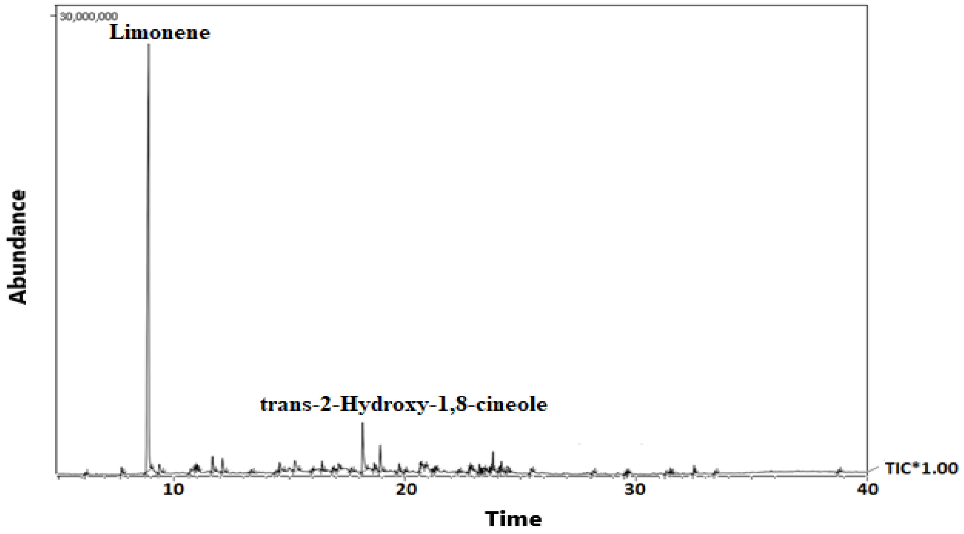
2.1. Gas Chromatography-Mass Spectrometry (GC-MS) Analysis

Essential oil from A. marmelos leaves produced 0.53% v/w of volatile compositions. The GC–MS chromatogram and the details of the volatile metabolites are shown in Figure 1 and Table 1, respectively. Limonene, a monoterpene (63.71%), was found to be the principal volatile component, along with the other monoterpenes trans-2-Hydroxy-1,8-cineole (6.85%), p-menth-2,8-dien-1-ol (2.29%) and menthyl acetate (2.02%). About 84.02% of the total volatile metabolites were monoterpenes, while the others were sesquiterpenes (6.01%), non-terpenes (8.01%), and Diterpene (0.28%).

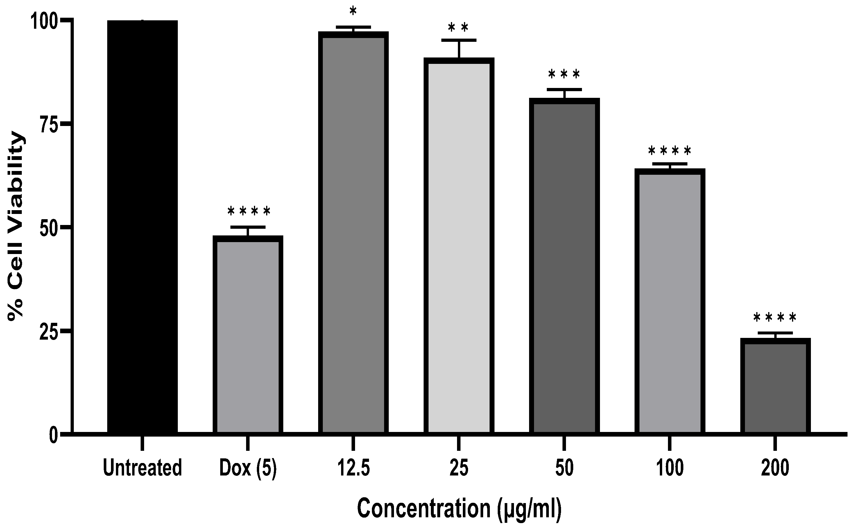
2.2. Anti-Carcinoma Effects of A. marmelos Leaves Essential Oil

Figure 2 shows the effects of A. marmelos leaf essential oil on cell viability at 12.5 µg/mL to 200 µg/mL by MTT assay in human epithelial carcinoma cells (KB cell line). The essential oil treatment significantly inhibited KB cell production after 24 h. In the present study, the IC50 value of essential oil was measured using the the linear regression equation y = mx + c, where y = 50 and m and c values were derived from the viability graph. The percentage of KB cells was significantly reduced by the oil dose-dependently, with an IC50 value of 132.52 µg/mL.
The MTT (3-(4,5-Dimethylthiazol-2-yl)-2,5-Diphenyltetrazolium Bromide) assay is a calorimetric assay for determining cytotoxicity and cell proliferation based on reducing tetrazolium dye MTT to formazan crystals. Mitochondrial lactate dehydrogenase (LDH) formed by living cells reduces MTT to insoluble formazan crystals (yellow colored). After dissolution in a suitable solvent, it showed a purple color; the intensity of the color is directly proportional to the number of viable cells. At 570 nm, it was measured spectrophotometrically. The outcomes of the statistical data of the cell cytotoxicity study by MTT demonstrated that A. marmelos leaf essential oil has significant cytotoxic potential against human epithelial carcinoma cells.

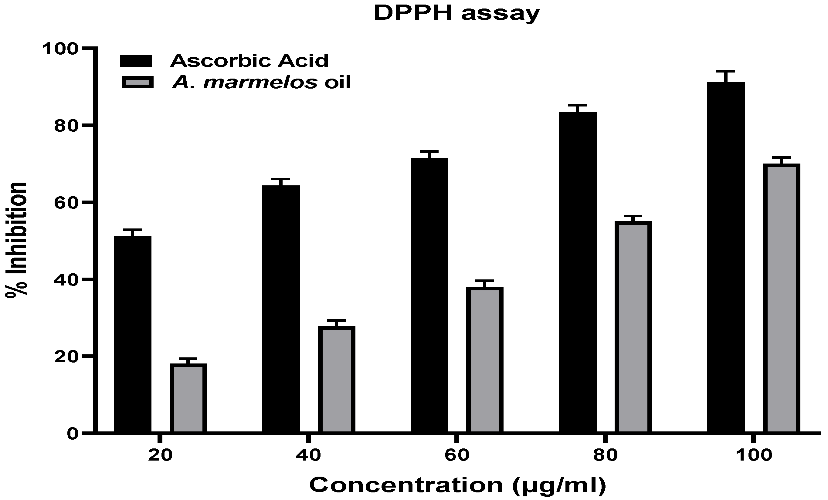
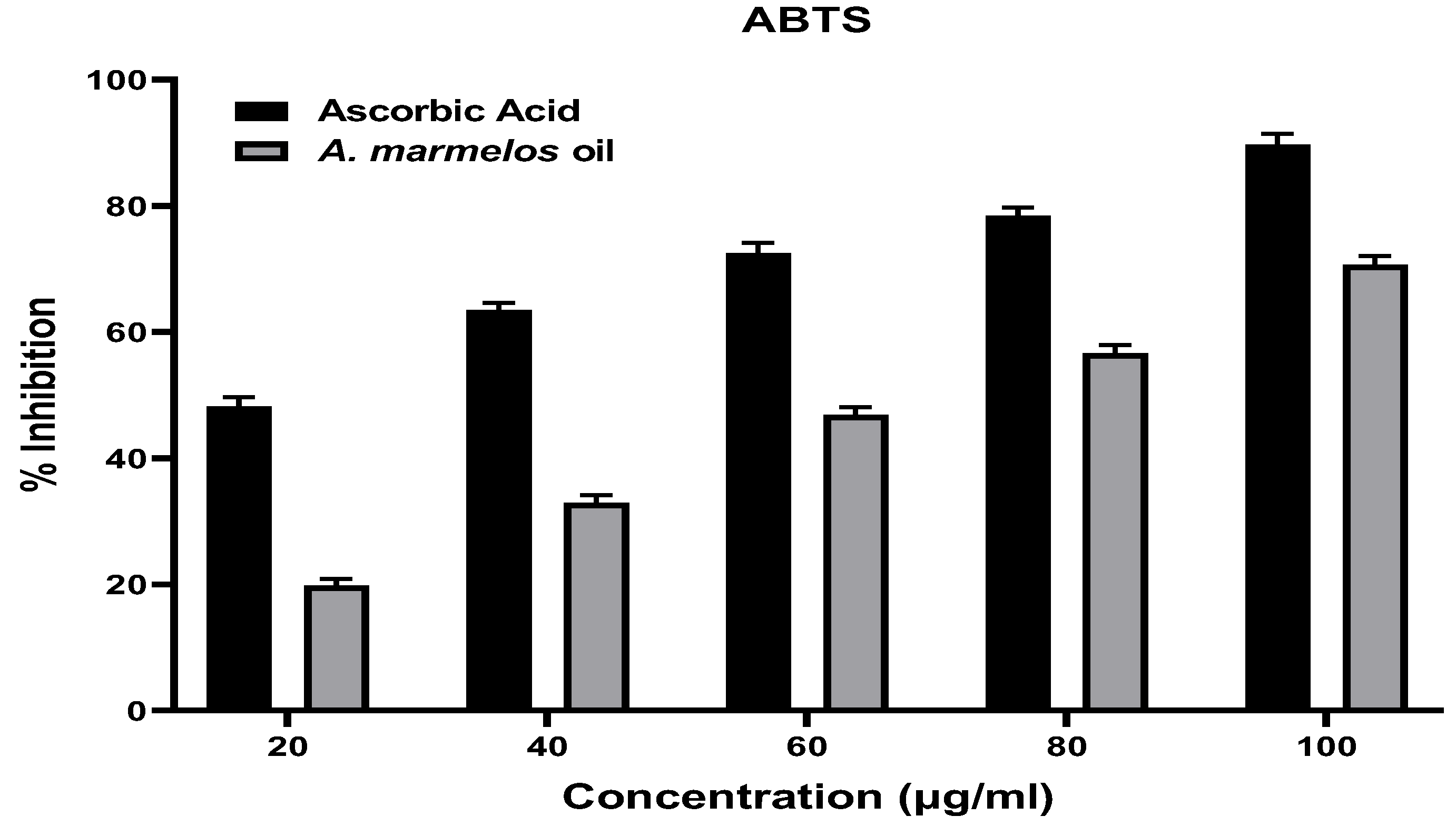
2.3. Antioxidant Activity of A. marmelos Leaves Essential Oil

To explore the antioxidant potential of A. marmelos leaf essential oil, the present study exhibited the DPPH and ABTS assays, and the outcomes of both free radical assays were reported in Figure 3. The percentage of inhibition of the DPPH-induced free radical capacity of A. marmelos leaf essential oil ranged from 18.05 ± 1.33 to 70.02 ± 1.6% at concentrations ranging from 20 µg/mL to 100 µg/mL. The range of inhibition of the ABTS-induced free radicals increased from 19.82 ± 1.05% to 70.7 ± 1.32% as the concentration increased from 20 µg/mL to 100 µg/mL. The inhibition of DPPH-induced free radicals by ascorbic acid ranged from 51.32 ± 1.55% to 91.11 ± 2.88% at concentrations ranging from 20 µg/mL to 100 µg/mL, while the range of inhibition of ABTS-induced free radicals increased from 48.27 ± 1.42% to 89.68 ± 1.72% as the concentration increased from 20 µg/mL to 100 µg/mL.

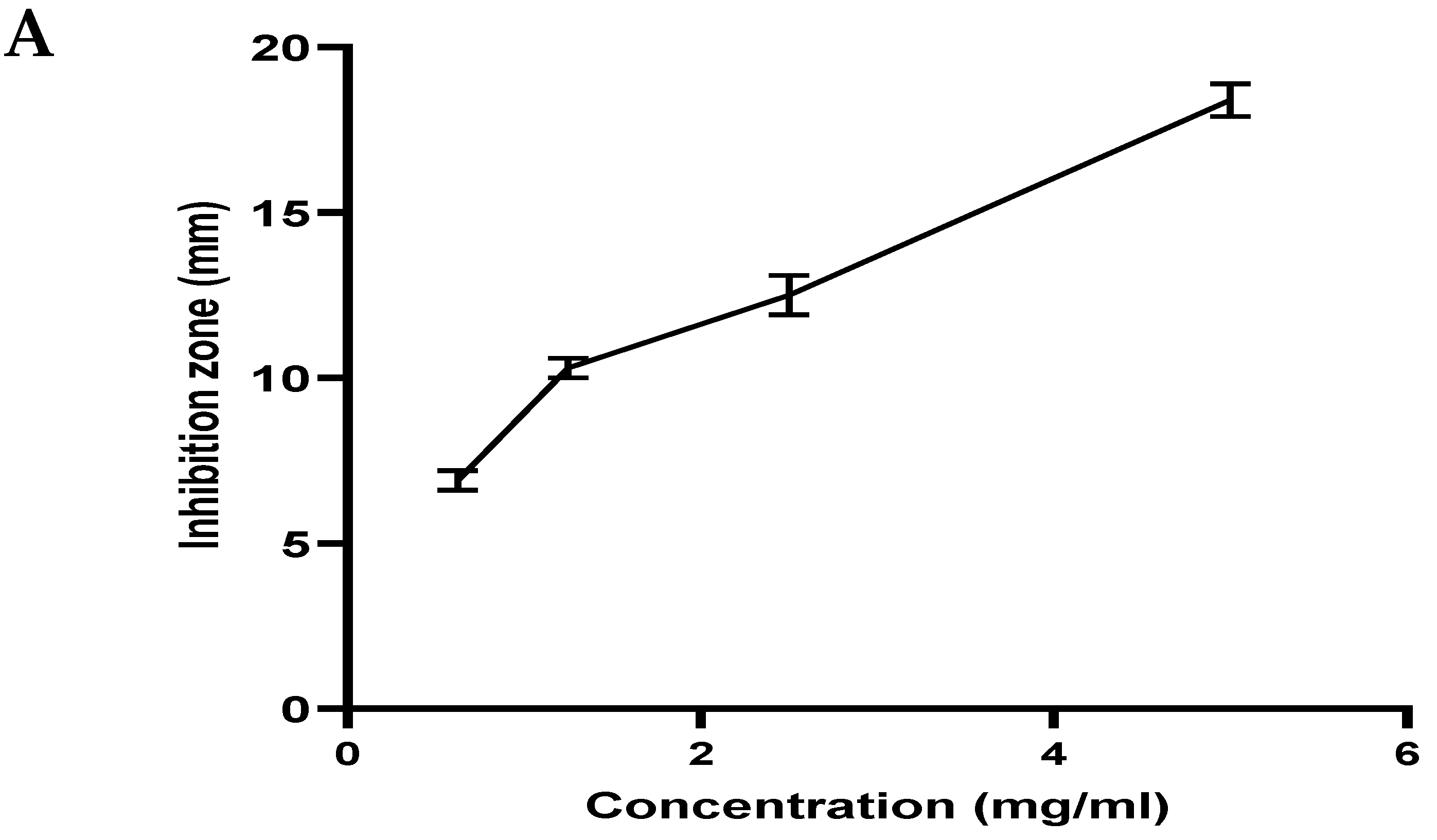
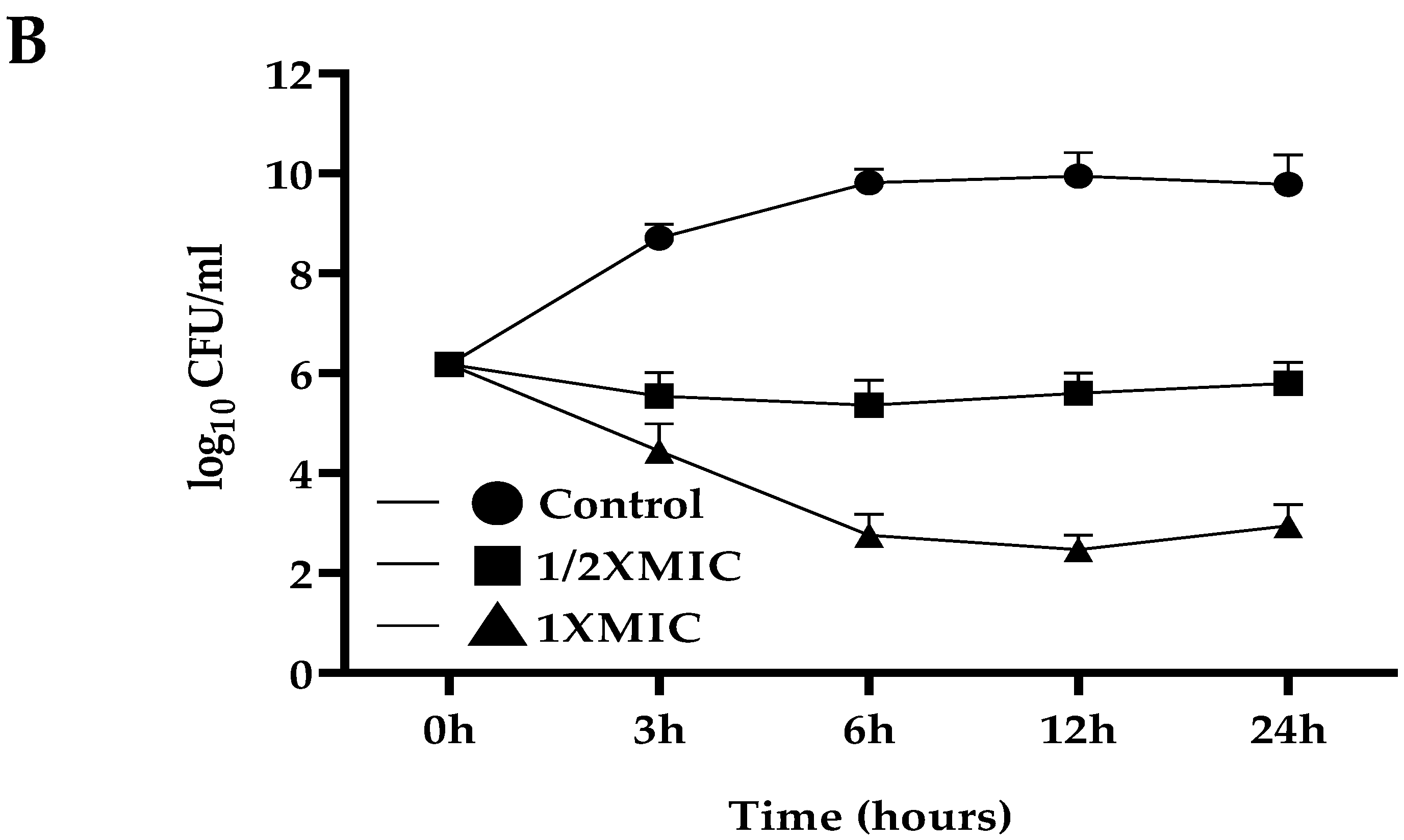
2.4. Anti-S. mutans Activity of A. marmelos Leaves Essential Oil

The agar diffusion assay explored the antibacterial activity of A. marmelos leaf essential oil. Figure 4A, shows the zone of inhibition of A. marmelos leaf essential oil against S. mutans. The zone of inhibition (ZOI) effect was observed from 0.625 mg/mL to 5 mg/mL against the S. mutans (18.4 ± 0.3 mm).
The cariogenic bacteria S. mutans exhibited a dose-dependent inhibition zone ranging from 6.9 to 18.48 mm, comparatively lower than the positive control, amoxicillin (at 10 µg/mL, inhibition zone 24.23 ± 0.8 mm). The MIC value of A. marmelos leaf essential oil against S. mutans was 0.25 mg/mL. The time-kill assay was performed using A. marmelos leaf essential oil against S. mutans, and the results are shown in Figure 4B. The outcomes of the control group (untreated S. mutans) exhibited approximately 6.5 to 9 (log10 CFU/mL) growth. In contrast, in the A. marmelos leaf essential oil 1/2 × MIC (0.125 mg/mL) treated group, the growth of S. mutans remained constant with 5.3 to 5.8 (log10 CFU/mL) growth. On the other hand, after the treatment with A. marmelos leaf essential oil 1 × MIC (0.25 mg/mL), the growth of S. mutans decreased drastically from 3 to 6 h, and it remained constant at approximately 2.7 to 2.59 (log10 CFU/mL) (Figure 4B). The plot deviated with 1 × MIC treatment inhibition of S. mutans growth, different from at 1/2 × MIC; hence, the results indicate that 1 × MIC of A. marmelos leaf essential oil showed a more rapid killing effect than 1/2 × MIC.

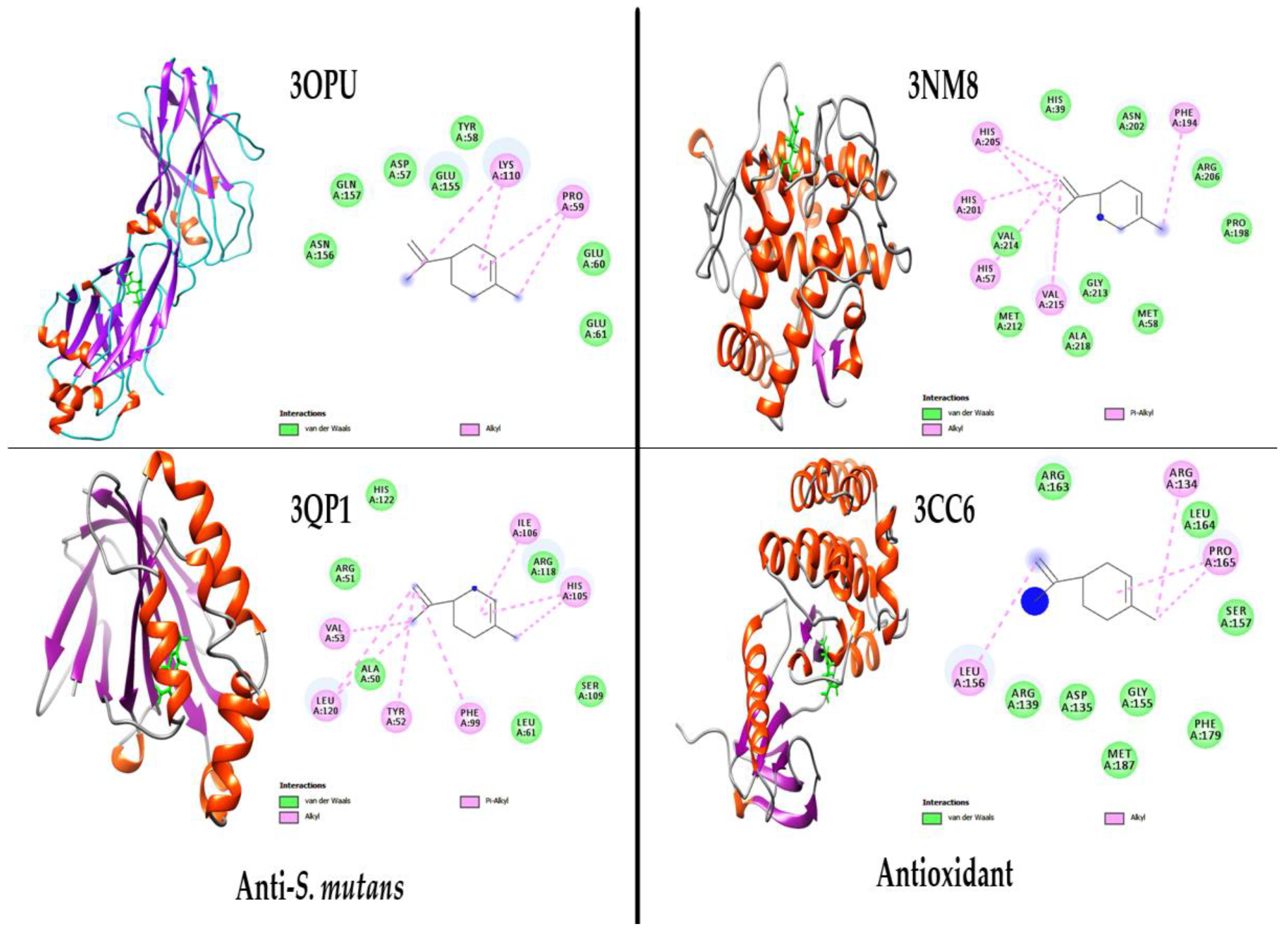
2.5. Molecular Docking Study of the Major Compound Limonene

Molecular docking studies were generally employed to assess the interaction between ligand–target proteins to explore the mechanism of action of the pharmacologically active compounds. The major volatile metabolite limonene was selected for the molecular docking study.
Molecular docking was performed against antioxidant target enzymes [tyrosinase (PDB: 3NM8) and tyrosine kinase 2 beta, PTK2B (PDB: 3CC6)], and antibacterial [C-terminal domain of S. mutans surface protein (PDB: 3OPU) and CviR protein (3QP1)] receptors were used to explore critical ligand-protein interactions (Figure 5). Table 2 shows the results of energy ΔG (kcal/mol), inhibition constant (µM), and amino acid residues.
The complexes formed between the tyrosinase (3NM8) of antioxidant enzymes and limonene ligands exhibit stable chemical bonds with the active sites of the enzymes PHE (A194), VAL (A215), HIS (A57), HIS (A201), and HIS (A205), with a high binding energy (−7.73 kcal/mol) and inhibition constant (898.74 µM). Similarly, the complexes formed between tyrosine kinase 2 beta (PTK2B) (3CC6), and limonene exhibit stable chemical bonds with the active sites of the enzymes ARG (A134), PRO (A165), and LEU (A156), with a high binding energy (−7.40 kcal/mol) and inhibition constant (859.54 µM).
The complexes formed between the C-terminal domain of Streptococcus mutans surface protein SpaP (PDB: 3OPU) and of anti-S. mutans protein and limonene ligands exhibit stable chemical bonds with the active sites of the enzymes PRO (A59) and LYS (A110), with a high binding energy (−7.31 kcal/mol) and inhibition constant (987.29 µM). Similarly, the complexes formed between the protein CviR ligand-binding domain, bound to the native ligand C6-HSL (PDB: 3QP1), and limonene exhibit stable chemical bonds with the active sites of the enzymes ILE (A106), HIS (A105), PHE (A99), TYR (A52), LEU (A120), and VAL (A53) with a high binding energy (−7.89 kcal/mol) and inhibition constant (764.34 µM).

3. Discussion

The volatile compounds in the present sample exhibited limonene (63.71%) as the primary component, with trans-2-hydroxy-1,8-cineole (6.85%), p-menth-2,8-dien-1-ol (2.29%), and menthyl acetate as minor components. On the other hand, the composition of Egyptian essential oil compounds of A. marmelos leaves exhibited α-phellandrene (20.97%), α-pinene (17.76%) and δ-care (16.37%) as the major volatile components, different from the present sample [32]. Verma and coworkers (2014) reported that the chemical composition of limonene (31.0–90.3%), along with α-phellandrene (E)-β-ocimene, α-pinene in the essential oil from Indian A. marmelos exhibited similar but different quality from the present samples [34]. In the other study, Singh and coworkers (2009) reported the GC-MS analysis of A. marmelos leaf essential oil and reported limonene as the primary component [33]. Pant and coworkers (2019) worked on Nepali A. marmelos leaf essential oil and reported that sesquiterpenes, β-Caryophyllene (26%) is a significant compound, different from the present analysis of volatile metabolites [12]. Jamal and coworkers (2017) worked on Bangladeshi A. marmelos leaf essential oil and reported that ledene oxide-(II) (18.16%) is a significant compound, different from the present analysis of volatile metabolites [30]. Interestingly, Kaur and coworkers (2006) reported monoterpenes limonene as the major volatile component and the presence of total monoterpene (93.7%) and sesquiterpene (3.1%), supporting the present finding of major component limonene as well as the volatile content of total monoterpenes (83.2%) and sesquiterpene (5.5%) [37]. Several other studies also supported the present finding that limonene is the volatile primary component of the essential oil of A. marmelos leaves [33,38].
Several studies focused on the anticancer effects of volatile oils as natural bioactive sources that can inhibit cancer cells [39]. The cytotoxic effect of A. marmelos leaf essential oil on the KB cells was studied using an MTT assay. The outcome suggested that the induction of A. marmelos inhibited the viability of KB cells and leaves the essential oil in a dose-dependent manner. The value of the standard doxorubicin compound IC50 (2.55 µg/mL) has been reported for the KB cells in the prior study and comparatively showed a very high IC50 value compared to the essential oil of the present sample [40]. Lampronti and coworkers (2019) reviewed Bangladeshi A. marmelos and its antiproliferative effects. They reported that it might inhibit the proliferation of human leukemic-K562, B-lymphoid Raji, T-lymphoid-Jurkat, erythro-leukemic HEL, melanoma-Colo38, and breast cancer-MCF7 and MDA-MB-231 tumor cell lines [41]. Poonkodi and coworkers (2019) studied Western Ghats Region-South India Aegle marmelos (L.) Correa essential oil that showed significant antiproliferative effects against HeLa (human cervical cancer cell line) and less sensitivity against the African Monkey-Kidney normal cell line, Vero [13]. Several other studies also supported the anticancer effects of the essential oil of A. marmelos leaves [2].
Veerappan and coworkers (2007) reported that intraperitoneal (I.P) administration of A. marmelos leaf extract at doses of 50–100 mg/kg body wt. for 14 successive days to male and female Wistar rats did not show any toxicity [42]. Limonene is a major compound found in the essential oil of A. marmelos leaves and it has reported nontoxic, non-carcinogenic activity, and no compound-related clinical signs or histopathologic lesions were observed at doses up to 500 mg/kg body wt. in both rats and mice [43]. Hence, the essential oil extracted from the leaves of A. marmelos may have nontoxic effects with significant anticancerous potential, possibly due to the presence of the major metabolite limonene [44,45].
Both antioxidant methods involve the reduction of a colored oxidant that is produced due to an electron transfer, and it is easily monitored via UV. The DPPH assay is based on reducing DPPH (purple) radical to 1,1-diphenyl-2-picryl hydrazine. In contrast, ABTS involves the reduction of ABTS (blue/green) to a colorless sulfonic acid [46]. The ascorbic acid’s (standard) antioxidant potential was the highest in both methods. A. marmelos leaf essential oil presented a significant antioxidant potential, with IC50 values of 72.71 μg/mL for the DPPH assay and 67.33 μg/mL for the ABTS assay. The antioxidant activity of A. marmelos leaf essential oil exhibited dose-dependent scavenging activities in both assays, representing the significant antioxidant potential of A. marmelos leaf essential oil. Several studies have been conducted to determine the antioxidant properties of A. marmelos leaf essential oil [3,47]. The antioxidant activity may be due to limonene or other compounds in A. marmelos leaf essential oil [13,48].
Aegle marmelos can treat several conditions, including cancer, infections, and pain [5,6]. The present studies highlight the use of Aegle marmelos leaf essential oil in the treatment of oral cancer and dental infections. Both types of bacteria (Gram-positive and Gram-negative) are responsible for oral or dental infections. A scientific report suggested that Gram-positive bacteria from the genus Streptococcus are the most responsible [49]. S. mutans of the Streptococcus genus are most responsible for oral infections and dental caries [50]. The outcome of the present antimicrobial study of A. marmelos leaf essential oil showed significant inhibition against S. mutans, with MIC values of 0.25 mg/mL. Several scientific studies have shown the antimicrobial effects of Aegle marmelos leaf essential oil against Gram-positive and Gram-negative bacteria and fungi [33,38]. The antimicrobial effect of Aegle marmelos leaf essential oil are not restricted to S. mutans only, they also affect other types of oral microorganisms, such as Bacillus cereus, S. aureus, Corynebacterium diphtheriae, Candida albicans, Bacillus subtilis, Escherichia coli and Candida albicans, which are responsible for oral infections and dental caries [32,38]. The biological activities of Aegle marmelos leaf essential oil may be due to a high content of volatile metabolite limonene (63.71%) or several other metabolites such as trans-2-Hydroxy-1,8-cineole (6.85%), Methyl acetate (2.83%), p-Menth-2,8-dien-1-ol (2.29%) [51,52,53,54]. In the literature and review, several studies have reported that the natural monoterpene limonene has antioxidant and antibacterial activities [55].
The aim of this study was to explore the anticancerous, antioxidant, and antibacterial activity of Aegle marmelos leaf essential oil against oral disease conditions. Hence, a molecular docking study of limonene was simulated with tyrosinase (3NM8), tyrosine kinase 2 beta, PTK2B (3CC6), C-terminal domain of Streptococcus mutans surface protein SpaP (3OPU), and CviR ligand-binding domain bound to the native ligand C6-HSL (3QP1) targets proteins. The binding energy inhibition constant with several residues of H-bonding outcomes of enzyme tyrosinase with ligand limonene interaction supported the antioxidant properties of the A. marmelos leaf essential oil, mainly due to the presence of limonene. The inhibition of tyrosinase enzyme played a pivotal role in preventing melanin biosynthesis in the skin and hydroxylation of L-tyrosine to DOPA (3,4-dihydroxyphenylalanine) and further oxidation of DOPA to dopaquinone [56]. Therefore, tyrosinase inhibitors are an attractive target for evaluating antioxidant potential. Similarly, the binding energy inhibition constant with several residues of H-bonding outcomes of receptor tyrosine kinase 2 beta, PTK2B with ligand limonene interaction supported the antioxidant properties due to the presence of limonene. The antioxidant potential is mainly due to the inhibition of tyrosinase and tyrosine kinase 2 beta [57]. The antioxidant vitamin C has been reported previously to inhibit tyrosinase production [58].
To predict the anti-S. mutans mechanism of Aegle marmelos leaf essential oil, the major metabolite limonene was docked to the C-terminal domain of Streptococcus mutans surface protein SpaP. The results showed a high binding energy and inhibition constant with few H-bonding residues. The inhibition of the C-terminal domain of the Streptococcus mutant’s surface protein was essential in preventing dental caries [59]. Therefore, Streptococcus mutans surface protein inhibitor limonene may be an attractive ligand for anti-S. mutans or anticaries potential. Similarly, the binding energy and inhibition constant, with several residues of the H-bonding outcome of receptor CviR ligand-binding domain, bound to the native ligand C6-HSL with several residues conferring H-bonding stability to the complex, were explored with ligand limonene interaction and supported the antibacterial potential of limonene. The previous study suggested that the antibacterial potential of limonene is mainly due to the inhibition of the CviR ligand-binding receptor [60].

4. Materials and Methods

4.1. Chemicals

The cell line (oral adenocarcinoma cell line KB—human) was obtained from the National Center for Cell Science (NCCS, Pune, India), along with Dulbecco’s modified Eagle’s medium (DMEM), high glucose medium (Cat No: AL007, HiMedia, (Mumbai, Maharashtra, India), MTT reagent (HiMedia), fetal bovine serum (FBS), and Dulbecco’s phosphate buffered saline (D-PBS, Himedia). Dimethyl sulfoxide (DMSO), doxorubicin, ascorbic acid, and amoxicillin were purchased from Sigma-Aldrich (Mumbai, India). All media used for the antimicrobial studies were purchased from HiMedia Laboratories (Mumbai, India). 96-well plates for cell culture (Kennebunk, ME, USA) and all other chemicals (Analytical grade) used in the present study were purchased from HiMedia and Sigma Aldrich (Mumbai, India).

4.2. Methods

4.2.1. Extraction of Essential Oil

Aegle marmelos (Indian Bael) leaves were collected from the northern part of India (Lucknow, Utter Pradesh, India) close to the Integral University campus area in August 2022. The sample was authenticated (IU/PHAR-HRBD/22/02), and the sample specimen was deposited at the College of Pharmacy, Integral University (Lucknow, India). The leaf samples were shed, dried, powdered, and volatile metabolites were isolated using the hydro-distillation (Clevenger-type) method and purified by following the preceding method [29]. The purified A. marmelos leaf essential oil was weighed and transferred into an air-tight vial (amber-colored) under the deep refrigeration (−20 °C) until use.

4.2.2. Gas-Chromatography/Mass Spectrometry (GC-MS) Analysis of Essential Oil

The volatile components present in crude Aegle marmelos leaf essential oil were explored by GC/MS instrumentation investigation. The GC/MS-QP2010 equipped with an auto-sampler auto-injector (AOC-20si) instrument (Shimadzu, Tokyo, Japan) was used in this study. The conditions, such as the temperature of the column oven, injection volume, and documentation of compounds, were adopted with slight modifications to the reported analysis conditions [29]. Then, 100 mg of AMLEO was dissolved in 10 mL hexane (10 mg·mL−1), and 1 µL solution was injected in a split-mode (ratio, 1:10) in an RTX-5MS column where the length was 30 m, the diameter was 0.25 mm, and the film thickness of the stationary phase was 0.25 µm (Shimadzu, Japan). The system was operated at 70 eV, helium was used as a carrier gas, and the gas flow rate was set at 1.21 mL/min, with the column head pressure of 69 kPa. The injection temperature was fixed at 260.00 °C. The oven temperature was first initiated at 50 °C for 2 min; then, the temperature was increased to 180 °C (with the rate of 5 °C/min), held for 1 min, and then to 280 °C (with the rate of 15 °C/min), at which the column temperature was maintained for 15 min. The scan mode 40–650 m/z was used to explore the spectra of volatile compounds. The documentation of Aegle marmelos leaf volatile metabolites was identified using the National Institute of Standards and Technology MS-library (NIST 14). The percentage area and retention indices (RIs) of each volatile compound were calculated and observed relative to n-alkanes homologous series (C7–C30) and by comparing their mass spectra libraries (NIST) with the literature [61].

4.2.3. Cytotoxicity Assay of Essential Oil

In the present study (KB) cells (human mouth epidermal carcinoma) were maintained in a high supplement of glucose medium with fetal bovine serum (10% FBS) and antibiotic-antifungal solution (1%) in an atmosphere of CO2 (5%), O2 (18–20%) at 37 °C in a CO2 incubator (Heal-force, Shanghai, China) and every two days they were sub-cultured. The KB cell line was performed preceding the earlier method [62] with some modifications. The cell suspension (200 μL) was seeded into a 96-well plate (2 × 10⁴ cells/well) and, for approximately 24 h, it was allowed to grow. An appropriate Aegle marmelos oil sample concentration was added and incubated for 24 h at 37 °C. Plates were removed from the CO2 incubator, and MTT reagent (3-(4,5-dimethylthiazol-2-yl)-2,5-diphenyl-2H-tetrazolium bromide) was added following the concentration of 0.05% of total volume. The plate was further wrapped in aluminum foil and incubated for 3 h. After 3 h, MTT reagents were removed, and 100 μL DMSO was added carefully. The absorbance was measured at 570 nm using an enzyme-linked immunosorbent assay plate reader (ELISA, Biorad-PW41, (Hercules, CA, USA). Cell viability percentage was calculated using the given formula (Equation (1)), and using a linear regression equation (Y = mx + c) the IC50 was determined. From the viability diagram, Y = 50, and M and C values were identified.
%   C e l l   V i a b i l i t y = ( A b s o r b a n c e   o f   T r e a t e d   C e l l / A b s o r b a n c e   o f   C o n t r o l   C e l l s ) × 100
Morphological changes of cells were studied at different concentrations (12.5–200 μg/mL) after treatment of KB with Aegle marmelos oil samples using fluorescence microscopy (Leica Microsystems Inc., Buffalo Grove, IL, USA) at a magnification of 100×.

4.2.4. Free Radical Scavenging Activity of Essential Oil

The method of 2,2-diphenyl-1-picrylhydrazyl (DPPH) is based on the free radical (DPPH•) produced by the oxidation of DPPH with methanol to confirm by development of purple/violet color. The antioxidant compounds have a hydrogen denoting ability to change from violet to a yellow color. In the present study, free radical scavenging powers of Aegle marmelos oil were analyzed using the earlier DPPH assay [63], with some modifications. Concisely, a stock solution (1000 µg/mL) of samples and the standard solution was prepared. The DPPH-cation solution (0.1 mM) was prepared in methanol. Into the 5 mL test tubes, 2 mL of DPPH was added and then the standard and samples were added separately to make a final concentration of 20–100 µg/mL. The reaction mixtures were correctly mixed and incubated in the dark for 30 min at 25 °C. The absorbance was recorded at 517 nm using UV-Visible-Spectrophotometer (Double-beam Systronics India (Pvt.) Ltd., Ahmedabad, Gujarat, India). The percentage inhibition (% I) was documented as mean ± standard deviation of triplicate.
The method of 2,2′-azino-bis (3-ethylbenzothiazoline-6-sulfonic acid) (ABTS) is based on the free radical (ABTS•+) produced by the oxidation of ABTS with potassium persulfate, confirmed by development of bluish green color. The antioxidant compounds have a hydrogen denoting ability to change from bluish green to colourless. In the present study, free radical scavenging powers of Aegle marmelos oil were analyzed using the earlier ABTS assay [64], with some modifications. Consciously, 7.5 mmol/L ABTS and 2.5 mmol/L potassium persulfate were prepared in methanol. ABTS∙+ (radical cation) was created by mixing ABTS and potassium persulfate, allowing it to keep in the dark for 16 h at room temperature before use. Later, the free radical cation stock solution (ABTS∙+) was diluted with methanol (UV Absorption: 0.90 ± 0.02 at 734 nm). Similar to the DPPH assay, in the 5 mL test tube, 1.9 mL of ABTS∙+ solution and standard samples were added to make a final concentration of 20–100 µg/mL to each test tube. The absorbance was recorded after 7 min at 737 using UV-Visible-Spectrophotometer (Double-beam, Systronics India (Pvt.) Ltd., Ahmedabad, Gujarat, India). The percentage radical scavenging (%RA) was documented as mean ± standard deviation of triplicate (Equation (2)).
% RA = [(Ab (DPPH/ABTS − Ab (sample/standard)/Ab (DPPH/ABTS) × 100)]
A graph plotting the percentage of inhibition versus concentration was used to calculate the IC50 value (the essential oil concentration providing 50% inhibition) of antioxidant activity.

4.2.5. Anti-S. mutans Activity of Essential Oil

The well-diffusion method determined the antibacterial potential of Aegle marmelos oil [54]. Plates containing nutrient agar (Hi-media, Mumbai, India) media were used to test Aegle marmelos oil effects on Streptococcus mutans (MTCC 389). The inoculum of the selected bacterial suspension was prepared in 10 mL nutrient broth. The suspension turbidity was adjusted to 1.5 × 10⁶ colony-forming units/mL using 0.5 McFarland. Then, 50 μL of this suspension was poured onto fresh solidified agar media plates. Four wells of 6 mm diameter were bored in the inoculated media. Each board was filled with 50 μL of various concentrations (50, 100, and 200 μg/mL) of Aegle marmelos oil and allowed to diffuse for 30 min, followed by incubation at 37 °C for 24 h. The inhibition of growth zones around the well was measured in mm, and means ± standard deviation were calculated. The solutions dimethyl sulfoxide (10%) and amoxicillin (10 μg/mL) were also used as a negative and positive controls, respectively. For the minimum inhibitory concentration (MIC), different concentrations (1.25, 2.5, 5, to 10 μg/mL) of AMLEO were prepared in 10% DMSO. The MIC was determined using the broth dilution CLSI method [33]. To each diluted tube, 100 µL of bacterial inoculum was seeded in nutrient broth (NB). Aliquots (100 μL) of each dilution were added in each tube and incubated at 37 °C for 24 h. The lowermost concentration that produced no visible growth was recorded as MIC. To investigate the bactericidal anti-S. mutans activity of Aegle marmelos oil, the time-kill assay of Aftab et al. was performed [33]. The cultures of S. mutans were incubated on the broth of nutrient agar and cultured at 37 °C for 10 h.
The suspension of S. mutans was centrifuged and resuspended in saline at 1.5 × 10⁶ CFU/mL. The S. mutans suspensions were treated with broth medium containing different concentrations of Aegle marmelos oil according to the MIC, mixed, and cultured at 37 °C. At the selected intervals (0, 3, 6, 12, and 24 h), samples (1/2 × MIC and 1 × MIC) were removed, diluted with saline, and re-cultured and incubation of the plates at 37 °C for 24 h in a broth medium. After that, the CFU/mL (colony forming unit/mL) was calculated, and a graph, log 10 CFU/mL versus time, was plotted to explore the time-kill assay.

4.2.6. Molecular Docking Effects of Major Compound Limonene

The molecular docking study of the ligands (D-limonene) and the 3D structure of the protein molecules were performed using ADT to determine their potential binding affinities. The tool ADT (AutoDock) created the file from the conventional RCSB PDB files [65,66]. For the molecular docking study, 3NM8: tyrosinase X-ray structure of B. megaterium tyrosinase [67] and 3CC6: protein tyrosine kinase 2 beta (PTK2B) were selected for the prediction of antioxidant properties of limonene [68] For the anti-S. mutans docking study, 3OPU: C-terminal domain of Streptococcus mutans surface proteins SpaP and 3QP1: CviR ligand-binding domains bound to the native ligand C6-HSL were selected for the inhibitory effect of limonene [69,70].
To draw the 3D input files, only the PDB structure of the protein part was taken by removing additional ions, atoms, and molecules [71]. All protein targets were actively minimized by using the CHARMM software tool [72]. The 2D structures of D-limonene (CID: 440917) ligands from the PubChem database in the standard data format (SDF) were retrieved [33]. The standard 2D data format was transformed to PDB-3D using the BIOVIA discovery studio visualizer [73]. The ligands and receptors, a file of grid parameters, and the PDF file of docking parameters were produced to execute the molecular docking. The grid box size for the coordinates x, y, and z was 40 Å, with a grid spacing of 0.4200 Å, and center of the grid box in the x (28.095 Å), y (27.0005 Å), and z (8.4735 Å). Around the protein molecule, the grid frame was drawn with different grid points on the axes of x, y, and z and a maximum distance (1.00) between two successive grids to deliver sufficient space for ligand movement.
Ten (10) runs were executed for the protein and ligand. The minimum free binding energy (ΔG) and inhibition constant (Ki) were considered selective parameters to pick one of the best positions of the ligands bound to the binding cleft of the receptor [74].

4.3. Statistical Analysis

Statistics of the present study were explored using GraphPad Prism 9.5.0 software (GraphPad Software, Inc., San Diego, CA, USA) and are presented as mean ± standard deviation. The test sample concentration that created 50% inhibition (IC50 value) was evaluated using the graph by plotting the inhibition (%) versus the concentration. A significant p-value (* p < 0.05, ** p < 0.01, *** p < 0.001) was determined when the test sample was compared with a standard compound using a paired t-test.

5. Conclusions

In conclusion, the research conducted on Aegle marmelos leaves indicates that they possess valuable natural therapeutic properties for managing oral cancer and infections. The essential oil extracted from these leaves exhibits significant anticancer, antioxidant, and anti-cariogenic effects. Specifically, the volatile metabolites in the oil inhibit DPPH- and ABTS-induced free radicals as well as Streptococcus mutans bacteria growth in in vitro studies. Molecular modelling of limonene compounds present in Aegle marmelos leaves demonstrated theoretical inhibitory effects on proteins associated with antioxidant and antibacterial activity, which supports our experimental results indicating strong ligand-protein-binding energy interactions (ΔG) and inhibition constants (µM). Therefore, it is suggested that Aegle marmelos leaves can be considered an alternative source for managing human mouth epidermal carcinoma as well as bacterial infections due to their potent anticarcinoma potential against KB cells along with remarkable performance against S.mutans. Therefore, A. marmelos leaves could be considered as an alternative treatment option to conventional medicines for oral cancer and infections with their well-established traditional medicinal benefits combined with modern scientific validation through this study’s results.

Author Contributions

Conceptualization, A.H.A. and A.A.; Methodology, M.J.A. and M.F.B.; Software, M.M.K. and M.J.A.; Validation, T.J. and M.M.K.; Formal analysis, M.J.A. and A.A.; Investigation, M.F.B., A.A. and T.J.; Resources, T.J. and M.F.B.; Data curation, M.J.A. and M.F.B.; Writing—original draft, A.A. and M.M.K.; Writing—review & editing, A.H.A. and M.J.A.; Visualization, M.M.K. and A.A.; Supervision, A.H.A. and A.A.; Project administration, M.J.A. and T.J.; Funding acquisition, A.H.A. and M.F.B. All authors have read and agreed to the published version of the manuscript.

Funding

This research work received no external funding.

Institutional Review Board Statement

Not applicable.

Informed Consent Statement

Not applicable.

Data Availability Statement

The data presented in this study are available on request from the corresponding author.

Acknowledgments

This study is supported via funding from Prince Sattam Bin Abdulaziz University project number (PSAU/2023/R/1444).

Conflicts of Interest

The authors declare no conflict of interest.

References

  1. Brijesh, S.; Daswani, P.; Tetali, P.; Antia, N.; Birdi, T. Studies on the antidiarrhoeal activity of Aegle marmelos unripe fruit: Validating its traditional usage. BMC Complement. Altern. Med. 2009, 9, 47. [Google Scholar] [CrossRef] [PubMed]
  2. Pathirana, C.K.; Madhujith, T.; Eeswara, J. Bael (Aegle marmelos L. Corrêa), a Medicinal Tree with Immense Economic Potentials. Adv. Agric. 2020, 2020, 8814018. [Google Scholar] [CrossRef]
  3. Rahman, S.; Parvin, R. Therapeutic potential of Aegle marmelos (L.)-An overview. Asian Pacific J. Trop. Dis. 2014, 4, 71–77. [Google Scholar] [CrossRef]
  4. Sarkar, T.; Salauddin, M.; Chakraborty, R. In-depth pharmacological and nutritional properties of bael (Aegle marmelos): A critical review. J. Agric. Food Res. 2020, 2, 100081. [Google Scholar] [CrossRef]
  5. Jagetia, G.C.; Venkatesh, P.; Baliga, M.S. Aegle marmelos (L.) Correa inhibits the proliferation of transplanted Ehrlich ascites carcinoma in mice. Biol. Pharm. Bull. 2005, 28, 58–64. [Google Scholar] [CrossRef] [PubMed]
  6. Dhankhar, S.; Ruhil, S.; Balhara, M.; Dhankhar, S.; Chhillar, A.K. Aegle marmelos (Linn.) Correa: A potential source of Phytomedicine. J. Med. Plants Res. 2011, 5, 1497–1507. [Google Scholar]
  7. Iqbal, T.; Hussain, A.I.; Chatha, S.A.S.; Naqvi, S.A.R.; Bokhari, T.H. Antioxidant activity and volatile and phenolic profiles of essential oil and different extracts of wild mint (Mentha longifolia) from the Pakistani flora. J. Anal. Methods Chem. 2013, 2013, 536490. [Google Scholar] [CrossRef]
  8. Xu, X.; Liu, A.; Hu, S.; Ares, I.; Martínez-Larrañaga, M.R.; Wang, X.; Martínez, M.; Anadón, A.; Martínez, M.A. Synthetic phenolic antioxidants: Metabolism, hazards and mechanism of action. Food Chem. 2021, 353, 129488. [Google Scholar] [CrossRef]
  9. Lugo-Flores, M.A.; Quintero-Cabello, K.P.; Palafox-Rivera, P.; Silva-Espinoza, B.A.; Cruz-Valenzuela, M.R.; Ortega-Ramirez, L.A.; Gonzalez-Aguilar, G.A.; Ayala-Zavala, J.F. Plant-derived substances with antibacterial, antioxidant, and flavoring potential to formulate oral health care products. Biomedicines 2021, 9, 1669. [Google Scholar] [CrossRef]
  10. Salman, B.N.; Darvish, S.; Goriuc, A.; Mazloomzadeh, S.; Hossein Poor Tehrani, M.; Luchian, I. Salivary oxidative stress markers’ relation to oral diseases in children and adolescents. Antioxidants 2021, 10, 1540. [Google Scholar] [CrossRef]
  11. Pytko-Polończyk, J.; Stawarz-Janeczek, M.; Kryczyk-Poprawa, A.; Muszyńska, B. Antioxidant-rich natural raw materials in the prevention and treatment of selected oral cavity and periodontal diseases. Antioxidants 2021, 10, 1848. [Google Scholar] [CrossRef] [PubMed]
  12. Pant, P.; Sut, S.; Castagliuolo, I.; Gandin, V.; Maggi, F.; Gyawali, R.; Dall’Acqua, S. Sesquiterpene rich essential oil from Nepalese Bael tree (Aegle marmelos (L.) Correa) as potential antiproliferative agent. Fitoterapia 2019, 138, 104266. [Google Scholar] [CrossRef]
  13. Poonkodi, K.; Vimaladevi, K.; Suganthi, M.; Gayathri, N. Essential Oil Composition and Biological Activities of Aegle marmelos (L.) Correa Grown in Western Ghats Region-South India. J. Essent. Oil-Bear. Plants 2019, 22, 1013–1021. [Google Scholar] [CrossRef]
  14. Nocini, R.; Lippi, G.; Mattiuzzi, C. The worldwide burden of smoking-related oral cancer deaths. Clin. Exp. Dent. Res. 2020, 6, 161–164. [Google Scholar] [CrossRef] [PubMed]
  15. Pires, F.R.; Ramos, A.B.; de Oliveira, J.B.C.; Tavares, A.S.; de Luz, P.S.R.; dos Santos, T.C.R.B. Oral squamous cell carcinoma: Clinicopathological features from 346 cases from a single oral pathology service during an 8-year period. J. Appl. Oral Sci. 2013, 21, 460–467. [Google Scholar] [CrossRef]
  16. Rivera, C.; Venegas, B. Histological and molecular aspects of oral squamous cell carcinoma. Oncol. Lett. 2014, 8, 7–11. [Google Scholar] [CrossRef] [PubMed]
  17. Sharifi-Rad, J.; Sureda, A.; Tenore, G.C.; Daglia, M.; Sharifi-Rad, M.; Valussi, M.; Tundis, R.; Sharifi-Rad, M.; Loizzo, M.R.; Oluwaseun Ademiluyi, A.; et al. Biological activities of essential oils: From plant chemoecology to traditional healing systems. Molecules 2017, 22, 70. [Google Scholar] [CrossRef] [PubMed]
  18. Lok, B.; Babu, D.; Tabana, Y.; Dahham, S.S.; Adam, M.A.A.; Barakat, K.; Sandai, D. The Anticancer Potential of Psidium guajava (Guava) Extracts. Life 2023, 13, 346. [Google Scholar] [CrossRef]
  19. Marsh, P.D. Dental plaque as a biofilm and a microbial community—Implications for health and disease. BMC Oral Health 2006, 6, S14. [Google Scholar] [CrossRef]
  20. Qiu, W.; Zhou, Y.; Li, Z.; Huang, T.; Xiao, Y.; Cheng, L.; Peng, X.; Zhang, L.; Ren, B. Application of Antibiotics/Antimicrobial Agents on Dental Caries. BioMed Res. Int. 2020, 2020, 5658212. [Google Scholar] [CrossRef]
  21. Kumar, M.; Prakash, S.; Radha; Kumari, N.; Pundir, A.; Punia, S.; Saurabh, V.; Choudhary, P.; Changan, S.; Dhumal, S.; et al. Beneficial role of antioxidant secondary metabolites from medicinal plants in maintaining oral health. Antioxidants 2021, 10, 1061. [Google Scholar] [CrossRef] [PubMed]
  22. Deo, P.N.; Deshmukh, R. Oral microbiome: Unveiling the fundamentals. J. Oral Maxillofac. Pathol. 2019, 23, 122. [Google Scholar] [CrossRef] [PubMed]
  23. Bertolini, M.; Costa, R.C.; Barão, V.A.R.; Cunha Villar, C.; Retamal-Valdes, B.; Feres, M.; Silva Souza, J.G. Oral Microorganisms and Biofilms: New Insights to Defeat the Main Etiologic Factor of Oral Diseases. Microorganisms 2022, 10, 2413. [Google Scholar] [CrossRef] [PubMed]
  24. Llor, C.; Bjerrum, L. Antimicrobial resistance: Risk associated with antibiotic overuse and initiatives to reduce the problem. Ther. Adv. Drug Saf. 2014, 5, 229–241. [Google Scholar] [CrossRef] [PubMed]
  25. Thosar, N.; Basak, S.; Bahadure, R.N.; Rajurkar, M. Antimicrobial efficacy of five essential oils against oral pathogens: An in vitro study. Eur. J. Dent. 2013, 7, S071–S077. [Google Scholar] [CrossRef]
  26. Zomorodian, K.; Ghadiri, P.; Saharkhiz, M.J.; Moein, M.R.; Mehriar, P.; Bahrani, F.; Golzar, T.; Pakshir, K.; Fani, M.M. Antimicrobial activity of seven essential oils from Iranian aromatic plants against common causes of oral infections. Jundishapur J. Microbiol. 2015, 8, e17766. [Google Scholar] [CrossRef]
  27. Tambur, Z.; Miljković-Selimović, B.; Opačić, D.; Vuković, B.; Malešević, A.; Ivančajić, L.; Aleksić, E. Inhibitory effects of propolis and essential oils on oral bacteria. J. Infect. Dev. Ctries. 2021, 15, 1027–1031. [Google Scholar] [CrossRef]
  28. Bardají, D.K.R.; Reis, E.B.; Medeiros, T.C.T.; Lucarini, R.; Crotti, A.E.M.; Martins, C.H.G. Antibacterial activity of commercially available plant-derived essential oils against oral pathogenic bacteria. Nat. Prod. Res. 2016, 30, 1178–1181. [Google Scholar] [CrossRef] [PubMed]
  29. Alam, A.; Jawaid, T.; Alsanad, S.M.; Kamal, M.; Balaha, M.F. Composition, Antibacterial Efficacy, and Anticancer Activity of Essential Oil Extracted from Psidium guajava (L.) Leaves. Plants 2023, 12, 246. [Google Scholar] [CrossRef]
  30. Jamal, M.A.H.M.; Rahman, M.S.; Hossain, M.B.; Sharma, S.P.; Chung, H.J.; Kim, H.J.; Hong, S.T. Antibacterial Properties and Chemical Composition of Essential Oil from Aegle marmelos (L.) Corr. Leaves Growing in Bangladesh. J. Essent. Oil-Bear. Plants 2017, 20, 155–174. [Google Scholar] [CrossRef]
  31. Balakumar, S.; Rajan, S.; Thirunalasundari, T.; Jeeva, S. Antifungal activity of Aegle marmelos (L.) Correa (Rutaceae) leaf extract on dermatophytes. Asian Pac. J. Trop. Biomed. 2011, 1, 309–312. [Google Scholar] [CrossRef] [PubMed]
  32. Ibrahim, N.A.; El-Sakhawy, F.M.; Mohammed, M.M.D.; Farid, M.A.; Abdel-Wahed, N.A.M.; Deabes, D.A.H. Chemical composition, antimicrobial and antifungal activities of essential oils of the leaves of Aegle marmelos (L.) Correa growing in Egypt. J. Appl. Pharm. Sci. 2015, 5, 001–005. [Google Scholar] [CrossRef]
  33. Singh, P.; Kumar, A.; Dubey, N.K.; Gupta, R. Essential Oil of Aegle marmelos as a Safe Plant-Based Antimicrobial Against Postharvest Microbial Infestations and Aflatoxin Contamination of Food Commodities. J. Food Sci. 2009, 74, M302–M307. [Google Scholar] [CrossRef] [PubMed]
  34. Verma, R.S.; Padalia, R.C.; Chauhan, A. Essential oil composition of Aegle marmelos (L.) Correa: Chemotypic and seasonal variations. J. Sci. Food Agric. 2014, 94, 1904–1913. [Google Scholar] [CrossRef] [PubMed]
  35. Ekins, S.; Mestres, J.; Testa, B. In silico pharmacology for drug discovery: Methods for virtual ligand screening and profiling. Br. J. Pharmacol. 2007, 152, 9–20. [Google Scholar] [CrossRef]
  36. Alam, A.; Jawaid, T.; Alam, P. In vitro antioxidant and anti-inflammatory activities of green cardamom essential oil and in silico molecular docking of its major bioactives. J. Taibah Univ. Sci. 2021, 15, 757–768. [Google Scholar] [CrossRef]
  37. Kaur, H.P.; Garg, S.N.; Sashidhara, K.V.; Yadav, A.; Naqvi, A.A.; Khanuja, S.P.S. Chemical composition of the essential oil of the twigs and leaves of Aegle marmelos (L.) correa. J. Essent. Oil Res. 2006, 18, 288–289. [Google Scholar] [CrossRef]
  38. Fawzi Mahomoodally, M.; Mollica, A.; Stefanucci, A.; Zakariyyah Aumeeruddy, M.; Poorneeka, R.; Zengin, G. Volatile components, pharmacological profile, and computational studies of essential oil from Aegle marmelos (Bael) leaves: A functional approach. Ind. Crops Prod. 2018, 126, 13–21. [Google Scholar] [CrossRef]
  39. Asma, S.T.; Acaroz, U.; Imre, K.; Morar, A.; Shah, S.R.A.; Hussain, S.Z.; Arslan-Acaroz, D.; Demirbas, H.; Hajrulai-Musliu, Z.; Istanbullugil, F.R.; et al. Natural Products/Bioactive Compounds as a Source of Anticancer Drugs. Cancers 2022, 14, 6203. [Google Scholar] [CrossRef]
  40. Chimsook, T. Phytochemical screening, total phenolic content, antioxidant activities and cytotoxicity of Dendrobium signatum leaves. In MATEC Web of Conferences; EDP Sciences: Les Ulis, France, 2016; Volume 62. [Google Scholar]
  41. Lampronti, I.; Martello, D.; Bianchi, N.; Borgatti, M.; Lambertini, E.; Piva, R.; Jabbar, S.; Choudhuri, M.S.K.; Khan, M.T.H.; Gambari, R. In vitro antiproliferative effects on human tumor cell lines of extracts from the Bangladeshi medicinal plant Aegle marmelos Correa. Phytomedicine 2003, 10, 300–308. [Google Scholar] [CrossRef]
  42. Veerappan, A.; Miyazaki, S.; Kadarkaraisamy, M.; Ranganathan, D. Acute and subacute toxicity studies of Aegle marmelos Corr., an Indian medicinal plant. Phytomedicine 2007, 14, 209–215. [Google Scholar] [CrossRef] [PubMed]
  43. National Toxicology Program. TR-347 Toxicology and Carcinogenesis Studies of d-Limonene (CAS No. 5989-27-5) in F344/N Rats and B6C3F1Mice (Gavage Studies). Available online: https://pubmed.ncbi.nlm.nih.gov/12704437/ (accessed on 15 March 2023).
  44. Zhou, J.; Azrad, M.; Kong, L. Effect of Limonene on Cancer Development in Rodent Models: A Systematic Review. Front. Sustain. Food Syst. 2021, 5, 725077. [Google Scholar] [CrossRef]
  45. Mandal, D.; Patel, P.; Verma, S.K.; Sahu, B.R.; Parija, T. Proximal discrepancy in intrinsic atomic interaction arrests G2/M phase by inhibiting Cyclin B1/CDK1 to infer molecular and cellular biocompatibility of d-limonene. Sci. Rep. 2022, 12, 18184. [Google Scholar] [CrossRef] [PubMed]
  46. Gulcin, İ. Antioxidants and antioxidant methods: An updated overview. Arch. Toxicol. 2020, 94, 651–715. [Google Scholar] [CrossRef] [PubMed]
  47. Reddy, V.P.; Urooj, A. Antioxidant properties and stability of aegle marmelos leaves extracts. J. Food Sci. Technol. 2013, 50, 135–140. [Google Scholar] [CrossRef]
  48. Roberto, D.; Micucci, P.; Sebastian, T.; Graciela, F.; Anesini, C. Antioxidant activity of limonene on normal murine lymphocytes: Relation to H2O2 modulation and cell proliferation. Basic Clin. Pharmacol. Toxicol. 2010, 106, 38–44. [Google Scholar] [CrossRef]
  49. Ahmadi, H.; Ebrahimi, A.; Ahmadi, F. Antibiotic Therapy in Dentistry. Int. J. Dent. 2021, 2021. [Google Scholar] [CrossRef]
  50. Wang, H.; Ren, D. Controlling Streptococcus mutans and Staphylococcus aureus biofilms with direct current and chlorhexidine. AMB Express 2017, 7, 204. [Google Scholar] [CrossRef]
  51. Miyazawa, M.; Hashimoto, Y. Antimicrobial and bactericidal activities of esters of 2-endo-hydroxy-1,8-cineole as new aroma chemicals. J. Agric. Food Chem. 2002, 50, 3522–3526. [Google Scholar] [CrossRef]
  52. Soković, M.D.; Vukojević, J.; Marin, P.D.; Brkić, D.D.; Vajs, V.; Van Griensven, L.J.L.D. Chemical composition of essential oils of Thymus and Mentha species and their antifungal activities. Molecules 2009, 14, 238–249. [Google Scholar] [CrossRef]
  53. Madi, Y.F.; Choucry, M.A.; Meselhy, M.R.; El-Kashoury, E.S.A. Essential oil of Cymbopogon citratus cultivated in Egypt: Seasonal variation in chemical composition and anticholinesterase activity. Nat. Prod. Res. 2021, 35, 4063–4067. [Google Scholar] [CrossRef]
  54. Benzaid, C.; Belmadani, A.; Tichati, L.; Djeribi, R.; Rouabhia, M. Effect of Citrus aurantium L. Essential oil on Streptococcus mutans growth, biofilm formation and virulent genes expression. Antibiotics 2021, 10, 54. [Google Scholar] [CrossRef] [PubMed]
  55. Roy, D.N. Terpenoids Against Human Diseases, 1st ed.; CRC Press: Boca Raton, FL, USA, 2019. [Google Scholar]
  56. Foudah, A.I.; Alqarni, M.H.; Alam, A.; Salkini, M.A.; Ross, S.A.; Yusufoglu, H.S. Phytochemical Screening, In Vitro and In Silico Studies of Volatile Compounds from Petroselinum crispum (Mill) Leaves Grown in Saudi Arabia. Molecules 2022, 27, 934. [Google Scholar] [CrossRef] [PubMed]
  57. Silveira, J.P.S.; Seito, L.N.; Eberlin, S.; Dieamant, G.C.; Nogueira, C.; Pereda, M.C.V.; Di Stasi, L.C. Photoprotective and antioxidant effects of Rhubarb: Inhibitory action on tyrosinase and tyrosine kinase activities and TNF-α, IL-1α and α-MSH production in human melanocytes. BMC Complement. Altern. Med. 2013, 13, 49. [Google Scholar] [CrossRef] [PubMed]
  58. De Dormael, R.; Bastien, P.; Sextius, P.; Gueniche, A.; Ye, D.; Tran, C.; Chevalier, V.; Gomes, C.; Souverain, L.; Tricaud, C. Vitamin C prevents ultraviolet-induced pigmentation in healthy volunteers: Bayesian meta-analysis results from 31 randomized controlled versus vehicle clinical studies. J. Clin. Aesthet. Dermatol. 2019, 12, E53. [Google Scholar]
  59. Liao, Y.; Zhang, M.; Lin, X.; Yan, F. Diaryl Urea Derivative Molecule Inhibits Cariogenic Streptococcus mutans by Affecting Exopolysaccharide Synthesis, Stress Response, and Nitrogen Metabolism. Front. Cell. Infect. Microbiol. 2022, 12, 904488. [Google Scholar] [CrossRef]
  60. Ghannay, S.; Aouadi, K.; Kadri, A.; Snoussi, M. GC-MS Profiling, Vibriocidal, Antioxidant, Antibiofilm, and Anti-Quorum Sensing Properties of Carum carvi L. Essential Oil: In Vitro and In Silico Approaches. Plants 2022, 11, 1072. [Google Scholar] [CrossRef]
  61. Adams, R.P. Identification of Essential Oil Components by Gas Chromatography/Mass Spectrometry; Allured Pub. Corp.: Carol Stream, IL, USA, 2007; Volume 8. [Google Scholar]
  62. Manosroi, J.; Dhumtanom, P.; Manosroi, A. Anti-proliferative activity of essential oil extracted from Thai medicinal plants on KB and P388 cell lines. Cancer Lett. 2006, 235, 114–120. [Google Scholar] [CrossRef]
  63. Alam, A.; Singh, V. Composition and pharmacological activity of essential oils from two imported Amomum subulatum fruit samples. J. Taibah Univ. Med. Sci. 2021, 16, 231–239. [Google Scholar] [CrossRef]
  64. Xiao, Z.; He, L.; Hou, X.; Wei, J.; Ma, X.; Gao, Z.; Yuan, Y.; Xiao, J.; Li, P.; Yue, T. Relationships between structure and antioxidant capacity and activity of glycosylated flavonols. Foods 2021, 10, 849. [Google Scholar] [CrossRef]
  65. Zhang, S.; Krumberger, M.; Morris, M.A.; Parrocha, C.M.T.; Kreutzer, A.G.; Nowick, J.S. Structure-based drug design of an inhibitor of the SARS-CoV-2 (COVID-19) main protease using free software: A tutorial for students and scientists. Eur. J. Med. Chem. 2021, 218, 113390. [Google Scholar] [CrossRef] [PubMed]
  66. Majumdar, M.; Singh, V.; Misra, T.K.; Roy, D.N. In silico studies on structural inhibition of SARS-CoV-2 main protease Mpro by major secondary metabolites of Andrographis paniculata and Cinchona officinalis. Biologia 2022, 77, 1373–1389. [Google Scholar] [CrossRef] [PubMed]
  67. Rocchitta, G.; Rozzo, C.; Pisano, M.; Fabbri, D.; Dettori, M.A.; Ruzza, P.; Honisch, C.; Dallocchio, R.; Dessì, A.; Migheli, R.; et al. Inhibitory Effect of Curcumin-Inspired Derivatives on Tyrosinase Activity and Melanogenesis. Molecules 2022, 27, 7942. [Google Scholar] [CrossRef] [PubMed]
  68. Weng, M.S.; Chang, J.H.; Hung, W.Y.; Yang, Y.C.; Chien, M.H. The interplay of reactive oxygen species and the epidermal growth factor receptor in tumor progression and drug resistance. J. Exp. Clin. Cancer Res. 2018, 37, 61. [Google Scholar] [CrossRef] [PubMed]
  69. Alafeef, M.; Dighe, K.; Moitra, P.; Pan, D. Monitoring the Viral Transmission of SARS-CoV-2 in Still Waterbodies Using a Lanthanide-Doped Carbon Nanoparticle-Based Sensor Array. ACS Sustain. Chem. Eng. 2022, 10, 245–258. [Google Scholar] [CrossRef]
  70. Martins, F.G.; Melo, A.; Sousa, S.F. Identification of new potential inhibitors of quorum sensing through a specialized multi-level computational approach. Molecules 2021, 26, 2600. [Google Scholar] [CrossRef]
  71. Burley, S.K.; Berman, H.M.; Duarte, J.M.; Feng, Z.; Flatt, J.W.; Hudson, B.P.; Lowe, R.; Peisach, E.; Piehl, D.W.; Rose, Y.; et al. Protein Data Bank: A Comprehensive Review of 3D Structure Holdings and Worldwide Utilization by Researchers, Educators, and Students. Biomolecules 2022, 12, 1425. [Google Scholar] [CrossRef]
  72. Steffen, C.; Thomas, K.; Huniar, U.; Hellweg, A.; Rubner, O.; Schroer, A. CHARMM: The Biomolecular Simulation Program. J. Comput. Chem. 2010, 31, 1545–1614. [Google Scholar]
  73. Wróbel, A.; Baradyn, M.; Ratkiewicz, A.; Drozdowska, D. Synthesis, biological activity, and molecular dynamics study of novel series of a trimethoprim analogs as multi-targeted compounds: Dihydrofolate reductase (dhfr) inhibitors and dna-binding agents. Int. J. Mol. Sci. 2021, 22, 3685. [Google Scholar] [CrossRef]
  74. Terefe, E.M.; Ghosh, A. Molecular Docking, Validation, Dynamics Simulations, and Pharmacokinetic Prediction of Phytochemicals Isolated from Croton dichogamus Against the HIV-1 Reverse Transcriptase. Bioinform. Biol. Insights 2022, 16. [Google Scholar] [CrossRef]
Figure 1. GC-MS chromatogram of A. marmelos leaf essential oil.
Antibiotics 12 00835 g001
Figure 2. Effect of A. marmelos leaf essential oil on cell viability, where * p < 0.05, as compared with control; ** p < 0.01, *** p < 0.001 and **** p < 0.0001, as compared with control when treated with cancer cells at various concentrations (12.5 to 200 µg/mL).
Antibiotics 12 00835 g002
Figure 3. Percentage (%) inhibition of standard ascorbic acid and A. marmelos leaf essential oil on DPPH- and ABTS-induced free radical assay, where a significant p-value was determined when essential oil compared with standard ascorbic using GraphPad Prism 9.5.0, paired t-test.
Antibiotics 12 00835 g003aAntibiotics 12 00835 g003b
Figure 4. Zone (mm) of inhibition of (A) A. marmelos leaf essential against S. mutans, and time-kill assays of (B) A. marmelos leaf essential against S. mutans.
Antibiotics 12 00835 g004aAntibiotics 12 00835 g004b
Figure 5. Interaction of ligand D-Limonene with antibacterial PDB-Protein (3OPU and 3QP1) and antioxidant PDB protein (3NM8 and 3CC6). The left of each figure shows a 3D surface pose (red sticks). The right figure shows the 2D pose where D-Limonene binds with different residues of enzymes.
Antibiotics 12 00835 g005
Table 1. Chemical composition of the A. marmelos leaf essential oil.
CompositionRI (Lit)RI (Obs)Area %
Monoterpenes
α-Pinene9329480.19
Myrcene9889580.73
Limonene97297663.71
β-Ocimene102210181.73
cis-Sabinene hydrate105910410.35
Linalool108210820.45
p-Menth-2,8-dien-1-ol113811402.29
D-Carvone121011901.76
cis-Carveol120912061.10
(-)-Myrtenol, TMS derivative122112211.06
trans-2-Hydroxy-1,8-cineole124712476.85
Menthyl acetate129413042.83
trans-p-menth-8-ene-1,2-diol134013460.95
Sesquiterpenes
γ-Elemene143014310.19
trans-β-Caryophyllene141714141.05
α-Bisabolene152115180.66
Globulol1548.115300.71
Ledene alcohol157015610.54
Caryophyllene oxide158215872.04
Humulene-1,2-epoxide160815920.34
Rosifoliol160015980.48
Diterpene
Geranyl-α-terpinene196019620.28
Other compounds
3-Methyl-2-(2-methyl-2-butenyl)-furan109911090.45
Trimethyl[2-(phenylthio)ethoxy]silane121412280.94
6-Allyl-2-cresol131613160.40
Pentanoic acid, 2-methyl-, anhydride1347.513392.92
(+)-3-Carene, 2-(acetylmethyl)-138013840.29
(2E)-7-Ethoxy-3,7-dimethyl-2-octen-1-ol146314310.24
beta-Ionone epoxide145614360.48
10,12-Hexadecadien-1-ol188018700.39
9,12,15-Octadecatrienoic acid, ethyl ester, (Z,Z,Z)-214522010.25
9,12-Octadecadiynoic acid, trimethylsilyl ester220122210.53
13-Hexyloxacyclotridec-10-en-2-one2071.223250.58
Cholesta-3,5-diene258023900.23
Monoterpenes (84.02%)
Sesquiterpenes (6.01%)
Diterpene (0.28%)
Non-terpene compounds (8.01%)
Total % Area (98.32%)
RI (Lit) = Retention Index determined in literature (NIST and PubChem) and RI (Obs) = Retention Index determined in reference to a homologous series of n-alkanes (C7–C29).
Table 2. The molecular docking score, ki, and residues for limonene with target antioxidant and antimicrobial proteins.
EnzymesPDB: IDBinding Energy ΔG (kcal/mol)Inhibition Constant (µM)Hydrogen Bond Interaction
Tyrosinase3NM8−7.73898.74PHE A: 194, VAL A: 215, HIS A: 57, HIS A: 201, HIS A: 205
Protein tyrosine kinase 2 beta (PTK2B)3CC6−7.40859.54ARG A: 134, PRO A: 165, LEU A: 156
C-terminal domain of S. mutans surface protein 3OPU−7.31987.29PRO A: 59, LYS A: 110
CviR ligand-binding domain bound to the native ligand C6-HSL3QP1−7.89764.34ILE A: 106, HIS A: 105, PHE A: 99, TYR A: 52, LEU A: 120, VAL A: 53
Disclaimer/Publisher’s Note: The statements, opinions and data contained in all publications are solely those of the individual author(s) and contributor(s) and not of MDPI and/or the editor(s). MDPI and/or the editor(s) disclaim responsibility for any injury to people or property resulting from any ideas, methods, instructions or products referred to in the content.

