Review

Regulation of Host Defense Peptide Synthesis by Polyphenols

Department of Animal and Food Sciences, Oklahoma State University, Stillwater, OK 74078, USA
*
Author to whom correspondence should be addressed.
Antibiotics 2023, 12(4), 660; https://doi.org/10.3390/antibiotics12040660
Submission received: 10 March 2023 / Revised: 24 March 2023 / Accepted: 26 March 2023 / Published: 28 March 2023
(This article belongs to the Special Issue Reviews on Antimicrobial Peptides)

Abstract

:
The rise of antimicrobial resistance has created an urgent need for antibiotic-alternative strategies for disease control and prevention. Host defense peptides (HDPs), which have both antimicrobial and immunomodulatory properties, are an important component of the innate immune system. A host-directed approach to stimulate the synthesis of endogenous HDPs has emerged as a promising solution to treat infections with a minimum risk for developing antimicrobial resistance. Among a diverse group of compounds that have been identified as inducers of HDP synthesis are polyphenols, which are naturally occurring secondary metabolites of plants characterized by the presence of multiple phenol units. In addition to their well-known antioxidant and anti-inflammatory activities, a variety of polyphenols have been shown to stimulate HDP synthesis across animal species. This review summarizes both the in vitro and in vivo evidence of polyphenols regulating HDP synthesis. The mechanisms by which polyphenols induce HDP gene expression are also discussed. Natural polyphenols warrant further investigation as potential antibiotic alternatives for the control and prevention of infectious diseases.

Graphical Abstract

1. Introduction

Antimicrobial resistance has emerged as a major world health concern over past decades [1]. Excessive usage of antibiotics in human medicine and agriculture production has driven the emergence of resistant bacterial strains [1]. This issue has led to a significant increase in deaths associated with infections that were once treatable with antibiotics [1]. Development of novel antibiotics and antimicrobial approaches has become an intensive area of research [2]. Host defense peptides (HDPs), also known as antimicrobial peptides, elicit strong antimicrobial activity and regulate the immune response to protect against infections while minimizing inflammation [3,4].
Although synthetic and recombinant HDPs have shown clinical promise [5] and are also being used in livestock animals [6], direct administration of HDPs may be problematic due to the fact of poor pharmacokinetics, high production costs, and lack of efficacy in vivo [5]. Thus, the identification of small-molecule compounds capable of stimulating the synthesis of endogenous HDPs may provide a cost-effective, antibiotic-alternative approach to treat infections [7]. In fact, a number of HDP inducers have been identified and several have been proven effective in disease alleviation [7,8,9,10]. Recent evidence has shown that polyphenols, a large group of natural metabolites produced by plants, have the ability to upregulate HDP production with potential to mitigate antimicrobial resistance. This review is aimed at summarizing the current findings on polyphenols regulating HDP production and discussing their possible mechanisms of action.

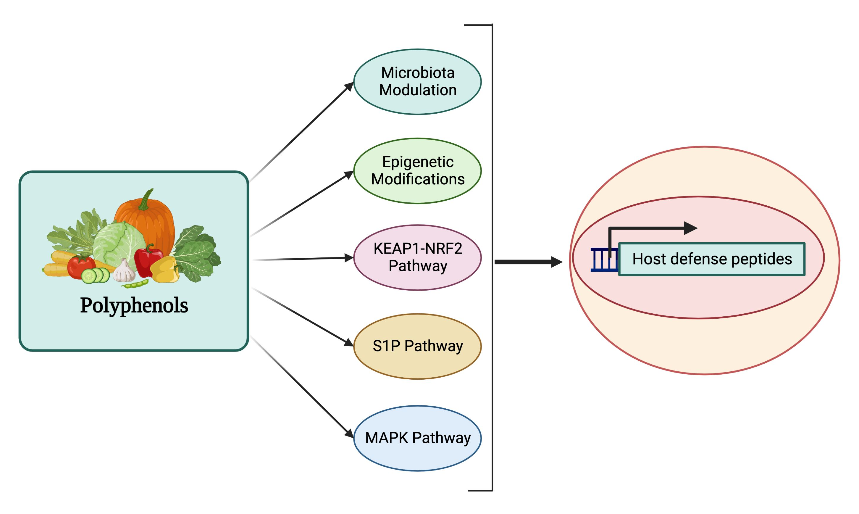
2. Host Defense Peptides

The innate immune system, consisting of physical, chemical, and cellular barriers, serves as the host’s first line of defense against a wide variety of insults. As an important chemical barrier of the innate immune system, HDPs are small peptides (approximately 15–45 amino acids) found in virtually all organisms including mammals, birds, reptiles, amphibians, plants, and fungi [3,4,11].

2.1. Classifications of HDPs

Two major HDP families in vertebrate animals are cathelicidins and defensins [3,4]. Cathelicidins are produced abundantly in myeloid cells such as neutrophils and monocytes as well as skin keratinocytes and epithelial cells lining the respiratory, gastrointestinal, and urogenital tracts [12]. Synthesized initially as a precursor, cathelicidins are proteolytically cleaved from the cathelin pro-segment to become biologically active and released upon activation [12]. Defensins are characterized by the presence of six cysteine residues in defined positions [13]. Based on the location and spacing pattern of the cysteines, defensins are further divided into three subfamilies, namely α-, β-, and θ-defensins [13]. The α-defensins are produced abundantly by neutrophils and intestinal Paneth cells, while β-defensins are produced largely by the skin keratinocytes and epithelial cells of the gastrointestinal and urogenital tracts [13]. On the other hand, θ-defensins have a unique cyclic structure and have only been found in primates [14]. Like cathelicidins, α- and θ-defensins are translated as inactive precursors that are proteolytically cleaved at highly conserved sites to become mature active peptides to be stored in the granules of neutrophils or Paneth cells [14].

2.2. Mechanism of Antimicrobial Action of HDPs

HDPs are active against both Gram-positive and Gram-negative bacteria, fungi, parasites, and enveloped viruses, as well as cancer cells [3,4]. Several models, such as barrel stave, carpet, and toroidal pore, have been proposed to explain the antibacterial activity of HDPs [3,4] (Figure 1). The most accepted mechanism is based on the cationic and amphipathic natures of HDPs, which allow these peptides to interact with the outer membrane of bacteria composed of negatively charged phospholipid head groups. The cationicity of HDPs drives their attraction to the bacterial membrane causing disruption of the membrane structure leading to the leakage of cellular contents and bacterial lysis [3,4]. Additionally, other antibacterial mechanisms have been revealed with several HDPs (Figure 1). For example, human α-defensin 6 (HD6) forms a nanonet structure to entrap bacteria to prevent translocation into intestinal epithelium [15]. Proline-rich HDPs, such as indolicidin, kill bacteria by binding to intracellular targets such as DNA without membrane lysis [16]. Proline-arginine rich PR-39 directly inhibits or degrades proteins involved in DNA replication in the bacterial nucleus [17]. Furthermore, human β-defensin 3 (HBD3), has been shown to inhibit bacterial cell wall synthesis by interacting with the cell wall building block, lipid II [18].

2.3. Role of HDPs in Innate and Adaptive Immunity

In addition to direct antimicrobial activity, HDPs possess immunomodulatory activities [3,12] (Figure 1). HDPs are capable of directly recruiting different types of immune cells to the site of infection or indirectly through stimulation of chemokine production. For example, human cathelicidin LL-37 is chemotactic for neutrophils, monocytes, and T lymphocytes through engaging formyl peptide receptor 2 [19], while human β-defensins such as HBD2 and HBD3 chemoattract neutrophils and monocytes through interacting with chemokine receptor CCR2 [20] and they recruit dendritic cells and T cells through CCR6 [21]. Additionally, HDPs such as LL-37 and HBD3 can induce the expression of a variety of chemokines, such as CCL2/monocyte chemoattractant protein-1, CCL3/macrophage inflammatory protein-1α, CCL4/macrophage inflammatory protein-1β, CXCL1/Gro-α, and CCL22/macrophage-derived chemokine, which attract monocytes, macrophages, neutrophils, dendritic cells, and T cells subsequently to the sites of inflammation [22]. In addition to chemoattraction, many HDPs further induce maturation and activation of the cells involved in innate and adaptive immunity. For example, β-defensins induce the expression of co-stimulatory molecules, CD80, CD86, and CD40, on monocytes and dendritic cells in a Toll-like receptor-dependent manner [23,24].
Although they are capable of eliciting proinflammatory and immune responses to facilitate the clearance of pathogens, HDPs mediate largely anti-inflammatory responses during inflammation and infection by inhibiting the expression of proinflammatory cytokines such as interleukin (IL)-1β, IL-6, and tumor necrosis factor-α (TNF-α), while stimulating the production of anti-inflammatory cytokines such as IL-10 [25,26] (Figure 1). Furthermore, cationic HDPs also bind to anionic lipopolysaccharides (LPS) to neutralize its effect on inflammation [3,12].

2.4. Transcriptional Regulation of HDPs
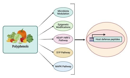
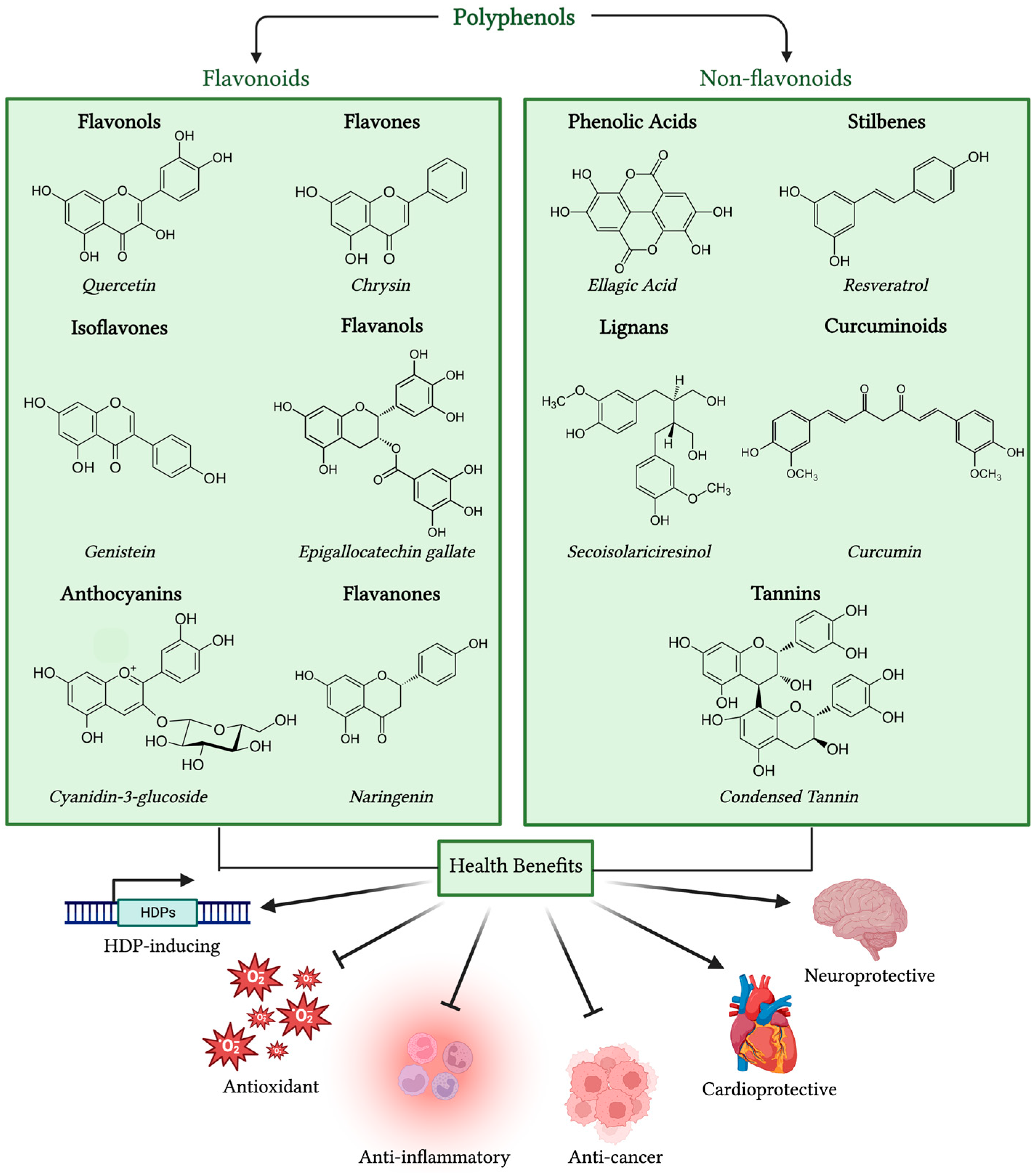
As an important component of innate defense, certain HDPs such as human α-defensins and HBD1 are constitutively produced, while other HDPs like HBD2, HBD3, and HBD4 are induced upon infection and injury [27,28]. Pathogen-associated molecular patterns (PAMPs), such as LPS, bacterial DNA, and flagellin, can induce the expression of HDPs [27]. For example, Psuedomonas aeruginosa rhamnolipids act as a PAMP to activate transcription factors, such as nuclear factor κB (NF-κB) and activator protein-1 (AP-1), causing induction of HBD2 in keratinocytes [29,30]. Proinflammatory cytokines such as TNF-α and IL-1α are known to induce the expression of HDPs such as HBD2 [31].
Additionally, a diverse group of small-molecule compounds have been shown to induce expression of HDPs [7,8,9,10]. Enhancing HDP synthesis by short-chain fatty acids (SCFAs) such as acetate, propionate, and butyrate has been well documented [10]. Medium-chain fatty acids are also capable of regulating HDP synthesis, albeit with reduced efficacy [32,33]. Amino acids such as L-arginine, L-isoleucine, leucine, and valine can induce β-defensin synthesis in the intestinal epithelium [34]. Histone deacetylase (HDAC) inhibitors, such as entinostat, valproic acid, and trichostatin, are very effective at upregulating HDP synthesis [9]. Other compounds such as vitamin D3, zinc, β-glucan, fructan, and lactose are also capable of inducing HDP synthesis [8]. Furthermore, probiotic bacteria such as Lactobacillus strains have the ability to upregulate β-defensin synthesis as well [35]. Recent development of several high-throughput screening assays has led to identification of a number of HDP-inducing compounds and their efficacy in disease control and prevention is being characterized [36,37,38,39,40,41].

3. Polyphenols

3.1. Classifications of Polyphenols

Polyphenols are secondary metabolites produced abundantly in fruits, vegetables, tea, coffee, wine, and nuts as a mechanism of defense against pathogen invasion [42]. Polyphenols are structurally diverse, but share a foundational structure consisting of an aromatic ring with multiple hydroxyl groups [42]. Based on the structure, polyphenols are broadly divided into flavonoids and non-flavonoids. Flavonoids are further divided into six subcategories, namely, flavonols, flavones, isoflavones, flavanones, anthocyanidins, and flavanols, while non-flavonoid polyphenols mainly include phenolic acids, stilbenes, lignans, tannins, and curcuminoids [43] (Figure 2).

3.2. Antioxidant and Anti-Inflammatory Activities of Polyphenols

Polyphenols are well-known natural antioxidants and anti-inflammatory agents [42,43,44]. They act as scavengers of free radicals owing to the presence of an aromatic structure and multiple hydroxyl groups [44]. Polyphenols also attenuate oxidative stress by inhibiting enzymes involved in oxidation, such as NADPH oxidase, nitric oxide synthase, and xanthine oxidase [44]. On the other hand, polyphenols suppress inflammation mainly through inhibition of NF-κB, the arachidonic acid signaling pathway, and the mitogen-activated protein kinase (MAPK) pathway [45]. For example, resveratrol and curcumin alleviate oxidative stress by inhibiting NADPH oxidase activity [46,47], while quercetin inhibits the synthesis of proinflammatory cytokines in murine intestinal epithelial cells by inhibiting recruitment of NF-κB to target gene promoters [48].

3.3. Role of Polyphenols in Cancer, Cardiovascular, and Neurological Diseases

Polyphenols are also known to have anticancer activities and improve cardiovascular, metabolic, and neuronal health as supported by multiple epidemiological studies [49,50,51]. Modifications of key signaling intermediates involved in cancer-driving pathways such as PI3K, Akt, mTOR, RAS, MAPK, and p53 are suggested to be the main anticancer mechanism of polyphenols [49,52]. A possible explanation for polyphenol-mediated cardiovascular benefits is likely due to the fact that polyphenols are strong inhibitors of low-density lipoprotein oxidation, thereby reducing the risk of developing atherosclerosis [42]. Additionally, antioxidant and anti-inflammatory effects of polyphenols, together with their ability to enhance high-density lipoprotein levels and endothelial functions, also contribute to the cardio-protective property of polyphenols [42,50]. Similarly, the strong antioxidant effects of polyphenols are the major contributor to their ability to slow or inhibit neurodegenerative processes [51,53]. Polyphenols have been shown to prevent the development of diseases, such as Parkinson’s disease and Alzheimer’s disease, though the inhibition of acetylcholinesterase activity and β-amyloid aggregation within the brain, processes that are intensified by oxidative stress [51,53].

4. Regulation of Host Defense Peptides by Polyphenols

A number of polyphenols have been shown with the capacity to enhance HDP expression at mRNA and/or protein levels both in vitro and in vivo across a number of animal species including humans, mice, pigs, chickens, fishes, and insects (Table 1).

4.1. Regulation of HDPs by Flavonols

Quercetin is capable of inducing the synthesis of HDPs in multiple animal species. Quercetin augmented the mRNA expression of hepcidin antimicrobial peptide (HAMP) by 4-fold in human HepG2 hepatocytes and enhanced HAMP mRNA expression by over 600-fold in the liver of rats when injected intraperitoneally [54]. Zebrafish supplemented with quercetin had a significant increase in mRNA expression of defensin and liver-expressed antimicrobial peptide (LEAP2) in the liver [55]. Additionally, defensin and cecropin A mRNA expression was enhanced in the larvae of Bombyx mori, a silkworm, in response to dietary supplementation of quercetin [56]. Likewise, quercetin promoted the expression of multiple avian β-defensin (AvBD) genes in chicken HTC macrophage cells, further showing a strong synergy with butyrate [57]. Moreover, dietary supplementation of quercetin upregulated the expression of multiple AvBD genes in the chicken ileum [58].

4.2. Regulation of HDPs by Flavones

Flavone induced human cathelicidin (CAMP) expression in SW620 colon epithelial cells [59]. Similarly, chrysin enhanced CAMP expression by approximately 100-fold in A549 cells, a human lung epithelial cell line [60]. Saponarin, a flavone glucoside, also significantly increased CAMP expression, but decreased DEFB1 expression in human HaCaT keratinocytes [61]. Apigenin, an active compound of chrysanthemums, promoted DEFB1, DEFB2, DEFB3, and CAMP mRNA expression in human epidermal keratinocytes [62]. Further, mice treated topically with apigenin for 9 days increased the expression of CAMP and MBD3 proteins, which was accompanied with increased production of lamellar bodies in the epidermis of mice [63]. Interestingly, flavones appear to play an opposite role during inflammation by downregulating HDP expression. For example, chrysin significantly decreased mRNA expression of CAMP, DEFB2, S100A7 (psoriasin), S100A8 (calprotectin A), and S100A9 (calprotectin B) in human NHEK epidermal keratinocytes in response to stimulation with proinflammatory cytokines TNF-α, IL-17A, or IL-22 [64].

4.3. Regulation of HDPs by Isoflavones

Genistein, a soy isoflavone, upregulated DEFB1 expression by approximately 30-fold in human LNCaP prostate cancer cell line [65] and also increased CAMP expression by 3-fold in human HaCaT keratinocytes [66]. Similarly, genistein and daidzein, another soy isoflavone, were found to promote the expression of porcine β-defensin 2 (PBD2) and PBD3 in endometrial epithelial cells [67] and increase secretion of PBD1 and PBD2 in endometrial tissues [68]. Genistein- and daidzein-mediated induction of PBD2 and PBD3 was further enhanced in porcine endometrial epithelial cells treated with polyinosinic:polycytidylic acid (Poly (I:C)) [67], suggesting the possibility of these polyphenols to further stimulate HDP production during viral infections. Calycosin, an isoflavone found in the root of Astragalus membranaceus, triggered the expression of multiple porcine HDP genes, PBD2, PEP2C, and PG1-5, in 3D4/31 alveolar macrophages [39]. Moreover, calycosin stimulated PBD2, PBD3, and PEP2C expression in porcine IPEC-J2 cells and also enhanced PBD3 expression in jejunal explants [39].

4.4. Regulation of HDPs by Flavanols

Epigallocatechin gallate (EGCG), a flavanol of green tea, and theaflavins, flavanols of black tea, significantly enhanced mRNA expression and protein secretion of HBD1 and HBD2 in human gingival epithelial cells and oral epithelial cells, respectively [69,70]. EGCG also induced mRNA and protein expression of HBD3 in bronchial epithelial cells, leading to an enhanced activity against H1N1 influenza virus [71]. In the same study, silencing the DEFB3 gene abolished the antiviral effect of EGCG, indicating that EGCG-induced upregulation of HBD3 synthesis is essential for the antiviral activity of EGCG [71]. Additionally, EGCG induced AvBD9 mRNA expression by 50-fold in chicken peripheral blood mononuclear cells (PBMCs) and further synergized with butyrate to induce AvBD9 expression by over 3500-fold [57]. In porcine jejunal epithelial cells, EGCG promoted PBD1 and PBD2 mRNA and protein expression in a dose-dependent manner [72]. Moreover, EGCG reversed suppression of the mRNA expression of two human α-defensins, HD5 and HD6, in human Caco-2 colonic epithelial cells treated with Tegafur, an oral anticancer drug [73].

4.5. Regulation of HDPs by Anthocyanins

Anthocyanins such as cyanidin 3-O-glucoside increased total β-defensin protein levels in the intestinal mucosa of mice in an ovalbumin-induced food allergy model [74]. A blueberry powder, high in anthocyanins, supplemented in a high fat diet of rats for 8 weeks significantly increased Defb2 mRNA expression by 2-fold in the ileum compared to rats on a low-fat diet [75]. Mice fed a high fat diet supplemented with an anthocyanin-rich grape pomace extract for 8 weeks restored the expression of regenerating family member 3 gamma (REG3G) to levels similar to rats on a low-fat diet [76]. However, delphinidin, an anthocyanin abundantly in pigmented vegetables and fruits, suppressed the induction of S100A7 and S100A15 (koebnerisin), together with many other proinflammatory cytokines, in inflamed psoriatic keratinocytes [77].

4.6. Regulation of HDPs by Phenolic Acids

Ellagic acid, a hydroxybenzoic acid, induced both mRNA and protein expression levels of HBD2 in primary human gingival epithelial cells [78]. Oral administration of caffeic acid phenethyl ester (CAPE) increased the mRNA expression of Defb3 in mice by nearly 10-fold and reduced the colonization of Candida albicans in the oral cavity [79]. In the same study, CAPE also enhanced the expression of two antifungal defensins, galiomycin and galeriomycin, in the larvae of the greater wax moth Galleria mellonella [79]. Anacardic acid, a phenolic acid, drastically induced AvBD9 mRNA expression by 100-fold in chicken PBMCs and further synergized with sodium butyrate to induce AvBD9 mRNA expression over 1600-fold [57].

4.7. Regulation of HDPs by Stilbenes

Resveratrol, a well-known stilbenoid of grapes, has been found to regulate HDP expression both in vitro and in vivo across animal species. For example, resveratrol induced CAMP mRNA and protein expression in human U937 monocytes, further showing a strong synergy with 1,25-dihydroxyvitamin D3 [80]. Resveratrol caused a significant increase in murine CAMP mRNA and protein expression in both cultured human keratinocytes and mouse skin and further blocked invasion of Staphylococcus aureus into mouse skin [81]. Similarly, dietary supplementation of resveratrol enhanced mRNA expression of Defa3, Defa5, and Defa20 in the ileum of mice [82]. Resveratrol also induced HAMP mRNA expression in liver cells by 4-fold as well as an 8-fold mRNA increase in the liver of rats [54]. Additionally, resveratrol upregulated AvBD9 mRNA expression and also synergized with sodium butyrate to induce AvBD9 expression in chicken PBMCs [57]. While resveratrol further upregulated Streptococcus pneumonia-induced expression of DEFB2 in human A549 lung epithelial cells [83], the opposite occurred in A549 cells infected with Pseudomonas aeruginosa or nontypeable Haemophilus influenzae [84,85]. Pterostilbene and pinostilbene, a metabolite of pterostilbene, promoted the expression of PBD3 and PG1-5 in porcine 3D4/31 alveolar macrophages [39]. Isorhapontigenin, a methoxylated stilbene analog, was found to induce PBD3, PEP2C, and PG1-5 mRNA expression in porcine IPEC-J2 intestinal epithelial cells and porcine jejunal explants [39].

4.8. Regulation of HDPs by Other Polyphenols

Several other classes of polyphenols have also been shown to regulate HDP expression. For example, curcumin, a curcuminoid isolated from turmeric, was found to cause a 3-fold induction of CAMP mRNA in human U937 monocytes and HT-29 intestinal epithelial cells [86]. Dietary supplementation of curcumin upregulated the mRNA expression of DEFB1, HAMP, and LEAP2 in the liver and blood and also improved the survival of grass carp infected with Aeromonas hydrophilla [87]. However, condensed tannins and gossypol polyphenols were shown to downregulate DEFB1, HAMP, LEAP2A, and LEAP2B mRNA expression in the intestine and exacerbate A. hydrophila infection in grass carp [88,89]. Naringenin, a flavanone, promoted the expression of HAMP in HepG2 cells [54]. Chalcone polyphenol, isoliquiritigenin, induced DEFB3 mRNA expression by 30-fold in human colonic epithelial cells [90]. More than 20 polyphenolic compounds were recently identified from a high-throughput screening with activity to induce PBD3 expression in porcine IPEC-J2 cells [39]. Of those identified, xanthohumol and deoxyshikonin were revealed to be among the most potent inducers of PBD3 in porcine IPEC-J2 intestinal epithelial cells, 3D4/31 alveolar macrophages, and jejunal explants [39].
Several studies have also revealed the benefits of polyphenol-rich foods, rather than an individual polyphenol, on HDP regulation. For instance, supplementation of pinto beans, which contain high amounts of phenolic compounds such as kaempferol, p-coumaric, gallic acid, vanilic acid, and trans-cinnamic acid [91], caused a 5-fold induction in REG3B and 2-fold increase in REG3G mRNA expression in the ileum of mice [92]. Dietary supplementation of another polyphenol-rich food, gold kiwifruit, also increased colonic expression of Defb1 and Defb2, as well as improved barrier junction in mice [93].
Table 1. In vitro and in vivo regulation of host defense peptide expression by polyphenols.
ClassPolyphenolModelHDPs Affected 1References
FlavonolsQuercetinHuman HepG2 hepatocytesHAMP[54]
Rat liverHAMP[54]
Zebrafish liver↑ defensin, LEAP2[55]
Silkworm larvae↑ defensin and cecropin A[56]
Chicken HTC macrophagesAvBD1, AvBD2, AvBD3, AvBD4, AvBD5, AvBD6, AvBD7, AvBD9, AvBD14[57]
Chicken ileumAvBD3, AvBD4, AvBD6, AvBD7, AvBD9, AvBD10, AvBD11[58]
FlavonesFlavoneHuman SW620 colon epithelialCAMP[59]
ChrysinHuman A549 lung epithelialCAMP[60]
Human keratinocytes stimulated with TNF-α, IL-17A, or IL-22S100A7, S100A8, S100A9, DEFB2, CAMP[64]
SaponarinHuman HaCaT keratinocytesCAMP, ↓ DEFB1[61]
ApigeninHuman HaCaT keratinocytesDEFB1, DEFB2, DEFB3, CAMP[62]
Mouse epidermisCamp, Defb3[63]
IsoflavonesGenisteinHuman LNCaP prostate cancerDEFB1[65]
Human HaCaT keratinocytesCAMP[66]
Porcine endometrial epithelial PBD2, PBD3[67,68]
DaidzeinPorcine endometrial epithelial PBD2, PBD3[67,68]
CalycosinPorcine 3D4/31 macrophagesPBD2, PEP2C, PG1-5[39]
Porcine IPEC-J2 intestinal epithelialPBD2, PEP2C, PBD3[39]
Porcine jejunal explantsPBD3[39]
FlavanolsEpigallocatechin gallateHuman B11 oral epithelial DEFB1, DEFB2[69]
Human OBA-9 gingival epithelialDEFB1, DEFB2[70]
BEAS-2B bronchial epithelialDEFB3[71]
Chicken PBMCAvBD9[57]
Porcine IPEC-J2 intestinalPBD1, PBD2[72]
Human Caco-2 colonic epithelial stimulated with TegafurDEFA5, DEFA6[73]
Antho-
cyanins
Cyanidin 3-O-glycosideMouse intestinal mucosa↑ total β-defensin protein[74]
Blueberry powderRat ileumDefb2[75]
Grape pomace extractMouse colonREG3G[76]
DelphinidinHuman psoriatic keratinocytes S100A7, S100A15[77]
Phenolic
Acids
Ellagic acidHuman gingival epithelialDEFB2[78]
Caffeic acid phenethyl esterMouse tongueDefb3[79]
Galleria mellonella larvae↑ galiomycin, galeriomycin[79]
Anacardic acidChicken PBMCAvBD9[57]
StilbenesResveratrolHuman U937 monocytesCAMP[80]
Human HaCaT keratinocytesCAMP[80]
Mouse epidermisCAMP[81]
Mouse ileumDefa3, Defa5, Defa20[82]
Human HepG2 hepatocytesHAMP[54]
Rat liverHAMP[54]
Chicken PBMCAvBD9[57]
Streptococcus pneumonia-infected human A549 lung epithelialDEFB2[83]
Pseudomonas aeruginosa-infected A549 lung epithelialDEFB2[84,85]
PterostilbenePorcine 3D4/31 macrophagesPBD3, PG1-5[39]
PinostilbenePorcine 3D4/31 macrophagesPBD3, PG1-5[39]
IsorhapontigeninPorcine IPEC-J2 intestinal epithelialPBD3, PEP2C, PG1-5[39]
Porcine jejunal explantsPBD3, PG1-5[39]
CurcuminoidCurcuminHuman U937 monocytesCAMP[86]
Human HT29 colonic epithelialCAMP[86]
Grass carp liver and bloodDEFB1, HAMP, LEAP2[87]
TanninsCondensed tanninsGrass carp intestineHAMP, LEAP2A, LEAP2B, DEFB1[88]
FlavanonesNaringeninHuman HepG2 hepatocytesHAMP[54]
ChalconesIsoliquiritigeninHuman Caco-2 colonic epithelialDEFB3[90]
XanthohumolPorcine IPEC-J2 intestinalPEP2C, PBD3, PG1-5[39]
Porcine 3D4/31 macrophagesPEP2C, PBD3, PG1-5[39]
Porcine jejunal explantsPBD3, PG1-5[39]
OthersGossypolGrass carp intestineHAMP, LEAP2B, DEFB1[89]
DeoxyshikoninPorcine IPEC-J2 intestinal epithelial
Porcine 3D4/31 macrophages
Porcine jejunal explants
PEP2C, PBD3, PG1-5
PEP2C, PBD3, PG1-5
PBD3, PG1-5
[39]
[39]
[39]
Pinto beansMouse ileumREG3B, REG3G[92]
Gold kiwifruitRat colonDefb1, Defb2[93]
1 Abbreviations: HAMP, hepcidin antimicrobial peptide; AvBD, avian β-defensin; LEAP2, liver-expressed antimicrobial peptide 2; CAMP, cathelicidin antimicrobial peptide; DEFB, β-defensin; PBD, porcine β-defensin; DEFA, α-defensin; REG3, regenerating family member 3. “↑” denotes increased gene expression, while “↓” denotes decreased gene expression.

5. Mechanism of Action for Polyphenols

The mechanisms by which structurally diverse polyphenols regulate HDP expression are beginning to be revealed. The MAPK signaling pathway, the sphingosine-1-phopshate (S1P) pathway, the Kelch-like ECH-associated protein 1- nuclear factor erythroid 2-related factor 2 (KEAP1-NRF2) pathway, and epigenetics are involved in the regulation of certain HDP genes. In live animals, polyphenol-mediated alternations of intestinal bacterial metabolites are likely to contribute to enhanced HDP synthesis as well.

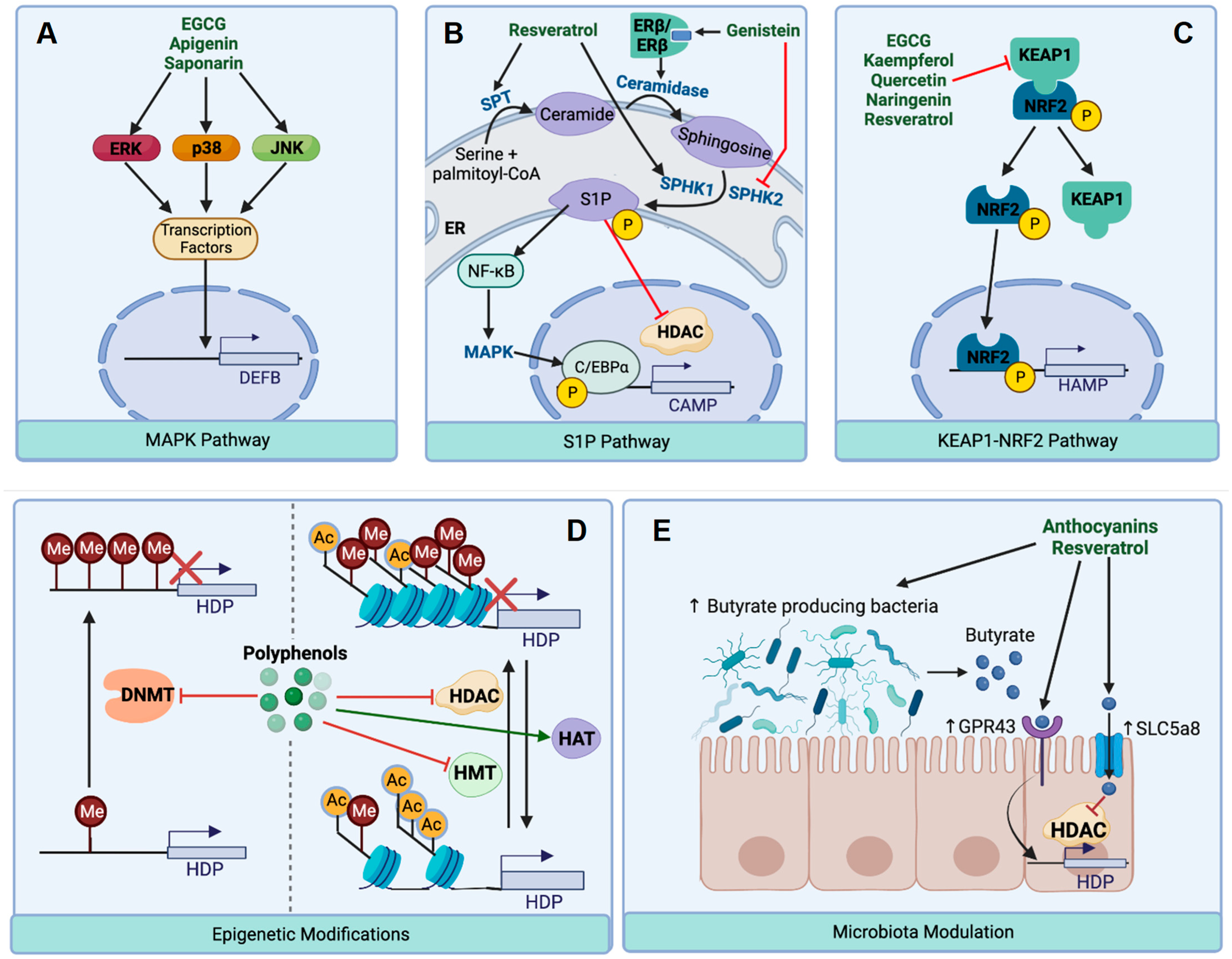
5.1. HDP Regulation by the MAPK Signaling Pathway

The MAPK signaling pathway is essential for a variety of cellular processes such as stress response and cell proliferation and differentiation [94]. The MAPK family consists of three major serine/threonine protein kinases namely, extracellular signal-regulated protein kinase 1/2 (ERK1/2), c-Jun N-terminal kinase (JNK), and p38 MAPK [94]. Once activated by external stimuli, these kinases will phosphorylate downstream proteins including transcription factors to regulate gene expression [94]. The MAPK pathways have been shown to be involved in polyphenol-mediated enhancement of HDP gene expression (Figure 3A). For example, EGCG-induced DEFB1 and DEFB2 expression is diminished in the presence of ERK/12 and p38 MAPK inhibitors in human gingival epithelial cells [69]. Similarly, EGCG enhances phosphorylation of ERK/12, JNK, and p38 MAPK in lung epithelial cells, while each of the three MAPK inhibitors suppresses EGCG-mediated DEFB3 induction [71]. Induction of PBD2 expression by EGCG is significantly reduced in porcine IPEC-J2 cells upon inhibition of p38 MAPK, but not ERK1/2 or JNK [72]. While they appear to activate the MAPK pathways in quiescent cells [69,71], polyphenols such as EGCG, apigenin, and saponarin clearly suppress the inflammatory response and the phosphorylation of ERK1/2, JNK, and p38 MAPK in cells during inflammation or infection [61,62,71].

5.2. HDP Regulation by the S1P Signaling Pathway

S1P is produced intracellularly from sphingosine on the cell membrane, acting as a signaling molecule involved in many cellular processes such as cell growth and cell trafficking [95]. Genistein-mediated increase in CAMP/LL-37 synthesis in keratinocytes is dependent upon estrogen receptor β (ERβ) [66] (Figure 3B). Engagement of genistein with ERβ induces the gene expression of ceramidase, which in turn stimulates S1P synthesis by hydrolyzing ceramide to form sphingosine. Increased S1P subsequently activates NF-κB and MAPK, leading to the phosphorylation of C/EBPα and initiation of CAMP gene expression [66]. Resveratrol has also shown to target the S1P pathway through activation of serine C-palmitoyltransferase (SPT) to stimulate S1P synthesis, resulting in NF-κB activation, C/EBPα phosphorylation, and enhanced CAMP gene expression [81]. Two sphingosine kinase isoforms, sphingosine kinase 1 (SPHK1) and SPHK2, synthesize S1P by phosphorylating sphingosine [95]. While S1P produced by SPHK1 stimulates CAMP gene expression, SIP generated by SPHK2 appears to be a negative regulator of CAMP expression [96]. It is not surprising that genistein specifically suppresses SPHK2 mRNA expression [66], and resveratrol induces SPHK1 mRNA expression [81].

5.3. HDP Regulation by the KEAP1-NRF2 Pathway

KEAP1 is a repressor of NRF2, a major transcription factor responsible for transcription of many genes with antioxidant properties [97]. Therefore, the KEAP1-NRF2 pathway plays a major role in regulating oxidative stress [97]. Polyphenols such as EGCG, kaempferol, naringenin, quercetin, and resveratrol activate the HAMP gene promotor by inhibiting KEAP1 to allow increased binding of NRF2 to the antioxidant response element (ARE) site of target gene promoters [54] (Figure 3C). Many other polyphenols such as curcumin and xanthohumol also activate NRF2 for enhanced antioxidant response [98,99]. However, whether NRF2 is involved in the ability of polyphenols to promote the transcription of other HDP genes remains to be investigated.

5.4. HDP Regulation through Epigenetic Modifications

5.4.1. Inhibition of Histone Acetylation

Epigenetic modifications of DNA and histones play a very prominent role in gene expression [100]. Histone acetylation and methylation of DNA and histones are among the most common epigenetic modifications that alter chromatin accessibility and gene transcription [100,101]. Lysine residues in the histone tails can be modified by the addition or removal of acetyl groups by two opposing classes of enzymes known as histone acetyltransferases (HATs) and histone deacetylases (HDACs), respectively [102]. Increased histone acetylation is often associated with increased gene expression, while histone deacetylation is commonly associated with diminished gene expression [102]. Polyphenols such as resveratrol, quercetin, EGCG, genistein, and delphinidin are known to inhibit HDACs [103] (Figure 3D). For example, genistein increases the acetylation of histones H3 and H4 through enhancing HAT activity while suppressing HDAC activity [104]. EGCG has been shown to decrease the expression of proteins involved in heterochromatin formation such as HP1α and HP1γ as well as chromatin assembly factor 1A, thereby promoting a more relaxed chromatin structure [105], a condition favorable for HDP transcription. These observations are consistent with the fact that HDAC inhibitors are well-known HDP inducers [9]. In fact, S1P also inhibits HDAC1 and HDAC2 activity [106], which provides another explanation for increased S1P leading to enhanced HDP transcription.
Besides Zn-dependent classical HDACs, sirtuins are a family of NAD+-dependent HDACs [107]. Many polyphenols such as resveratrol, pterostilbene, quercetin, EGCG, and curcumin are sirtuin modulators [107]. Resveratrol further augmented Streptococcus pneumonia-induced DEFB2 expression in human lung epithelial cells, while inhibition of sirtuin activity attenuated S. pneumonia-induced DEFB2 [83], suggestive of the potential for natural polyphenolic compounds to modulate HDP synthesis.

5.4.2. Inhibition of DNA and Histone Methylation

DNA methylation occurs mainly at cytosines by DNA methyltransferases (DNMTs), which leads to a condensed chromatin structure associated frequently with diminished gene transcription [101]. Methylation of the DEFB1 promoter has been shown to cause a decrease in DEFB1 mRNA and conversely, demethylation with a known DNMT inhibitor, 5-aza-2′-deoxycytidine, causes an increase in DEFB1 transcription [108,109]. 5-Azacytidine and SGI-1027, two DNMT inhibitors, also induce AvBD9 mRNA expression [110]. Polyphenols such as EGCG, quercetin, curcumin, and genistein are known to inhibit DNMTs [111,112], suggesting the possibility of these natural polyphenolic compounds to induce HDPs by acting as DNMT inhibitors (Figure 3D). Similar to DNA, histones can also be methylated to regulate gene expression [100,101]. Histones are methylated at the lysine or arginine residues by histone methyltransferases (HMT) [113]. Depending upon the position of lysine/arginine and the number of methyl groups to be transferred, histone methylation is associated with either transcriptional activation or repression [113]. Inhibition of histone methylation by specific inhibitors of G9a HMT (BIX01294) and EZH1/2 HMT (UNC1999) significantly enhanced chicken HDP gene expression in chicken HTC macrophage cells [110]. However, genistein increased histone H3 trimethylation at lysine 4 (H3K4me3) [104], a common marker of relaxed chromatin, which is consistent with the positive role of genistein in HDP expression [65,66].

5.5. HDP Regulation through Modulation of Gut Microbial Metabolites

Most dietary polyphenol intake is unabsorbed in the small intestine, thus the gut microbiota is critically important for biotransformation and metabolism of these products [114]. Polyphenol consumption is known to cause changes in the gut microbiota leading to alterations in bacterial metabolites such as SCFAs [114], which are well-known HDP inducers [7,8,9,10]. For example, polyphenol supplementation enriches butyrate-producing bacteria such as Allobaculum, Roseburia, Butyricicoccus, Bifidobacterium, and Anaerotruncus along with increasing SCFA production in mice [76,82], which may be partially responsible for polyphenol-mediated increases in HDP synthesis in live animals (Figure 3E). In addition to enhanced SCFA production, increased butyrate influx and signaling are likely to occur in vivo upon polyphenol supplementation. A dramatic increase in the mRNA expression of SLC5a8, a butyrate transporter, was observed in the colon of mice supplemented with polyphenol-rich extracts of cinnamon bark and grape pomace [76]. Supplementation of blueberries, rich in anthocyanins, caused an increase in mRNA expression of both Defb2 and SCFA receptor, Gpr43, in mice [75]. GPR43 is in fact utilized by SCFAs to upregulate REG3G and DEFB expression in intestinal epithelial cells [115]. Therefore, polyphenol-induced SCFA synthesis by gut microbiota is likely to be involved in the in vivo induction of HDP synthesis.

6. Future Prospects and Conclusions

The use of HDPs as antibiotic alternatives to infectious disease control and prevention is promising due to the beneficial antimicrobial and immunomodulatory properties of these peptides [3,4,11]. Exogenous administration of HDPs have been found to be effective against various infections in both laboratory and livestock animals and a number of these peptides are in different stages of clinical trials [3,4,11]. However, because of high production cost and instability associated intrinsically with most peptide-based drugs, new technologies are being explored to improve HDP stability and efficacy such as nanotechnology-based formulation and delivery, peptide modifications, and non-peptide mimetics [3,11]. HDPs are also being tried as adjuvants to antibiotics and vaccines [3]. Alternatively, modulation of endogenous HDP synthesis is a promising host-directed approach to disease control and prevention in the age of antimicrobial resistance and a large number of natural and synthetic small-molecule compounds, such as polyphenols, have been found to be capable of inducing HDP synthesis both in vitro and in vivo [7,8,9,10].
Natural polyphenols are known to be antioxidant, anti-inflammatory, anticancer, cardio-protective, and neuro-protective. Current research further highlights the ability of polyphenols to boost HDP synthesis and innate defense, suggesting its potential for anti-infective application. Further studies on the in vivo efficacy of these polyphenols in different animal disease models are required to demonstrate the utility of these natural compounds as effective antibiotic alternatives. The MAPK signaling pathway, the S1P pathway, and the KEAP1-NRF2 pathway have been shown to be involved in polyphenol-mediated induction of certain HDP genes. In addition, epigenetic modifications such as histone acetylation, histone methylation, and DNA methylation by polyphenols play a critical role in HDP induction as well. Moreover, bacterial metabolites, SCFAs in particular, contribute to HDP gene induction in vivo upon polyphenol consumption. Further investigations on the mechanisms of action of these polyphenols are warranted to fully realize their potential in antimicrobial therapy.
Although polyphenols demonstrate many alluring health benefits, their use in human medicine has been limited due to the low oral bioavailability of many polyphenols [116]. Polyphenol absorption is inefficient due to chemical and physical characteristics causing low solubility and instability in the gastrointestinal tract [116]. On the other hand, polyphenols are known to be extensively metabolized by the intestinal microbiota and different metabolites of polyphenols exhibits various degrees of antioxidant and anti-inflammatory activities on the host [114]. However, it remains largely unknown how different polyphenol metabolites regulate HDP synthesis. It is perhaps more effective to use those metabolites that may demonstrate a superior HDP-inducing activity than the parental polyphenol compounds. It is also beneficial to encapsulate polyphenols or their metabolites for protection and extended release to the gastrointestinal tract. In fact, nanoencapsulation has improved absorption and bioavailability of polyphenols such as EGCG, curcumin, and resveratrol to increase their efficacy in vivo [117,118]. It is exciting to explore the potential of polyphenols as antibiotic alternatives to improve human and animal health while mitigating antimicrobial resistance.

Author Contributions

Conceptualization, G.Z.; writing—original draft preparation, I.T.; writing—review and editing, G.Z.; funding acquisition, G.Z. All authors have read and agreed to the published version of the manuscript.

Funding

This research was funded by the USDA National Institute of Food and Agriculture, grant numbers 2018-68003-27462, 2020-67016-31619, and 2023-67015-39095, the Ralph F. and Leila W. Boulware Endowment Fund, and Oklahoma Agricultural Experiment Station Project H-3112. I.T. is also supported by the Graduate Research Training Initiative for Student Enhancement (G-RISE) from the National Institutes of Health to Oklahoma State University (T32 GM140953).

Institutional Review Board Statement

Not applicable.

Informed Consent Statement

Not applicable.

Data Availability Statement

All data generated during this study are included in this published article.

Acknowledgments

We are grateful for all former and current laboratory members for their contributions to this line of research. All graphs were created with BioRender.com.

Conflicts of Interest

The authors declare no conflict of interest. The funders had no role in the design of the study; in the collection, analyses, or interpretation of data; in the writing of the manuscript; or in the decision to publish the results.

Abbreviations

AP-1, activator protein-1; ARE, antioxidant response element; AvBD, avian β-defensin; CAMP, cathelicidin antimicrobial peptide; CAPE, caffeic acid phenethyl ester; COX, cyclooxygenase; DEFA, human α-defensin; DEFB/HBD, human β-defensin; DNMT, DNA methyltransferase; EGCG, epigallocatechin gallate; ERK, extracellular signal-related protein kinase; HAMP, hepcidin antimicrobial peptide; HAT, histone acetyltransferase; HD5, human α-defensin 5; HD6, human α-defensin 6; HDAC, histone deacetylase; HDP, host defense peptides; HMT, histone methyltransferase; JNK, c-Jun N-terminal kinase; KEAP1, Kelch-like ECH-associated protein 1; LEAP2, liver-expressed antimicrobial peptide; LPS, lipopolysaccharide; MAPK, mitogen-activated protein kinase; MBD2, murine β-defensin 2; NF-ᴋB, nuclear factor ᴋB; NRF2, nuclear factor erythroid 2-related factor 2; NSAID, nonsteroidal anti-inflammatory drug; PAMP, pathogen-associated molecular pattern; PBD, porcine β-defensin; PBMCs, peripheral blood mononuclear cells; Poly (I:C), polyinosinic:polycytidylic acid; REG3, regenerating family member 3; REG3G, regenerating family member 3 gamma; S1P, sphingosine-1-phosphate; SCFA, short-chain fatty acid; SPHK, sphingosine kinase; SPT, serine C-palmitoyltransferase

References

  1. Watkins, R.R.; Bonomo, R.A. Overview: The Ongoing Threat of Antimicrobial Resistance. Infect. Dis. Clin. N. Am. 2020, 34, 649–658. [Google Scholar] [CrossRef]
  2. Schrader, S.M.; Vaubourgeix, J.; Nathan, C. Biology of antimicrobial resistance and approaches to combat it. Sci. Transl. Med. 2020, 12, eaaz6992. [Google Scholar] [CrossRef] [PubMed]
  3. Mookherjee, N.; Anderson, M.A.; Haagsman, H.P.; Davidson, D.J. Antimicrobial host defence peptides: Functions and clinical potential. Nat. Rev. Drug Discov. 2020, 19, 311–332. [Google Scholar] [CrossRef] [PubMed]
  4. Magana, M.; Pushpanathan, M.; Santos, A.L.; Leanse, L.; Fernandez, M.; Ioannidis, A.; Giulianotti, M.A.; Apidianakis, Y.; Bradfute, S.; Ferguson, A.L.; et al. The value of antimicrobial peptides in the age of resistance. Lancet Infect. Dis. 2020, 20, e216–e230. [Google Scholar] [CrossRef] [PubMed]
  5. Dijksteel, G.S.; Ulrich, M.M.W.; Middelkoop, E.; Boekema, B.K.H.L. Review: Lessons Learned From Clinical Trials Using Antimicrobial Peptides (AMPs). Front. Microbiol. 2021, 12, 616979. [Google Scholar] [CrossRef]
  6. Rodrigues, G.; Souza Santos, L.; Franco, O.L. Antimicrobial Peptides Controlling Resistant Bacteria in Animal Production. Front. Microbiol. 2022, 13, 874153. [Google Scholar] [CrossRef]
  7. Bergman, P.; Raqib, R.; Rekha, R.S.; Agerberth, B.; Gudmundsson, G.H. Host Directed Therapy Against Infection by Boosting Innate Immunity. Front. Immunol. 2020, 11, 1209. [Google Scholar] [CrossRef]
  8. Chen, J.; Zhai, Z.; Long, H.; Yang, G.; Deng, B.; Deng, J. Inducible expression of defensins and cathelicidins by nutrients and associated regulatory mechanisms. Peptides 2020, 123, 170177. [Google Scholar] [CrossRef]
  9. Rodriguez-Carlos, A.; Jacobo-Delgado, Y.M.; Santos-Mena, A.O.; Rivas-Santiago, B. Modulation of cathelicidin and defensins by histone deacetylase inhibitors: A potential treatment for multi-drug resistant infectious diseases. Peptides 2021, 140, 170527. [Google Scholar] [CrossRef]
  10. Robinson, K.; Ma, X.; Liu, Y.; Qiao, S.; Hou, Y.; Zhang, G. Dietary modulation of endogenous host defense peptide synthesis as an alternative approach to in-feed antibiotics. Anim. Nutr. 2018, 4, 160–169. [Google Scholar] [CrossRef]
  11. Thakur, A.; Sharma, A.; Alajangi, H.K.; Jaiswal, P.K.; Lim, Y.B.; Singh, G.; Barnwal, R.P. In pursuit of next-generation therapeutics: Antimicrobial peptides against superbugs, their sources, mechanism of action, nanotechnology-based delivery, and clinical applications. Int. J. Biol. Macromol. 2022, 218, 135–156. [Google Scholar] [CrossRef] [PubMed]
  12. Alford, M.A.; Baquir, B.; Santana, F.L.; Haney, E.F.; Hancock, R.E.W. Cathelicidin Host Defense Peptides and Inflammatory Signaling: Striking a Balance. Front. Microbiol. 2020, 11, 1902. [Google Scholar] [CrossRef] [PubMed]
  13. Xu, D.; Lu, W. Defensins: A Double-Edged Sword in Host Immunity. Front. Immunol. 2020, 11, 764. [Google Scholar] [CrossRef] [PubMed]
  14. Selsted, M.E.; Ouellette, A.J. Mammalian defensins in the antimicrobial immune response. Nat. Immunol. 2005, 6, 551–557. [Google Scholar] [CrossRef] [PubMed]
  15. Chu, H.; Pazgier, M.; Jung, G.; Nuccio, S.P.; Castillo, P.A.; de Jong, M.F.; Winter, M.G.; Winter, S.E.; Wehkamp, J.; Shen, B.; et al. Human alpha-defensin 6 promotes mucosal innate immunity through self-assembled peptide nanonets. Science 2012, 337, 477–481. [Google Scholar] [CrossRef] [Green Version]
  16. Gennaro, R.; Zanetti, M.; Benincasa, M.; Podda, E.; Miani, M. Pro-rich antimicrobial peptides from animals: Structure, biological functions and mechanism of action. Curr. Pharm. Des. 2002, 8, 763–778. [Google Scholar] [CrossRef]
  17. Cardoso, M.H.; Meneguetti, B.T.; Costa, B.O.; Buccini, D.F.; Oshiro, K.G.N.; Preza, S.L.E.; Carvalho, C.M.E.; Migliolo, L.; Franco, O.L. Non-Lytic Antibacterial Peptides That Translocate Through Bacterial Membranes to Act on Intracellular Targets. Int. J. Mol. Sci. 2019, 20, 4877. [Google Scholar] [CrossRef] [Green Version]
  18. Sass, V.; Schneider, T.; Wilmes, M.; Körner, C.; Tossi, A.; Novikova, N.; Shamova, O.; Sahl, H.-G. Human beta-Defensin 3 Inhibits Cell Wall Biosynthesis in Staphylococci. Infect. Immun. 2010, 78, 2793–2800. [Google Scholar] [CrossRef] [Green Version]
  19. Yang, D.; Chen, Q.; Schmidt, A.P.; Anderson, G.M.; Wang, J.M.; Wooters, J.; Oppenheim, J.J.; Chertov, O. Ll-37, the Neutrophil Granule–And Epithelial Cell–Derived Cathelicidin, Utilizes Formyl Peptide Receptor–Like 1 (Fprl1) as a Receptor to Chemoattract Human Peripheral Blood Neutrophils, Monocytes, and T Cells. J. Exp. Med. 2000, 192, 1069–1074. [Google Scholar] [CrossRef] [Green Version]
  20. Rohrl, J.; Yang, D.; Oppenheim, J.J.; Hehlgans, T. Human beta-defensin 2 and 3 and their mouse orthologs induce chemotaxis through interaction with CCR2. J. Immunol. 2010, 184, 6688–6694. [Google Scholar] [CrossRef] [Green Version]
  21. Yang, D.; Chertov, O.; Bykovskaia, S.N.; Chen, Q.; Buffo, M.J.; Shogan, J.; Anderson, M.; Schroder, J.M.; Wang, J.M.; Howard, O.M.; et al. Beta-defensins: Linking innate and adaptive immunity through dendritic and T cell CCR6. Science 1999, 286, 525–528. [Google Scholar] [CrossRef]
  22. Petrov, V.; Funderburg, N.; Weinberg, A.; Sieg, S. Human β defensin-3 induces chemokines from monocytes and macrophages: Diminished activity in cells from HIV-infected persons. Immunology 2013, 140, 413–420. [Google Scholar] [CrossRef]
  23. Biragyn, A.; Ruffini, P.A.; Leifer, C.A.; Klyushnenkova, E.; Shakhov, A.; Chertov, O.; Shirakawa, A.K.; Farber, J.M.; Segal, D.M.; Oppenheim, J.J.; et al. Toll-Like Receptor 4-Dependent Activation of Dendritic Cells by β-Defensin 2. Science 2002, 298, 1025–1029. [Google Scholar] [CrossRef]
  24. Funderburg, N.; Lederman, M.M.; Feng, Z.; Drage, M.G.; Jadlowsky, J.; Harding, C.V.; Weinberg, A.; Sieg, S.F. Human -defensin-3 activates professional antigen-presenting cells via Toll-like receptors 1 and 2. Proc. Natl. Acad. Sci. USA 2007, 104, 18631–18635. [Google Scholar] [CrossRef] [Green Version]
  25. Liu, H.; Yu, H.; Gu, Y.; Xin, A.; Zhang, Y.; Diao, H.; Lin, D. Human beta-defensin DEFB126 is capable of inhibiting LPS-mediated inflammation. Appl. Microbiol. Biotechnol. 2013, 97, 3395–3408. [Google Scholar] [CrossRef]
  26. Mookherjee, N.; Brown, K.L.; Bowdish, D.M.E.; Doria, S.; Falsafi, R.; Hokamp, K.; Roche, F.M.; Mu, R.; Doho, G.H.; Pistolic, J.; et al. Modulation of the TLR-Mediated Inflammatory Response by the Endogenous Human Host Defense Peptide LL-37. J. Immunol. 2006, 176, 2455–2464. [Google Scholar] [CrossRef] [Green Version]
  27. Contreras, G.; Shirdel, I.; Braun, M.S.; Wink, M. Defensins: Transcriptional regulation and function beyond antimicrobial activity. Dev. Comp. Immunol. 2020, 104, 103556. [Google Scholar] [CrossRef]
  28. Lyu, W.; Curtis, A.R.; Sunkara, L.T.; Zhang, G. Transcriptional Regulation of Antimicrobial Host Defense Peptides. Curr. Protein Pept. Sci. 2015, 16, 672–679. [Google Scholar] [CrossRef]
  29. Wehkamp, K.; Schwichtenberg, L.; Schröder, J.-M.M.; Harder, J. Pseudomonas aeruginosa- and IL-1β-Mediated Induction of Human β-Defensin-2 in Keratinocytes Is Controlled by NF-κB and AP-1. J. Investig. Dermatol. 2006, 126, 121–127. [Google Scholar] [CrossRef] [Green Version]
  30. Gerstel, U.; Czapp, M.; Bartels, J.; Schröder, J.-M. Rhamnolipid-induced shedding of flagellin from Pseudomonas aeruginosa provokes hBD-2 and IL-8 response in human keratinocytes. Cell. Microbiol. 2009, 11, 842–853. [Google Scholar] [CrossRef]
  31. O’Neil, D.A.; Porter, E.M.; Elewaut, D.; Anderson, G.M.; Eckmann, L.; Ganz, T.; Kagnoff, M.F. Expression and Regulation of the Human β-Defensins hBD-1 and hBD-2 in Intestinal Epithelium. J. Immunol. 1999, 163, 6718–6724. [Google Scholar] [CrossRef] [PubMed]
  32. Jiang, W.; Sunkara, L.T.; Zeng, X.; Deng, Z.; Myers, S.M.; Zhang, G. Differential regulation of human cathelicidin LL-37 by free fatty acids and their analogs. Peptides 2013, 50, 129–138. [Google Scholar] [CrossRef] [PubMed]
  33. Sunkara, L.T.; Jiang, W.; Zhang, G. Modulation of antimicrobial host defense peptide gene expression by free fatty acids. PLoS ONE 2012, 7, e49558. [Google Scholar] [CrossRef] [Green Version]
  34. Ren, M.; Zhang, S.; Liu, X.; Li, S.; Mao, X.; Zeng, X.; Qiao, S. Different Lipopolysaccharide Branched-Chain Amino Acids Modulate Porcine Intestinal Endogenous β-Defensin Expression through the Sirt1/ERK/90RSK Pathway. J. Agric. Food Chem. 2016, 64, 3371–3379. [Google Scholar] [CrossRef] [PubMed]
  35. Schlee, M.; Harder, J.; Koten, B.; Stange, E.F.; Wehkamp, J.; Fellermann, K. Probiotic lactobacilli and VSL#3 induce enterocyte beta-defensin 2. Clin. Exp. Immunol. 2008, 151, 528–535. [Google Scholar] [CrossRef]
  36. Lyu, W.; Deng, Z.; Zhang, G. High-Throughput Screening for Epigenetic Compounds That Induce Human beta-Defensin 1 Synthesis. Antibiotics 2023, 12, 186. [Google Scholar] [CrossRef]
  37. Lyu, W.; Mi, D.; Vinson, P.N.; Zhang, G. Large-scale Identification of Multiple Classes of Host Defense Peptide-Inducing Compounds for Antimicrobial Therapy. Int. J. Mol. Sci. 2022, 23, 8400. [Google Scholar] [CrossRef]
  38. Deng, Z.; Lyu, W.; Zhang, G. High-Throughput Identification of Epigenetic Compounds to Enhance Chicken Host Defense Peptide Gene Expression. Antibiotics 2022, 11, 933. [Google Scholar] [CrossRef]
  39. Wang, J.; Lyu, W.; Zhang, W.; Chen, Y.; Luo, F.; Wang, Y.; Ji, H.; Zhang, G. Discovery of natural products capable of inducing porcine host defense peptide gene expression using cell-based high throughput screening. J. Anim. Sci. Biotechnol. 2021, 12, 14. [Google Scholar] [CrossRef]
  40. Lyu, W.; Deng, Z.; Sunkara, L.T.; Becker, S.; Robinson, K.; Matts, R.; Zhang, G. High Throughput Screening for Natural Host Defense Peptide-Inducing Compounds as Novel Alternatives to Antibiotics. Front. Cell. Infect. Microbiol. 2018, 8, 191. [Google Scholar] [CrossRef] [Green Version]
  41. Nylen, F.; Miraglia, E.; Cederlund, A.; Ottosson, H.; Stromberg, R.; Gudmundsson, G.H.; Agerberth, B. Boosting innate immunity: Development and validation of a cell-based screening assay to identify LL-37 inducers. Innate Immun. 2014, 20, 364–376. [Google Scholar] [CrossRef] [Green Version]
  42. Rana, A.; Samtiya, M.; Dhewa, T.; Mishra, V.; Aluko, R.E. Health benefits of polyphenols: A concise review. J. Food Biochem. 2022, 46, e14264. [Google Scholar] [CrossRef]
  43. Durazzo, A.; Lucarini, M.; Souto, E.B.; Cicala, C.; Caiazzo, E.; Izzo, A.A.; Novellino, E.; Santini, A. Polyphenols: A concise overview on the chemistry, occurrence, and human health. Phytother. Res. 2019, 33, 2221–2243. [Google Scholar] [CrossRef] [Green Version]
  44. Rudrapal, M.; Khairnar, S.J.; Khan, J.; Dukhyil, A.B.; Ansari, M.A.; Alomary, M.N.; Alshabrmi, F.M.; Palai, S.; Deb, P.K.; Devi, R. Dietary Polyphenols and Their Role in Oxidative Stress-Induced Human Diseases: Insights Into Protective Effects, Antioxidant Potentials and Mechanism(s) of Action. Front. Pharmacol. 2022, 13, 806470. [Google Scholar] [CrossRef]
  45. Yahfoufi, N.; Alsadi, N.; Jambi, M.; Matar, C. The Immunomodulatory and Anti-Inflammatory Role of Polyphenols. Nutrients 2018, 10, 1618. [Google Scholar] [CrossRef] [Green Version]
  46. Chow, S.-E.; Hshu, Y.-C.; Wang, J.-S.; Chen, J.-K. Resveratrol attenuates oxLDL-stimulated NADPH oxidase activity and protects endothelial cells from oxidative functional damages. J. Appl. Physiol. 2007, 102, 1520–1527. [Google Scholar] [CrossRef] [Green Version]
  47. Deby-Dupont, G.; Mouithys-Mickalad, A.; Serteyn, D.; Lamy, M.; Deby, C. Resveratrol and curcumin reduce the respiratory burst of Chlamydia-primed THP-1 cells. Biochem. Biophys. Res. Commun. 2005, 333, 21–27. [Google Scholar] [CrossRef]
  48. Ruiz, P.A.; Braune, A.; Holzlwimmer, G.; Quintanilla-Fend, L.; Haller, D. Quercetin inhibits TNF-induced NF-kappaB transcription factor recruitment to proinflammatory gene promoters in murine intestinal epithelial cells. J. Nutr. 2007, 137, 1208–1215. [Google Scholar] [CrossRef] [Green Version]
  49. Sharma, E.; Attri, D.C.; Sati, P.; Dhyani, P.; Szopa, A.; Sharifi-Rad, J.; Hano, C.; Calina, D.; Cho, W.C. Recent updates on anticancer mechanisms of polyphenols. Front. Cell Dev. Biol. 2022, 10, 1005910. [Google Scholar] [CrossRef]
  50. Wang, S.; Du, Q.; Meng, X.; Zhang, Y. Natural polyphenols: A potential prevention and treatment strategy for metabolic syndrome. Food Funct. 2022, 13, 9734–9753. [Google Scholar] [CrossRef]
  51. Caruso, G.; Torrisi, S.A.; Mogavero, M.P.; Currenti, W.; Castellano, S.; Godos, J.; Ferri, R.; Galvano, F.; Leggio, G.M.; Grosso, G.; et al. Polyphenols and neuroprotection: Therapeutic implications for cognitive decline. Pharmacol. Ther. 2022, 232, 108013. [Google Scholar] [CrossRef] [PubMed]
  52. Chairez-Ramirez, M.H.; de la Cruz-Lopez, K.G.; Garcia-Carranca, A. Polyphenols as Antitumor Agents Targeting Key Players in Cancer-Driving Signaling Pathways. Front. Pharmacol. 2021, 12, 710304. [Google Scholar] [CrossRef] [PubMed]
  53. Islam, F.; Islam, M.M.; Khan Meem, A.F.; Nafady, M.H.; Islam, M.R.; Akter, A.; Mitra, S.; Alhumaydhi, F.A.; Emran, T.B.; Khusro, A.; et al. Multifaceted role of polyphenols in the treatment and management of neurodegenerative diseases. Chemosphere 2022, 307, 136020. [Google Scholar] [CrossRef] [PubMed]
  54. Bayele, H.K.; Balesaria, S.; Srai, S.K. Phytoestrogens modulate hepcidin expression by Nrf2: Implications for dietary control of iron absorption. Free Radic. Biol. Med. 2015, 89, 1192–1202. [Google Scholar] [CrossRef] [Green Version]
  55. Wang, J.; Zhang, C.; Zhang, J.; Xie, J.; Yang, L.; Xing, Y.; Li, Z. The effects of quercetin on immunity, antioxidant indices, and disease resistance in zebrafish (Danio rerio). Fish Physiol. Biochem. 2020, 46, 759–770. [Google Scholar] [CrossRef]
  56. Shi, G.; Kang, Z.; Ren, F.; Zhou, Y.; Guo, P. Effects of Quercetin on the Growth and Expression of Immune-Pathway-Related Genes in Silkworm (Lepidoptera: Bombycidae). J. Insect Sci. 2020, 20, 23. [Google Scholar] [CrossRef]
  57. Yang, Q.; Burkardt, A.C.; Sunkara, L.T.; Xiao, K.; Zhang, G. Natural Cyclooxygenase-2 Inhibitors Synergize with Butyrate to Augment Chicken Host Defense Peptide Gene Expression. Front. Immunol. 2022, 13, 819222. [Google Scholar] [CrossRef]
  58. Ying, L.; Wu, H.; Zhou, S.; Lu, H.; Ding, M.; Wang, B.; Wang, S.; Mao, Y.; Xiao, F.; Li, Y. Toll-Like Receptors Signaling Pathway of Quercetin Regulating Avian Beta-Defensin in the Ileum of Broilers. Front. Cell Dev. Biol. 2022, 10, 816771. [Google Scholar] [CrossRef]
  59. Schauber, J.; Svanholm, C.; Termen, S.; Iffland, K.; Menzel, T.; Scheppach, W.; Melcher, R.; Agerberth, B.; Luhrs, H.; Gudmundsson, G.H. Expression of the cathelicidin LL-37 is modulated by short chain fatty acids in colonocytes: Relevance of signalling pathways. Gut 2003, 52, 735–741. [Google Scholar] [CrossRef] [Green Version]
  60. Xin, Y.; Chen, S.; Tang, K.; Wu, Y.; Guo, Y. Identification of Nifurtimox and Chrysin as Anti-Influenza Virus Agents by Clinical Transcriptome Signature Reversion. Int. J. Mol. Sci. 2022, 23, 2372. [Google Scholar] [CrossRef]
  61. Min, S.Y.; Park, C.H.; Yu, H.W.; Park, Y.J. Anti-Inflammatory and Anti-Allergic Effects of Saponarin and Its Impact on Signaling Pathways of RAW 264.7, RBL-2H3, and HaCaT Cells. Int. J. Mol. Sci. 2021, 22, 8431. [Google Scholar] [CrossRef]
  62. Park, C.H.; Min, S.Y.; Yu, H.W.; Kim, K.; Kim, S.; Lee, H.J.; Kim, J.H.; Park, Y.J. Effects of Apigenin on RBL-2H3, RAW264.7, and HaCaT Cells: Anti-Allergic, Anti-Inflammatory, and Skin-Protective Activities. Int. J. Mol. Sci. 2020, 21, 4620. [Google Scholar] [CrossRef]
  63. Hou, M.; Sun, R.; Hupe, M.; Kim, P.L.; Park, K.; Crumrine, D.; Lin, T.K.; Santiago, J.L.; Mauro, T.M.; Elias, P.M.; et al. Topical apigenin improves epidermal permeability barrier homoeostasis in normal murine skin by divergent mechanisms. Exp. Dermatol. 2013, 22, 210–215. [Google Scholar] [CrossRef] [Green Version]
  64. Li, H.-J.; Wu, N.-L.; Pu, C.-M.; Hsiao, C.-Y.; Chang, D.-C.; Hung, C.-F. Chrysin alleviates imiquimod-induced psoriasis-like skin inflammation and reduces the release of CCL20 and antimicrobial peptides. Sci. Rep. 2020, 10, 2932. [Google Scholar] [CrossRef] [Green Version]
  65. Merchant, K.; Kumi-Diaka, J.; Rathinavelu, A.; Esiobu, N.; Zoeller, R.; Hormann, V. Genistein modulation of immune-assoicated genes in LNCaP prostate cancer cell line. Open Prostate Cancer J. 2012, 5, 1–7. [Google Scholar] [CrossRef] [Green Version]
  66. Park, K.; Kim, Y.I.; Shin, K.O.; Seo, H.S.; Kim, J.Y.; Mann, T.; Oda, Y.; Lee, Y.M.; Holleran, W.M.; Elias, P.M.; et al. The dietary ingredient, genistein, stimulates cathelicidin antimicrobial peptide expression through a novel S1P-dependent mechanism. J. Nutr. Biochem. 2014, 25, 734–740. [Google Scholar] [CrossRef] [Green Version]
  67. Srisomboon, Y.; Poonyachoti, S.; Deachapunya, C. Soy isoflavones enhance β-defensin synthesis and secretion in endometrial epithelial cells with exposure to TLR3 agonist polyinosinic-polycytidylic acid. Am. J. Reprod. Immunol. 2017, 78, e12694. [Google Scholar] [CrossRef]
  68. Srisomboon, Y.; Poonyachoti, S.; Deachapunya, C. Enhanced Secretion of Beta-Defensins in Endometrial Tissues and Epithelial Cells by Soy Isoflavones. J. Med. Assoc. Thai. 2016, 99 (Suppl. S8), S142–S149. [Google Scholar]
  69. Lombardo Bedran, T.B.; Feghali, K.; Zhao, L.; Palomari Spolidorio, D.M.; Grenier, D. Green tea extract and its major constituent, epigallocatechin-3-gallate, induce epithelial beta-defensin secretion and prevent beta-defensin degradation by Porphyromonas gingivalis. J. Periodontal Res. 2014, 49, 615–623. [Google Scholar] [CrossRef]
  70. Lombardo Bedran, T.B.; Morin, M.P.; Palomari Spolidorio, D.; Grenier, D. Black Tea Extract and Its Theaflavin Derivatives Inhibit the Growth of Periodontopathogens and Modulate Interleukin-8 and β-Defensin Secretion in Oral Epithelial Cells. PLoS ONE 2015, 10, e0143158. [Google Scholar] [CrossRef]
  71. Mou, Q.; Jiang, Y.; Zhu, L.; Zhu, Z.; Ren, T. EGCG induces β-defensin 3 against influenza A virus H1N1 by the MAPK signaling pathway. Exp. Ther. Med. 2020, 20, 3017–3024. [Google Scholar] [CrossRef] [PubMed]
  72. Wan, M.L.; Ling, K.H.; Wang, M.F.; El-Nezami, H. Green tea polyphenol epigallocatechin-3-gallate improves epithelial barrier function by inducing the production of antimicrobial peptide pBD-1 and pBD-2 in monolayers of porcine intestinal epithelial IPEC-J2 cells. Mol. Nutr. Food Res. 2016, 60, 1048–1058. [Google Scholar] [CrossRef] [PubMed]
  73. Takahashi, N.; Kobayashi, M.; Ogura, J.; Yamaguchi, H.; Satoh, T.; Watanabe, K.; Iseki, K. Immunoprotective effect of epigallocatechin-3-gallate on oral anticancer drug-induced α-defensin reduction in Caco-2 cells. Biol. Pharm. Bull. 2014, 37, 490–492. [Google Scholar] [CrossRef] [PubMed] [Green Version]
  74. Li, J.; Zou, C.; Liu, Y. Amelioration of Ovalbumin-Induced Food Allergy in Mice by Targeted Rectal and Colonic Delivery of Cyanidin-3-O-Glucoside. Foods 2022, 11, 1542. [Google Scholar] [CrossRef] [PubMed]
  75. Lee, S.; Keirsey, K.I.; Kirkland, R.; Grunewald, Z.I.; Fischer, J.G.; de La Serre, C.B. Blueberry Supplementation Influences the Gut Microbiota, Inflammation, and Insulin Resistance in High-Fat-Diet-Fed Rats. J. Nutr. 2018, 148, 209–219. [Google Scholar] [CrossRef] [Green Version]
  76. Van Hul, M.; Geurts, L.; Plovier, H.; Druart, C.; Everard, A.; Ståhlman, M.; Rhimi, M.; Chira, K.; Teissedre, P.L.; Delzenne, N.M.; et al. Reduced obesity, diabetes, and steatosis upon cinnamon and grape pomace are associated with changes in gut microbiota and markers of gut barrier. Am. J. Physiol. Endocrinol. Metab. 2018, 314, E334–E352. [Google Scholar] [CrossRef]
  77. Chamcheu, J.C.; Pal, H.C.; Siddiqui, I.A.; Adhami, V.M.; Ayehunie, S.; Boylan, B.T.; Noubissi, F.K.; Khan, N.; Syed, D.N.; Elmets, C.A.; et al. Prodifferentiation, anti-inflammatory and antiproliferative effects of delphinidin, a dietary anthocyanidin, in a full-thickness three-dimensional reconstituted human skin model of psoriasis. Skin Pharmacol. Physiol. 2015, 28, 177–188. [Google Scholar] [CrossRef] [Green Version]
  78. Promsong, A.; Chung, W.O.; Satthakarn, S.; Nittayananta, W. Ellagic acid modulates the expression of oral innate immune mediators: Potential role in mucosal protection. J. Oral Pathol. Med. 2015, 44, 214–221. [Google Scholar] [CrossRef]
  79. de Barros, P.P.; Rossoni, R.D.; Garcia, M.T.; Kaminski, V.L.; Loures, F.V.; Fuchs, B.B.; Mylonakis, E.; Junqueira, J.C. The Anti-Biofilm Efficacy of Caffeic Acid Phenethyl Ester (CAPE) In Vitro and a Murine Model of Oral Candidiasis. Front. Cell Infect. Microbiol. 2021, 11, 700305. [Google Scholar] [CrossRef]
  80. Guo, C.; Sinnott, B.; Niu, B.; Lowry, M.B.; Fantacone, M.L.; Gombart, A.F. Synergistic induction of human cathelicidin antimicrobial peptide gene expression by vitamin D and stilbenoids. Mol. Nutr. Food Res. 2014, 58, 528–536. [Google Scholar] [CrossRef] [Green Version]
  81. Park, K.; Elias, P.M.; Hupe, M.; Borkowski, A.W.; Gallo, R.L.; Shin, K.-O.; Lee, Y.-M.; Holleran, W.M.; Uchida, Y. Resveratrol Stimulates Sphingosine-1-Phosphate Signaling of Cathelicidin Production. J. Investig. Dermatol. 2013, 133, 1942–1949. [Google Scholar] [CrossRef] [Green Version]
  82. Zhuang, Y.; Huang, H.; Liu, S.; Liu, F.; Tu, Q.; Yin, Y.; He, S. Resveratrol Improves Growth Performance, Intestinal Morphology, and Microbiota Composition and Metabolism in Mice. Front. Microbiol. 2021, 12, 726878. [Google Scholar] [CrossRef]
  83. Lin, L.; Wen, S.H.; Guo, S.Z.; Su, X.Y.; Wu, H.J.; Chong, L.; Zhang, H.L.; Zhang, W.X.; Li, C.C. Role of SIRT1 in Streptococcus pneumoniae-induced human β-defensin-2 and interleukin-8 expression in A549 cell. Mol. Cell. Biochem. 2014, 394, 199–208. [Google Scholar] [CrossRef]
  84. Cerqueira, A.M.; Khaper, N.; Lees, S.J.; Ulanova, M. The antioxidant resveratrol down-regulates inflammation in an in-vitro model of Pseudomonas aeruginosa infection of lung epithelial cells. Can. J. Physiol. Pharmacol. 2013, 91, 248–255. [Google Scholar] [CrossRef] [Green Version]
  85. Euba, B.; López-López, N.; Rodríguez-Arce, I.; Fernández-Calvet, A.; Barberán, M.; Caturla, N.; Martí, S.; Díez-Martínez, R.; Garmendia, J. Resveratrol therapeutics combines both antimicrobial and immunomodulatory properties against respiratory infection by nontypeable Haemophilus influenzae. Sci. Rep. 2017, 7, 12860. [Google Scholar] [CrossRef] [Green Version]
  86. Guo, C.; Rosoha, E.; Lowry, M.B.; Borregaard, N.; Gombart, A.F. Curcumin induces human cathelicidin antimicrobial peptide gene expression through a vitamin D receptor-independent pathway. J. Nutr. Biochem. 2013, 24, 754–759. [Google Scholar] [CrossRef] [Green Version]
  87. Ming, J.; Ye, J.; Zhang, Y.; Xu, Q.; Yang, X.; Shao, X.; Qiang, J.; Xu, P. Optimal dietary curcumin improved growth performance, and modulated innate immunity, antioxidant capacity and related genes expression of NF-κB and Nrf2 signaling pathways in grass carp (Ctenopharyngodon idella) after infection with Aeromonas hydrophila. Fish Shellfish Immunol. 2020, 97, 540–553. [Google Scholar] [CrossRef]
  88. Li, M.; Feng, L.; Jiang, W.D.; Wu, P.; Liu, Y.; Jiang, J.; Kuang, S.Y.; Tang, L.; Zhou, X.Q. Condensed tannins decreased the growth performance and impaired intestinal immune function in on-growing grass carp (Ctenopharyngodon idella). Br. J. Nutr. 2020, 123, 737–755. [Google Scholar] [CrossRef]
  89. Wang, K.-Z.; Feng, L.; Jiang, W.-D.; Wu, P.; Liu, Y.; Jiang, J.; Kuang, S.-Y.; Tang, L.; Zhang, Y.-A.; Zhou, X.-Q. Dietary gossypol reduced intestinal immunity and aggravated inflammation in on-growing grass carp (Ctenopharyngodon idella). Fish Shellfish Immunol. 2019, 86, 814–831. [Google Scholar] [CrossRef]
  90. Sechet, E.; Telford, E.; Bonamy, C.; Sansonetti, P.J.; Sperandio, B. Natural molecules induce and synergize to boost expression of the human antimicrobial peptide β-defensin-3. Proc. Natl. Acad. Sci. USA 2018, 115, E9869–E9878. [Google Scholar] [CrossRef] [Green Version]
  91. Yang, Q.-Q.; Gan, R.-Y.; Ge, Y.-Y.; Zhang, D.; Corke, H. Polyphenols in Common Beans (Phaseolus vulgaris L.): Chemistry, Analysis, and Factors Affecting Composition. Compr. Rev. Food Sci. Food Saf. 2018, 17, 1518–1539. [Google Scholar] [CrossRef] [PubMed] [Green Version]
  92. Ojo, B.A.; Lu, P.; Alake, S.E.; Keirns, B.; Anderson, K.; Gallucci, G.; Hart, M.D.; El-Rassi, G.D.; Ritchey, J.W.; Chowanadisai, W.; et al. Pinto beans modulate the gut microbiome, augment MHC II protein, and antimicrobial peptide gene expression in mice fed a normal or western-style diet. J. Nutr. Biochem. 2021, 88, 108543. [Google Scholar] [CrossRef] [PubMed]
  93. Paturi, G.; Butts, C.A.; Bentley-Hewitt, K.L.; Ansell, J. Influence of green and gold kiwifruit on indices of large bowel function in healthy rats. J. Food Sci. 2014, 79, H1611–H1620. [Google Scholar] [CrossRef] [PubMed]
  94. Rezatabar, S.; Karimian, A.; Rameshknia, V.; Parsian, H.; Majidinia, M.; Kopi, T.A.; Bishayee, A.; Sadeghinia, A.; Yousefi, M.; Monirialamdari, M.; et al. RAS/MAPK signaling functions in oxidative stress, DNA damage response and cancer progression. J. Cell. Physiol. 2019, 234, 14951–14965. [Google Scholar] [CrossRef] [PubMed]
  95. Maceyka, M.; Harikumar, K.B.; Milstien, S.; Spiegel, S. Sphingosine-1-phosphate signaling and its role in disease. Trends Cell Biol. 2012, 22, 50–60. [Google Scholar] [CrossRef] [Green Version]
  96. Park, K.; Elias, P.M.; Shin, K.O.; Lee, Y.M.; Hupe, M.; Borkowski, A.W.; Gallo, R.L.; Saba, J.; Holleran, W.M.; Uchida, Y. A novel role of a lipid species, sphingosine-1-phosphate, in epithelial innate immunity. Mol. Cell. Biol. 2013, 33, 752–762. [Google Scholar] [CrossRef] [Green Version]
  97. Deshmukh, P.; Unni, S.; Krishnappa, G.; Padmanabhan, B. The Keap1-Nrf2 pathway: Promising therapeutic target to counteract ROS-mediated damage in cancers and neurodegenerative diseases. Biophys. Rev. 2017, 9, 41–56. [Google Scholar] [CrossRef] [Green Version]
  98. Balogun, E.; Hoque, M.; Gong, P.; Killeen, E.; Green, C.J.; Foresti, R.; Alam, J.; Motterlini, R. Curcumin activates the haem oxygenase-1 gene via regulation of Nrf2 and the antioxidant-responsive element. Biochem. J. 2003, 371, 887–895. [Google Scholar] [CrossRef] [Green Version]
  99. Yao, J.; Zhang, B.; Ge, C.; Peng, S.; Fang, J. Xanthohumol, a Polyphenol Chalcone Present in Hops, Activating Nrf2 Enzymes to Confer Protection against Oxidative Damage in PC12 Cells. J. Agric. Food Chem. 2015, 63, 1521–1531. [Google Scholar] [CrossRef]
  100. Portela, A.; Esteller, M. Epigenetic modifications and human disease. Nat. Biotechnol. 2010, 28, 1057–1068. [Google Scholar] [CrossRef]
  101. Perri, F.; Longo, F.; Giuliano, M.; Sabbatino, F.; Favia, G.; Ionna, F.; Addeo, R.; Della Vittoria Scarpati, G.; Di Lorenzo, G.; Pisconti, S. Epigenetic control of gene expression: Potential implications for cancer treatment. Crit. Rev. Oncol. Hematol. 2017, 111, 166–172. [Google Scholar] [CrossRef]
  102. Verdone, L.; Caserta, M.; Mauro, E.D. Role of histone acetylation in the control of gene expression. Biochem. Cell Biol. 2005, 83, 344–353. [Google Scholar] [CrossRef]
  103. Bouyahya, A.; El Hachlafi, N.; Aanniz, T.; Bourais, I.; Mechchate, H.; Benali, T.; Shariati, M.A.; Burkov, P.; Lorenzo, J.M.; Wilairatana, P.; et al. Natural Bioactive Compounds Targeting Histone Deacetylases in Human Cancers: Recent Updates. Molecules 2022, 27, 2568. [Google Scholar] [CrossRef]
  104. Majid, S.; Dar, A.A.; Shahryari, V.; Hirata, H.; Ahmad, A.; Saini, S.; Tanaka, Y.; Dahiya, A.V.; Dahiya, R. Genistein reverses hypermethylation and induces active histone modifications in tumor suppressor gene B-Cell translocation gene 3 in prostate cancer. Cancer 2010, 116, 66–76. [Google Scholar] [CrossRef] [Green Version]
  105. Ciesielski, O.; Biesiekierska, M.; Balcerczyk, A. Epigallocatechin-3-gallate (EGCG) Alters Histone Acetylation and Methylation and Impacts Chromatin Architecture Profile in Human Endothelial Cells. Molecules 2020, 25, 2326. [Google Scholar] [CrossRef]
  106. Hait, N.C.; Allegood, J.; Maceyka, M.; Strub, G.M.; Harikumar, K.B.; Singh, S.K.; Luo, C.; Marmorstein, R.; Kordula, T.; Milstien, S.; et al. Regulation of histone acetylation in the nucleus by sphingosine-1-phosphate. Science 2009, 325, 1254–1257. [Google Scholar] [CrossRef] [Green Version]
  107. Mayack, B.K.; Sippl, W.; Ntie-Kang, F. Natural Products as Modulators of Sirtuins. Molecules 2020, 25, 3287. [Google Scholar] [CrossRef]
  108. Noh, Y.-H.; Lee, J.; Seo, S.J.; Myung, S.C. Promoter DNA methylation contributes to human β-defensin-1 deficiency in atopic dermatitis. Anim. Cells Syst. 2018, 22, 172–177. [Google Scholar] [CrossRef] [Green Version]
  109. Sun, C.Q.; Arnold, R.; Fernandez-Golarz, C.; Parrish, A.B.; Almekinder, T.; He, J.; Ho, S.-m.; Svoboda, P.; Pohl, J.; Marshall, F.F.; et al. Human β-Defensin-1, a Potential Chromosome 8p Tumor Suppressor: Control of Transcription and Induction of Apoptosis in Renal Cell Carcinoma. Cancer Res. 2006, 66, 8542–8549. [Google Scholar] [CrossRef] [Green Version]
  110. Whitmore, M.A.; Li, H.; Lyu, W.; Khanam, S.; Zhang, G. Epigenetic Regulation of Host Defense Peptide Synthesis: Synergy Between Histone Deacetylase Inhibitors and DNA/Histone Methyltransferase Inhibitors. Front. Immunol. 2022, 13, 874706. [Google Scholar] [CrossRef]
  111. Saldivar-Gonzalez, F.I.; Gomez-Garcia, A.; Chavez-Ponce de Leon, D.E.; Sanchez-Cruz, N.; Ruiz-Rios, J.; Pilon-Jimenez, B.A.; Medina-Franco, J.L. Inhibitors of DNA Methyltransferases From Natural Sources: A Computational Perspective. Front. Pharmacol. 2018, 9, 1144. [Google Scholar] [CrossRef] [PubMed] [Green Version]
  112. Akone, S.H.; Ntie-Kang, F.; Stuhldreier, F.; Ewonkem, M.B.; Noah, A.M.; Mouelle, S.E.M.; Muller, R. Natural Products Impacting DNA Methyltransferases and Histone Deacetylases. Front. Pharmacol. 2020, 11, 992. [Google Scholar] [CrossRef] [PubMed]
  113. Greer, E.L.; Shi, Y. Histone methylation: A dynamic mark in health, disease and inheritance. Nat. Rev. Genet. 2012, 13, 343–357. [Google Scholar] [CrossRef] [PubMed] [Green Version]
  114. Wang, X.; Qi, Y.; Zheng, H. Dietary Polyphenol, Gut Microbiota, and Health Benefits. Antioxidants 2022, 11, 1212. [Google Scholar] [CrossRef]
  115. Zhao, Y.; Chen, F.; Wu, W.; Sun, M.; Bilotta, A.J.; Yao, S.; Xiao, Y.; Huang, X.; Eaves-Pyles, T.D.; Golovko, G.; et al. GPR43 mediates microbiota metabolite SCFA regulation of antimicrobial peptide expression in intestinal epithelial cells via activation of mTOR and STAT3. Mucosal Immunol. 2018, 11, 752–762. [Google Scholar] [CrossRef] [Green Version]
  116. Bertelli, A.; Biagi, M.; Corsini, M.; Baini, G.; Cappellucci, G.; Miraldi, E. Polyphenols: From Theory to Practice. Foods 2021, 10, 2595. [Google Scholar] [CrossRef]
  117. Hu, B.; Liu, X.; Zhang, C.; Zeng, X. Food macromolecule based nanodelivery systems for enhancing the bioavailability of polyphenols. J. Food Drug Anal. 2017, 25, 3–15. [Google Scholar] [CrossRef] [Green Version]
  118. Zhang, Z.; Qiu, C.; Li, X.; McClements, D.J.; Jiao, A.; Wang, J.; Jin, Z. Advances in research on interactions between polyphenols and biology-based nano-delivery systems and their applications in improving the bioavailability of polyphenols. Trends. Food Sci. Technol. 2021, 116, 492–500. [Google Scholar] [CrossRef]
Figure 1. Antimicrobial and immunomodulatory activities of host defense peptides (HDPs). HDPs possess direct antimicrobial activity against bacteria, fungi, viruses, and parasites mainly through membrane disruption. A few HDPs also inhibit bacteria by forming nanonets or targeting bacterial DNA, RNA, proteins, or lipid II of the cell wall. Immunomodulatory effects of HDPs mainly include recruitment and activation of immune cells and regulation of inflammatory response.
Antibiotics 12 00660 g001
Figure 2. Classifications and health benefits of polyphenols. One representative compound is shown in each category for illustrative purposes.
Antibiotics 12 00660 g002
Figure 3. Molecular mechanisms of polyphenol-mediated induction of host defense peptide (HDP) genes. Certain polyphenols are shown to activate the MAPK pathway to upregulate the expression of human β-defensin (DEFB) genes (A). Resveratrol and genistein enhance sphingosine-1-phopshate (S1P) synthesis and NF-κB activation to induce cathelicidin antimicrobial peptide (CAMP) gene expression (B). Several polyphenols inhibit KEAP1 to allow nuclear translocation of NRF2 to induce the expression of hepcidin antimicrobial peptide (HAMP) (C). Many polyphenols function as inhibitors of histone deacetylase (HDAC), DNA methyltransferase (DNMT), or histone methyltransferase (HMT) to increase histone acetylation while reducing DNA and histone methylation to promote a relaxed chromatin structure in favor of HDP transcription (D). A few polyphenols directly enhance the histone acetyltransferase (HAT) activity to promote HDP transcription (D). HDP gene expression may be enhanced by polyphenols through modulating the synthesis of metabolites, such as butyrate, by the intestinal microbiota (E).
Antibiotics 12 00660 g003
Disclaimer/Publisher’s Note: The statements, opinions and data contained in all publications are solely those of the individual author(s) and contributor(s) and not of MDPI and/or the editor(s). MDPI and/or the editor(s) disclaim responsibility for any injury to people or property resulting from any ideas, methods, instructions or products referred to in the content.

