
Determination of the Prevalence and Antimicrobial Resistance of Enterococcus faecalis and Enterococcus faecium Associated with Poultry in Four Districts in Zambia

 ,
,  and
and
Abstract
:1. Introduction
2. Results
2.1. Identification
2.1.1. Identification of Enterococci Using Analytical Profile Index (API)
2.1.2. Identification of Enterococci Using Polymerase Chain Reaction (PCR)
2.1.3. Comparing API and PCR Identification
2.2. Prevalence of Enterococci
2.2.1. Overall Prevalence
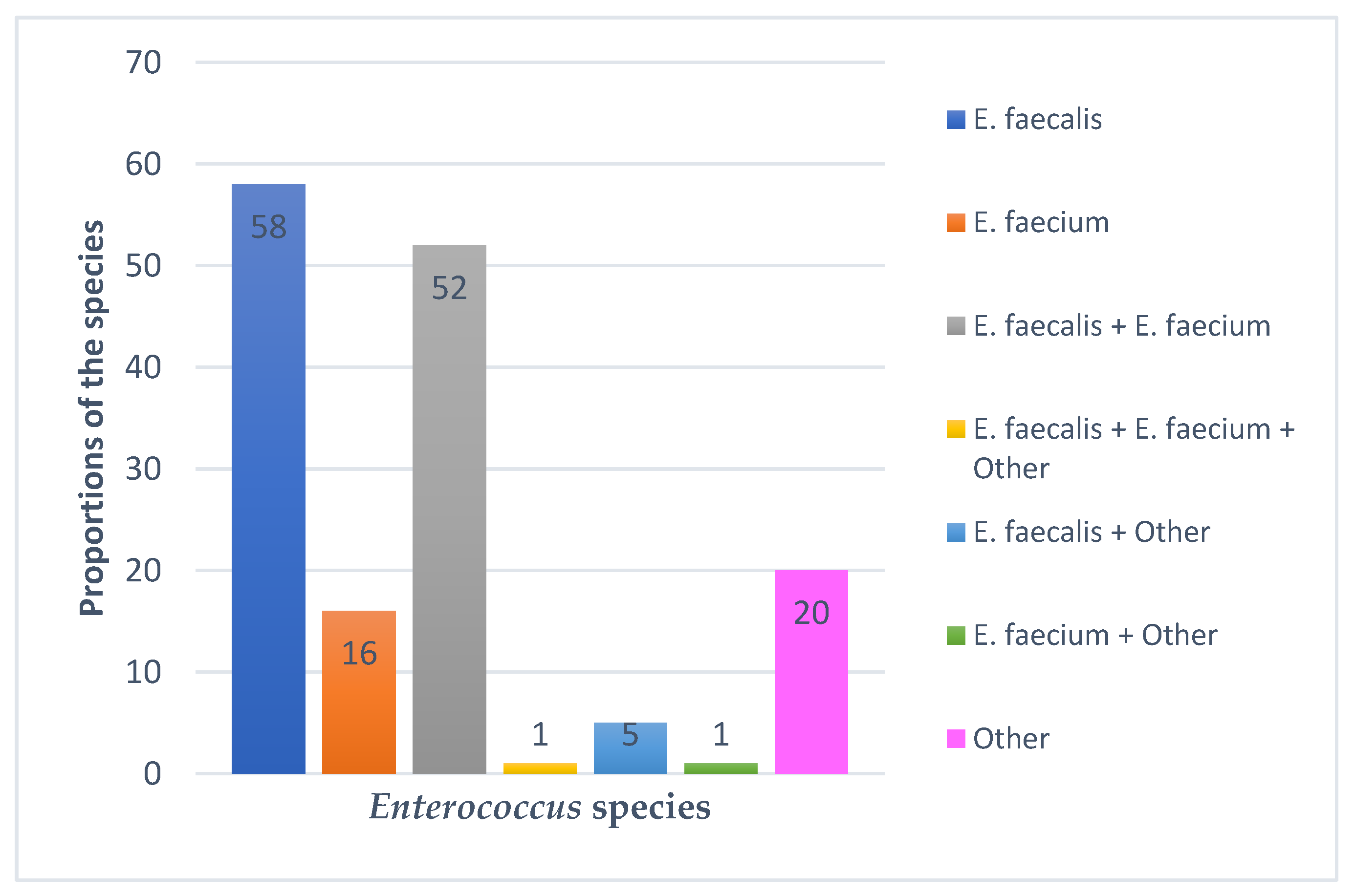
2.2.2. Species-Specific Prevalence of Isolates
2.3. Antimicrobial Susceptibility Test Results
2.3.1. Antimicrobial Susceptibility of Enterococci
2.3.2. Number of Enterococcus Isolates Resistant to One, Two, Three or More Antimicrobial Classes
2.4. Detected Antimicrobial Resistance Genes
2.4.1. Presence of Antimicrobial Resistance Genes in E. faecalis and E. faecium
2.4.2. Resistance Genes in E. faecalis and E. faecium isolates across the Study Area
2.5. Association between Antimicrobials and Resistance Genes
3. Discussion
4. Materials and Methods
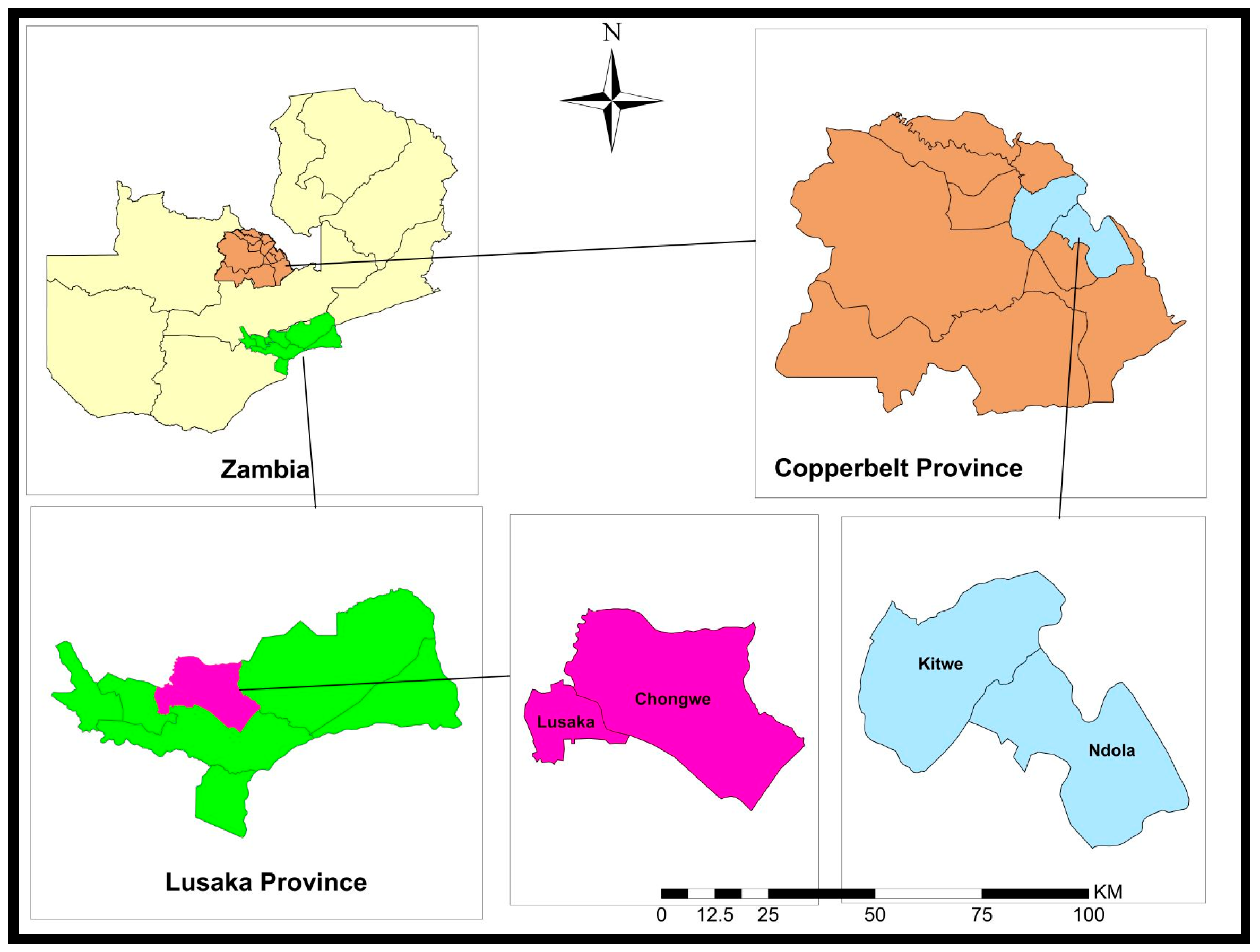
4.1. Study Design and Sites
4.2. Sample Collection
4.3. Laboratory Investigations
4.3.1. Isolation of Enterococci
4.3.2. Identification of Enterococci Using Analytical Profile Index (API)
4.3.3. DNA Extraction
4.3.4. Molecular Identification of Enterococci
4.3.5. Determination of Antimicrobial Resistance Levels
4.3.6. Detection of Antimicrobial Resistant Genes (ARG)
4.4. Data Analysis
Author Contributions
Funding
Institutional Review Board Statement
Informed Consent Statement
Data Availability Statement
Acknowledgments
Conflicts of Interest
References
- Ludwig, W.; Schleifer, K.H.; Whitman, W.B. Revised road map to the phylum Firmicutes. In Bergey’s Manual® of Systematic Bacteriology; Springer: New York, NY, USA, 2009; pp. 1–13. [Google Scholar]
- Lebreton, F.; Willems, R.J.L.; Gilmore, M.S. Enterococcus Diversity, Origins in Nature, and Gut Colonization. In Enterococci: From Commensals to Leading Causes of Drug Resistant Infection; Gilmore, M.S., Clewell, D.B., Ike, Y., Shankar, N., Eds.; Massachusetts Eye and Ear Infirmary: Boston, MA, USA, 2014; pp. 21–61. [Google Scholar]
- Soodmand, J.; Zeinali, T.; Kalidari, G.; Hashemitabar, G.; Razmyar, J. Antimicrobial Susceptibility Profile of Enterococcus Species Isolated from Companion Birds and Poultry in the Northeast of Iran. Arch. Razi. Inst. 2018, 73, 207–213. [Google Scholar]
- Byappanahalli, M.N.; Nevers, M.B.; Korajkic, A.; Staley, Z.R.; Harwood, V.J. Enterococci in the environment. Microbiol. Mol. Biol. Rev. 2012, 76, 685–706. [Google Scholar] [CrossRef] [Green Version]
- Boehm, A.B.; Sassoubre, L.M. Enterococci as Indicators of Environmental Fecal Contamination. In Enterococci: From Commensals to Leading Causes of Drug Resistant Infection; Massachusetts Eye and Ear Infirmary: Boston, MA, USA, 2014. [Google Scholar]
- Ahmed, M.O.; Baptiste, K.E. Vancomycin-Resistant Enterococci: A Review of Antimicrobial Resistance Mechanisms and Perspectives of Human and Animal Health. Microb. Drug Resist. 2018, 24, 590–606. [Google Scholar] [CrossRef] [Green Version]
- Eaton, T.J.; Gasson, M.J. Molecular screening of enterococcus virulence determinants and potential for genetic exchange between food and medical isolates. Appl. Environ. Microbiol. 2001, 67, 1628–1635. [Google Scholar] [CrossRef] [Green Version]
- Giraffa, G. Enterococci from foods. FEMS Microbiol. Rev. 2002, 26, 163–171. [Google Scholar] [CrossRef] [PubMed]
- Partridge, S.R.; Kwong, S.M.; Firth, N.; Jensen, S.O. Mobile Genetic Elements Associated with Antimicrobial Resistance. Clin. Microbiol. Rev. 2018, 31, e00088-17. [Google Scholar] [CrossRef] [Green Version]
- Agudelo Higuita, N.I.; Huycke, M.M. Enterococcal Disease, Epidemiology, and Implications for Treatment. Curr. Infect. Dis. Rep. 2014, 16, 385. [Google Scholar]
- Abat, C.; Huart, M.; Garcia, V.; Dubourg, G.; Raoult, D. Enterococcus faecalis urinary-tract infections: Do they have a zoonotic origin? J. Infect. 2016, 73, 305–313. [Google Scholar] [CrossRef] [PubMed]
- Souillard, R.; Laurentie, J.; Kempf, I.; Le Caër, V.; Le Bouquin, S.; Serror, P.; Allain, V. Increasing incidence of Enterococcus-associated diseases in poultry in France over the past 15 years. Vet. Microbiol. 2022, 269, 109426. [Google Scholar] [CrossRef]
- Pillay, S.; Zishiri, O.T.; Adeleke, M.A. Prevalence of virulence genes in Enterococcus species isolated from companion animals and livestock. Onderstepoort J. Vet. Res. 2018, 85, e1–e8. [Google Scholar] [CrossRef] [Green Version]
- Daniel, D.S.; Lee, S.M.; Dykes, G.A.; Rahman, S. Public health risks of multiple-drug-resistant Enterococcus spp. in Southeast Asia. Appl. Environ. Microbiol. 2015, 81, 6090–6097. [Google Scholar] [CrossRef] [Green Version]
- Borst, L.B.; Suyemoto, M.M.; Sarsour, A.H.; Harris, M.C.; Martin, M.P.; Strickland, J.D.; Oviedo, E.O.; Barnes, H.J. Pathogenesis of Enterococcal Spondylitis Caused by Enterococcus cecorum in Broiler Chickens. Vet. Pathol. 2017, 54, 61–73. [Google Scholar] [CrossRef] [Green Version]
- Rehman, M.A.; Yin, X.; Zaheer, R.; Goji, N.; Amoako, K.K.; McAllister, T.; Pritchard, J.; Topp, E.; Diarra, M.S. Genotypes and phenotypes of Enterococci isolated from broiler chickens. Front. Sustain. Food Syst. 2018, 2, 83. [Google Scholar] [CrossRef]
- Adams, D.J.; Eberly, M.D.; Goudie, A.; Nylund, C.M. Rising vancomycin-resistant Enterococcus infections in hospitalized children in the United States. Hosp. Pediatr. 2016, 6, 404–411. [Google Scholar] [CrossRef] [PubMed] [Green Version]
- Chiang, H.-Y.; Perencevich, E.N.; Nair, R.; Nelson, R.E.; Samore, M.; Khader, K.; Chorazy, M.L.; Herwaldt, L.A.; Blevins, A.; Ward, M.A.; et al. Incidence and Outcomes Associated with Infections Caused by Vancomycin-Resistant Enterococci in the United States: Systematic Literature Review and Meta-Analysis. Infection Control & Hospital Epidemiology. Camb. Univ. Press 2017, 38, 203–215. [Google Scholar]
- Jung, A.; Rautenschlein, S. Comprehensive report of an Enterococcus cecorum infection in a broiler flock in Northern Germany. BMC Vet. Res. 2014, 10, 311. [Google Scholar] [CrossRef] [Green Version]
- Shahbandeh, M. Meat Consumption Worldwide 1990–2021, by Type 2022. Available online: https://www.statista.com/statistics/274522/global-per-capita-consumption-of-meat/ (accessed on 19 January 2023).
- Chai, S.J.; Cole, D.; Nisler, A.; Mahon, B.E. Poultry: The most common food in outbreaks with known pathogens, United States, 1998–2012. Epidemiol. Infect. 2017, 145, 316–325. [Google Scholar] [CrossRef] [Green Version]
- Kousar, S.; Rehman, N.; Javed, A.; Hussain, A.; Naeem, M.; Masood, S.; Ali, H.A.; Manzoor, A.; Khan, A.A.; Akrem, A.; et al. Intensive Poultry Farming Practices Influence Antibiotic Resistance Profiles in Pseudomonas Aeruginosa Inhabiting Nearby Soils. Infect. Drug Resist. 2021, 29, 4511–4516. [Google Scholar] [CrossRef]
- Rice, L.B. Federal Funding for the Study of Antimicrobial Resistance in Nosocomial Pathogens: No ESCAPE. J. Infect. Dis. 2008, 197, 1079–1081. [Google Scholar] [CrossRef]
- Lee, T.; Pang, S.; Abraham, S.; Coombs, G.W. Molecular characterization and evolution of the first outbreak of vancomycin-resistant enterococcus faecium in Western Australia. Int. J. Antimicrob. Agents 2019, 53, 814–819. [Google Scholar] [CrossRef]
- Zambia National Public Health Institute. Government of the Republic of Zambia Multisectoral National Action Plan on Antimicrobial Resistance; WHO: Geneva, Switzerland, 2017; pp. 24–27. Available online: https://www.afro.who.int/publications/multi-sectoral-national-action-plan-antimicrobial-resistance-2017-2027 (accessed on 21 November 2022).
- Dolka, B.; Gołębiewska–Kosakowska, M.; Krajewski, K.; Kwieciński, P.; Nowak, T.; Szubstarski, J.; Wilczyński, J.; Szeleszczuk, P. Occurrence of Enterococcus spp. in poultry in Poland based on 2014–2015 data. Med. Water 2017, 73, 220–224. [Google Scholar] [CrossRef] [Green Version]
- Getachew, Y.M.; Hassan, L.; Zakaria, Z.; Saleha, A.A.; Kamaruddin, M.I.; Che Zalina, M.Z. Characterization of vancomycin-resistant Enterococcus isolates from broilers in Selangor, Malaysia. Trop Biomed. 2009, 26, 280–288. [Google Scholar] [PubMed]
- Ayeni, F.A.; Odumosu, B.T.; Oluseyi, A.E.; Ruppitsch, W. Identification and prevalence of tetracycline resistance in enterococci isolated from poultry in Ilishan, Ogun State, Nigeria. J. Pharm. Bioallied Sci. 2016, 8, 69–73. [Google Scholar] [PubMed]
- Mudenda, S.; Matafwali, S.K.; Malama, S.; Munyeme, M.; Yamba, K.; Katemangwe, P.; Siluchali, G.; Mainda, G.; Mukuma, M.; Bumbangi, F.N.; et al. Prevalence and antimicrobial resistance patterns of Enterococcus species isolated from laying hens in Lusaka and Copperbelt provinces of Zambia: A call for AMR surveillance in the poultry sector. JAC-Antimicrob. Resist. 2022, 4, 126. [Google Scholar] [CrossRef]
- Eldaly, E.A.; Rasha, M.; Elazam, R.A. Prevalence of Enterococcus species in chicken meat in Sharkia Governorate. Egypt. J. Appl. Sci. 2019, 34, 317–323. [Google Scholar] [CrossRef]
- Ferede, Z.T.; Tullu, K.D.; Derese, S.G.; Yeshanew, A.G. Prevalence and antimicrobial susceptibility pattern of Enterococcus species isolated from different clinical samples at Black Lion Specialized Teaching Hospital, Addis Ababa, Ethiopia. BMC Res. Notes 2018, 11, 793. [Google Scholar] [CrossRef]
- Ali, S.A.; Hasan, K.A.; Bin Asif, H.; Abbasi, A. Environmental enterococci: I. Prevalence of Virulence, Antibiotic Resistance and species distribution in poultry and its related environment in Karachi, Pakistan. Lett. Appl. Microbiol. 2014, 58, 423–432. [Google Scholar] [CrossRef]
- Noenchat, P.; Nhoonoi, C.; Srithong, T.; Lertpiriyasakulkit, S.; Sornplang, P. Prevalence and Multidrug Resistance of Enterococcus Species isolated from Chickens at Slaughterhouses in Nakhon Ratchasima Province, Thailand. Vet. World 2022, 15, 2535–2542. [Google Scholar] [CrossRef]
- Suyemoto, M.M.; Barnes, H.J.; Borst, L.B. Culture methods impact recovery of antibiotic-resistant Enterococci including Enterococcus cecorum from pre-and postharvest chicken. Lett. Appl. Microbiol. 2017, 64, 210–216. [Google Scholar] [CrossRef]
- Chingwaru, W.; Mpuchane, S.F.; Gashe, B.A. Enterococcus faecalis and Enterococcus faecium isolates from milk, beef, and chicken and their antibiotic resistance. J. Food Prot. 2003, 66, 931–936. [Google Scholar] [CrossRef]
- Çitak, S.; Yucel, N.; Orhan, S. Antibiotic resistance and incidence of Enterococcus species in Turkish white cheese. Int. J. Dairy Technol. 2004, 57, 27–31. [Google Scholar] [CrossRef]
- Stępień-Pyśniak, D.; Marek, A.; Banach, T.; Adaszek, Ł.; Pyzik, E.; Wilczyński, J.; Winiarczyk, S. Prevalence and antibiotic resistance of Enterococcus strains isolated from poultry. Acta Vet. Hung. 2016, 64, 148–163. [Google Scholar] [CrossRef] [PubMed] [Green Version]
- Karunarathna, R.; Popowich, S.; Wawryk, M.; Chow-Lockerbie, B.; Ahmed, K.A.; Yu, C.; Liu, M.; Goonewardene, K.; Gunawardana, T.; Kurukulasuriya, S.; et al. Increased Incidence of enterococcal infection in nonviable broiler chicken embryos in Western Canadian hatcheries as detected by matrix-assisted laser desorption/ionization-time-of-flight mass spectrometry. Avian Dis. 2017, 61, 472–480. [Google Scholar] [CrossRef]
- Gong, J.; Forster, R.J.; Yu, H.; Chambers, J.R.; Wheatcroft, R.; Sabour, P.M.; Chen, S. Molecular analysis of bacterial populations in the ileum of broiler chickens and comparison with bacteria in the cecum. FEMS Microbiol. Ecol. 2002, 41, 171–179. [Google Scholar] [CrossRef] [PubMed]
- Hayes, J.R.; English, L.L.; Carter, P.J.; Proescholdt, T.; Lee, K.Y.; Wagner, D.D.; White, D.G. Prevalence and antimicrobial resistance of Enterococcus species isolated from retail meats. Appl. Environ. Microbiol. 2003, 69, 7153–7160. [Google Scholar] [CrossRef] [PubMed] [Green Version]
- Rakotovao-Ravahatra, Z.D.; Antilahy, J.A.; Rakotovao-Ravahatra, J.N.; Rakotovao, A.L. Comparison of Bis-Plus-D and API 20 Strep for the identification of streptococci in the Laboratory of the University Hospital of Befelatanana Antananarivo Madagascar. J. Anal. Tech. Res. 2022, 4, 130–134. [Google Scholar] [CrossRef]
- Gomes, B.C.; Esteves, C.T.; Palazzo, I.C.; Darini AL, C.; Franco, B.D.; de Martinis, E.C. Correlation between API 20 STREP and multiplex PCR for identification of Enterococcus spp. isolated from Brazilian foods. Braz. J. Microbiol. 2007, 38, 617–619. [Google Scholar] [CrossRef] [Green Version]
- Facklam, R.; Teixeira, L.M. Enterococcus. In Manual of Clinical Microbiology, 8th ed.; Murray, P.R., Baron, E.J., Jorgensen, J.H., Pfaller, M.A., Yolken, R.H., Eds.; ASM Press: Washington, DC, USA, 2003; pp. 422–433. [Google Scholar]
- Velasco, D.; Perez, S.; Peña, F.; Dominguez, M.A.; Cartelle, M.; Molina, F.; Moure, R.; Villanueva, R.; Bou, G. Lack of correlation between phenotypic techniques and PCR-based genotypic methods for identification of Enterococcus spp. Diagn. Microbiol. Infect. Dis. 2004, 49, 151–156. [Google Scholar] [CrossRef]
- Winston, L.G.; Pang, S.; Haller, B.L.; Wong, M.; Chambers, H.F., III; Perdreau-Remington, F. API 20 STREP identification system may incorrectly speciate enterococci with low level resistance to vancomycin. Diagn. Microbiol. Infect. Dis. 2004, 48, 287–288. [Google Scholar] [CrossRef]
- World Health Organization. Critically Important Antimicrobials for Human Medicine: Ranking of Antimicrobial Agents for Risk Management of Antimicrobial Resistance Due to Non-Human Use. 2017. Available online: https://apps.who.int/iris/bitstream/handle/10665/255027/978?sequence=1 (accessed on 20 January 2023).
- Kim, M.H.; Moon, D.C.; Kim, S.J.; Mechesso, A.F.; Song, H.J.; Kang, H.Y.; Choi, J.H.; Yoon, S.S.; Lim, S.K. Nationwide surveillance on antimicrobial resistance profiles of Enterococcus faecium and Enterococcus faecalis isolated from healthy food animals in South Korea, 2010 to 2019. Microorganisms 2021, 9, 925. [Google Scholar] [CrossRef]
- Kolář, M.; Pantůček, R.; Bardoň, J.; Vágnerová, I.; Typovska, H.; Válka, I.; Doškař, J. Occurrence of antibiotic-resistant bacterial strains isolated in poultry. Veterinární Med. 2002, 47, 52–59. [Google Scholar] [CrossRef] [Green Version]
- Furtula, V.; Jackson, C.R.; Farrell, E.G.; Barrett, J.B.; Hiott, L.M.; Chambers, P.A. Antimicrobial resistance in Enterococcus spp. isolated from environmental samples in an area of intensive poultry production. Int. J. Environ. Res. Public Health 2013, 10, 1020–1036. [Google Scholar] [CrossRef] [PubMed]
- Tremblay, C.L.; Letellier, A.; Quessy, S.; Boulianne, M.; Daignault, D.; Archambault, M. Multiple-antibiotic resistance of Enterococcus faecalis and Enterococcus faecium from cecal contents in broiler chicken and turkey flocks slaughtered in Canada and plasmid colocalization of tetO and ermB genes. J. Food Prot. 2011, 74, 1639–1648. [Google Scholar] [CrossRef] [Green Version]
- Šeputiene, V.; Bogdaite, A.; Ružauskas, M.; Sužiedeliene, E. Antibiotic resistance genes and virulence factors in Enterococcus faecium and Enterococcus faecalis from diseased farm animals: Pigs, cattle, and poultry. Pol. J. Vet. Sci. 2012, 15, 431–438. [Google Scholar] [PubMed] [Green Version]
- Liu, Y.; Liu, K.; Lai, J.; Wu, C.; Shen, J.; Wang, Y. Prevalence and antimicrobial resistance of Enterococcus species of food animal origin from Beijing and Shandong Province, China. J. Appl. Microbiol. 2013, 114, 555–563. [Google Scholar] [CrossRef]
- Sung, C.H.; Chon, J.W.; Kwak, H.S.; Kim, H.; Seo, K.H. Prevalence and antimicrobial resistance of Enterococcus faecalis and Enterococcus faecium isolated from beef, pork, chicken, and sashimi. Korean J. Food Sci. Anim. Resour. 2013, 33, 133–138. [Google Scholar] [CrossRef] [Green Version]
- Maasjost, J.; Mühldorfer, K.; De Jäckel, S.C.; Hafez, H.M. Antimicrobial susceptibility patterns of Enterococcus faecalis and Enterococcus faecium isolated from poultry flocks in Germany. Avian Dis. 2015, 59, 143–148. [Google Scholar] [CrossRef]
- Kim, Y.J.; Park, J.H.; Seo, K.H. Comparison of the loads and antibiotic-resistance profiles of Enterococcus species from conventional and organic chicken carcasses in South Korea. Poult. Sci. 2018, 97, 271–278. [Google Scholar] [CrossRef]
- Noh, E.B.; Kim, Y.B.; Seo, K.W.; Son, S.H.; Ha, J.S.; Lee, Y.J. Antimicrobial resistance monitoring of commensal Enterococcus faecalis in broiler breeders. Poult. Sci. 2020, 99, 2675–2683. [Google Scholar] [CrossRef]
- Fracalanzza, S.A.P.; Scheidegger, E.M.D.; Dos Santos, P.F.; Leite, P.C.; Teixeira, L.M. Antimicrobial resistance profiles of enterococci isolated from poultry meat and pasteurized milk in Rio de Janeiro, Brazil. Memórias do Inst. Oswaldo Cruz 2007, 102, 853–859. [Google Scholar] [CrossRef] [Green Version]
- Yoshimura, H.; Ishimaru, M.; Endoh, Y.S.; Kojima, A. Antimicrobial susceptibilities of enterococci isolated from faeces of broiler and layer chickens. Lett. Appl. Microbiol. 2000, 31, 427–432. [Google Scholar] [CrossRef]
- Butaye, P.; Devriese, L.A.; Haesebrouck, F. Differences in antibiotic resistance patterns of Enterococcus faecalis and Enterococcus faecium strains isolated from farm and pet animals. Antimicrob. Agents Chemother. 2001, 45, 1374–1378. [Google Scholar] [CrossRef] [Green Version]
- Depardieu, F.; Perichon, B.; Courvalin, P. Detection of the van alphabet and identification of enterococci and staphylococci at the species level by multiplex PCR. J. Clin. Microbiol. 2004, 42, 5857–5860. [Google Scholar] [CrossRef] [Green Version]
- Xu, X.; Lin, D.; Yan, G.; Ye, X.; Wu, S.; Guo, Y.; Zhu, D.; Hu, F.; Zhang, Y.; Wang, F.; et al. vanM, a new glycopeptide resistance gene cluster found in Enterococcus faecium. Antimicrob. Agents Chemother. 2010, 54, 4643–4647. [Google Scholar] [CrossRef] [PubMed] [Green Version]
- Lebreton, F.; Depardieu, F.; Bourdon, N.; Fines-Guyon, M.; Berger, P.; Camiade, S.; Leclercq, R.; Courvalin, P.; Cattoir, V. D-Ala-D-Ser VanN-type transferable vancomycin resistance in Enterococcus faecium. Antimicrob. Agents Chemother. 2011, 55, 4606–4612. [Google Scholar] [CrossRef] [PubMed] [Green Version]
- Facklam, R.R.; Collins, M.D. Identification of Enterococcus species isolated from human infections by conventional test scheme. J. Clin. Microbiol. 1989, 27, 731–734. [Google Scholar] [CrossRef] [PubMed] [Green Version]
- Li, X.; Xing, J.; Li, B.; Wang, P.; Liu, J. Use of tuf as a target for sequence-based identification of Gram positive cocci of the genus Enterococcus, Streptococcus, coagulase-negative Staphylococcus, and Lactococcus. Annu. Clin. Microbiol. Antimicrob. 2012, 11, 31. [Google Scholar] [CrossRef] [Green Version]
- Ke, D.; Picard, F.; Martineau, F.; Menard, C.; Roy, P.; Ouellette, M.; Bergeron, M. Development of a PCR assay for rapid detection of enterococci. J. Clin. Microbiol. 1999, 37, 3497–3503. [Google Scholar] [CrossRef] [PubMed] [Green Version]
- Vilela, M.A.; Souz, S.L.; Palazzo, I.C.V.; Ferreira, J.C.; Morais, M.A., Jr.; Darini, A.L.C.; Morais, M.M.C. Identification and molecular characterization of Van A-type vancomycin-resistant Enterococcus faecalis in Northeast of Brazil. Memórias do Inst. Oswaldo Cruz 2006, 101, 716–719. [Google Scholar] [CrossRef] [Green Version]
- Bensalah, F.; Flores, M.J.A.; Mouats, A. Rapid PCR based method to distinguish between Enterococcus species by using degenerate and species-specific sodA gene primers. Afr. J. Biotechnol. 2006, 5, 697–702. [Google Scholar]
- Clinical and Laboratory Standards Institute (CLSI). Performance Standards for Antimicrobial Susceptibility Testing; Tech. Rep. M100-S22; PA Publication: Wayne, PA, USA, 2012. [Google Scholar]
- Sabouni, F.; Movahedi, Z.; Mahmoudi, S.; Pourakbari, B.; Keshavarz, V.S.; Mamishi, S. High frequency of vancomycin resistant Enterococcus faecalis in children: An alarming concern. J. Prev. Med. Hyg. 2016, 57, E201–E204. [Google Scholar] [PubMed]
- Goudarzi, G.; Tahmasbi, F.; Anbari, K.; Ghafarzadeh, M. Distribution of genes encoding resistance to macrolides among Staphylococci isolated from the nasal cavity of hospital employees in Khorramabad, Iran. Iran. Red Crescent Med. J. 2016, 18, e25701. [Google Scholar] [CrossRef] [PubMed] [Green Version]
- Sutcliffe, J.; Grebe, T.; Tait-Kamradt, A.; Wondrack, L. Detection of erythromycin-resistant determinants by PCR. Antimicrob. Agents Chemother. 1996, 40, 2562–2566. [Google Scholar] [CrossRef] [Green Version]
- Aarestrup, F.M.; Agerso, Y.; Gerner–Smidt, P.; Madsen, M.; Jensen, L.B. Comparison of antimicrobial resistance phenotypes and resistance genes in Enterococcus faecalis and Enterococcus faecium from humans in the community, broilers, and pigs in Denmark. Diagn. Microbiol. Infect. Dis. 2000, 37, 127–137. [Google Scholar] [CrossRef] [PubMed]
- Ng, L.K.; Martin, I.; Alfo, M.; Mulvey, M. Multiplex PCR for the detection of tetracycline resistance genes. Mol. Cell. Probes 2001, 15, 209–215. [Google Scholar] [CrossRef]
- Sting, R.; Richter, A.; Popp, C.; Hafez, H.M. Occurrence of vancomycin-resistant enterococci in turkey flocks. Poult. Sci. 2013, 92, 346–351. [Google Scholar] [CrossRef]


| Study ID | PCR ID | API ID | Study ID | PCR ID | API ID |
|---|---|---|---|---|---|
| 80 | E. faecalis | E. faecalis | 454 | E. faecalis + E. faecium | E. faecium |
| 82 | E. faecium | E. faecium | 455 | E. faecalis + E. faecium | E. faecalis |
| 84 | E. faecalis + E. faecium | E. faecalis | 476 | E. faecalis + E. faecium | E. faecium |
| 87 | E. faecalis | E. faecalis | 477 | E. faecalis + E. faecium | E. faecalis |
| 88 | E. faecalis + Other | E. faecium | 501 | Other enterococci | Not identified |
| 89 | Other enterococci | Not identified | 552 | E. faecalis + E. faecium | E. faecalis |
| 90 | E. faecium | E. faecium | 555 | E. faecium | E. faecium |
| 92 | Other enterococci | E. durans1 | 576 | E. faecalis + E. faecium | E. faecalis |
| 93 | E. faecalis | E. faecalis | 585 | E. faecalis + E. faecium | E. faecium |
| 94 | E. faecium | E. faecium | 619 | E. faecalis | E. faecalis |
| 96 | E. faecalis | E. faecalis | 627 | E. faecium | E. faecium |
| 99 | E. faecalis + E. faecium | E. faecium | 630 | E. faecalis + E. faecium | E. faecalis |
| 100 | E. faecalis + E. faecium | E. faecium | 702 | E. faecalis + E. faecium | E. faecium |
| 101 | E. faecalis + E. faecium | E. faecium | 704 | E. faecalis + E. faecium | E. faecalis |
| 102 | E. faecalis | E. faecalis | 714 | E. faecalis + E. faecium | E. faecalis |
| 106 | E. faecalis | E. faecalis | 718 | E. faecalis + E. faecium | E. faecium |
| 107 | E. faecium | E. faecium | 725 | E. faecalis | E. faecalis |
| 361 | E. faecalis | E. faecalis | 734 | E. faecalis | E. faecalis |
| 399 | E. faecalis | E. faecalis |
| Factor | Categories | n Tested | n Positive | Prevalence (%) | 95% CI |
|---|---|---|---|---|---|
| Overall | Positivity | 492 | 153 | 31.1 | 27.1–35.4 |
| Province | Lusaka | 107 | 33 | 30.8 | 22.5–40.6 |
| Copperbelt | 385 | 120 | 31.2 | 26.6–36.1 | |
| District | Lusaka | 50 | 22 | 44.0 | 30.3–58.7 |
| Chongwe | 57 | 11 | 19.3 | 10.5–32.3 | |
| Kitwe | 245 | 78 | 31.8 | 26.1–38.1 | |
| Ndola | 140 | 42 | 30.0 | 22.7–38.4 | |
| Enterococci isolates | E. faecium | 153 | 16 | 10.5 | 6.3–16.7 |
| E. faecalis | 153 | 58 | 37.9 | 30.3–46.1 | |
| All other Enterococcus species | 153 | 79 | 51.6 | 43.5–59.7 |
| Factors | Categories | Species | n Tested | n Positive | Prevalence (%) | 95% CI |
|---|---|---|---|---|---|---|
| Districts | Chongwe | E. faecium | 57 | 1 | 1.8 | 0.1–10.6 |
| E. faecalis | 57 | 5 | 8.8 | 3.3–20.0 | ||
| E. faecalis + E. faecium | 57 | 1 | 1.8 | 0.1–10.6 | ||
| E. faecalis + other | 57 | 2 | 3.5 | 0.6–13.2 | ||
| Other | 57 | 2 | 3.5 | 0.6–13.2 | ||
| Total | 57 | 11 | 19.3 | 10.5–32.3 | ||
| Lusaka | E. faecium | 50 | 5 | 10.0 | 3.7–22.6 | |
| E. faecalis | 50 | 8 | 16.0 | 7.6–29.7 | ||
| E. faecalis + E. faecium | 50 | 4 | 8.0 | 2.6–20.1 | ||
| E. faecium + other | 50 | 1 | 2.0 | 0.1–12.0 | ||
| E. faecalis + other | 50 | 1 | 2.0 | 0.1–12.0 | ||
| Other | 50 | 3 | 6.0 | 1.6–17.5 | ||
| Total | 50 | 22 | 44.0 | 30.3–58.7 | ||
| Kitwe | E. faecium | 245 | 8 | 3.3 | 1.5–6.6 | |
| E. faecalis | 245 | 26 | 10.6 | 7.2–15.3 | ||
| E. faecalis + E. faecium | 245 | 39 | 15.9 | 11.7–21.2 | ||
| Other | 245 | 5 | 2.0 | 0.8–5.0 | ||
| Total | 245 | 78 | 31.8 | 26.1–38.1 | ||
| Ndola | E. faecium | 140 | 2 | 1.4 | 0.2–5.6 | |
| E. faecalis | 140 | 19 | 13.6 | 8.6–20.6 | ||
| E. faecalis + E. faecium | 140 | 8 | 5.7 | 2.7–11.3 | ||
| E. faecalis + E. faecium + other | 140 | 1 | 0.7 | 0.0–4.5 | ||
| E. faecalis + other | 140 | 2 | 1.4 | 0.2–5.6 | ||
| Other | 140 | 10 | 7.1 | 3.7–13.1 | ||
| Total | 140 | 42 | 30.0 | 22.7–38.4 | ||
| Species | Overall | E. faecium | 492 | 16 | 3.3 | 1.9–5.3 |
| E. faecalis | 492 | 58 | 11.8 | 9.1–15.1 | ||
| E. faecalis + E. faecium | 492 | 52 | 10.6 | 8.1–13.7 | ||
| E. faecalis + E. faecium + other | 492 | 1 | 0.2 | 0.0–1.3 | ||
| E. faecium + other | 492 | 1 | 0.2 | 0.0–1.3 | ||
| E. faecalis + other | 492 | 5 | 1.0 | 0.4–2.5 | ||
| Other | 492 | 20 | 4.1 | 2.6–6.3 | ||
| Total Isolates | 492 | 153 | 31.1 | 27.1–35.4 |
| Species | Susceptibility Test Result | AMP n (%) | CHL n (%) | CIP n (%) | ERY n (%) | NIT n (%) | PEN n (%) | TET n (%) | VAN n (%) |
|---|---|---|---|---|---|---|---|---|---|
| E. faecalis | Resistant | 37 (63.8) | 35 (60.3) | 44 (75.9) | 54 (93.1) | 37 (63.8) | 31 (53.4) | 58 (100) | 19 (32.8) |
| Susceptible | 21 (36.2) | 23 (39.7) | 14 (24.1) | 4 (6.9) | 21 (36.2) | 27 (46.6) | 0 | 39 (67.2) | |
| E. faecium | Resistant | 14 (87.5) | 6 (37.5) | 13 (81.3) | 16 (100) | 8 (50) | 8 (50) | 14 (87.5) | 7 (43.7) |
| Susceptible | 2 (12.5) | 10 (62.5) | 3 (18.7) | 0 | 8 (50) | 8 (50) | 2 (12.5) | 9 (56.3) | |
| Total | E. faecalis | 51 (68.9) | 41 (55.4) | 57 (77.0) | 70 (94.6) | 45 (60.8) | 40 (54.1) | 72 (97.3) | 26 (35.1) |
| E. faecium | 23 (31.1) | 33 (44.6) | 17 (23.0) | 4 (5.4) | 29 (39.2) | 34 (46.0) | 2 (2.7) | 48 (64.9) |
| Isolate (Total Number) | All Susceptible n (%) | Resistant to One Class of Antibiotic, n (%) | Resistant to Two Classes of Antibiotics, n (%) | Resistant to Three or More Classes of Antibiotics, n (%) |
|---|---|---|---|---|
| All Efs and Efm (74) | 0 (0) | 2 (2.7%) | 5 (6.8%) | 67 (90.5%) |
| E. faecalis (58) | 0 (0) | 2 (3.5%) | 4 (6.9%) | 52 (89.7%) |
| E. faecium (16) | 0 (0) | 0 (0) | 1 (6.3%) | 15 (93.8%) |
| Resistance Gene | Total Isolates Tested | Detected | Undetected | |||
|---|---|---|---|---|---|---|
| E. faecalis (58) | Proportion (E. faecalis) | E. faecium (16) | Proportion (E. faecium) | |||
| aac(6′)-Ie-aph(2″)-LA | 74 | 33 | 44.6% | 12 | 16.2% | 29 |
| ermA | 74 | 1 | 0.01% | 1 | 0.01% | 72 |
| ermB | 74 | 35 | 47.3% | 10 | 13.5% | 29 |
| tetK | 74 | 40 | 54.1% | 8 | 10.8% | 26 |
| tetM | 74 | 45 | 60.8% | 10 | 13.5% | 19 |
| tetL | 74 | 41 | 55.4% | 8 | 10.8% | 25 |
| tetX | 74 | 3 | 0.04% | 2 | 0.03% | 69 |
| vanA | 74 | 2 | 0.03% | 0 | 0 | 72 |
| Area | Species | #RG | Resistance Genes | TRG | |||||||||||||||
|---|---|---|---|---|---|---|---|---|---|---|---|---|---|---|---|---|---|---|---|
| aac | ermA | ermB | tetK | tetL | tetM | tetX | vanA | ||||||||||||
| Pos | Neg | Pos | Neg | Pos | Neg | Pos | Neg | Pos | Neg | Pos | Neg | Pos | Neg | Pos | Neg | ||||
| Lusaka Province | E. faecalis | 13 | 10 | 3 | 0 | 15 | 8 | 5 | 12 | 1 | 13 | 0 | 13 | 0 | 1 | 12 | 1 | 12 | 13 |
| E. faecium | 6 | 6 | 0 | 0 | 6 | 5 | 1 | 4 | 2 | 5 | 1 | 4 | 2 | 1 | 5 | 0 | 6 | 6 | |
| Copperbelt Province | E. faecalis | 45 | 23 | 22 | 1 | 44 | 27 | 18 | 28 | 17 | 29 | 16 | 32 | 13 | 2 | 43 | 1 | 44 | 32 |
| E. faecium | 10 | 6 | 4 | 1 | 9 | 5 | 5 | 4 | 6 | 3 | 7 | 6 | 4 | 1 | 9 | 0 | 10 | 6 | |
| Chongwe | E. faecalis | 5 | 4 | 1 | 0 | 5 | 3 | 2 | 4 | 1 | 5 | 0 | 5 | 0 | 0 | 5 | 0 | 4 | 5 |
| E. faecium | 1 | 1 | 0 | 0 | 1 | 1 | 0 | 1 | 0 | 1 | 0 | 1 | 0 | 0 | 1 | 0 | 1 | 1 | |
| Lusaka | E. faecalis | 8 | 6 | 2 | 0 | 8 | 5 | 3 | 8 | 0 | 7 | 1 | 8 | 0 | 1 | 7 | 0 | 8 | 8 |
| E. faecium | 5 | 5 | 0 | 0 | 5 | 4 | 1 | 3 | 2 | 4 | 1 | 3 | 2 | 1 | 4 | 0 | 5 | 4 | |
| Ndola | E. faecalis | 20 | 13 | 7 | 0 | 20 | 12 | 8 | 11 | 9 | 15 | 5 | 16 | 4 | 0 | 20 | 0 | 20 | 16 |
| E. faecium | 2 | 1 | 1 | 0 | 2 | 2 | 0 | 1 | 1 | 2 | 0 | 2 | 0 | 0 | 2 | 0 | 2 | 2 | |
| Kitwe | E. faecalis | 25 | 10 | 15 | 1 | 24 | 15 | 10 | 17 | 8 | 14 | 11 | 16 | 9 | 2 | 23 | 1 | 24 | 17 |
| E. faecium | 8 | 5 | 3 | 1 | 7 | 5 | 5 | 3 | 5 | 1 | 7 | 4 | 4 | 1 | 7 | 0 | 5 | 5 | |
| Antibiotic | Genes | X2—Value | p-Value |
|---|---|---|---|
| TET | tet | 3.945 | 0.047 *** |
| ERY | erm | 6.947 | 0.008 *** |
| VAN | vanA | 3.795 | 0.051 |
| IDENTIFICATION PRIMERS | ||||
|---|---|---|---|---|
| Target Gene | Primer Name | Primer Sequence 5′-3′ | Amplicon Size bp | References |
| tuf | tuf-F | TACTGACAAACCATTCATGATG | 112 | [65] |
| tuf-R | AACTTCGTCACCAACGCGAAC | |||
| ddl | ddlF | CACCTGAAGAAACAGGC | 475 | [66] |
| ddlR | ATGGCTACTTCAATTTCACG | |||
| sodAEfm | sodAEfm1 | CAGCAATTGAGAAATAC | 190 | [67] |
| sodAEfm2 | CTTCTTTTATTTCTCCTGTA | |||
| sodAEfs | sodAEfs1 | CTGTAGAAGACCTAATTTCA | 209 | [67] |
| sodAEfs2 | CAGCTGTTTTGAAAGCAG | |||
| PRIMERS FOR RESISTANCE GENES | ||||
|---|---|---|---|---|
| Target Gene | Primer Name | Primer Sequence 5′-3′ | Amplicon Size (bp) | References |
| aac(6′)-Ie-aph(2″)-LA | aacF | CAGGAATTTATCGAAAATGGTAGAAAAG | 369 | [69] |
| aacR | CACAATCGACTAAAGAGTACCAATC | |||
| ermA | ermAF | TATCTTATCGTTGAGAAGGGATT | 139 | [70] |
| ermAR | CTACACTTGGCTTAGGATGAAA | |||
| ermB | ermB-1 | GAAAAGTACTCAACCAAATA | 639 | [71] |
| ermB-2 | AGTAACGGTACTTAAATTGTTTA | |||
| tetK | tetK-1 | TTAGGTGAAGGGTTAGGTCC | 697 | [72] |
| tetK-2 | GCAAACTCATTCCAGAAGCA | |||
| tetM | tetM-1 | GTTAAATAGTGTTCTTGGAG | 576 | [72] |
| tetM-2 | CTAAGATATGGCTCTAACAA | |||
| tetL | tetL-1 | CATTTGGTCTTATTGGATCG | 456 | [72] |
| tetL-2 | ATTACACTTCCGATTTCGG | |||
| tetX | tetXF | CAATAATTGGTGGTGGACCC | 468 | [73] |
| tetXR | TTCTTACCTTGGACATCCCG | |||
| vanA | vanAF | CTGCAATAGAGATAGCCGCTAACA | 751 | [74] |
| vanAR | TGTATCCGTCCTCGCTCCTC | |||
Disclaimer/Publisher’s Note: The statements, opinions and data contained in all publications are solely those of the individual author(s) and contributor(s) and not of MDPI and/or the editor(s). MDPI and/or the editor(s) disclaim responsibility for any injury to people or property resulting from any ideas, methods, instructions or products referred to in the content. |
© 2023 by the authors. Licensee MDPI, Basel, Switzerland. This article is an open access article distributed under the terms and conditions of the Creative Commons Attribution (CC BY) license (https://creativecommons.org/licenses/by/4.0/).
Share and Cite
Mwikuma, G.; Kainga, H.; Kallu, S.A.; Nakajima, C.; Suzuki, Y.; Hang’ombe, B.M. Determination of the Prevalence and Antimicrobial Resistance of Enterococcus faecalis and Enterococcus faecium Associated with Poultry in Four Districts in Zambia. Antibiotics 2023, 12, 657. https://doi.org/10.3390/antibiotics12040657
Mwikuma G, Kainga H, Kallu SA, Nakajima C, Suzuki Y, Hang’ombe BM. Determination of the Prevalence and Antimicrobial Resistance of Enterococcus faecalis and Enterococcus faecium Associated with Poultry in Four Districts in Zambia. Antibiotics. 2023; 12(4):657. https://doi.org/10.3390/antibiotics12040657
Chicago/Turabian StyleMwikuma, Grace, Henson Kainga, Simegnew Adugna Kallu, Chie Nakajima, Yasuhiko Suzuki, and Bernard Mudenda Hang’ombe. 2023. "Determination of the Prevalence and Antimicrobial Resistance of Enterococcus faecalis and Enterococcus faecium Associated with Poultry in Four Districts in Zambia" Antibiotics 12, no. 4: 657. https://doi.org/10.3390/antibiotics12040657