Article

Evaluation of the Efficiency of Random and Diblock Methacrylate-Based Amphiphilic Cationic Polymers against Major Bacterial Pathogens Associated with Cystic Fibrosis

1
CNRS, Aix-Marseille Univ, LISM UMR7255, IMM FR3479, 13402 Marseille, France
2
Aix Marseille Univ, CNRS, Centrale Marseille, iSm2 (UMR7313), 13013 Marseille, France
3
Aix Marseille Univ, CNRS, ICR, UMR7273, 13397 Marseille, France
4
Laboratoire Associé au CNR de la Résistance aux Antibiotiques, Centre Hospitalier Universitaire de Besançon, Boulevard Fleming, 25000 Besançon, France
*
Authors to whom correspondence should be addressed.
These authors contributed equally to this work.
Antibiotics 2023, 12(1), 120; https://doi.org/10.3390/antibiotics12010120
Submission received: 15 December 2022 / Revised: 2 January 2023 / Accepted: 5 January 2023 / Published: 8 January 2023

Abstract

:
Cystic fibrosis (CF) is associated with repeated lung bacterial infection, mainly by Pseudomonas aeruginosa, Staphylococcus aureus, and Mycobacterium abscessus, all known to be or becoming resistant to several antibiotics, often leading to therapeutic failure and death. In this context, antimicrobial peptides and antimicrobial polymers active against resistant strains and less prompt to cause resistance, appear as a good alternative to conventional antibiotics. In the present study, methacrylate-based copolymers obtained by radical chemistry were evaluated against CF-associated bacterial strains. Results showed that the type (Random versus Diblock) and the size of the copolymers affected their antibacterial activity and toxicity. Among the different copolymers tested, four (i.e., Random10200, Random15000, Random23900, and Diblock9500) were identified as the most active and the safest molecules and were further investigated. Data showed that they inserted into bacterial lipids, leading to a rapid membranolytic effect and killing of the bacterial. In relation with their fast bactericidal action and conversely to conventional antibiotics, those copolymers did not induce a resistance and remained active against antibiotic-resistant strains. Finally, the selected copolymers possessed a preventive effect on biofilm formation, although not exhibiting disruptive activity. Overall, the present study demonstrates that methacrylate-based copolymers are an interesting alternative to conventional antibiotics in the treatment of CF-associated bacterial infection.

1. Introduction

Cystic fibrosis (CF) is a recessive genetic disease affecting between 70,000 and 100,000 people worldwide [1], with an average incidence between 1/3000 and 1/6000 in populations of European descent [2,3]. Although complications are possible in nearly all organs, respiratory disease is the most severe symptom, and the most frequent cause of death [4]. For most of CF patients, the thickening of the airway mucus leads to recurrent and/or persistent colonization of the lung by microorganisms (pathogenic or opportunistic) eventually leading to airway inflammation, respiratory failure, and death [5]. Although many pathogenic microorganisms can colonize the lung of CF patients, Pseudomonas aeruginosa and Staphylococcus aureus are the most frequently found bacteria [6,7]. The prevalence of P. aeruginosa increases with age, ranging from ~25% in children and up to 70–80% in adult CF patients [8]. This latter pathogen is known for its high level of antibiotic resistance related to its high rate of mutation. That allows it to rapidly evolve and adapt to a multitude of conditions and drugs, resulting in recalcitrant and relapsing infections [9,10]. Approximately 50% of adult CF patients are infected by S. aureus but, in contrast to P. aeruginosa, its prevalence is higher in children, exceeding 60% [8]. Importantly, the prevalence of S. aureus infection in the adult CF patients increased steadily during the last decades, whereas the prevalence of P. aeruginosa tends to be constant or to decrease [8]. More alarming is the increasing diffusion in CF patients of antibiotic-resistant strains of S. aureus, mainly methicillin-resistant S. aureus (MRSA) strains [11]. Last, with the increase in the life span of CF patients, in addition to S. aureus and P. aeruginosa, new pathogens have now become clinically relevant. Nontuberculous mycobacteria (NTM) are among such opportunistic pathogens of which Mycobacterium abscessus represents the most significant example [12], with a prevalence ranging from 2% to 28% [8]. Infections by NTM, and notably M. abscessus, represent a serious problem for CF patients. M. abscessus exists as two distinct colony morphotypes, a smooth (S) variant and a rough (R) one [13], which may evolve differently in infected patients [14]. The S variant is responsible for the primo infection, while the R variant is often associated with late stage severe pulmonary infection [15,16]. The main difference between the two variants is the presence of surface-associated glycopeptidolipids (GPL) in the S form, which are absent in the R one [13]. Moreover, M. abscessus is intrinsically resistant not only to nearly all conventional anti-tuberculosis drugs, but also to most currently available antibiotics. This makes infections due to this mycobacterium, referred to as an “antibiotic nightmare” [17], very difficult to treat and eradicate [18,19,20]. Furthermore, postoperative severe complications are often observed in patients with M. abscessus infection prior to lung transplantation [21]. With nearly ⅓ of adult CF patients co-infected by P. aeruginosa and S. aureus [22,23], and since M. abscessus may also be present in these patients, the development of novel efficient drug treatments active against each of these strains is urgently needed. However, due to their intrinsic and/or acquired resistance to already available antibiotics, the chances for a drug to be active on the three bacterial species at the same time are very limited [24]. The development of alternative antimicrobial agents with a large spectrum able to act through different mechanisms compared to conventional antibiotics, can thus offer new therapeutic options and thwart current multidrug resistance. In this context, naturally occurring antimicrobial peptides (AMPs), found in all living organisms, show particular interest either for administration as a monotherapy or combined with other drugs [25,26]. Contrary to conventional antibiotics that classically target proteins and enzymes, most of the AMPs act on the bacterial membrane [25,26]. The original mechanism of action of AMPs differing from the one of conventional antibiotics allows them to remain active against bacteria resistant to antibiotics and to be less prompt at causing resistance [27,28]. However, although some AMPs succeeded in clinical trials, their cost/time of production, lability, and possible cytotoxicity, have prevented the development of systemic applications of AMPs-based therapeutics [29]. In this context, different molecules inspired from AMPs have been designed in order to maintain the benefits and erase the limitations of AMPs [30,31]. Most of these synthetic molecules, either based on a peptidic or non-peptidic scaffold, have been designed to mimic the cationic and amphiphilic properties of native AMPs, which is the key determinant of their antibacterial activity [31,32]. Among them, non-peptidic cationic amphiphilic polymers recently received growing attention as alternative antimicrobial agents [29,33,34,35,36,37,38,39]. Similar to AMPs, cationic antimicrobial polymers are active against different bacterial species including the ones already resistant to conventional antibiotics and do not seem to cause resistance [29,40,41,42,43,44,45,46,47]. In addition, these polymers also exhibit some inherent advantages over AMPs, such as easy and low cost/fast production allowing large scale synthesis (in kilograms), versatile chemical structures, unique self-assembly properties, low cytotoxicity, prolonged antimicrobial activity, and stability to protease degradation [29,47,48]. In the present study, different methacrylate-based cationic copolymers were synthetized by a radical polymerization using dimethylaminoethyl methacrylate (DMAEMA) as hydrophilic monomer and butyl methacrylate (BMA) as hydrophobic monomer (Figure 1). More precisely, Diblock copolymers named poly(BMA)-b-poly(DMAEMA)(PBMA-b-PDMAEMA), and Random copolymers (PBMA-co-DMAEMA), from 2800 to 23,900 g/mol, were synthetized.
Those Random and Diblock copolymers of different sizes were tested for their antimicrobial activity against the three major bacteria found in CF patients, i.e., P. aeruginosa, S. aureus, and M. abscessus. Their innocuity and their mechanism of action were also investigated, allowing for the identification of four promising candidates for the treatment of infections found in CF patient.

2. Results

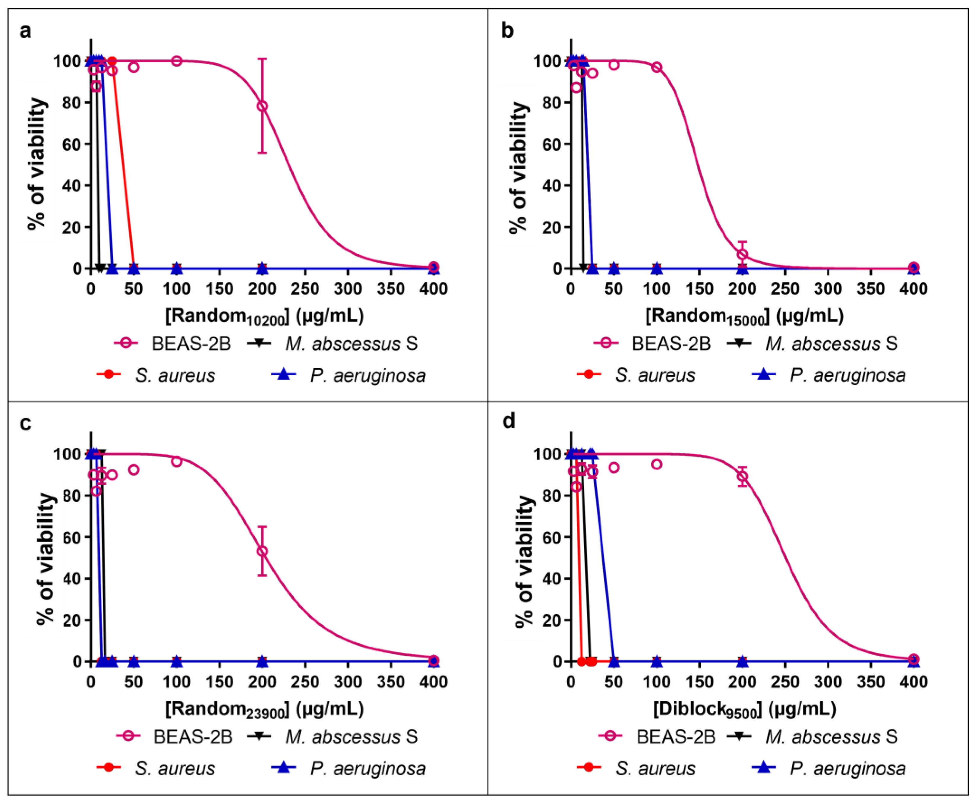
2.1. Antimicrobial Activity of the Copolymers

The characteristics of the Random and Diblock copolymers, i.e., molecular weight and FDMAEMA corresponding to the DMAEMA molar ratio in the final copolymers, are given in Table 1.
The bacterial activity of the Random and Diblock copolymers of different sizes was evaluated through determination of their minimum inhibitory concentrations (MIC). The comparison of the antibacterial activity of the different copolymers shows that their activity has no correlation with their FDMAEMA. As an example, Random10200, Diblock18400, and Diblock20500, all having a FDMAEMA of 0.64, display different MIC values (Table 2); this is also true for Random9300, Random14600, and Diblock6800 or with Diblock9500 and Diblock17900 sharing the same FDMAEMA values but giving different MIC (Table 2). Results demonstrated that copolymer activity rather relies on their type (i.e., Random or Diblock) and their size/molecular weight. Regarding Random copolymers (Table 2), MIC values on M. abscessus S range from 6.25 to 25 µg/mL with a tendency of decreased/weaker activity correlated to increasing sizes. For example, the most active Random copolymer on M. abscessus S (i.e., the one with the lowest MIC value) is Random2800 with a MIC of 6.25 µg/mL, whereas Random copolymers with a size ranging from 14,600 to 23,900 g/mol possess MIC of 25 µg/mL. Oppositely, for P. aeruginosa and S. aureus, Random copolymers display a tendency of increased/improved activity when their size increases (MIC ranging from 12.5 to 400 µg/mL and from 12.5 to 200 µg/mL for P. aeruginosa and S. aureus, respectively). Indeed, the MIC values of Random2800 on P. aeruginosa and S. aureus are 200 µg/mL, whereas Random23900 possesses a MIC of 12.5 µg/mL on both strains.
Regarding Diblock copolymers (Table 2), as for Random ones, the MIC values on M. abscessus S also display decreased activity with increase in their size (from 25 to 100 µg/mL for Diblock6800 and Diblock21900, respectively). In the case of P. aeruginosa and S. aureus, the size of the Diblock copolymers has little influence on the MIC values, ranging from 50 to 100 µg/mL and from 12.5 to 25 µg/mL, respectively.
Finally, it should be mentioned that none of the tested Random and Diblock copolymers possesses antibacterial effect on M. abscessus R (MIC >400 µg/mL).

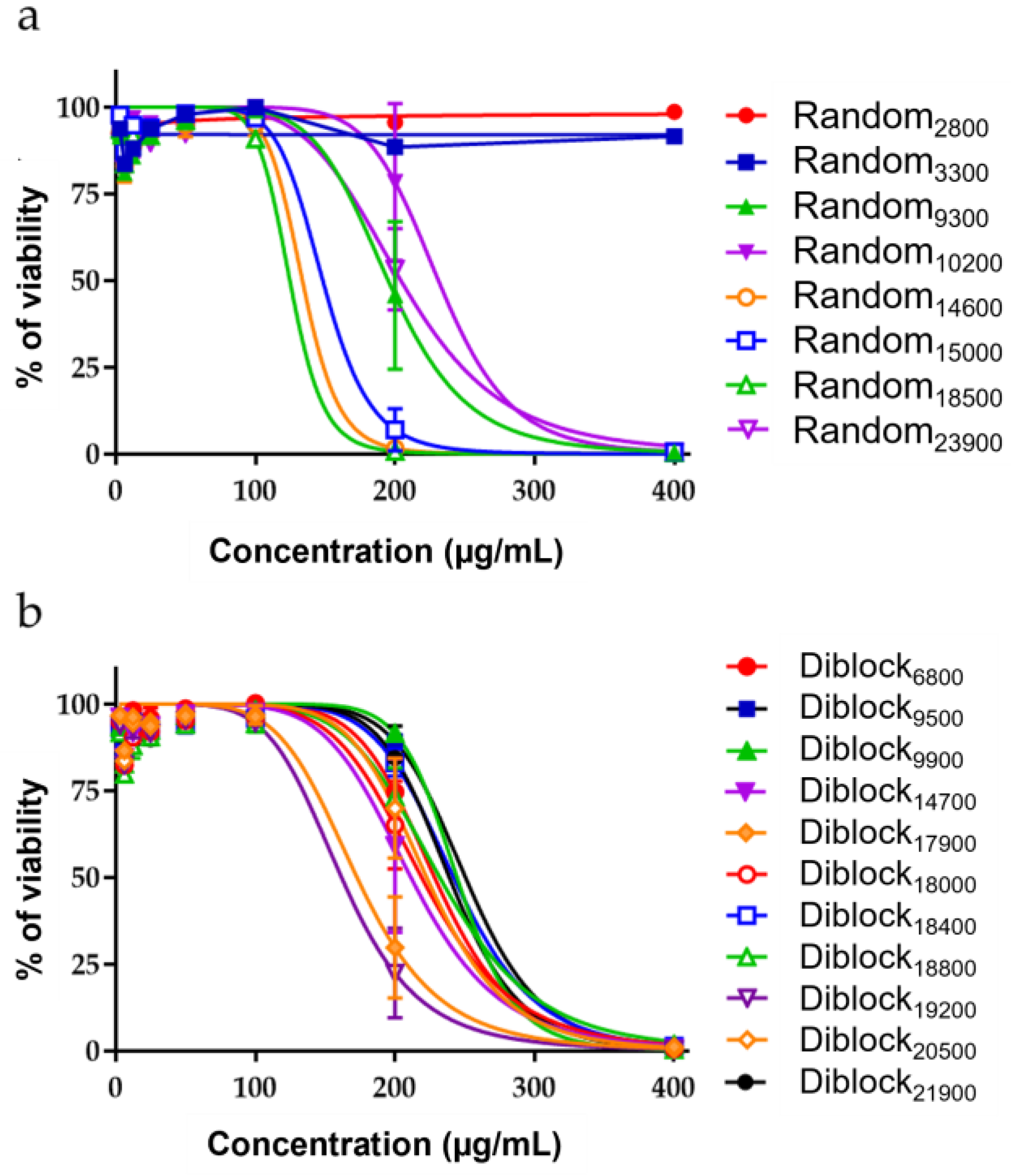
2.2. Innocuity of the Copolymers

The cytotoxic activity of the copolymers was measured after 48 h exposure of non-tumorigenic human lung epithelial cells (BEAS-2B cells) to increasing concentrations of Random or Diblock copolymers (Figure 2). This assay allowed the determination of the IC50 (Inhibitory Concentration 50%), corresponding to the concentration of compound that inhibits 50% of cell survival compared to untreated cells. Regarding Random copolymers, their toxicity shows a tendency to increase with their size (IC50 values from 124.7 to >400 µg/mL) (Figure 2a and Table 3).
With Diblock copolymers, the relationship between their size and their toxicity is less evident, with IC50 values ranging from 161.8 to 250.6 µg/mL (Figure 2b and Table 3).
In addition to IC50 values, the innocuity of the copolymers must be evaluated through the determination of their therapeutic index (TI), i.e., the relation between their IC50 on BEAS-2B and their MIC on the bacterial strains. It is admitted that the TI must be higher than 1 and that the higher the TI, the safer the compound is. As shown in Table 3, for all tested copolymers and regardless of the bacterial strain, all TI are >1. The highest TI is >64 for the Random copolymers (Random2800 on M. abscessus S; Table 3) and 20 for the Diblock copolymers (Diblock9500 on S. aureus; Table 3).
Based on the MIC and the TI values, Random10200, Random15000, Random23900, and Diblock9500 copolymers, which display good antibacterial activity against the three bacteria associated with low toxicity (Figure 3), were selected for further investigations. It has to be noted that, although Random2800 had the lowest cytotoxicity on BEAS-2B cells (i.e., IC50 > 400 µg/mL) and a very potent activity on M. abscessus S (MIC of 6.25 µg/mL), this copolymer was found to be weakly active on P. aeruginosa and S. aureus, explaining why it was not further evaluated in the present study.

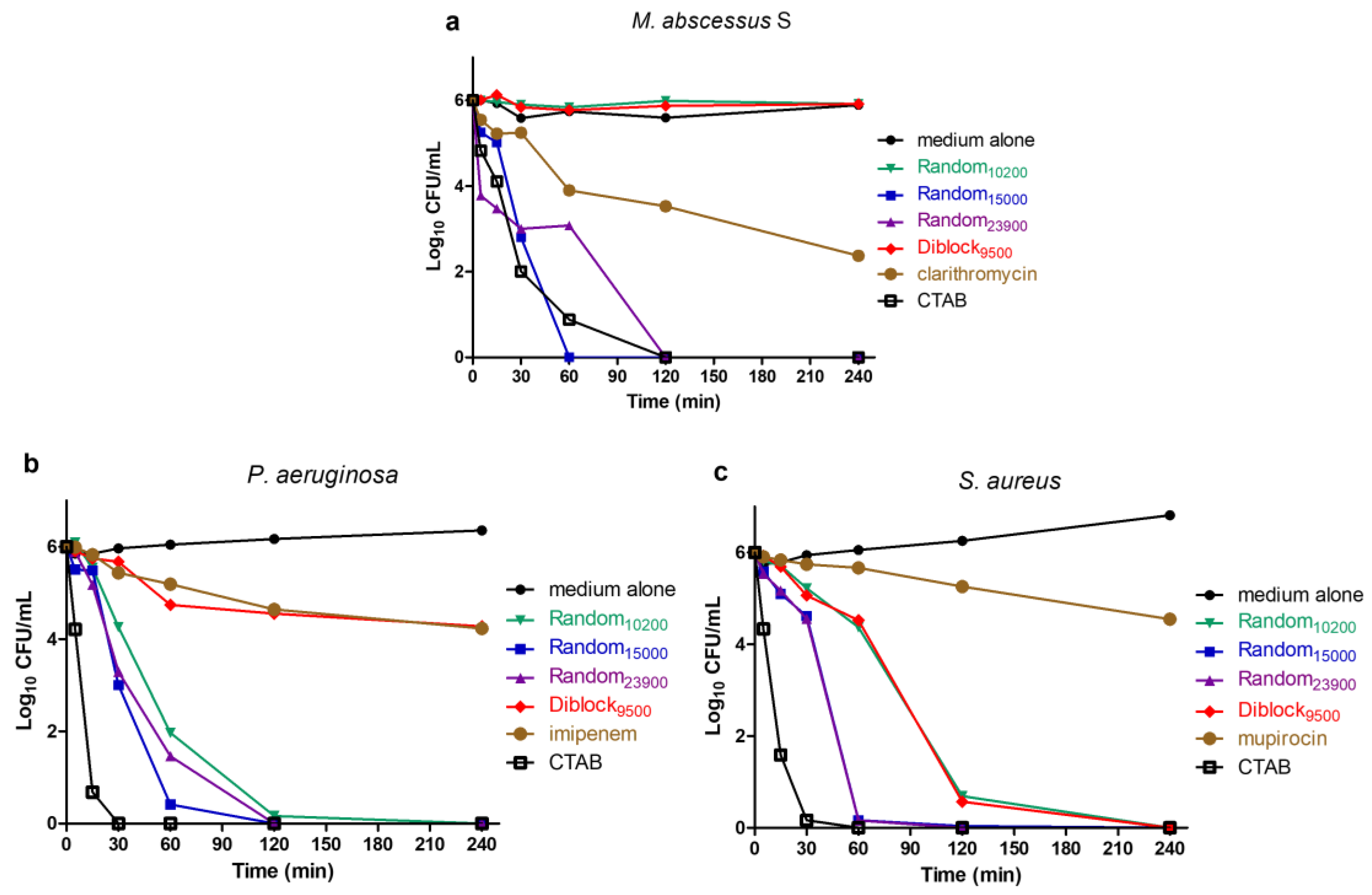
2.3. Determination of Time-Kill Kinetics

A time-kill assay was performed to evaluate the kinetics of action of the four selected copolymers over time (Figure 4). Bacteria were incubated with each copolymer at 4× MIC and took off at different times to be spread on agar plates for colonies forming units (CFU) counting. In addition to the copolymers, the quaternary ammonium cetyltrimethylammonium bromide (CTAB) was used as a control, this cationic surfactant being able to rapidly insert into the bacterial membrane, leading to a fast membranolytic effect [49,50,51]. Conventional bactericidal antibiotics were also used for comparison (i.e., clarithromycin for M. abscessus, imipenem for P. aeruginosa, and mupirocin for S. aureus). Results show that the copolymers Random15000 and Random23900 display a strong and rapid killing effect against the three bacteria tested. Indeed, complete killing of all bacteria (6 log reduction) is achieved in 120 min, with already a ≥3 log reduction in the CFU count within 60 min (Figure 4). This fast killing activity is similar to the one of CTAB, reinforcing the idea that these copolymers, as most AMPs, act through a membranolytic effect. Importantly, these copolymers act more rapidly and strongly than control conventional bactericidal antibiotics leading to less than 1 log reduction in CFU count within 60 min and reaching approximatively 2–4 log reduction after 240 min exposure (Figure 4). The Random10200 compound also exhibits a fast bactericidal activity on P. aeruginosa and S. aureus with a >5 log CFU/mL reduction within 120 min (Figure 4b and c, respectively) but with no killing effect on M. abscessus S during the time course of the assay (Figure 4a). Concerning Diblock9500, although a quick bactericidal activity is observed against S. aureus with a >5 log CFU/mL reduction in 120 min (Figure 4c), the results show that this copolymer possesses no bactericidal activity during the assay against M. abscessus S (Figure 4a) and only a slow killing activity on P. aeruginosa, which is similar to the conventional bactericidal antibiotic imipenem (Figure 4b).

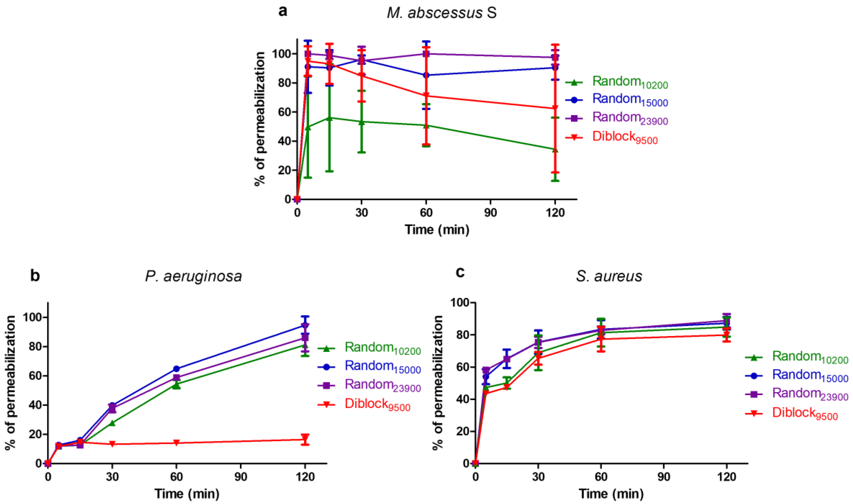
2.4. Mechanism of Action

Since the killing assay suggested that most of the tested copolymers display a rapid bactericidal activity similarly to the membranolytic detergent CTAB, the effect of copolymer on membrane integrity was then evaluated using a propidium iodide assay (Figure 5). Indeed, the cell impermeable dye propidium iodide can enter bacteria and form a highly fluorescent complex with DNA/RNA only if the cell membrane is damaged, allowing for monitoring of the membrane’s integrity. The membranolytic detergent CTAB was used as a positive control giving 100% permeabilization [49,50,51,52]. In M. abscessus S, the copolymers induce a very rapid permeabilization, with more than 50% permeabilization in 5 min (Figure 5a). Within 120 min, more than 90% permeabilization are obtained with Random15000 and Random23900, the permeabilization activity being lower for Random10200 and Diblock9500 (approximately 34% and 62% permeabilization, respectively) (Figure 5a). In P. aeruginosa, the permeabilization activity of Random15000, Random23900, and Random10200 is more progressive, but these three compounds induce more than 80% permeabilization within 120 min exposure (Figure 5b). On the contrary, Diblock9500, which does not have any bactericidal activity on P. aeruginosa (Figure 5b), is unable to permeabilize P. aeruginosa cells during the 120 min of the assay (Figure 5b). Last, on S. aureus, the four copolymers induce a relatively rapid permeabilization, with more than 65% permeabilization within 30 min and ≥80% after 120 min exposure (Figure 5c).

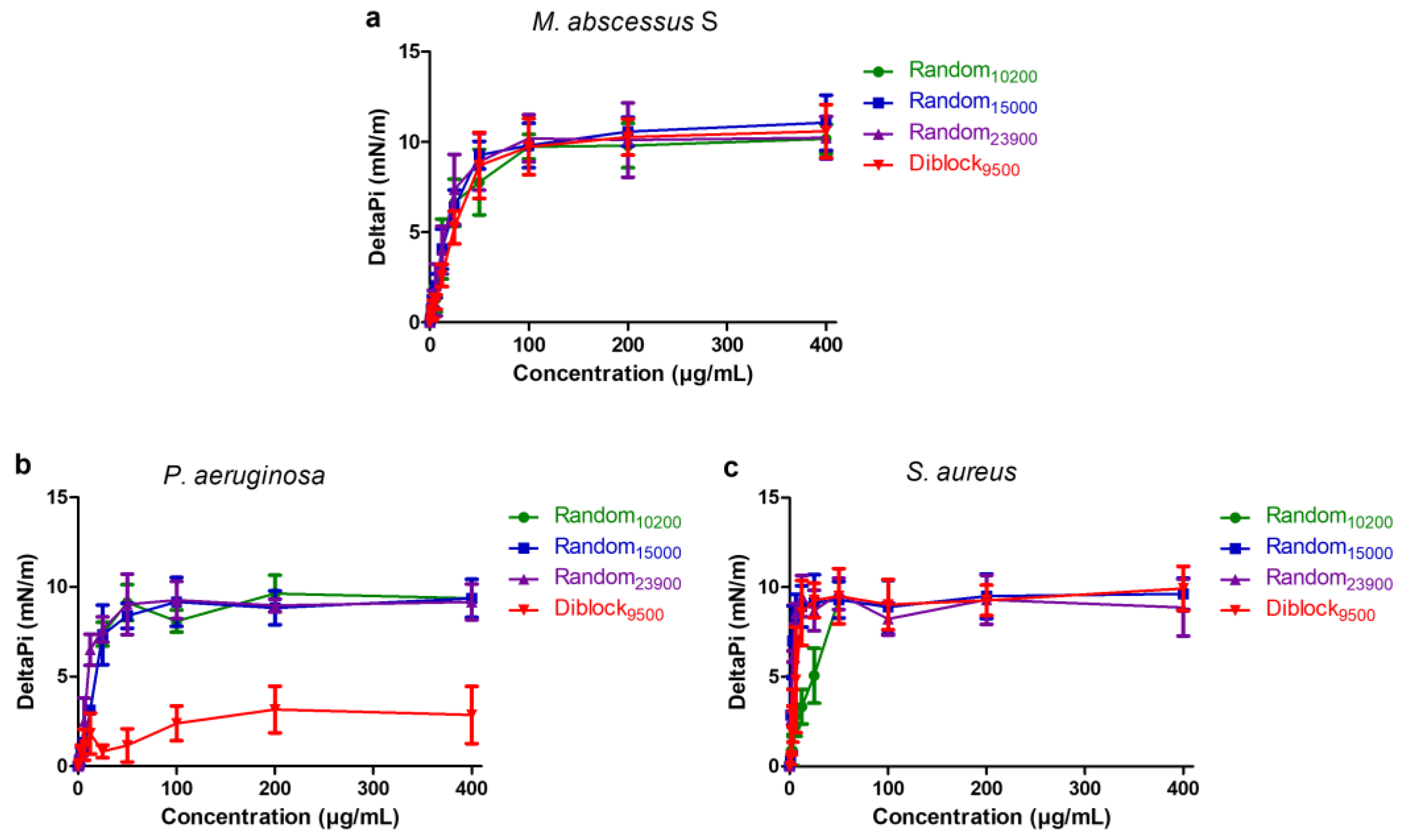
2.5. Insertion in Lipid Monolayers

The ability of the selected copolymers to insert into the bacterial membranes was further evaluated using Langmuir lipid monolayer assay (Figure 6). Total membrane lipids were first extracted from each strain. The extracted total lipids were spread at the air-water interface to reach a surface pressure of 30 ± 0.5 mN/m corresponding to a lipid packing density theoretically equivalent to that of the outer leaflet of the bacterial cell membrane [53]. Increasing concentrations of copolymer were then injected into the aqueous phase below the lipid monolayers. Their ability to insert into the films, causing variations in the surface pressure (deltaPi), was measured using surface microtensiometer. In accordance with propidium iodide assay results (Figure 6), data show that Random10200, Random15000, and Random23900 are able to efficiently insert into lipids extracted from M. abscessus S, P. aeruginosa, and S. aureus, with a maximal insertion observed at concentrations close or superior to the MIC values, i.e., at 1 to 4× MIC (Figure 6). The same result is obtained in the case of Diblock9500, except for the total lipids extracted from P. aeruginosa for which lipid insertion is weaker, reflecting the fact that Diblock9500 only causes a limited membrane permeabilization in P. aeruginosa (Figure 6b).

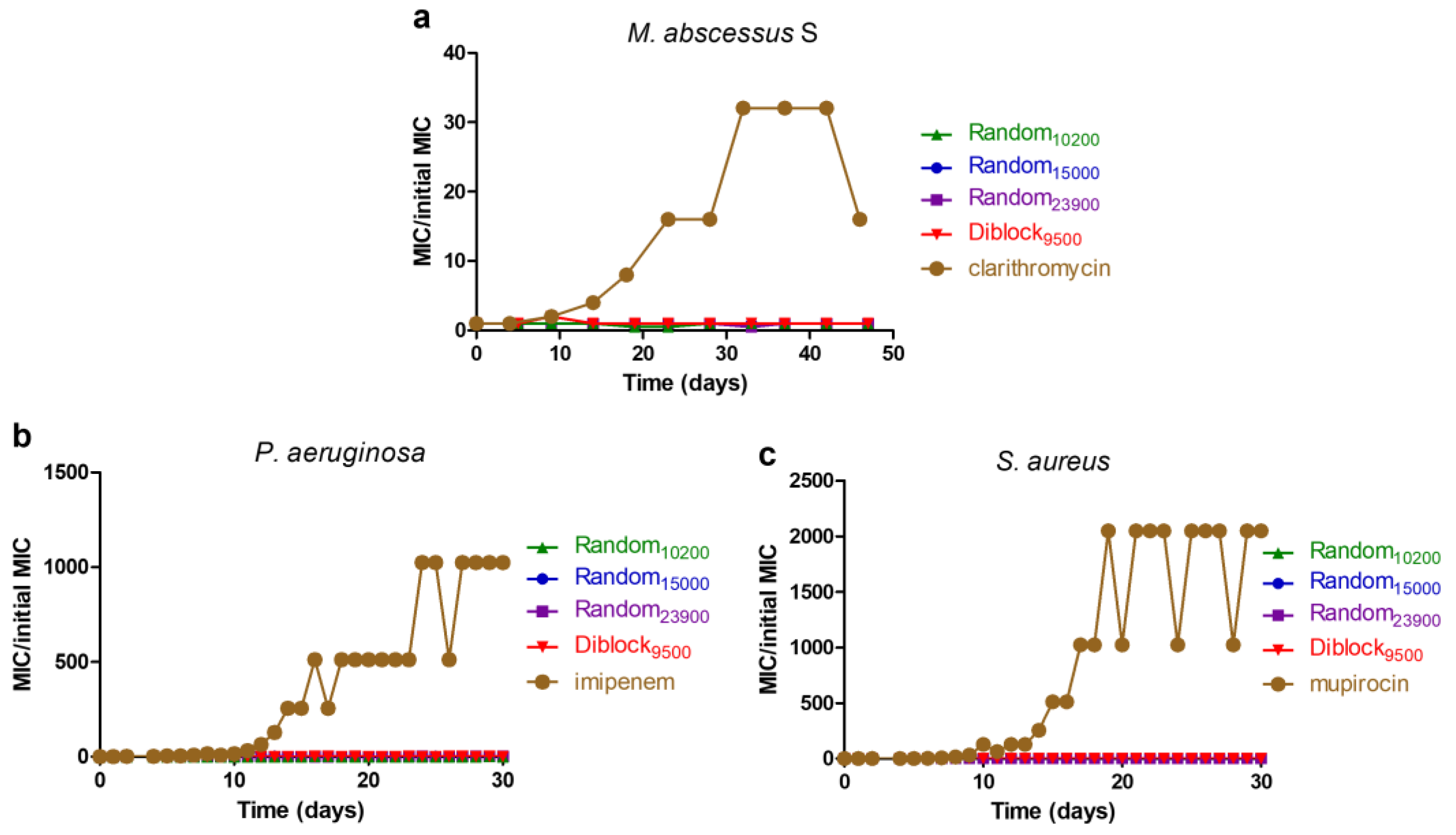
2.6. Resistance Induction Assay

Results obtained above showed that the selected copolymers target the bacterial membrane. Such membranolytic activity, also present in cationic AMPs, suggests that these copolymers should be less prompt at selecting resistant bacteria compared to conventional antibiotics targeting enzyme/proteins for which mutations may induce resistance. To confirm this hypothesis, the appearance of resistant bacteria was evaluated through repeated MIC testing during 30–45 days (Figure 7).
Results show that none of the tested copolymers are able to cause increased MIC values typical of an induction of resistance even after 30–45 days of continuous exposure. By contrast, conventional antibiotics known to be prompt to cause resistance in bacteria select resistant strains after 10–15 days of exposure. Importantly, copolymers fully retain their activity against those generated antibiotic-resistant bacteria with unchanged MIC values.

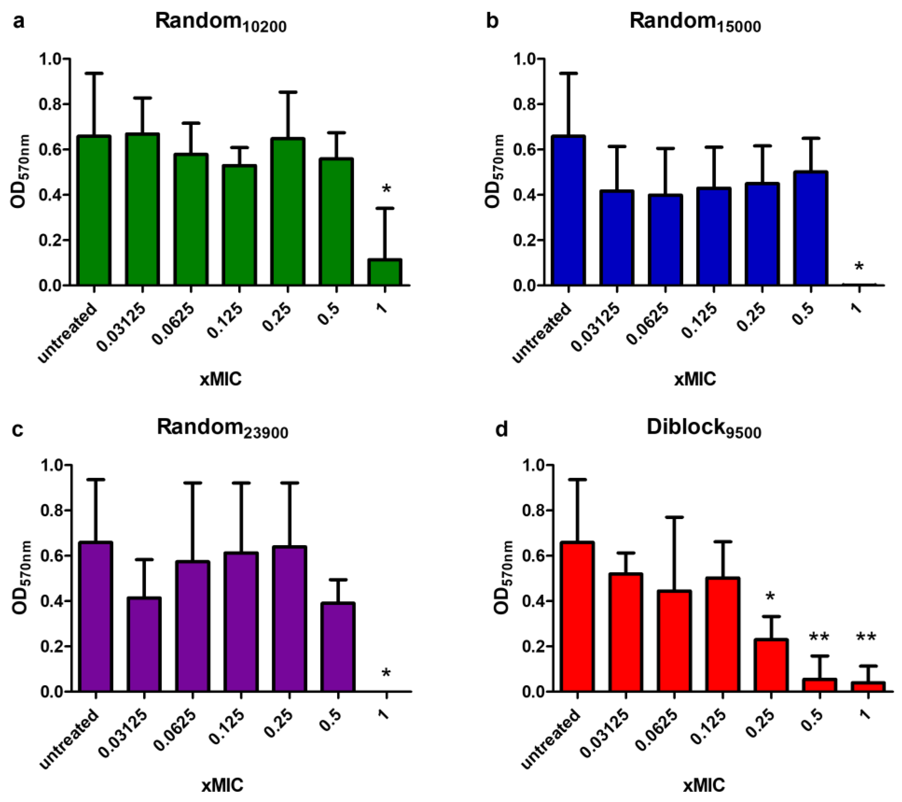
2.7. Anti-Biofilm Activity

The difficulty to treat P. aeruginosa infections is essentially linked to its ability to form biofilms [54]. As a consequence, the formation of highly structured biofilms allows P. aeruginosa to compete, survive, and overtakes other bacteria in the lung of CF patients colonized by a great variety of microorganisms [55]. Therefore, the effect of selected copolymers on biofilm formation was measured. As shown in Figure 8, Random10200, Random15000, Random23900, and Diblock9500 dose-dependently prevent biofilm formation. Diblock9500 is the most interesting one with a statistically significant preventing effect observed with concentration as low as 0.25× MIC. Unfortunately, copolymers were found inactive against preformed/established biofilms.

3. Discussion

Currently, a major global challenge for health is to fight against antibiotic resistance. Indeed, we are facing the dramatic appearance in resistance to one or more antibiotics in a vast number of bacteria infecting humans, combined with the decline in the discovery of new antibiotics [56,57,58]. Furthermore, patients infected with antibiotic-resistant bacteria have generally worse clinical outcomes, higher probability of death, and consume more healthcare resources than patients infected with non-resistant bacteria [56]. In other words, antibiotic resistance increases morbidity, mortality, and hospital costs [56,57,58]. Therefore, there is an urgent need to develop new antibiotics, notably antibiotics against which bacteria develop low or no resistance [56,57,58]. This problem of drug resistance is particularly important for CF patients for which lung complications are often the cause of death, notably lung colonization by pathogenic bacteria [4]. In this study, we identified four methacrylate-based cationic copolymers that could have a great potential for the treatment of CF patients. These copolymers possess a methacrylate backbone, with DMAEMA as the hydrophilic monomer and BMA the hydrophobic monomer. Three of the selected copolymers are Random copolymers (Random10200, Random15000, and Random23900) while one is a Diblock copolymer (Diblock9500). All four copolymers display low cytotoxicity on human lung epithelial cells and exhibit a very good antibacterial activity with relatively high TI values on P. aeruginosa and S. aureus, the most frequently found bacteria in CF patients. The copolymers are also active against M. abscessus S variant, a bacteria often found later in the disease course and nicknamed the “antibiotic nightmare” [6,7,17]. Among the selected copolymers, Random15000 and Random23900 have a strong and rapid killing effect on P. aeruginosa, S. aureus as well as on M. abscessus S. Our data strongly support that this bactericidal activity could be due to the permeabilization of the bacterial membrane leading to their lysis and death. This certainly results from the interaction between the cationic part of the copolymer (due to the presence of DMAEMA) and the negatively charged surface of the bacteria, before insertion of the copolymer into the bacterial membrane thanks to its hydrophobic part (due to the presence of BMA). The fact that these copolymers have no effect on M. abscessus R variant, suggests that the surface-exposed GPL present only in the S variant may play a key role in the interactions governing the antibacterial activity of the copolymers. It is now admitted that CF patients are first infected by M. abscessus S variant, which could evolve into the R form able to form cords and leading to a severe and almost incurable pulmonary infection. In this context, these copolymers could represent a promising alternative strategy to kill this mycobacterium at the early stage of the infection, thus preventing the formation of cords by R variants. Moreover, given their pore-forming mode of action, the probability for the bacteria to develop resistance against these compounds is very low, rendering these copolymers particularly interesting in the treatment of CF patients. This was confirmed by the absence of appearance of resistance against these four copolymers after 30–45 days continuous exposure as previously described for similar copolymers [47]. In addition, the copolymers were found active on bacteria trained to become resistant to conventional antibiotics. Interestingly, it has to be noted that Diblock9500, although found membranolytic for M. abscessus S and S. aureus, does not cause membrane permeabilization in P. aeruginosa. This suggests another mechanism of action for this copolymer on this strain that would be investigated in the future. Finally, similar to numerous AMPs [56,59,60], the tested copolymers were able to prevent biofilm formation implying a major impact of our molecule at the early stage of the infection. Unfortunately, selected copolymers were inactive on established biofilms, a limitation also observed with several AMPs [56,59,60], only few AMPs being shown to both inhibit biofilm formation and disrupt established ones [59].
Overall, this study shows that the Random and Diblock copolymers display the same strengths than AMPs in the treatment of CF-associated bacteria [60], notably their capacity to rapidly kill bacteria through a membranolytic activity, without causing resistance. However, unlike AMPs, Random and Diblock copolymers are insensitive to proteases, not immunogenic, and easy to produce in a large industrial scale. Future studies, including in vivo assays, will be necessary however to confirm their potential in the treatment of bacterial infections in CF patients.

4. Materials and Methods

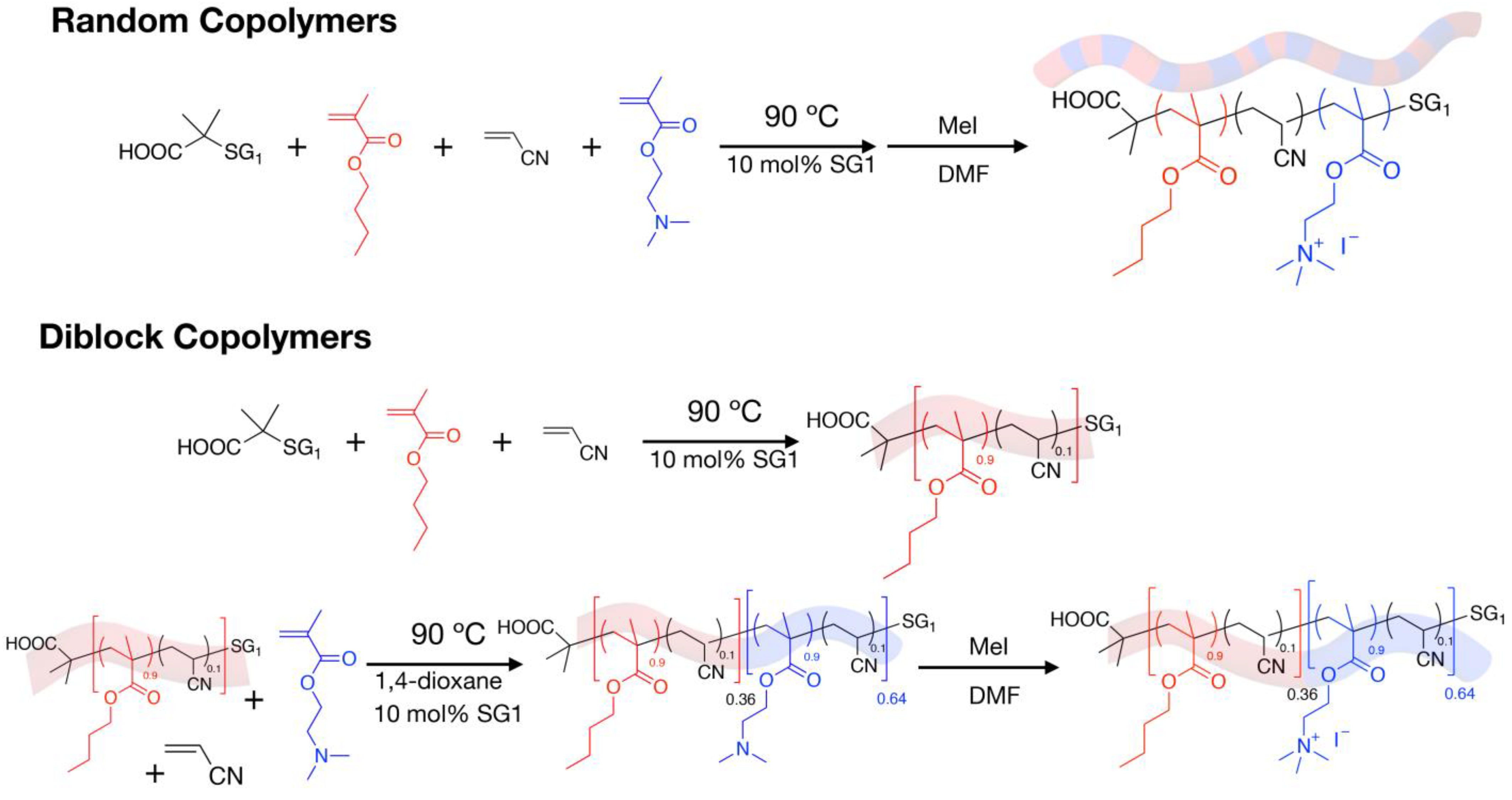
4.1. Synthesis and Characterization of the Cationic Amphiphilic Polymers

The synthesis of the different Random and Diblock copolymers by nitroxide-mediated polymerization (NMP) has already been described [47]. A brief description of their synthesis is given below. The synthesis of the PBMA-b-PDMAEMA Diblock copolymers is a two-step procedure. BMA, acrylonitrile (ACN, comonomer of the NMP, 10mol% vs. BMA), BlocBuilder (initiator) and SG1 (control agent) were introduced in a 100 mL two-necked round bottom glass flask fitted with a magnetic stir bar. After 30 min of nitrogen bubbling at RT, the flask was heated at 90 °C under Argon. The reaction was stopped when the conversion is close to 45%. The conversion was determined by 1H NMR spectroscopy. The fist block was recovered by precipitation into MeOH/H2O and analyzed by SEC/DMF to obtain the molecular weight (Mn) and dispersity (Ð) values. After drying, the PBMA was used as a macroinitiator for the polymerization of the DMAEMA to produce the corresponding PBMA-b-PDMAEMA Diblock copolymer. Typically, PBMA, DMAEMA, ACN (10 mol% vs. DMAEMA), 1,4-dioxane and SG1 were introduced in a 25 mL two-necked round bottom glass flask fitted with a magnetic stir bar. The solution was deoxygenated by nitrogen bubbling for 30 min at room temperature before heating at 90 °C under argon. The reaction was stopped when the conversion reached 40%. The final PBMA-b-PDMAEMA Diblock copolymer was purified by precipitation in pentane and dried under vacuum before analysis by 1H NMR spectroscopy and SEC/DMF to obtain the composition (with FDMAEMA the DMAEMA molar composition in the final copolymer) and the number-average molecular weight Mn), respectively. The synthesis of the Random copolymers is performed in one step. Typically, BMA, ACN, DMAEMA, BlocBuilder, and SG1 were introduced in a 25 mL two-necked round-bottom flask fitted with a magnetic stir bar. After 30 min of deoxygenation by argon bubbling at room temperature, the solution was heated at 90 °C under argon. Samples were taken periodically to determine the conversion (1H NMR spectroscopy/CDCl3/400 MHz) and the molecular weights (SEC/DMF). The reaction was stopped when the conversion was close to 50%. The final P(BMA-co-DMAEMA) Random copolymer was isolated by precipitation in pentane, then dried under vacuum before analysis. The permanent quaternization of the Random and Diblock copolymers (quaternary ammonium ion) was achieved by methylation of the amine group using methyl iodide (MeI). Typically, after dissolution of the copolymers in THF, 2 mol equivalents of MeI versus the number of amine function of DMAEMA were slowly added. After 18 h of stirring at room temperature, the antibacterial copolymers were recovered by precipitation in diethyl ether and then dried under vacuum.

4.2. Bacterial Strains and Growth Conditions

Reference strains used in this study were obtained either from the American Type Culture Collection (ATCC, Molsheim Cedex France) or the French Pasteur Institute (CIP, Paris, France). Pseudomonas aeruginosa (ATCC 9027) and Staphylococcus aureus (ATCC 6538P) were routinely grown on Luria Bertani (LB) agar plates and LB broth at 37 °C. M. abscessus S and R (CIP 104536T) were grown in 7H9 medium supplemented with 0.05% Tween 80, 0.2% glycerol, and 10% oleic acid–albumin–dextrose–catalase (OADC enrichment; BD Difco) (7H9-SOADC) at 37 °C under agitation (200 rpm).

4.3. Minimal Inhibitory Concentration (MIC) Determination

Antimicrobial activity of the polymers was evaluated by determination of their MIC, i.e., the lowest concentration of polymer that inhibits visible growth of the organism compared to a control, following the National Committee of Clinical Laboratory Standards (NCCLS, 1997). Briefly, for P. aeruginosa and S. aureus, single colonies of the different bacterial strains cultured on agar plates were used to inoculate 3 mL of Mueller–Hinton broth (MH). Tubes were incubated overnight (for approximately 16 h) at 37 °C under stirring (200 rpm). The bacterial suspensions were adjusted to OD600 nm = 1 with the medium, then diluted 1/100 in 3 mL of fresh medium and incubated at 37 °C, 200 rpm until bacteria reached log phase growth (OD600 nm approximately 0.6). Then, 100 μL of bacterial suspension (105 cells/mL) were added per well into sterile polypropylene 96 wells microplates (Greiner BioOne, Dominique Dutscher, Brumath, France) containing a serial two-fold dilution of polymers’ increasing concentrations ranging from 0 to 400 µg/mL. Plates were incubated at 37 °C for 18–24 h and OD600 nm measurements were taken using microplate reader (Biotek, Synergy Mx, Colmar, France) to determine the MIC. Experiments were conducted in independent triplicate (n = 3). Susceptibility testing for M. abscessus was performed using the Middlebrook 7H9 broth microdilution method in 96-well flat-bottom Nunclon Delta Surface microplates with lid (Thermo-Fisher Scientific, Illkirch-Graffenstaden, France). Briefly, log-phase bacteria were diluted to a cell density of 5 × 10⁶ cells/mL in 7H9-SOADC medium; then, 100 μL of this bacterial suspension was added to each well containing 100 μL of serial two-fold dilutions of the copolymers or controls to a final volume of 200 μL. Plates were incubated at 37 °C for 3–5 days, and MIC was recorded by absorbance measurement at 560 nm. At least three independent experiments were conducted for each copolymer.

4.4. Evaluation of the Cytotoxic Effect of Polymers Using Human Cells

The toxicity of the polymers on human cells was evaluated as previously described [61] using human non tumorigenic bronchial epithelial cells BEAS-2B (ATCC CRL-9609) as model. Cells were routinely grown on 25 cm2 flasks in DMEM medium supplemented with 10% Fetal Bovine Serum (FBS) and 1% antibiotics (all from Thermo Fisher Scientific, Illkirch-Graffenstaden, France) in a 5% CO2 incubator at 37 °C. For cytotoxicity assay, cells were detached using trypsin–EDTA solution (from Thermo Fisher Scientific, Illkirch-Graffenstaden, France). Cells were counted using Malassez counting chamber and seeded into 96-well cell culture plates (Greiner bio-One, Dominique Dutscher, Brumath, France) at approximately 10,000 cells per well. The cells were left to grow for 48–72 h at 37 °C in a 5% CO2 incubator until they reached confluence. Medium from wells was then aspirated and cells were treated with 100 μL of culture medium containing increasing concentrations of polymers obtained by serial dilution (from 0 to 400 µg/mL, 1:2 dilution). After 48 h incubation at 37 °C in a 5% CO2 incubator, the cell viability was evaluated using a resazurin based in vitro toxicity assay kit (Sigma-Aldrich, Lyon, France), following manufacturer’s instructions. Briefly, the culture medium was removed and cells were treated with 100 μL of the resazurin diluted 1:10 in sterile phosphate buffer saline (PBS) containing calcium and magnesium (PBS++, pH 7.4). After 4 h incubation at 37 °C, fluorescence intensity (λexem = 530/590 nm) was measured using a microplate reader (Biotek, Synergy Mx, Colmar, France). The fluorescence values were normalized by the negative controls and expressed as percentage of viability. The IC50 value of the copolymers (i.e., the concentration of polymers causing a reduction of 50% of the cell viability as compared to the control) was calculated using GraphPad® Prism 7 software (San Diego, CA, USA). Experiments were conducted in triplicate (n = 3).

4.5. Time–Kill Assays

Time-kill assay was performed as previously described [60,62]. Log-phase bacteria were incubated in wells of a 96 wells microplate at a final concentration of 106 cell/mL with either medium alone (growth control), or 150 µM cetyltrimethylammonium bromide (CTAB), or copolymer at 4× MIC. Conventional bacteriolytic antibiotics (at 4× MIC) were used as controls, i.e., clarithromycin for M. abscessus, imipenem for P. aeruginosa, and mupirocin for S. aureus. The plate was incubated at 37 °C in a humidity chamber. At defined time intervals (5, 15, 30, 60, 120 and 240 min), samples were taken from each well and serial dilutions were prepared and plated on agar plates. The number of CFU/mL was then evaluated after 5 days (for M. abscessus) or overnight (for P. aeruginosa and S. aureus) incubation at 37 °C. Experiments were conducted at least in triplicate (n ≥ 3).

4.6. Bacterial Membrane Permeabilization Assay

Membrane permeabilization by copolymers was evaluated using propidium iodide, a cell-impermeable DNA/RNA probe, as previously described [63]. Logarithmic growing bacterial suspensions of P. aeruginosa or S. aureus were prepared from overnight bacterial suspension by 1 in 100 dilutions. After 3 h incubation at 37 °C, 200 rpm, bacterial suspensions were centrifuged for 5 min at 3000 rpm. Cell pellets were then resuspended in MH at approximately 109 cells/mL. Similar protocol was used for M. abscessus with log-phase bacteria, the cell pellet being resuspended in 7H9-SOADC. Propidium iodide (Sigma Aldrich, Lyon, France) was added to these suspensions at a final concentration of 1 mg/mL. Then, 100 μL of this suspension were transferred into Greiner polypropylene 96-well black plates (Greiner bio-One, Dominique Dutscher, Brumath, France) containing copolymers at 4× MIC, or 150 µM CTAB as positive control leading to 100% permeabilization. A negative control was included, with bacteria and propidium iodide but without copolymer. Bacteria were incubated at 37 °C and fluorescence (λexem = 530/590 nm) was recorded over time using a microplate reader (Tecan Spark™, Tecan Group Ltd., France for M. abscessus, and Biotek, Synergy Mx, Colmar, France for P. aeruginosa and S. aureus). Results were expressed in percentage of permeabilization compared to the maximum fluorescence obtained with CTAB, after subtraction of fluorescence of the negative control. All experiments were independently performed at least three times.

4.7. Induction of Resistance Assay

Induction of bacterial resistance by copolymers was evaluated as previously described [60,62,64]. Briefly, bacterial cultures were continuously exposed to copolymers or conventional antibiotics over 45 consecutive days for M. abscessus and 30 consecutive days for P. aeruginosa and S. aureus, through repeated broth microdilution susceptibility testing and determination of MIC values. After each determination of the MIC values, the well containing the highest concentration of copolymer allowing a bacterial growth was diluted 1:1000 in medium and used as inoculum for the next MIC assay.

4.8. Lipid Insertion Assay

Interaction of polymers with bacterial lipids was measured using reconstituted lipid monolayer at the air-water interface as previously described [65]. Total lipids were extracted from bacterial suspensions of P. aeruginosa or S. aureus using Folch procedure [61,64]. With M. abscessus, total lipids were extracted as previously described [66,67] with slight modifications. Briefly, total lipids from dry extract were incubated for 16 h with CHCl3-MeOH (1:2; v/v) at room temperature under shaking. Residual lipids were re-extracted, first with CHCl3-MeOH (1:1, v/v) and then for 16 h with the same solvents but using an alternative ratio (2:1, v/v). All the three organic phases were pooled and concentrated under reduced pressure. Samples were re-suspended in CHCl3-MeOH (3:1, v/v), and washed with 0.3% (w/v) NaCl solution. Organic and aqueous phases were separated by centrifugation 5000× g for 15 min and the organic phase was dried over MgSO4. Each sample of extracted total lipids was dried, resolubilized in CHCl3-MeOH (2:1, v/v) and stored at –20 °C under nitrogen. For lipid interaction assay, total lipid extract was spread using a 50 µL Hamilton’s syringe at the surface of 800 µL of sterile ultra-pure water creating a lipid monolayer at the air-water interface. Lipids were added until the surface pressure reached 30 ± 0.5 mN/m, a value corresponding to a lipid packing density theoretically equivalent to that of the outer leaflet of the cell membrane [53]. After a delay of 5–10 min to allow for solvent evaporation and stabilization of the initial surface pressure, 8 µL of polymers more or less diluted in sterile ultra-pure water were injected into the ultra-pure water sub-phase (volume 800 µL) using a 10 µL Hamilton syringe to reach the desired final concentration of polymer. The variation of the surface pressure caused by polymer insertion was then continuously monitored using a fully automated microtensiometer (µTROUGH SX, Kibron Inc., Helsinki, Finland) until reaching equilibrium (within 90 min of continuous recording). All experiments were carried out in a controlled atmosphere at 20 °C ± 1 °C, and data were analyzed using the Filmware 2.5 program (Kibron Inc., Helsinki, Finland).

4.9. Antibiofilm Formation Assay

P. aeruginosa ATCC 9027 was grown in Mueller Hinton (MH) broth at 37 °C with agitation at 200 rpm until OD600nm reached 0.2–0.3. Cells were then diluted to 105 CFU/mL in MH and 150 µL of cell suspension was added per well of the Calgary biofilm device (CBD). Sterile polymers were added at a maximum concentration of 400 µg/mL and twofold series dilutions were performed in cell suspension to a minimal concentration of 0.8 µg/mL. As it has been demonstrated that an edge effect affects the biofilm formation, all the edges of the plates were filled with MH and used as sterility and negative controls [59]. Conversely, untreated wells containing bacterial suspension were used as a positive control for biofilm formation. After 24 h of incubation at 37 °C with agitation reduced to 110 rpm, the lid of the CBD was removed and the pegs were rinsed twice in PBS and then fixed in 100% methanol for 15 min. Pegs were rinsed with PBS once more and then air-dried before being stained with crystal violet at 0.2% for 15 min. Pegs were rinsed with PBS twice before being air-dried and then destained in methanol for 15 min before being discarded. Absorbance at 570 nm was measured to evaluate biofilm formation. All the rinsing, staining, and destaining steps were performed in 96-well plates with a volume of 200 µL per well in order for the biofilm to be totally soaked. All experiments were performed in independent triplicates.

4.10. Antibiofilm Disruption Assay

P. aeruginosa ATCC 9027 was grown in Luria Bertani (LB) broth at 37 °C with agitation at 200 rpm until OD600nm reached 0.2–0.3. Cells were then diluted to 105 CFU/mL in LB, and 150 µL of cell suspension was added per well of the Calgary biofilm device without the addition of polymers and taking into account the edge effect previously mentioned. After 24 h of growth at 37 °C under stirring at 110 rpm, the lid of the CBD was transferred into a new 96-well plate containing fresh MH media supplemented either with polymers at concentrations ranging from 0.8 to 400 µg/mL or not. Wells were filled with 200 µL to make sure the pegs were totally soaked. Then, after 24 h of incubation in the same condition, the pegs were rinsed, fixed, stained, and destained as described above. A volume of 300 µL was used per well. Absorbance at 570 nm was measured to evaluate biofilm formation and disruption.

4.11. Statistical Analysis

All assays were performed in triplicate (n = 3). Data were plotted and analyzed using GraphPad® Prism 7 software (San Diego, CA, USA). Statistical significancy of the data was evaluated using a Mann-Whitney test.

5. Conclusions

In this study, various Random and Diblock methacrylate-based cationic copolymers ranging from 2800 to 23,900 g/mol were tested against the three major bacterial species colonizing CF patients (i.e., P. aeruginosa, S. aureus, and M. abscessus). Evaluation of the antibacterial activity and innocuity on human cells allowed the identification of four copolymers, i.e., Random10200, Random15000, Random23900, and Diblock9500, as the best molecules. Mechanistic approaches demonstrated that the copolymers, due to their amphipathic character and cationic charge, preferentially interact with the bacterial membrane, leading to a rapid membranolytic effect and the death of the bacteria. Random and Diblock methacrylate-based cationic copolymers thus display the same strengths as AMPs against bacteria, notably their fast killing action related to a membranolytic activity preventing the development of resistance and allowing them to remain active on bacteria already resistant to antibiotics. In addition, their insensitivity to proteases, the absence of immunogenicity, and their ease of production make those copolymers good candidates as antibacterial agents. Although in vivo confirmation will be necessary, our results already demonstrate the potential value of the described antimicrobial amphiphilic cationic copolymers in the fight against bacteria, particularly the ones infecting CF patients.

Author Contributions

Conceptualization, M.C., J.-F.C., S.C. (Stéphane Canaan), Y.G., C.L. and M.M.; methodology, M.C., J.-F.C., S.C. (Stéphane Canaan), Y.G., M.L., C.L., J.P., S.H., V.P., C.R. and M.M.; validation, M.C. and M.M.; formal analysis, M.C. and M.M.; investigation, M.C., H.O., S.H., C.R., J.-F.C., K.J., B.C., J.P., S.C. (Siméon Charriau), M.L.,Y.G., S.C. (Stéphane Canaan), V.P., C.L. and M.M.; writing—original draft preparation, M.C. and M.M.; writing—review and editing, M.C., H.O., S.H., C.R., J.-F.C., K.J., B.C., J.P., S.C. (Siméon Charriau), M.L.,Y.G., S.C. (Stéphane Canaan), V.P., C.L. and M.M.; supervision, C.L., Y.G., M.C. and M.M.; project administration, M.C. and M.M.; funding acquisition, M.C., Y.G., C.L., S.C. (Stéphane Canaan), and M.M. All authors have read and agreed to the published version of the manuscript.

Funding

This research was funded by Amidex Pépinière d’Excellence.

Institutional Review Board Statement

Not applicable.

Informed Consent Statement

Not applicable.

Data Availability Statement

Data available on request due to restrictions eg privacy or ethical. The data presented in this study are available on request from the corresponding author. The data are not publicly available due to privacy.

Conflicts of Interest

The authors declare no conflict of interest.

References

  1. Shteinberg, M.; Haq, I.J.; Polineni, D.; Davies, J.C. Cystic Fibrosis. Lancet 2021, 397, 2195–2211. [Google Scholar] [CrossRef]
  2. Southern, K.W.; Munck, A.; Pollitt, R.; Travert, G.; Zanolla, L.; Dankert-Roelse, J.; Castellani, C.; ECFS CF Neonatal Screening Working Group. A Survey of Newborn Screening for Cystic Fibrosis in Europe. J. Cyst. Fibros 2007, 6, 57–65. [Google Scholar] [CrossRef] [Green Version]
  3. Scotet, V.; Gutierrez, H.; Farrell, P.M. Newborn Screening for CF across the Globe-Where Is It Worthwhile? Int. J. Neonatal Screen 2020, 6, 18. [Google Scholar] [CrossRef] [PubMed] [Green Version]
  4. Goetz, D.; Ren, C.L. Review of Cystic Fibrosis. Pediatr. Ann. 2019, 48, e154–e161. [Google Scholar] [CrossRef] [PubMed]
  5. McElvaney, O.J.; Wade, P.; Murphy, M.; Reeves, E.P.; McElvaney, N.G. Targeting Airway Inflammation in Cystic Fibrosis. Expert Rev. Respir. Med. 2019, 13, 1041–1055. [Google Scholar] [CrossRef] [PubMed]
  6. Lobo, L.J.; Noone, P.G. Respiratory Infections in Patients with Cystic Fibrosis Undergoing Lung Transplantation. Lancet Respir. Med. 2014, 2, 73–82. [Google Scholar] [CrossRef] [PubMed]
  7. Jones, A.M. Which Pathogens Should We Worry About? Paediatr. Respir. Rev. 2019, 31, 15–17. [Google Scholar] [CrossRef]
  8. Lipuma, J.J. The Changing Microbial Epidemiology in Cystic Fibrosis. Clin. Microbiol. Rev. 2010, 23, 299–323. [Google Scholar] [CrossRef] [PubMed] [Green Version]
  9. Chirgwin, M.E.; Dedloff, M.R.; Holban, A.M.; Gestal, M.C. Novel Therapeutic Strategies Applied to Pseudomonas Aeruginosa Infections in Cystic Fibrosis. Materials 2019, 12, 4093. [Google Scholar] [CrossRef] [PubMed] [Green Version]
  10. Pang, Z.; Raudonis, R.; Glick, B.R.; Lin, T.-J.; Cheng, Z. Antibiotic Resistance in Pseudomonas Aeruginosa: Mechanisms and Alternative Therapeutic Strategies. Biotechnol. Adv. 2019, 37, 177–192. [Google Scholar] [CrossRef] [PubMed]
  11. Ren, C.L.; Morgan, W.J.; Konstan, M.W.; Schechter, M.S.; Wagener, J.S.; Fisher, K.A.; Regelmann, W.E. Presence of Methicillin Resistant Staphylococcus Aureus in Respiratory Cultures from Cystic Fibrosis Patients Is Associated with Lower Lung Function. Pediatr. Pulmonol. 2007, 42, 513–518. [Google Scholar] [CrossRef] [PubMed]
  12. Griffith, D.E.; Aksamit, T.R. Understanding Nontuberculous Mycobacterial Lung Disease: It’s Been a Long Time Coming. F1000Res 2016, 5, 2797. [Google Scholar] [CrossRef] [PubMed] [Green Version]
  13. Howard, S.T.; Rhoades, E.; Recht, J.; Pang, X.; Alsup, A.; Kolter, R.; Lyons, C.R.; Byrd, T.F.Y. Spontaneous Reversion of Mycobacterium Abscessus from a Smooth to a Rough Morphotype Is Associated with Reduced Expression of Glycopeptidolipid and Reacquisition of an Invasive Phenotype. Microbiology 2006, 152, 1581–1590. [Google Scholar] [CrossRef] [Green Version]
  14. Roux, A.-L.; Viljoen, A.; Bah, A.; Simeone, R.; Bernut, A.; Laencina, L.; Deramaudt, T.; Rottman, M.; Gaillard, J.-L.; Majlessi, L.; et al. The Distinct Fate of Smooth and Rough Mycobacterium Abscessus Variants inside Macrophages. Open Biol. 2016, 6, 160185. [Google Scholar] [CrossRef] [PubMed] [Green Version]
  15. Catherinot, E.; Clarissou, J.; Etienne, G.; Ripoll, F.; Emile, J.-F.; Daffé, M.; Perronne, C.; Soudais, C.; Gaillard, J.-L.; Rottman, M. Hypervirulence of a Rough Variant of the Mycobacterium Abscessus Type Strain. Infect. Immun. 2007, 75, 1055–1058. [Google Scholar] [CrossRef] [Green Version]
  16. Catherinot, E.; Roux, A.-L.; Macheras, E.; Hubert, D.; Matmar, M.; Dannhoffer, L.; Chinet, T.; Morand, P.; Poyart, C.; Heym, B.; et al. Acute Respiratory Failure Involving an R Variant of Mycobacterium Abscessus. J. Clin. Microbiol. 2009, 47, 271–274. [Google Scholar] [CrossRef] [Green Version]
  17. Nessar, R.; Cambau, E.; Reyrat, J.M.; Murray, A.; Gicquel, B. Mycobacterium Abscessus: A New Antibiotic Nightmare. J. Antimicrob. Chemother. 2012, 67, 810–818. [Google Scholar] [CrossRef] [Green Version]
  18. Sanguinetti, M.; Ardito, F.; Fiscarelli, E.; La Sorda, M.; D’Argenio, P.; Ricciotti, G.; Fadda, G. Fatal Pulmonary Infection Due to Multidrug-Resistant Mycobacterium Abscessus in a Patient with Cystic Fibrosis. J. Clin. Microbiol. 2001, 39, 816–819. [Google Scholar] [CrossRef] [Green Version]
  19. Jeon, K.; Kwon, O.J.; Lee, N.Y.; Kim, B.-J.; Kook, Y.-H.; Lee, S.-H.; Park, Y.K.; Kim, C.K.; Koh, W.-J. Antibiotic Treatment of Mycobacterium Abscessus Lung Disease. Am. J. Respir. Crit. Care Med. 2009, 180, 896–902. [Google Scholar] [CrossRef]
  20. Ballarino, G.J.; Olivier, K.N.; Claypool, R.J.; Holland, S.M.; Prevots, D.R. Pulmonary Nontuberculous Mycobacterial Infections: Antibiotic Treatment and Associated Costs. Respir. Med. 2009, 103, 1448–1455. [Google Scholar] [CrossRef]
  21. Osmani, M.; Sotello, D.; Alvarez, S.; Odell, J.A.; Thomas, M. Mycobacterium Abscessus Infections in Lung Transplant Recipients: 15-Year Experience from a Single Institution. Transpl. Infect. Dis. 2018, 20, e12835. [Google Scholar] [CrossRef] [PubMed]
  22. Yung, D.B.Y.; Sircombe, K.J.; Pletzer, D. Friends or Enemies? The Complicated Relationship between Pseudomonas Aeruginosa and Staphylococcus Aureus. Mol. Microbiol. 2021, 116, 1–15. [Google Scholar] [CrossRef]
  23. Camus, L.; Briaud, P.; Bastien, S.; Elsen, S.; Doléans-Jordheim, A.; Vandenesch, F.; Moreau, K. Trophic Cooperation Promotes Bacterial Survival of Staphylococcus Aureus and Pseudomonas Aeruginosa. ISME J. 2020, 14, 3093–3105. [Google Scholar] [CrossRef]
  24. Brooks, B.D.; Brooks, A.E. Therapeutic Strategies to Combat Antibiotic Resistance. Adv. Drug Deliv. Rev. 2014, 78, 14–27. [Google Scholar] [CrossRef] [PubMed]
  25. Mahlapuu, M.; Håkansson, J.; Ringstad, L.; Björn, C. Antimicrobial Peptides: An Emerging Category of Therapeutic Agents. Front. Cell Infect. Microbiol. 2016, 6, 194. [Google Scholar] [CrossRef] [PubMed] [Green Version]
  26. Fox, J.L. Antimicrobial Peptides Stage a Comeback. Nat. Biotechnol. 2013, 31, 379–382. [Google Scholar] [CrossRef] [PubMed]
  27. Duplantier, A.J.; van Hoek, M.L. The Human Cathelicidin Antimicrobial Peptide LL-37 as a Potential Treatment for Polymicrobial Infected Wounds. Front. Immunol. 2013, 4, 143. [Google Scholar] [CrossRef] [PubMed] [Green Version]
  28. Fjell, C.D.; Hiss, J.A.; Hancock, R.E.W.; Schneider, G. Designing Antimicrobial Peptides: Form Follows Function. Nat. Rev. Drug Discov. 2011, 11, 37–51. [Google Scholar] [CrossRef]
  29. Shen, W.; He, P.; Xiao, C.; Chen, X. From Antimicrobial Peptides to Antimicrobial Poly(α-Amino Acid)s. Adv. Healthc. Mater. 2018, 7, 1800354. [Google Scholar] [CrossRef]
  30. Molchanova, N.; Hansen, P.R.; Franzyk, H. Advances in Development of Antimicrobial Peptidomimetics as Potential Drugs. Molecules 2017, 22, 1430. [Google Scholar] [CrossRef]
  31. Azmi, F.; Skwarczynski, M.; Toth, I. Towards the Development of Synthetic Antibiotics: Designs Inspired by Natural Antimicrobial Peptides. Curr. Med. Chem. 2016, 23, 4610–4624. [Google Scholar] [CrossRef] [PubMed]
  32. Zou, P.; Laird, D.; Riga, E.K.; Deng, Z.; Dorner, F.; Perez-Hernandez, H.-R.; Guevara-Solarte, D.L.; Steinberg, T.; Al-Ahmad, A.; Lienkamp, K. Antimicrobial and Cell-Compatible Surface-Attached Polymer Networks–How the Correlation of Chemical Structure to Physical and Biological Data Leads to a Modified Mechanism of Action. J. Mater. Chem. B 2015, 3, 6224–6238. [Google Scholar] [CrossRef] [PubMed] [Green Version]
  33. Tew, G.N.; Scott, R.W.; Klein, M.L.; DeGrado, W.F. De Novo Design of Antimicrobial Polymers, Foldamers, and Small Molecules: From Discovery to Practical Applications. Acc. Chem. Res. 2010, 43, 30–39. [Google Scholar] [CrossRef] [PubMed] [Green Version]
  34. Al-Ahmad, A.; Laird, D.; Zou, P.; Tomakidi, P.; Steinberg, T.; Lienkamp, K. Nature-Inspired Antimicrobial Polymers--Assessment of Their Potential for Biomedical Applications. PLoS ONE 2013, 8, e73812. [Google Scholar] [CrossRef] [PubMed] [Green Version]
  35. Kuroda, K.; Caputo, G.A. Antimicrobial Polymers as Synthetic Mimics of Host-Defense Peptides. Wiley Interdiscip Rev. Nanomed Nanobiotechnol. 2013, 5, 49–66. [Google Scholar] [CrossRef] [PubMed] [Green Version]
  36. Carmona-Ribeiro, A.M.; de Melo Carrasco, L.D. Cationic Antimicrobial Polymers and Their Assemblies. Int. J. Mol. Sci. 2013, 14, 9906–9946. [Google Scholar] [CrossRef] [Green Version]
  37. Deka, S.R.; Sharma, A.K.; Kumar, P. Cationic Polymers and Their Self-Assembly for Antibacterial Applications. Curr. Top. Med. Chem. 2015, 15, 1179–1195. [Google Scholar] [CrossRef]
  38. Ergene, C.; Palermo, E.F. Antimicrobial Synthetic Polymers: An Update on Structure-Activity Relationships. Curr. Pharm. Des. 2018, 24, 855–865. [Google Scholar] [CrossRef]
  39. Konai, M.M.; Bhattacharjee, B.; Ghosh, S.; Haldar, J. Recent Progress in Polymer Research to Tackle Infections and Antimicrobial Resistance. Biomacromolecules 2018, 19, 1888–1917. [Google Scholar] [CrossRef]
  40. Makobongo, M.O.; Gancz, H.; Carpenter, B.M.; McDaniel, D.P.; Merrell, D.S. The Oligo-Acyl Lysyl Antimicrobial Peptide C12K-2β12 Exhibits a Dual Mechanism of Action and Demonstrates Strong In Vivo Efficacy against Helicobacter Pylori. Antimicrob. Agents Chemother. 2012, 56, 378–390. [Google Scholar] [CrossRef]
  41. Chakrabarty, S.; King, A.; Kurt, P.; Zhang, W.; Ohman, D.E.; Wood, L.F.; Lovelace, C.; Rao, R.; Wynne, K.J. Highly Effective, Water-Soluble, Hemocompatible 1,3-Propylene Oxide-Based Antimicrobials: Poly[(3,3-Quaternary/PEG)-Copolyoxetanes]. Biomacromolecules 2011, 12, 757–769. [Google Scholar] [CrossRef] [PubMed]
  42. King, A.; Chakrabarty, S.; Zhang, W.; Zeng, X.; Ohman, D.E.; Wood, L.F.; Abraham, S.; Rao, R.; Wynne, K.J. High Antimicrobial Effectiveness with Low Hemolytic and Cytotoxic Activity for PEG/Quaternary Copolyoxetanes. Biomacromolecules 2014, 15, 456–467. [Google Scholar] [CrossRef] [PubMed]
  43. Kozon, D.; Mierzejewska, J.; Kobiela, T.; Grochowska, A.; Dudnyk, K.; Głogowska, A.; Sobiepanek, A.; Kuźmińska, A.; Ciach, T.; Augustynowicz-Kopeć, E.; et al. Amphiphilic Polymethyloxazoline–Polyethyleneimine Copolymers: Interaction with Lipid Bilayer and Antibacterial Properties. Macromol. Biosci. 2019, 19, 1900254. [Google Scholar] [CrossRef] [PubMed]
  44. Cheng, J.; Chin, W.; Dong, H.; Xu, L.; Zhong, G.; Huang, Y.; Li, L.; Xu, K.; Wu, M.; Hedrick, J.L.; et al. Biodegradable Antimicrobial Polycarbonates with In Vivo Efficacy against Multidrug-Resistant MRSA Systemic Infection. Adv. Healthc. Mater. 2015, 4, 2128–2136. [Google Scholar] [CrossRef]
  45. Tan, J.P.K.; Coady, D.J.; Sardon, H.; Yuen, A.; Gao, S.; Lim, S.W.; Liang, Z.C.; Tan, E.W.; Venkataraman, S.; Engler, A.C.; et al. Broad Spectrum Macromolecular Antimicrobials with Biofilm Disruption Capability and In Vivo Efficacy. Adv. Healthc. Mater. 2017, 6, 1601420. [Google Scholar] [CrossRef]
  46. Yang, C.; Lou, W.; Zhong, G.; Lee, A.; Leong, J.; Chin, W.; Ding, B.; Bao, C.; Tan, J.P.K.; Pu, Q.; et al. Degradable Antimicrobial Polycarbonates with Unexpected Activity and Selectivity for Treating Multidrug-Resistant Klebsiella Pneumoniae Lung Infection in Mice. Acta Biomater. 2019, 94, 268–280. [Google Scholar] [CrossRef]
  47. Benkhaled, B.T.; Hadiouch, S.; Olleik, H.; Perrier, J.; Ysacco, C.; Guillaneuf, Y.; Gigmes, D.; Maresca, M.; Lefay, C. Elaboration of Antimicrobial Polymeric Materials by Dispersion of Well-Defined Amphiphilic Methacrylic SG1-Based Copolymers. Polym. Chem. 2018, 9, 3127–3141. [Google Scholar] [CrossRef]
  48. Zou, Y.-J.; He, S.-S.; Du, J.-Z. ε-Poly(L-Lysine)-Based Hydrogels with Fast-Acting and Prolonged Antibacterial Activities. Chin. J. Polym. Sci. 2018, 36, 1239–1250. [Google Scholar] [CrossRef]
  49. Ioannou, C.J.; Hanlon, G.W.; Denyer, S.P. Action of Disinfectant Quaternary Ammonium Compounds against Staphylococcus Aureus. Antimicrob. Agents Chemother. 2007, 51, 296–306. [Google Scholar] [CrossRef] [Green Version]
  50. Jagatia, H.; Cantillon, D. DNA Isolation from Mycobacteria. Mycobact. Protoc. 2021, 59–75. [Google Scholar] [CrossRef]
  51. Pietsch, F.; Heidrich, G.; Nordholt, N.; Schreiber, F. Prevalent Synergy and Antagonism Among Antibiotics and Biocides in Pseudomonas Aeruginosa. Front. Microbiol. 2021, 11, 615618. [Google Scholar] [CrossRef] [PubMed]
  52. Dengler, W.A.; Schulte, J.; Berger, D.P.; Mertelsmann, R.; Fiebig, H.H. Development of a Propidium Iodide Fluorescence Assay for Proliferation and Cytotoxicity Assays. Anti-Cancer Drugs 1995, 6, 522–532. [Google Scholar] [CrossRef] [PubMed]
  53. Dennison, S.R.; Kim, Y.S.; Cha, H.J.; Phoenix, D.A. Investigations into the Ability of the Peptide, HAL18, to Interact with Bacterial Membranes. Eur. Biophys. J. 2008, 38, 37. [Google Scholar] [CrossRef] [PubMed]
  54. Thi, M.T.T.; Wibowo, D.; Rehm, B.H.A. Pseudomonas Aeruginosa Biofilms. Int. J. Mol. Sci. 2020, 21, 8671. [Google Scholar] [CrossRef]
  55. Oluyombo, O.; Penfold, C.N.; Diggle, S.P. Competition in Biofilms between Cystic Fibrosis Isolates of Pseudomonas Aeruginosa Is Shaped by R-Pyocins. mBio 2019, 10, e01828-18. [Google Scholar] [CrossRef] [Green Version]
  56. Chellat, M.F.; Raguž, L.; Riedl, R. Targeting Antibiotic Resistance. Angew. Chem. Int. Ed. 2016, 55, 6600–6626. [Google Scholar] [CrossRef]
  57. Baym, M.; Stone, L.K.; Kishony, R. Multidrug Evolutionary Strategies to Reverse Antibiotic Resistance. Science 2016, 351, aad3292. [Google Scholar] [CrossRef] [Green Version]
  58. Hutchings, M.I.; Truman, A.W.; Wilkinson, B. Antibiotics: Past, Present and Future. Curr. Opin. Microbiol. 2019, 51, 72–80. [Google Scholar] [CrossRef]
  59. Roblin, C.; Chiumento, S.; Jacqueline, C.; Pinloche, E.; Nicoletti, C.; Olleik, H.; Courvoisier-Dezord, E.; Amouric, A.; Basset, C.; Dru, L.; et al. The Multifunctional Sactipeptide Ruminococcin C1 Displays Potent Antibacterial Activity In Vivo as Well as Other Beneficial Properties for Human Health. Int. J. Mol. Sci. 2021, 22, 3253. [Google Scholar] [CrossRef]
  60. Mulkern, A.J.; Oyama, L.B.; Cookson, A.R.; Creevey, C.J.; Wilkinson, T.J.; Olleik, H.; Maresca, M.; da Silva, G.C.; Fontes, P.P.; Bazzolli, D.M.S.; et al. Microbiome-Derived Antimicrobial Peptides Offer Therapeutic Solutions for the Treatment of Pseudomonas Aeruginosa Infections. npj Biofilms Microbiomes 2022, 8, 70. [Google Scholar] [CrossRef]
  61. Olleik, H.; Yacoub, T.; Hoffer, L.; Gnansounou, S.M.; Benhaiem-Henry, K.; Nicoletti, C.; Mekhalfi, M.; Pique, V.; Perrier, J.; Hijazi, A.; et al. Synthesis and Evaluation of the Antibacterial Activities of 13-Substituted Berberine Derivatives. Antibiotics 2020, 9, 381. [Google Scholar] [CrossRef] [PubMed]
  62. Oyama, L.B.; Olleik, H.; Teixeira, A.C.N.; Guidini, M.M.; Pickup, J.A.; Hui, B.Y.P.; Vidal, N.; Cookson, A.R.; Vallin, H.; Wilkinson, T.; et al. In Silico Identification of Two Peptides with Antibacterial Activity against Multidrug-Resistant Staphylococcus Aureus. npj Biofilms Microbiomes 2022, 8, 58. [Google Scholar] [CrossRef] [PubMed]
  63. Olleik, H.; Nicoletti, C.; Lafond, M.; Courvoisier-Dezord, E.; Xue, P.; Hijazi, A.; Baydoun, E.; Perrier, J.; Maresca, M. Comparative Structure–Activity Analysis of the Antimicrobial Activity, Cytotoxicity, and Mechanism of Action of the Fungal Cyclohexadepsipeptides Enniatins and Beauvericin. Toxins 2019, 11, 514. [Google Scholar] [CrossRef] [PubMed] [Green Version]
  64. Oyama, L.B.; Girdwood, S.E.; Cookson, A.R.; Fernandez-Fuentes, N.; Privé, F.; Vallin, H.E.; Wilkinson, T.J.; Golyshin, P.N.; Golyshina, O.V.; Mikut, R.; et al. The Rumen Microbiome: An Underexplored Resource for Novel Antimicrobial Discovery. npj Biofilms Microbiomes 2017, 3, 33. [Google Scholar] [CrossRef] [PubMed] [Green Version]
  65. Mahfoud, R.; Maresca, M.; Santelli, M.; Pfohl-Leszkowicz, A.; Puigserver, A.; Fantini, J. PH-Dependent Interaction of Fumonisin B1 with Cholesterol: Physicochemical and Molecular Modeling Studies at the Air-Water Interface. J. Agric. Food Chem. 2002, 50, 327–331. [Google Scholar] [CrossRef] [PubMed]
  66. Santucci, P.; Point, V.; Poncin, I.; Guy, A.; Crauste, C.; Serveau-Avesque, C.; Galano, J.M.; Spilling, C.D.; Cavalier, J.-F.; Canaan, S. LipG a Bifunctional Phospholipase/Thioesterase Involved in Mycobacterial Envelope Remodeling. Biosci. Rep. 2018, 38, BSR20181953. [Google Scholar] [CrossRef] [Green Version]
  67. Meniche, X.; Labarre, C.; de Sousa-d’Auria, C.; Huc, E.; Laval, F.; Tropis, M.; Bayan, N.; Portevin, D.; Guilhot, C.; Daffé, M.; et al. Identification of a Stress-Induced Factor of Corynebacterineae That Is Involved in the Regulation of the Outer Membrane Lipid Composition. J. Bacteriol. 2009, 191, 7323–7332. [Google Scholar] [CrossRef] [PubMed]
Figure 1. Synthetic routes and structures of Random and Diblock methacrylate-based copolymers. The synthesis of the copolymers detailed in the Materials and Methods section involves one (for Random) or two steps (for Diblock) synthesis. In all cases, the synthesis involves two monomers: butyl methacrylate (BMA, hydrophobic monomer, in red) and dimethylaminoethyl methacrylate (DMAEMA, hydrophilic monomer, in blue), and acrylonitrile (ACN), BlocBuilder (initiator), and SG1 (control agent) to initiate the polymerization. The permanent quaternization of the Random and Diblock copolymers (quaternary ammonium ion) is achieved by the methylation of the amine group in the presence of methyl iodide (MeI).
Antibiotics 12 00120 g001
Figure 2. Evaluation of the toxicity of Random and Diblock copolymers. Non tumorigenic human airway epithelial cells (BEAS-2B) were exposed for 48 h to increasing concentrations of Random (a) or Diblock (b) copolymers. At the end of the incubation, the cell viability (expressed as % of viability of control cells) was measured using the resazurin assay. Data were analyzed using GraphPad Prism (n = 3).
Antibiotics 12 00120 g002
Figure 3. Comparative toxicity and antibacterial activity of the selected copolymers. The four more interesting copolymers, i.e., Random10200 (a), Random15000 (b), Random23900 (c), and Diblock9500 (d), were tested in terms of toxicity on human cells (BEAS-2B cells) and activity on bacteria. Results are expressed as the mean ± SD of the percentage of cell viability (n = 3).
Antibiotics 12 00120 g003
Figure 4. Time-dependent killing activity of the copolymers. Bacterial strains (M. abscessus S (a), P. aeruginosa (b) and S. aureus (c), at approximatively 106 CFU/mL initial bacterial density) were incubated with Random or Diblock copolymers at 4× MIC concentrations. Results are expressed as the mean of logarithm of the number of CFU/mL as a function of time (n = 3). To facilitate visualization, SD are not represented. Conventional bactericidal antibiotics (i.e., clarithromycin, imipenem, and mupirocin for M. abscessus S, P. aeruginosa, and S. aureus, respectively) in addition to CTAB (a quaternary ammonium inducing bacterial killing by membranolytic effect) were used as controls.
Antibiotics 12 00120 g004
Figure 5. Effect of the copolymers on bacterial membrane integrity. The effect of the copolymers on membrane integrity of M. abscessus S (a), P. aeruginosa (b), and S. aureus (c) was measured as described in the Materials and Methods. Results are expressed as mean percentage of permeabilization ± SD, the maximum fluorescence obtained with CTAB (at 150 µM) being used as a positive control giving 100% of permeabilization (n = 3).
Antibiotics 12 00120 g005
Figure 6. Evaluation of the insertion of the copolymers in lipid monolayers. The insertion of the copolymers in lipid monolayers from M. abscessus S (a), P. aeruginosa (b), and S. aureus (c) was evaluated by measuring the surface pressure (deltaPi) as a function of copolymer concentration. Mean ± SD of deltaPi (mN/m) is represented (n = 3).
Antibiotics 12 00120 g006
Figure 7. Evaluation of the acquisition of resistance caused by copolymers. Induction of resistance caused by copolymers was evaluated by repeated MIC testing over 30 or 45 days exposure of M. abscessus S (a), P. aeruginosa (b), and S. aureus (c). Conventional antibiotics prompt to cause resistance were used as positive controls (i.e., clarithromycin, imipenem, and mupirocin for M. abscessus S, P. aeruginosa, and S. aureus, respectively).
Antibiotics 12 00120 g007
Figure 8. Quantification of anti-biofilm activity of copolymers. OD values at 570 nm from which blank values have been removed, were measured after peptide treatment at different concentrations (expressed as a function of MIC) before biofilm formation. Mean ± SD (n ≥ 3) is represented for Random10200 (a), Random15000 (b), Random23900 (c), and Diblock9500 (d). *: p < 0.05 and **: p < 0.01 compared to untreated cells (Mann–Whitney test).
Antibiotics 12 00120 g008
Table 1. Characteristics of the Random and Diblock copolymers with Mn the number average molecular weight and FDMAEMA the DMAEMA molar ratio in the final copolymer.
Copolymer NameMn (g/mol)
(SEC/DMF)
FDMAEMA
Random280028000.67
Random330033000.63
Random930093000.65
Random1020010,2000.64
Random1460014,6000.65
Random1500015,0000.56
Random1850018,5000.68
Random2390023,9000.61
Diblock680068000.65
Diblock950095000.62
Diblock990099000.72
Diblock1470014,7000.78
Diblock1790017,9000.62
Diblock1800018,0000.8
Diblock1840018,4000.64
Diblock1880018,8000.57
Diblock1920019,2000.74
Diblock2050020,5000.64
Diblock2190021,9000.58
Table 2. Evaluation of the antimicrobial activity of the Random and Diblock copolymers. MIC of Random and Diblock copolymers were measured as described in Materials and Methods section and are expressed in µg/mL (n ≥ 3).
Copolymer NameM. abscessus SP. aeruginosaS. aureus
Random28006.25200 200
Random330025400 200
Random93002525 50
Random1020012.525 50
Random146002525 12.5
Random150002525 25
Random185002512.5 12.5
Random239002512.5 12.5
Diblock680025100 12.5
Diblock95005050 12.5
Diblock99005050 12.5
Diblock147005050 12.5
Diblock1790010050 12.5
Diblock180005050 12.5
Diblock1840010050 25
Diblock1880010050 12.5
Diblock192005050 12.5
Diblock2050050100 12.5
Diblock2190010050 12.5
Table 3. Evaluation of the innocuity of Random and Diblock copolymers. IC50 values of Random and Diblock copolymers (expressed in µg/mL) on BEAS-2B viability were calculated using data from Figure 2a. Therapeutic Index (TI) was calculated by dividing the IC50 of the copolymer by their MIC value for each bacterial strain.
Copolymer NameIC50 BEAS-2BTI M. abscessus STI P. aeruginosaTI S. aureus
Random2800>400>64>2>2
Random3300>400>16>1>2
Random9300195.17.87.83.9
Random10200231.118.59.24.6
Random14600133.75.35.310.7
Random15000148.35.95.95.9
Random18500124.75.010.010.0
Random23900204.28.216.316.3
Diblock6800228.49.12.318.3
Diblock9500250.65.05.020.0
Diblock9900244.54.94.919.6
Diblock14700211.54.24.216.9
Diblock17900172.81.73.413.8
Diblock18000219.24.44.417.5
Diblock18400242.12.44.89.7
Diblock18800232.12.34.618.6
Diblock19200161.83.23.212.9
Diblock20500222.74.42.217.8
Diblock219002392.44.819.1
Disclaimer/Publisher’s Note: The statements, opinions and data contained in all publications are solely those of the individual author(s) and contributor(s) and not of MDPI and/or the editor(s). MDPI and/or the editor(s) disclaim responsibility for any injury to people or property resulting from any ideas, methods, instructions or products referred to in the content.

