Article

Antimicrobial Susceptibility of Streptococcus suis Isolated from Diseased Pigs in Thailand, 2018–2020

by
Kamonwan Lunha
1,*,
Wiyada Chumpol
1,
Sukuma Samngamnim
2,
Surasak Jiemsup
1,
Pornchalit Assavacheep
2 and
Suganya Yongkiettrakul
1,*
1
National Center for Genetic Engineering and Biotechnology, National Science and Technology Development Agency, Pathum Thani 12120, Thailand
2
Department of Veterinary Medicine, Faculty of Veterinary Science, Chulalongkorn University, Bangkok 10330, Thailand
*
Authors to whom correspondence should be addressed.
Antibiotics 2022, 11(3), 410; https://doi.org/10.3390/antibiotics11030410
Submission received: 10 February 2022 / Revised: 15 March 2022 / Accepted: 15 March 2022 / Published: 18 March 2022
(This article belongs to the Special Issue Antimicrobial Resistance in Animal and Zoonotic Pathogens)

Abstract

:
Streptococcus suis is a porcine and zoonotic pathogen that causes severe systemic infection in humans and pigs. The treatment of S. suis infection relies on antibiotics; however, antimicrobial resistance (AMR) is an urgent global problem, pushing research attention on the surveillance of antibiotic-resistant S. suis to the fore. This study investigated the antimicrobial susceptibility of 246 S. suis strains isolated from diseased pigs in Thailand from 2018–2020. The major sources of S. suis strains were lung and brain tissues. PCR-based serotyping demonstrated that the most abundant serotype was serotype 2 or ½, followed by serotypes 29, 8, 9, and 21. To the best of our knowledge, this is the first report describing the distribution of AMR S. suis serotype 29 in diseased pigs. The antimicrobial susceptibility test was performed to determine the minimum inhibitory concentrations of 35 antimicrobial agents. The results showed that important antimicrobial agents for human use, amoxicillin/clavulanic acid, daptomycin, ertapenem, meropenem, and vancomycin, were the most effective drugs. However, a slight decrease in the number of S. suis strains susceptible to amoxicillin/clavulanic acid and vancomycin raised awareness of the AMR problem in the future. The data indicated a tendency of reduced efficacy of available veterinary medicines, including ampicillin, cefepime, cefotaxime, ceftiofur, ceftriaxone, chloramphenicol, florfenicol, gentamicin, penicillin, and tiamulin, for the treatment of S. suis infection, thus emphasizing the importance of the prudent use of antibiotics. The widespread of multidrug-resistant S. suis strains was identified in all serotypes and from different time periods and different regions of the country, confirming the emergence of the AMR problem in the diseased pig-isolated S. suis population.

1. Introduction

Streptococcus suis is an important swine pathogen that is responsible for a variety of infections, such as meningitis, septicemia, arthritis, endocarditis, and sudden death, leading to high mortality and considerable economic losses. Moreover, as an emerging zoonotic pathogen, it also has public health implications in humans who come in contact with infected pigs or contaminated pork products [1,2]. Based on the capsular polysaccharides, there are 35 recognized serotypes (serotypes 1–34 and ½) of S. suis. However, recent taxonomic studies using DNA-based approaches have reclassified six of those serotypes as Streptococcus orisratti (serotypes 32 and 34), Streptococcus parasuis (serotypes 20, 22, and 26), and Streptococcus ruminantium (serotype 33) [2,3]. Serotype 2 is the most prevalent in pathogenic S. suis infection, and the other serotypes, including serotypes 1, 3, 4, 5, 7, 8, 9, 14, 16, 21, 24, and 31, have also been associated with diseases in pigs and humans [2,4].
Because of antigenic variations, no effective vaccines against specific serotypes of S. suis are available. Therefore, antimicrobial agents play an important role in treating and controlling S. suis infection [2], while an evident overlap between the use of antimicrobials in the livestock sector and human medicine exists [5]. Beta-lactams, tetracyclines, sulfonamides, and macrolides are the most frequently used antimicrobials for the treatment of streptococcal infections in humans and pigs. However, a large body of knowledge has shown the growing trend of resistance in a zoonotic pathogen S. suis to commonly used antibiotics, including tetracyclines and macrolides, worldwide [6,7,8].
Despite intensive use of antimicrobials and growing evidence of the emergence of antimicrobial resistance in S. suis worldwide, there have been limited investigations on the prevalence and antibiotic resistance of this organism in Thailand. Recently, Yongkiettrakul et al. [9] reported on the antimicrobial resistance (AMR) profiles of S. suis strains obtained from asymptomatic pigs, diseased pigs, and human patients in Thailand, highlighting the zoonotic transmission of AMR S. suis in Thailand. While beta-lactams, vancomycin, chloramphenicol, and florfenicol remained effective agents with low levels of resistance, high rates of intermediate susceptibility to penicillin were observed, suggesting a progressive trend of rising antibiotic resistance among S. suis. Antimicrobial resistance levels can differ among different countries, serotypes, and over a period of time [2,7]. Therefore, it is essential to monitor the antimicrobial susceptibility of S. suis, especially in different endemic areas and time periods, for monitoring the emergence of resistant strains and optimizing the available therapeutic options. This study therefore aimed to conduct AMR surveillance to monitor the current emerging AMR situation of S. suis isolated from diseased pigs in Thailand.

2. Results

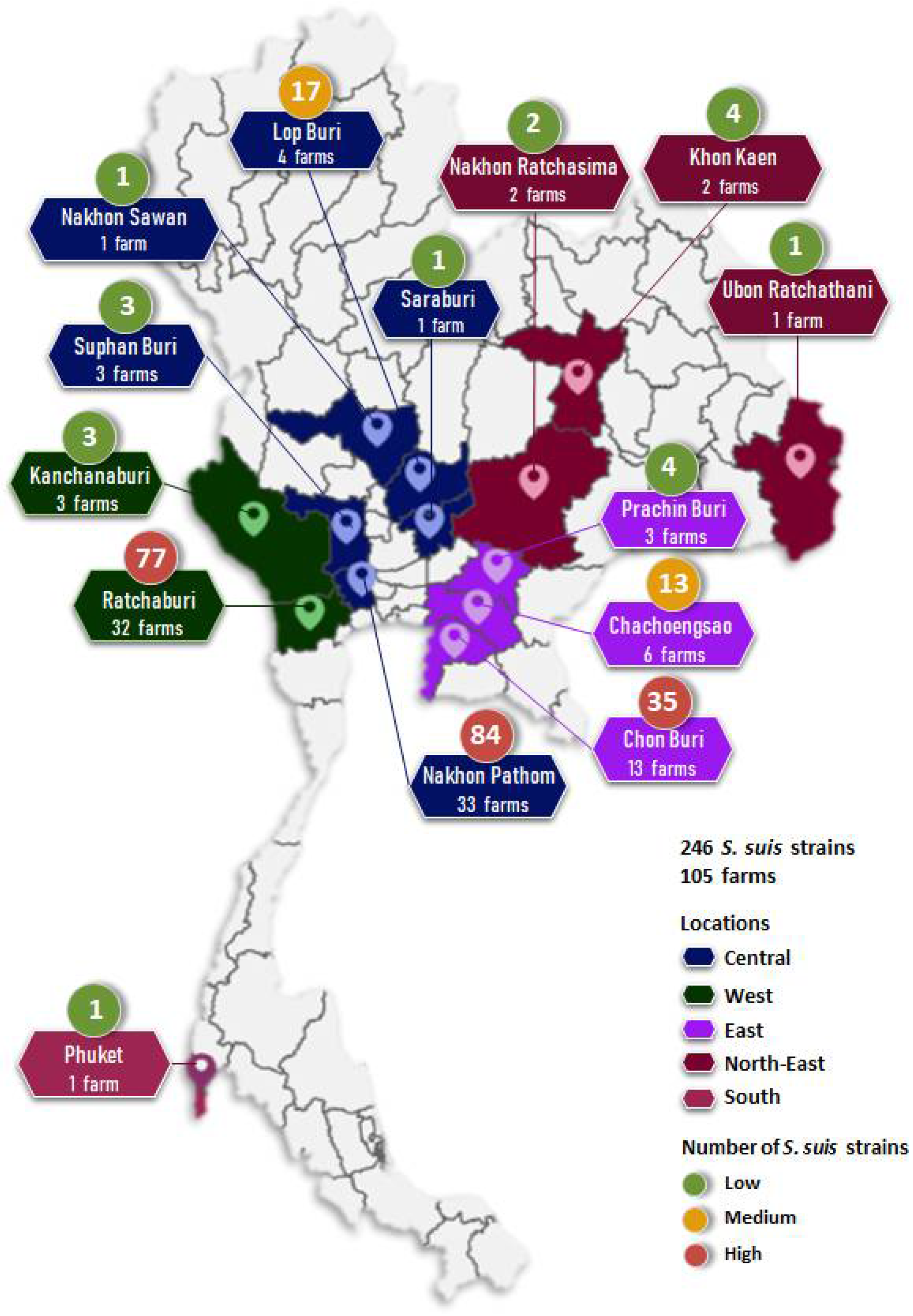
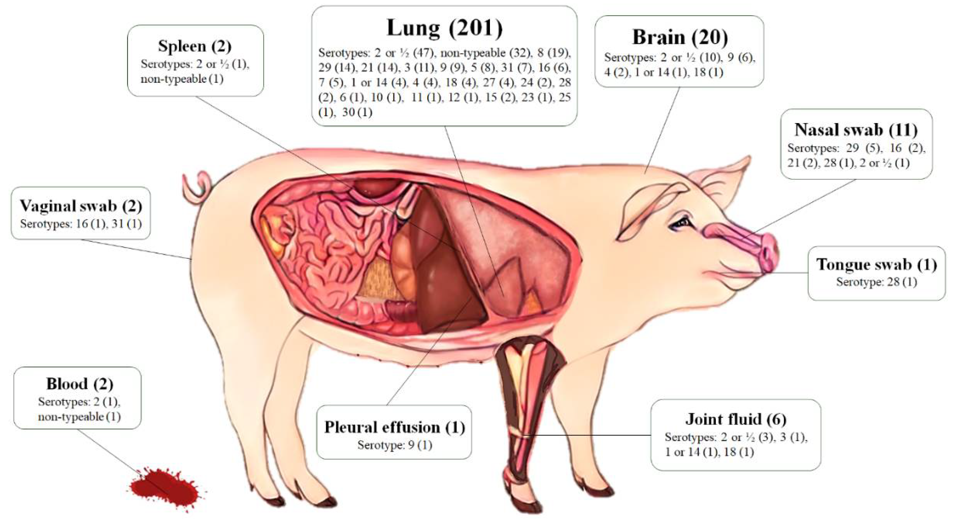
2.1. Bacterial Sampling

A total of 246 S. suis strains were recovered from diseased pigs from 2018–2020. The pig specimens were derived from 105 farms localized in 14 provinces across Thailand (Figure 1 and Table S1), including Nakhon Pathom (33 farms, 84 strains), Ratchaburi (32 farms, 77 strains), Chon Buri (13 farms, 35 strains), Chachoengsao (6 farms, 13 strains), Lop Buri (4 farms, 17 strains), Prachin Buri (3 farms, 4 strains), Kanchanaburi (3 farms, 3 strains), Suphan Buri (3 farm, 3 strains), Khon Kaen (2 farms, 4 strains), Nakhon Ratchasima (2 farms, 2 strains), Nakhon Sawan (1 farm, 1 strain), Phuket (1 farm, 1 strain), Saraburi (1 farm, 1 strain), and Ubon Ratchathani (1 farm, 1 strain). S. suis strains were isolated from various organs of diseased pigs, including lung (81.7%), brain (8.1%), nasal swab (4.5%), joint fluid (2.4%), blood, spleen, vaginal swab (0.8% each), pleural effusion, and tongue swab (0.4% each) (Figure 2).

2.2. Serotyping

PCR-based serotyping revealed that most S. suis strains (62.6%) belonged to serotype 2 or ½ (25.6%), followed by serotypes 8 and 29 (7.7% each), 9 and 21 (6.5% each), 3 (4.9%), 16 (3.7%), and other serotypes (23.6%). There were 34 non-typeable strains (13.8%) that could not provide any specific band to all tested multiplex PCR reactions, and no S. suis serotypes 13, 17, and 19 were identified from this study (Table 1). It is noteworthy that this PCR-based serotyping protocol did not enable the differentiation of serotype 2 from ½ and 1 from 14.
Lung (n = 201, 81.7%), brain (n = 20, 8.1%), nasal swab (n = 11, 4.5%), joint fluid (n = 6, 2.4%), blood (n = 2, 0.8%), spleen (n = 2, 0.8%), vaginal swab (n = 2, 0.8%), pleural effusion (n = 1, 0.4%), and tongue swab (n = 1, 0.4%) samples were collected.
Regarding the serotypes and anatomical sites of isolation, lung, brain, and nasal swabs comprised 94.3% of the isolation sites. Serotype 2 or ½ was the most prevalent in the isolates recovered from lung (23.4%), followed by serotypes 8 (9.5%), 21, and 29 (7.0% each). The main serotype recovered from the brain was serotype 2 or ½ (50.0%) and 9 (30.0%), while serotype 29 (45.5%) was the most common serotype for nasal swabs. In addition, serotype 2 or ½ was found in almost all types of tissues except tongue, vaginal, and pleural effusion (Figure 2 and Table S2).

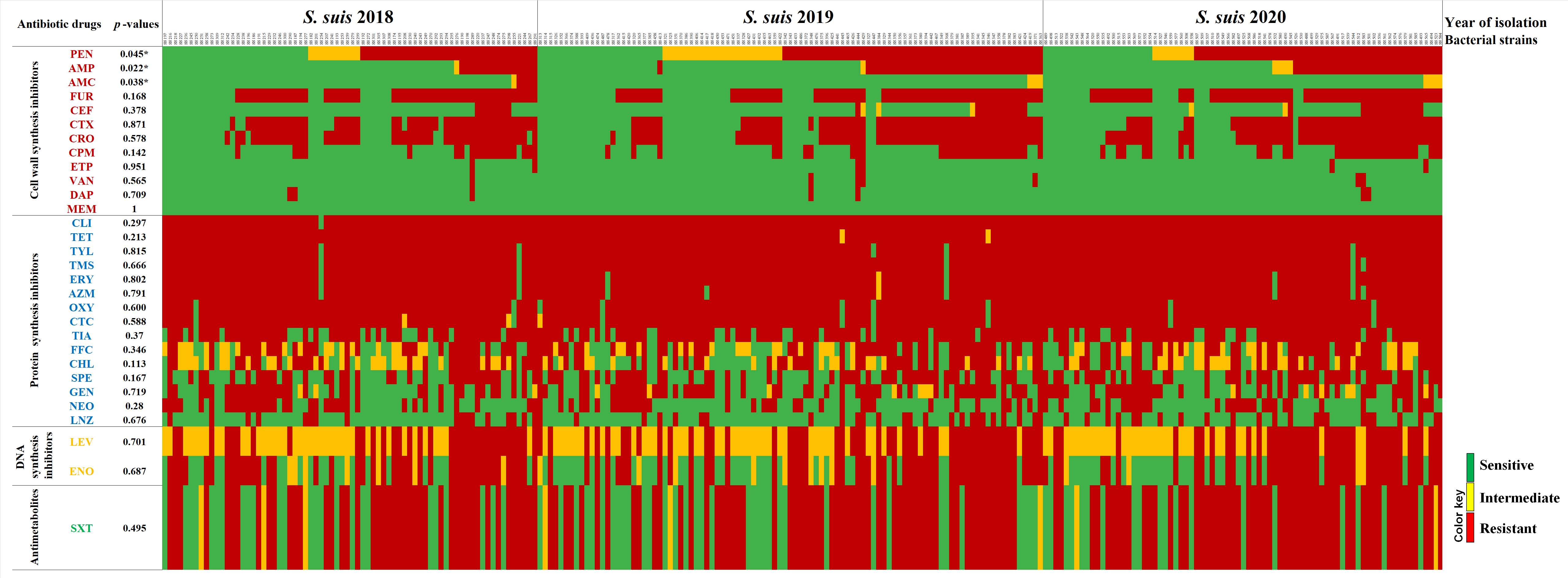
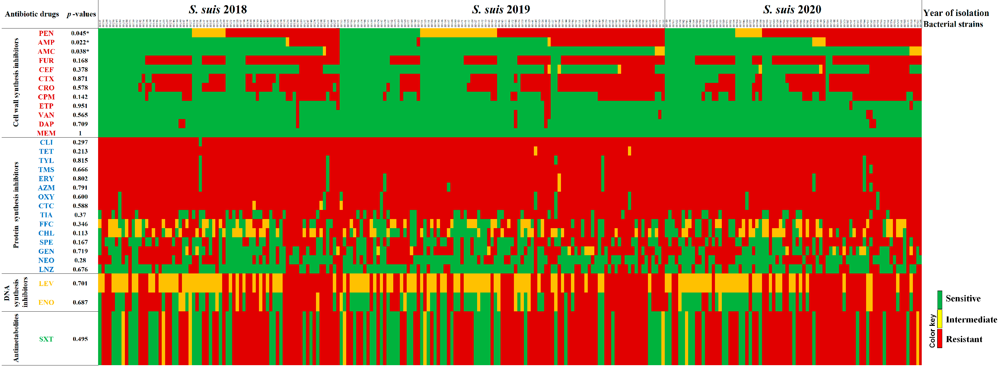
2.3. Antimicrobial Susceptibility Profiles

The distributions, MIC50, MIC90, and rate of antimicrobial resistance against the 246 S. suis strains isolated from samples collected during 2018 (n = 72), 2019 (n = 97) and 2020 (n = 77) are presented in Table 2. Antimicrobial resistance of zoonotic bacteria is a source of concern, as it may compromise the effective treatment of the infection in humans. In this study, the antimicrobial susceptibility test (AST) was therefore performed to determine minimum inhibitory concentrations (MICs) of 35 antimicrobial agents, covering both veterinary and human medicines. No MIC breakpoints were observed for danofloxacin, sulphadimethoxine, and tulathromycin for Streptococcus spp., while concentrations of moxifloxacin and tigecycline used in this study were out of the recommended MIC breakpoint values; therefore, the AST interpretation could be made for 30 of 35 antibiotics according to CLSI veterinary breakpoints, EUCAST, FDA, and previously reported data (Table S3).
The results revealed that S. suis strains obtained from diseased pigs remained highly susceptible to meropenem (100%), ertapenem (97.6%), daptomycin (97.2%), vancomycin (97.2%), amoxicillin/clavulanic acid (95.1%), and ceftiofur (85.4%), but considerably resistant to clindamycin (99.6%), tetracycline (99.2%), tilmicosin (98.0%), tylosin tartrate (98.0%), erythromycin (97.2%), azithromycin (96.1%), oxytetracycline (96.3%), and chlortetracycline (95.5%). A high prevalence of S. suis resistant to tiamulin (79.3%), cefuroxime (67.9%), trimethoprim/sulfamethoxazole (64.6%), ceftriaxone (62.6%), cefotaxime (59.8%), spectinomycin (55.3%), and enrofloxacin (54.9%) was also detected.
From 2018–2020, intermediate susceptibility to levofloxacin (50.0%), chloramphenicol (29.3%), florfenicol (23.6%), and penicillin (16.7%) were determined (Table 2). In 2020, intermediate susceptibility and resistance to levofloxacin were relatively high (48.1% and 52.0%, respectively), and no susceptible strain to levofloxacin was found (Table 3). In addition, the results demonstrated the high prevalence of penicillin-resistant S. suis during 2018–2020 with an increasing penicillin MIC50 value from 0.5 µg/mL (in 2018) to 2.0 µg/mL (in 2020) and a constant MIC90 value at >8 µg/mL. Among beta-lactam antibiotics, ertapenem and meropenem exhibited the highest activity, with >97.0% of the isolates being susceptible. A high prevalence of antimicrobial susceptibility to amoxicillin/clavulanic acid (93.0–96.9%), vancomycin (95.9–98.6%), and daptomycin (96.0–98.0%) was also determined (Tables S4–S6). Among the 3rd generation cephalosporins, ceftiofur was the most effective drug (80.5–90.3%), whereas a low prevalence of antimicrobial susceptibility to ceftriaxone (33.8–41.2%), and cefotaxime (38.9–42.3%) were reported. However, resistance against ceftiofur emerged (9.7–16.9%). Furthermore, the results demonstrated the presence of strains resistant to the 4th generation cephalosporin, cefepime (26.4–40.3%).
The AMR profile revealed 208 different antibiograms (patterns of antibiotic resistance), including 152 patterns for 173 S. suis strains that exhibited multidrug resistance (MDR) and resistance to at least one agent in three or more antimicrobial categories [10]. None of the S. suis isolated strains used in this study was susceptible to all tested antibiotic drugs (Figure 3). The AMR profiles suggested that antibiotics inhibiting cell wall synthesis were the most effective therapeutic drugs (Figure 3). Of 246 S. suis strains, 34 strains (13.8%) exhibited antimicrobial susceptibility to all cell wall synthesis inhibitors. There were 71 strains (28.9%) susceptible to all 5 cephalosporins, and a high prevalence of antimicrobial susceptibility to ceftiofur, the 3rd generation drug commonly used in veterinary medicine, was determined.
Regarding antibiotic drugs inhibiting protein synthesis, more than half of the isolates were susceptible to linezolid (73.6%), followed by neomycin (59.8%). For fluoroquinolones, DNA synthesis inhibitor drugs, no S. suis strain exhibited antimicrobial susceptibility to levofloxacin, and only 91 strains (37.0%) were susceptible to enrofloxacin. The prevalence of MDR S. suis isolated in 2018, 2019, and 2020 was 52 (72.2%), 61 (62.9%), and 60 (77.9%), respectively. MDR S. suis strains were found in different regions of the country. The results also revealed a significant association between the isolation period and susceptibility of S. suis to amoxicillin/clavulanic acid (p = 0.022), ampicillin (p = 0.038), and penicillin (p = 0.045) (Table 3).
Serotype 2 or ½ was the most frequently identified AMR pattern with resistance to 9–25 antimicrobial agents. The MDR S. suis strains were identified in all major serotypes, including serotype 2 or ½ (57.1%), 3 (25.0%), 8 (31.6%), 9 (68.8%), 16 (100%), 21 (87.5%), and 29 (84.2%). There were significant associations between bacterial serotypes and the susceptibility patterns toward ampicillin, cefepime, cefotaxime, ceftiofur, ceftriaxone, cefuroxime, penicillin, azithromycin, chloramphenicol, florfenicol, neomycin, spectinomycin, tylosin tartrate, enrofloxacin, levofloxacin, and trimethoprim/sulfamethoxazole (Table 4).

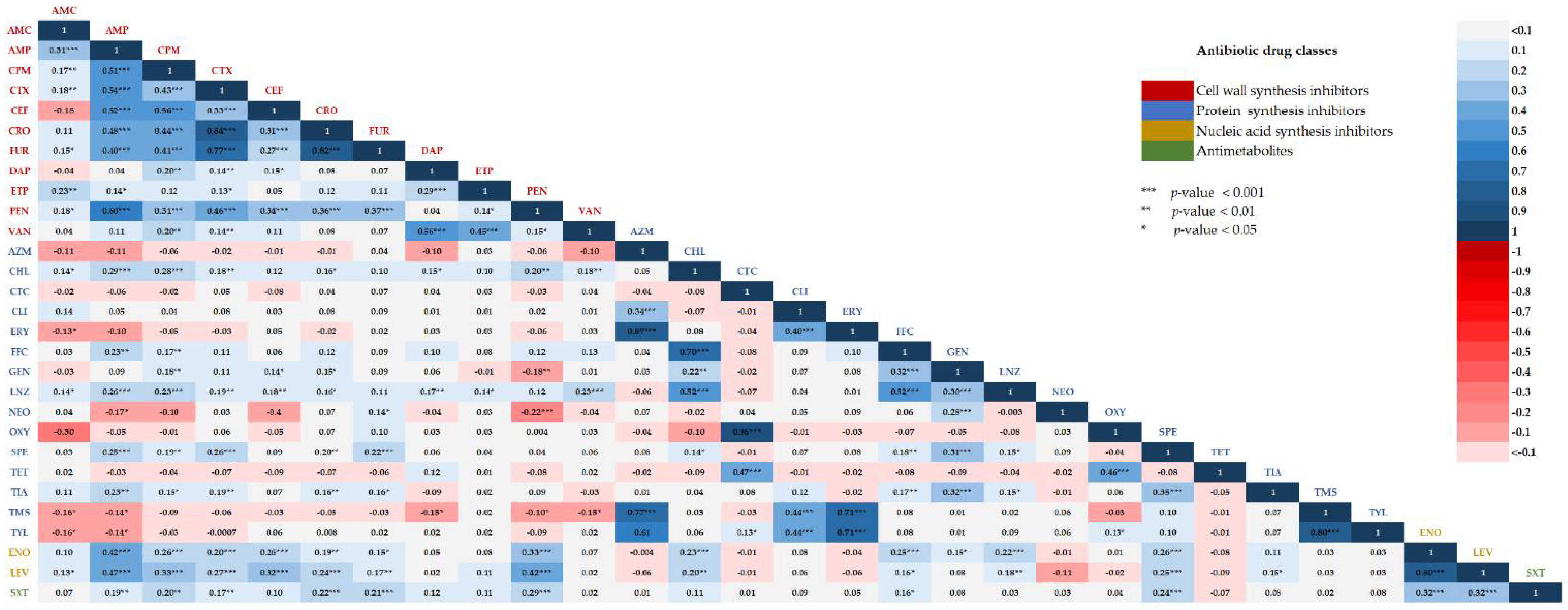
2.4. Correlations between Two Different Antibiotic Susceptibility Statuses among Isolates

The results of pairwise correlation analysis revealed varying degrees of correlation between resistance to the different antibiotics tested (Figure 4). Positive correlation refers to similarity in susceptibility or resistance of two antibiotics, while negative correlation refers to the correlation between the susceptibility of an individual drug and the resistance of another drug. Among antibiotics inhibiting cell wall synthesis, cefotaxime resistance significantly exhibited the strongest positive correlation with the resistance to ceftriaxone and cefuroxime with Pearsons’ correlation of 0.84 and 0.77, respectively (p < 0.001). In addition, ampicillin resistance was positively correlated with the resistance to penicillin, cefotaxime, ceftiofur, cefepime, ceftriaxone, and cefuroxime with Pearson’s correlation of 0.60, 0.54, 0.52, 0.51, 0.48, and 0.40, respectively (p < 0.001). In the DNA synthesis inhibitor class, enrofloxacin had the highest correlation coefficient to levofloxacin (0.80, p < 0.001). All cell wall synthesis and antimetabolite drugs showed a positive correlation with those of the DNA synthesis inhibitor antibiotics. A significant negative correlation was found among different drug classes, especially between protein synthesis inhibitors and cell wall synthesis inhibitors. Protein synthesis inhibitors, neomycin, were significantly negatively correlated with cell wall synthesis inhibitors, penicillin (−0.22, p < 0.001) and ampicillin (−0.17, p < 0.05). Gentamycin was also significantly negatively correlated with penicillin (−0.18, p < 0.01).

3. Discussion

S. suis is a major swine pathogen with the greatest impact on pig production worldwide [2]. It has considerable zoonotic potential for humans, especially in southeast Asian countries, including China, Vietnam, and Thailand [8,9,11]. Encapsulated extracellular S. suis is a highly invasive pathogen that causes septicemia, meningitis, endocarditis, pneumonia, and arthritis. After penetration of host mucosal barriers, it can reach and survive in the blood and finally invade multiple organs, including the lung, spleen, liver, kidney, heart, and brain [2]. In this study, a high prevalence of diseased pig-isolated S. suis strains was found in lung (81.7%) and brain tissues (8.1%). This data supports that the lungs and brain were major target organs for S. suis infection and that S. suis infection severely caused systemic dissemination in pigs [6].
It is known that S. suis serotype 2 is the most pathogenic and significantly associated with disease in both pigs and humans worldwide. However, the serotype distribution can differ over time and geographical area. In North America, multiple serotypes, such as serotypes ½, 2, 3, 8, 4, and 7, have been recorded in diseased pigs. In contrast, serotypes 2, 3, and 9 predominate in Europe and Asia [2,12]. Among diseased pigs in China, serotype 2 (66.0%) was commonly found [13], while serotype 29 (9.4%) was the most prevalent in healthy pigs, followed by serotype 2 (5.8%) and serotype 21 (4%) [9]. S. suis serotypes 3 (15.8%) and 2 (15.0%) were the most predominant in slaughtered and diseased pigs in South Korea [4], whereas serotype 2 (8.0%) was the most prevalent in slaughterhouse pigs in southern Vietnam [11]. Recent evidence demonstrated a higher frequency of serotype 29 among S. suis isolated from healthy pigs (15.4%) and pigs with respiratory disease (1.7%) in Germany [12]. In northern Thailand, serotypes 2 (19.1%) was the most common, followed by serotype 7 (15.7%), 9 (14.2%), 16 (9.3%), and 14 (7.3%) from pig tonsils at a slaughterhouse [14], while serotype 16 (11.0%) was the most frequent serotype, followed by serotypes 8 (7.0%), 9 (6.0%), and 3 (5.0%) from healthy pigs in central Thailand [15]. In agreement with previous reports, the data obtained from this study demonstrated that most S. suis isolates from diseased pigs were serotype 2 or ½ (25.6%), followed by serotypes 8 (9.0%) and 29 (7.1%). Serotype 29 has also been reported from S. suis isolated from healthy pigs in northern Thailand with a small abundance (1.0%) [16]. To the best of our knowledge, this is the first report on the high prevalence of serotype 29 identified in a large collection of S. suis strains isolated from diseased pigs in Thailand. The data confirmed that the distribution of different serotypes of S. suis in pigs could be varied by geographical localizations.
Serotypes 13, 17, and 19 have been reported from both healthy and diseased pigs, albeit with a relatively low prevalence (0.7–1.9%) [4,6,16]. However, no S. suis serotypes 13, 17, and 19 were found in this study, suggesting lower virulence capacities of these serotypes compared to the other common serotypes. The isolation of some uncommon S. suis serotypes from diseased pigs could be explained by the S. suis acting as an opportunistic pathogen while the inclusion of other bacterial infections being a primary cause of disease [2]. Regarding the sample sources, serotypes 2, ½, and 8 S. suis isolates from diseased pigs were mainly recovered from the lungs (23.4 and 9.5%, respectively), and serotype 29 was frequently isolated from the upper respiratory track (45.5%). In addition, serotype 29 was also recovered from the lung tissues (7.0%) of diseased pigs. It was possible that this serotype might be a potentially virulent serotype responsible for infections. However, virulence can also vary within serotypes [17]. Thus, further studies are needed to assess the virulence and pathogenicity of S. suis serotype 29. The study also revealed the dissemination of serotype 9 S. suis through different organs of diseased pigs, including lung, brain, and spleen, suggesting that serotype 9 is associated with invasive disease in pigs [18]. Taken together, the data suggested the dissemination of both serotype 2 and non-serotype 2 in the pig-isolated S. suis population in Thailand.
In southeast Asian countries, antimicrobials are freely available over the counter for use in both humans and animals, which likely contributes to the extensive use of antimicrobials in livestock sectors, leading to widespread AMR [19]. Thailand is one of the top ten veterinary antimicrobial users (4.2%) [20]. The most common drugs used are amoxicillin (39.6%), enrofloxacin (22.9%), tetracycline (12.5%), and penicillin (12.5%) [5]. For the treatment of S. suis infection, beta-lactams (amoxicillin/clavulanic acid, ampicillin, ceftiofur, ceftriaxone, and penicillin) and fluoroquinolones (enrofloxacin) are still the drugs of choice. However, the global trend of increasing antimicrobial resistance among streptococcal species is becoming more problematic [2].
In this study, the resistance of S. suis to commonly used antibiotics was relatively high. All isolates were resistant to at least one class of antibiotics, and 70.3% were resistant to three or more drug classes, which indicated substantial multidrug resistance (MDR). High frequencies of resistance were observed for protein synthesis inhibitors, such as clindamycin, tetracycline, erythromycin, and chlortetracycline, which was consistent with previous reports [6,8,9]. Among the primary drugs against S. suis infection, the prevalence of isolates resistant to penicillin (0–27%), ampicillin (0.6–23%), and ceftiofur (0–23%) was generally low [2,7,9]. The findings from this study are consistent with previous literature suggesting S. suis susceptibility to cell wall synthesis inhibitors, including beta-lactam antibiotics [6,7]. By contrast, the statistical analysis obtained from this study indicated a significant increase of antimicrobial resistance against penicillin from 47.4% to 64.3%, which was slightly higher than those from previous data in healthy pig-isolated (10.9%) and diseased pig-isolated (27.0%) S. suis strains in Thailand, during 2006–2007 and 2012–2015 [9]. In addition, the proportion of isolates with high penicillin MIC values increased over time, which was reflected in an increase in MIC50 value of 0.5 µg/mL to 2.0 µg/mL, and MIC90 value of >8 µg/mL. This evidence clearly confirmed the emergence and widespread nature of penicillin-resistant S. suis strains in Thailand. Whereas 12.6% of S. suis strains were resistant against the 3rd generation cephalosporin (ceftiofur), 29.7–38.1% of them were resistant to the 4th generation cephalosporin (cefepime), raising concerns that inappropriate use of cephalosporins could further accelerate widespread resistance to cephalosporins. Moreover, the presence of ampicillin resistance and the increasing prevalence of intermediate susceptible against amoxicillin/clavulanic raised awareness of the spread of resistance strains. The use of different antibiotic drugs that belong to the same categories can favor the cross-resistance of bacteria under the same resistance mechanism [21]. Recently, transferable resistance genes cfr and optrA have been identified from S. suis of animal origin under the selection of phenicols and other ribosomal-targeted antibiotics, which are broadly used in veterinary medicine [8,21]. However, these resistance genes were not only associated with resistance to phenicols but also conferred resistance to oxazolidinone (linezolid and tedizolid), available antibiotic drugs used only in humans. This evidence suggests the impact of antibiotic-resistant selection on farms to human health. In this study, the emergence of pig-isolated S. suis strains resistant to drugs used for humans, such as linezolid, vancomycin, and meropenem, was found. This finding raised serious concern about the transmission of antibiotic-resistant S. suis strains among animals and humans, causing clinical or epidemiological problems in the near future. The overuse of different antimicrobial substances in pig farming could induce more variation of antimicrobial resistance in S. suis of human origin. Therefore, proper use of antibiotic drugs for prophylaxis and treatment in swine production systems is highly recommended to avoid further spread of AMR S. suis in both animals and humans.
A remarkably high prevalence of S. suis isolates resistant to antibiotics inhibiting protein synthesis was determined in this study. A high prevalence of tetracycline-, macrolide-, and lincosamide-resistant S. suis isolates was observed in various countries in Asia, Europe, North America, and Africa [2,7,9]. Such great resistance is undoubtedly related to their intensive use in swine industries, and it could be associated with the acquisition and dissemination of AMR genes through mobile-genetic elements (MGEs). The acquisition and dissemination of AMR genes in streptococci is strongly associated with MGEs, mainly integrative and conjugative elements (ICEs) and prophages. A variety of AMR determinants for tetracyclines [tet(M), tet(L), tet(O), and tet(40)], macrolides [erm(B)], aminoglycosides (aphA3, sat, ant6, and aadE), and phenicols (cat) have been located in the ICESa2603 family [22]. For fluoroquinolones, the prevalence of enrofloxacin- and levofloxacin-resistant S. suis isolates was significant. A high frequency of intermediate susceptibility to levofloxacin (45.2–54.3%), suggesting the continued use of fluoroquinolones, could eventually lead to the emergence of resistance. In addition, resistance to macrolide and fluoroquinolone drugs could immensely limit the therapeutic use of these antibiotics for the treatment of S. suis infection. A more comprehensive investigation and characterization of the genetic determinants and understanding of the AMR mechanisms of S. suis strains in Thailand are needed for effective monitoring and preventing the spread of AMR in this region.
The AMR problem drastically impairs the effectiveness of the therapeutic use of existing antibiotics. Antibiotic combination therapy with different modes of action is a far more effective approach for combating MDR pathogens and preventing the emergence of resistance commonly found with monotherapy [23]. The combination of beta-lactams with gentamicin, displaying a strong synergistic effect against Streptococcus pneumoniae infection, has been reported [24]. In addition, Yu et al. [25] demonstrated a marked synergistic activity of the two combination regimens, including ampicillin plus apramycin and tiamulin plus spectinomycin, for treatment of S. suis infection. In this study, a significant negative correlation was found among different drug classes, especially between cell wall synthesis inhibitors, penicillin, and protein synthesis inhibitors, including neomycin, and gentamicin. Combination therapy of penicillin or ampicillin plus neomycin or gentamicin may be used as a treatment option for S. suis infection. For further study, to determine the effectiveness of combined drugs used against zoonotic S. suis infection, investigation, and verification of possible drug combinations should be performed for both animal and human antimicrobial agents.

4. Materials and Methods

4.1. Bacterial Collection

A total of 246 non-duplicate S. suis strains were collected from specimens (organs, tissues, and swabs) of diseased pigs across Thailand from 2018–2020 as a part of routine laboratory tests at the Veterinary Diagnostic Laboratory, Large Animal Teaching Hospital, Faculty of Veterinary Science, Chulalongkorn University. The S. suis strains were isolated on Columbia blood agar (5% sheep blood) at 37 °C in 5% CO2 for 24–48 h. The isolates with alpha hemolytic colonies were further identified by conventional biochemical tests [26]. Subsequently, the colonies were confirmed to be S. suis by the PCR-based approach targeting the glutamate dehydrogenase (gdh) gene [3] and the recombination/repair protein (recN) gene. The PCR primers used for recN identification were SuisRecNsy01_F (5′-TTA TCT GTC TTG AAA CAG ATT GGG-3′) and SuisRecNsy01_R (5′-TCT TTC TCT AAG TTC TTA AGC TGA AC-3′). The PCR conditions comprised an initial denaturation step at 95 °C for 5 min, followed by 35 cycles of 95 °C for 30 s, 54 °C for 1 min, and 72 °C for 1 min, with a final extension step at 72 °C for 7 min.

4.2. Multiplex PCR-Based Serotyping

Identification of S. suis serotypes was conducted using a multiplex PCR-based method [3]. The PCR reactions were carried out independently in four sets; the first set included primers for serotypes ½, 1, 2, 3, 7, 9, 11, 14, and 16; the second for serotypes 4, 5, 8, 12, 18, 19, 24, and 25; the third for serotypes 6, 10, 13, 15, 17, 23, and 31; and the fourth for serotypes 21, 27, 28, 29, and 30. PCR amplification of the S. suis species-specific PCR targeting the gdh gene was also carried out as a positive control of the reaction. The oligonucleotide primer sequences are listed in Table S7. The following PCR conditions were used: initial denaturation at 95 °C for 3 min, followed by 30 cycles of 95 °C for 20 s, 62 °C for 1.30 min, and 62 °C for 1.30 min, and a final extension at 72 °C for 5 min.

4.3. Antimicrobial Susceptibility Testing

The MICs of different antimicrobial agents were determined by the broth microdilution method using a semi-automatic system (Sensititre, Trek Diagnostic Systems Ltd., West Sussex, UK) in accordance with the Clinical and Laboratory Standards Institute (CLSI) recommendations [27]. The MIC test was performed with two sets of commercially prepared, dehydrated 96-well microtiter plates, including Sensititre Vet Bovine/Swine BOPO6F plate and Sensititre Streptococcus species STP6F plate, containing antibiotics for veterinary and human usages, respectively. A total of 35 antibiotics from different drug classes and mechanisms of action were included in this study (Table S3). The MIC test conditions were performed according to the manufacturer’s guidelines with minor modifications. In brief, isolates were cultured on Columbia blood agar at 37 °C in 5% CO2 incubator overnight. Selected colonies were suspended in Sensititre cation-adjusted Mueller-Hinton broth (CAMHBT) and adjusted to be a 0.5 McFarland standard. Subsequently, a 100-µL aliquot of the suspension was transferred into a tube of CAMHBT and CAMHBT with lysed Horse blood (CAMHBT+LHB) for BOPO6F and STP6F panels, respectively, to obtain an inoculum density of 5 × 10⁵ CFU/mL. The BOPO6F and STP6F panels were reconstituted by adding 50 µL and 100 µL/well, respectively, and the plates were covered with an adhesive seal and incubated at 35 ± 2 °C in Sensititre ARISTM 2X for 20–24 h. The MIC value, the lowest drug concentration inhibiting visible growth, was read automatically on the Sensititre ARISTM 2X and read visually using a manual viewbox according to the instructions in Sensititre SWIN software.
Streptococcus pneumoniae ATCC 49619, Staphylococcus aureus ATCC 29213, Escherichia coli ATCC 25922, and S. suis serotype 2-P1/7 (UK) were used as control and reference strains, and MICs were within the accepted quality control ranges. The results were interpreted according to CLSI veterinary breakpoints [28], EUCAST [29], FDA [30], and previously reported data when available (Table S3).

4.4. Statistical Analysis

Statistical differences were determined by performing chi-square tests with the STATA statistical package v14.0 (Stata Corporation, College Station, TX, USA). Pairwise analysis of the correlation between the antimicrobial susceptibility status (susceptible, intermediate, and resistant) to the different antibiotics was investigated using Pearson’s correlation analysis. A p-value of <0.05 was considered to be statistically significant.

5. Conclusions

The predominance of serotype 2 or ½, followed by 8, 29, 9, and 21, was identified from diseased pig-isolated S. suis strains in different regions of Thailand from 2018–2020. To the best of our knowledge, this is the first report describing the distribution of S. suis serotype 29 in a large population of diseased pigs. The surveillance of antimicrobial resistance confirmed widespread AMR and MDR S. suis strains against different commonly available antibiotic drug classes. Although drugs inhibiting cell wall synthesis were the most effective antibiotics, a tendency toward reduced efficacy of these drugs was observed. In addition, the proportion of intermediate susceptibility and resistance to many antibiotics increased over time. As a result, effective antibiotic drugs for the treatment of S. suis infection in both animals and humans could be limited in the near future. In this study, pairwise correlation between two antimicrobial susceptibility statuses suggested that the combination of cell wall synthesis inhibitors (penicillin) with protein synthesis inhibitors (neomycin and gentamicin) may be used as a choice for treatment of S. suis infection; this therapeutic approach deserves additional study. Taken together, the knowledge gained from this study underlined the resistance selective pressure in livestock systems, raising an awareness of prudent and efficient use of therapeutic options for S. suis infection in both public and veterinary healthcare. Continuous surveillance is required to monitor the prevalence of AMR and MDR in S. suis and to guide decisions regarding appropriate treatment. Further research focusing on the understanding of AMR mechanisms would be helpful and necessary for developing effective preventive measures for S. suis infection. In addition, effective prevention and infection control strategies should be made to prevent the dissemination of AMR and MDR in S. suis in the country.

Supplementary Materials

The following supporting information can be downloaded at: https://www.mdpi.com/article/10.3390/antibiotics11030410/s1, Table S1: Sources of S. suis strains isolated from diseased pigs from 2018–2020; Table S2: Distribution of S. suis serotypes in different sources of specimens; Table S3: MIC breakpoints and interpretative categories of antimicrobial susceptibility test; Table S4: Minimum inhibitory concentration (MIC) value distribution, MIC50 and MIC90 values, and resistance rates of 72 S. suis strains in 2018; Table S5: Minimum inhibitory concentration (MIC) value distribution, MIC50 and MIC90 values, and resistance rates of 97 S. suis strains in 2019; Table S6: Minimum inhibitory concentration (MIC) value distribution, MIC50 and MIC90 values, and resistance rates of 77 S. suis strains in 2020; Table S7: Oligonucleotide primer sequences.

Author Contributions

Conceptualization, S.Y. and K.L.; methodology, S.Y., K.L., W.C. and S.J.; validation, S.Y., K.L., W.C., S.J., S.S. and P.A.; formal analysis, K.L. and W.C.; investigation, S.Y. and S.J.; resources, S.Y., S.S. and P.A.; data curation, K.L. and W.C.; writing—original draft preparation, K.L.; writing—review and editing, S.Y., K.L., W.C. and S.J.; visualization, S.S. and P.A.; supervision, S.Y.; project administration, S.Y. and K.L.; funding acquisition, S.Y. All authors have read and agreed to the published version of the manuscript.

Funding

This research was funded by an RDI Research Grant, grant number P20-50-967.

Institutional Review Board Statement

Not applicable.

Informed Consent Statement

Not applicable.

Data Availability Statement

Not applicable.

Acknowledgments

The authors would like to thank Darin Kongkasuriyachai for her constructive discussion and internal reviews for this study. We are grateful to Potjanee Srimanote for providing S. suis serotype 2-P1/7 (UK) and S. pneumoniae ATCC 49618 reference strains and to Krissana Maneerat for technical assistance.

Conflicts of Interest

The authors declare no conflict of interest. The funders had no role in the design of the study; in the collection, analyses, or interpretation of data; in the writing of the manuscript, or in the decision to publish the results.

References

  1. Lun, Z.R.; Wang, Q.P.; Chen, X.G.; Li, A.X.; Zhu, X.Q. Streptococcus suis: An emerging zoonotic pathogen. Lancet Infect. Dis. 2007, 7, 201–209. [Google Scholar] [CrossRef]
  2. Segura, M.; Aragon, V.; Brockmeier, S.L.; Gebhart, C.; Greeff, A.; Kerdsin, A.; O’Dea, M.A.; Okura, M.; Saléry, M.; Schultsz, C.; et al. Update on Streptococcus suis research and prevention in the era of antimicrobial restriction: 4th International Workshop on S. suis. Pathogens 2020, 9, 374. [Google Scholar] [CrossRef] [PubMed]
  3. Kerdsin, A.; Akeda, Y.; Hatrongjit, R.; Detchawna, U.; Sekizaki, T.; Hamada, S.; Gottschalk, M.; Oishi, K. Streptococcus suis serotyping by a new multiplex PCR. J. Med. Microbiol. 2014, 63, 824–830. [Google Scholar] [CrossRef] [PubMed] [Green Version]
  4. Oh, S.I.; Jeon, A.B.; Jung, B.Y.; Byun, J.W.; Gottschalk, M.; Kim, A.; Kim, J.W.; Kim, H.Y. Capsular serotypes, virulence-associated genes and antimicrobial susceptibility of Streptococcus suis isolates from pigs in Korea. J. Vet. Med. Sci. 2017, 79, 780–787. [Google Scholar] [CrossRef] [PubMed] [Green Version]
  5. Lekagul, A.; Tangcharoensathien, V.; Mills, A.; Rushton, J.; Yeung, S. How antibiotics are used in pig farming: A mixed-methods study of pig farmers, feed mills and veterinarians in Thailand. BMJ Glob. Health 2020, 5, e001918. [Google Scholar] [CrossRef] [PubMed] [Green Version]
  6. Petrocchi-Rilo, M.; Martínez-Martínez, S.; Aguarón-Turrientes, Á.; Roca-Martínez, E.; García-Iglesias, M.J.; Pérez-Fernández, E.; González-Fernández, A.; Herencia-Lagunar, E.; Gutiérrez-Martín, C.B. Anatomical site, typing, virulence gene profiling, antimicrobial susceptibility and resistance genes of Streptococcus suis isolates recovered from pigs in Spain. Antibiotics 2021, 10, 707. [Google Scholar] [CrossRef]
  7. Tan, M.F.; Tan, J.; Zeng, Y.B.; Li, H.Q.; Yang, Q.; Zhou, R. Antimicrobial resistance phenotypes and genotypes of Streptococcus suis isolated from clinically healthy pigs from 2017 to 2019 in Jiangxi Province, China. J. Appl. Microbiol. 2020, 130, 797–806. [Google Scholar] [CrossRef]
  8. Zhang, C.; Zhang, P.; Wang, Y.; Fu, L.; Liu, L.; Xu, D.; Hou, Y.; Li, Y.; Fu, M.; Wang, X.; et al. Capsular serotypes, antimicrobial susceptibility, and the presence of transferable oxazolidinone resistance genes in Streptococcus suis isolated from healthy pigs in China. Vet. Microbiol. 2020, 247, 108750. [Google Scholar] [CrossRef]
  9. Yongkiettrakul, S.; Maneerat, K.; Arechanajan, B.; Malila, Y.; Srimanote, P.; Gottschalk, M.; Visessanguan, W. Antimicrobial susceptibility of Streptococcus suis isolated from diseased pigs, asymptomatic pigs, and human patients in Thailand. BMC Vet. Res. 2019, 15, 5. [Google Scholar] [CrossRef] [Green Version]
  10. Magiorakos, A.P.; Srinivasan, A.; Carey, R.B.; Carmeli, Y.; Falagas, M.E.; Giske, C.G.; Harbarth, S.; Hindler, J.F.; Kahlmeter, G.; Olsson-Liljequist, B.; et al. Multidrug-resistant, extensively drug-resistant and pandrug-resistant bacteria: An international expert proposal for interim standard definitions for acquired resistance. Clin. Microbiol. Infect. 2012, 18, 268–281. [Google Scholar] [CrossRef] [Green Version]
  11. Hoa, N.T.; Chieu, T.T.; Nghia, H.D.; Mai, N.T.; Anh, P.H.; Wolbers, M.; Baker, S.; Campbell, J.I.; Chau, N.V.; Hien, T.T.; et al. The antimicrobial resistance patterns and associated determinants in Streptococcus suis isolated from humans in southern Vietnam, 1997–2008. BMC Infect. Dis. 2011, 11, 6. [Google Scholar] [CrossRef] [PubMed] [Green Version]
  12. Prüfer, T.L.; Rohde, J.; Verspohl, J.; Rohde, M.; de Greeff, A.; Willenborg, J.; Valentin-Weigand, P. Molecular typing of Streptococcus suis strains isolated from diseased and healthy pigs between 1996–2016. PLoS ONE 2019, 14, e0210801. [Google Scholar] [CrossRef] [PubMed]
  13. Zhang, C.; Zhang, Z.; Song, L.; Fan, X.; Wen, F.; Xu, S.; Ning, Y. Antimicrobial resistance profile and genotypic characteristics of Streptococcus suis capsular type 2 isolated from clinical carrier sows and diseased pigs in China. Biomed. Res. Int. 2015, 2015, 284303. [Google Scholar] [PubMed]
  14. Kerdsin, A.; Takeuchi, D.; Nuangmek, A.; Akeda, Y.; Gottschalk, M.; Oishi, K. Genotypic comparison between Streptococcus suis isolated from pigs and humans in Thailand. Pathogens 2020, 9, 50. [Google Scholar] [CrossRef] [Green Version]
  15. Meekhanon, N.; Kaewmongkol, S.; Phimpraphai, W.; Okura, M.; Osaki, M.; Sekizaki, T.; Takamatsu, D. Potentially hazardous Streptococcus suis strains latent in asymptomatic pigs in a major swine production area of Thailand. J. Med. Microbiol. 2017, 66, 662–669. [Google Scholar] [CrossRef]
  16. Thongkamkoon, P.; Kiatyingangsulee, T.; Gottschalk, M. Serotypes of Streptococcus suis isolated from healthy pigs in Phayao Province, Thailand. BMC Res. Notes 2017, 10, 53. [Google Scholar] [CrossRef] [Green Version]
  17. Segura, M.; Fittipaldi, N.; Calzas, C.; Gottschalk, M. Critical Streptococcus suis virulence factors: Are they all really critical? Trends Microbiol. 2017, 25, 585–599. [Google Scholar] [CrossRef]
  18. Zheng, H.; Du, P.; Qiu, X.; Kerdsin, A.; Roy, D.; Bai, X.; Xu, J.; Vela, A.I.; Gottschalk, M. Genomic comparisons of Streptococcus suis serotype 9 strains recovered from diseased pigs in Spain and Canada. Vet. Res. 2018, 49, 1. [Google Scholar] [CrossRef] [Green Version]
  19. Coyne, L.; Arief, R.; Benigno, C.; Giang, V.N.; Huong, L.Q.; Jeamsripong, S.; Kalpravidh, W.; McGrane, J.; Padungtod, P.; Patrick, I.; et al. Characterizing antimicrobial use in the livestock sector in three South East Asian countries (Indonesia, Thailand, and Vietnam). Antibiotics 2019, 8, 33. [Google Scholar] [CrossRef] [Green Version]
  20. Tiseo, K.; Huber, L.; Gilbert, M.; Robinson, T.P.; Van Boeckel, T.P. Global trends in antimicrobial use in food animals from 2017 to 2030. Antibiotics 2020, 9, 918. [Google Scholar] [CrossRef]
  21. Du, F.; Lv, X.; Duan, D.; Wang, L.; Huang, J. Characterization of a linezolid- and vancomycin-resistant Streptococcus suis isolate that harbors optrA and vanG operons. Front. Microbiol. 2019, 10, 2026. [Google Scholar] [CrossRef] [PubMed]
  22. Huang, J.; Ma, J.; Shang, K.; Hu, X.; Liang, Y.; Li, D.; Wu, Z.; Dai, L.; Chen, L.; Wang, L. Evolution and diversity of the antimicrobial resistance associated mobilome in Streptococcus suis: A probable mobile genetic elements reservoir for other streptococci. Front. Cell. Infect. Microbiol. 2016, 6, 118. [Google Scholar] [CrossRef] [PubMed] [Green Version]
  23. Dai, C.; Zhao, T.; Yang, X.; Xiao, X.; Velkov, T.; Tang, S. Pharmacokinetics and relative bioavailability of an oral amoxicillin-apramycin combination in pigs. PLoS ONE 2017, 12, e0176149. [Google Scholar] [CrossRef] [PubMed]
  24. Tateda, K.; Matsumoto, T.; Miyazaki, S.; Yamaguchi, K. Efficacy of beta-lactam antibiotics combined with gentamicin against penicillin-resistant pneumococcal pneumonia in CBA/J mice. J. Antimicrob. Chemother. 1999, 43, 367–371. [Google Scholar] [CrossRef] [PubMed] [Green Version]
  25. Yu, Y.; Fang, J.T.; Zheng, M.; Zhang, Q.; Walsh, T.R.; Liao, X.P.; Sun, J.; Liu, Y.H. Combination therapy strategies against multiple-resistant Streptococcus suis. Front. Pharmacol. 2018, 9, 489. [Google Scholar] [CrossRef]
  26. Tarradas, C.; Arenas, A.; Maldonado, A.; Luque, I.; Miranda, A.; Perea, A. Identification of Streptococcus suis isolated from swine: Proposal for biochemical parameters. J. Clin. Microbiol. 1994, 322, 578580. [Google Scholar] [CrossRef] [PubMed] [Green Version]
  27. Clinical and Laboratory Standards Institute. Performance Standards for Antimicrobial Susceptibility Testing; Twenty-Fifth Informational Supplement, 30th ed.; CLSI Supplement M100; Clinical and Laboratory Standards Institute: Wayne, PA, USA, 2020. [Google Scholar]
  28. Clinical and Laboratory Standards Institute. Performance Standards for Antimicrobial Disk and Dilution Susceptibility Tests for Bacteria Isolated From Animals, 5th ed.; CLSI Supplement VET01S; Clinical and Laboratory Standards Institute: Wayne, PA, USA, 2020. [Google Scholar]
  29. European Committee on Antimicrobial Susceptibility Testing. Breakpoint Tables for Interpretation of MICs and Zone Diameters; Version 10.0; EUCAST: Basel, Switzerland, 2020; Available online: http://www.eucast.org (accessed on 15 September 2021).
  30. Food and Drug Administration. Antibacterial Susceptibility Test Interpretive Criteria; FDA: Silver Spring, MD, USA, 2019. Available online: https://www.fda.gov/drugs/development-resources/antibacterial-susceptibility-test-interpretive-criteria (accessed on 15 September 2021).
Figure 1. Geographic distribution of S. suis strains isolated from diseased pigs from 2018–2020.
Antibiotics 11 00410 g001
Figure 2. Anatomical origin of 246 S. suis strains and their serotype distribution.
Antibiotics 11 00410 g002
Figure 3. Heat map showing antimicrobial susceptibility profiles of S. suis strains. Rows represent antibiotics and columns represent bacterial strains, where green blocks indicate antibiotic susceptibility, yellow blocks indicate intermediate, and red blocks indicate resistance action of the antibiotics. Cell wall synthesis inhibitor antibiotics: AMC, Amoxicillin/Clavulanic acid; AMP, Ampicillin; CEF, Ceftiofur; CPM, Cefepime; CRO, Ceftriaxone; CTX, Cefotaxime; DAP, Daptomycin; ETP, Ertapenem; FUR, Cefuroxime; MEM, Meropenem; PEN, Penicillin; VAN, Vancomycin. Protein synthesis inhibitor antibiotics: AZM, Azithromycin; NEO, Neomycin; CHL, Chloramphenicol; CLI, Clindamycin; CTC, Chlortetracycline; ERY, Erythromycin; FFC, Florfenicol; GEN, Gentamicin; LNZ, Linezolid; NEO, Neomycin; SPE, Spectinomycin; TET, Tetracycline; TMS, Tilmicosin; TYL, Tylosin tartrate; TIA, Tiamulin; OXY, Oxytetracycline. Nucleic acid synthesis inhibitor antibiotics: ENO, Enrofloxacin; LEV, Levofloxacin. Antimetabolite antibiotic: SXT, Trimethoprim/sulfamethoxazole. S, susceptible; I, intermediate; R, resistant. * p-value < 0.05.
Antibiotics 11 00410 g003
Figure 4. Pairwise correlation between two antimicrobial susceptibility statuses. Positive correlations are visualized in blue blocks and negative correlations in red blocks. The color intensity of the text labels is proportional to the correlation coefficients. Significant p-values corresponding to the correlation coefficient are indicated with asterisk (*** p-value < 0.001; ** p-value < 0.01; * p-value < 0.05). Cell wall synthesis inhibitor antibiotics: AMC, Amoxicillin/Clavulanic acid; AMP, Ampicillin; CEF, Ceftiofur; CPM, Cefepime; CRO, Ceftriaxone; CTX, Cefotaxime; DAP, Daptomycin; ETP, Ertapenem; FUR, Cefuroxime; MEM, Meropenem; PEN, Penicillin; VAN, Vancomycin. Protein synthesis inhibitor antibiotics: AZM, Azithromycin; NEO, Neomycin; CHL, Chloramphenicol; CLI, Clindamycin; CTC, Chlortetracycline; ERY, Erythromycin; FFC, Florfenicol; GEN, Gentamicin; LNZ, Linezolid; NEO, Neomycin; SPE, Spectinomycin; TET, Tetracycline; TMS, Tilmicosin; TYL, Tylosin tartrate; TIA, Tiamulin; OXY, Oxytetracycline. Nucleic acid synthesis inhibitor antibiotics: ENO, Enrofloxacin; LEV, Levofloxacin. Antimetabolite antibiotic: SXT, Trimethoprim/sulfamethoxazole.
Antibiotics 11 00410 g004
Table 1. Distribution of S. suis strains according to serotype and isolation period.
SerotypesYear of Isolation, n (%)p-ValuesTotal
2018
n = 72
2019
n = 97
2020
n = 77
Serotype 2 or ½18 (25.0)26 (26.8)19 (24.7)0.94163 (25.6)
Serotype 34 (5.6)4 (4.1)4 (5.2)0.90212 (4.9)
Serotype 84 (5.6)11 (11.3)4 (5.2)0.22919 (7.7)
Serotype 93 (4.2)6 (6.2)7 (9.1)0.47016 (6.5)
Serotype 164 (5.6)2 (2.1)3 (3.9)0.4859 (3.7)
Serotype 213 (4.2)7 (7.2)6 (7.8)0.62616 (6.5)
Serotype 294 (5.6)7 (7.2)8 (10.4)0.52819 (7.7)
Other serotypes a21 (29.2)21 (21.6)16 (20.8)0.41058 (23.6)
Non-typeable11 (15.3)13 (13.4)10 (13.0)0.91034 (13.8)
a Other serotypes, including serotype 1 or 14 (n = 6), 4 (n = 6), 5 (n = 8), 6 (n = 1), 7 (n = 5), 10 (n = 1), 11 (n = 1), 12 (n = 1), 15 (n = 2), 18 (n = 6), 23 (n = 1), 24 (n = 2), 25 (n = 1), 27 (n = 4), 28 (n = 4), 30 (n = 1), and 31 (n = 8).
Table 2. Minimum inhibitory concentration (MIC) values distribution, MIC50 and MIC90 values, and resistance rates of S. suis strains from 2018–2020.
Antibiotic DrugsMIC Breakpoints (µg/mL)MIC Values (µg/mL) aMIC50MIC90S (%)I (%)R (%)MIC Ranges
SIR0.0080.0160.030.060.1250.250.51248163264128256512
Amoxicillin/Clavulanic acid≤8/416/8≥32/16 194132784 ≤2895.13.31.6≤2–>16
Ampicillin≤0.51≥2 150116151371331 ≤0.25>1665.42.432.1≤0.25–>16
Cefepime≤24≥8 168261818106 ≤0.5468.3ND31.7≤0.5–>8
Cefotaxime≤0.5-≥1 27205264281243 1>440.2ND59.8≤0.12–>4
Ceftiofur≤0.5-≥1 14224291551516 ≤0.25885.42.012.6≤0.25–>8
Ceftriaxone≤0.5-≥1 301646682264 1>237.4ND62.6≤0.12–>2
Cefuroxime≤0.5-≥1 797638746 1>432.1ND67.9≤0.5–>4
Daptomycin≤1-≥2 22981076616 0.250.2597.2ND2.8≤0.06–>2
Ertapenem≤0.5-≥1 240321 ≤0.5≤0.597.6ND2.4≤0.5–4
Meropenem≤2-≥4 24141 ≤0.25≤0.25100.0ND0.0≤0.25–1
Penicillin≤0.250.5≥1 15102028411916271654 1>829.716.753.7≤0.03–>8
Vancomycin≤1-≥2 2372313 ≤0.5≤0.597.2ND2.8≤0.5–>4
Azithromycin≤0.51≥2 7112235 >2>23.30.496.3≤0.25–>2
Chloramphenicol≤48≥16 56472413826 8>3228.029.342.72–>32
Chlortetracycline≤24≥8 44 317218 >8>83.31.295.5≤0.5–>8
Clindamycin≤0.51–2≥4 1 223238 >16>160.40.099.60.25–>16
Erythromycin≤0.250.5≥1 6136230 >2>22.40.497.2≤0.25–>2
Florfenicol≤24≥8 6585814110 4>826.023.650.41–>8
Gentamicin≤48≥16 2435471611113 8>1643.16.550.4≤1–>16
Linezolid≤2-≥4 22598565213 1473.6ND26.4≤0.25–>4
Neomycin≤16-≥32 2670513366 16>3259.8ND40.2≤4–>32
Oxytetracycline≤4-≥8 324 6231 >8>83.7ND96.3≤0.5–>8
Spectinomycin≤64-≥128 1746407136 >64>6444.7ND55.3≤8–>64
Tetracycline≤0.51≥2 23 2239 >8>80.00.899.2≤1–>8
Tiamulin≤16-≥32 981227137188 >32>3220.7ND79.3≤0.5–>32
Tigecycline≤0.25-≥0.5 2195368104 0.12>0.12NDNDND≤0.02–>0.12
Tilmicosin≤16-≥32 32 1 240 >64>642.0ND98.0≤4–>64
TulathromycinNDNDND 132 224232 >64>64NDNDND≤1–>64
Tylosin tartrate≤4-≥8 41 1240 >32>322.0ND98.01–>32
DanofloxaciNDNDND 285153132 >1>1NDNDND≤0.12–>1
Enrofloxacin≤0.51≥2 120702012123 2>237.08.154.9≤0.12–>2
Levofloxacin≤0.010.03–2≥4 8332812111 2>40.050.050.0≤0.5–>4
Moxifloxacin≤0.5-≥1 20627121 ≤12NDNDND≤1–8
SulphadimethoxineNDNDND 13233>256>256NDNDND≤256–>256
Trimethoprim/sulfamethoxazole≤0.5/9.51/19–2/38≥4/76 795326133 >2>432.13.364.6≤0.5–>2
a White cells indicate the dilution range tested. Green and red vertical lines, respectively, describe the susceptible and resistant clinical breakpoints recommended by the CLSI (Vet01S, 2020), EUCAST (EUCAST, 2020), FDA (FDA, 2019), and previously reported data. MIC, minimum inhibitory concentration values, which are interpreted as susceptible (S), intermediate (I), and resistant (R). MIC50, the MIC that inhibits 50% of the isolates tested; MIC90, the MIC that inhibits 90% of the isolates tested; ND, no data/not determined.
Table 3. Antimicrobial susceptibility of S. suis strains according to their isolation period.
Antibiotic DrugsAntimicrobial Susceptibility, n (%)p-Values
2018
n = 72
2019
n = 97
2020
n = 77
SIRSIRSIR
Amoxicillin/Clavulanic acid67 (93.1)1 (1.4)4 (5.6)94 (96.9)3 (3.1)0 (0.0)73 (94.8)4 (5.2)0 (0.0)0.022 *
Ampicillin56 (77.8)1 (1.4)15 (20.8)61 (62.9)1 (1.0)35 (36.1)44 (57.1)4 (5.2)29 (37.7)0.038 *
Cefepime53 (73.6)ND19 (26.4)69 (71.1)ND28 (28.9)46 (59.7)ND31 (40.3)0.142
Cefotaxime28 (38.9)ND44 (61.1)41 (42.3)ND56 (57.7)30 (39.0)ND47 (61.0)0.872
Ceftiofur65 (90.3)0 (0.0)7 (9.7)83 (85.6)3 (3.1)11 (11.3)62 (80.5)2 (2.6)13 (16.9)0.373
Ceftriaxone26 (36.1)ND46 (63.9)40 (41.2)ND57 (58.8)26 (33.8)ND51 (66.2)0.578
Cefuroxime23 (31.9)ND49 (68.1)37 (38.1)ND60 (61.9)19 (24.7)ND58 (75.3)0.168
Daptomycin69 (95.8)ND3 (4.2)95 (97.9)ND2 (2.1)75 (97.4)ND2 (2.6)0.709
Ertapenem70 (97.2)ND2 (2.8)95 (97.9)ND2 (2.1)75 (97.4)ND2 (2.6)0.951
Meropenem72 (100.0)ND0 (0.0)97 (100.0)ND0 (0.0)77 (100.0)ND0 (0.0)1.000
Penicillin28 (38.9)10 (13.9)34 (47.2)24 (24.7)23 (23.7)50 (51.5)21 (27.3)8 (10.4)48 (62.3)0.045 *
Vancomycin71 (98.6)ND1 (1.4)93 (95.9)ND4 (4.1)75 (97.4)ND2 (2.6)0.565
Azithromycin2 (2.8)0 (0.0)70 (97.2)3 (3.1)1 (1.0)93 (95.9)3 (3.9)0 (0.0)74 (96.1)0.791
Chloramphenicol16 (22.2)28 (38.9)28 (38.9)34 (35.1)21 (21.6)42 (43.3)19 (24.7)23 (29.9)35 (45.5)0.113
Chlortetracycline2 (2.8)2 (2.8)68 (94.4)3 (3.1)1 (1.0)93 (95.9)2 (2.6)0 (0.0)75 (97.4)0.588
Clindamycin1 (1.4)0 (0.0)71 (98.6)0 (0.0)0 (0.0)97 (100.0)0 (0.0)0 (0.0)77 (100.0)0.297
Erythromycin2 (2.8)0 (0.0)70 (97.2)2 (2.1)1 (1.0)94 (96.9)2 (2.6)0 (0.0)75 (97.4)0.802
Florfenicol20 (27.8)20 (27.8)32 (44.4)29 (29.9)18 (18.6)50 (51.5)15 (19.5)20 (26.0)42 (54.5)0.346
Gentamicin35 (48.6)3 (4.2)34 (47.2)38 (39.2)7 (7.2)52 (53.6)33 (42.9)6 (7.8)38 (49.4)0.719
Linezolid55 (76.4)ND17 (23.6)72 (74.2)ND25 (25.8)54 (70.1)ND23 (29.9)0.676
Neomycin38 (52.8)ND34 (47.2)63 (64.9)ND34 (35.1)46 (59.7)ND31 (40.3)0.280
Oxytetracycline2 (2.8)ND70 (97.2)5 (5.2)ND92 (94.8)2 (2.6)ND75 (97.4)0.600
Spectinomycin37 (51.4)ND35 (48.6)45 (46.4)ND52 (53.6)28 (36.4)ND49 (63.6)0.167
Tetracycline0 (0.0)0 (0.0)72 (100.0)0 (0.0)2 (2.1)95 (97.9)0 (0.0)0 (0.0)77 (100.0)0.213
Tiamulin19 (26.4)ND53 (73.6)18 (18.6)ND79 (81.4)14 (18.2)ND63 (81.8)0.370
Tilmicosin2 (2.8)ND70 (97.2)1 (1.0)ND96 (99.0)2 (2.6)ND75 (97.4)0.666
Tylosin tartrate2 (2.8)ND70 (97.2)2 (2.1)ND95 (97.9)1 (1.3)ND76 (98.7)0.815
Enrofloxacin23 (31.9)7 (9.7)42 (58.3)38 (39.2)9 (9.3)50 (51.5)30 (39.0)4 (5.2)43 (55.8)0.687
Levofloxacin0 (0.0)39 (54.2)33 (45.8)0 (0.0)47 (48.5)50 (51.5)0 (0.0)37 (48.1)40 (51.9)0.701
Trimethoprim/sulfamethoxazole26 (36.1)3 (4.2)43 (59.7)35 (36.1)3 (3.1)59 (60.8)19 (24.7)2 (2.6)56 (72.7)0.495
ND: no data/not determined; S: susceptible; I: intermediate; R: resistant; * p-value < 0.05.
Table 4. Antimicrobial susceptibility of S. suis strains according to serotype.
Antibiotic DrugsAntimicrobial Susceptible, n (%)p-Values
Serotype
2 or ½
Serotype
3
Serotype
8
Serotype
9
Serotype
16
Serotype
21
Serotype
29
Other
Serotypes a
Non-
Typeable
n = 63n = 12n = 19n = 16n = 9n = 16n = 19n = 58n = 34
Amoxicillin/Clavulanic Acid62 (98.4)12 (100.0)19 (100.0)16 (100.0)9 (100.0)15 (93.8)17 (89.5)55 (94.8)29 (85.3)0.566
Ampicillin50 (79.4)12 (100.0)17 (89.5)12 (75.0)4 (44.4)10 (62.5)8 (42.1)38 (65.5)10 (29.4)<0.001 *
Cefepime52 (82.5)11 (91.7)17 (89.5)12 (75.0)3 (33.3)11 (68.8)13 (68.4)36 (62.1)13 (38.2)<0.001 *
Cefotaxime24 (38.1)6 (50.0)16 (84.2)8 (50.0)4 (44.4)6 (37.5)6 (31.6)24 (41.4)5 (14.7)0.001 *
Ceftiofur59 (93.7)12 (100.0)18 (94.7)16 (100.0)6 (66.7)12 (75.0)16 (84.2)49 (84.5)22 (64.7)0.013 *
Ceftriaxone23 (36.5)6 (50.0)16 (84.2)6 (37.5)3 (33.3)6 (37.5)6 (31.6)19 (32.8)7 (20.6)0.003 *
Cefuroxime19 (30.2)5 (41.7)15 (78.9)3 (18.8)4 (44.4)5 (31.3)3 (15.8)19 (32.8)6 (17.6)0.001 *
Daptomycin61 (96.8)12 (100.0)18 (94.7)16 (100.0)9 (100.0)14 (87.5)19 (100.0)57 (98.3)33 (97.1)0.461
Ertapenem62 (98.4)12 (100.0)19 (100.0)16 (100.0)9 (100.0)15 (93.8)18 (94.7)58 (100.0)31 (91.2)0.233
Meropenem63 (100.0)12 (100.0)19 (100.0)16 (100.0)9 (100.0)16 (100.0)19 (100.0)58 (100.0)34 (100.0)ND
Penicillin30 (47.6)9 (75.0)8 (42.1)5 (31.3)3 (33.3)3 (18.8)0 (0.0)13 (22.4)2 (5.9)<0.001 *
Vancomycin61 (96.8)12 (100.0)19 (100.0)16 (100.0)9 (100.0)14 (87.5)19 (100.0)57 (98.3)32 (94.1)0.341
Azithromycin1 (0.02)0 (0.0)1 (5.3)0 (0.0)2 (22.2)2 (12.5)2 (10.5)0 (0.0)0 (0.0)0.025*
Chloramphenicol30 (47.6)1 (8.3)2 (10.5)7 (43.8)1 (11.1)7 (43.8)8 (42.1)9 (15.5)4 (11.8)<0.001 *
Chlortetracycline1 (1.6)1 (8.3)0 (0.0)1 (6.3)0 (0.0)0 (0.0)1 (5.3)4 (6.9)0 (0.0)0.256
Clindamycin0 (0.0)0 (0.0)1 (5.3)0 (0.0)0 (0.0)0 (0.0)0 (0.0)0 (0.0)0 (0.0)0.151
Erythromycin1 (1.6)0 (0.0)1 (5.3)0 (0.0)1 (11.1)1 (6.3)2 (10.5)0 (0.0)0 (0.0)0.264
Florfenicol19 (30.2)2 (16.7)3 (15.8)7 (43.8)1 (11.1)6 (37.5)8 (42.1)11 (19.0)7 (20.6)0.012 *
Gentamicin19 (30.2)5 (41.7)9 (47.4)7 (43.8)2 (22.2)6 (37.5)12 (63.2)33 (56.9)13 (38.2)0.078
Linezolid47 (74.6)8 (66.7)12 (63.2)15 (93.8)6 (66.7)9 (56.3)15 (78.9)47 (81.0)22 (64.7)0.216
Neomycin27 (42.9)7 (58.3)11 (57.9)8 (50.0)4 (44.4)7 (43.8)16 (84.2)41 (70.7)26 (76.5)0.004 *
Oxytetracycline1 (1.6)1 (8.3)1 (5.3)1 (6.3)0 (0.0)0 (0.0)1 (5.3)4 (6.9)0 (0.0)0.641
Spectinomycin23 (36.5)7 (58.3)14 (73.7)9 (56.3)3 (33.3)5 (31.3)10 (52.6)28 (48.3)11 (32.4)0.071
Tetracycline0 (0.0)0 (0.0)0 (0.0)0 (0.0)0 (0.0)0 (0.0)0 (0.0)0 (0.0)0 (0.0)0.978
Tiamulin11 (17.5)5 (41.7)4 (21.1)2 (12.5)0 (0.0)4 (25.0)6 (31.9)15 (25.9)4 (11.8)0.216
Tilmicosin1 (1.6)0 (0.0)1 (5.3)0 (0.0)0 (0.0)1 (6.3)2 (10.5)0 (0.0)0 (0.0)0.149
Tylosin tartrate1 (1.6)0 (0.0)1 (5.3)1 (6.3)0 (0.0)0 (0.0)2 (10.5)0 (0.0)0 (0.0)0.149
Enrofloxacin27 (42.9)9 (75.0)9 (47.4)11 (68.8)1 (11.1)2 (12.5)6 (31.6)21 (36.2)5 (14.7)<0.001 *
Levofloxacin0 (0.0)0 (0.0)0 (0.0)0 (0.0)0 (0.0)0 (0.0)0 (0.0)0 (0.0)0 (0.0)<0.001 *
Trimethoprim/sulfamethoxazole28 (44.4)10 (83.3)12 (63.2)6 (37.5)0 (0.0)3 (18.8)1 (5.3)13 (22.4)6 (17.6)<0.001 *
a Other serotypes including serotype 1 or 14 (n =6), 4 (n = 6), 5 (n = 8), 6 (n =1), 7 (n = 5), 10 (n =1), 11 (n = 1), 12 (n = 1), 15 (n = 2), 18 (n = 6), 23 (n = 1), 24 (n = 2), 25 (n = 1), 27 (n = 4), 28 (n = 4), 30 (n = 1), and 31 (n = 8). * p-value < 0.05.
Publisher’s Note: MDPI stays neutral with regard to jurisdictional claims in published maps and institutional affiliations.

