Article

Quantification of Total and Viable Cells and Determination of Serogroups and Antibiotic Resistance Patterns of Listeria monocytogenes in Chicken Meat from the North-Western Iberian Peninsula

by
Cristina Rodríguez-Melcón
1,2,
Alexandra Esteves
3,4,
Sarah Panera-Martínez
1,2,
Rosa Capita
1,2 and
Carlos Alonso-Calleja
1,2,*
1
Department of Food Hygiene and Technology, Veterinary Faculty, University of León, E-24071 León, Spain
2
Institute of Food Science and Technology, University of León, E-24071 León, Spain
3
Department of Veterinary Sciences, School of Agrarian and Veterinary Sciences, University of Trás-os-Montes e Alto Douro (UTAD), 5000-801 Vila Real, Portugal
4
Veterinary and Animal Research Centre (CECAV), University of Trás-os-Montes e Alto Douro (UTAD), 5000-801 Vila Real, Portugal
*
Author to whom correspondence should be addressed.
Antibiotics 2022, 11(12), 1828; https://doi.org/10.3390/antibiotics11121828
Submission received: 12 October 2022 / Revised: 19 November 2022 / Accepted: 29 November 2022 / Published: 16 December 2022

Abstract

:
Twenty samples of minced chicken meat procured from butcher’s shops in León (Spain; 10 samples) and Vila Real (Portugal; 10 samples) were analyzed. Microbial concentrations (log10 cfu/g) of 7.53 ± 1.02 (viable aerobic microbiota), 7.13 ± 1.07 (psychrotrophic microorganisms), and 4.23 ± 0.88 (enterobacteria) were found. The detection method described in the UNE-EN ISO 11290-1 standard (based on isolation from the chromogenic medium OCLA) with confirmation by the polymerase chain reaction (PCR; lmo1030) (OCLA–PCR), revealed Listeria monocytogenes in 14 samples (70.0% of the total), nine of Spanish origin and five of Portuguese (p > 0.05). The levels of viable and inactivated L. monocytogenes in the samples were determined with a q-PCR using propidium monoazide (PMAxx) as a viability marker. Seven samples tested positive both with the OCLA–PCR and with the q-PCR, with estimated concentrations of viable cells varying between 2.15 log10 cfu/g (detection limit) and 2.94 log10 cfu/g. Three samples tested negative both with the OCLA–PCR and with the q-PCR. Seven samples were positive with the OCLA–PCR, but negative with the q-PCR, and three samples tested negative with the OCLA–PCR and positive with the q-PCR. The percentage of viable cells relative to the total ranged between 2.4% and 86.0%. Seventy isolates of L. monocytogenes (five from each positive sample) were classified in PCR serogroups with a multiplex PCR assay. L. monocytogenes isolates belonged to serogroups IIa (52 isolates; 74.3%), IIc (7; 10.0%), IVa (2; 2.9%), and IVb (9; 12.9%). The susceptibility of the 70 isolates to 15 antibiotics of clinical interest was tested. The strains presented resistance to between three and eight antibiotics. The average number of resistances was greater (p < 0.001) among strains isolated from Spanish samples (6.20 ± 1.08), than in those from Portugal (5.00 ± 1.08). In both groups of strains, a prevalence of resistance higher than 95% was observed for oxacillin, cefoxitin, cefotaxime, and cefepime. The need to handle minced chicken meat correctly, taking care to cook it sufficiently and to avoid cross-contamination, so as to reduce the danger of listeriosis, is emphasized. A combination of culture-dependent and culture-independent methods offers complementary routes for the detection in food of the cells of L. monocytogenes in various different physiological states.

Graphical Abstract

1. Introduction

World per capita consumption of poultry stood at 15.2 kg in 2017, only exceeded by pork (15.7 kg) [1]. This high level of consumption of poultry products may be attributed to its variety, versatility, and low-fat content. Moreover, it is an inexpensive food, easily cooked, offers pleasant sensory qualities, and is acceptable to almost all cultures and religions [2].
A certain proportion of meat is eaten in the form of meat preparations. Regulation (EC) 853/2004 defines meat preparations (e.g., minced meat), as fresh meat, including meat that has been reduced to fragments, which has had foodstuffs, seasonings or additives added to it, or which has undergone processes insufficient to modify the internal muscle fibre structure of the meat, and thus to eliminate the characteristics of fresh meat [3]. Meat preparations are suitable for a range of cooking techniques, and thus satisfy the demands of consumers, who prefer meat products ready to cook, since saving time in the preparation of food has become a priority for most families [4,5].
The considerable consumption of poultry is a good cause for taking an interest in ensuring that any products of this nature offered for sale are safe and have an appropriate texture, flavour, color, and general appearance. Items excessively contaminated with microorganisms are undesirable both financially and from the perspective of Public Health [6].
The muscle of a healthy, living animal is essentially sterile, but even under conditions of strict hygiene it can become contaminated with pathogenic or spoilage bacteria during slaughter and processing [7]. There are several groups of microorganisms, such as viable aerobic microbiota, psychrotrophic bacteria, and enterobacteria, whose counts in meat allow an evaluation of its microbiological safety, the hygiene conditions during processing, any spoilage of products, and their remaining shelf-life [6,8].
Listeria monocytogenes is a Gram-positive bacterium in the shape of a bacillus, a facultative anaerobe, and is psychrotrophic and not spore-forming. This microorganism is responsible for listeriosis, an infection whose main route of transmission to humans is via contaminated foodstuffs [9,10]. Each year there are some 23,000 cases of invasive listeriosis worldwide [11]. In the European Union, 1876 cases of invasive listeriosis were recorded in 2020, with a notification rate of 0.42 cases per 100,000 inhabitants and a mortality rate of 13.0%, the highest among all food-borne illnesses [12]. These facts make listeriosis one of the most serious bacteria which is transmitted in foodstuffs. Although 13 serotypes of L. monocytogenes have been described, only three (1/2a, 1/2b, and 4b) have been associated with more than 98% of human listeriosis cases [13,14]. Strains from group 1/2 have been associated with sporadic cases of listeriosis, while the serotype 4b is responsible for most outbreaks of disease [15,16]. Therefore, serotype designation is associated with virulence potential.
Official standard methods for detecting pathogenic microorganisms, like L. monocytogenes in foodstuffs, are based on enrichment and culturing in a selective medium. They have some drawbacks, such as their long analysis times and problems with the presence of bacterial cells that are viable but not culturable, because these cells would not be detected with such culturing methods [17]. Several speedier techniques, like the quantitative polymerase chain reaction (q-PCR), are effective alternatives for the detection and quantification of pathogenic microorganisms in foods. Furthermore, if they are used with a viability marker, it becomes possible to quantify exclusively viable cells, not just the overall total of cells.
A phenomenon observed over recent years is the increase occurring in the resistance of bacteria to antibiotics, involving not only all the principal pathogenic microorganisms, but also a wide range of antimicrobial substances. The presence in food of bacteria resistant to antibiotics is a worrying matter, in view of the chance of an infection occurring either through handling contaminated foodstuffs, or through eating them when they are inadequately cooked or when there has been cross-contamination. Furthermore, bacteria resistant to antimicrobials may constitute a reservoir of resistance genes transferrable to other bacteria in the food chain [18].
It is estimated that within three decades infections by bacteria resistant to antibiotics will become the principal source of mortality, causing some ten million deaths per year worldwide [19,20]. To grasp the magnitude of this problem, these figures must be compared with the 700,000 deaths attributable to antibiotic resistance that occurred in 2014 [19,20]. The financial consequences of resistance to antibiotics are also very heavy; these infections having been estimated to cost the health systems of the United States and the countries forming the European Union of some EUR 1.1 thousand million every year [21].
The objective of this research work was to determine the prevalence, levels of total and viable cells, serogroups, and patterns of resistance to antibiotics of L. monocytogenes from samples of minced chicken from the north-western of the Iberian Peninsula. This study also determined the amounts of several groups of microorganisms that are indicators of hygiene standards. In order to reveal any differences between countries, the samples were obtained from both Spain and Portugal.

2. Results

2.1. Levels of Microorganisms Indicating Quality of Hygiene

The average levels (log10 cfu/g) found were 7.53 ± 1.02 for viable aerobic microbiota, 7.13 ± 1.07 for psychrotrophic microorganisms, and 4.23 ± 0.88 for enterobacteria (Table 1). No statistically significant difference (p > 0.05) was observed between the two countries with respect to viable aerobic microbiota, with 7.81 ± 0.85 log10 cfu/g found in Spain and 7.29 ± 1.12 log10 cfu/g in Portugal. The same was true for enterobacteria, the Spanish value being 4.55 ± 0.96 log10 cfu/g and the Portuguese 4.02 ± 0.78 log10 cfu/g. However, samples acquired in Portugal presented lower (p < 0.05) levels of psychrotrophic microorganisms, at 6.64 ± 1.10 log10 cfu/g, than those procured from Spanish sources, at 7.56 ± 0.86 log10 cfu/g.
Counts for viable aerobic microbiota were similar (p > 0.05) to those for psychrotrophic microorganisms. This was true both if the samples from each country were taken separately and if all the samples studied were treated as a single set. Moreover, levels of enterobacteria were lower (p < 0.05) than those of the other groups of microorganisms in all cases.
Psychrotrophic microorganisms represented 56.2% of total viable aerobic counts in the samples procured in Spain, 22.4% in those acquired in Portugal, and 39.8% in the set of samples analyzed as a whole.

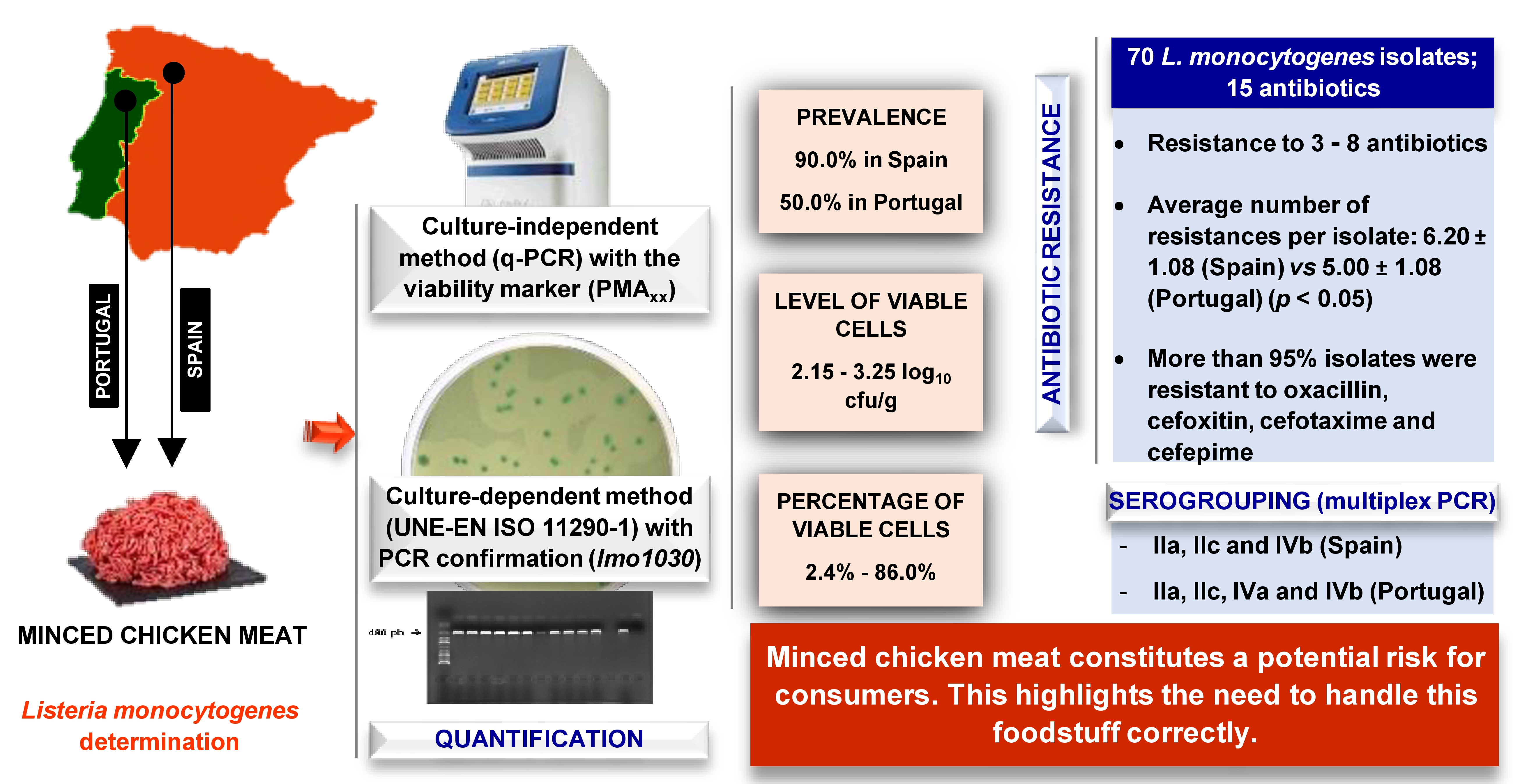
2.2. Prevalence and Levels of Listeria monocytogenes

Colonies with a typical L. monocytogenes morphology, greenish blue with a halo on the OCLA medium, were isolated from 14 samples, nine from Spain and five from Portugal. All the colonies isolated, a total of 70, comprising five from each positive sample, were identified as L. monocytogenes by PCR. Thus, the prevalence of viable culturable cells of L. monocytogenes was 70.0% overall, 90.0% in the samples acquired in Spain and 50.0% in those from Portugal (p = 0.070).
The samples of minced chicken meat were examined by q-PCR. In each amplification cycle of the q-PCR the quantity of DNA in the sample is doubled. The larger the amount of bacterial DNA present in a sample, the fewer the number of amplification cycles will be necessary for detecting it. A sample is deemed positive when it goes above the fluorescence threshold, set at a value of 0.3. At this point, the equipment indicates a value for Ct, the cycle in which this value is exceeded (Figure 1).
Seven samples, four from Spain and three from Portugal, tested positive with both the OCLA–PCR (isolation from chromogenic medium OCLA and confirmation by PCR) and the q-PCR. Three samples from Portugal yielded negative results with both the OCLA–PCR and q-PCR. Seven samples, five from Spain and two from Portugal, showed positive with the OCLA–PCR, but negative with the q-PCR. Three samples, one from Spain and two from Portugal, tested negative with the OCLA–PCR, but positive with the q-PCR. Thus, the number of samples with L. monocytogenes was 14 (considering the OCLA–PCR), 10 (q-PCR), or 17 (combining the results of both culture-dependent and culture-independent methods).
Table 2 shows the results obtained from amplification in the q-PCR in terms of Ct, ng of DNA in the reaction tube and log10 cfu/g in the sample, recording both total cells and viable cells. The levels of contamination by L. monocytogenes ranged between <2.15 log10 cfu/g (limit of detection) and 4.32 log10 cfu/g. For the viable cells, concentrations varied between <2.15 log10 cfu/g and 3.25 log10 cfu/g. The percentage of viable L. monocytogenes cells relative to the total lay between 2.4% and 86.0%.
The conventional OCLA–PCR method and the q-PCR technique were compared with respect to their capacity to detect L. monocytogenes. The classic method was taken as the reference technique. Use of the q-PCR yielded values for sensitivity (the ability to pick up positive instances), specificity (the capacity to detect negative cases), and efficiency (the probability of results being correct) of 50.0%. The predictive value for a positive test was 70.0%, and for a negative it was 30.0%. Finally, agreement, in terms of the kappa coefficient, was 0.0.

2.3. Serogroups of Listeria monocytogenes

A total of 70 isolates of L. monocytogenes, five from each of the samples yielding positive on the OCLA medium and confirmed by PCR, were classified in PCR serogroups with a multiplex PCR assay. The strains were distributed across four serogroups: IIa (52 isolates; 74.3% of total), IIc (7; 10.0%), IVa (2; 2.9%), and IVb (9; 12.9%). In Spain, figures were 84.4% (serogroup IIa), 4.4% (IIc), and 11.1% (IVb). Data from Portugal were 56.0% (IIa), 20.0% (IIc), 8.0% (IVa), and 16.0% (IVb).

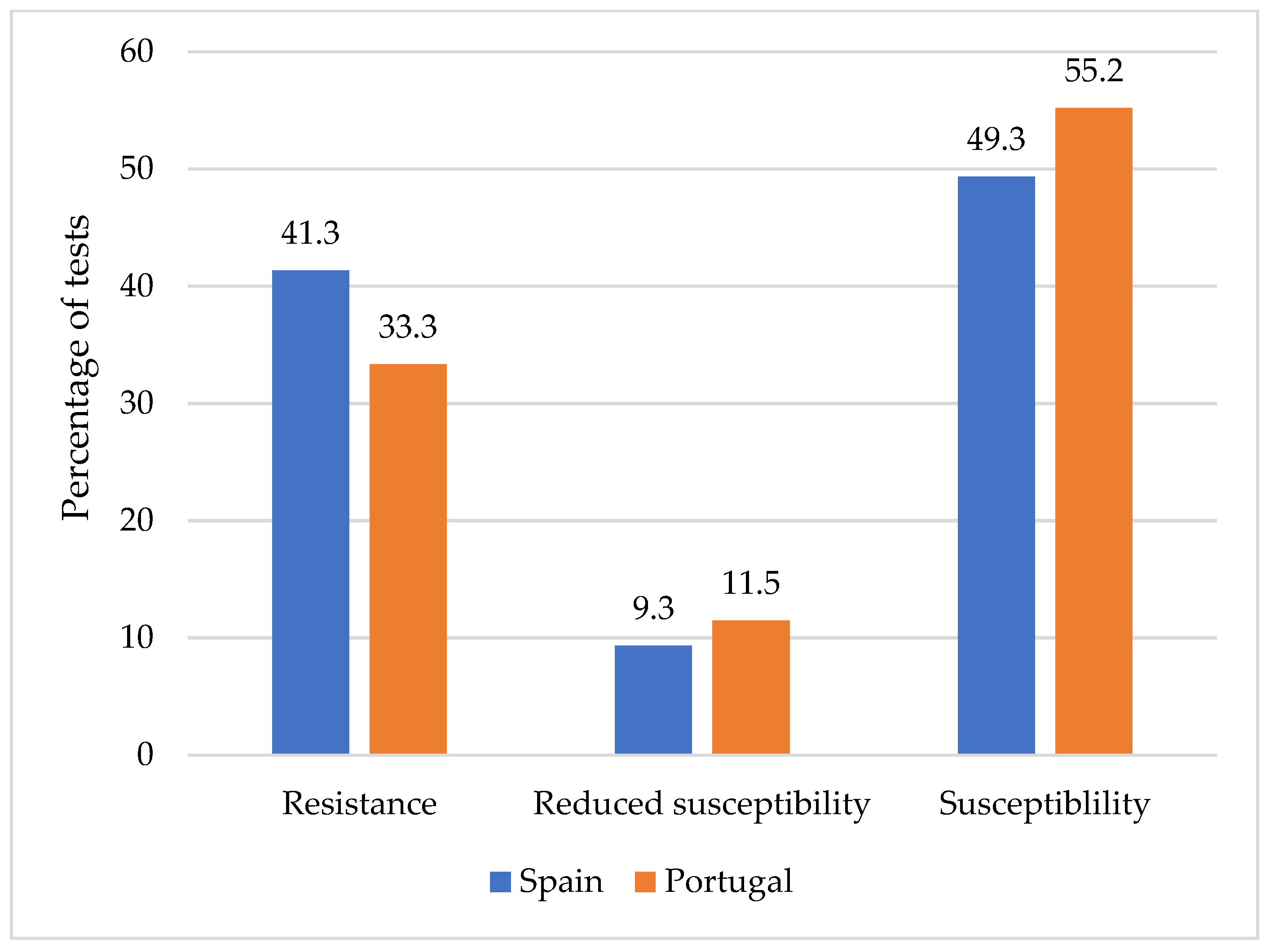
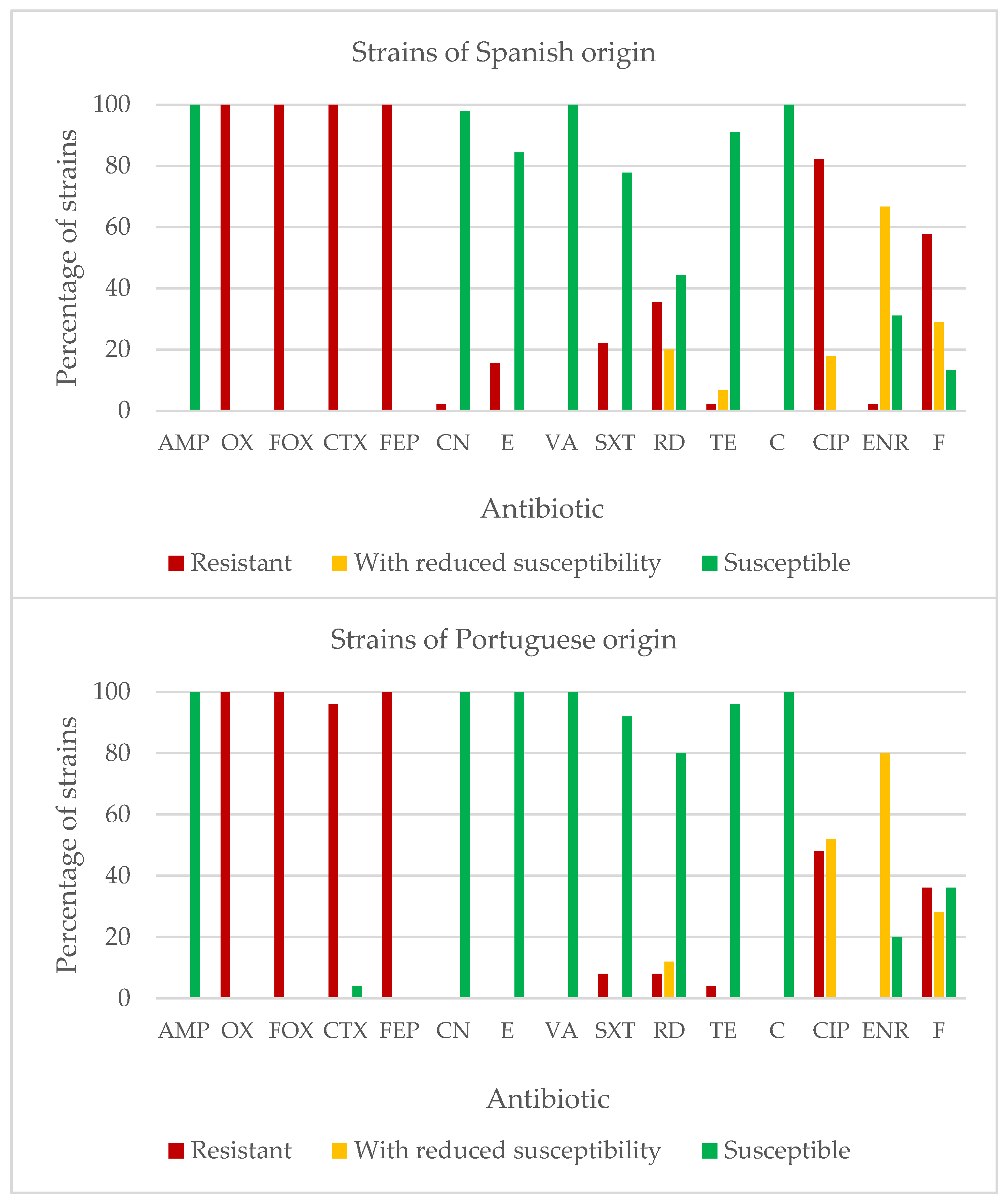
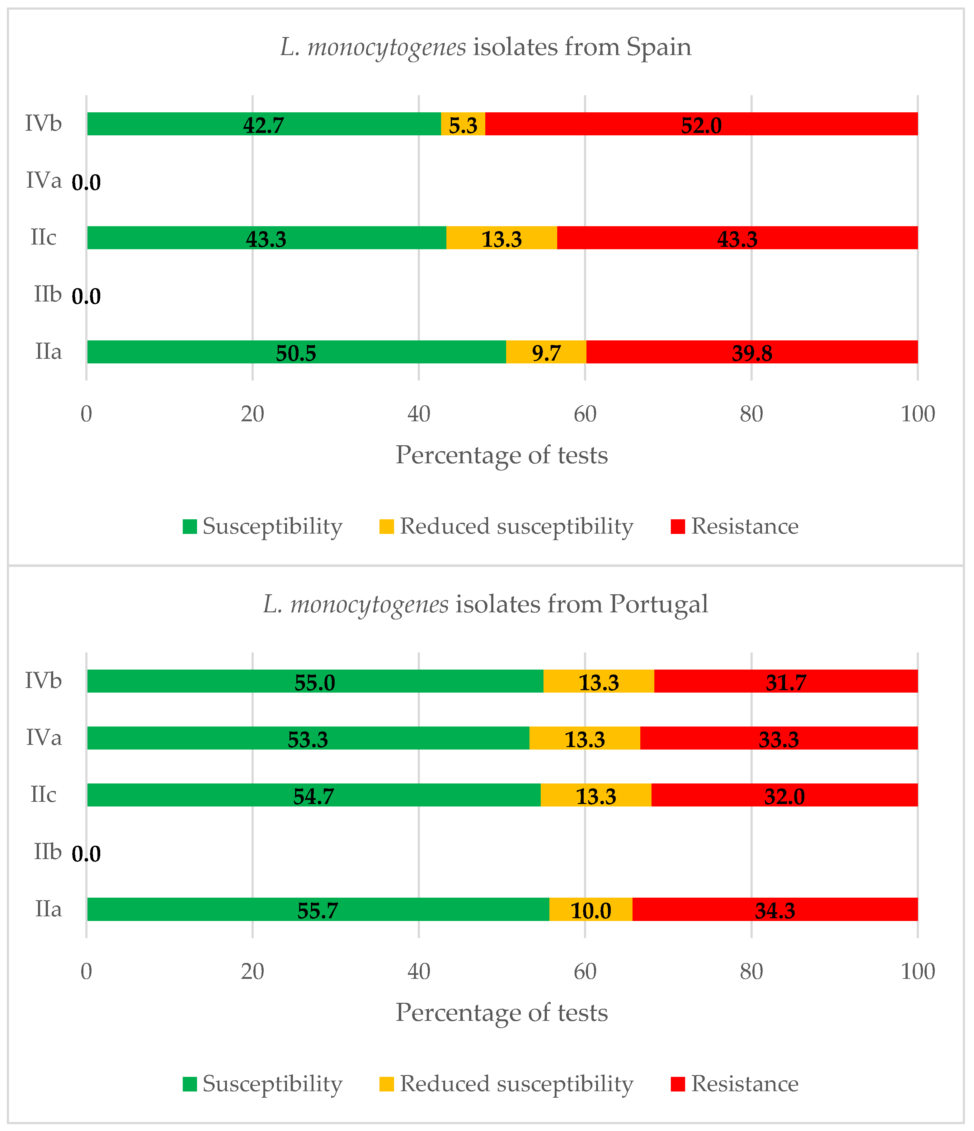
2.4. Susceptibility to Antibiotics of Listeria monocytogenes

The 70 L. monocytogenes isolates were analyzed to determine their susceptibility to a panel of 15 antibiotics of veterinary and human clinical importance. In all, 1050 tests were undertaken, which was the product of the numbers of the sets of strains and the antibiotics tested. No differences (p > 0.05) were observed between the two countries in the percentage of tests in which resistance, reduced susceptibility, and susceptibility were recorded (Figure 2).
All the strains were multi-resistant to between three and eight antibiotics. The average number of resistances per isolate was 5.77 ± 1.22 overall. This average was higher (p < 0.001) among strains isolated in Spain, at 6.20 ± 1.08, than in those from Portugal, for which the figure was 5.00 ± 1.08. If resistance and reduced susceptibility are taken together, the figures are 7.29 ± 1.16 for the whole set of strains, 7.60 ± 1.10 for strains of Spanish origin, and 6.72 ± 1.06 for strains from Portugal.
Table 3 displays the patterns of resistance detected in the 70 strains of L. monocytogenes studied. The most commonly observed pattern was resistance to OX-FOX-CTX-FEP-CIP-F, present in 14 strains (10 Spanish and 4 from Portugal). Other phenotypes seen with some frequencies were OX-FOX-CTX-FEP-CIP, noted in seven strains of Spanish origin and six from Portugal, and OX-FOX-CTX-FEP, seen in seven strains from Spain and one from Portugal.
Figure 3 shows the percentages of strains of L. monocytogenes resistant to each of the antibiotics considered. The results were similar for the two countries, with percentages of resistant strains in excess of 95.0% in oxacillin, cefoxitin, cefotaxime, and cefepime. On the other hand, percentages of resistant strains below 25.0% were observed for the following antibiotics (percentages in brackets are for Spain and Portugal, respectively): ampicillin (0.0% and 0.0%); gentamycin (2.2% and 0.0%); erythromycin (15.6% and 0.0%); vancomycin (0.0% and 0.0%); trimethoprim-sulfamethoxazole (22.2% and 8.0%); tetracycline (2.2% and 4.0%); and chloramphenicol (0.0% and 0.0%). For the remaining antibiotics, intermediate percentages of resistance were noted in Spain and Portugal, respectively: ciprofloxacin (17.8% and 52.0% strains), rifampicin (20.0% and 12.0%), and nitrofurantoin (28.9% and 28.0%).
When the strains with resistance and with reduced susceptibility were grouped together as a single cohort, a value of 100% of resistant strains, in both Spain and Portugal, was recorded for oxacillin, cefoxitin, and cefepime. Other percentages found for this combined grouping for Spain and Portugal, respectively, were 100% and 96.0% for cefotaxime; 55.6% and 20.0% for rifampicin; 22.2% and 8.0% for trimethoprim-sulfamethoxazole; 2.2% and 0.0% for gentamycin; 15.6% and 0.0% for erythromycin; and, finally, 8.9% in Spain and 4.0% in Portugal for tetracycline. No strains presented any resistance or reduced susceptibility to ampicillin, vancomycin, or chloramphenicol.
The strains isolated in Spain presented a greater prevalence of resistance to erythromycin (p < 0.001), trimethoprim-sulfamethoxazole (p < 0.01), rifampicin (p < 0.001), ciprofloxacin (p < 0.001), and nitrofurantoin (p < 0.001). Only for gentamycin was a greater prevalence of resistance noted in the strains isolated in Portugal (p < 0.001). The place of origin of the strains had no influence on the prevalence of resistance to the remaining antibiotics tested.
When the strains with resistance were grouped together with those having reduced susceptibility, the prevalence was higher in Spain with regard to rifampicin (p < 0.001), trimethoprim-sulfamethoxazole (p < 0.01), and erythromycin (p < 0.001). In contrast, the strains isolated in Portugal presented a greater percentage of resistance or reduced susceptibility to ciprofloxacin (p < 0.001) than those from Spain.
Figure 4 shows the percentages of reactions which are either resistant, intermediate, or susceptible for the strains from Spain and Portugal in each serogroup. Considering simultaneously the isolates with resistance or reduced susceptibility, the average values of 48.1%, 48.6%, 46.7%, and 51.9% were observed for serogroups IIa, IIc, IVa, and IVb, respectively.

3. Discussion

3.1. Levels of Microorganisms Indicating the Quality of Hygiene

The study being reported here investigated the microbiological quality of samples of minced chicken procured from various retail outlets in Spain and Portugal. For this purpose, the levels, expressed in log10 cfu/g, of viable aerobic microbiota, of psychrotrophic microorganisms, and of enterobacteria were determined.
Counts of viable aerobic microbiota are widely used to estimate overall microbial contamination of foodstuffs. Although high levels of these microorganisms do not necessarily imply a potential risk for human health, their importance lies in the fact that they are indicators of the quality of hygiene in the areas where food is processed and of the products themselves [6]. The viable aerobic microbiota count has been utilized as a parameter in predicting the shelf life of meat, since the presence of these microbes in large quantities can trigger rapid spoilage of products [22]. These microorganisms can also act as indicators of inappropriate processing, so that quantifying them is one way of monitoring good manufacturing practices (GMPs).
Guidelines and recommendations have been drawn up to check the microbial quality of meat preparations. According to the norms for GMPs, the overall level of microbiological contamination (viable aerobic microbiota) in raw meat preparations should not exceed 5, or at the very most 7 log10 cfu/g [23,24], figures which are lower than those noted in the present research. Moreover, a formula for sampling three categories for viable aerobic microbiota in mince at the end of the manufacturing process, n = 5, c = 3, m = 5.70 log10 cfu/g, and M = 6.70 log10 cfu/g, is in application at the current time in the European Union [25]. In accordance with the criteria stated, “n” is the number of units making up the sample and “c” is the number of units in a sample in which values higher than “m”, but never going above “M”, are permitted. The results are deemed satisfactory if all the values observed (n) are below m, acceptable if a maximum of the c values lie between the m and M, and unsatisfactory if any value exceeds M or if more than the c values go above m. The average figures noted in the present study surpassed the value of M. Hence, on the basis of the microbiological criteria noted, it may be stated that the counts recorded are excessively high.
The levels, expressed in log10 cfu/g, of viable aerobic microbiota in the samples, at 7.81 ± 0.85 for those acquired in Spain and 7.29 ± 1.12 for those from Portugal (p > 0.05), were higher than those previously recorded in poultry in Spain, which ranged from 6.29 ± 0.64 to 7.28 ± 0.51 [4,8]. They were also higher than in other zones in the European Union, such as France, where a figure of 6.05 ± 0.18 was noted [26]. Such high levels, especially in the samples procured in Spain, may be due to inappropriate processing or a lack of cleanliness in the equipment and installations utilized. With reference to this, it should also be emphasized that the mincing of meat contributes to its contamination, as a consequence of touching different surfaces, modification to the structure of tissues, the increase in the surface area, and the contact with air as chopping occurs [27]. Furthermore, in the present study, the time elapsed since slaughter, at the very least several days, may to some extent be an explanation for the high counts recorded [28].
Psychrotrophic microorganisms are of particular relevance when products are kept under refrigeration because such storage conditions still allow them to multiply [22]. The results from the current study fall within the broad range of values, running from 3.5 to 10.7 log10 cfu/g, recorded by other authors [29] for poultry products, including mince. Nevertheless, the average counts in log10 cfu/g for psychrotrophic microorganisms observed in this work, at 7.13 ± 1.07, are higher than those previously encountered by members of the same research group in samples of poultry preparations, at 6.66 ± 1.09 [4], in chicken carcasses, at 4.84 ± 0.60 [30], and in chicken legs, ranging between 4.34 ± 0.77 and 7.07 ± 1.07 [6,8]. It must be noted that all the samples in this research exceeded the maximum limit established by Pascual-Anderson [31] for poultry in Spain, which stands at 5 log10 cfu/g. It is probable that the levels of psychrotrophic microorganisms grew relative to the initial counts during the time spent in refrigerated storage in the shop prior to purchase, as previously noted [4,6].
Differences (p < 0.05) were observed between the counts of psychrotrophic microorganisms recorded in samples of chicken mince from Spain, at 7.56 ± 0.86, and from Portugal, at 6.64 ± 1.10. The psychrotroph counts in the samples of mince acquired in Portugal were close to the levels previously seen in turkey preparations, with a figure of 6.27 ± 1.17 log10 cfu/g being quoted by Castaño-Arriba et al. [32]. It should be noted that none of the samples acquired showed signs of spoilage, even though some researchers [33] have stated that levels of this group of microbes of between 6 and 8 log10 cfu/g are enough to change the smell and appearance of meat.
The low percentage of psychrotrophic microorganisms relative to viable aerobic microbiota, standing at 39.8% for the set of samples as a whole, and especially in the samples procured in Portugal, at just 22.4%, suggests that there were irregularities in maintaining temperatures at some point during the processing, storage, transport, distribution, or display in the shops of these products, as suggested previously [28]. In other studies of refrigerated chicken, the concentration of psychrotrophic microorganisms was higher than that of viable aerobic microbiota [30,34,35].
Most of the enterobacteria found in fresh meat come from contamination with faeces, so that their presence in large quantities may point to poor hygiene at the abattoir from which the product originates, inappropriate storage, or a combination of both [34,36,37]. Hence, the level of enterobacteria has been used as an indicator of faecal contamination in fresh meat. The average counts for enterobacteria in the present study, at 4.23 ± 0.88 log10 cfu/g, lay above the limits used as microbiological criteria for free-range poultry in Spain, which are set at 2 log10 cfu/g [31].
No significant differences were observed for enterobacteria as a group between samples analyzed in Spain, with 4.55 ± 0.96 log10 cfu/g, and in Portugal, with 4.02 ± 0.78 log10 cfu/g. Various studies refer to lower levels of enterobacteria in poultry, both in Spain, where a figure of 2.89 ± 0.77 log10 cfu/g was recorded by Buzón-Durán et al. [4], and in other areas within the European Union, with 2 log10 cfu/g noted by Fraqueza et al. [38]. Previous studies also carried out in north-western Spain [3] indicated a lower load of enterobacteria in meat preparations based on beef, at 1.99 ± 0.99 log10 cfu/g, and pork, at 1.96 ± 1.44 log10 cfu/g. This greater count of enterobacteria in poultry products when compared to foodstuffs of other types may have been an outcome of the higher initial pH in birds’ muscle meat [39].

3.2. Prevalence and Levels of Listeria monocytogenes

It proved possible to assign 100% of the colonies with the characteristic morphology of L. monocytogenes on OCLA medium to that species using the PCR technique, detecting the lmo1030 gene. These results coincide with those of several other studies, in which a comparison of the classic method of double enrichment and inoculation onto a solid medium with a PCR approach for detecting L. monocytogenes found figures for agreement between the two ranging from 80% to 100% [40,41,42].
The strains of L. monocytogenes were isolated from 70.0% of the samples of minced chicken. They were found in 90.0% of the samples obtained from Spanish outlets and in 50.0% of those from Portugal. This prevalence falls within the broad range of values recorded by other authors, with between 0% and 99% of samples of poultry testing positive for Listeria spp. [43]. In previous work done by members of the research group undertaking this study, the prevalence observed for L. monocytogenes was 24.5% [44] or 32% [30], much lower than the values from the present case. Nevertheless, it must be noted that those other research studies investigated either chicken carcasses or chicken legs. As the mincing of meat increases the risk of contamination by L. monocytogenes [45], it was to be expected that the prevalence found in the current work would be higher than in previous investigations. With regard to the prevalence of L. monocytogenes in samples of food in Portugal, the value noted, 50.0%, is similar to the range observed by other authors in raw chicken in that country, with published prevalence values of 19.3% [46], 41.3% [47], or 60% [48].
In the seven samples that tested positive with both OCLA–PCR and q-PCR, estimated concentrations ranged between 2.25 and 4.32 log10 cfu/g. Such levels of contamination are similar to those observed by other authors in raw chicken, where the detected values did not exceed the value of 3 log10 cfu/g of sample [49]. Other researchers have found concentrations above 3 log units, but only in a small percentage of samples [50]. The results of the present study are also similar to what was encountered by other authors in fermented sausages, where the range was 2.85 to 3.38 log10 cfu/g [51], or in fresh cheese, for which 3.60 log10 cfu/g was the figure quoted [52].
According to the model utilized by the European Food Safety Authority (EFSA), 92% of cases of invasive listeriosis are to be attributed to doses of more than 5 log10 cfu per portion consumed. If the average size of a portion is taken to be 50 g, this would equate to a concentration of L. monocytogenes in ready-to-eat foods in excess of 3.30 log10 cfu/g at the point of consumption. Nonetheless, a small proportion of cases are associated with such foodstuffs having lower levels than this of L. monocytogenes [53]. Regulation (EC) num. 2073/2005 [25] establishes as the upper limit of shelf life a concentration of 2 log10 cfu/g in ready-to-eat foods on sale.
It is true that chicken preparations are cooked before consumption. However, the risk arising from the presence of viable cells of L. monocytogenes in such foodstuffs, especially when it is at a high level, derives from the possibility of inadequate cooking or of incidents of cross-contamination affecting other foods. Moreover, L. monocytogenes is a psychrotrophic microorganism, so that its concentration in mince can grow over the course of refrigerated storage.
Three samples of minced chicken tested negative both with the classic OCLA–PCR isolation method and with the q-PCR. Either these samples had no L. monocytogenes, or they were contaminated only with cells that were inactivated, or viable, but not culturable (and at a concentration lower than the detection limit for the q-PCR technique, 2.15 log10 cfu/g). If culturable cells were present in the samples, their concentration was below 1 ufc/25 g. Seven samples tested positive with the OCLA–PCR and negative with the q-PCR. This may have been due to the fact that 40 amplification cycles, equating to 2.15 log10 cfu/g, taken as the limit for detection by the technique, were not enough to get over the fluorescence threshold. Hence, these samples were deemed to be contaminated by viable culturable cells of L. monocytogenes, but at a concentration below 2.15 log10 cfu/g. It should be noted that the detection limit adopted was lower than those set by other authors, lying between 3 and 4 log10 cfu/g or cfu/mL [52,54,55]. Finally, three samples tested negative with the OCLA–PCR and positive with the q-PCR, which may be an outcome of the fact that the cells of L. monocytogenes were viable but not culturable or that the DNA came from inactivated bacteria. If culturable cells were present, their concentration was below 1 ufc/25 g.
The limited sensitivity of the q-PCR with regard to the OCLA–PCR (50.0%) shows clearly that this method is not of any use in detecting L. monocytogenes in food when contamination levels are very low, because of the detection threshold for the technique, at 2.15 log10 cfu per gram of sample. Hence, the predictive value of a negative test is no more than 30.0%. In contrast, the q-PCR does have the advantage over the classic method of allowing detection of cells inactivated and cells that are viable, but not culturable. This is what leads to the low specificity of the technique (50.0%), since the classic method used as a benchmark rate as negative any samples contaminated with L. monocytogenes cells that are not culturable or are inactivated. In particular, three samples in the present study gave evidence of the detection by the q-PCR (using the viability marker PMAxx) of viable L. monocytogenes cells that could not be cultured on OCLA medium. The detection and quantification of cells that are viable, but not culturable, is of interest in the context of food safety, since these are living cells that could cause infections in consumers.
The percentages of viability recorded in this research varied between 2.4% and 86.0%. A search of published works did not find any research into L. monocytogenes allowing a comparison of these percentages. The results do lie within the range of values obtained by Zhang et al. [56] for other pathogenic Gram-positive microorganisms like Staphylococcus aureus, the concentration of viable cells of which in powdered milk was 10% (3 × 10² cfu/g relative to a total 3 × 10³ cfu/g).

3.3. Serogroups of Listeria monocytogenes

The serogroup IIa was the most prevalent in minced chicken meat from both Spain and Portugal (84.4% and 56.0% of isolates, respectively). The strains in this serogroup (which includes serotype 1/2a) have shown extensive distribution in foodstuffs and food-processing environments around the world, thereby indicating its robust ecological adaptability [57,58,59,60,61,62]. It has been indicated that the strains of the dominant serovar from this serogroup (serotype 1/2a) appears to contain more plasmids than other serotypes. Considering that plasmids frequently carry genes that confer resistance to antimicrobial agents, including sanitizers used in processing operations, bacteria harbouring such plasmids would have an increased ability to survive and develop in these environments [16]. Moreover, isolates from serotype 1/2a present a high prevalence of various virulence genes and are often isolated from human clinical samples [63].
The strains belonging to serogroups IIc (including serotype 1/2c) and IVb (including serotype 4b), which were detected in the study being processed here, have been previously detected in food samples [64,65,66]. The presence of L. monocytogenes isolates from serogroup IVb is a matter of concern because the serotype 4b strains exhibit the strongest epidemiological association with human listeriosis [67]. It should be noted that no strains from serogroup IIb were found in minced chicken meat, which does not coincide with results from most authors consulted. It has been suggested that differences in geographical region and in the investigated type of food may cause variations in serogrouping [67]. On the other hand, serogroup IVa (strains 4a–4c), to which 8.0% of isolates from Portugal belonged, is considered very infrequent in foodstuffs, and its presence is linked exclusively to animals. The strains of this serogroup are rarely reported with clinical relevance for humans [59].

3.4. Susceptibility of Antibiotics

The susceptibility of 70 isolates of L. monocytogenes obtained from chicken mince was checked against 15 antibiotics of clinical significance. The average number of resistances per isolate (5.77 ± 1.22; 6.20 ± 1.08 for the Spanish strains and 5.00 ± 1.08 for those from Portugal), was much higher than the values recorded in previous work with the strains of L. monocytogenes obtained from poultry in north-western Spain, in which the number of resistances per strain observed was 1.60 in 1993 and 4.24 in 2006 [44]. Although L. monocytogenes is a bacterium that in the past has been sensitive to the majority of antimicrobials employed to treat infections by Gram-positive organisms, in recent years a striking increase has occurred in the prevalence of resistance in this microorganism [68], a situation that is also highlighted by the results of the present study, where the strains showed resistance to antibiotics used for the treatment of human listeriosis (e.g., erythromycin, trimethoprim-sulfamethoxazole, fluoroquinolones, and rifampicin) [16]. Among other causes, this growth in resistance in L. monocytogenes is due to its gradual acquisition from the cells of various different genera of bacteria of mobile genetic elements, such as plasmids or transposons [69]. It should be noted that resistance to antibiotics has been commonplace for some years in other Gram-positive bacteria. Thus, the average number of resistances per strain noted in this work is similar to what was found previously for Gram-positive bacteria in poultry from north-western Spain, where figures of 4.50 [32] or 5.58 [70] were observed for enterococci, and 6.35 for S. aureus [4].
A group of international experts established under a joint initiative of the European Centre for Disease Prevention and Control (ECDC) and the Centers for Disease Control and Prevention (CDC) in the United States devised the standard definitions for phenotypes, which were seen as multidrug-resistant (MDR), extensively drug-resistant (XDR), and pan-drug-resistant (PDR) in bacteria of interest for Public Health. The MDR phenotype is defined as acquired non-susceptibility to at least one agent in three or more antimicrobial categories, with one or more antibiotics from each category being applied. The XDR phenotype is defined as non-susceptibility to at least one agent in all but two or fewer antimicrobial categories, so that the bacterial isolates remain susceptible to only one or two categories. Finally, the PDR phenotype refers to a lack of susceptibility affecting all agents in all antimicrobial categories [71]. These criteria were used in characterizing the profile of resistance to antibiotics on the part of the strains trialled in the present study.
No pan-susceptible strains were found, nor any that were resistant to just one antibiotic. One strain (1.4% of the total) showed resistance to three antibiotics, eight strains (11.4%) to four antibiotics, twenty-three strains (32.9%) to five, and thirty-eight strains (54.3%) presented a multidrug-resistant phenotype (MDR). The isolates assigned to the MDR category presented resistance to six (20 strains; 28.6% of the total), seven (10 strains; 14.3%), or eight (8 strains; 11.4%) antimicrobial compounds. The presence of strains with resistance to various antibiotics constitutes a crucial challenge for Public Heath because many antimicrobials would consequently be ruled out as therapeutic options [18].
More than 50% of the strains presented resistance to oxacillin, cefoxitin, cefotaxime, cefepime, ciprofloxacin, enrofloxacin, and nitrofurantoin. The strains of Spanish origin showed, in addition, intermediate levels of resistance to tetracycline (6.7% of strains), rifampicin (20.0%), ciprofloxacin (17.8%), enrofloxacin (66.7%), and nitrofurantoin (28.9%). These findings are worrying, since some of the substances mentioned are habitually employed in treating listeriosis, for which a beta-lactam, generally ampicillin, would be the antibiotic of choice, administered alone or in combination with gentamycin. Where an allergy to beta-lactams occurs, possible alternatives include erythromycin, vancomycin, trimethoprim-sulfamethoxazole, and fluoroquinolones. Occasionally, treatment for listeriosis is undertaken with rifampicin, tetracycline, and chloramphenicol [16].
Furthermore, certain substances to which the strains in the present study showed a high prevalence of resistance are classified by the World Health Organization as antimicrobial agents that are “critically important” (AMP, CIP, ENR, FOX, CTX, FEP, and RD), “highly important” (OX and SXT), or “important” (F) for human medicine [72]. It should also be noted that the other antibiotics to which the strains studied presented some resistance, even if to an extent below 15%, are classified as “critically important” (CN, E, and VA) or “highly important” (TE) in human medicine. In the list published by the World Organization for Animal Health (OIE), AMP, OX, CIP, ENR, SXT, CN, and TE are considered antibiotics that are “critically important”, and RD is “highly important” in veterinary medicine [73].
Various authors have found the strains of L. monocytogenes resistant to one or more of the antibiotics to which resistance was noted in the current study [16,44,69,74,75,76,77,78]. It must be noted that some of the antibiotics to which resistance was observed are substances that are widely used in animal production (e.g., fluoroquinolones) [79,80,81]. Hence, the selective pressure exerted by the use of antibiotics (particularly when incorrectly employed at sub-inhibitory doses) has been identified as the principal cause of the marked growth in the prevalence of resistance to antibiotics that has taken place over recent decades [18]. Resistance was also observed to nitrofurantoin, a drug that has been banned in the European Union in the 1990s because of its toxicological risks for consumers [82]. Despite the fact that this antimicrobial has not been used on European poultry farms for years, cross-resistance or co-resistance mechanisms could be the cause of the resistance observed to this drug [82]. On the other hand, a very low prevalence of resistance was observed for tetracycline, although it is a compound widely used in European avian farms [81]. The low prevalence of resistance to tetracycline in chicken meat has been observed in previous reports [16].
The high prevalence of resistance observed among the strains of the PCR-serogroup IVb is a finding that is coincidental with observations from other researchers [83].

4. Material and Methods

4.1. Sample Collection

Twenty samples of minced chicken meat, each weighing approximately 400 g, were analyzed. They were procured from various different retail outlets in the cities of León in Spain (ten samples) and of Vila Real in Portugal (ten samples). The chicken meat was minced in the retail outlets. All of the samples were transported in their individual wrappings to the laboratories in the two locations, where they were processed immediately upon arrival.

4.2. Counts of Microorganisms Indicating the Quality of Hygiene

From each sample, 25 g was taken and placed with 225 mL of 0.1% peptone water into a sterile bag with a filter. Homogenization was performed over 120 s in a paddle homogenizer (Stomacher, IUL Instruments, Barcelona, Spain). Decimal dilutions were carried out with the same diluent. Table 4 shows the culture media, the incubation conditions, and the references used for each of the microbial groups studied. All the culture media used in this research were products of the company Oxoid Ltd. (Hampshire, United Kingdom).

4.3. Isolation and Identification of Listeria monocytogenes

In detecting L. monocytogenes, the method specified by the UNE-EN ISO 11290-1 standard was used. Samples weighing 25 g each were placed in sterile bags with filters and homogenized with an IUL Stomacher in 225 mL of Half Fraser broth for 120 s. After 24 h of incubation at 30 °C, quantities of 10 µL were transferred to tubes with 10 mL of Fraser broth, these being incubated at 37 °C for 24 h. Following this, they were streak-plated on the Oxoid Chromogenic Listeria Agar OCLA. A further 48 h of incubation at 37 °C followed, after which five greenish blue colonies with haloes, assumed to be the strains of L. monocytogenes, were taken for later identification. The strains were kept at −50 °C in tryptone soya broth (TSB) with 20% glycerol (PanReac AppliChem, Darmstadt, Germany). The culture media were from Oxoid Ltd. (Hampshire, United Kingdom).
The identification of the isolates was carried out by means of a PCR, utilizing primers and conditions specifically for detecting the gene lmo1030 (Table 5).
The DNA of the strains was extracted from a TSB culture broth incubated for between 18 and 24 h at 37 °C, centrifuged at 13,000 rpm for 60 s twice, and followed by exposure to 100 °C for 30 min in a water bath. The purity and concentration of the DNA were determined using a Nano-Drop One spectrophotometer (Thermo Scientific, Wilmington, Delaware, USA); the wavelength being set at 260 nm. Concentrations of between 80 ng/µL and 180 ng/µL were seen as suitable.
The PCR reactions were carried out in an overall volume of 25 µL, incorporating 5 µL of DNA, 0.5 μM of each primer (Isogen Life Sciences, Barcelona, Spain), a mixture of 0.2 mM of deoxynucleoside triphosphates (dNTPs) obtained from EURx (Gdansk, Poland), a reaction buffer at a 1× concentration (BIORON, Diagnostics GbmH, Ludwigshafen, Germany), MgCl2 with a concentration of 3 mM (BIORON), Taq DNA polymerase (BIORON), and sterile distilled water to make up a final volume of 25 µL.
DNA amplification was performed in a thermocycler by Bio-Rad (Hercules, CA, USA). This was programmed as follows: an initial denaturation cycle at 95 °C for five minutes, then 35 amplification cycles (denaturation for 30 s, annealing for 30 s, and elongation at 72 °C for 45 s), followed by a final elongation period of five minutes. Amplification products were separated by means of horizontal electrophoresis on agarose gel (BIORON) at 1% (weight/volume) in a 1× tris-acetate-EDTA buffer stained with SimplySafe (EURx) and diluted to 1:10,000. A loading buffer of glycerol (PanReac AppliChem, Darmstadt, Germany) was used together with a bromophenol blue dye (Panreac Química S.L.U., Barcelona, Spain). In the visualization of the electrophoresis bands an ultra-violet transilluminator (Gel Doc EZ System, Bio-Rad) was used. The size of each PCR product was estimated using markers with a standard molecular weight (10 kb DNA Ladder from BIORON). All the PCR trials included controls, which were both negative (MilliQ water) and positive (L. monocytogenes ATCC 13932).

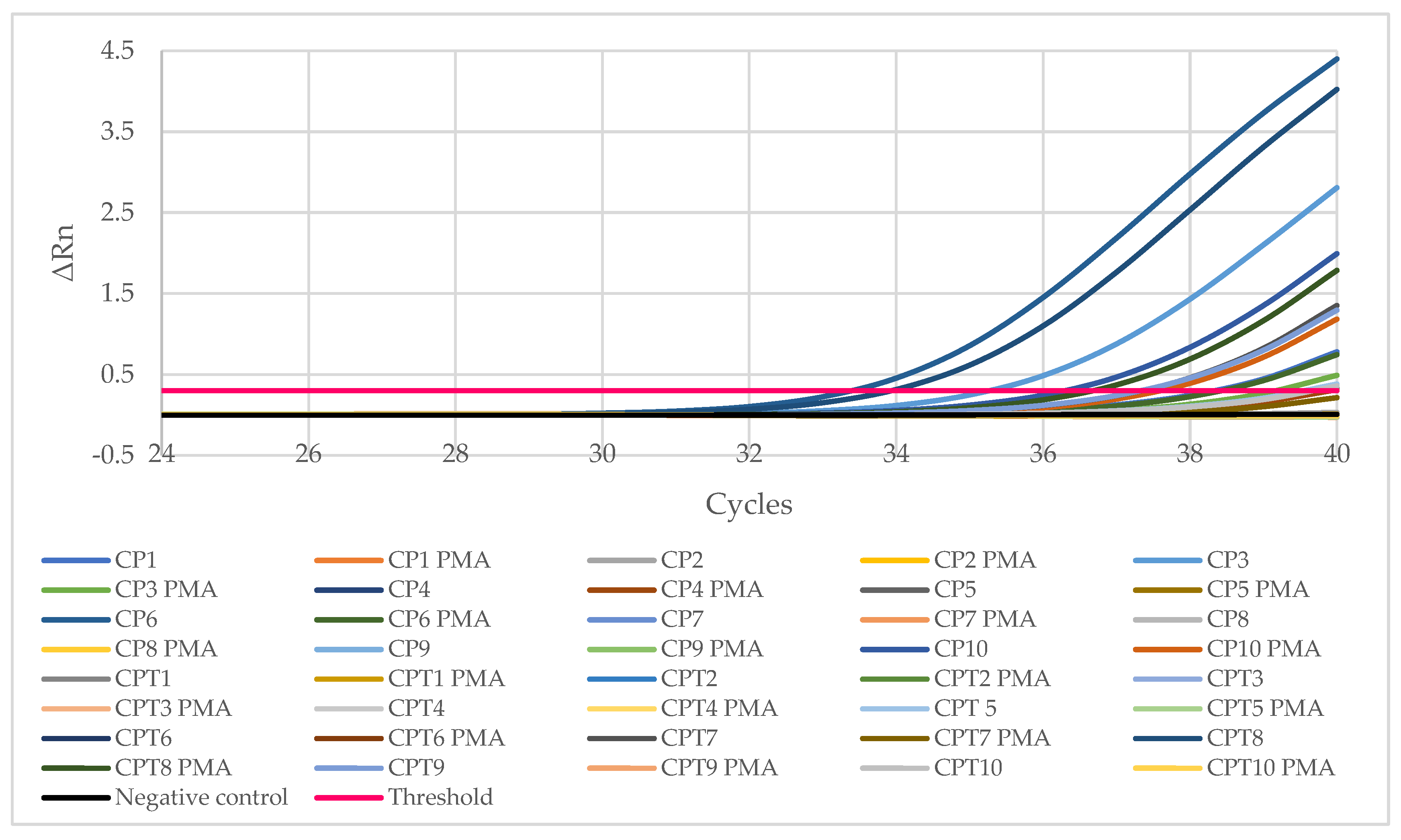
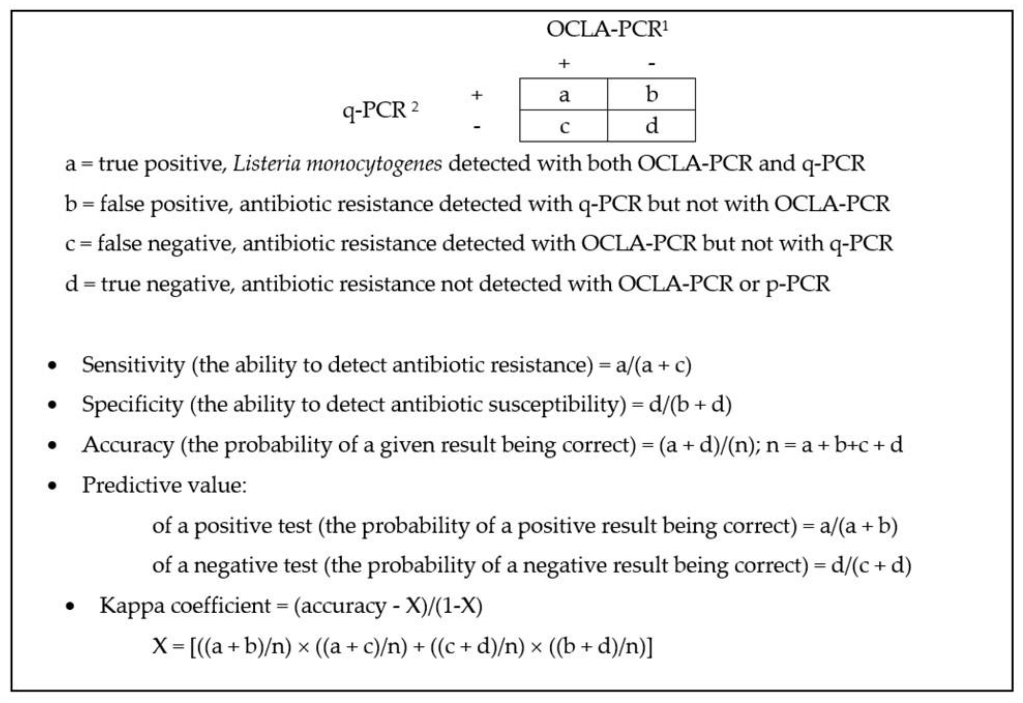
4.4. Detecion and Quantification of Listeria monocytogenes by q-PCR

To detect and quantify L. monocytogenes in minced chicken by means of a q-PCR, 1.6 mL of the contents of the homogenization bags was taken together with 0.1% peptone water (Oxoid) and deposited in a 5 mL Falcon™ round-bottom tube (Thermo Fisher Scientific, Waltham, MA, USA) and shaken with a vortex agitator. Thereafter, a measured quantity of 750 μL of the contents of the tube was removed. DNA extraction was performed with the aid of a PrepSEQTM Rapid Spin Sample Preparation Kit with Proteinase K (Thermo Fisher Scientific). For amplification by means of the q-PCR, 30 µL of the extracts of DNA were deposited in each of the wells of a MicroSEQ™ L. monocytogenes detection kit (Thermo Fisher Scientific). Both the extraction and amplification of the DNA were performed in accordance with the manufacturer’s instructions.
Subsequently, a quantity of 800 µL was transferred from the Falcon™ tube (Thermo Fisher Scientific) to a sterile Eppendorf tube and a volume of 1 µL of the viability marker PMAxx (Biotium, Fremont, CA, USA) was added, yielding a final dye concentration of 25 µM, this mix being stirred with the pipette. The samples with PMAxx were incubated at 42 °C for 30 min in darkness and manually rotated several times to encourage binding of the colourant to the DNA of the damaged cells. The dyed samples were next exposed to a halogen light source for 15 min at a distance of approximately 20 cm while placed on a block of ice covered in aluminium foil to enhance light reflection. A measured quantity of 750 μL was then taken from the contents of the Eppendorf tube and DNA extraction was carried out using a PrepSEQTM Rapid Spin Sample Preparation Kit with Proteinase K (Thermo Fisher Scientific). To amplify them using the q-PCR, 30 µL of the extracts of DNA were put into each of the wells of a MicroSEQ™ L. monocytogenes detection kit (Thermo Fisher Scientific). Both the extraction and the amplification of DNA were carried out in accordance with the manufacturer’s instructions.
The samples were placed into the thermal cycler of a StepOneTM Real-Time PCR System (Applied Biosystems, Foster City, California, United States); the fluorescence threshold being set at 0.3. To convert amplification cycles into quantities of DNA, a standard straight line (y = −3.0525x + 23.206; R2 = 0.966), derived from samples with known amounts of L. monocytogenes DNA, was used [87]. To extrapolate the quantity of DNA in terms of bacterial concentrations (log10 cfu per gram of sample) the size of the genome of L. monocytogenes was considered, where 1 ng of DNA was deemed approximately equivalent to 340,000 cfu [88]. Calculations were performed on the basis of the following equation [87]:
L .   m o n o c y t o g e n e s   concentration   ( Log 10 cfu g ) = Log 10   ( 10 Ct 23.206 3.0525 × 340 , 000 × 10 5 750 ) cfu / g
In establishing this equation, various items were considered. These were: (1) the total volume of the homogenization bag (250 mL, or 250,000 µL), (2) the decimal dilution performed to produce the homogenate (25 g of sample in 225 mL of diluent), (3) the fact that the reaction tube receives one-tenth of the total amount of DNA extracted (30 µL out of 300 µL), and (4) that the DNA was extracted solely from 750 µL.
In comparing L. monocytogenes detection data obtained with the classic method (OCLA–PCR) and those from the q-PCR, the sensitivity, specificity, efficiency, and predictive value of the second method were calculated. Since the degree of contamination of samples was unknown, the calculation method assumed that the conventional OCLA–PCR technique yielded the correct results, acting as a benchmark method. In addition, the two methods were compared by calculating the kappa co-efficient, or agreement [30]. The definitions and way of working out these parameters are shown in Figure 5.

4.5. Multiplex PCR Serogrouping of Listeria monocytogenes Isolates

L. monocytogenes isolates were further confirmed and classified in PCR groups of serotypes with a multiplex PCR assay, in accordance with the method described by Doumith et al. [89], with minimal modifications. As PCR templates, three to five bacterial colonies grown on tryptone soya agar (TSA) plates (Oxoid) were emulsified in tubes with 50 µL of a solution formed by Tris-HCl 10 mM and EDTA 1 mM (pH = 8.0) and incubated at 99 °C for 15 min. Subsequently, the solutions were cooled in ice and 200 µL of distilled water was added to each mixture. The tubes were centrifuged at 13.000 rpm for 5 min and 50 µL of the supernatant were taken. PCR reactions were carried out incorporating 1 µL of DNA; 1 μM of each of the primers for lmo0737, ORF2819, and ORF2110; 3 μM of the primer for lmo1118; 0.2 μM of primer for prs (Macrogren Humanizing Genomics, Seoul, Republic of Korea) (Table 6); DNA AmpliTools Multiplex Master Mix 2× (BIOTOOLS, Madrid, Spain); and sterile distilled water to make up a final volume of 20 µL.
The PCR was performed with an initial denaturation step at 94 °C for 3 min, which included 35 cycles of 94 °C for 40 s, 53 °C for 1 min 15 s, and 72 °C for 1 min 15 s; one final cycle of 72 °C for 7 min in a thermocycler ProFlex™ (Applied Biosystems, Waltham, Massachusetts, EEUU). The amplification products (10 µL of the reaction mixture was used) were separated as indicated in previous paragraphs for L. monocytogenes identification, but with slight modifications (agarose gel at 2%). Table 7 shows the correlation of multiplex PCR fragment amplification and conventional serotyping.

4.6. Antibiotic Susceptibility Tests

The susceptibility of seventy isolates of L. monocytogenes (five colonies isolated from the OCLA medium for each positive sample) to a panel of fifteen antibiotics was determined. The disc diffusion method described by the Clinical and Laboratory Standards Institute [90] was employed. Initially, the strains were cultured in tubes holding 9 mL of Mueller Hinton broth (MHB, Oxoid), 20 µL being taken with an inoculation loop from each of the cultures kept frozen. These tubes were incubated for 11 h at 37 °C and their contents were used to inoculate Petri dishes with Mueller Hinton agar (MHA, Oxoid) using a spread plate technique, producing a lawn of culture with a sterile swab. The antibiotic discs were then placed, five on each dish, with sterile tweezers. Antibiotics of 10 categories were tested. These were (1) beta-lactams (ampicillin, AMP, 10 µg; oxacillin, OX, 1 µg; cefoxitin, FOX, 30 µg; cefotaxime, CTX, 30 µg; cefepime, FEP, 30 µg), (2) aminoglycosides (gentamycin, CN, 10 µg), (3) macrolides (erythromycin; E, 15 µg), (4) glycopeptides (vancomycin, VA, 30 µg), (5) sulphonamides (trimethoprim-sulfamethoxazole, SXT, 25 µg), (6) ansamycins (rifampicin, RD, 5 µg), (7) tetracyclines (tetracycline, TE, 30 µg), (8) amphenicols (chloramphenicol, C, 30 µg), (9) fluoroquinolones (ciprofloxacin, CIP, 5 µg; enrofloxacin, ENR, 5 µg), and (10) nitrofuran derivatives (nitrofurantoin, F, 300 µg). All antibiotic discs were purchased from Oxoid.
The plates were incubated for 24 h at 37 °C in an inverted position. The inhibition zones were measured and the strains were classified as sensitive, having reduced sensitivity, or resistant, as a function of the size of the inhibition halo in accordance with the guidelines published by the European Committee on Antibiotic Susceptibility Testing (EUCAST) in 2020 [91] for CN, E, SXT, RD, TE, C, and CIP, and by the CLSI in 2018 [90] for AMP, FOX, CTX, FEP, VA, ENR, and F.

4.7. Statistical Analysis

For the purposes of statistical analyses, microbial counts were converted into logarithmic units (log10 cfu/g) and compared using analysis of variance (ANOVA), the means being separated with Duncan’s multiple range test. The prevalence levels of L. monocytogenes and of the resistance to antibiotics in the samples from the two countries were compared using Fisher’s exact test. In the comparison of the number of resistances per strain in the various groups of samples, the nonparametric Mann–Whitney U test was used. Significant differences were established with a probability level of 95% (p < 0.05). All statistical analyses were performed with the aid of the Statistica® 8.0 software package (StatSoft Inc., Tulsa, OK, USA).

5. Conclusions

The results of this research work indicate that chicken mince is a foodstuff of dubious quality from the point of view of hygiene, given the considerable amounts of viable aerobic microbiota, psychrotrophic microorganisms, and enterobacteria found. Moreover, this sort of product provides a large reservoir of strains of L. monocytogenes belonging to the PCR-serogroups frequently associated with human listeriosis and showing resistance to multiple antibiotics of clinical importance, a worrying fact in the context of food safety. This entails a need for those handling such foodstuffs to be thoroughly trained in food hygiene, with the aim of avoiding bad practices, like inadequate cooking or cross-contamination, thus reducing the risk to consumers.
The results obtained highlight the limitations of both the classic method for isolating by two-fold enrichment and inoculation onto a selective, differential solid medium, which confirms the bacterial species with a conventional PCR, and also of the q-PCR for detecting L. monocytogenes in samples of poultry. The classic method does not permit the detection of L. monocytogenes cells that are viable, but not culturable. On the other hand, the q-PCR method is not suitable for detecting low levels of L. monocytogenes, in the light of this technique’s high threshold for detection (2.15 log10 cfu/g). These findings indicate the usefulness of combining the two techniques (OCLA–PCR and q-PCR) with a view to enhancing the detection of L. monocytogenes in food.

Author Contributions

Conceptualization, C.R.-M. and R.C.; methodology, C.R.-M., A.E., S.P.-M., and C.A.-C.; validation, C.R.-M., S.P.-M., and C.A.-C.; formal analysis, C.R.-M., S.P.-M., A.E., and R.C.; investigation, C.R.-M., A.E., R.C., and C.A.-C.; resources, A.E., R.C., and C.A.-C.; data curation, C.R.-M.; writing—original draft preparation, C.R.-M. and R.C.; writing—review and editing, R.C. and C.A.-C.; supervision, A.E., R.C., and C.A.-C.; project administration, R.C.; funding acquisition, R.C. and C.A.-C. All authors have read and agreed to the published version of the manuscript.

Funding

This research was funded by the Junta de Castilla y León (Consejería de Educación, Spain, grant number LE018P20), the Ministerio de Ciencia, Innovación y Universidades (Spain, grant number RTI2018-098267-R-C33), and the Universidad de León (Spain, grant number 18BB282).

Institutional Review Board Statement

Not applicable.

Informed Consent Statement

Not applicable.

Data Availability Statement

Not applicable.

Conflicts of Interest

The authors declare no conflict of interest.

References

  1. FAOSTAT. Consumo Carne de Pollo. Available online: http://www.fao.org/faostat/es/#data/QL (accessed on 22 September 2021).
  2. Cardoso Pereira, P.M.C.; Baltazar Vicente, A.F.R. Meat nutritional composition and nutritive role in the human diet. Meat Sci. 2013, 93, 586–592. [Google Scholar] [CrossRef] [PubMed] [Green Version]
  3. Capita, R.; Castaño-Arriba, A.; Rodríguez-Melcón, C.; Igrejas, G.; Poeta, P.; Alonso-Calleja, C. Diversity, antibiotic resistance, and biofilm-forming ability of enterobacteria isolated from red meat and poultry preparations. Microorganisms 2020, 8, 1226. [Google Scholar] [CrossRef] [PubMed]
  4. Buzón-Durán, L.; Capita, R.; Alonso-Calleja, C. Microbial loads and antibiotic resistance patterns of Staphylococcus aureus in different types of raw poultry-based meat preparations. Poult. Sci. 2017, 96, 4046–4052. [Google Scholar] [CrossRef] [PubMed]
  5. Selvan, P.; Naremdra, B.R.; Sureshkumar, S.; Vankataramanujam, V. Microbial quality of retail meat products available in Chennai city. Am. J. Food Technol. 2007, 2, 55–59. [Google Scholar]
  6. Del Río, E.; Panizo-Morán, M.; Prieto, M.; Alonso-Calleja, C.; Capita, R. Effect of various chemical decontamination treatments on natural microflora and sensory characteristics of poultry. Int. J. Food Microbiol. 2007, 115, 268–280. [Google Scholar] [CrossRef] [PubMed]
  7. Rodríguez-Melcón, C.; Alonso-Calleja, C.; Capita, R. Lactic acid concentrations that reduce microbial load yet minimally impact colour and sensory characteristics of beef. Meat Sci. 2017, 129, 169–175. [Google Scholar] [CrossRef]
  8. Álvarez-Astorga, M.; Capita, R.; Alonso-Calleja, C.; Moreno, B.; García-Fernández, M.C. Microbiological quality of retail chicken by-products in Spain. Meat Sci. 2002, 62, 45–50. [Google Scholar] [CrossRef]
  9. Rodríguez-Melcón, C.; Riesco-Peláez, F.; Carballo, J.; García-Fernández, C.; Capita, R.; Alonso-Calleja, C. Structure and viability of 24- and 72-h-old biofilms formed by four pathogenic bacteria on polystyrene and glass contact surfaces. Food Microbiol. 2018, 76, 513–517. [Google Scholar] [CrossRef]
  10. Rodríguez-Melcón, C.; Riesco-Peláez, F.; García-Fernández, C.; Alonso-Calleja, C.; Capita, R. Susceptibility of Listeria monocytogenes planktonic cultures and biofilms to sodium hypochlorite and benzalkonium chloride. Food Microbiol. 2019, 82, 533–540. [Google Scholar] [CrossRef]
  11. Maertens de Noordhout, C.; Devleesschauwer, B.; Angulo, F.J.; Verbeke, G.; Haagsma, J.; Kirk, M.; Havelaar, A.; Speybroeck, N. The global burden of listeriosis: A systematic review and meta-analysis. Lancet Infect. Dis. 2014, 14, 1073–1082. [Google Scholar] [CrossRef] [Green Version]
  12. EFSA; ECDC. The European Union One Health 2019 Zoonoses Report. EFSA J. 2021, 19, e06406. [Google Scholar]
  13. Muchaamba, F.; Eshwar, A.K.; Stevens, M.J.A.; Stephan, R.; Tasara, T. Different shades of Listeria monocytogenes: Strain, serotype, and lineage-based variability in virulence and stress tolerance profiles. Front. Microbiol. 2022, 4, 792162. [Google Scholar] [CrossRef] [PubMed]
  14. Tîrzu, E.; Herwman, V.; Nichita, I.; Morar, A.; Imre, M.; Ban-Cucerzan, A.; Bucur, I.; Tîrziu, A.; Mateiu-Petrec, O.C.; Imre, K. Diversity and antibiotic resistance profiles of Listeria monocytogenes serogroups in different food products from the Transylvania Region of Central Romania. J. Food Prot. 2022, 85, 54–59. [Google Scholar] [CrossRef]
  15. Alonso-Calleja, C.; Gómez-Fernández, S.; Carballo, J.; Capita, R. Prevalence, molecular typing, and determination of the biofilm-forming ability of Listeria monocytogenes serotypes from poultry meat and poultry preparations in Spain. Microorganisms 2019, 7, 529. [Google Scholar] [CrossRef] [Green Version]
  16. Capita, R.; Felices-Mercado, A.; García-Fernández, C.; Alonso-Calleja, C. Characterization of Listeria monocytogenes originating from the Spanish meat-processing chain. Foods 2019, 8, 542. [Google Scholar] [CrossRef] [PubMed] [Green Version]
  17. Barretta, C.; Verruck, S.; Maran, M.B.; Mauricio, L.S.; Mirotto, L.; Vieira, C.R.W.; Prudencio, E.S. Listeria monocytogenes survival in raw Atlantic salmon (Salmo salar) fillet under in vitro simulated gastrointestinal conditions by culture, qPCR and PMA-qPCR detection methods. LWT 2019, 107, 132–137. [Google Scholar] [CrossRef]
  18. Capita, R.; Alonso-Calleja, C. Antibiotic-resistant bacteria: A challenge for the food industry. Crit. Rev. Food Sci. Nutr. 2013, 53, 11–48. [Google Scholar] [CrossRef]
  19. O’Neill, J. Antimicrobial Resistance: Tackling a Crisis for the Health and Wealth of Nations. 2014. Available online: https://amr-review.org/sites/default/files/AMR%20Review%20Paper%20-%20Tackling%20a%20crisis%20for%20the%20health%20and%20wealth%20of%20nations_1.pdf (accessed on 14 November 2022).
  20. O’Neill, J. Tackling Drug-Resistant Infections Globally: Final Report and Recommendations. 2016. Available online: https://amr-review.org/sites/default/files/160518_Final%20paper_with%20cover.pdf (accessed on 22 September 2021).
  21. OCDE. Antimicrobial Resistance. Tackling the Burden in the European Union. 2019. Available online: https://www.oecd.org/health/health-systems/AMR-Tackling-the-Burden-in-the-EU-OECD-ECDC-Briefing-Note-2019.pdf (accessed on 22 September 2021).
  22. González-Gutiérrez, M.; García-Fernández, C.; Alonso-Calleja, C.; Capita, R. Microbial load and antibiotic resistance in raw beef preparations from northwest Spain. Food Sci. Nutr. 2019, 8, 777–785. [Google Scholar] [CrossRef]
  23. ICMSF. Microorganisms in Foods 8. Use of Data for Assessing Process Control and Product Acceptance; Springer: New York, NY, USA, 2011. [Google Scholar]
  24. IFST. Development and use of microbiological criteria for foods. Food Sci. Technol. Today 1997, 11, 137–176. [Google Scholar]
  25. OJEU. Commission regulation (EC) N° 2073/2005 of 15 November 2005 on microbiological criteria for foodstuffs. Off. J. Eur. Union 2005, L338, 1–26. [Google Scholar]
  26. Lerasle, M.; Federighi, M.; Simonin, H.; Anthoine, V.; Rezé, S.; Chéret, R.; Guillou, S. Combined use of modified atmosphere packaging and high pressure to extend the shelf-life of raw poultry sausage. Innov. Food Sci. Emerg. Technol. 2014, 23, 54–60. [Google Scholar] [CrossRef]
  27. Łaszkiewicz, B.; Szymański, P.; Kołożyn-Krajewska, D. The effect of selected lactic acid bacterial strains on the technological and microbiological quality of mechanically separated poultry meat cured with a reduced amount of sodium nitrite. Poult. Sci. 2021, 100, 263–272. [Google Scholar] [CrossRef] [PubMed]
  28. Alonso-Calleja, C.; Martínez-Fernández, B.; Prieto, M.; Capita, R. Microbiological quality of vacuum-packed retail ostrich meat in Spain. Food Microbiol. 2004, 21, 241–246. [Google Scholar] [CrossRef]
  29. Gashe, B.A.; Mpuchane, S. Prevalence of salmonellae on beef products at butcheries and their antibiotic resistance profiles. J. Food Sci. 2000, 65, 880–883. [Google Scholar] [CrossRef]
  30. Capita, R.; Alonso-Calleja, C.; Moreno, B.; García-Fernández, M.C. Occurrence of Listeria species in retail poultry meat and comparison of a cultural/immunoassay for their detection. Int. J. Food Microbiol. 2001, 65, 75–82. [Google Scholar] [CrossRef]
  31. Pascual-Anderson, M.R. Microbiología Alimentaria. Metodología Analítica Para Alimentos Y Bebidas; Diaz de Santos: Madrid, Spain, 1992. [Google Scholar]
  32. Castaño-Arriba, A.; González-Machado, C.; Igrejas, G.; Poeta, P.; Alonso-Calleja, C.; Capita, R. Antibiotic resistance and biofilm-forming ability in enterococcal isolates from red meat and poultry preparations. Pathogens 2020, 9, 1021. [Google Scholar] [CrossRef]
  33. Dainty, R.H.; Mackey, B.M. The relationship between the phenotypic properties of bacteria from chill-stored meat and spoilage processes. J. Appl. Bacteriol. Symp. Suppl. 1992, 73, 103S–114S. [Google Scholar] [CrossRef]
  34. Capita, R.; Alonso-Calleja, C.; García-Arias, M.T.; Moreno, B.; García-Fernández, M.C. Methods to detect the occurrence of various indicator bacteria on the surface of retail poultry in Spain. J. Food Sci. 2002, 67, 765–771. [Google Scholar] [CrossRef]
  35. Jay, J.M. A Review of aerobic and psychrotrophic plate count procedures for fresh meat and poultry products. J. Food Prot. 2002, 65, 1200–1206. [Google Scholar] [CrossRef]
  36. Andritsos, N.D.; Mataragas, M.; Mavrou, E.; Stamatiou, A.; Drosinos, E.H. The microbiological condition of minced pork prepared at retail stores in Athens, Greece. Meat Sci. 2012, 91, 480–489. [Google Scholar] [CrossRef]
  37. Wong, T.; Whyte, R.J.; Cornelius, A.J.; Hudson, J.A. Enumeration of Campylobacter and Salmonella on chicken packs. Br. Food J. 2004, 106, 651–662. [Google Scholar] [CrossRef]
  38. Fraqueza, M.J.; Ferreira, M.C.; Barreto, A.S. Spoilage of light (PSE-like) and dark turkey meat under aerobic or modified atmosphere package: Microbial indicators and their relationship with total volatile basic nitrogen. Br. Poult. Sci. 2008, 49, 12–20. [Google Scholar] [CrossRef] [PubMed] [Green Version]
  39. Säde, E.; Murros, A.; Björkroth, J. Predominant enterobacteria on modified-atmosphere packaged meat and poultry. Food Microbiol. 2013, 34, 252–258. [Google Scholar] [CrossRef] [PubMed]
  40. Burbano, E.M.; Carrascal, A.K.; Mercado, M.; Poutou, R. Validación de PCR para Listeria monocytogenes en Leches. Aliment. Hoy 2011, 10, 1–10. [Google Scholar]
  41. De la Rosa Zariñana, A.E.; Crosby-Galván, M.M.; Ramírez-Guzmán, M.E.; Hernández-Sánchez, D.; Mata-Espinoza, M.A. Standardization of PCR technique for detecting Listeria monocytogenes in chicken, beef and pork. Ecosistemas y Recur. Agropecu. 2018, 5, 25–34. [Google Scholar] [CrossRef]
  42. Poutou, R.M.; Burbano, S.; Sierra, K.; Torres, A.; Carrascal, K.; Mercado, M. Estandarización de la extracción de ADN y validación de la PCR múltiple para detectar Listeria monocytogenes en queso, leche, carne de res y pollo. Univ. Sci. 2005, 10, 61–78. [Google Scholar]
  43. Jamshidi, A.; Zeinali, T. Significance and characteristics of Listeria monocytogenes in poultry products. Int. J. Food Sci. 2019, 2019, 7835253. [Google Scholar] [CrossRef] [Green Version]
  44. Alonso-Hernando, A.; Prieto, M.; García-Fernández, C.; Alonso-Calleja, C.; Capita, R. Increase over time in the prevalence of multiple antibiotic resistance among isolates of Listeria monocytogenes from poultry in Spain. Food Control. 2012, 23, 37–41. [Google Scholar] [CrossRef]
  45. Oliveira, T.S.; Varjão, L.M.; da Silva, L.N.N.; Pereira, R.C.L.; Hofer, E.; Vallim, D.C.; de Castro Almeida, R.C. Listeria monocytogenes at chicken slaughterhouse: Occurrence, genetic relationship among isolates and evaluation of antimicrobial susceptibility. Food Control. 2012, 88, 131–138. [Google Scholar] [CrossRef]
  46. Gonçalves-Tenório, A.; Nunes Silva, B.; Rodrigues, V.; Cadavez, V.; Gonzales-Barron, U. Prevalence of pathogens in poultry meat: A meta-analysis of European published surveys. Foods 2018, 7, 69. [Google Scholar] [CrossRef] [Green Version]
  47. Antunes, P.; Réu, C.; Sousa, J.C.; Pestana, N.; Peixe, L. Incidence of susceptibility to antimicrobial agents of Listeria spp. and Listeria monocytogenes isolated from poultry carcasses in Porto, Portugal. J. Food Prot. 2002, 65, 1888–1893. [Google Scholar] [CrossRef] [PubMed]
  48. Mena, C.; Almeida, G.; Carneiro, L.; Teixeira, P.; Hogg, T.; Gibbs, P.A. Incidence of Listeria monocytogenes in different food products commercialized in Portugal. Food Microbiol. 2004, 21, 213–216. [Google Scholar] [CrossRef]
  49. Sugiri, Y.D.; Gölz, G.; Meeyam, T.; Baumann, M.P.O.; Kleer, J.; Chaisowwong, W.; Alter, T. Prevalence and antimicrobial susceptibility of Listeria monocytogenes on chicken carcasses in Bandung, Indonesia. J. Food Prot. 2014, 77, 1407–1410. [Google Scholar] [CrossRef] [PubMed]
  50. Rørvik, L.M.; Yndestad, M. Listeria monocytogenes in foods in Norway. Int. J. Food Microbiol. 1991, 13, 97–104. [Google Scholar] [CrossRef]
  51. Martín, B.; Jofré, A.; Garriga, M.; Hugas, M.; Aymerich, T. Quantification of Listeria monocytogenes in fermented sausages by MPN-PCR method. Lett. Appl. Microbiol. 2004, 39, 290–295. [Google Scholar] [CrossRef]
  52. Rantsiou, K.; Alessandria, V.; Urso, R.; Dolci, P.; Cocolin, M. Detection quantification and vitality of Listeria monocytogenes in food as determined by quantitative PCR. Int. J. Food Microbiol. 2008, 21, 99–105. [Google Scholar] [CrossRef]
  53. AESAN. Listeriosis. 2019. Available online: https://www.aesan.gob.es/AECOSAN/web/seguridad_alimentaria/subdetalle/listeria.htm (accessed on 21 September 2021).
  54. Rodríguez-Lázaro, D.; Jofré, A.; Aymerich, T.; Hugas, M.; Pla, M. Rapid quantitative detection of Listeria monocytogenes in meat products by Real-Time PCR. Appl. Environ. Microbiol. 2004, 70, 6299–6301. [Google Scholar] [CrossRef] [Green Version]
  55. Rodríguez-Lázaro, D.; Jofré, A.; Aymerich, T.; Garriga, M.; Pla, M. Rapid quantitative detection of Listeria monocytogenes in salmon products: Evaluation of Pre–Real-Time PCR strategies. J. Food Prot. 2005, 68, 1467–1471. [Google Scholar] [CrossRef]
  56. Zhang, Z.; Liu, W.; Xu, H.; Aguilar, Z.P.; Nagendra, P.S.; Wei, H. Propidium monoazide combined with real-time PCR for selective detection of viable Staphylococcus aureus in milk powder and meat products. J. Dairy Sci. 2015, 98, 1625–1633. [Google Scholar] [CrossRef] [Green Version]
  57. Korsak, D.; Borek, A.; Daniluk, S.; Grabowska, A.; Pappelbaum, K. Antimicrobial susceptibilities of Listeria monocytogenes strains isolated from food and food processing environment in Poland. Int. J. Food Microbiol. 2012, 158, 203–208. [Google Scholar] [CrossRef]
  58. Chen, M.; Cheng, J.; Zhang, J.; Chen, Y.; Zeng, H.; Xue, L.; Lei, T.; Pang, R.; Wu, S.; Wu, H.; et al. Isolation, potential virulence, and population diversity of Listeria monocytogenes from meat and meat products in China. Front. Microbiol. 2019, 10, 946. [Google Scholar] [CrossRef] [PubMed] [Green Version]
  59. Braga, V.; Vázquez, S.; Vico, V.; Pastorino, V.; Mota, M.I.; Legnani, M.; Schelotto, F.; Lancibidad, G.; Varela, G. Prevalence and serotype distribution of Listeria monocytogenes isolated from foods in Montevideo-Uruguay. Braz. J. Microbiol. 2017, 48, 689–694. [Google Scholar] [CrossRef] [PubMed]
  60. Raschle, S.; Stephan, R.; Stevens, M.J.A.; Cernela, N.; Zurfuh, K.; Muchaamba, F.; Nüesch-Inderbinen, M. Environmental dissemination of pathogenic Listeria monocytogenes in fowing surface waters in Switzerland. Sci. Rep. 2021, 11, 9066. [Google Scholar] [CrossRef] [PubMed]
  61. Zhang, H.; Chen, W.; Wang, J.; Xu, B.; Liu, H.; Dong, Q.; Zhang, X. 10-year molecular surveillance of Listeria monocytogenes using whole-genome sequencing in Shanghai, China, 2009–2019. Front. Microbiol. 2020, 11, 551020. [Google Scholar] [CrossRef]
  62. Wu, S.; Wu, Q.; Zhang, J.; Chen, M.; Yan, Z.; Hu, H. Listeria monocytogenes prevalence and characteristics in retail foods in China. PLoS ONE 2015, 10, e0136682. [Google Scholar] [CrossRef]
  63. Lachtara, B.; Wieczorek, K.; Osek, J. Genetic diversity and relationships of Listeria monocytogenes serogroup IIa isolated in Poland. Microorganisms 2022, 10, 532. [Google Scholar] [CrossRef]
  64. Shimojima, Y.; Ida, M.; Nishino, Y.; Ishitsuka, R.; Kuroda, S.; Hirai, A.; Sadamasu, K.; Nakama, A.; Kai, A. Multiplex PCR serogrouping of Listeria monocytogenes isolated in Japan. J. Vet. Med. Sci. 2016, 78, 477–479. [Google Scholar] [CrossRef] [Green Version]
  65. Varsaki, A.; Ortiz, S.; Santorum, P.; López, P.; López-Alonso, V.; Hernández, M.; Abad, D.; Rodríguez-Grande, J.; Ocampo-Sosa, A.A.; Martínez-Suárez, J.V. Prevalence and population diversity of Listeria monocytogenes isolated from dairy cattle farms in the Cantabria Region of Spain. Animals 2022, 12, 2477. [Google Scholar] [CrossRef]
  66. Amajoud, N.; Leclercq, A.; Soriano, J.M.; Bracq-Dieye, H.; El Maadoudi, M.; Senhaji, N.S.; Kounnoun, A.; Moura, A.; Lecuit, M.; Abrini, J. Prevalence of Listeria spp. and characterization of Listeria monocytogenes isolated from food products in Tetouan, Morocco. Food Control. 2018, 84, 436–441. [Google Scholar] [CrossRef] [Green Version]
  67. Acciari, V.A.; Ruolo, A.; Torresi, M.; Ricci, L.; Pompei, A.; Marfoglia, C.; Valente, F.M.; Centorotola, G.; Conte, A.; Salini, R.; et al. Genetic diversity of Listeria monocytogenes strains contaminating food and food producing environment as single based sample in Italy (retrospective study). Int. J. Food Microbiol. 2022, 366, 109562. [Google Scholar] [CrossRef]
  68. Fallah, A.A.; Saei-Dehkordi, S.S.; Rahnama, M.; Tahmasby, H.; Mahzounieh, M. Prevalence and antimicrobial resistance patterns of Listeria species isolated from poultry products marketed in Iran. Food Control. 2012, 28, 327–332. [Google Scholar] [CrossRef]
  69. Olaimat, A.N.; Al-Holy, M.A.; Shahbaz, H.M.; Al-Nabusli, A.A.; Abu Goush, M.H.; Osaili, T.M.; Ayyash, M.M.; Holley, R.A. Emergence of antibiotic resistance in Listeria monocytogenes isolated from food products: A comprehensive review. Compr. Rev. Food Sci. Food Saf. 2018, 17, 1277–1292. [Google Scholar] [CrossRef] [PubMed] [Green Version]
  70. Cordero, J.; Alonso-Calleja, C.; García-Fernández, C.; Capita, R. Microbial load and antibiotic resistance patterns of Escherichia coli and Enterococcus faecalis isolates from the meat of wild and domestic pigeons. Foods 2019, 8, 536. [Google Scholar] [CrossRef] [PubMed] [Green Version]
  71. Magiorakos, A.-P.; Srinivasan, A.; Carey, R.B.; Carmeli, Y.; Falagas, M.E.; Giske, C.G.; Harbarth, S.; Hindler, J.F.; Kahlmeter, G.; Olsson-Liljequist, B.; et al. Multidrug-resistant, extensively drug-resistant and pandrug-resistant bacteria: An international expert proposal for interim standard definitions for acquired resistance. Clin. Microbiol. Infect. 2012, 18, 268–281. [Google Scholar] [CrossRef] [Green Version]
  72. WHO. Critically Important Antimicrobials for Human Medicine, 6th ed.; World Health Organization: Geneva, Switzerland, 2019.
  73. OIE. OIE List of Antimicrobial Agents of Veterinary Importance. 2018. Available online: http://www.oie.int/fileadmin/Home/eng/Our_scientific_expertise/docs/pdf/Eng_OIE_List_antimicrobials_May2015.pdf (accessed on 23 September 2021).
  74. Adzitey, F.; Ali, G.R.R.; Huda, N.; Cogan, T.; Corry, J. Prevalence, antibiotic resistance and genetic diversity of Listeria monocytogenes isolated from ducks, their rearing and processing environments in Penang, Malaysia. Food Control. 2013, 163, 607–614. [Google Scholar] [CrossRef]
  75. Bae, D.; Mezal, E.H.; Smiley, R.D.; Cheng, C.-M.; Khan, A.A. The sub-species characterization and antimicrobial resistance of Listeria monocytogenes isolated from domestic and imported food products from 2004 to 2011. Food Res. Int. 2014, 64, 656–663. [Google Scholar] [CrossRef]
  76. Doménech, E.; Jimenez-Belenguer, A.; Amoros, J.A.; Ferrus, M.A.; Escriche, I. Prevalence and antimicrobial resistance of Listeria monocytogenes and Salmonella strains isolated in ready-to-eat foods in Eastern Spain. Food Control. 2015, 47, 120–125. [Google Scholar] [CrossRef]
  77. Komora, N.; Bruschi, C.; Magalhães, R.; Ferreira, V.; Teixeira, P. Survival of Listeria monocytogenes with different antibiotic resistance patterns to food-associated stresses. Int. J. Food Microbiol. 2017, 245, 79–87. [Google Scholar] [CrossRef]
  78. Wang, X.-M.; Lü, X.-F.; Yin, L.; Liu, H.-F.; Zhang, W.-J.; Si, W.; Tu, S.-Y.; Shao, M.-L.; Liu, S.-G. Occurrence and antimicrobial susceptibility of Listeria monocytogenes isolates from retail raw foods. Food Control. 2013, 32, 153–158. [Google Scholar] [CrossRef]
  79. De Briyne, N.; Atkinson, J.; Pokludová, L.; Borriello, S.P. Antibiotics used most commonly to treat animals in Europe. Vet. Rec. 2014, 175, 325. [Google Scholar] [CrossRef] [Green Version]
  80. Cameron, A.; McAllister, A.A. Antimicrobial usage and resistance in beef production. J. Anim. Sci. Biotechnol. 2016, 7, 68. [Google Scholar] [CrossRef] [PubMed] [Green Version]
  81. Roth, N.; Käsbohrer, A.; Mayrhofer, S.; Zitz, U.; Hofacre, C.; Doming, K.J. The application of antibiotics in broiler production and the resulting antibiotic resistance in Escherichia coli: A global overview. Poult. Sci. 2019, 98, 1791–1804. [Google Scholar] [CrossRef] [PubMed]
  82. Álvarez-Fernández, E.; Alonso-Calleja, C.; García-Fernández, C.; Capita, R. Prevalence and antimicrobial resistance of Salmonella serotypes isolates from poultry in Spain: Comparison between 1993 and 2006. Int. J. Food Microbiol. 2012, 153, 281–287. [Google Scholar] [CrossRef]
  83. Noll, M.; Kleta, S.; Al Dahouk, S. Antibiotic susceptibility of 259 Listeria monocytogenes strains isolated from food, food-processing plants and human samples in Germany. J. Infect. Public Health 2018, 11, 572–577. [Google Scholar] [CrossRef] [PubMed]
  84. Cousin, M.A.; Jay, J.M.; Vasavada, P.C. Psychrotrophic microorganisms. In Compendium of Methods for the Microbiological Examination of Foods, 4th ed.; Downes, F.P., Ito, K., Eds.; American Public Health Association: Washington, DC, USA, 2001; pp. 159–166. [Google Scholar]
  85. Baird, R.M.; Corry, J.E.J.; Curtis, G.D.W. Pharmacopeia of culture media for food microbiology. Int. J. Food Microbiol. 1987, 5, 221–222. [Google Scholar]
  86. Ryu, J.; Park, S.H.; Yeom, Y.S.; Shrivastav, A.; Lee, S.H.; Kim, Y.R.; Kim, H.Y. Simultaneous detection of Listeria species isolated from meat processed foods using multiplex PCR. Food Control 2013, 32, 659–664. [Google Scholar] [CrossRef]
  87. Panera-Martínez, S.; Rodríguez-Melcón, C.; Serrano-Galán, V.; Alonso-Calleja, C.; Capita, R. Prevalence, quantification and antibiotic resistance of Listeria monocytogenes in poultry preparations. Food Control 2022, 135, 108609. [Google Scholar] [CrossRef]
  88. Glaser, P.; Frangeul, L.; Buchrieser, C.; Rusniok, C.; Amend, A.; Baquero, F.; Berche, P.; Bloecker, H.; Brandt, P.; Chakraborty, T.; et al. Comparative genomics of Listeria species. Science 2001, 294, 849–852. [Google Scholar] [CrossRef] [Green Version]
  89. Doumith, M.; Buchrieser, C.; Glaser, P.; Jacquet, C.; Martin, P. Differentiation of the major Listeria monocytogenes serovars by multiplex PCR. J. Clin. Microbiol. 2004, 42, 3819–3822. [Google Scholar] [CrossRef]
  90. CLSI. Performance Standards for Antimicrobial Disk and Dilution Susceptibility Test for Bacteria Isolated from Animals; CLSI VET08-ED4:2018; National Committee for Clinical Laboratory Standards: Wayne, PA, USA, 2018. [Google Scholar]
  91. EUCAST. European Committee on Antimicrobial Susceptibility Testing. 2021. Available online: https://www.eucast.org/clinical_breakpoints/ (accessed on 3 September 2021).
Figure 1. Chart of the amplification of samples of Listeria monocytogenes with q-PCR. All twenty samples of minced chicken, with and without the PMAxx marker, are included, together with a negative control, and the value for the threshold of fluorescence.
Antibiotics 11 01828 g001
Figure 2. Percentages of tests recording resistance, reduced susceptibility, and susceptibility in strains of Listeria monocytogenes from Spanish and Portuguese minced chicken samples.
Antibiotics 11 01828 g002
Figure 3. Percentages of strains of Listeria monocytogenes which are either resistant, have reduced susceptibility, or are susceptible to each antibiotic tested: AMP (ampicillin, 10 µg), OX (oxacillin, 1 µg), FOX (cefoxitin, 30 µg), CTX (cefotaxime, 30 µg), FEP (cefepime, 30 µg), CN (gentamycin, 10 µg), E (erythromycin, 15 µg), VA (vancomycin, 30 µg), SXT (trimethoprim-sulfamethoxazole, 25 µg), RD (rifampicin, 5 µg), TE (tetracycline, 30 µg), C (chloramphenicol, 30 µg), CIP (ciprofloxacin, 5 µg), ENR (enrofloxacin, 5 µg), and F (nitrofurantoin, 300 µg).
Antibiotics 11 01828 g003
Figure 4. Percentages of strains of Listeria monocytogenes which are either resistant, have reduced susceptibility, or are susceptible in each serogroup.
Antibiotics 11 01828 g004
Figure 5. The definition and calculation of the sensitivity, specificity, efficiency, predictive value, and kappa coefficient of the q-PCR for detection of Listeria monocytogenes in minced poultry. OCLA–PCR 1, isolation by the method specified by the UNE-EN ISO 11290-1 standard (on selective OCLA medium) with identification of the presumptive strains by polymerase chain reaction (PCR); 2 detection by quantitative PCR.
Antibiotics 11 01828 g005
Table 1. Microbial counts (log10 cfu/g) recorded in samples of minced chicken from Spain and Portugal.
Microbial GroupOrigin of the Minced Chicken Samples
Spain
(n = 10)
Portugal
(n = 10)
All the Samples
(n = 20)
Viable aerobic microbiota7.81 ± 0.85 aa7.29 ± 1.12 aa7.53 ± 1.02 a
Psychrotrophic microorganisms7.56 ± 0.86 aa6.64 ± 1.10 ba7.13 ± 1.07 a
Enterobacteria4.55 ± 0.96 ab4.02 ± 0.78 ab4.23 ± 0.88 b
Values (average ± standard deviation) in the same row that share any superscript letter have no significant differences one from another (p > 0.05). Figures in the same column sharing any subscript letter have no significant differences one from another (p > 0.05).
Table 2. The transformed results of the quantification of Listeria monocytogenes in samples of minced chicken by q-PCR.
SAMPLEResults with q-PCR 1Detection with OCLA–PCR 2
Total CellsViable Cells
Ct 3DNA (ng) in the Reaction TubeLog10 cfu/g in the SampleCt PMADNA (ng) in the Reaction TubeLog10 cfu/g in the Sample% of Total Cells
SAMPLES FROM SPAINCP138.280.0000122.7240.000.0000032.1527.3+
CP2>40<0.000003<2.15>40<0.000003<2.15-+
CP335.260.0001123.7139.150.0000062.435.3+
CP439.720.0000042.2539.920.0000032.1886.0
CP5>40<0.000003<2.15>40<0.000003<2.15-+
CP633.400.0004584.3238.370.0000112.692.4+
CP7>40<0.000003<2.15>40<0.000003<2.15-+
CP8>40<0.000003<2.15>40<0.000003<2.15-+
CP9>40<0.000003<2.15>40<0.000003<2.15-+
CP1036.290.0000523.3737.590.0000192.9437.5+
SAMPLES FROM PORTUGALCPT1>40<0.000003<2.15>40<0.000003<2.15-
CPT2>40<0.000003<2.15>40<0.000003<2.15-+
CPT3>40<0.000003<2.15>40<0.000003<2.15-
CPT4>40<0.000003<2.15>40<0.000003<2.15-+
CPT539.530.0000042.3140.000.0000032.1570.2+
CPT6>40<0.000003<2.15>40<0.000003<2.15-
CPT737.340.0000233.0340.000.0000032.1513.5+
CPT833.930.0003074.1436.670.0000393.2512.7
CPT937.330.0000243.0340.000.0000032.1513.3
CPT1039.660.0000042.2740.000.0000032.1577.4+
1, detection by quantitative PCR. 2, OCLA–PCR isolation by the method specified by the UNE-EN ISO 11290-1 standard (on selective OCLA medium) with identification of the presumptive strains by polymerase chain reaction (PCR; lmo1030 gene); 3, Ct = threshold cycle.
Table 3. Antibiotic resistance patterns shown by 70 isolates of Listeria monocytogenes from Spanish and Portuguese minced chicken samples.
Antibiotic Resistance PatternNumber of Isolates
From SpainFrom PortugalTotal
OX-FOX-FEP011
OX-FOX-CTX-FEP178
OX-FOX-CTX-FEP-SXT011
OX-FOX-CTX-FEP-CIP7613
OX-FOX-CTX-FEP-F336
OX-FOX-CTX-FEP-E101
OX-FOX-CTX-FEP-RD112
OX-FOX-CTX-FEP-SXT-CIP202
OX-FOX-CTX-FEP-RD-CIP303
OX-FOX-CTX-FEP-CIP-F10414
OX-FOX-CTX-FEP-E-RD101
OX-FOX-CTX-FEP-SXT-CIP-F202
OX-FOX-CTX-FEP-RD-CIP-F516
OX-FOX-CTX-FEP-E-CIP-F101
OX-FOX-CTX-FEP-E-RD-CIP101
OX-FOX-CTX-FEP-SXT-TE-CIP-F011
OX-FOX-CTX-FEP-CN-E-SXT-CIP101
OX-FOX-CTX-FEP-E-RD-CIP-F101
OX-FOX-CTX-FEP-SXT-CIP-ENR-F101
OX-FOX-CTX-FEP-SXT-RD-CIP-F303
OX-FOX-CTX-FEP-E-SXT-RD-TE101
OX (oxacillin, 1 µg), FOX (cefoxitin, 30 µg), CTX (cefotaxime, 30 µg), FEP (cefepime, 30 µg), CN (gentamycin, 10 µg), E (erythromycin, 15 µg), SXT (trimethoprim-sulfamethoxazole, 25 µg), RD (rifampicin, 5 µg), TE (tetracycline, 30 µg), CIP (ciprofloxacin, 5 µg), ENR (enrofloxacin, 5 µg), and F (nitrofurantoin, 300 µg).
Table 4. Culture media, incubation conditions, and references for each microbial group studied.
Microbial GroupCulture MediaIncubationReference
TimeTemperature (°C)
Viable aerobic microbiotaPCA 172 h30 °C[35]
Psychrotrophic microorganismsPCA 110 days7 °C[84]
EnterobacteriaVRBGA 2,324 h37 °C[85]
1 plate count agar; spread-plate technique (0.1 mL); 2 crystal violet, neutral red, bile salts, glucose, and agar; pour-plate technique (1 mL); 3 with overlay. Inoculations were performed in duplicate.
Table 5. The gene and primers used to identify strains of Listeria monocytogenes by PCR [86].
GenePrimerSequence (5′ → 3′)Temperature (°C)Size (bp)
lmo1030Lmo1030-FGCTTGTATTCACTTGGATTTGTCTGG62509
Lmo1030-RACCATCCGCATATCTCAGCCAACT
Table 6. Nucleotide sequences of the primer sets used in this study [89].
Gene TargetPrimer Sequence (5′ → 3′) Product Size (bp)Serovar Specificity
lmo0737F: AGGGCTTCAAGGACTTACCC
R: ACGATTTCTGCTTGCCATTC
691L. monocytogenes serovars 1/2a, 1/2c, 3a, and 3c
lmo1118F: AGGGGTCTTAAATCCTGGAA
R: CGGCTTGTTCGGCATACTTA
906L. monocytogenes serovars 1/2c and 3c
ORF2819F: AGCAAAATGCCAAAACTCGT
R: CATCACTAAAGCCTCCCATTG
471L. monocytogenes serovars 1/2b, 3b, 4b, 4d, and 4e
ORF2110F: AGTGGACAATTGATTGGTGAA
R: CATCCATCCCTTACTTTGGAC
597L. monocytogenes serovars 4b, 4d, and 4e
prsF: GCTGAAGAGATTGCGAAAGAAG
R: CAAAGAAACCTTGGATTTGCGG
370All Listeria species
Table 7. The correlation of multiplex PCR and conventional serotyping for Listeria monocytogenes strains.
Multiplex PCR Fragment AmplificationSerogroupListeria monocytogenes SerovarControl Strain
lmo1118 (906 bp)lmo0737 (691 bp)ORF2110 (597 bp)ORF2819 (471 bp)prs (370 bp)
++IIa1/2a, 3aATCC 1 19111 (serovar 1/2a)
++IIb1/2b, 3b, 7STCC 2 936 (serovar 1/2b)
+++IIc1/2c, 3cSTCC 938 (serovar 3c)
+IVa4a, 4cATCC 19114 (serovar 4a)
+++IVb4b, 4d, 4eATCC 13932 (serovar 4b)
1 American Type Culture Collection; 2 Spanish Type Culture Collection.
Publisher’s Note: MDPI stays neutral with regard to jurisdictional claims in published maps and institutional affiliations.

