Article

PurN Is Involved in Antibiotic Tolerance and Virulence in Staphylococcus aureus

1
Department of Pathogenic Biology, School of Basic Medical Sciences, Lanzhou University, No. 199, Donggang West Rd., Lanzhou 730000, China
2
Department of Infectious Diseases, Shanghai Key Laboratory of Infectious Diseases and Biosafety Emergency Response, National Medical Center for Infectious Diseases, Huashan Hospital, State Key Laboratory of Genetic Engineering, School of Life Science, Fudan University, Shanghai 200040, China
3
State Key Laboratory for the Diagnosis and Treatment of Infectious Diseases, The First Affiliated Hospital, Zhejiang University School of Medicine, 79 Qingchun Rd., Hangzhou 310003, China
*
Authors to whom correspondence should be addressed.
These authors contributed equally to this work.
Antibiotics 2022, 11(12), 1702; https://doi.org/10.3390/antibiotics11121702
Submission received: 17 October 2022 / Revised: 13 November 2022 / Accepted: 21 November 2022 / Published: 25 November 2022

Abstract

:
Staphylococcus aureus can cause chronic infections which are closely related to persister formation. Purine metabolism is involved in S. aureus persister formation, and purN, encoding phosphoribosylglycinamide formyltransferase, is an important gene in the purine metabolism process. In this study, we generated a ΔpurN mutant of the S. aureus Newman strain and assessed its roles in antibiotic tolerance and virulence. The ΔpurN in the late exponential phase had a significant defect in persistence to antibiotics. Complementation of the ΔpurN restored its tolerance to different antibiotics. PurN significantly affected virulence gene expression, hemolytic ability, and biofilm formation in S. aureus. Moreover, the LD50 (3.28 × 10¹⁰ CFU/mL) of the ΔpurN for BALB/c mice was significantly higher than that of the parental strain (2.81 × 10⁹ CFU/mL). Transcriptome analysis revealed that 58 genes that were involved in purine metabolism, alanine, aspartate, glutamate metabolism, and 2-oxocarboxylic acid metabolism, etc., were downregulated, while 24 genes involved in ABC transporter and transferase activity were upregulated in ΔpurN vs. parental strain. Protein-protein interaction network showed that there was a close relationship between PurN and GltB, and SaeRS. The study demonstrated that PurN participates in the formation of the late exponential phase S. aureus persisters via GltB and regulates its virulence by activating the SaeRS two-component system.

1. Introduction

Staphylococcus aureus is a common pathogen and usually resides asymptomatically on the skin and mucous membranes of humans and animals [1]. S. aureus can synthesize and produce various virulence factors, such as fibronectin-, fibrinogen-, and immunoglobulin-cell wall binding proteins and capsular polysaccharides, pore-forming toxins, enterotoxins, toxic shock syndrome toxin-1 (TSST-1), exfoliative toxins, multiple tissue-damaging exoenzymes, etc. [2,3,4,5,6]. These virulence factors and the biofilm, which are established by attaching to medical implants and host tissues, are responsible for a variety of acute or chronic and relapsing suppurative infections such as impetigo, bacteremia, and endocarditis, pneumonia and empyema, osteomyelitis, infections of implanted devices, septic arthritis, etc. [7,8] and toxin-mediated diseases including scalded skin syndrome, food poisoning and toxic shock [6]. S. aureus has become a significant burden on the health care system and a major cause of nosocomial and community-acquired infections [8]. Due to the formation of persisters and the emerging resistance to antibiotics, the treatment of S. aureus infections, especially chronic and relapsing infections, has become quite challenging [9].
Persisters are a small subpopulation of bacterial cells in a genetically homogenous population that show tolerance to lethal doses of antibiotics without genetic mutations and present as phenotypic variants in a nongrowing dormant state [10]. Persister cells have been identified in every major pathogen [11,12], such as Borrelia burgdorferi, Mycobacterium tuberculosis, S. aureus, Escherichia coli, Pseudomonas aeruginosa, Salmonella typhimurium, etc. and are responsible for post-treatment relapse and can lead to chronic and recurrent infections [13,14,15,16,17,18].
Persisters are dormant cells [10,19]; however, there are similarities and differences in the mechanisms by which different bacteria form persisters. The mechanisms of persister formation and survival have been studied mainly in E. coli, and various genes and pathways have been confirmed to be involved in persister formation or survival [12]. The best-known pathways include toxin-antitoxin modules (HipA/B) [20]; energy production (SucB, UbiF) [21]; the trans-translation mediated pathway (SsrA and SmpB) [22]; the stringent response (RelA) [23]; the phosphate and cellular metabolism PhoU-mediated pathway [24]; SOS response/DNA repair (LexA) [25], etc. However, the mechanisms of persistence in S. aureus are not well understood. Recent studies have identified several pathways involved in persister formation in S. aureus, such as biosynthesis of amino acids (ArgJ) [26]; purine biosynthesis metabolism (PurF, PurB, and PurM) [27,28]; energy production (CtaB, SucA, SucB, SdhA, and SdhB) [29,30,31]; glycerol metabolism [32]; protein degradation (ClpX) [31]; and phosphate metabolism (PhoU) [33]. Numerous studies have demonstrated that persister formation in stationary phase bacteria is significantly higher than that of the bacteria in the exponential phase [10,12,34,35,36,37]. This indicates that there may be differences in the mechanisms of persister formation at different growth phases. Furthermore, multiple persistence-related genes such as argJ, lysR, phoU, and msaABCR [26,33,38,39] are involved in regulating S. aureus virulence, indicating that the persister formation mechanism is associated with virulence.
Previously, we found that purine metabolism plays a role in antibiotic tolerance and that PurB and PurM are involved in persister formation in S. aureus [27]. purN, encoding phosphoribosylglycinamide formyltransferase, is an important gene in the purine metabolism process. PurN catalyzes glycinamide ribonucleotide (GAR) to formylglycinamide ribonucleotide (fGAR), which is an important step to produce inosine monophosphate (IMP) [40]. In this study, we generated a purN mutant of the S. aureus Newman strain, and the effects of the purN deletion on bacterial growth, antibiotic tolerance, and virulence were investigated. Mutation analysis indicated that purN was important for persister formation and virulence in S. aureus. Our work provides new insights into the mechanisms of antibiotic tolerance and the factors affecting virulence in S. aureus and furnishes new therapeutic targets for improved treatment of S. aureus persistent infections.

2. Results

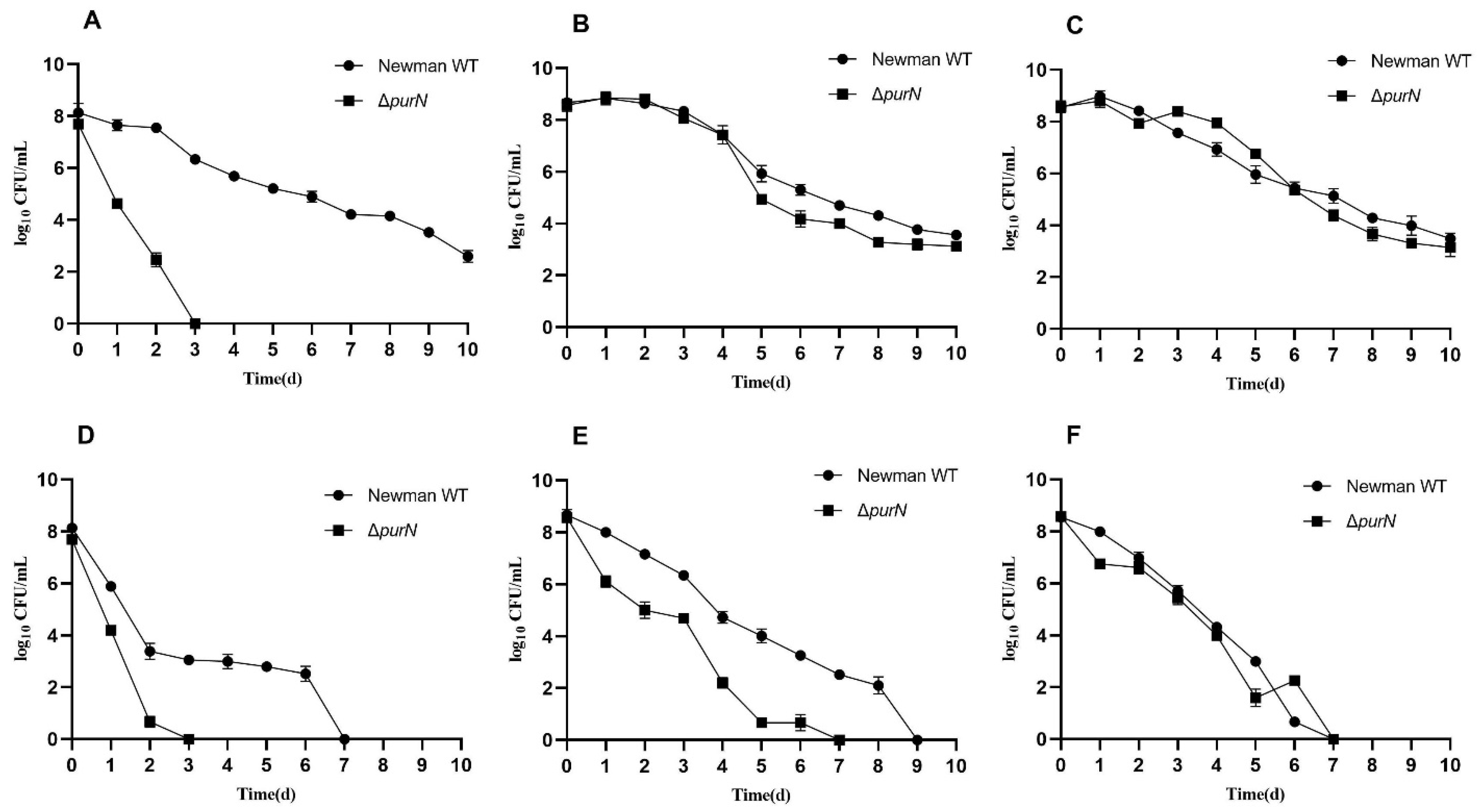
2.1. ΔpurN Had Significantly Decreased Antibiotic Tolerance

Based on our previous study, that PurB and PurM participated in purine metabolism and were involved in persister formation in S. aureus [27], we constructed a mutant strain of purN encoding phosphoribosylglycinamide formyltransferase in S. aureus Newman strain by homologous recombination to further explore the mechanisms by which purine metabolism regulates persister formation and virulence of S. aureus in this study.
In order to investigate the effect of the purN knockout on the formation of S. aureus persisters, antibiotic exposure tests at different culture time points were performed to determine the survival of the wild-type and ΔpurN. Compared to the parental strain, ΔpurN showed significantly increased susceptibility to ampicillin in 5-h cultures and was completely killed after 3 days of drug exposure, while the wild-type had approximately 106 CFU/ mL of viable cells remaining. Even on the 10th day of ampicillin treatment, the wild-type still had 102 CFU/mL of bacteria remaining (Figure 1A). There were no significant differences in the survival of the wild-type and ΔpurN strains upon ampicillin exposure when the bacteria were cultured for 9 and 18 h. Approximately 103 CFU/mL of bacteria remained after 10 days of drug exposure (Figure 1B,C).
Similar results were observed for levofloxacin exposure. Compared with the parental strain, ΔpurN showed increased sensitivity to levofloxacin when the bacteria were cultured for 5 and 9 h (Figure 1D,E). Among them, the most significant difference was observed in the 5-h cultures. After 3 days of levofloxacin exposure, ΔpurN exhibited no surviving bacteria, whereas more than 103 CFU/mL of bacteria remained for the parental strain. The persister level of S. aureus wild-type with levofloxacin exposure was similar to that of ΔpurN in 18-h cultures (Figure 1F).

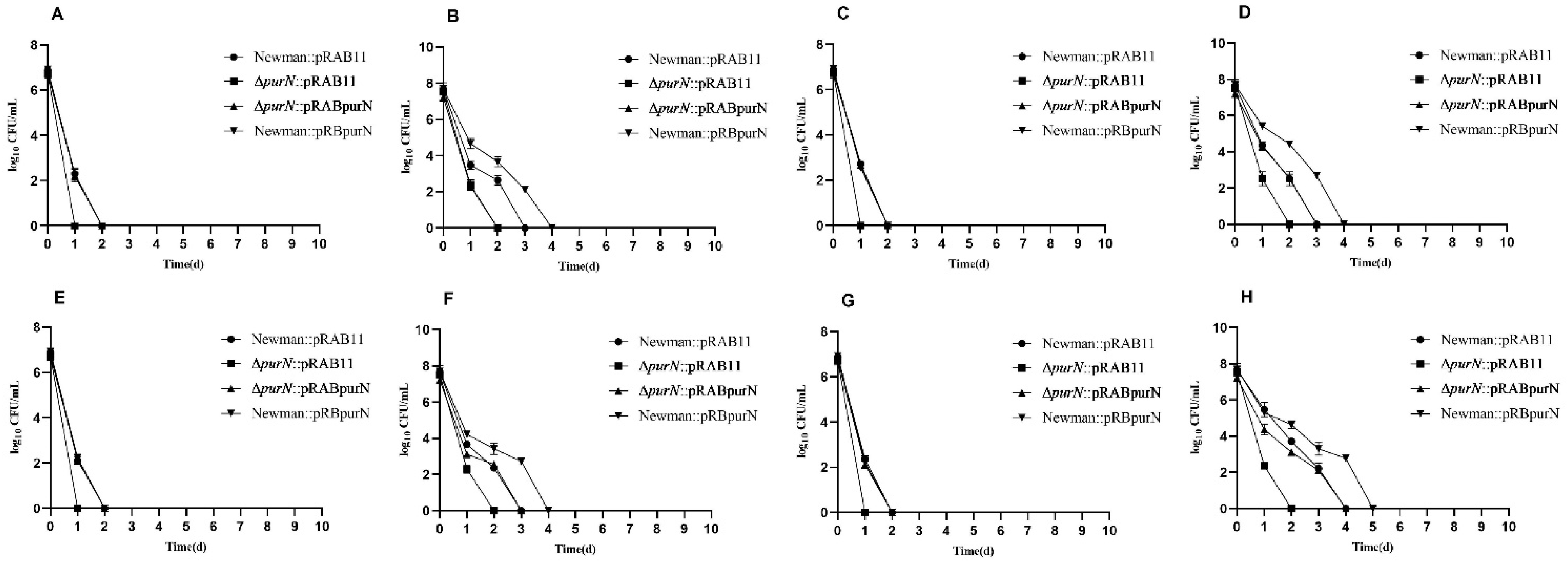
2.2. Complementation of the purN Restored Tolerance to Various Antibiotics

To further confirm the relationship between purN and S. aureus persister formation, the pRAB11 plasmid was used to complement ∆purN and the wild-type. Newman::pRAB11, ∆purN::pRAB11, ∆purN::pRABpurN, and Newman::pRBpurN were successfully obtained. The growth curves for these four strains indicated no differences in either the log phase or stationary phases under non-stressed conditions (Supplementary Figure S1). Compared with the Newman::pRAB11 strain, RT-qPCR confirmed that the expression levels of purN in the complemented ∆purN::pRABpurN strain (log2 fold change: 5.58 ± 0.16) and Newman::pRBpurN strain (log2 fold change: 6.48 ± 0.22) induced by anhydrotetracycline (Atc) were significantly higher than that of the wild-type with pRAB11 (p < 0.05).
An antibiotic exposure experiment was carried out for the constructed S. aureus strains. Due to the pRAB11 used in the complementation study being an Atc induced plasmid, all the complemented strains were cultured in TSB medium containing Atc (100 ng/mL) which can produce certain inhibition of S. aureus growth. The growth rates of each S. aureus complemented by pRAB11 or pRABpurN significantly decreased so that the numbers of live bacteria were still less than 108 CFU/mL after 9 h of culture, and they were still in the exponential phase. In 5-h culture, the antibiotics (e.g., ampicillin, vancomycin, gentamicin, and levofloxacin) exposure experiment demonstrated that ∆purN::pRAB11 all died after 24 h of drug treatment, while Newman::pRAB11, ∆purN::pRABpurN, and Newman::pRBpurN had more than 102 CFU/mL of bacteria remaining. After 48 h of antibiotic exposure, the Newman::pRAB11, ∆purN::pRABpurN, and Newman::pRBpurN strains had no viable bacteria (Figure 2A,C,E,G). Similar growth curves were observed in the 9-h cultures (Figure 2B,D,F,H). The purN complemented strain restored tolerance to antibiotics (e.g., vancomycin, gentamicin, and levofloxacin) except for ampicillin. However, for the 18-h cultures, except for the ∆purN::pRAB11, which had less than 103 CFU/mL of bacteria remaining after 10 days of levofloxacin exposure, the other strains showed significant tolerance to ampicillin, vancomycin, gentamicin, and levofloxacin, with many viable bacteria remaining after 10 days of antibiotic exposure (Supplementary Figure S2A–D).

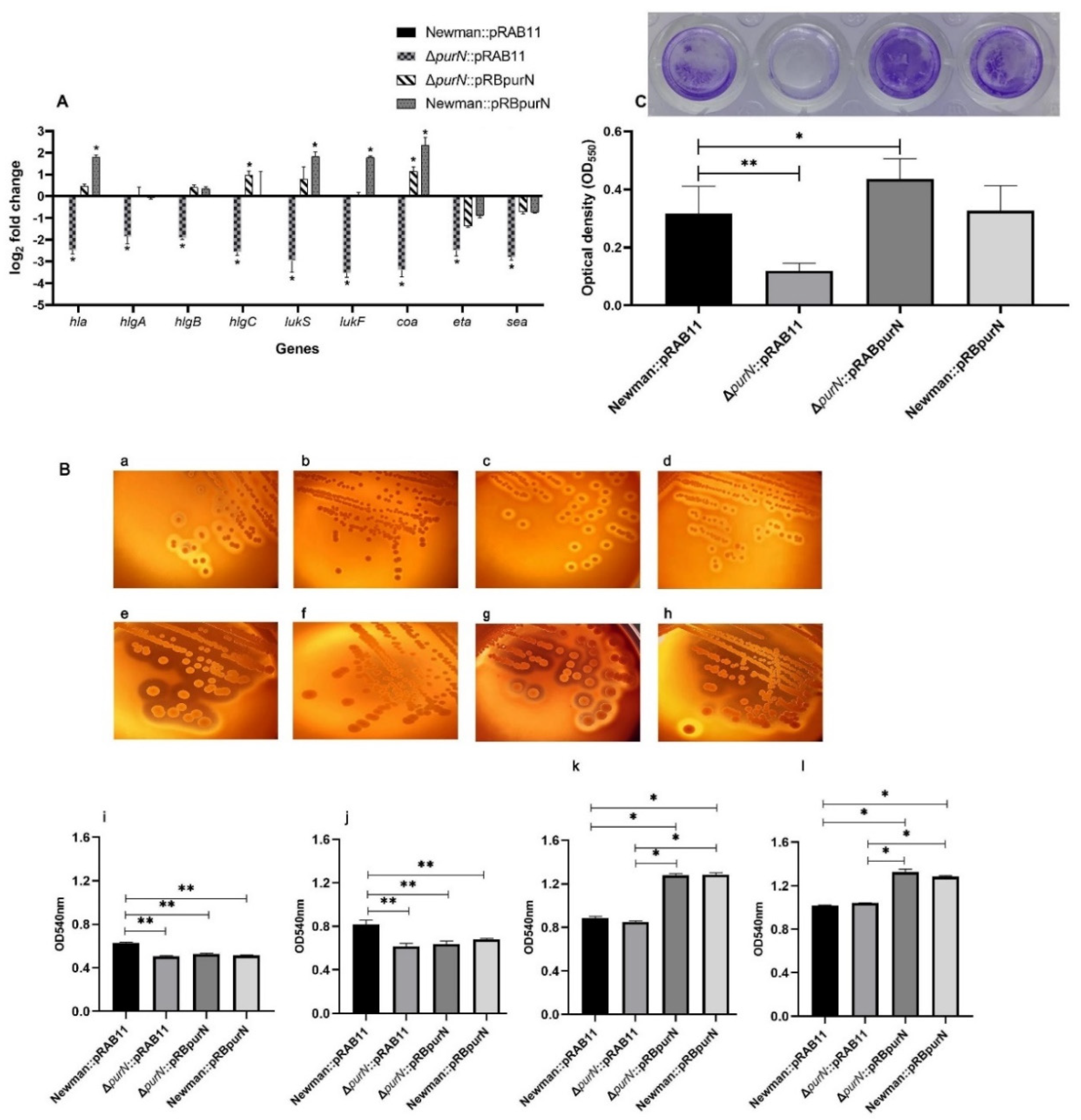
2.3. Knockout of purN Affected the Expression of Virulence Factors in S. aureus

To further investigate the effect of purN knockout on the expression of S. aureus virulence factors, RT-qPCR was used to compare the gene expression levels of the major virulence factors, including hla, hlgA, hlgB, hlgC, lukS, lukF, eta, sea, and coa, in the S. aureus Newman::pRAB11, ΔpurN::pRAB11, ΔpurN::pRBpurN, and Newman::pRBpurN strains. The expression levels of the major virulence genes in the ΔpurN::pRAB11 strain were significantly lower than those in the Newman::pRAB11 strain (p < 0.05). The complemented strain, ΔpurN::pRBpurN, exhibited restored expression levels of virulence genes. Moreover, the expression levels of hlgC and coa in ΔpurN::pRBpurN were significantly higher than those for Newman::pRAB11 (p < 0.05). In addition, the expression levels of hla, lukS, lukF, and coa in Newman::pRBpurN were significantly higher than those in Newman::pRAB11 (p < 0.05) (Figure 3A).

2.4. The Ability of the ∆purN to Lyse Sheep Erythrocytes Was Significantly Reduced

To investigate the effect of the purN mutation on the hemolysis characteristics of S. aureus, the Newman::pRAB11, ΔpurN::pRAB11, ΔpurN::pRBpurN, and Newman::pRBpurN strains were inoculated on sheep blood TSA plates containing Atc (100 ng/mL) at 37 °C for 10, 14 (images not shown), 24 and 48 h, respectively. The β-hemolytic rings around the colony of the Newman::pRAB11 colony (Figure 3(Ba,Be)) were larger and clearer than those of ΔpurN::pRAB11 (Figure 3(Bb,Bf)) at 24 and 48 h. In the 24- and 48-h cultures, the hemolytic rings of ΔpurN::pRBpurN (Figure 3(Bc,Bg)) and Newman::pRBpurN (Figure 3(Bd,Bh)) tended to be consistent with that of Newman::pRAB11. Hemolysis assays of each S. aureus culture indicated that at 10 and 14 h, the hemolyzing ability of Newman::pRAB11 cultures was significantly higher than that for ΔpurN::pRAB11 (p < 0.01, Figure 3(Bi,Bj)). With the prolongation of culture time and accumulation of hemolytic toxins, the differences in hemolytic ability between Newman::pRAB11 and ΔpurN::pRAB11 disappeared at 24 h and 48 h. However, when purN was overexpressed, compared with Newman::pRAB11 and ΔpurN::pRAB11, the hemolytic ability of the ΔpurN::pRBpurN and Newman::pRBpurN strains was enhanced (p < 0.05, Figure 3(Bk,Bl)).

2.5. Knockout of purN Affected Biofilm Formation in S. aureus

The biofilm formation abilities of Newman::pRAB11, ΔpurN::pRAB11, ∆purN::pRABpurN, and Newman::pRBpurN were measured in 96-well plates. The results showed that the biofilm formation ability of Newman::pRAB11 was significantly higher than that of ΔpurN::pRAB11 (p < 0.01, Figure 3C). After complementation, the biofilm formation ability of ΔpurN::pRBpurN was restored. Furthermore, when purN was overexpressed, the biofilm formation ability of ΔpurN::pRBpurN was significantly higher than that of Newman::pRAB11 (p < 0.05, Figure 3C). In addition, there were no significant differences in biofilm formation between Newman::pRAB11 and Newman::pRBpurN.

2.6. The LD50 Values of ΔpurN in Mice Were Significantly Higher Than That of Wild-Type S. aureus

To further explore the effect of the purN mutation on the virulence of S. aureus, we determined the LD50 of the S. aureus Newman strain and the ΔpurN in BALB/c mice. Different doses of the wild-type and ΔpurN bacterial suspensions were injected intraperitoneally. The LD50 values for the wild-type and ΔpurN in BALB/c mice were calculated according to the survival status of the mice, and the results showed that the LD50 of the ΔpurN mutant (3.28 × 10¹⁰ CFU/mL) was significantly higher than that of the wild-type (2.81 × 10⁹ CFU/mL).

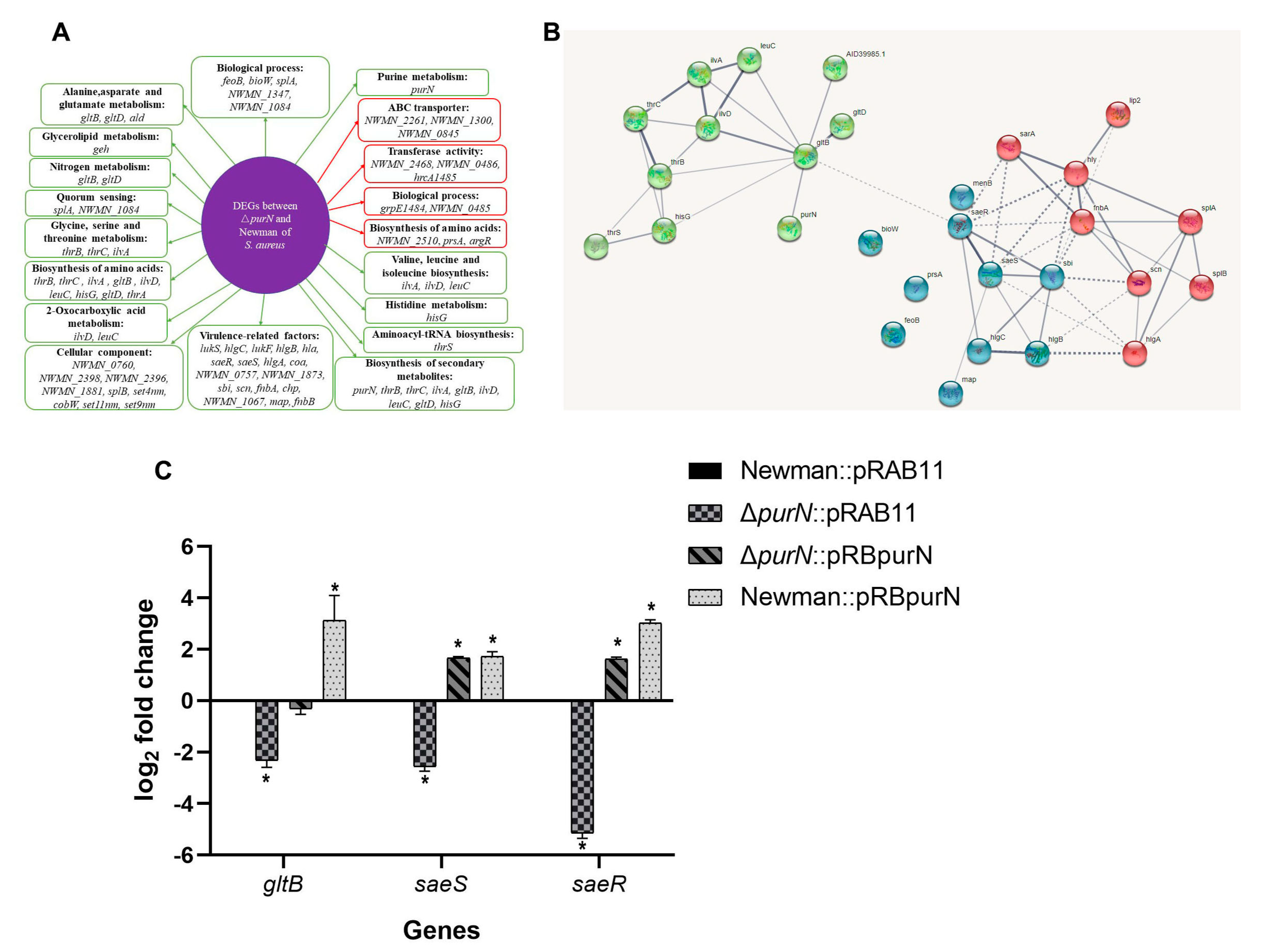
2.7. Comparative Transcriptome Analysis of the ΔpurN and the Wild-Type

To gain further insights into the molecular mechanisms by which PurN affects persister formation and virulence in S. aureus, the DEGs of the ΔpurN mutant and the wild-type strain were profiled by RNA-seq. Compared with its parental strain, 58 genes were downregulated, and 24 genes were upregulated in the ΔpurN mutant with a cutoff value of log2 fold change less than −2 or more than 2 (Supplementary Table S1). Thirteen DEGs were selected as target genes (e.g., saeS, saeR, ilvA, NWMN_1873, lukS, hla, hlgC, lukF, NWMN_2510, NWMN_2262, NWMN_2266, NWMN_0485, and NWMN_0845) for validation by RT-qPCR and the results confirmed the reliability of the transcriptome analysis (Supplementary Table S2). The DEGs were assigned to the following functional categories. KEGG pathway enrichment analysis suggested that these DEGs were mainly involved in purine metabolism, alanine, aspartate, and glutamate metabolism, 2-oxocarboxylic acid metabolism, histidine metabolism, biosynthesis of amino acids, ABC transporters, quorum sensing, etc. (Figure 4A). To evaluate the DEG associations, a PPI was constructed based on the STRING database, and the network showed that there were close relationships between purN and gltB and saeR and saeS (Figure 4B). Furthermore, compared with the wild-type, the transcription levels of virulence-related genes, including lukS, lukF, hlgA, hlgB, hlgC, and hla, were downregulated significantly in the ΔpurN mutant (Supplementary Table S1).
To further explore the relationships between purN and gltB and saeR and saeS, RT-qPCR was used to detect the gltB, saeR, and saeS expression in the Newman::pRAB11, ΔpurN::pRAB11, ΔpurN::pRBpurN, and Newman::pRBpurN strains. Compared with Newman::pRAB11, the expression level of gltB in ΔpurN::pRAB11 was significantly lower (p < 0.05), whereas in Newman::pRBpurN, it was higher (p < 0.05), and there was no significant difference in ΔpurN::pRBpurN. Meanwhile, compared with Newman::pRAB11, the expression levels of saeR and saeS in ΔpurN::pRAB11 were significantly lower (p < 0.05), whereas in purN overexpressed strains, ΔpurN::pRBpurN and Newman::pRBpurN were significantly higher (p < 0.05) (Figure 4C). purN affected the expression of gltB, saeR, and saeS in S. aureus and was consistent with the PPI network (Figure 4B).

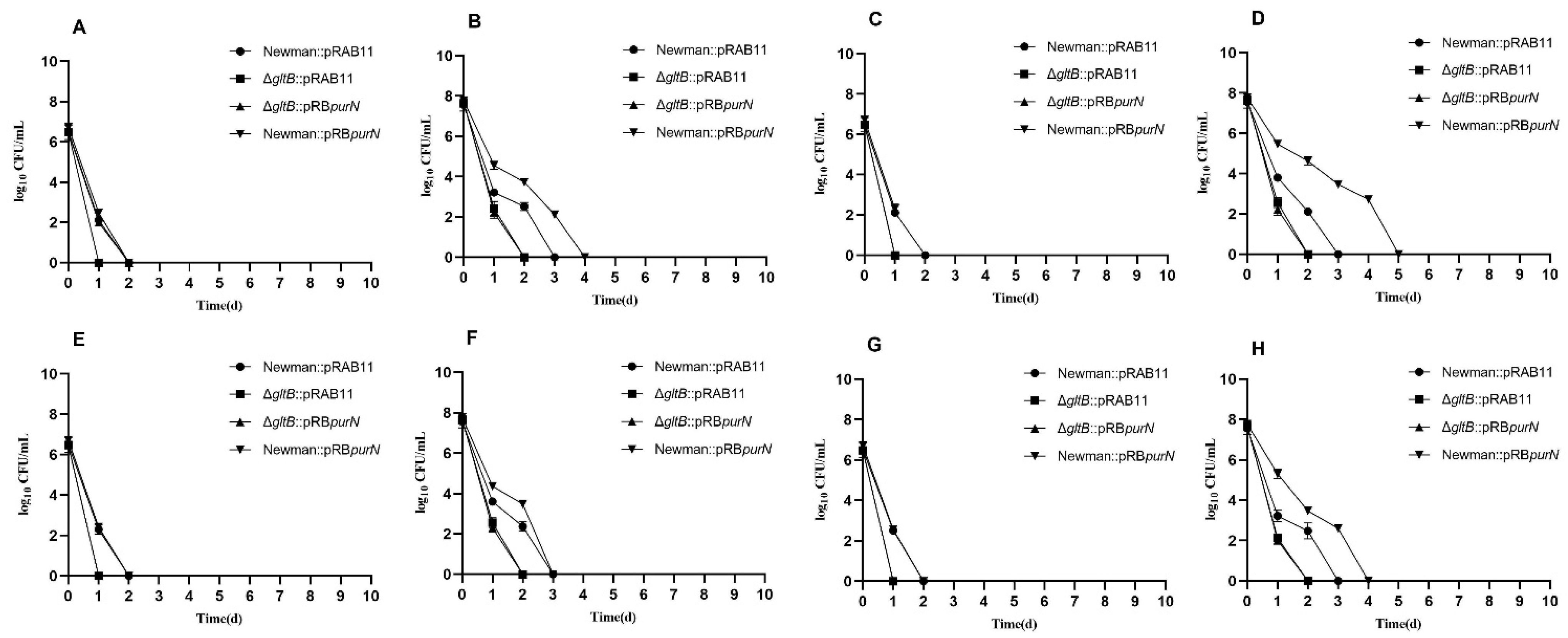
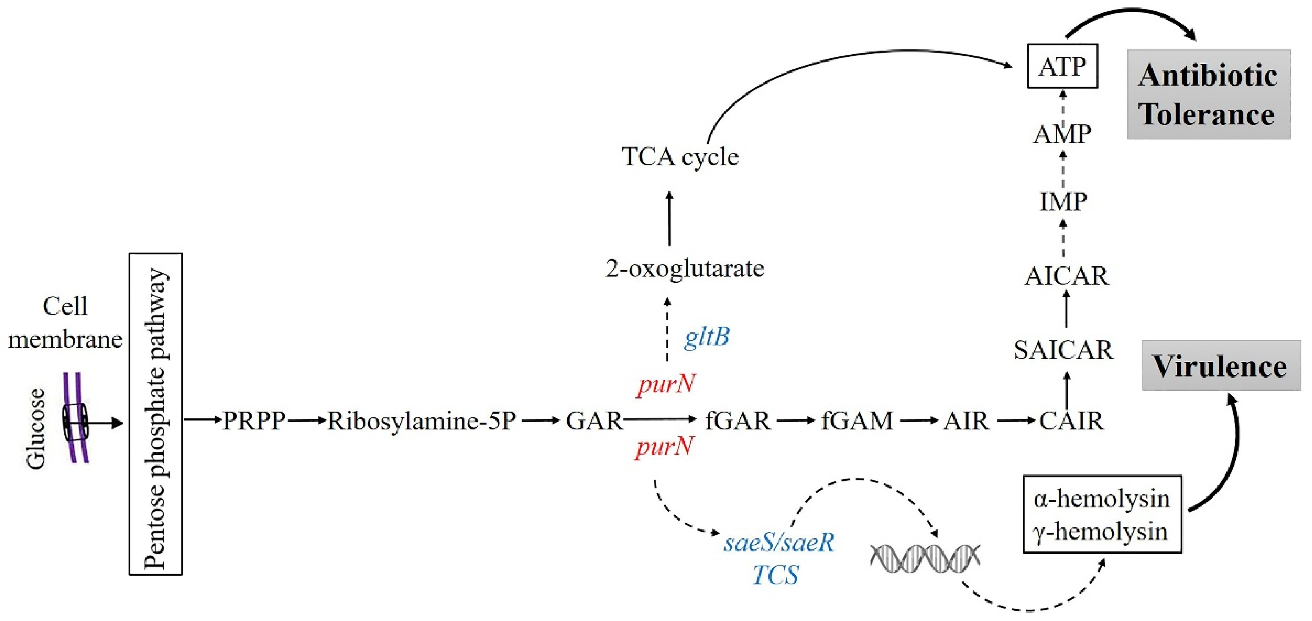
2.8. purN Affects the Persister Formation in S. aureus via gltB

To verify the PPI network based on the transcriptome analysis of the ΔpurN mutant and the wild-type, ΔgltB::pRAB11 and ΔgltB::pRBpurN were constructed. Further experiments showed that ΔgltB::pRAB11, ΔgltB::pRBpurN, Newman::pRBpurN, and ∆purN::pRABpurN had similar growth curves (Supplementary Figure S1). RT-qPCR confirmed that the expression level of purN in the ∆gltB::pRABpurN strain (log2fold change: 4.99 ± 0.016) was significantly higher than that in ΔgltB::pRAB11 (p < 0.05). To further explore the association between purN and gltB in the formation of S. aureus persisters, four strains, Newman::pRAB11, ΔgltB::pRAB11, ΔgltB::pRBpurN, and Newman::pRBpurN, were incubated for 5, 9, and 18 h, respectively. Each strain was exposed to lethal concentrations of antibiotics, including ampicillin (10 μg/mL), levofloxacin (20 μg/mL), vancomycin (40 μg/mL), and gentamicin (100 μg/mL), to observe the differences in persister formation ability. The results showed that the four strains in 5-h cultures were completely killed after 1–2 days of drug exposure (Figure 5A,C,E,G). However, after 9 h of incubation, the changing characteristics of the viable in ΔgltB::pRBpurN and ΔgltB::pRAB11 strains were similar and were completely killed after 2 days of antibiotic exposure, while the Newman::pRAB11 and Newman::pRBpurN strains retained more than 102 CFU/mL of viable bacteria. The Newman::pRAB11 was killed after 3 days of drug exposure, while Newman::pRBpurN was completely killed after approximately 4–5 days of antibiotic exposure (Figure 5B,D,F,H). In the 18-h cultures, the numbers of viable bacteria in ΔgltB::pRAB11 and ΔgltB::pRBpurN were less than those of Newman::pRAB11 and Newman::pRBpurN after 10 days of drug exposure (Supplementary Figure S3A–D). The results showed that when gltB was knocked out, overexpression of purN did not increase persister formation, indicating that purN affects persister formation in S. aureus via gltB.

3. Discussion

Persister formation in S. aureus is closely related to the growth phase of culture [10,12,35,36]. Previous studies have shown that purine biosynthesis plays an important role in persister formation in S. aureus [27]. purN is a crucial gene in the third step of purine biosynthesis. We analyzed the effect of the purN mutant of S. aureus and found that its mutation resulted in persister reduction in late exponential phase cultures, indicating PurN is important for persister formation.
purN participates in several important pathways, including purine metabolism, one carbon pool by folate, metabolic pathways, and biosynthesis of secondary metabolites. A large number of studies have confirmed that ATP production [16,41], alarmone ppGpp [10], amino acid synthesis, and metabolism in bacterial cells play important roles in the formation and regulation of persisters in bacteria [26,42,43].
It is well known that purine metabolism is crucial for ATP energy supply. PurN catalyzes GAR to fGAR, which is an important step in the purine metabolism process to produce IMP. IMP is converted to guanosine 5′-monophosphate (GMP) or adenosine 5′-monophosphate (AMP) by subsequent enzymes. In this process, both ribosylamine-5P produced by phosphoribosyl pyrophosphate (PRPP) and formylglycinamidine ribonucleotide (fGAM) produced by fGAR require glutamine to provide amido, and glutamate is also produced. At the same time, glycine is required to participate in the process of ribosylamine-5P to generate GAR, and aspartate is required to participate in the process of 5-amino-4-carboxyaminoimidazole ribonucleotide (CAIR) to generate N-succinylo-5-aminoimidazole-4-carboxamide ribonucleotide (SAICAR) [40] (Figure 6). The PPI network established by our data indicated that purN affected the persister formation in S. aureus via gltB (Figure 4B). gltB, encoding the large subunit of glutamate synthase, is the key gene in glutamine and glutamate metabolism, which catalyzes L-glutamine and 2-oxoglutarate into two molecules of L-glutamate [44]. Transcriptome analysis found that gltB expression decreased in the ΔpurN mutant (Figure 4A). Thus, glutamine and glutamate synthesis were reduced. The decrease of gltB resulted in an increase of 2-oxoglutartate, which has been shown to promote the TCA cycle and cause increased ATP production [45,46], which in turn would inhibit persister formation of the ΔpurN mutant (Figure 6).
Biofilm formation, a major virulence factor in S. aureus infections, accelerates bacterial colonization in host tissues and promotes persister formation and antimicrobial agents. Our data revealed that the purN mutant significantly decreased biofilm formation. In other previous studies, purine biosynthesis was shown to affect biofilm formation through the secondary messenger, cyclic di-AMP (c-di-AMP) [28,47]. PurN is involved in the third step of purine biosynthesis, which affects ATP production. c-di-AMP is synthesized by di-adenylate cyclase via the condensation of two ATPs, one of the final products of purine biosynthesis [48]. The ΔpurN mutant may inhibit c-di-AMP synthesis from preventing bacterial biofilm formation in S. aureus. However, the underlying mechanisms deserve future detailed studies.
S. aureus has a complex regulatory network to control its virulence [49]. The regulatory systems include the accessory gene regulator (agr) quorum-sensing system [50], SarA protein family regulators [51], two-component system (TCS) of the SaeRS [52], SrrAB [53], ArlRS [54], and the alternative sigma factors (SigB and SigH) [51]. Transcriptome analyses of ΔpurN and wild-type strains indicate that the expression levels of saeR and saeS encoding the SaeRS TCS were significantly decreased in the ΔpurN, and due to this, the expression levels of multiple virulence factors, including α-hemolysin, γ-hemolysin, PVL, and coagulase, were also significantly reduced. This is consistent with our mouse study, in which we found that the virulence of ΔpurN was significantly reduced, as well as the results of the hemolysis assay (Figure 3B). The SaeRS TCS is an important regulatory system for the virulence of S. aureus [52]. SaeS is the sensor histidine kinase, which can sense signals in the environment and autophosphorylate at the His131 residue and then the phosphoryl group is transferred to Asp51 of SaeR, and the phosphorylated SaeR (SaeR-P) binds to the SaeR binding sequence (SBS) to activate the transcription of the target genes [52,55,56]. Several Sae target genes have been discovered, most of which are related to the virulence of S. aureus, including coa, fnbA, eap, sbi, efb, fib, saeP, hla, hlb, and hlgC [57,58]. The currently reported signals of SaeRS TCS activation mainly include human neutrophil peptide 1, 2, and 3 (HNP1–3), calprotectin, hydrogen peroxide, etc. [59,60]. Our data showed that the expression levels of saeR and saeS were higher in the purN overexpressed strains (Figure 4C). The results confirmed the PPI networks (Figure 4B), which PurN may affect virulence through the SaeRS in S. aureus (Figure 6).
Our findings further suggest that there is a close relationship between persister formation and bacterial virulence. In addition to the reported multiple persistence-related genes, such as argJ, lysR, phoU, and msaABCR, which are involved in bacterial virulence [26,33,38,39], the PurN of S. aureus is another multifunctional factor that not only participates in persister formation but also participates in virulence regulation.
In summary, this study has demonstrated that PurN participates in the formation of the late exponential phase S. aureus persister formation via the key gene, gltB, in glutamate synthesis and regulates bacterial virulence by activating the SaeRS two-component system. Therefore, PurN can potentially serve as a novel therapeutic target to develop more effective treatments to control persistent S. aureus infections in the future.

4. Materials and Methods

4.1. Culture Media, Antibiotics, and Animals

Tryptic soy broth (TSB) and tryptic soy agar (TSA) were obtained from Becton Dickinson (BD). Luria-Bertani (LB) medium and anhydrotetracycline (Atc) were obtained from Solarbio (Beijing, China). The rationale for selecting the antibiotics used in antibiotics exposure experiments is based on clinically used antibiotics in treating S. aureus infections and three classes of bactericidal antibiotics commonly used for persister assays, i.e., cell wall inhibitors, aminoglycosides, and fluoroquinolones. Ampicillin, levofloxacin, rifampin, chloramphenicol, vancomycin, and gentamicin were obtained from Sangon Biotech (Shanghai, China), and their stock solutions were freshly prepared, filter-sterilized, and used at appropriate concentrations as indicated. BALB/c mice were purchased from Lanzhou University (China). The study was approved by the Ethics Committee of Lanzhou University.

4.2. Bacterial Strains and Culture Conditions

The bacterial strains and plasmids used in this study are listed in Table 1. All the S. aureus strains were cultivated in TSA and TSB. The E. coli DC10B strain was cultivated in LB. The shuttle vector, pRAB11, harbors a tet operator that is induced by Atc. In the process of inducing high expression of purN, S. aureuspurN::pRBpurN, Newman::pRABpurN, and ∆gltB::pRBpurN mutants, and the control strains, S. aureuspurN::pRB11, Newman::pRAB11, and ∆gltB::pRAB11 were all inoculated in TSB medium containing Atc (100 ng/mL). For the persister assays, antibiotics were used at the following concentrations: ampicillin, 10 μg/mL; levofloxacin, 20 μg/mL; vancomycin, 40 μg/mL; and gentamicin, 100 μg/mL.

4.3. Susceptibility of Mutants to Antibiotics

In order to assess the effects of purN knockout on persister formation, overnight cultures of the relevant S. aureus were diluted 1:1000 with TSB in bacterial culture tubes and cultured at 37 °C with shaking (180 rpm). At 5, 9, and 18 h of incubation, cultures were collected, and ampicillin (10 μg/mL), levofloxacin (20 μg/mL), vancomycin (40 μg/mL), and gentamicin (100 μg/mL) were added to assess persister survival. The cultures exposed to drugs were incubated without shaking at 37 °C for up to ten days. Aliquots of cultures exposed to antibiotics were taken at different time points and washed in TSB, and the number of viable cells was counted after serial dilutions.

4.4. Construction of Gene Knockout and Overexpression Strains

To construct purN knockout mutants of S. aureus, we followed the method described previously [15]. The plasmid, pMX10, was used for gene knockout in S. aureus. Q5 Master Mix PCR (New England BioLabs) was used for all PCR experiments, and restriction enzymes and T4 DNA Ligase (Thermo Fisher Scientific, Waltham, MA, USA) were used to construct the recombinant plasmids used in this study. The Primers used for purN of S. aureus gene knockout included purN-uf, purN-ur, purN-df, and purN-dr, and the primer sequences are listed in Supplementary Table S3.
To construct knockout mutants, upstream and downstream fragments of each gene were amplified with the corresponding primers using the genomic DNA of the S. aureus wild-type strain Newman as a template. Two fragments of each gene were then used as templates to amplify a fusion fragment with appropriate primers. The fusion fragment and pMX10 plasmid were digested with Kpn I and Mlu I, respectively, and ligated with T4 DNA ligase, and the recombinant plasmids were transformed into E. coli DC10B competent cells. The transformed DC10B was screened on LB agar plates containing ampicillin (100 μg/mL), and the positive clones were verified by restriction digestion and DNA sequencing. The recombinant plasmid was electrotransformed into the S. aureus Newman strain, as we described previously [32]. Mutants selection was carried out following the previously published protocol [61]. Using the same method, we also obtained gltB knockout mutants of S. aureus.
The pRAB11 plasmid was used for inducible overexpression of purN in S. aureus. The full sequence of purN of wild-type S. aureus Newman was amplified with the primers OEpurN-f and OEpurN-r (Supplementary Table S3). After digestion with KpnI and EcoRI, the fragment was inserted into pRAB11. The recombinant plasmid pRAB11-purN was transformed into E. coli DC10B competent cells. The recombinant plasmid pRAB11-purN, was verified by DNA sequencing and then electrotransformed into ΔpurN and ΔgltB mutants and Newman wild-type to obtain ∆purN::pRABpurN, ΔgltB::pRBpurN and Newman::pRBpurN, while the empty pRAB11 was transformed into ∆purN, ΔgltB mutants and Newman wild-type and ∆purN::pRAB11, ΔgltB::pRAB11 and Newman::pRAB11 were obtained.

4.5. RT-qPCR Detected Genes Expression

After the cultures of S. aureus were treated with lysostaphin (Shanghai Hi-tech Bioengineering Co., Ltd., Shanghai, China), total RNA was extracted using the Sangon RNeasy kit (Sangon Biotech, China), and the quality and concentration of the extracted RNA were analyzed using a NanoDrop spectrophotometer (Thermo Fisher Scientific, Wilmington, DE, USA). Reverse transcription was performed with SuperScript III First-Strand synthesis (Takara Bio, Japanese) using 1 μg of total RNA that was isolated according to the manufacturer’s instructions. RT-qPCR was performed using SYBR Green Supermix (Yeasen Biotech, Shanghai, China), and the relative fold changes in gene expression were calculated using 16S rRNA as an endogenous control gene [62]. The data represent the results from three independent experiments. The primers for each gene were designed using Primer Premier 5.0 software (PREMIER Biosoft International, San Francisco, USA), and the primer sequences are listed in Supplementary Table S3. All data were analyzed with GraphPad Prism 8.0 (GraphPad Software, San Diego, CA, USA) and compared using the independent-samples t-test. Differences with p-value < 0.05 were considered statistically significant.

4.6. Hemolysis Assay

S. aureus Newman::pRAB11, ∆purN::pRAB11, ∆purN::pRABpurN, and Newman::pRBpurN were inoculated on TSA plates containing 10% sheep blood and Atc (100 ng/mL), incubated at 37 °C for 10, 14, 24 and 48 h, and the hemolysis that formed around the colonies were observed. The hemolysis analysis was conducted as described previously [63] with some modifications. Briefly, Newman::pRAB11, ∆purN::pRAB11, ∆purN::pRABpurN, and Newman::pRBpurN were cultured in TSB medium with chloramphenicol (10 µg/mL) for 18 h at 180 rpm, diluted 1:1000 and cultured in 5 mL of TSB containing Atc (100 ng/mL) for 10, 14, 24 and 48 h. Each culture was centrifuged at 9000× g for 3 min. Then, 200 μL of supernatant was mixed with an equal volume of 4% (v/v) sheep red blood cells suspended in PBS buffer and incubated at 37 °C for 1.5 h with shaking at 180 rpm. Supernatants were collected after centrifugation (12,000× g for 1 min), and the optical density at 540 nm was measured with a spectrophotometer. All experiments were performed in triplicate.

4.7. Establishment of an In Vitro S. aureus Biofilm Model

The ability of the S. aureus strains to form biofilm was tested in a 96-well plate according to a previously published method [64]. Two hundred microliters of TSB with 0.25% glucose and Atc (100 ng/mL) were transferred to each of the wells on the microtiter plate. Two microliters of each overnight culture of S. aureus were transferred to the wells, except for the blank control. Each S. aureus strain was tested in three parallel wells. The 96-well plate was incubated at 37 °C for 24 h. The wells were then washed three times with 200 µL of PBS and left to dry at 60 °C for 60 min. Then, 200 µL of crystal violet (0.5% solution, Sigma Aldrich, St. Louis, MO, USA) was added and incubated at room temperature for 30 min. The wells were washed five times with 200 µL of tap water. In order to extract the crystal violet from the biofilm, 200 µL of 33% glacial acetic acid was added. The optical density of the solutions at 550 nm was measured.

4.8. Median Lethal Dose Determination

Seventy-five female BALB/c mice weighing approximately 18–22 g were randomly divided into 15 groups to measure the median lethal dose (LD50) of the S. aureus Newman wild-type strain and the ∆purN mutant. The overnight S. aureus Newman wild-type and the ΔpurN were diluted 1:100 in 100 mL TSB and shaken overnight at 37 °C. The cultures were centrifuged at 12,000 rpm for 3 min, and the pellets were washed twice with sterile PBS. After the removal of the supernatant, the pellets were resuspended in 10 mL PBS, and the viable bacteria in the suspension were counted by serial dilution. Then, the suspensions were diluted to form 7 concentration gradients using a double dilution method. Each mouse in each group was injected intraperitoneally with 0.6 mL of a bacterial suspension at doses ranging from 108–1010 CFU/mL. After 5 days of observation, the LD50 value of each strain was calculated by the Reed-Muench method [65].

4.9. Transcriptome Analysis

To identify the key genes regulating the differential responses between the parental Newman strain and ΔpurN mutant, triplicate samples cultured for 5 h in TSB after dilution of 1:1000 were collected and subjected to high-throughput mRNA transcriptome sequencing. Total RNA was extracted as mentioned above. Sequencing libraries were generated according to the manufacturer’s protocol (NEBNext®UltraTM RNA Library Prep Kit for Illumina®) [66]. Cleaved RNA fragments were copied into first-strand cDNA using reverse transcriptase and random primers. The cDNA library preferentially selected fragments of 200–250 bp in length, which were prepared by the AMPure XP system. Then, the fragment products were amplified by Illumina cBot and sequenced on an Illumina HiSeq 2500 system (Illumina, San Diego, CA USA). Library construction and sequencing were performed at the Shanghai Applied Protein Technology Co., Ltd. By using Hisat2 (v2.0.5) (https://daehwankimlab.github.io/hisat2/manual/ (accessed on 19 August 2020)), paired-end clean reads were aligned to the reference genome of S. aureus Newman on the NCBI website. The number of reads corresponding to each gene was calculated using Feature Counts v1.5.0-p3. Then, each gene fragment per kilobase million (FPKM) was calculated based on the gene lengths and read counts mapped to this gene. In order to control the false discovery rate, Benjamini and Hochberg’s approach was used to adjust the p-values to compare FPKM values between the mutant and wild-type groups. Genes with Padj < 0.05 and log2 fold change >2 or <−2 were defined as differentially expressed genes (DEGs). RT-qPCR, which was performed in triplicate, was used to confirm the RNA expression levels, and the primer sequences are listed in Supplementary Table S2.

4.10. Protein-Protein Interaction Network

In order to explore the interactive relationships among DEGs, the web portal for the STRING database (http://www.string-db.org/ (accessed on 1 April 2021)) was used for protein–protein interaction (PPI) network analysis. The following two criteria were applied to detect the important nodes: (1) medium confidence equal to 0.4 and (2) network clustering by K-means clustering.

Supplementary Materials

The following supporting information can be downloaded at: https://www.mdpi.com/article/10.3390/antibiotics11121702/s1, Table S1: DEGs between ΔpurN and its parental strain (log2 fold change greater than 2 or less than −2); Table S2: Oligonucleotide sequences of RT-qPCR primers used in this study. RT-qPCR verified DEGs between ΔpurN and its parental strain from transcriptome analysis. Results were normalized using 16S rRNA and expressed as fold change (mean ± SD, p < 0.05); Table S3: Primers and oligonucleotides used in this study; Figure S1: The growth curves for S. aureus Newman::pRAB11, ∆purN::pRAB11, ∆purN::pRABpurN, ∆gltB::pRAB11, ∆gltB::pRABpurN and Newman::pRABpurN strains; Figure S2: Drug exposure results of 18-h culture of Newman::pRAB11, ∆purN::pRAB11, ∆purN::pRABpurN, and Newman::pRBpurN to ampicillin (A), vancomycin (B), gentamicin (C) and levofloxacin (D); Figure S3: Drug exposure results of 18-h culture of Newman::pRAB11, ∆gltB::pRAB11, ∆gltB::pRABpurN, and Newman::pRBpurN to ampicillin (A), levofloxacin (B), gentamicin (C) and vancomycin (D).

Author Contributions

Conceptualization, J.H. and Y.Z.; methodology, Q.P., L.G., Y.D., T.B., H.W. and T.X.; formal analysis, Q.P., L.G. and Y.D.; writing—original draft preparation, L.G.; writing—review & editing, J.H. and Y.Z.; supervision, J.H. and Y.Z.; funding Acquisition, L.G., J.H., and Y.Z. All authors have read and agreed to the published version of the manuscript.

Funding

This research was supported by the National Natural Science Foundation of China (Grant Numbers: 81902098, 81571952, 81772231).

Institutional Review Board Statement

The study was conducted in accordance with the Declaration of Helsinki and approved by the Ethics Committee of Lanzhou University, China (protocol code jcyxy20190209b and 25 February 2019 approval).

Informed Consent Statement

Not applicable.

Data Availability Statement

Not applicable.

Conflicts of Interest

The authors declare no conflict of interest.

References

  1. Dayan, G.H.; Mohamed, N.; Scully, I.L.; Cooper, D.; Begier, E.; Eiden, J.; Jansen, K.U.; Gurtman, A.; Anderson, A.S. Staphylococcus aureus: The current state of disease, pathophysiology and strategies for prevention. Expert. Rev. Vaccines 2016, 15, 1373–1392. [Google Scholar] [CrossRef] [PubMed]
  2. Vandenesch, F.; Lina, G.; Henry, T. Staphylococcus aureus hemolysins, bi-component leukocidins, and cytolytic peptides: A redundant arsenal of membrane-damaging virulence factors? Front. Cell. Infect. Microbiol. 2012, 2, 12. [Google Scholar] [CrossRef] [PubMed] [Green Version]
  3. Gordon, C.P.; Williams, P.; Chan, W.C. Attenuating Staphylococcus aureus virulence gene regulation: A medicinal chemistry perspective. J. Med. Chem. 2013, 56, 1389–1404. [Google Scholar] [CrossRef] [PubMed]
  4. Bronner, S.; Monteil, H.; Prevost, G. Regulation of virulence determinants in Staphylococcus aureus: Complexity and applications. FEMS Microbiol. Rev. 2004, 28, 183–200. [Google Scholar] [CrossRef] [PubMed] [Green Version]
  5. Cheung, A.L.; Bayer, A.S.; Zhang, G.; Gresham, H.; Xiong, Y.Q. Regulation of virulence determinants in vitro and in vivo in Staphylococcus aureus. FEMS Immunol. Med. Microbiol. 2004, 40, 1–9. [Google Scholar] [CrossRef] [PubMed] [Green Version]
  6. Murray, P.R.; Rosenthal, K.S.; Pfaller, M.A. Medical Microbiology, 8th ed.; ELSEVIER Inc.: Philadelphia, PA, USA, 2016; pp. 173–175. [Google Scholar]
  7. Conlon, B.P. Staphylococcus aureus chronic and relapsing infections: Evidence of a role for persister cells: An investigation of persister cells, their formation and their role in S. aureus disease. Bioessays 2014, 36, 991–996. [Google Scholar] [CrossRef]
  8. Lister, J.L.; Horswill, A.R. Staphylococcus aureus biofilms: Recent developments in biofilm dispersal. Front. Cell. Infect. Microbiol. 2014, 4, 178. [Google Scholar] [CrossRef] [Green Version]
  9. Fisher, R.A.; Gollan, B.; Helaine, S. Persistent bacterial infections and persister cells. Nat. Rev. Microbiol. 2017, 15, 453–464. [Google Scholar] [CrossRef]
  10. Harms, A.; Maisonneuve, E.; Gerdes, K. Mechanisms of bacterial persistence during stress and antibiotic exposure. Science 2016, 354, aaf4268. [Google Scholar] [CrossRef]
  11. Defraine, V.; Fauvart, M.; Michiels, J. Fighting bacterial persistence: Current and emerging anti-persister strategies and therapeutics. Drug Resist. Updat. 2018, 38, 12–26. [Google Scholar] [CrossRef]
  12. Zhang, Y. Persisters, persistent infections and the Yin-Yang model. Emerg. Microbes. Infect. 2014, 3, e3. [Google Scholar] [CrossRef] [PubMed]
  13. Feng, J.; Li, T.; Yee, R.; Yuan, Y.; Bai, C.; Cai, M.; Shi, W.; Embers, M.; Brayton, C.; Saeki, H.; et al. Stationary phase persister/biofilm microcolony of Borrelia burgdorferi causes more severe disease in a mouse model of Lyme arthritis: Implications for understanding persistence, Post-treatment Lyme Disease Syndrome (PTLDS), and treatment failure. Discov. Med. 2019, 27, 125–138. [Google Scholar] [PubMed]
  14. Shi, W.; Zhang, X.; Jiang, X.; Yuan, H.; Lee, J.S.; Barry, C.E., 3rd; Wang, H.; Zhang, W.; Zhang, Y. Pyrazinamide inhibits trans-translation in Mycobacterium tuberculosis. Science. 2011, 333, 1630–1632. [Google Scholar] [CrossRef] [PubMed] [Green Version]
  15. Xu, T.; Wang, X.Y.; Cui, P.; Zhang, Y.M.; Zhang, W.H.; Zhang, Y. The Agr Quorum Sensing System Represses Persister Formation through Regulation of Phenol Soluble Modulins in Staphylococcus aureus. Front. Microbiol. 2017, 8, 2189. [Google Scholar] [CrossRef]
  16. Shan, Y.; Brown Gandt, A.; Rowe, S.E.; Deisinger, J.P.; Conlon, B.P.; Lewis, K. ATP-Dependent Persister Formation in Escherichia coli. mBio 2017, 8, e02267-16. [Google Scholar] [CrossRef] [Green Version]
  17. Hazan, R.; Maura, D.; Que, Y.A.; Rahme, L.G. Assessing Pseudomonas aeruginosa Persister/antibiotic tolerant cells. Methods Mol. Biol. 2014, 1149, 699–707. [Google Scholar] [CrossRef]
  18. Stapels, D.A.C.; Hill, P.W.S.; Westermann, A.J.; Fisher, R.A.; Thurston, T.L.; Saliba, A.E.; Blommestein, I.; Vogel, J.; Helaine, S. Salmonella persisters undermine host immune defenses during antibiotic treatment. Science 2018, 362, 1156–1160. [Google Scholar] [CrossRef] [Green Version]
  19. Lewis, K. Persister cells. Annu. Rev. Microbiol. 2010, 64, 357–372. [Google Scholar] [CrossRef]
  20. Moyed, H.S.; Bertrand, K.P. hipA, a newly recognized gene of Escherichia coli K-12 that affects frequency of persistence after inhibition of murein synthesis. J. Bacteriol. 1983, 155, 768–775. [Google Scholar] [CrossRef] [Green Version]
  21. Ma, C.; Sim, S.; Shi, W.; Du, L.; Xing, D.; Zhang, Y. Energy production genes sucB and ubiF are involved in persister survival and tolerance to multiple antibiotics and stresses in Escherichia coli. FEMS Microbiol. Lett. 2010, 303, 33–40. [Google Scholar] [CrossRef]
  22. Li, J.; Ji, L.; Shi, W.; Xie, J.; Zhang, Y. Trans-translation mediates tolerance to multiple antibiotics and stresses in Escherichia coli. J. Antimicrob. Chemother. 2013, 68, 2477–2481. [Google Scholar] [CrossRef] [PubMed] [Green Version]
  23. Korch, S.B.; Henderson, T.A.; Hill, T.M. Characterization of the hipA7 allele of Escherichia coli and evidence that high persistence is governed by (p)ppGpp synthesis. Mol. Microbiol. 2003, 50, 1199–1213. [Google Scholar] [CrossRef] [PubMed]
  24. Li, Y.; Zhang, Y. PhoU is a persistence switch involved in persister formation and tolerance to multiple antibiotics and stresses in Escherichia coli. Antimicrob. Agents Chemother. 2007, 51, 2092–2099. [Google Scholar] [CrossRef] [PubMed] [Green Version]
  25. Debbia, E.A.; Roveta, S.; Schito, A.M.; Gualco, L.; Marchese, A. Antibiotic persistence: The role of spontaneous DNA repair response. Microb. Drug Resist. 2001, 7, 335–342. [Google Scholar] [CrossRef]
  26. Yee, R.; Cui, P.; Shi, W.; Feng, J.; Wang, J.; Zhang, Y. Identification of a novel gene argJ involved in arginine biosynthesis critical for persister formation in Staphylococcus aureus. Discov. Med. 2020, 29, 65–77. [Google Scholar]
  27. Yee, R.; Cui, P.; Shi, W.; Feng, J.; Zhang, Y. Genetic Screen Reveals the Role of Purine Metabolism in Staphylococcus aureus Persistence to Rifampicin. Antibiotics 2015, 4, 627–642. [Google Scholar] [CrossRef] [Green Version]
  28. Li, L.; Li, Y.; Zhu, F.; Cheung, A.L.; Wang, G.; Bai, G.; Proctor, R.A.; Yeaman, M.R.; Bayer, A.S.; Xiong, Y.Q. New Mechanistic Insights into Purine Biosynthesis with Second Messenger c-di-AMP in Relation to Biofilm-Related Persistent Methicillin-Resistant Staphylococcus aureus Infections. mBio 2021, 12, e0208121. [Google Scholar] [CrossRef]
  29. Xu, T.; Han, J.; Zhang, J.; Chen, J.; Wu, N.; Zhang, W.; Zhang, Y. Absence of Protoheme IX Farnesyltransferase CtaB Causes Virulence Attenuation but Enhances Pigment Production and Persister Survival in MRSA. Front. Microbiol. 2016, 7, 1625. [Google Scholar] [CrossRef] [Green Version]
  30. Wang, Y.; Bojer, M.S.; George, S.E.; Wang, Z.; Jensen, P.R.; Wolz, C.; Ingmer, H. Inactivation of TCA cycle enhances Staphylococcus aureus persister cell formation in stationary phase. Sci. Rep. 2018, 8, 10849. [Google Scholar] [CrossRef] [Green Version]
  31. Wang, W.; Chen, J.; Chen, G.; Du, X.; Cui, P.; Wu, J.; Zhao, J.; Wu, N.; Zhang, W.; Li, M.; et al. Transposon Mutagenesis Identifies Novel Genes Associated with Staphylococcus aureus Persister Formation. Front. Microbiol. 2015, 6, 1437. [Google Scholar] [CrossRef] [Green Version]
  32. Han, J.; He, L.; Shi, W.; Xu, X.; Wang, S.; Zhang, S.; Zhang, Y. Glycerol uptake is important for L-form formation and persistence in Staphylococcus aureus. PLoS ONE 2014, 9, e108325. [Google Scholar] [CrossRef] [PubMed]
  33. Shang, Y.; Wang, X.; Chen, Z.; Lyu, Z.; Lin, Z.; Zheng, J.; Wu, Y.; Deng, Q.; Yu, Z.; Zhang, Y.; et al. Staphylococcus aureus PhoU Homologs Regulate Persister Formation and Virulence. Front. Microbiol. 2020, 11, 865. [Google Scholar] [CrossRef] [PubMed]
  34. Xu, T.; Wang, X.; Meng, L.; Zhu, M.; Wu, J.; Xu, Y.; Zhang, Y.; Zhang, W. Magnesium Links Starvation-Mediated Antibiotic Persistence to ATP. mSphere 2020, 5, e00862-19. [Google Scholar] [CrossRef] [Green Version]
  35. Mechler, L.; Herbig, A.; Paprotka, K.; Fraunholz, M.; Nieselt, K.; Bertram, R. A novel point mutation promotes growth phase-dependent daptomycin tolerance in Staphylococcus aureus. Antimicrob. Agents Chemother. 2015, 59, 5366–5376. [Google Scholar] [CrossRef] [Green Version]
  36. Keren, I.; Kaldalu, N.; Spoering, A.; Wang, Y.; Lewis, K. Persister cells and tolerance to antimicrobials. FEMS Microbiol. Lett. 2004, 230, 13–18. [Google Scholar] [CrossRef] [PubMed] [Green Version]
  37. Kamble, E.; Pardesi, K. Antibiotic Tolerance in Biofilm and Stationary-Phase Planktonic Cells of Staphylococcus aureus. Microb. Drug Resist. 2021, 27, 3–12. [Google Scholar] [CrossRef]
  38. Han, J.; Liu, Z.; Xu, T.; Shi, W.; Zhang, Y. A Novel LysR-Type Global Regulator RpvA Controls Persister Formation and Virulence in Staphylococcus aureus. bioRxiv 2019, 12, 861500. [Google Scholar]
  39. Sahukhal, G.S.; Pandey, S.; Elasri, M.O. msaABCR operon is involved in persister cell formation in Staphylococcus aureus. BMC Microbiol. 2017, 17, 218. [Google Scholar] [CrossRef] [Green Version]
  40. Nygaard, P.; Smith, J.M. Evidence for a novel glycinamide ribonucleotide transformylase in Escherichia coli. J. Bacteriol. 1993, 175, 3591–3597. [Google Scholar] [CrossRef] [Green Version]
  41. Conlon, B.P.; Rowe, S.E.; Gandt, A.B.; Nuxoll, A.S.; Donegan, N.P.; Zalis, E.A.; Clair, G.; Adkins, J.N.; Cheung, A.L.; Lewis, K. Persister formation in Staphylococcus aureus is associated with ATP depletion. Nat. Microbiol. 2016, 1, 16051. [Google Scholar] [CrossRef] [Green Version]
  42. Shan, Y.; Lazinski, D.; Rowe, S.; Camilli, A.; Lewis, K. Genetic basis of persister tolerance to aminoglycosides in Escherichia coli. mBio 2015, 6, e00078-15. [Google Scholar] [CrossRef] [PubMed] [Green Version]
  43. Yan, D.; Zhang, Q.; Fu, Q.; Sun, M.; Huang, X. Disruption of Fis reduces bacterial persister formation by regulating glutamate metabolism in Salmonella. Microb. Pathog. 2021, 152, 104651. [Google Scholar] [CrossRef] [PubMed]
  44. Castaño, I.; Bastarrachea, F.; Covarrubias, A.A. gltBDF operon of Escherichia coli. J. Bacteriol. 1988, 170, 821–827. [Google Scholar] [CrossRef] [PubMed] [Green Version]
  45. Joseph, A.; Aikawa, S.; Sasaki, K.; Teramura, H.; Hasunuma, T.; Matsuda, F.; Osanai, T.; Hirai, M.Y.; Kondo, A. Rre37 stimulates accumulation of 2-oxoglutarate and glycogen under nitrogen starvation in Synechocystis sp. PCC 6803. FEBS Lett. 2014, 588, 466–471. [Google Scholar] [CrossRef] [Green Version]
  46. Kim, J.N.; Méndez-García, C.; Geier, R.R.; Iakiviak, M.; Chang, J.; Cann, I.; Mackie, R.I. Metabolic networks for nitrogen utilization in Prevotella ruminicola 23. Sci. Rep. 2017, 7, 7851. [Google Scholar] [CrossRef] [Green Version]
  47. Zeden, M.S.; Kviatkovski, I.; Schuster, C.F.; Thomas, V.C.; Fey, P.D.; Gründling, A. Identification of the main glutamine and glutamate transporters in Staphylococcus aureus and their impact on c-di-AMP production. Mol. Microbiol. 2020, 113, 1085–1100. [Google Scholar] [CrossRef]
  48. Bowman, L.; Zeden, M.S.; Schuster, C.F.; Kaever, V.; Gründling, A. New Insights into the Cyclic Di-adenosine Monophosphate (c-di-AMP) Degradation Pathway and the Requirement of the Cyclic Dinucleotide for Acid Stress Resistance in Staphylococcus aureus. J. Biol. Chem. 2016, 291, 26970–26986. [Google Scholar] [CrossRef] [Green Version]
  49. Jenul, C.; Horswill, A.R. Regulation of Staphylococcus aureus Virulence. Microbiol. Spectr. 2019, 7, 29. [Google Scholar] [CrossRef]
  50. Dunman, P.M.; Murphy, E.; Haney, S.; Palacios, D.; Tucker-Kellogg, G.; Wu, S.; Brown, E.L.; Zagursky, R.J.; Shlaes, D.; Projan, S.J. Transcription profiling-based identification of Staphylococcus aureus genes regulated by the agr and/or sarA loci. J. Bacteriol. 2001, 183, 7341–7353. [Google Scholar] [CrossRef] [Green Version]
  51. Andrey, D.O.; Jousselin, A.; Villanueva, M.; Renzoni, A.; Monod, A.; Barras, C.; Rodriguez, N.; Kelley, W.L. Impact of the Regulators SigB, Rot, SarA and sarS on the Toxic Shock Tst Promoter and TSST-1 Expression in Staphylococcus aureus. PLoS ONE 2015, 10, e0135579. [Google Scholar] [CrossRef]
  52. Liu, Q.; Yeo, W.S.; Bae, T. The SaeRS Two-Component System of Staphylococcus aureus. Genes 2016, 7, 81. [Google Scholar] [CrossRef] [PubMed] [Green Version]
  53. Tiwari, N.; López-Redondo, M.; Miguel-Romero, L.; Kulhankova, K.; Cahill, M.P.; Tran, P.M.; Kinney, K.J.; Kilgore, S.H.; Al-Tameemi, H.; Herfst, C.A.; et al. The SrrAB two-component system regulates Staphylococcus aureus pathogenicity through redox sensitive cysteines. Proc. Natl. Acad. Sci. USA 2020, 117, 10989–10999. [Google Scholar] [CrossRef] [PubMed]
  54. Walker, J.N.; Crosby, H.A.; Spaulding, A.R.; Salgado-Pabón, W.; Malone, C.L.; Rosenthal, C.B.; Schlievert, P.M.; Boyd, J.M.; Horswill, A.R. The Staphylococcus aureus ArlRS two-component system is a novel regulator of agglutination and pathogenesis. PLoS Pathog. 2013, 9, e1003819. [Google Scholar] [CrossRef] [PubMed]
  55. Giraudo, A.T.; Calzolari, A.; Cataldi, A.A.; Bogni, C.; Nagel, R. The sae locus of Staphylococcus aureus encodes a two-component regulatory system. FEMS Microbiol. Lett. 1999, 177, 15–22. [Google Scholar] [CrossRef] [PubMed] [Green Version]
  56. Sun, F.; Li, C.; Jeong, D.; Sohn, C.; He, C.; Bae, T. In the Staphylococcus aureus two-component system sae, the response regulator SaeR binds to a direct repeat sequence and DNA binding requires phosphorylation by the sensor kinase SaeS. J. Bacteriol. 2010, 192, 2111–2127. [Google Scholar] [CrossRef] [Green Version]
  57. Mainiero, M.; Goerke, C.; Geiger, T.; Gonser, C.; Herbert, S.; Wolz, C. Differential target gene activation by the Staphylococcus aureus two-component system saeRS. J. Bacteriol. 2010, 192, 613–623. [Google Scholar] [CrossRef] [Green Version]
  58. Liang, X.; Yu, C.; Sun, J.; Liu, H.; Landwehr, C.; Holmes, D.; Ji, Y. Inactivation of a two-component signal transduction system, SaeRS, eliminates adherence and attenuates virulence of Staphylococcus aureus. Infect. Immun. 2006, 74, 4655–4665. [Google Scholar] [CrossRef] [Green Version]
  59. Geiger, T.; Goerke, C.; Mainiero, M.; Kraus, D.; Wolz, C. The virulence regulator Sae of Staphylococcus aureus: Promoter activities and response to phagocytosis-related signals. J. Bacteriol. 2008, 190, 3419–3428. [Google Scholar] [CrossRef] [Green Version]
  60. Cho, H.; Jeong, D.W.; Liu, Q.; Yeo, W.S.; Vogl, T.; Skaar, E.P.; Chazin, W.J.; Bae, T. Calprotectin Increases the Activity of the SaeRS Two Component System and Murine Mortality during Staphylococcus aureus Infections. PLoS Pathog. 2015, 11, e1005026. [Google Scholar] [CrossRef] [Green Version]
  61. Bae, T.; Schneewind, O. Allelic replacement in Staphylococcus aureus with inducible counter-selection. Plasmid 2006, 55, 58–63. [Google Scholar] [CrossRef]
  62. Han, J.; Shi, W.; Xu, X.; Wang, S.; Zhang, S.; He, L.; Sun, X.; Zhang, Y. Conditions and mutations affecting Staphylococcus aureus L-form formation. Microbiology 2015, 161, 57–66. [Google Scholar] [CrossRef] [PubMed] [Green Version]
  63. Larzábal, M.; Mercado, E.C.; Vilte, D.A.; Salazar-González, H.; Cataldi, A.; Navarro-Garcia, F. Designed coiled-coil peptides inhibit the type three secretion system of enteropathogenic Escherichia coli. PLoS ONE 2010, 5, e9046. [Google Scholar] [CrossRef] [PubMed]
  64. Stepanović, S.; Vuković, D.; Hola, V.; Di Bonaventura, G.; Djukić, S.; Cirković, I.; Ruzicka, F. Quantification of biofilm in microtiter plates: Overview of testing conditions and practical recommendations for assessment of biofilm production by staphylococci. APMIS 2007, 115, 891–899. [Google Scholar] [CrossRef] [PubMed]
  65. Reed, L.J.M.H. A simple method of estimating fifty percent endpoints. Am. J. Hyg. 1932, 27, 493–497. [Google Scholar]
  66. Mortazavi, A.; Williams, B.A.; McCue, K.; Schaeffer, L.; Wold, B. Mapping and quantifying mammalian transcriptomes by RNA-Seq. Nat. Methods 2008, 5, 621–628. [Google Scholar] [CrossRef] [PubMed]
Figure 1. Exposure assay results of S. aureus wild-type, ΔpurN to ampicillin (10 μg/mL, (AC)), and levofloxacin (20 μg/mL, (DF)) in cultures at different time points. 5 h point (A,D). 9 h point (B,E). 18 h point (C,F).
Antibiotics 11 01702 g001
Figure 2. Drug exposure results of Newman::pRAB11, ∆purN::pRAB11, ∆purN::pRABpurN, and Newman::pRBpurN to ampicillin (A,B), vancomycin (C,D), gentamicin (E,F) and levofloxacin (G,H) at different culture times. 5-h culture (A,C,E,G); 9-h culture (B,D,F,H).
Antibiotics 11 01702 g002
Figure 3. Comparison of the virulence of Newman::pRAB11, ∆purN::pRAB11, ∆purN::pRABpurN, and Newman::pRBpurN in S. aureus. (A) The virulence gene expression levels detected by RT-qPCR. (B) Variation of hemolysis in different strains. Hemolysis status of Newman::pRAB11 (a,e), ∆purN::pRAB11 (b,f), ∆purN::pRABpurN (c,g), and Newman::pRBpurN (d,h) cultured for 24 h (ad) and 48 h (eh) on blood TSA plates. The hemolysis assay of the four strains was measured in different time points cultures. (i)10 h, (j) 14 h, (k) 24 h, and (l) 48 h. (C) The biofilm formation abilities of the four S. aureus strains in 96-well plates. Comparison of OD550 and biofilm images in 96-well plate of different strains. * p < 0.05, ** p < 0.01.
Antibiotics 11 01702 g003
Figure 4. Comparative analyses of the transcriptomics of ΔpurN and wild-type, and the gltB, saeR, and saeS expression in Newman::pRAB11, ΔpurN::pRAB11, ΔpurN::pRBpurN, and Newman::pRBpurN strains. (A) DEGs and pathways involved in the comparison of ΔpurN and wild-type. The genes in the green box and red box are downregulated and upregulated genes, respectively. (B) Protein-protein interaction network of DEGs between ΔpurN and parental strain by STRING database. The line thickness of the network indicates the strength of association/binding. (C) Comparison of expression levels of the gltB, saeR, and saeS in the four S. aureus strains (* p < 0.05).
Antibiotics 11 01702 g004
Figure 5. Antibiotics exposure results of Newman::pRAB11, ∆gltB::pRAB11, ΔgltB::pRBpurN and Newman::pRBpurN to ampicillin (A,B), levofloxacin (C,D), gentamicin (E,F) and vancomycin (G,H) at different culture times. 5-h culture (A,C,E,G); 9-h culture (B,D,F,H).
Antibiotics 11 01702 g005
Figure 6. Pathways that indicate how purN is involved in persister formation and virulence in S. aureus.
Antibiotics 11 01702 g006
Table 1. Bacteria and plasmids used in this study.
Strains or PlasmidRelevant Genotype and PropertySource or Reference
S. aureus Strains
   NewmanClinical isolate, ATCC 25904ATCC
   ΔpurNNewman with a deletion of purNThis study
   Newman::pRAB11Newman with pRAB11This study
   ∆purN::pRAB11purN with pRAB11This study
   ∆purN::pRBpurN
   ∆gltB::pRAB11
   ∆gltB::pRBpurN
purN with pRAB11-purN
gltB with pRAB11
gltB with pRAB11-purN
This study
This study
This study
Escherichia coli strains
   DC10Bdcm in the DH10B background; Dam methylation only[33]
plasmids
   pMX10A pKOR1 derivate for gene knockout, CmR, AmpR[29]
   pRAB11Atc inducible shuttle plasmid, CmR, AmpR[15]
   pRAB11-purNOverexpression plasmid for purNThis study
CmR: Chloramphenicol resistance; AmpR: Ampicillin resistance. The antibiotics were used at the following concentrations: ampicillin at 100 μg/mL and chloramphenicol at 10 μg/mL to maintain the plasmids resistance.
Publisher’s Note: MDPI stays neutral with regard to jurisdictional claims in published maps and institutional affiliations.

