Communication

Staphylococcus lugdunensis Endophthalmitis: Case Series and Literature Review

1
Department of Ophthalmology, Chang Gung Memorial Hospital, Linkou branch, Taoyuan 333, Taiwan
2
College of Medicine, Chang Gung University, Taoyuan 333, Taiwan
3
Singapore National Eye Centre, Singapore 168751, Singapore
4
Department of Ophthalmology, Chang Gung Memorial Hospital, Keelung 204, Taiwan
*
Author to whom correspondence should be addressed.
Antibiotics 2022, 11(11), 1485; https://doi.org/10.3390/antibiotics11111485
Submission received: 24 September 2022 / Revised: 20 October 2022 / Accepted: 25 October 2022 / Published: 26 October 2022

Abstract

:
Staphylococcus lugdunensis endophthalmitis is an uncommon intraocular infection with potentially visually devastating consequences. S. lugdunensis endophthalmitis have been reported following cataract surgery, trauma, intravitreal injections of anti-vascular endothelial growth factor agents and dexamethasone implant. We report four cases of postoperative S. lugdunensis endophthalmitis after cataract extraction (three patients) and combined pars plana vitrectomy and cataract extraction (one patient). The onset of presentation of endophthalmitis was acute (within 2 weeks) in two patients, subacute (2 to 6 weeks) in one patient, and chronic (more than 6 weeks) in one patient. All patients had presenting visual acuity (VA) of hand motions or worse and were treated with pars plana vitrectomy with intravitreal antibiotics. The final VA was 20/50 in two patients, 4/200 in one patient with pre-existing myopic maculopathy, and no light perception in one patient with retinal detachment. In antibiotic susceptibility testing, S. lugdunensis isolates were resistant to penicillin (3/4, 75%), but all were susceptible to vancomycin, oxacillin, teicoplanin, tigecycline, and sulfamethoxazole-trimethoprim. S. lugdunensis may be associated with acute or chronic endophthalmitis. Favorable visual outcomes can be achieved with prompt diagnosis and management.

1. Introduction

Bacterial endophthalmitis is the most feared complication of intraocular surgery that can result in severe visual loss. Acute postoperative endophthalmitis is mainly caused by patients’ own flora on the ocular surface, with the majority being associated with Staphylococcus species. The incidence of postoperative endophthalmitis ranged from 0.02 to 0.2% after cataract extraction and from 0.016 to 0.083% after intravitreal injections of anti-vascular endothelial growth factor (VEGF) agents or corticosteroids [1,2]. Management of postoperative bacterial endophthalmitis is important to save the vision and decrease the burden of this complication. In the Endophthalmitis Vitrectomy Study (EVS), conventional culture techniques revealed that coagulase-negative staphylococci (CNS) accounted for 70% of all cases, and 9 (3.6%) of 250 intraocular specimens grew Staphylococcus lugdunensis [3,4]. The EVS also reported that S. lugdunensis accounted for a total of 5.9% of coagulase-negative Staphylococcus specimens cultured from both intraocular and extraocular sites, whereas S. epidermidis accounted for 81.9% [4]. S. lugdunensis is a Gram-positive, coagulase-negative bacterium, and can cause infective endocarditis, bone and joint infections, and skin and soft tissue infections [5,6,7]. Several reports of patients with S. lugdunensis endophthalmitis have been reported following cataract surgery, trauma, and intravitreal injections of anti-VEGF agents and dexamethasone implant [4,8,9,10,11,12,13], but it still remains an infrequent cause of post-operative endophthalmitis. In this study, we investigate the clinical presentation, antibiotic susceptibilities, and treatment outcomes of all patients with endophthalmitis caused by S. lugdunensis at a tertiary referral center in Northern Taiwan. We also conducted a literature review and compared the clinical characteristics with our study.

2. Methods and Materials
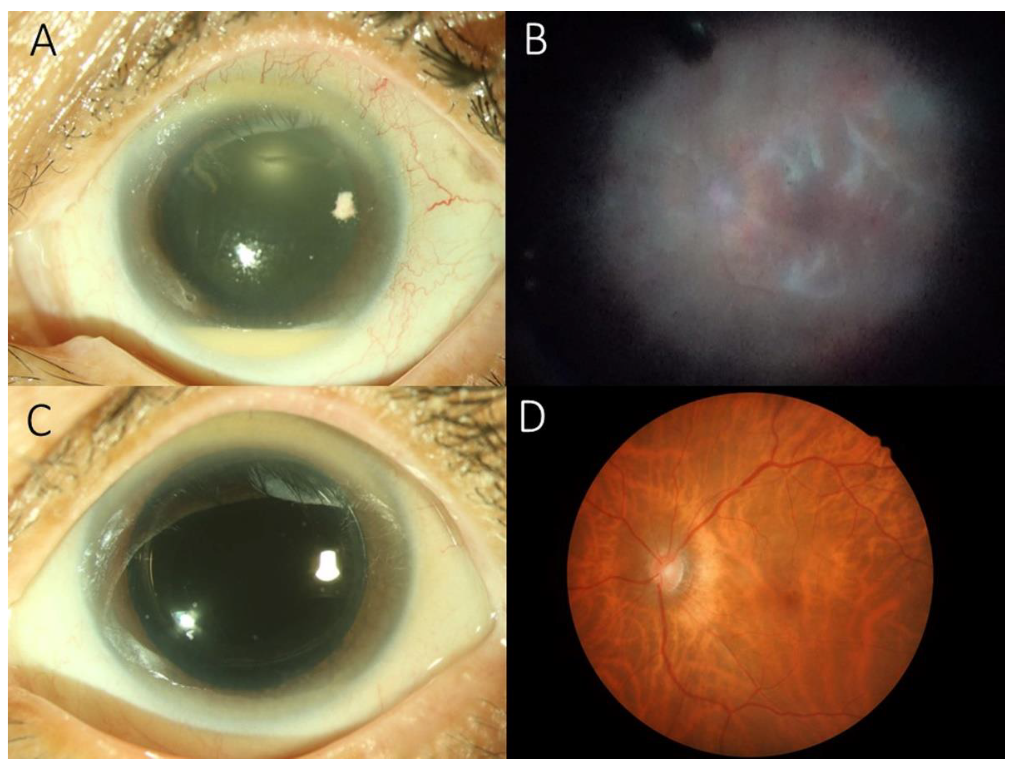
This study is a retrospective, consecutive case series of patients diagnosed with S. lugdunensis endophthalmitis from January 2009 to April 2019 at a tertiary referral center in Northern Taiwan. The institutional review board of Chang Gung Memorial Hospital in Taoyuan, Taiwan, approved this retrospective study protocol (201900614B0C601, 10 August 2019) and waived the need for written informed consent by these patients. All clinical procedures were conducted according to the principles of the Declaration of Helsinki. A review of all patients with a diagnosis of acute-onset endophthalmitis at the Chang Gung Memorial Hospital Microbiology Department was performed. Patients were included in this study if vitreous and/or aqueous samples taken at the time of diagnosis grew isolates of S. lugdunensis. The availability of medical records used electronic systems. Data collected and reviewed included demographic information, medical history, presenting signs and symptoms, duration of symptoms before diagnosis of endophthalmitis, intervals between event, and diagnosis of endophthalmitis, culture sites, antibiotic sensitivities and resistance patterns, management administered, and final visual acuity (VA). The doses of intravitreal agents were as follows: vancomycin, 1 mg/0.1 mL; ceftazidime, 2.25 mg/0.1 mL; and dexamethasone, 0.4 mg/0.1 mL. Poor visual outcomes were defined as VA worse than 20/200.
All microbiological investigations were performed at the Microbiology Department of Chang Gung Memorial Hospital, Taoyuan, Taiwan. Bacterial culture isolates were identified using conventional microbiological methods and matrix-assisted laser desorption/ionization-time of flight mass spectrometry (MALDI-TOF-MS). Conventional microbiological methods included Gram staining and biochemical tests. In MALDI-TOF-MS, automated measurement of the spectrum and comparative analysis against reference bacteria spectra were performed using an Ultraflextreme mass spectrometer and in MALDI Biotyper 3.0 software (Bruker Daltonics, Karlsruhe, Germany). The reliability of identification in the MALDI Biotyper system was expressed in terms of points. A log(score) of ≥2.0 indicated identification to the species level. The antibiotic susceptibility testing results were based on the choice of antibiotics tested in our hospital during the study period. The isolates were tested for susceptibility to various antibiotics using the Kirby–Bauer disc diffusion method on Mueller–Hinton blood agar. Clinical and Laboratory Standards Institute (Wayne, PA, USA) standards were used for interpretation and quality control for each corresponding year.

3. Results

A total of four patients were diagnosed with S. lugdunensis endophthalmitis during the study period. The mean age of patients was 71.0 ± 3.5 years (range: 67 to 75 years). All patients were female. Table 1 shows the clinical summary of the patients. Based on the interval between event (time of surgery) and diagnosis of endophthalmitis, the onset of presentation of endophthalmitis was classified as acute (with 2 weeks) in two patients, subacute (2 to 6 weeks) in one patient, and chronic (more than 6 weeks) in one patient. Ocular B-mode ultrasonography revealed moderate to dense, hyper-reflective echogenicity in vitreous cavity of all four patients. Fundal examination during vitrectomy showed diffuse retinal vasculitis and hemorrhages (Figure 1) in all patients. One patient (patient 3) had dense posterior pole preretinal exudates and retinal detachment. Three patients were successfully managed with pars plana vitrectomy. One patient developed retinal detachment even after vitrectomy. All patients were treated with topical either 0.5% levofloxacin or 2.5% vancomycin and 1 % prednisolone acetate after vitrectomy, but they did not receive oral antibiotics. In last visit, two eyes achieved favorable visual outcomes with 20/50 vision, but one eye had a final visual acuity of 4/200 that was limited by pre-existing myopic maculopathy.

Antibiotic Susceptibility Testing

Table 2 presents the antibiotic susceptibility of S. lugdunensis isolates. Most S. lugdunensis isolates were resistant to penicillin (3/4), but all were susceptible to vancomycin, which is currently being used empirically to treat Gram-positive bacterial endophthalmitis. The isolates were also susceptible to oxacillin, teicoplanin, tigecycline, and sulfamethoxazole-trimethoprim. One isolate was resistant to clindamycin and erythromycin.

4. Discussion

S. lugdunensis is a constituent of the human normal skin flora and an infrequent human pathogen. Early studies established S. lugdunensis as a skin, nasal cavity, conjunctival commensal [6,14,15,16]. It was often misidentified as S. aureus, but this has been rectified by recent routine use of matrix-assisted laser desorption ionization-time of flight mass spectrometry (MALDI-TOF MS) in diagnostic laboratories [16]. S. lugdunensis represents an ambiguous staphylococcal species, and, since its first description in 1988 [6], there is growing evidence that it should be considered a dangerous opportunistic pathogen rather than just as a harmless skin commensal [6,7]. The aggressive nature of S. lugdunensis may be related to its tendency to form a biofilm, which has a role in bacterial colonization and interferes with the phagocytosis-associated activities of neutrophils and the production of enzymes (esterase, fatty acid-modifying enzymes, protease, and lipase) [6]. Without doubt, S. lugdunensis represents the most aggressive species of CNS [16]. Compared with patients with other CNS endophthalmitis, patients with S. lugdunensis endophthalmitis often had a worse final visual outcome and a higher frequency of post-vitrectomy retinal detachment [8].
Previous studies showed that cases of S. lugdunensis endophthalmitis occurred within 3 weeks. However, in our series, two patients occurred within 2 weeks of surgery, and another two patients developed endophthalmitis on postoperative day 41 and 83. This could represent delayed onset endophthalmitis or secondary infection from unknown reasons. During vitrectomy, diffuse retinal vasculitis and hemorrhages were identified in all patients. One patient (patient 3) had condensed posterior pole preretinal exudates and retinal detachment, and became phthisic in last visit. We previously reported that acute endophthalmitis with severe vitritis are typically associated with condensed posterior pole preretinal exudates, which might be associated with poor visual outcomes [17].
The comparisons of our study with the literature are listed in Table 3. In a study of five patients with acute postoperative S. lugdunensis endophthalmitis following cataract surgery, Chiquet et al. [8] reported two patients with good visual outcome. One patient had intravitreal antibiotics alone and the other received both intravitreal antibiotics and vitrectomy. Three patients had poor visual outcomes after pars plana vitrectomy which resulted in retinal detachment [8]. In another report of six patients with acute-onset S. lugdunensis endophthalmitis caused by surgery (cataract surgery in three patients and intravitreal injection in one) or trauma (two patients), Garoon et al. [13] reported that the final visual acuity was ≥20/400 in 6/6 (100%) of eyes and ≥20/40 in 3/6 (50%) of cases after receiving intravitreal antibiotics (three patients) or pars plana vitrectomy (three patients). In previous reports [8,9,10,12] regarding S. lugdunensis endophthalmitis after intravitreal injection of anti-VEGF or dexamethasone implant (Ozurdex), a favorable visual outcome (≥20/200) was achieved in four patients receiving intravitreal antibiotics or pars plana vitrectomy. In our study, three eyes were successfully managed with pars plana vitrectomy, and two eyes achieved favorable visual outcomes. Given the mixed results, it is possible that pars plana vitrectomy may have impacted the final visual outcome in patients with worse presenting VA, and intravitreal injections of antibiotics alone could have resulted in favorable outcomes in patients with better presenting VA.
In our study, all S. lugdunensis isolates were susceptible to vancomycin, but most isolates (75%) were resistant to penicillin. All isolates were also susceptible to oxacillin, teicoplanin, tigecycline, and sulfamethoxazole-trimethoprim. In previous studies in patients with S. lugdunensis endophthalmitis, all isolates identified were susceptible to vancomycin [8,9,13]. Garoon et al. reported three of six isolates of S. lugdunensis demonstrated resistance to oxacillin and one isolate demonstrated resistance to ciprofloxacin [13]. All six isolates were susceptible to levofloxacin, and minimum inhibitory concentration values of four isolates were 0.25 μg/mL [13]. The susceptibility testing of fluoroquinolones for S. lugdunensis was not performed in our hospital; therefore, there was no susceptibility testing data available for levofloxacin.
The study has some limitations. First, this was a single-center retrospective small case series. Second, S. lugdunensis isolates were not routinely tested for fluoroquinolones at our hospital. Third, follow-up periods were not standardized. Despite these limitations, our findings contribute to the literature in providing detailed analysis and review of S. lugdunensis endophthalmitis.
In conclusion, S. lugdunensis may be associated with acute or chronic endophthalmitis. Prompt diagnosis and management can result in favorable visual outcomes. Even though vancomycin-resistant Staphylococcus species are emerging in certain regions, vancomycin provides consistent coverage against S. lugdunensis, which has no known resistance at present, and remain a first line agent in treating endophthalmitis caused by CNS [18,19].

Author Contributions

Conceptualization, K.-J.C.; Data curation, K.-J.C. and M.-H.S.; Formal analysis, K.-J.C.; Funding acquisition, K.-J.C.; Investigation, K.-J.C.; Methodology, K.-J.C.; Project administration, Writing—review and editing, K.-J.C., A.S.H.T., C.-C.S., C.-C.L. and W.-C.W. All authors have read and agreed to the published version of the manuscript.

Funding

This research was funded by a grant from the Taiwan Ministry of Science and Technology MOST 106-2314-B-182A-041, and Chang Gung Memorial Hospital CMRPG3 K1681.

Institutional Review Board Statement

The study was conducted according to the guidelines of the Declaration of Helsinki and approved by the Institutional Review Board of Chang Gung Memorial Hospital (201900614B0C601, 10 August 2019) in Taoyuan, Taiwan.

Informed Consent Statement

Patient consent was waived by the Institutional Review Board of Chang Gung Memorial Hospital due to a retrospective study.

Data Availability Statement

Not applicable.

Acknowledgments

None.

Conflicts of Interest

The authors declare no conflict of interest.

References

  1. Clarke, B.; Williamson, T.H.; Gini, G.; Gupta, B. Management of bacterial postoperative endophthalmitis and the role of vitrectomy. Surv. Ophthalmol. 2018, 63, 677–693. [Google Scholar] [CrossRef] [PubMed]
  2. Singh, R.; Davoudi, S.; Ness, S. Preventive factors, diagnosis, and management of injection-related endophthalmitis: A literature review. Graefe′s Arch. Clin. Exp. Ophthalmol. 2022, 260, 2399–2416. [Google Scholar] [CrossRef] [PubMed]
  3. Han, D.P.; Wisniewski, S.R.; Wilson, L.A.; Barza, M.; Vine, A.K.; Doft, B.H.; Kelsey, S.F. Spectrum and susceptibilities of microbiologic isolates in the Endophthalmitis Vitrectomy Study. Am. J. Ophthalmol. 1996, 122, 1–17. [Google Scholar] [CrossRef]
  4. Bannerman, T.L.; Rhoden, D.L.; McAllister, S.K.; Miller, J.M.; Wilson, L.A. The source of coagulase-negative staphylococci in the Endophthalmitis Vitrectomy Study. A comparison of eyelid and intraocular isolates using pulsed-field gel electrophoresis. Arch. Ophthalmol. 1997, 115, 357–361. [Google Scholar] [CrossRef] [PubMed]
  5. Sampathkumar, P.; Osmon, D.R.; Cockerill, F.R., 3rd. Prosthetic joint infection due to Staphylococcus lugdunensis. Mayo Clin. Proc. 2000, 75, 511–512. [Google Scholar] [CrossRef] [Green Version]
  6. Frank, K.L.; Del Pozo, J.L.; Patel, R. From clinical microbiology to infection pathogenesis: How daring to be different works for Staphylococcus lugdunensis. Clin. Microbiol. Rev. 2008, 21, 111–133. [Google Scholar] [CrossRef] [PubMed] [Green Version]
  7. Yeh, C.F.; Chang, S.C.; Cheng, C.W.; Lin, J.F.; Liu, T.P.; Lu, J.J. Clinical Features, Outcomes, and Molecular Characteristics of Community- and Health Care-Associated Staphylococcus lugdunensis Infections. J. Clin. Microbiol. 2016, 54, 2051–2057. [Google Scholar] [CrossRef] [PubMed] [Green Version]
  8. Chiquet, C.; Pechinot, A.; Creuzot-Garcher, C.; Benito, Y.; Croize, J.; Boisset, S.; Romanet, J.P.; Lina, G.; Vandenesch, F. Acute postoperative endophthalmitis caused by Staphylococcus lugdunensis. J. Clin. Microbiol. 2007, 45, 1673–1678. [Google Scholar] [CrossRef] [PubMed] [Green Version]
  9. Murad-Kejbou, S.; Kashani, A.H.; Capone, A., Jr.; Ruby, A. Staphylococcus lugdunensis endophthalmitis after intravitreal injection: A case series. Retin. Cases Brief Rep. 2014, 8, 41–44. [Google Scholar] [CrossRef] [PubMed]
  10. Salceanu, S.O.; Hamada, D.; Ursu, R.G.; Saad, A.A. Staphylococcus lugdunensis endophthalmitis following dexamethasone intravitreal implant. Indian J. Ophthalmol. 2019, 67, 424–426. [Google Scholar] [CrossRef] [PubMed]
  11. Veliev, A.; Nakipoglu, Y. Investigation of Staphylococcus lugdunensis and Selected Coagulase Negative Staphylococci Isolated from Blood Culture bottles and Determination of their Sensitivities to Antibiotics. Pak. J. Med. Sci. 2022, 38, 657–662. [Google Scholar] [CrossRef] [PubMed]
  12. Wani, V.B.; Al-Kandari, J.; Sabti, K.; Aljassar, F.; Qali, H.; Kumar, N.; Uboweja, A.; Al-Sabah, K.; Diab, F.A.; Al-Rashidi, S. Incidence of Endophthalmitis after Intravitreal Bevacizumab using Aliquots Prepared On-site in 2 Operating Rooms in Kuwait. Middle East Afr. J. Ophthalmol. 2016, 23, 64–70. [Google Scholar] [CrossRef] [PubMed]
  13. Garoon, R.B.; Miller, D.; Flynn, H.W., Jr. Acute-onset endophthalmitis caused by Staphylococcus lugdunensis. Am. J. Ophthalmol. Case Rep. 2018, 9, 28–30. [Google Scholar] [CrossRef] [PubMed]
  14. Xu, S.; Zhang, H. Bacteriological profile of conjunctiva bacterial Flora in Northeast China: A hospital-based study. BMC Ophthalmol. 2022, 22, 223. [Google Scholar] [CrossRef] [PubMed]
  15. Bieber, L.; Kahlmeter, G. Staphylococcus lugdunensis in several niches of the normal skin flora. Clin. Microbiol. Infect. 2010, 16, 385–388. [Google Scholar] [CrossRef] [Green Version]
  16. Heilbronner, S.; Foster, T.J. Staphylococcus lugdunensis: A Skin Commensal with Invasive Pathogenic Potential. Clin. Microbiol. Rev. 2021, 34, e00205-20. [Google Scholar] [CrossRef] [PubMed]
  17. Chen, K.J.; Chen, Y.P.; Wang, N.K.; Sun, M.H.; Sun, C.C.; Wu, W.C.; Lai, C.C. Retinal Vasculitis and Posterior Pole Preretinal Exudates in Exogenous Bacterial Endophthalmitis: Management and Visual Outcomes. Ocul. Immunol. Inflamm. 2022, 30, 491–496. [Google Scholar] [CrossRef] [PubMed]
  18. Relhan, N.; Albini, T.A.; Pathengay, A.; Kuriyan, A.E.; Miller, D.; Flynn, H.W. Endophthalmitis caused by Gram-positive organisms with reduced vancomycin susceptibility: Literature review and options for treatment. Br. J. Ophthalmol. 2016, 100, 446–452. [Google Scholar] [CrossRef] [PubMed] [Green Version]
  19. Chen, K.-J.; Sun, M.-H.; Hou, C.-H.; Chen, H.-C.; Chen, Y.-P.; Wang, N.-K.; Liu, L.; Wu, W.-C.; Chou, H.-D.; Kang, E.Y.-C.; et al. Susceptibility of bacterial endophthalmitis isolates to vancomycin, ceftazidime, and amikacin. Sci. Rep. 2021, 11, 15878. [Google Scholar] [CrossRef] [PubMed]
Figure 1. (A) Slit-lamp biomicroscopic examination showing corneal edema with a 2-mm hypopyon in patient 4. (B) During pars plana vitrectomy, posterior pole showing diffuse retinal vasculitis. (C) Four weeks later, Slit-lamp biomicroscopic examination showing clear cornea without hypopyon. (D) Fundoscopic examination revealing myopic tessellated retina with resolution of retinal vasculitis.
Antibiotics 11 01485 g001
Table 1. Clinical summary of patients with postoperative endophthalmitis caused by S. lugdunensis.
No.Sex/Age
/Eye
Interval (Days)Symptom (Days)EtiologyIOP
(mmHg)
Culture SitesSystemic DiseasesInitial VATreatmentFinal VAOther Eye ConditionFollow-Up (Months)
PrimarySecondary
1F/67/OS72CE + IOL10AC, VCHF, HT, DM, CVAHM 10 cmPPV + IVABIVAB(1M)20/50M-NPDR36
2F/75/OD411PPV + CE + IOL12AC, V HM 30 cmPPV + IVAB 4/200MM16
3F/74/OD833CE + IOL30AC, VGoiterLP 10 cmPPV + IVABIVAB(3D)NLPRD3
4F/68/OS31CE + IOL7VMiller-Fisher syndromeHM 40 cmPPV + IVAB 20/50 54
AC, anterior chamber; CE, cataract extraction; CHF, congestive heart failure; CVA, cerebrovascular accident; D, day; DM, diabetes mellitus; HM, hand motions; IOL, intraocular lens implantation; IOP, intraocular pressure; IVAB, intravitreal antibiotics; LP, light perception; M, month; MM, high myopic maculopathy with foveoschisis; M-NPDR, moderate non-proliferative diabetic retinopathy; NLP, no light perception; PPV, pars plana vitrectomy, RD, retinal detachment; HT, hypertension; V, vitreous.
Table 2. Antibiotic susceptibility testing results of Staphylococcus lugdunensis isolates.
AntibioticsPatient
1234
PenicillinRRSR
OxacillinS SS
TeicoplaninSSSS
VancomycinSSSS
TigecyclineSSSS
SMX/TMPSSSS
LinezolidS
Clindamycin SSRS
ErythromycinSSRS
R, resistant; S, susceptible; SMX/TMP, sulfamethoxazole-trimethoprim.
Table 3. Comparisons of published and current studies of Staphylococcus lugdunensis endophthalmitis.
No.AuthorNationYear
(Published)
No. of EyesInterval (Days)EtiologyInitial VAVANTreatmentFinal VA Cause of Poor VA
1Chiquet et al. [8]France2004–2005 (2007)57CEHMSIVAB, PPV20/40
6CELPSIVAB, PPVHMRD
5CE + IOLLPSIVAB, PPVNLPRD
12CE + IOL20/100SIVAB 20/20
7CE + IOLHMSIVAB, PPVCFRD
2Garoon et al. [13]US1990–2017 (2018)610CE + IOLCFSIVAB 20/30
8CE + IOLHMSIVAB 20/40
21IVICFSIVAB 20/40
6CE + IOLHMSIVAB, PPV20/40
2TraumaHMSPPV + IVAB20/400
3TraumaLPSPPV + IVAB20/100
3Salceanu et al. [10]UK201914IVI (Ozurdex)CFNAPPV + IVAB20/120
4Wani et al. [12]KuwaitNA (2016)13IVI (Avastin)20/150NAIVAB20/30
5Veliev et al. [11]Turkey NA (2022)1NApostoperativeNANANANA
6Murad-Kejbou et al. [9]US2011–2012 (2014)32IVI (Lucentis)LPSPPV + IVABHM
3IVI (Lucentis)20/200SIVAB20/200
2IVI (Avastin)20/200SIVAB, PPV20/100
7Bannerman et al. [4]US1990–1996 (1997)4NApostoperativeNANANANA
8Current study
Chen et al.
Taiwan2010–201947CE + IOLHMSPPV + IVAB20/50
41PPV + CE + IOLHMSPPV + IVAB4/200MM
83CE + IOLLPSPPV + IVABNLPRD
3CE + IOLHMSPPV + IVAB20/50
CE, cataract extraction; CF, counting fingers; HM, hand motions; IOL, intraocular lens implantation; IVAB, intravitreal antibiotics; IVI, intravitreal injection; LP, light perception; MM, myopic maculopathy; NA, not available; NLP, no light perception; PPV, pars plana vitrectomy, RD, retinal detachment; S, susceptible, VA, visual acuity, VAN, vancomycin.
Publisher’s Note: MDPI stays neutral with regard to jurisdictional claims in published maps and institutional affiliations.

